Note: Descriptions are shown in the official language in which they were submitted.
CA 02483442 2004-10-22
WO 03/090833 PCT/US03/09737
METHOD AND SYSTEM FOR DELIVERY OF A MEDICAL ELECTRICAL
LEAD WITHIN A VENOUS SYSTEM
RELATED APPLICATIONS
This application is a continuation-in-part of commonly assigned U.S. Patent
Application Serial Number 09/822,678 filed March 31, 2001, which is related
to, and
claims the benefit of provisionally-ale US Patent Application No. 60/193, 695,
fled
March 31, 2000, and entitled "Intraluminal Visualization System with
Deflectable
Mechanism", both of which are incorporated herein by reference in their
entireties.
Cross-reference is hereby made to commonly assigned related U.S. Applications,
filed concurrently herewith, docket number P-10017.02 CIP1, entitled "IMPROVED
SYSTEM AND METHOD FOR POSITIONING IMPLANTABLE MEDICAL DEVICES
WITHIN CORONARY VEINS", and docket number P-10017.04 CIP3, entitled
"METHOD AND SYSTEM FOR DELIVERING A MEDICAL ELECTRICAL LEAD
WITHIN A VENOUS SYSTEM".
BACKGROUND OF THE INVENTION
The present invention relates generally to delivery of various devices or
agents into
a targeted area of the body, and in particular, the present invention relates
to a method and
system for accurately delivering medical devices such as leads,
electrophysiology
catheters, and therapeutic agents into large-organ vessel systems such as the
coronary
vasculature.
In treating conditions such as arrhythmia, one technique is to destroy or
damage
heart tissue that causes or is involved with the arrhythmia by suitably
heating the tissue,
e.g., by applying a laser beam or high-frequency electrical energy such as
radio-frequency
(RF) or microwave energy.
For such treatment to be effective, the location of the tissue site causing or
involved with the arrhythmia must be accurately determined in order to be able
to contact
heart tissue adjacent the desired location with a tissue-destroying device. A
high degree of
accuracy in determining this site is paramount so that an excessive amount of
viable tissue
is not destroyed adjacent the site. For example, the average arrhythmogenic
site consists
CA 02483442 2004-10-22
WO 03/090833 PCT/US03/09737
2
of about 1.4cm2 of endocardial tissue, whereas a re-entrant site might be much
larger. RF
ablation techniques produce lesions about 0.5 cm2 of diameter, so a number of
lesions are
typically generated in order to ablate the area of interest. If the site is
not accurately
mapped, much of the viable tissue surrounding the site will be unnecessarily
destroyed.
To determine the location of the tissue to be ablated, it is widely known to
use
elongated intravascular signal sensing devices that are advanced through the
patient's
vasculature until the distal portions of the device are disposed within one or
more of the
patient's heart chambers, with one or more electrodes on the distal portion of
the device in
contact with the endocardial lining. Such devices may also be advanced within
a patient's
coronary artery, coronary sinus, or cardiac vein. Sensing devices such as
those disclosed
in U.S. Patent No. 5,967,978 to Littmann et al., and combination sensing-
ablation devices
such as those disclosed in U.S. Patent No. 6,002,956 to Schaer are typical.
Guiding catheters such as those disclosed in U.S. Patent Nos. 6,021,340 and
5,775,327 to Randolph et al, may be used to rapidly advance such devices into
a patient's
cardiac vein dxaining into the coronary sinus. A particular advantage of the
catheters
disclosed in these references is the presence of an inner lumen and distal
port on the
catheter shaft, which, in conjunction with a distal balloon, allows fox the
deployment of
contrast fluid distal to the distal end of the catheter for visualizing the
venous structure.
The following U.S. Patents discuss related devices and methods for their use:
U.S.
Patent Nos. 5,509,411, 5,645,064, 5,682,885, 5,699,796, 5,706,809, and
5,701,298, each to
Littmann et al; U.S. Patent Nos. 5,881,732 and 5,645,082, each to Sung et al;
U.S. Patent
No. 5,766,152 to Morely et al; U.S. Patent Nos. 5,782,760 and 5,863,291, each
to Schaer;
U.S. Patent No. 5,882,333 to Schaer et al., and U.S. Patent Number 6,122,552
to Tockman
et al.
However, despite the advantages of these sensing devices and guiding
catheters, it
remains quite difficult to accurately and reliably contact the various curved
shapes one
encounters in the endocardial lining. This is due to the frequent inability to
customize the
shape of their distal portion, or at least the inability to instantaneously
and accurately
adjust their shape upon demand during deployment to conform to the shape of
the tissue of
interest.
CA 02483442 2004-10-22
WO 03/090833 PCT/US03/09737
3
Concerns similar to those described above are associated with the placement of
leads within the heart and other areas of the coronary vasculature. For
example,
pacemakers, defibrillator/cardioverters, and other implantable medical device
(IMDs) may
employ one or more electrodes that are maintained in contact with a patient's
heart muscle
and through which electrical stimulation of the heart muscle is achieved. Such
devices
typically employ a flexible conductive lead that connects a remotely
positioned and
implanted power source to the one or more electrodes. Secure placement of the
electrodes
in the selected heart chamber (typically the right atrium) or in a coronary
vein or artery is
required to assure appropriate and reliable depolarization or "capture" of
cardiac tissue by
electrical stimuli delivered by the IMD.
Many problems exist with reliably and accurately placing medical electrical
leads
and other similar devices such as catheters within the heart and associated
vasculature.
For instance, when placing transvenous leads or catheters, it is often
difficult to engage the
coronary sinus and sub-select the proper vessel into which the lead or
catheter is to
eventually be placed. Moreover, once placed, transvenous devices suffer from a
relatively
high rate of dislodgment from sites adjacent to, or on, the epicardium. Such
dislodgement
may result in a loss of capture or, at best, a reduction of the degree of
electrical coupling
between the electrode and the myocardium. More accurate and secure placement
of the
lead or catheter would not only reduce the difficulty and time associated with
lead
placement, but would reduce the risk of subsequent dislodgment as well.
There thus is a need for a method and system for placing intralumenally-
deployed
devices such as electxophysiology catheters and leads into selected areas of
the coronary
vasculature in a highly accurate and reliable fashion.
SUMMARY OF THE INVENTION
The present invention is directed to a system for delivering a medical
electrical
lead within a coronary venous system that includes an introducer kit for
establishing
venous access and a plurality of delivery sheaths, each corresponding to a
desired
approach to a coronary sinus of the coronary venous system and insertable
within the
coronary venous system through the navigation pathway. A hemostasis valve is
coupled
to a delivery sheath of the plurality of delivery sheaths, and a guide wire is
inserted within
CA 02483442 2004-10-22
WO 03/090833 PCT/US03/09737
4
the lead lumen, guiding delivery of the distal tip of the medical electrical
lead to a target
site within the coronary venous system through the hemostasis valve and the
delivery
sheath. Subsequent to the distal tip being delivered to the target sight, the
hemostasis
valve is advanced over a connector pin of the medical electrical lead to
xemove the
hemostasis valve from the medical electrical lead.
According to an embodiment of the present invention, a system for delivering a
medical electrical lead within a coronary venous system includes an introducer
kit that
establishes venous access to the coronary venous system, and a plurality of
delivery
sheaths, each corresponding to a desired approach to a coronary sinus of the
coronary
venous system and insertable within the coronary venous system through the
navigation
pathway. An anchoring sleeve is positioned along the medical electrical lead
and a
hemostasis valve is coupled to a delivery sheath of the plurality of delivery
sheaths. A
guide wire is inserted within the lead lumen, guiding delivery of the distal
tip of the
medical electrical lead to a target site within the coronary venous system
though the
hemostasis valve and the delivery sheath. Subsequent to the distal tip being
delivered to
the target sight, the hemostasis valve is advanced over a connector pin of the
medical
electrical lead and the anchoring sleeve of the medical electrical lead to
remove the
hemostasis valve from the medical electrical lead.
According to yet another embodiment of the present invention, the guide wire
is a
stylet having a stylet knob, and the hemostasis valve is advanced over the
stylet knob to
remove the hemostasis valve from the medical electrical lead.
BRIEF DESCRIPTION OF THE DRAWINGS
Figure 1A is a side cutaway view of a delivery sheath of the present
invention.
Figure 1B is a cross-sectional view of a delivery sheath of the present
invention.
Figures ZA-2B are side and cross-sectional views, respectively, of a balloon
catheter of the present invention.
Figure 3 is as side view illustrating components included in both the
deflection
mechanism and micro-deflection mechanism of the present invention.
Figures 4A-4B are various views of a deflection mechanism handle of the
present
invention.
CA 02483442 2004-10-22
WO 03/090833 PCT/US03/09737
Figure 5 is a cross-sectional side view of three components of the present
invention: a deflection mechanism, an outer sheath, and a balloon catheter
with an inflated
distal balloon and a deflected distal end.
Figures 6A-6D are various views of a micro-deflection mechanism handle of the
present invention.
Figures 7A-7B axe two embodiments of deflection and micro-deflection
mechanisms detailing two notch configurations.
Figures 8A-8D are additional embodiments of deflection and micro-deflection
mechanisms of the present invention, detailing additional notch
configurations.
Figure 8E is a cross-sectional view of a deflection and micro-deflection
mechanism
having a tubular member with an irregular wall thickness to provide a
preferred bending
direction.
Figures 9-11 depict a method for accurately placing an endocardial lead into
the
cardiac venous system through the coronary sinus ostium using a system of the
pxesent
invention.
Figure I2 is a plan view of a steerable catheter that may be used as an
alternative
deflection mechanism to navigate the balloon catheter 200 into the coronary
sinus.
Figures 12A through 12C illustrate various deflection positions of the distal
tip of
the steexable catheter of Figure 12.
Figure 13 is a schematic diagram of a tool kit used to establish venous access
in a
system for delivering medical devices within a coronary venous system
according to the
present invention.
Figure 14 is a schematic diagram of a guide wire clip of a tool kit according
to the
present invention.
Figure 15 is a schematic diagram of a wire clip of a tool kit according to the
present invention.
Figure 16 is a schematic diagram of a rotatable hemostasis valve (RHV) of a
tool
kit according to the present invention.
Figure 17 is a schematic diagram of a delivery sheath for delivering a medical
electrical device within a coronary venous system, according to the present
invention,
from a right-sided venous access point to a coronary sinus.
CA 02483442 2004-10-22
WO 03/090833 PCT/US03/09737
6
Figure 18 is a schematic diagram of a delivery sheath for delivering medical
devices within a coronary venous system, according to the present invention,
from a left-
sided venous access point to a coronary sinus.
Figure 19 is a plan view of a medical electrical lead having a lumen for
receiving a
stylet or a guide wire for delivering a medical electrical device within a
coronary venous
system according to the present invention.
Figure 20 is a schematic of a guide wire atraumatic formable tip protruding
from a
lead distal tip of a medical electrical lead and navigating from the coronary
sinus into a
branch vein.
Figure 21 is a planar view of a stylet inserted within an over-the-wire
medical
electrical lead in a system for delivering medical devices within a venous
system
according to the present invention.
Figure 22 is planar side view of a medical electrical lead having a lumen for
receiving a stylet wire and a guide wire in a system for delivering medical
devices within a
venous system according to the present invention.
Figure 23 is a cross-sectional side view of a lead distal tip of the medical
electrical
lead of Figure 22.
Figure 24 is a schematic diagram of a loading tool in a system for delivering
medical devices within a venous system according to the present invention.
Figure 25 is a cross-sectional view of the loading tool of Figure 24.
Figure 26 is a schematic diagram of a lead connector fixedly inserted within
the
loading tool of Figure 24.
Figure 27 is an isometric diagram of an alternate embodiment of a loading tool
in a
system for delivering medical devices within a venous system according to the
present
invention.
Figure 28 is a front planar view of the loading tool of Figure 27 in a closed
position.
Figure 29 is a cross-sectional side view of a loading tool according to the
present
invention, taken along cross-sectional lines VII-VII of Figure 28,
Figure 30 is a front planar view of the loading tool of Figure 27 in an open
position.
CA 02483442 2004-10-22
WO 03/090833 PCT/US03/09737
7
Figure 31 is a top perspective view of a loading tool for loading a guide wire
within a medical electrical lead according to the present invention.
Figure 32 is a cross-sectional side view of a loading tool according to the
present
invention, taken along cross-sectional line IV-IV of Figure 31.
Figure 33 is a top planax view illustrating insertion of a guide wire within a
medical electric lead using a loading tool according to the present invention.
Figure 34 is a cross-sectional side view of a loading tool according to the
present
invention, taken along cross-sectional lines V-V of Figure 33.
Figure 35 is a top planar view illustrating insertion of a guide wire within a
medical electric lead using an alternate embodiment of a loading tool
according to the
present invention.
Figure 36 is a schematic diagram of positioning of a guide wire 46 within a
branch
vein.
Figure 37 is a schematic diagram of a hemostasis valve according to the
present
invention in an attached position.
Figure 38 is a schematic diagram of a hernostasis valve according to the
present
invention in an unattached position.
Figure 39 is partial section plan view of a hemostasis valve according to the
present invention.
Figure 40 is a flowchart of a method of delivering a medical electrical lead
within a
coronary sinus according to the present invention.
DETAILED DESCRIPTION OF THE INVENTION
This invention is a method and system for intralumenal visualization and
deployment of implantable medical devices (IMDs) such as transvenous leads,
electrophysiology catheters and the like to various targeted regions of the
body. The
inventive system includes a sheath, a balloon catheter and associated
deflection
mechanism, and a micro-deflection device for highly accurate placement of the
lead,
catheter, or other device once the area of interest has been visualized.
In the following pages we provide a component-by-component description of a
preferred variation of the invention followed by a description of a procedure
for using this
system to place a transvenous lead into the coronary veins. Although we detail
an
CA 02483442 2004-10-22
WO 03/090833 PCT/US03/09737
exemplary set of system components and a method for its use, additional system
configurations, adaptations, and methods of use, some of which are also
described herein,
are within the scope of the invention.
In general, the intralumenal visualization system and micro-deflection device
of
the present invention includes a deflectable catheter that includes an
inflatable member
such as a balloon, and is insertable into a lumen of a delivery sheath. This
sheath may be
inserted into the body via a typical introducer as will be described in more
detail. In a
preferred use, a balloon catheter is guided by a deflection mechanism so that
it may
engage the coronary sinus ostium. A balloon catheter is inserted through the
delivery
sheath and into the coronary sinus or through a delivery sheath over a guide
wire so that an
occlusive venogram may be rendered and the balloon catheter is removed. Next,
a lead
with a micro-deflection mechanism is inserted into the sheath lumen so that
the Lead may
be deployed at the desired location in the coronary veins. The micro-
deflection
mechanism disposed within the lead is used to provide rigidity to the lead and
to allow a
means to sub-select coronary vessels. The sheath preferably may be splittable
along its
longitudinal length so that it may be removed around the lead without
disturbing it.
Delivery Sheath
Figure lA is a cutaway side view depicting a variation of the delivery sheath
described above. As best seen in Figure lA, sheath 100 comprises an elongate
shaft 102
containing a central lumen 104 throughout its length. The working length of
sheath 100
comprises a distal section 110 and a proximal section 120, each of which
comprises a
polymeric material having differing flexibilities as described below. A distal
end 1 I2 of
sheath 100 disposed adjacent distal section 110 also comprises the working
length.
Near the proximal end of sheath 100, a hub 114 may be affixed to proximal
section
120 by an adhesive or other suitable means. We prefer an ultraviolet-curable
adhesive
sold by Loctite Corp. of Rocky Hill, Connecticut under the name W 4201. We
also
prefer an adhesive sold by Dymax core. of Trorrington, Connecticut under the
trademark
DYMAX. Hub 114 is made from any suitable medical-grade polymer, and is
preferably
injection molded and Longitudinally scored or perforated so that it may be
removed from
CA 02483442 2004-10-22
WO 03/090833 PCT/US03/09737
9
around a device without disturbing that device. It may be molded ira situ onto
the proximal
section 120 of shaft 102.
Hub 114 has an opening large enough to accommodate a special rotatable
hemostatic valve (RHV) 118, to which it is detachably secured by, e.g. , an
annular ring on
the valve 118 inner diameter. A central lumen 124 in RHV 118 is aligned and in
fluid
communication with the lumen of shaft 102. Lumen 124 has a diameter large
enough to
accommodate a balloon catheter and a typical lead connector, such as an IS-1-
type
connector. An optional side arm (not shown) may be disposed on RHV 118 in
fluid
communication with Iumen 124. RHV 118 may also be splittable via a scoring or
perforation as described above.
An annular polymeric collar 1 I6 is disposed on the outside diameter of RHV
118
distal portion proximal to the point where hub 114 meets RHV 118. In this
embodiment,
rotation of collar 116 locks the RHV 118 to the hub 114.
Figure 1B is a cross-sectional view of the delivery sheath of Figure lA. As
shown
in Figure 1B, a cross-section of shaft 102 in the distal section 110 reveals
shaft lumen 104.
The inner diameter of shaft 102 will vary depending on the outer diameter of
the balloon
catheter and the lead, each of which should be capable of passing through
lumen 104.
Typically the shaft inner diameter is between about 0.080 and 0.110 inch; more
preferably
it is about 0.098 inch. Likewise, the outer diameter of shaft 102 is typically
between about
0.090 and 0.130 inch; more preferably it is about 0.118 inch. We prefer the
outer diameter
of shaft 102 to be as small as possible while still maintaining acceptable
performance
levels according to the application for which the shaft is used. We also
prefer that shaft
102 generally maintains a constant inner diameter throughout its length to
provide a
smooth and continuous step-free profile for the passage of various devices and
materials
therethrough as described herein.
Tubing comprising distal section 110 and proximal section 120 will typically
be
polymeric, and is preferably any typical medical grade, biocompatible tubing
with the
appropriate performance characteristics as described herein. An especially
desirable
material is an extruded polyether block amide of the type sold by Atochem
North
America, Inc., Philadelphia, Pennsylvania under the trademark PEBAX.
CA 02483442 2004-10-22
WO 03/090833 PCT/US03/09737
Distal and proximal sections I 10 and 120, respectively, are constructed of
tubing
having a durometer hardness ranging from about 20D to I OOD (shore). The
working
length of shaft 102 preferably is composed of materials having two or more
stiffnesses;
although shaft 102, having a single stiffness value throughout its length is
within the scope
5 of the invention.
In one embodiment, proximal section 120 comprises a relatively high stiffness
material (typically about 72D) in comparison to the more flexible distal
section 110
(typically about 40D). Although not shown in the view of Figure 1B, distal
section 110
and proximal section 120 may be comprised of a DACRON (E.I. du Pont de Nemours
and
10 Company, Wilmington, DE) bxaid with a TEFLON (E.I. du Pont de Nemours and
Company, Wilmington, DE) liner. The braid is surrounded by the PEBAX tubing as
described above, which renders the proximal section 120 of shaft 102 generally
stiffer and
less flexible than distal portion I I0.
Distal end 112 is preferably a soft, atraumatic tip made form a relatively low
stiffness polymeric material so to prevent injury to the intima of the vessel
walls or to
other tissue. We have found an effective material for distal end I 12. A
material well-
suited for the distal end is a thermoplastic polyurethane elastomer such as
PELLETHANE
(Dow Chemical Co., Midland, MI) or the like.
According to one aspect of the invention, distal portion 110 may be
radiopaque.
This can be achieved by the inclusion of radiopaque metals or their alloys
into the
structure, or more preferably by incorporating radiopaque powders such s BaSO,
BiCO,
etc. into the polymer comprising distal portion 110. Distal end 112 is
preferably more
radiopaque than distal portion I 10. This can be achieved by the incorporation
of greater
quantities of radiopaque powder, for instance, into the tubing, or by the use
of a different
material having greater radiopacity than that used in distal portion 110. This
radiopaque
feature allows the user to more readily visualize these portions of sheath 100
under
fluoroscopy.
The entire length of shaft 102 (from distal end 112 to the far proximal end of
RHV
118) is typically between about 40 and 60 cm, and is preferably about 55 cm.
Distal end
112 rnay be between about 0.2 cm and 0.5 cm long, while distal section 110 is
generally
CA 02483442 2004-10-22
WO 03/090833 PCT/US03/09737
11
between about 5 and 10 cm long, and is preferably about 8 cm long. Proximal
section 120
is between about 35 and 50 cm long; preferably about 42 cm.
Both the working length of shaft 102 as well as the attached hub 114 may
contain a
perforation or score 126 along their longitudinal axes. Alternatively, they
may be
otherwise configured to split so that they may be opened and removed from
around an
inserted device such as a lead or electrophysiology catheter without having to
axially slide
the sheath 100 relative to the device. A special tool may be used to
facilitate such
splitting, or the sheath/hub (and even RHV 114) combination may be split by
hand without
the aid of any special device. The splittable valve and sheath combinations as
described in
U.S. Patent No. 5,312,355 to Lee is exemplary.
Balloon Catheter
Tuniing now to Figures 2A-2B, a balloon catheter 200 of the present invention
is
shown in side view and distal cross-sectional view, respectively. This
catheter is largely
similar to the guiding catheters disclosed in U.S. Patent Nos. 6,021,340 and
5,775,327 to
Randolph et al, the entirety of each of which are incorporated herein by
reference, as well
as the VUEPORT family of balloon occlusion guiding catheters sold by Cardima,
Inc. of
Fremont CA.
Catheter 200 is designed to pass through the central lumen 104 of deployment
sheath 100, and reach the therapeutic site as a combined unit with sheath 100
and
deflection mechanism 300.
As shown in Figures 2A and 2B, balloon catheter 200 generally includes an
elongated shaft 202, a distal shaft section 204, a proximal shaft section 206,
and an inner
lumen 208. A female luer lock 210 may be disposed on the proximal end of shaft
202 and
secured by a suitable adhesive 212, such as UV-curable Loctite 4201.
A distal port 214 is provided in the distal end 216 of the catheter shaft that
is in
fluid communication with the inner lumen 208. Proximal of distal end 216 is an
occlusion
balloon 211 axially disposed in the distal section 204 about catheter shaft
202. The
catheter shaft 202 is provided with an inflation lumen 209 that extends
through the shaft
202 to the interior of the balloon 211 to direct inflation fluid therein.
CA 02483442 2004-10-22
WO 03/090833 PCT/US03/09737
12
On the proximal end of catheter 200, proximal to luer lock 210, is a multiarm
adapter or hub 222 that terminates in a Y-adapter or hernostasis valve 232 and
a proximal
port 218 for passage of a deflection mechanism therethrough as described
later.
A first sidearm ox port 224 on adapter 222 (shown in partial cross section in
Figure
2A) facilitates introduction of inflation fluid into inflation lumen 209. A
stopcock 228 on
first sidearm 224 that allows balloon 221 to stay inflated once the proper
volume of fluid
(such as air) has been introduced via syringe 230 is disposed adjacent
stopcock 228.
Inflation lumen 209 is disposed in port 224 and extends distally into shaft
224 to facilitate
inflation of balloon 211 as described above.
A second sidearm or port 226 may also be disposed on hub 222, and may be in
direct fluid communication with large inner lumen 208. Inner lumen 208 is used
for
housing devices such as a deflection mechanism or the like. Once balloon 211
is inflated,
the second port 226 may be used for introducing contrast media or similar
material
through lumen 208 and out the distal port 214 for visualization of a section
of interest in
the body, such as an organ lumen or the cardiac venous system, for instance.
Not shown is a rotatable hemostatic valve (RHV) that may be housed in the
proximal center port 218 and that can accept devices such as a deflection
mechanism
described below. This RHV is capable of sealing onto the deflection mechanism
to
prevent fluid leakage and may be part of a duostat modified to comprise a
single RHV and
two sideports. Other configurations, of course, are possible.
Shaft 202 of balloon catheter 200 is of a sufficient size so that it may
readily pass
through the lumen 104 of sheath 100. Ideally, we prefer the outer diameter of
shaft 202 to
be between approximately 0.050 inch and 0.100 inch. More preferably, it is
between
0.060 inch and 0.080 inch, and most preferably is about 0.074 inch.
The diameter of inner lumen 208 preferably is large enough to allow free
passage
of contrast media or other material therethrough so that venograms and similax
diagnostic
procedures may be readily accomplished. It should also be large enough for the
passage
of a deflection mechanism as discussed below in greater detail. Finally, lumen
208 should
allow the free passage of contrast media or other agents therethrough while
occupied by a
device such as a deflection mechanism. In general, we prefer that inner lumen
have a
diameter of between 0.030 inch and 0.080 inches, and is preferably about 0.048
inch.
CA 02483442 2004-10-22
WO 03/090833 PCT/US03/09737
13
Likewise, inflation lumen 209 preferably has a diameter of between about 0.005
inch and
0.020 inch, and preferably is about 0.014 inch.
The balloon catheter shaft 202 preferably comprises PEBAX tubing having a
durometer hardness of between about 60D and 80D, preferably about 72D.
Pxeferably,
shaft proximal section 206 has a heat shrink tubing disposed on the outer
surface thereof.
Preferably, this heat shrink tubing is polymeric and is comprised of clear
polyoleftn or the
like. Distal tip 216 is preferably a soft, atraumatic tip made of a relatively
flexible
polymeric material similar in composition and stiffness to distal tip 112 of
sheath 100. In
one embodiment, distal tip is radiopaque.
The working length of balloon catheter shaft 202, which includes the distal
tip 216,
distal section 204, and proximal section 206, should be between about 50 cm
and 90 cm,
although it may be longer or shorter depending upon the application. We
especially prefer
a working length of approximately 70 cm which can accommodate a distal tip 216
of
approximately 0.5 cm, a distal section 204 of approximately 6 cm, and a
pxoximal section
206 of approximately 63.5 cm.
The length of the entire catheter 200 in this embodiment (the working length
of
shaft 202 and the components disposed proximal of proximal section 206
discussed above)
should be about 77.5 cm. In general, we prefer that the balloon catheter shaft
202 be
between about 15 cm and 20 cm longer than sheath 100.
Of course, the absolute and relative lengths of each component of catheter 200
may
vary considerably. The particular application in which catheter 200 and the
entire system
of the present invention is to be used will dictate the particular dimensions
and materials
for its various components (as well as each of the components of the inventive
system)
described herein.
Occlusion balloon 211, when inflated, should have a diameter sufficient to
seal the
coronary sinus ostium. This inflated diameter will typically be between about
0.2 inch and
1.0 inches, and more preferably, between about 0.4 inch and 0.8 inches. We
prefer
balloon 211 to comprise an inelastic or elastic polymeric material.
Polyurethane (e.g.
PELLETHANE 80A durometer, World Medical, Inc., Miami FL) is especially
preferable.
The inner diameter of the uninflated balloon 211 typically will be between
about 0.04 inch
and 0.08 inches, and more preferably between about 0.056 inch and 0.070
inches. The
CA 02483442 2004-10-22
WO 03/090833 PCT/US03/09737
14
balloon wall thickness typically will be between about 0.002 inch and 0.006
inches, and
more preferably about 0.004 inches. Finally, the balloon 211 length typically
will be
between about 6mm and l4mm, and more preferably between about 8mm and l2mm.
Deflection Mechanisms and Micro-Deflection Mechanism
The deflection mechanism and the micro-deflection mechanism are two separate
components of the present invention. Deflection mechanism 300 is designed for
use in the
balloon catheter 200, and is similax in many respects to the micro-deflection
mechanism
400, only larger. Micro-deflection mechanism 400 is designed for use in a
variety of
applications where precise control and deflection of a device such as a lead,
electrophysiology catheter, or other similar IMDs, is needed. Its small size
relative to
deflection mechanism 300 renders it useful in a wide range of applications in
which its
small size and flexibility may be relied upon.
Figure 3 is a plan view illustrating components of both the deflection and
micro-
deflection mechanisms, although it will be described in terms of the
deflection mechanism
300 for discussion purposes. Deflection mechanism 300 generally comprises a
proximal
section 304, a distal section 306, and a distal tip 308. Adjacent the proximal
section 304 is
handle 310, a preferred variation of which is shown in detail in Figures 4A
and 4B.
Deflection mechanism 300 is designed to be place through proximal port 218 of
the balloon catheter 200 and into the inner lumen 208 such that the deflection
mechanism
distal tip 308 generally reaches distal section 204, and preferably distal tip
216, of balloon
catheter shaft 202. When the handle 310 is activated, the distal section 306
of deflection
mechanism 300 deflects in a predetermined fashion, thus deflecting the distal
section 204
of the balloon catheter in a similar fashion. In this way, balloon catheter
200 (or any
device into which deflection mechanism 300 is disposed) may be torqued to
conform to
the particular lumen or cavity into which it is disposed.
Shaft 302 of deflection mechanism 300 comprises a tubular member such as
hypotube 312, preferably made of metallic biocompatible material such as
medical grade
stainless steel, titanium, nitinol, alloys of these, or any suitable material
as known to those
of skill in the art. Hypotube 312 preferably has an outside diameter small
enough to fit
within inner lumen 208 of catheter 200 and is preferably less than 0.048 inch.
As shown
CA 02483442 2004-10-22
WO 03/090833 PCT/US03/09737
in Figure 3, hypotube 312 is beveled to form a strain relief 316 at the distal
end of
hypotube 312. Of course, this particular configuration of hypotube 312, as
well as other
aspects of the Figure 3 deflection mechanism 300, is merely exemplary. Other
configurations that serve the purposes of this invention are within the scope
of this
S disclosure as well.
Disposed within a central lumen of hypotube 312 is a pull wire 320, which can
be a
stainless steel, titanium, nitinol or other metal or alloy or even polymeric
wire which when
pulled activates the deflection of distal section 306 of deflection mechanism
300. Pull
wire 320 is attached to a flat spring 322, which is disposed in the distal
section 306 of
10 deflection mechanism 300. Spring 322 is attached to hypotube 312 using any
suitable
attachment method, such as welding, brazing, soldering, adhesives, or the like
as is known
to those of skill in the art. Spring 322 may be brazed to hypotube 312 along
braze zone
314 as seen in Figure 3. Likewise, any similar suitable attachment techniques
may be used
to attach pull wire 320 to spring 322. In one embodiment, the pull wire and
spring are
15 brazed to one another in braze zone 318 as seen in Figure 3.
Distal deflection region 306 is preferably covered with compliant polymeric
medical grade tubing, such as polyester, PEBAX, and tetrafluoroethylene.
Especially
preferred is a polymer of tetrafluoroethylene hexafluoropropylene and
vinylidene fluoride
known by its acronym as THV. This prevents fluid intrusion into the deflection
mechanism.
Tn an especially useful variation of the invention in which the system is used
for
implanting a lead, the balloon deflection mechanism 300 will be of sufficient
diameter to
provide rigidity to the balloon catheter 200 during introduction into the
coronary sinus
ostium. The curve reach and deflection range should be sufficient to provide
easy '
introduction into the coronary sinus ostium, and the entire assembly should
provide
adequate pull strength to deflect and torque the distal portion 204 of balloon
catheter shaft
202 during manipulation into the coronary sinus ostium.
Turning now to Figures 4A-4B, a useful variation of handle 310 for
manipulating
deflection mechanism 300 is shown. Handle 310 includes body 324 and activation
mechanism 326. Activation mechanism 326 may be manipulated by pushing distally
or
pulling proximally along a longitudinal axis of handle 310. The machined parts
of these
CA 02483442 2004-10-22
WO 03/090833 PCT/US03/09737
16
components may be polymeric. For example, a thermoplastic such as the acetyl
homopolymer DELRIN (E.I. du Pont de Nemours and Company, Wilmington, DE) may
be used for this purpose. The molded parts may be formed of polymeric
materials such as
ABS (acrylonitrile butadiene styrene) or the like. A proximal end of pull wire
320 is
disposed in a central lumen 328 of handle 310 and affixed into handle by means
known to
those of skill in the art.
Handle 310 is preferably lightweight and ergonomically configured for simple,
one-handed operation. The deflection range (the maximum angular displacement
the
distal tip 308 undergoes when displaced from a straight and undeflected zero-
degree
position) may be between about 90 degrees and 180 degrees, preferably between
about
100 degrees and 135 degrees. Further details of the features and versatility
of distal
section 306 will be described in greater detail below, as well a detailed
description of how
deflection is achieved.
Figure 5 depicts three components of the inventive system described above in
a.
partial cross-section. Deflection mechanism 300 with handle 310 is shown
disposed in the
inner lumen of balloon catheter shaft 202 via the proximal port 218 as
previously
described. In turn, the combination deflection mechanism 300 and balloon
catheter 200
are disposed in the lumen 104 of sheath 100. In Figure 5, the distal section
of balloon
catheter shaft 202 is shown in a deflected state via the action of the
hypotube/pull wire
mechanism. Notice also that distal balloon 211 is inflated with fluid provided
through
balloon fluid port 224. An RHV 118 for outer peel-away sheath 100 as discussed
herein is
seen as a flush port 130 disposed on RHV 118. For purpose of clarity, sheath
hub 114 is
not shown.
In general, there is no limit to the size of the deflection mechanisms
described
herein. All of the related components are readily scalable to larger or
smaller sizes than
those disclosed here as would be apparent to one of ordinary skill in the art
and as the
particular application demands.
Turning now to a more specific discussion of micro-deflection mechanism 400
depicted generally in Figure 3, the features of this element are largely
similar to those of
deflection mechanism 300. The features are generally smaller so that they may
be used
CA 02483442 2004-10-22
WO 03/090833 PCT/US03/09737
17
within devices such as leads, electrophysiology catheters, and the like as
will be described
below.
The micro-deflection mechanism utilizes a hypotube configuration as shown in
Figures 7A, 7B, and 8A through 8E. We prefer the outer diameter of the micro-
deflection
mechanism hypotube (not shown) to be between about 0.012 inch and 0.030 inch;
preferably between about 0.014 inch and 0.026 inch; most preferably about
0.015 inch.
This will allow introduction of the hypotube into a conventional IS-1 lead
connector, as
well as allow for movement of the hypotube within the entire length of the
central lumen
of a lead body without causing any undue stress or damage to any of the lead
or catheter
components.
We also prefer that the micro-deflection mechanism 400 pull wire, which is
also
preferably stainless steel or nitinol, have an outer diameter of between .005
and .015
inches, and more preferably between about .006 and 0.010 inches. Most
preferably, the
outer diameter is about 0.008 inch.
During deflection, we prefer that the distal-most 10 mm to 30 mm of the
assembly
400 deflect, which in a preferred application, will allow the lead into which
assembly 400
is placed to engage the coronary sinus ostium. Due to the smaller size and
greater
maneuverability, assembly 400 may deflect through angles as high 360 degrees
and even
450 degrees or more. Such a high angular deflection capability allows the
mechanism 400
(and the device into which it may be deployed) to create a tight loop. These
high-angle
deflections are especially useful in electrophysiology applications in which
the micro-
deflection mechanism 400 may be deployed in a mapping/ablation micxocatheter
to effect
circumferential ablation patterns and the like in areas such as the cardiac
pulmonary vein.
Figures 6A-6D depict various components of an especially useful variation of
micro-deflection mechanism 400 handle 414. As shown in Figuxe 6A, handle 414
includes a body 416 and an activation mechanism 418 that may be manipulated by
pushing distally or pulling proximally axially along a longitudinal axis of
handle 310. The
handle has a relatively small preferred length that may be in the xange of 2
inches. This
scales well with the other, smaller components of micro-deflection mechanism
400, and
CA 02483442 2004-10-22
WO 03/090833 PCT/US03/09737
18
also allows for simple, one-hand fingertip operation by a physician. Of
course, the sizes
may be sized as needed in a manner discussed above.
Micro-deflection mechanism 400 can be used to replace the fixed-curve stylet
generally used to provide a deflectable lead or catheter. This deflectable
lead or catheter
may be more precisely placed in the targeted region of the cardiac venous
system,
overcoming the problems of state-of the-art systems. In addition, the micro-
deflection
mechanism may be used in conjunction with the other components of the
inventive system
describe herein for deflectable electrophysiological catheters.
Turning now to features that axe common to both the deflection mechanism 300
and micro-deflection mechanism 400 (hereinafter referred to in this generic
discussion as
simply "deflection mechanism"), each operates on the same principal based on a
hypotube/pull wire assembly. The pull wire runs through the middle of the
hypotube and
is attached, via brazing or the like, at the distal end of the deflection
mechanism.
The hypotube is allowed to deflect in a predetermined pattern by a series of
slots,
ox kerfs, cut into the hypotube distal section. U.S. Patent Nos. 5,507,725 to
Savage et al,
5,921,924 and 5,441,483 both to Avitall, 4,911,148 to Snowski et al, 5,304,131
to Paskar,
the entirety of each which axe hereby incorporated by reference, describe
various medical
devices in which some type of notch is used to effect deflection
Figures 7 and 8 depict two variations of notch patterns that are useful in the
present
invention. Because of the scalability of these features, they axe useful in
both the
deflection assembly 300 as well as micro-deflection assembly 400.
In reference to Figures 7 and 8, and the following discussion, note that due
to the
drawing space constraints, the "proximal section" of the hypotube refers to a
portion of the
deflection mechanism that is proximal only in that it is disposed proximal to
the
corresponding distal section. It is possible that a considerable length of the
hypotubes
depicted in Figures 7 and 8 exists proximal to the so-marked "proximal
section".
In Figures 7A and 7B, two hypotube/pull wire combinations are shown in top and
side views, starting from the top of the page, respectively. Figure 7A depicts
an assembly
700 in which a pull wire 704 is brazed, soldered, or otherwise affixed to the
distal end of
hypotube 702 at hypoW be distal section 708. Note that pull wire 704 is
deployed inside
hypotube 702. The pull wire is disposed in the interior of hypotube 702 all
the way to the
CA 02483442 2004-10-22
WO 03/090833 PCT/US03/09737
19
hypotube distal section 708 where it is affixed to hypotube 702 as described
above. In
general, pull wire 704 is affixed in handle 310 such that when the handle is
activated,
hypotube distal section 708 will deflect on the same side on which notches 710
(or as
discussed below, the reduced wall thickness of hypotube) are Located.
Each notch or kerf 710 is progressively deeper as one moves from the proximal
end 706 of hypotube 702 to the distal end 708. This particular feature will
cause the
hypotube to deflect in a smooth consistent curve. Note that the spacing
between notches
710 is constant, and the only dimension of each notch 710 that changes its
depth. The
width remains constant. Each of these parameters may waxy as performance
requires.
Further, the centroids of each notch are aligned along a single, straight
Liner
longitudinal axis as one moves from proximal section 706 to distal section
708. This axis
along which the notches are aligned may be nonlinear. For instance, the axis
may be
sinusoidal to effect a serpentine deflection profile, with a constant or
varying pitch, or the
axis may have some other curvilinear or even stepwise shape. Regardless of
whether the
notch centroids are aligned along a linear or nonlinear axis, the centroid of
each notch
does not have to Line up along such an axis.
Note also that the distance between adjacent notches as one moves from one end
of
a notch to the other end of hypotube of Figure 7A remains constant. That is,
the
longitudinal axes of the notches are parallel to one another. This aspect of
the notches or
kerfs rnay also change depending upon the application.
Another variable that may affect the shape and performance characteristics of
the
assembly 700 is the depth to which the notches 710 are cut into the hypotube.
For
instance, in the assemblies of Figures 7A and 7B, the notches are cut
completely through
the wall thickness of hypotube 702. This need not be the case. It is within
the scope of
the invention to provide notches in hypotube 702 in which a discrete amount of
material is
removed from the hypotube without penetrating through the hypotube thickness.
A Wide
variety of depth profiles and patterns in etching each notch is therefore
envisioned. .
Taking this concept one step further, hypotube 702 need not contain a series
of
notches or kerfs to achieve the desired preferential distance deflection shape
and response.
For instance, it is within the scope of the invention to preferentially
machine or etch the
CA 02483442 2004-10-22
WO 03/090833 PCT/US03/09737
bulk of hypotube 702 in an asymmetric fashion so that when the pull wire 704
is activated,
the distal section 708 of hypotube 702 deflects in a predetermined pattern. In
other words,
the wall thickness of hypotube 702 can be made to vary a function of length
and/or
circumferential position in patterns ranging from a simple. tapering pattern
to complex
5 patterns in which correspondingly intricate and complex deflection shapes
and resources
rnay be had. Such a concept can be used alone or in conjunction with the use
of notches or
kerfs~as described herein.
Each of the parameters described above, as well as other parameters such as
hypotube wall thickness, material selection, etc. may be chosen to effect a
particular
10 deflection pattern and response depending upon the application for which
the
hypotube/pull wire assembly (such as assembly 700) is intended. Furthermore,
variations
in many of these parameters from notch-to-notch may also be made. For
instance, one
notch may have a rectangular profile, while another notch on the same hypotube
may have
a circular profile, etc.
15 Software may be utilized to aid the designer, by way of mathematical
algorithms
and the like, to ascertain the optimal profile for hypotube 702 given a
desired deflection
shape, etc. For instance, a designer may be able to choose the application for
which the
assembly is to be used, and the software may select a number of alternative
shapes from
which the designer may choose. Once a deflection shape is chosen, the software
will then
20 calculate the optimal hypotube profile.
Figure 7B shows an assembly 750 in which hypotube 752 and pull wire 754 are
arranged in a similar fashion to those described above and shown in Figure 7A.
The only
difference in the assembly of Figure 7B is that the constant spacing between
the notches
756 is larger than that in the assembly of Figure 7A. This increased but
constant spacing
between notches 756 results in hypotube 752 being slightly heavier, since less
material has
been cut away from the hypotube. When assembly 750 is deflected, this means
that distal
section 760 will deflect through a smaller angle with a larger curve diameter
(although the
deflection shape will generally be similar as that of the deflected assembly
700 due to the
similar size, shape, and orientation of the notches in each assembly) than
that experienced
by assembly 700 in Figure 7A for a given deflection force.
CA 02483442 2004-10-22
WO 03/090833 PCT/US03/09737
21
Turning now to Figures 8A through 8E, additional variations of a notch pattern
are
shown (the pull wire is omitted for clarity). In Figure 8A, hypotube 810 with
proximal
section 812 and distal section 814 contains a series of linear notches 816
similar to those
of Figures 7A and 7B, except that each end of notches 816 contain a secondary
notch 818
oriented generally perpendicular to notch 816. This notch design causes the
distal section
814 of hypotube 810 to deflect in a similar fashion as described above,
possibly with a
tighter curve diameter.
The hypotube of Figure 8B is identical to that of Figure 8A, except that the
notch
pattern begins closer to the proximal section 822 of hypotube 820. A longer
length of
hypotube distal section 824 will therefore deflect when activated by the pull
wire.
Figure 8G is a plan view depicting an embodiment of deflection mechanism
wherein the notches are arranged in a non-linear manner. For example, a
sinusoidal
pattern is depicted, although many other types of patterns are possible.
Figure 8D is a plan view depicting an embodiment of deflection mechanism
wherein the notches are of different shapes and sizes. For example, the
notches may be ,
circular, triangular, rectangular, or any other pattern desired to allow the
deflection
mechanism to assume a desired shape when tension is applied to the pull wire.
The
notches may all have a uniform shape and size, or alternatively, may have
different shapes
and/or sizes.
Figure 8E is a cross-sectional view depicting an embodiment of the deflection
member wherein the hypotube has walls that are not of a consistent thickness.
The thinner
region of the wall defines a preferred bending direction when tension is
applied to the pull
wire. In one embodiment, both a thinner wall thickness and the creation of
notches in the
thinner region may be used to provide the deflection mechanism in the hypotube
or other
tubular member.
The notches or kerfs described herein and shown in the figures, as well as the
varying wall thickness of the hypotube, may be created by any means know to
those of
skill in the art. They may be machines by traditional, laser, electron-
discharge, or similar
machining methods, they may be chemically etched, etched using known
photolithographic techniques, etc.
CA 02483442 2004-10-22
WO 03/090833 PCT/US03/09737
22
A particularly useful feature in the deflection mechanisms described herein is
the
active control feature of the deflection mechanism handle (both handle 310 as
well as
handle 414). Once the handle activation mechanism is engaged to deflect the
distal
section as described above, the deflection can be reversed only by the
positive input of a
user to disengage the same activation mechanism. In one embodiment of the
deflection
mechanism described above and shown in Figures 4A-4B and Figures 6A-6D,
release of
the activation mechanisms 326 and 418 after these mechanism axe deployed
results in the
distal section remaining in a deflected position. Reversal of this deflection
requires that
the physician-user retract the activation mechanism, whereupon the distal
section 306 will
resume the undeflected state until the handle is activated once again. This
feature allows
the physician-user to manipulate other portions of the inventive system or to
perform other
tasks while the distal section 204 of balloon catheter 200, for example,
remains in the
intended deflected or undeflected state. Of course, it is within the scope of
the invention
to design the handle so that activation to deflect distal section is
automatically reversed to
return the distal portion to a default undeflected state. This may be
accomplished by a bias
spring or equivalent mechanism that activates when the physician releases the
positive
input causing the initial deflection. Such a design rnay also bias the distal
end of the
deflection mechanism to automatically reverse to a default deflected position.
Another feature common to both handles 310 and 414 is the presence of one or
more limit stops that may be built into the handle. These limit stops are
designed to
prevent over-deflection of the deflection mechanism.
Deployment of Cardiac Lead
Turning now to Figures 9-11, a particularly useful application for the system
herein
described is shown and is discussed below. In particular, a method for
intravascularly
deploying the system into the coronary sinus, obtaining an occlusive venogram,
arid
accurately subselecting a venous branch and placing a cardiac lead therein is
described.
To prepare for the procedure, balloon catheter 200 is inserted within the
lumen 104
of outer sheath 100 to create a sheath/catheter combination. A deflection
mechanism 300
is advanced into the large lumen 208 of the balloon catheter via proximal port
218 so that
the distal tip 308 of the deflection mechanism shaft 308 is generally disposed
in balloon
catheter shaft 202 near shaft distal tip 216 as previously describe. This
creates a
CA 02483442 2004-10-22
WO 03/090833 PCT/US03/09737
23
combination sheath/catheter/deflection mechanism system as shown in Figure 5.
Typically, a portion of shaft 202 will extend out through and beyond the lumen
104 at the
sheath 100 distal end 112 for some length.
This three-component system is introduced into the patient's venous system
through the cephalic, subclavian or femoral vein via a conventional introducer
as known to
those of skill in the art. The physician uses the introducer to dilate the
selected vein and
then advance the system through the introducer into the selected vein.
Typically under fluoroscopic guidance, the physician navigates the three
component system through the vasculature to and through the superior vena cava
910 or
inferior vena cava 940 (see Figure 9) and into the heart 900 right atrium 920.
At this
point, the distal tip 216 of shaft 202 and distal balloon 211 engage the
coronary sinus
ostium. The deflection mechanism is used to help steer the shaft 202 distal
tip 216 into
place. Balloon 211 is then inflated, and contrast is injected into the
coronary veins
through the distal port 214 of shaft 202. This creates an occlusive venogram
for
visualizing the coronary veins in advance of placing the lead in the desired
location.
Next, while balloon 211 is still in the coronary sinus, the outer sheath 100
is
advanced into the coronary sinus over the catheter shaft 202 so that it may be
available. as
a conduit for lead placement. Once the sheath 100 is in place, the balloon 211
is deflated
and the balloon catheter 200 and the associated deflection mechanism 300 are
proximally
withdrawn from sheath 100, leaving sheath 100 alone in place in the coxonary
sinus as
shown in Figure 10.
Next, the micro-deflection mechanism 400 is placed into a central lumen of a
lead
600 so that the deflectable distal section of micro-deflection mechanism 400
generally
engages the distal section of the lead 600. The combination of these
components is then
advanced into the lumen 104 of sheath 100 and into the coronary sinus ostium
as seen in
Figure I 1. From here, the physician will activate the deflection mechanism to
steer the
lead/micro-deflection mechanism combination. In one embodiment, the micro-
deflection
mechanism may be used to subselect a venous branch into which the lead is to
be
permanently placed. Of course, the particular deflection shape and
characteristics of
micro-deflection mechanism have been selected by the physician for optimal use
in
CA 02483442 2004-10-22
WO 03/090833 PCT/US03/09737
24
navigating the venous system and creating the shape for the lead to assume
during lead
placement.
Once the lead 600 is placed and the pacing thresholds are acceptable, the RHV
118
is removed from the sheath and slid over the lead connector (alternatively,
RHV 118 may
be split). Next, preferably with the aid of a special slitting tool such as a
customized razor
blade attached to the sheath 100, the sheath 100 and hub 114 are split along
score 126 as
the sheath is pulled away from the lead 600 and removed from the body.
Micro-deflection mechanism 400 may be withdrawn from the lead 600, after which
the lead 600 is the only component left in the body. Lead 600 remains in
place, and may
be coupled to a pulse generator, cardioverter/defibrillator, drug delivery
device, or another
type of IMD.
As discussed throughout the specification, the method outlined above is merely
exemplary of one way to deploy a cardiac lead according to the present
invention. Many
alternative applications for the invention are possible. Significant
variations from this
technique may occur within the scope of the present invention.
For example, in one embodiment, the deflection mechanism that is adapted to be
inserted within the balloon catheter is a steerable catheter such as an
electrophysiology
(EP) catheter. One example of a catheter having a suitable steering mechanism
is the
Marinr catheter commercially available from Medtronic Corporation.
Figure 12 is a plan view of a steerable catheter that may be used to navigate
the
balloon catheter 200 into the coronary sinus. The catheter 1000 is an
anatomically-
conforming, dual curve EP catheter used to sense electrical signals in the
heart and
associated vasculature. The catheter includes a shaft 1004 having an
atraumatic distal end
1006 and a proximal end 1008. Shaft 1004 may have an outside diameter of less
than
approximately 0.06 inches and a length of about 50 mm to 110 mm. Proximal end
1008 is
mounted to a handle 1010 having axially slidable manipulator rings IOI2 and
1013, and a
rotatable lateral deflection ring 1014 operably connected to proximal and
distal
manipulator wires carried by the body of the catheter. Sliding manipulator
rings 1012 and
1013 cause a deflectable tip 1020 of catheter shaft 1004 to deflect as shown
in FIGS. 12A
and 12B between, for example, the solid-line and dashed-line positions of FIG.
12B.
CA 02483442 2004-10-22
WO 03/090833 PCT/US03/09737
Rotating ring 1014 causes lateral deflection of tip 1020 thxough the torquing
action of a
core wire as shown in Figures 12C.
A steerable EP catheter of the type shown in Figures 12 through 12C is adapted
to
be inserted within the inner lumen of the balloon catheter, which in turn, is
inserted within
5 the lumen 104 of the outer sheath 100 to create an alternative
sheath/catheter combination.
As previously described, this assembly may be advanced into the chambers of
the heart.
Next, the EP catheter distal tip may be advanced beyond the distal end of the
outer sheath
to guide the balloon catheter into the coronary sinus. The range of motion
provided by the
steerable catheter as noted above makes it particularly suitable for
cannulating the
10 coronary sinus and utilizing the balloon catheter to obtain a venogram in
the manner
discussed above. Then the balloon catheter and the steerable catheter are
removed from
the sheath so that the sheath may be used to place an IMD with a
microdeflection
mechanism in the manner discussed above.
According to another aspect of the invention, the system described herein may
be
15 used for deploying a wide array of devices in the coronary venous
structure, the
pulmonary venous structure, or any organ with large enough vessels for the
introduction of
the system. In addition, the system can be used in extravascular applications
such as in the
deployment of coehlear implants, in body cavities, muscle tissue, and the
like.
The balloon catheter 200 can be used for the introduction of drugs or other
media
20 or agents within a very discrete region of a vessel. Note that the balloon
on the balloon
catheter 200 described herein is optional. The deflectable catheter may be
used without a
balloon, for improved access and maneuverability.
With respect to the micro-deflection mechanism 400, due to its ability to be
scaled
to a very small size, it may be used for interventions into the spinal column,
tiny vessels in
25 the brain, liver, kidney, or any other suitable organ. In addition, sensor
such as electrodes
for recording signals and possibly ablatir_g tissue may be incorporated into
the micro-
deflection mechanism 400. Fiber optics for the introduction of light for
visualization or
optical recording or sensing may be incorporated into either deflection
mechanism.
The deflection mechanism may also be used to deliver dings or other
therapeutic or
diagnostic agents or materials as described above.
CA 02483442 2004-10-22
WO 03/090833 PCT/US03/09737
26
The intralumenal visualization system of the present invention may
alternatively
be defined in ternzs of a navigation pathway tool kit. The tool kit provides
the operator
with a choice of tools to select an approach for the delivery of a medical
electrical lead
that is best suited for the patient receiving the lead. The navigation pathway
is defined as
the combination of the delivery sheath, positioned to provide access to the
coronary sinus,
and the venogram that serves as a map of the coronary veins. The present
invention also
includes additional lead accessory tools, with unique features, to facilitate
both lead
delivery and stable lead implant while the delivery sheath is being removed.
Navigation Pathway Tool Kit
Figure 13 is a schematic diagram of a tool kit used to establish venous access
in a
system for delivering medical devices within a coronary venous system
according to the
present invention. According to the present invention, a tool kit 10 for
establishing venous
access includes a percutaneous introducer kit 5, used to gain venous access
via the known
Seldinger technique, and including a needle 1, a syringe 3, an introducer
guide wire 4, an
introducer sheath 7, an introducer dilator 9, and an introducer slitter 11.
According to the present invention tool kit 10 also includes at least two
different
types of delivery sheaths, such as a right-sided venous access delivery sheath
21 and a left-
sided venous access delivery sheath 23, a delivery sheath dilator 22, a guide
wire clip 6,
and a delivery sheath slitter 24. Delivery sheath 21, which has a length of
approximately
40 cm, extends from a proximal portion 14 to a distal portion 12 formed into a
curvature
suited for an approach to the coronary sinus from a right-sided venous access
point, while
delivery sheath 23, which has a length of approximately 45 cm, extends from a
proximal
portion to a distal portion 13 formed into a curvature suited for an approach
to coronary
sinus from a left-sided venous access point. The general construction of such
delivery
sheaths is described above in conjunction with Figure lA and 1B.
Delivexy sheath dilator 22 is inserted within a lumen 37 at proximal portion
14 of
delivery sheath 21, 23 in order to stiffen and straighten distal portion 12,
13 for insertion
of delivery sheath 21, 23 into a venous system after access has been gained
using
percutaneous introducer kit 5.
Dilator 22 has a central lumen that extends along the entire length of dilator
22; is
open at both ends, and is of sufftcient diameter to slide over introducer
guide wire 4 once
CA 02483442 2004-10-22
WO 03/090833 PCT/US03/09737
27
introducer guide wire 4 is inserted within the central lumen of dilator 22.
Introducer guide
wire 4, which is approximately .035" in diameter and has a j-shaped tip 18, is
sufficiently
long, at minimum approximately 100 cm, in order to cannulate the coronary
sinus.
Following introduction of guide wire 4 within the coronary vein using the
Selldinger technique, and once dilator 22 is inserted within delivery sheath
21 or 23 and
delivery sheath 21 or 23, with dilator 22 therein, has been inserted over
introducer guide
wire 4, dilator 22 is removed. A distal tip 15 of delivery sheath 21, 23 is
then directed into
the coronary sinus. In order to prevent dissection of the coronary sinus when
advancing
delivery sheath 21 or 23, tip 18 of introducer guide wire 4 is first advanced
distally
through delivery sheath 21 or 23 and extended outward from distal tip 15 of
delivery
sheath 21 or 23 and advanced within the coronary vein. Once guide wire 4 is
positioned
within the coronary vein, delivery sheath 21 or 23 is advanced over guide wire
4 with
distal tip 15 being directed over introducer guide wire 4 through the coronary
sinus and
away from a wall of the coronary sinus.
Figure 14 is a schematic diagram of a guide wire clip of a tool kit according
to the
present invention. As illustrated in Figure 14, according to a preferred
embodiment of the
present invention, guide wire clip 6, such as product number 35110,
commercially
available from Qosina Components, includes a first engagement arm 200 and a
second
engagement arm 202 extending from a compression portion 204. Engagement arms
200
and 202 each include a number of engagement tabs 206 and 208, respectively,
positioned
along a respective front portion 210 and 212 of engagement arms 200 and 202.
When
guide wire clip 6 is in a non-engaging open position, as illustrated in Figure
14, a back
portion 214 of engagement arm 200 is engaged against a back portion 216 of
engagement
arm 202.
Figure 15 is a schematic diagram of a wire clip of a tool kit according to the
present invention. By applying appropriately directed pressure at gripping
portions 2I 8
and 220, engagement arms 200 and 202 are re-positioned to grip guide wire 4
between
engagement tabs 206 and 208 in an engaging closed position, as illustrated in
Figure 15.
As a result, excess length of guide wire 4 can be looped and clipped to
surgical drapes, for
example, so that guide wire clip 6 secures the excess length of guide wire 4
to prevent the
excess length of guide wire 4 from entering the sterile field when guide wire
clip 6 is in
CA 02483442 2004-10-22
WO 03/090833 PCT/US03/09737
28
the closed position. At the same time, while pressuxe applied by engagement
tabs 206 and
208 on guide wire 4 when guide wire clip 6 is in the closed position attaches
guide wire 4
to surgical drapes, for example, engagement tabs 206 and 208 minimize the
pressure
exerted by guide wire clip 6 on guide wire 4 so that guide wire clip 6 does
not prevent
some movement of guide wire 4 through engagement tabs 206 and 208. In this
way, guide
wire 4 can be repositioned without having to be removed from guide wire clip
6.
It is understood that although guide wire 4 is shown in Figure 15 as being
looped
through engagement tabs 206 and 208, guide wire 4 could also be positioned
between
engagement tabs 206 and 208 in a non-looped manner. As a result, guide wire
clip 6
assists in positioning excess length of guide wire 4, in either a looped or a
non-looped .
manner, to prevent the excess length from entering the stexile field, while
allowing guide
wire 4 to be re-positioned relative to guide wire clip 6.
According to an alternative embodiment of the present invention, distal
portions
12, 13 of delivery sheaths 21 and 23 may be straight. A steerable catheter
1002, illustrated
in Figures 12, and 12A-C, is included in this alternate embodiment of tool kit
10.
Steerable catheter 1002, inserted within a lumen of straight delivery sheath
imparts
selectable curvature to delivery sheath distal segment for directing delivery
sheath distal
tip 15 to the ostium of the coronary sinus. Steerable catheter 1002 may
replace dilator 22
and introducer guide wire 4 as a means for inserting delivery sheath 2I, 23
into the venous
system and directing distal tip 15 to the coronary sinus.
As illustrated in Figure 13, tool kit 10 of the present invention also
includes a
venogram balloon catheter 20. Balloon catheter 20 is delivered to the coronary
sinus
within lumen of delivery catheter 21, 23 in order to obtain a fluoroscopic
map, or
venogram, of the coronary venous system. The general construction of balloon
catheter 20
and method of use was described above in conjunction with Figures 2A-B.
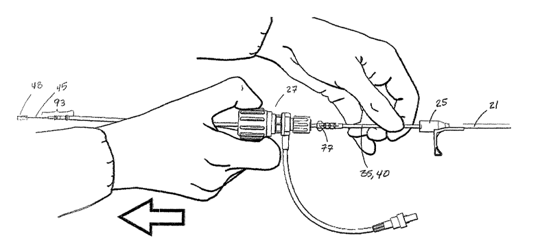
Figure 16 is a schematic diagram of a rotatable hemostasis valve (RITE of a
tool
kit according to the present invention. As illustrated in Figure 16, according
to the present
invention, a rotatable hemostasis valve (RHV) 27 of tool kit 10 includes a non-
standard
Touhy Borst valve 28, a side arm flush port assembly 26, and a non-standard
male luer
fitting 16 (Figure 39) within a locking collar 8. Proximal portion 14 of
delivery sheath 21,
23 is terminated with a slittable hub 25 of delivery sheath 21 or 23, such as
the slittable
CA 02483442 2004-10-22
WO 03/090833 PCT/US03/09737
29
hub described in U.S. Patent 6,159,198 to Gardeski et al., which is
incorporated in its
entirety herein. Slittable hub 25 includes non-standard female Iuer fitting 37
for the
connection of RHV 27. RHV 27 is connected to hub 25 prior to inserting
delivery sheath
21, 23 into venous system. According to the present invention, non-standard
male and
female luer fittings 16 and 37 have a diameter approximately twice that of
standard leer
fittings that are well known in the art. Furthermore, Touhy Borst valve 28 has
a Larger
maximum inner diameter (not shown) than standard Touhy Borst valves also well
known
in the art. The advantage of larger diameter luer fittings and Touhy Borst
valve 28 will be
presented, With a more detailed description of RHV 27, below, in conjunction
with Figures
38 and 39.
Hub 25 has an opening large enough to accommodate a special rotatable
hemostatic valve (RHV) 27, to which it is detachably secured by, e.g. , an
annular ring on
the inner diameter of valve 27. A central lumen 33 in RHV 27 is aligned and in
fluid
communication with the lumen within a shaft 36. Lumen 33 has a diameter large
enough
to accommodate a balloon catheter and a typical lead connector, such as an IS-
1-type
connector, for example. An optional side arm 26 may be disposed on RHV 27 in
fluid
communication with lumen 33. RHV 27 rnay also be splittable via a scoring or
perforation as described above.
An annular polymeric locking collar 8 is disposed on the outside diameter of
RHV
27 distal portion proximal to the point where hub 25 meets RHV 27. In this
embodiment,
rotation of collar 8 locks RHV 27 to hub 25.
Figure 17 is a schematic diagram of a delivery sheath for delivering a medical
electrical device within a coronary venous system, according to the present
invention,
from a right-sided venous access point to a coronary sinus. Figure 18 is a
schematic
diagram of a delivery sheath for delivering medical devices within a coronary
venous
system, according to the present invention, from a Left-sided venous access
point to a
coronary sinus. Figures 17 and 18 illustrate the right and left sided
approaches, after distal
tip 15 of delivery sheath 21, 23 has been seated in the coronary sinus 930.
Intxoducer .
guide wire 4 or steerable catheter 1002 has been removed from Lumen of
delivery sheath
21, 23. As illustrated in Figures 17 and 18, left-sided venous access point
960 is a greater
distance from the ostium of coronary sinus 930 than right-sided venous access
point 950,
CA 02483442 2004-10-22
WO 03/090833 PCT/US03/09737
and the approach to the coronary sinus 930, from left-sided venous access
point, is not as
direct. Left-sided venous access point 960 may be selected because venous
anatomy
communicating from right-sided access point 950 may be blocked or because a
preferred
implant site 970 for a medical device that is to be connected with a medical
electrical lead
5 is on a left side.
Once a passageway formed by lumen of delivery sheath 21, 23 has been
established, as illustrated in Figures 17 or 18, balloon catheter 20 may be
advanced down
lumen of delivery sheath 21, 23 and into coronary sinus 930 to obtain a
venogram. A
smaller guide wire or a smaller steerable catheter or deflection mechanism may
be used,
10 within a lumen of balloon catheter 20 in order to guide balloon catheter 20
distally into
coronary sinus 930 from distal tip 15 of delivery sheath 21, 23. After
obtaining venogram,
balloon catheter 20 is removed from delivery sheath 21, 23. A navigation
pathway
established for delivery of a medical electrical lead is a combination of
passageway
through delivery sheath 21, 23, into coronary sinus 930, and venogram obtained
using
15 balloon catheter 20.
Medical Electrical Leads and Accessory Tools
Figure 19 is a plan view of a medical electrical lead having a lumen for
receiving a
siylet or a guide wire for delivering a medical electrical device within a
coronary venous
20 system according t~ the present invention. As illustrated in Figure 19, a
guide wire 46 for
introducing a medical electrical lead 40 within the venous system, which is
significantly
smaller and of a different construction than introducer guide wire 4, is used
with delivery
sheath 21 or 23. Guide wire 46, which includes an atraumatic formable tip 47,
is the same
type used with either occlusion balloon catheter 20 or an angioplasty balloon
catheter
25 having a construction well known in the art.
Figure 19 illustrates guide wire 46 inserted into a lumen 34 of lead 40 with
formable atraumatic tip 47 protruding from a distal tip 41 of lead 40. Lumen
34 of lead
has a diameter between approximately 0.014 inches and 0.022 inches and extends
from
a proximal opening 38 at a connector pin 93 of a connector 50 of lead 40 to a
distal
30 opening 39 in distal tip 41 of lead 40. An anchoring sleeve 77 can also be
used to reduce
corruption of the lead body caused by suturing once lead 40 has been properly
positioned
CA 02483442 2004-10-22
WO 03/090833 PCT/US03/09737
31
within the venous system. Guide wire 46 is used to steer and guide lead distal
tip 41 to a
target site in coronary veins by advancing lead 40 over guide wire 46. Such an
embodiment of lead 40, called an "over-the-wire lead", is disclosed in
commonly assigned
TJ.S. Patent 6,192,280 B1, which is incorporated by reference herein its
entirety. A length
of guide wire 46 to be used with lead 40 exceeds a length of lead 40, so that
tip 47 of
guide wire 46 protrudes from distal tip 41 of lead 40, while a proximal
portion of guide
wire 46 extends proximally from connector pin 93 A guide wire steering tool 94
may be
attached to a pxoximal portion of guide wire 46 to facilitate steering of
guide wire 46.
According to a preferred embodiment of the present invention, the maximum
diameter~of
guide wire 46 is between approximately 0.012 inches and 0.020 inches.
Figure 20 is a schematic of a guide wire atraumatic formable tip protruding
from a
lead distal tip of a medical electrical lead and navigating from the coronary
sinus into a
branch vein. As illustrated in Figure 20, guide wire 46, shown by a dashed
line, may have
been loaded into lumen of lead 40, illustrated in Figure 19, with loading
device 51,
illustrated in Figures 24-26, then lead 40 and guide wire 46, together, were
advanced
through delivery sheath 21 to coronary sinus 930. On the other hand, lead 40
could
initially be positioned using a stylet wire (Figure 21), which is then
replaced by guide wire
46, or lead 40 could initially be advanced through delivery sheath 21 or 23
and guide wire
inserted later. It is therefore understood that many possible ordering of the
steps could be
used to delivery a medical electrical lead, all of which are merely a matter
of operatox
preference, and therefore the present invention is not intended to be limited
to preferred
ordering of the steps utilizing the aspects of the present invention, by
rather is intended to
include the steps performed in any order that is merely a matter of user
preference.
A contrast agent could have been injected down lumen of lead 40 to provide
xeal-
time fluoroscopic guidance as guide wire tip 47 is manipulated to sub-select
branch vein
932. According to the present invention, Figure 20 illustrates a means for
navigating lead
tip 41 into branch vein 932. A distal bend 42 of lead 40 provides both
guidance and back-
up support for guide wire atraumatic formable tip 47 to advance into branch
vein 932.
Once guide wire tip 47 has cannulated branch vein 932 and is seated deep
enough, lead tip
41 can be pushed forward over guide wire 46 to target site in branch vein 932.
Guide wire
CA 02483442 2004-10-22
WO 03/090833 PCT/US03/09737
32
clip 6, illustrated in Figure 13, may also be used to manage excess length of
guide wire 46
in a similar manner to that previously described for introducer guide wire 4
of Figure 13.
Figure 21 is a planar view of a stylet wire inserted within a medical
electrical lead
in a system for delivering medical devices within a venous system according to
the present
invention. As illustrated in Figure 21, a stylet wire 45, which typically has
a greater
stiffness than guide wire 46, is insertable within central lumen 34 of medical
electrical
lead 40 in place of guide wire 46 to assist in the insertion of lead 40 within
venous system.
Stylet wire 45 includes a distal portion 44, along with a stylet knob 48
attached to a
proximal end of stylet wire 45. Stylet wire 45 has a length relative to lead
40 such that
once stylet wire 45 is fully inserted within lumen 34, knob 48 of stylet wire
45 engages
against connector pin 93 at the proximal end of connector pin 50. As a result,
knob 48 of
stylet wire 46 prevents further insertion of stylet wire 45 within lumen 34 so
that distal
portion 44 of stylet wire 45 does not extend outward from distal tip 41 of
lead 40. Once
fully inserted within lumen 34, stylet wire 45 is subsequently utilized to
assist in directing
insertion of lead 40 within the venous system.
Figure 22 is planar side view of a medical electrical lead having a lumen for
receiving a stylet wire and a guide wire in a system for delivering medical
devices within a
venous system according to the present invention. Figure 23 is a cross-
sectional side view
of a lead distal tip of the medical electrical lead of Figure 22. As
illustrated in Figures 22
and 23, guide wire 46 and stylet wire 45 of tool kit 10 are also insertable
within distal tip
of a side-lumen lead 35. Similar to over-the-wire lead 40, side-lumen lead 35
includes
connector pin 50 and central lumen 34. However, distal tip 30 of lead 35
differs from
distal tip 41 of lead 40 since distal tip 30 includes a side lumen 32 that
extends from a side
25 lumen distal end 33 to a side lumen proximal end 43. As illustrated in
Figure 23, guide
wire 46 is insertable within side lumen 32 by first being inserted at lumen
distal end 33 of
side lumen 32 in a direction indicated by arrow C, and exiting side lumen 32
at lumen
proximal end 43. Once inserted within lumen 32 of lead 35, tip 47 of guide
wire 46 is
advanced within venous system, so that once tip 47 is positioned at a desired
location
30 within the coronary sinus, lead 35 is advanced over guide wire 46 to
subsequently position
lead distal tip 30 at the desired location, as described below.
CA 02483442 2004-10-22
WO 03/090833 PCT/US03/09737
33
In addition, as illustrated in Figures 22 and 23, stylet wire 45 may also be
inserted
within central lumen 34 of lead 35 at opening 49 of connector pin 93 and
advanced
through lumen 34 to provide additional stiffness fox advancing lead 35 within
the venous
system. As illustrated in Figures 22 and 23, lumen 34 of lead 35 extends from
opening 49
at connector pin 93 at the proximal end of connector 50 to a lumen end wall 91
located
inside distal tip 30 of lead 35. As a result, once stylet wire 4S is fully
inserted within
lumen 34, distal portion 44 of stylet wire 45 engages against end wall 91,
preventing stylet
wire 45 from being advanced outward from distal portion 30 of lead 35. Once
inserted
within lumen 34, stylet wire 45 provides further assistance in directing
insertion of lead 35
within the venous system by providing the additional stiffness to lead 35 when
advancing
distal tip 30 along guide wire 46.
Figure 24 is a schematic diagram of a loading device in a system for
delivering
medical devices within a venous system according to the present invention.
Figure 25 is a
cross-sectional view of the loading device of Figure 24. As illustrated in
Figures 24 and
25, a loading device 51 in a system for delivering medical devices within a
venous system
according to the present invention includes a navigation portion 54 having an
opening 59
formed at a proximal end of loading device 51, an alignment lumen 55
positioned within
an alignment shaft 52, and an engagement cavity 58 positioned at a distal end
of loading
device 51. Opening 59 of navigation portion 54 directs a formable atraumatic
tip 47 of
guide wire 46 or distal portion 44 of stylet wire 45 within navigation portion
54, which
then directs tip 47 or distal portion, respectively, into aligrunent lumen 55
through a
proximal lumen opening 53 of alignment lumen 55.
An inner diameter of engagement cavity 58 is sized to snap-fit connector pin
93 so
that a distal lumen opening 56 of loading device 51 is aligned with an opening
49 of lumen
34 of lead 35, 40 at connector pin 93 for continuity between alignment lumen
55 of
loading device 51 and lead lumen 34 of lead 35, 40. According to a preferred
embodiment
of the present invention, engagement cavity 58 includes an inner diameter
between
approximately 0.059 inches and 0.061 inches and a Length between approximately
0.1
inches and 0.2 inches. An engagement cavity wall 59 of engagement cavity 58
forms an
opening 74 so that engagement cavity wall 59 does not completely enclose
connector pin
93 when connector pin 93 is inserted within engagement cavity 58. As a result,
electrical
CA 02483442 2004-10-22
WO 03/090833 PCT/US03/09737
34
contact can be made with connector pin 93 when loading device 51 and connector
pin 93
axe fixedly engaged.
Figure 26 is a schematic diagram of a lead connector fixedly inserted within
the
loading device of Figure 24. In particular, as illustrated in Figure 26, once
connector pin
93 of connector 50 is fixedly inserted within cavity 58 of loading device 51,
tip 47 of
guide wire 46 or distal portion 44 of stylet wire 45 is inserted at opening 59
of navigation
portion 54 in a direction shown by arrow Y, and is directed within lumen
opening 53 of
alignment lumen 55 by navigation portion 54. Tip 47 or distal portion 44 is
then directed
through alignment lumen 55 towards distal lumen opening 56. Since opening 56
is
aligned with opening 49 of lumen 34 of connector 50 at connector pin 93, tip
47 or distal
portion 44 passes through openings 56 and 49 and into lumen 34 of connector
50.
According to the present invention, if loading tool 51 is utilized to load
stylet wire
45, loading tool 51 and hemostasis valve 27 are sized so that hemostasis valve
27 can be
advanced over loading tool 51 and stylet knob 48 to remove hemostasis valve 27
from lead
35, 40 once lead 35, 40 is advanced within the coronary venous system to a
target site, as
described below.
Figure 27 is an isometric diagram of an alternate embodiment of a loading
device
in a system fox delivering medical devices within a venous system according to
the present
invention. As illustrated in Figure 27, a loading device 151, according to an
alternate
preferred embodiment of the present invention is similar to loading device 51
described
above in reference to Figures 24 and 25 above. Accordingly, loading device 151
fixedly
engages with connector pin 93 in a manner as described above in reference t~
Figures 24
and 25, and therefore a description of the similar features, indicated by like
reference
numerals, is omitted merely for brevity.
Figure 28 is a front planar view of the loading deviee of Figure 27 in a
closed
position. Figure 29 is a cross-sectional side view of a loading device
according to the
present invention, taken along cross-sectional lines VII-VII of Figure 28. As
illustrated in
Figures 27-29, similar to loading device S 1, loading device 151 includes
engagement
cavity 58, alignment shaft 52, and navigation portion 54 forming opening 59.
However,
according to an alternate embodiment of the present invention, loading device
151
includes a slot 1 S2 extending from the distal end of alignment shaft 52 to
the proximal end
CA 02483442 2004-10-22
WO 03/090833 PCT/US03/09737
of navigation portion 54 at opening 59. As illustrated in Figures 27 and 28,
slot 152
extends through an outer wall 153 of navigation portion 54 and through
alignment shaft 52
to alignment lumen 55 (Figure 29) and is defined by a first side wall 154 of
alignment
shaft 52 adjacent a second side wall 156 of alignment shaft 52, and a first
side wall 158 of
outer wall 153 of navigation portion 54 adjacent a second side wall 160 of
outer wall 153
of navigation portion 54. Loading device 151 also includes spaced flange
portions I62
and 164 extending from the distal end of alignment shaft 52 and terminating
along outer
wall 153 of navigation portion 54. A distance between side walls 154-160 of
slot 152 is
less than the diameter of guide wire 46 or stylet wire 45 when slot 152 of
loading device
10 151 is in a closed position, illustrated in Figure 28. As a result, loading
device 151 cannot
be removed directly from guide wire 46 or stylet wire 45 when slot 152 of
loading device
151 is in the closed position.
Figure 30 is a front planar view of the loading device of Figure 27 in an open
position. As illustrated in Figure 30, once pressure is applied at flange
portions 162 and
15 164 in a direction shown by arrows P and P', respectively, the applied
pressure causes
flange portions 162 and 164 to be displaced relative to each other so that a
distance
between flange portions 162 and 164 is reduced, and slot 152 of loading device
151 is
moved from the closed position of Figure 28 to an open position, shown in
Figure 30. The
relative displacement of flange portions 162 and 164 causes side walls 154-I60
of slot 152
20 to be displaced so that the distance between side walls 154-160 of slot 152
is greater than
the diameter of guide wire 46 ox stylet wixe 45, so that loading device 151
can be directly
removed from guide wire 46 or from stylet wire 45 through slot 152 when slot
152 is in
the open position.
Figure 31 is a top perspective view of an alternate embodiment of a loading
device
25 for loading a guide wire within a medical electrical lead according to the
present
invention. As illustrated in Figure 31, a loading device 60 according to an
alternate
embodiment of the present invention includes a navigating portion 64 extending
from a
front end 80 to a back end 82. Navigation portion 64 includes an outer portion
94 and a
first side wall 95 spaced apart from a second side wall 96 to form a slot 65
that extends
30 from front end 80 to back end 82 of navigation portion 64. An opening 75 is
formed at
back end 82 of navigation portion 64 and couples navigation portion 64 with an
CA 02483442 2004-10-22
WO 03/090833 PCT/US03/09737
36
engagement cavity 66. Navigation portion 64 receives guide wire 46 as guide
wire 46 is
inserted within an opening 97 formed at front end 80 and directs guide wire
46, as guide
wire 46 is inserted within navigation portion 64, towards opening 75 at back
end 82.
Guide wire 46 is then directed within engagement cavity 66 through opening 75
at back
end 82.
Loading device 60 also includes engagement cavity 66 and a lead slot 68 for
receiving and positioning lead tip 30 and a lead body distal portion 31,
respectively, within
loading device 60. An insertion guide 70 is formed on an upper surface 62 of
loading
device 60 to assist the user in properly positioning lead 35 for insertion
within engagement
cavity 66 and lead slot 68. Engagement cavity 66 is shaped to form a line-to-
line or
minimum clearance fit around lead distal tip 30 to orientate lead distal tip
30 to be in a
position corresponding to the orientation indicated by insertion guide 70 in
order to
prevent lead distal tip 30 from being corrupted when lead distal tip 30 is
inserted within
engagement cavity 66. For example, according to a preferred embodiment of the
present
invention, engagement cavity 66 is sized to extend approximately 0.002 inches
from lead
distal tip 30 when lead distal tip 30 is positioned within engagement cavity.
On the other
hand, lead slot 68 is shaped to engage lead body distal portion 31 to snap-fit
lead body
distal portion 31 within lead slot 68.
Back end 82 of navigation portion 64 is located along engagement cavity 66 so
that
back end 82 is aligned with side lumen 32 of lead tip 30 at lumen distal end
33 when lead
tip 30 is inserted within engagement cavity 66 in a position corresponding to
the
orientation indicated by insertion guide 70 and lead body distal portion 31 is
snap-fit to be
fixedly engaged within lead slot 68. As a result, loading device 60 of the
present
invention enables guide wire 46 to be more easily inserted within side lumen
32 of lead tip
30, as will be described below.
Figure 32 is a cross-sectional side view of a loading device according to the
present invention, taken along cross-sectional line IV-IV of Figure 31. As
illustrated in
Figures 31 and 32, a ramp portion 63 is formed in engagement cavity 66 of
loading device
60. Ramp portion 63 extends from a lower end 67 to an upper end 69 to form an
upward
extending surface 71 that directs guide wire 46 out of engagement cavity 66 as
guide wire
46 is inserted within navigation portion 64 of loading device 60 and through
side lumen 32
CA 02483442 2004-10-22
WO 03/090833 PCT/US03/09737
37
of lead 35 once lead 35 is inserted within engagement cavity 66 and lead slot
68 of loading
device 60, as described below.
Figure 33 is a top planar view illustrating insertion of a guide wire within a
medical electric lead using a loading device according to the present
invention. Figure 34
is a cross-sectional side view of a loading device according to the present
invention, taken
along cross-sectional lines V-V of Figure 33. As illustrated in Figures 33 and
34, lead 35
is inserted Within loading device 60 by positioning lead tip 30 to match an
orientation
depicted by insertion guide 70, and once lead tip 30 is oriented in the same
position as
shown by insertion guide 70, lead 35 is positioned within loading device 60 by
inserting
lead tip 30 within engagement cavity 66 and snap-fitting lead body distal
portion 31 within
lead slot 68 of loading device 60 to fixedly engage lead body distal portion
31 within
loading device 60.
Once positioned within engagement cavity 66, distal end 33 of side lumen 32 of
lead 35 is aligned with opening 75 of navigation portion 64. After lead 35 is
inserted
within loading device 60, guide wire 46 is inserted within navigation portion
64 in a
direction A. By aligning opening 75 of navigation portion 64 with distal end
33 of side
lumen 32, once navigation portion 64 guides wire 46 to be advanced through
opening 75,
loading device 60 directs guide wire 46 to be advanced within lumen distal end
33 of side
lumen 32 of lead 35. As guide wire 46 is advanced through side lumen 32, and
guide wire
46 subsequently exits side lumen 32 at lumen proximal end 43, ramp portion 63
directs
guide wire 46 out of engagement cavity 66 as guide wire 46 is extended through
side
lumen 32 of lead tip 30.
According to a preferred embodiment of the present invention, upper surface 62
of
loading device 60 which is approximately one inch square with a thickness
between
approximately 0.15 inches and 0.25 inches, provides a platform that can easily
be held by
an operator while engaging lead body distal portion 31 and lead distal tip 30,
and directing
guide wire 46 into side lumen 32. Suxface of handling interface 62 is also
large enough to
fit etched insertion guide 70 depicting lead body distal poxtion 31 and distal
tip 30 at a 1:1
scale. Insertion guide 70 aids operator in correct placement of lead distal
tip 30 into
engagement cavity 66.
CA 02483442 2004-10-22
WO 03/090833 PCT/US03/09737
38
According to the present invention, first side wall 9S is spaced from second
side
wall 96 at a distance that enables guide wire 46 to be advanced between first
side wall 95
and second side wall 96. As a result, once guide wire 46 is positioned within
side lumen
32, lead 3S is removed from loading device 60 with guide wire 46 positioned
through side
S lumen 32 by removing distal portion 31 and lead distal tip 30 of lead 3S
from lead slot 68
and engagement cavity 66, respectively, and removing guide wire 46 from within
navigation portion 64 by advancing guide wire 46 through slot 6S.
Figure 3 S is a top planar view illustrating insertion of a guide wire within
a
medical electric lead using an alternate embodiment of a loading device
according to the
present invention. The alternate embodiment of loading device 60 differs in
that loading
device 60 is formed to enable loading of guide wire 46 within an over-the-wire
lead 40
having a lead distal tip 41 with a lumen 43 centrally located to extend
through lead 40
from a distal end 61 of lead distal tip 41. In particular, as illustrated in
FIG. 35, according
to the alternate embodiment of the present invention, engagement cavity 66 of
loading
1 S device 60 is formed to receive lead distal tip 41 to assist in the
insertion of guide wire 46
within lead 40. Lead 40 is inserted within loading device 60 by positioning
lead distal tip
41 to match an orientation depicted by insertion guide 70, and once lead
distal tip 41 is
oriented in the same position as shown by insertion guide 70, lead 40 is
positioned within
loading device 60 by inserting lead tip 41 within engagement cavity 66 and a
lead body
distal portion 72 within lead slot 68 of loading device 60.
Engagement cavity 66 is shaped to form a line-to-line or minimum clearance fit
around lead distal tip 41 to orientate lead distal tip 41 to be in a position
corresponding to
the orientation indicated by insertion guide 70 in order to prevent lead
distal tip 4I from
being corrupted when inserted within engagement cavity 66. For example,
according to a
2S preferred embodiment of the present invention, engagement cavity 66 is
sized to extend
approximately 0.002 inches from lead distal tip 41 when lead distal tip 41 is
positioned
within engagement cavity 66. Qn the other hand, lead slot 68 is shaped to
engage lead
body distal portion 72 to snap-fit lead body distal portion 72 within lead
slot 68. In
addition, similar to the preferred embodiment described above in reference to
Figures 31-
34, back end 82 of navigation portion 64 is located along engagement cavity 66
so that
back end 82 is aligned with lumen 43 at lumen distal end 61 when lead distal
tip 41 is
CA 02483442 2004-10-22
WO 03/090833 PCT/US03/09737
39
inserted within engagement cavity 66 in a position corresponding to the
orientation
indicated by insertion guide 70 and lead body distal poxtion 72 is snap-ftt to
be fixedly
engaged within lead slot 68. As a result, loading device 60 of the present
invention
enables guide wire 46 to be rnoxe easily inserted within lumen 43 of lead
distal tip 41.
After lead 40 is inserted within loading device 60, guide wire 46 is inserted
within
navigation portion 64 at opening 97 in direction B, with navigation portion 64
directing
guide wire 46 towards opening 75 so that guide wire 46 is inserted within lead
distal tip 41
at lumen distal end 61 of lumen 43. Guide wire 46 is directed into lumen 43 at
distal tip
41, passes through lumen 43, and travels out proximal opening in connector pin
50 (Figure
21). Once guide wire 46 is positioned within Iumen 43, lead 40 is removed from
loading
device 60 with guide wire 46 positioned through lumen 43 by removing lead
distal tip 41
and distal portion 72 of lead 40 from engagement cavity 66 and lead slot 68,
respectively,
and removing guide wire 46 from within navigation portion 64 by advancing
guide wire
46 through slot 65.
Figure 36 is a schematic diagram of positioning of a guide wire 46 within a
branch
vein. According to the present invention, Figure 36 illustrates a situation in
which guide
wire 46 was used to deliver venogram balloon catheter 20, illustrated in
Figure 13, through
delivery sheath 21. After obtaining venogram with balloon catheter 20,
atraumatic
fonnable tip 47 of guide wire 46 is advanced to a target site in branch vein
933;
positioning of atraumatic formable tip 47 may have been facilitated by
additional
injections of a contrast agent down a lumen of balloon catheter 20. Guide wire
46 is left in
position when balloon catheter 20 is removed. Proximal end of guide wire 46
may be
directed into side lumen 32 of lead tip 30, illustrated in Figures 21 and 22
and 32, using
loading device 60 as illustrated in Figure 31. Lead tip 30 is pushed along
guide wire 46
2S until lead tip 30 reaches the target site in branch vein 933. As
illustrated in Figures 21 and
22, stylet wire 45 may be inserted into central lumen 34 of lead 35 to provide
additional
stiffness for moving lead distal tip 30 along guide wire 46.
Guide wire 46 and /or stylet wire 45 may be removed before delivery sheath 21
is
removed, however a preferred method is to retain guide wire 46 and /or stylet
wire 45 until
after delivery sheath 21 is removed. A retained stylet wire 45 helps maintain
stiffness in
lead 35 that can be used to hold lead tip 30 in position while delivery sheath
21 is being
CA 02483442 2004-10-22
WO 03/090833 PCT/US03/09737
removed. If removal of delivery sheath 21 dislodges lead tip 30, a retained
guide wire 46
will help to re-position lead tip 30.
Figures 37 and 38 are schematic diagrams illustrating removal of a delivery
sheath
and a rotatable hernostasis valve from an implanted lead body according to the
pxesent
invention. Once insertion of the lead has been completed, delivery sheath 21,
23 must be
slit and peeled off from lead body 35, 40 since outer diameters of an industry
standard IS-
1 connector 50 and an anchoring sleeve 77, mounted on lead body 73, are
significantly
larger than a diameter of lead body 35, 40 and will not fit through lumen of
delivery
sheath 21. In addition, hemostasis valve 27 must also be removed from lead
body 35, 40.
10 According to the present invention, hemostasis valve 27 is first removed
from hub 25 of
sheath 21, 23 by rotating collar 37 to unlock hemostasis valve 27 from hub 25.
Once
unlocked from hub 25, hemostasis valve 27 is slid over lead 35, 40 to advance
hemostasis
valve 27 from an attached position, illustrated in Figure 37, to an unattached
position,
illustrated in Figure 38, so that hemostasis valve 27 is slid over anchoring
sleeve 77 and
15 connector 50, and over knob 48 of stylet wire 45, if stylet wire 45 is
utilized.
Figure 39 is partial section plan view of a hemostasis valve according to the
present invention. As illustrated in Figure 39, in order to enable hemostasis
valve 27 to be
removed from lead 35, 40 by being slid over connector 50, hemostasis valve 27
of the
present invention includes a non-standard Touhy Borst valve 28 having an
adjustable
20 lumen 29 and a non-standard male luer fitting 16 within a locking collar 8.
According to
the present invention, a minimum internal diameter of adjustable lumen 29 is
small
enough to seal on lead body 63, approximately .050 inches in diameter, and a
maximum
internal diameter of adjustable lumen is approximately .2 inches in diameter.
Furthermore, an inner diameter (shown with dashed line) of non-standard male
luer fitting
25 16 is approximately 0.2 inches. Both the maximum diameter of adjustable
lumen 29 and
inner diameter of male luer fitting 16 are large enough to allow passage of
lead anchoring
sleeve 77 and lead connector 50 as RHV 27 is removed from delivery sheath 21,
23.
Returning to Figure 38, according to the present invention, stylet wire 45
includes
knob 48 having a diameter large enough so that knob 48 cannot pass into lumen
of lead
30 but small enough so that knob 48 can pass through maximum diameter of
adjustable
lumen 29 of Touhy Borst valve 28 and inner diameter of male luer fitting 16.
CA 02483442 2004-10-22
WO 03/090833 PCT/US03/09737
41
Figure 40 is a flowchart of a method of delivering a medical electrical lead
within a
coronary sinus according to the present invention. As illustrated in Figure
40, a method of
delivering a medical electrical lead within a coronary sinus according to the
present
invention includes establishing venous access, Step 500. Once venous access is
established, a delivery sheath is chosen corresponding to the desired approach
to the
coronary sinus, Step 502. For example, if a right-sided approach is preferred,
delivery
sheath 21 is chosen, and if a left-sided approach is preferred, delivery
sheath 23 would be
chosen. Rotatable hemostasis valve 27 is coupled at proximal portion 14 of
delivery
sheath 21 or 23, Step 504, and delivery sheath 21 or 23 is inserted within the
venous
access over guide wire 4 of tool kit 5, Step 506. Once guide wire 46 is
inserted within
lead lumen 34, 32 of distal tip 41, 30 of lead 35, 40, respectively, Step 508
and a distal tip
41, 30 of lead 35, 40 is inserted through hemostasis valve 27 and within the
delivery
sheath 21 or 23, Step SIO, distal tip 41, 30 is advanced to a target site
within the coronary
venous system using guide wire or stylet wire 45 ox both, Step 512. Once
distal tip 41, 30
of lead 35, 40 is advanced using guide wire 46 and/or stylet wire 45 to
position distal tip
41, 30 at the target site, hemostasis valve 27 is decoupled from hub 25 of
delivery sheath
21, 23 and advanced over connector 50 of lead 35, 40 in order to remove
hemostasis valve
27 from lead 35, 40, Step 516. Delivery sheath 21, 23 is subsequently removed
from the
venous system, Step 518, using known slitting techniques, for example.
In addition, according to the present invention, when either stylet wire 45 is
utilized to guide delivery of distal tip 41, 30 or anchoring sleeve 77 is
utilized, alone, or in
combination, hemostasis valve 27 of the present invention is also advanced
over the
utilized inserted stylet wire 45 and/or anchoring sleeve 77. Furthermore, if
loading tool 51
is utilized to load stylet wire 45 within lead 35 or 40 or to load guide wire
within lead 40,
hemostasis valve 27 is also advanced over loading tool 51.
The method of delivering a medical electrical lead within a coronary sinus
according to the present invention may also include obtaining a venogram using
balloon
catheter 200, as described above.
Many alterations and modifications may be made by those having ordinary skill
in
the art without departing from the spirit and scope of the invention. The
illustrated
variations have been used only for the purposes of clarity and should not be
taken as
CA 02483442 2004-10-22
WO 03/090833 PCT/US03/09737
42
limiting the invention as defined by the following claims. For example,
although delivery
sheaths 21, 23 are described with distal portions 12, 13 illustrated in Figure
13,
combinations of delivery sheaths with other styles of distal curvature that
axe well known
in the art, such as Judkins and Amplatz, may be included in alternative
embodiments of
tool kit 10.