Note: Descriptions are shown in the official language in which they were submitted.
CA 02483719 2011-06-10
APPLIER FOR FASTENER FOR SINGLE LUMEN ACCESS
ANASTOMOSIS
Field of the Invention
[0002] The present invention relates, in general, to devices and methods for
surgically
modifying organs and vessels. More particularly, it relates to anastomosis
devices for
joining two organs such as, for example, two separate lengths of small bowel
to each
other, a section of small bowel to the stomach, or the common bile duct to the
duodenum
in a procedure called a choledochoduodenostomy. Vascular anastomosis may be
performed as well.
Background of the Invention
[0003] The percentage of the world population suffering from morbid obesity is
steadily
increasing. Severely obese persons are susceptible to increased risk of heart
disease,
stroke, diabetes, pulmonary disease, and accidents. Because of the effect of
morbid
obesity to the life of the patient, methods of treating morbid obesity are
being researched.
[0004] Numerous non-operative therapies for morbid obesity have been tried
with
virtually no permanent success. Dietary counselling, behavior modification,
wiring a
patient's jaws shut, and pharmacological methods have all been tried, and
though
- 1-
CA 02483719 2004-09-29
temporarily effective, failed to correct the condition. Further, introducing
an object in
the stomach, such as an esophago-gastric balloon, to fill the stomach have
also been
used to treat the condition; however, such approaches tend to cause irritation
to the
stomach and are not effective long-term.
[00051 Surgical treatments of morbid obesity have been increasingly used with
greater success. These approaches may be generalized as those that reduce the
effective size of the stomach, limiting the amount of food intake, and those
that create
malabsorption of the food that it is eaten. For instance, some patients
benefit from
adjustable gastric bands (AGB) that are advantageously laparoscopically placed
about
the stomach to form a stoma of a desired size that allows food to fill an
upper portion
of the stomach, causing a feeling of satiety. To allow adjustment of the size
of the
stoma after implantation, a fluid conduit communicates between an inwardly
presented fluid bladder of the AGB to a fluid injection port subcutaneously
placed in
front of the patient's sternum. A syringe needle may then inject or withdraw
fluid as
desired to adjust the AGB.
100061 Although an effective approach to obesity for some, other patients may
find
the lifestyle changes undesirable, necessitated by the restricted amount of
food intake.
In addition, the medical condition of the patient may suggest the need for a
more
permanent solution. To that end, surgical approaches have been used to alter
the
portions of the stomach and/or small intestine available for digesting food.
Creating
an anastomosis, or the surgical formation of a passage between two normally
distinct
vessels, is a critical step of many surgical procedures. This is particularly
true of
gastric bypass procedures in which two portions of small intestine are joined
together
and another portion of small intestine is joined to the stomach of the
patient. This is
also true of surgery to alleviate blockage in the common bile duct by draining
bile
from the duct to the small intestine during surgery for pancreatic cancer.
(00071 With particular reference to gastric bypass procedures, current methods
of
performing a laparoscopic anastomoses for a gastric bypass include stapling,
suturing,
and placing biofragmentable rings, each having significant challenges. For
instance,
suturing is time consuming, as well as being technique and dexterity
dependent.
Stapling requires placement of an anvil, which is a large device that cannot
be
-2-
CA 02483719 2011-06-10
introduced through a trocar port. Having to introduce the port through a
laparotomy
presents an increased incidence of wound site infection associated with
Intralumenal
content being dragged to the laparotomy entry site.
[00081 As an example of the latter approach, in U.S. Pat. No. 6,543,456 a
method for
gastric bypass surgery includes the insertion of proximal and distal
anastomosis
members (e.g., anvils) transorally with grasping forceps. The stomach and the
small
intestine are transected endoscopically by a surgical severing and stapling
instrument
to create a gastric pouch, a drainage loop, and a Roux limb. An endoscopically
inserted circular stapler attaches to the distal anastomosis member to join
the drainage
loop to a distal portion of the intestine, and the circular stapler attaches
to the
proximal anastomosis member to join the Roux limb to the gastric pouch.
Thereafter,
the anastomosis members are removed to create an orifice between joined
portions of
the stomach and intestine. This method reduces the number of laparoscopic
ports,
avoids a laparoscopic insertion of an anastomosis instrument (e.g., circular
stapler)
into an enlarged surgical port, and eliminates the need for an enterotomy and
an
enterotomy closure.
[00091 For many anastomoses, surgeons use circular staplers, linear staplers,
or
manual sutures. However, to reduce incision size and to make the surgical
process
less technically demanding and time consuming, an anastomotic device that
deforms
to hold tissue portions together when the device is ejected from a
constraining
enclosure has been described. Such an approach is described in U.S. Pat. Appl.
Publ.
No. US 2003/0032967 and PCT application WO 03/000142 both to Adrian Park et
al,
describes such a device. Therein, gastrointestinal or enteric (including
biliary)
anastomosis is achieved by insertion of a sheath that perforates the walls of
two tissue
passages, such as the stomach and small intestine. A three-dimensional woven
tube of
wire having a thermal shape memory effect (SME) ("generally-known nitinol ring
device") is presented by a cannula of the sheath on both sides of the
openings.
Deployment of the woven tube causes the outer loops or ends of the tube to
fold or loop
back to hold the luminal interface of the anastomosis site in apposition.
Thereby, the
need for a mechanical compression component in a delivery system is reduced or
avoided, reducing the size and complexity of the delivery device.
-3-
CA 02483719 2004-09-29
100101 The anastomotic device disclosed in WO 03/000142 is constrained by a
retractable sheath to an advantageous small-diameter tubular shape. A surgeon
applies
the anastomotic device by maneuvering the sheath through the tissue portions
requiring anastomosis and retracting the sheath. Retracting the sheath removes
the
constraint on the device, allowing the device to assume a roughly hourglass
shape.
The larger ends of the hourglass shape hold the two tissue portions together
in an
effective anastomosis.
100111 The constrained anastomotic device, which may be made of a shape memory
material such as nitinol, exerts a force against the inner diameter of the
sheath and
tends to warp towards its roughly hourglass-shaped deployed position. When the
sheath is retracted proximally, the forces generated by the device in
transition from a
tubular shape to an hourglass shape urge the anastomotic device distally. This
device
movement makes surgical control harder to achieve when placing the device
through
the otomies of two tissue portions requiring anastomosis.
(00121 While the generally-known nitinol ring device is a significant
advancement in
the treatment of morbid obesity, it is believed that further improvements
would be
desirable. For instance, weaving the wire strands and fastening together the
ends and
heat treating the woven tubes into an SME device is expensive. In addition, it
may
tend to be difficult to maintain two lumens that are to be anastomotized in
extremely
close contact in order for the generally-known nitinol ring device to
successfully
attach to both sides. Having to insert one or more grasping tools along with
the
anastomosis ring applier tends to mitigate the advantages of a single lumen
anastomosis by requiring multiple access ports. Moreover, even if the lumens
are
proximately position, the generally-known nitinol ring device tends to actuate
slowly,
if at all, by being limited to SME actuation.
100131 Consequently, there is a general need for an device for single lumen
access
anastomosis that can be used in existing trocar ports (e.g., 12 mm size) and
that
reliably and effectively creates an anastomotic attachment between lumens,
eliminating the need for surgical stapling and suturing to form an
anastomosis.
-4-
CA 02483719 2011-06-10
Brief Summary of the Invention
[0014] The invention overcomes the above-noted and other deficiencies of the
prior art
by providing an applier for an absorbable ring for a single lumen access
anastomosis, the
combination being suitable and sufficient to perform lumen control and
apposition as
well as enterotomy control. The applier that may be inserted through a trocar
and applied
without any additional parts such as an anvil. The applier holds the
absorbable ring that
has distal and proximal arm segments that the applier individually actuates to
enhance
control. For instance, the distal arm segments may be expanded in a distal
lumen, which
is then drawn back into closer contact with the proximal lumen before
actuating the
proximal arm segment. Alternatively, the proximal arm segments may be expanded
first
and the first lumen positioned relative to the second lumen. Thereby,
positioning the two
lumens to be anastomotized is simplified.
[0015] In one aspect of the invention, an applier has an implement portion
that receives
an anastomosis ring device with an unactuated shape of a cylinder with a
proximal ring at
one end and a distal ring at the other. The ring device further has proximal
arms that are
attached to the proximal ring and has distal arms are attached to the distal
ring. Inwardly
directed ends of the distal arms are coupled to inwardly directed ends of the
proximal
arms at a center ring such that the arms will outwardly actuate when the rings
are drawn
closer together during actuation. The ring device has a latching mechanism
that locks the
rings in this actuated shape of a rivet. The applier engages the ring such
that the spacing
of the distal ring to the center ring and the spacing of the proximal ring to
the center ring
may be reduced, causing actuation and latching. Then, the implement portion is
removed
from the ring device. Thus, a single lumen procedure is achieved without the
need for a
separate anvil device to actuate the device. Moreover, since the applier
affirmatively
actuates the ring device, limitations of generally-known shape memory effect
(SME)
actuation of a wire stent like ring device are avoided.
[0016] More particularly, there is provided a system comprising an applier
with an
anastomosis ring device having proximal, center, and distal rings connected
respectively
-5-
CA 02483719 2011-06-10
by proximal and distal hinged arms, the ring device having a generally
cylindrical shape
when unactuated and a rivet shape when actuated, the applier comprising:
an elongate implement portion;
a handle connected to the implement portion;
a first actuating member of the elongate implement portion having a first set
of prongs
internally engaged to the distal ring of the anastomosis device, the first set
of prongs
being configured to deflectably disengage from the distal ring of the
anastomosis ring
device when the anastomosis ring device is actuated;
an arresting member of the elongate implement portion engaged to the proximal
ring of
the anastomosis device;
a second actuating member of the elongate implement portion having a second
set of
prongs internally engaged to the center ring of the anastomosis device, the
second set of
prongs being configured to deflectably disengage from the center ring of the
anastomosis
ring device when the anastomosis ring device is actuated; and
first control coupled to the handle operably configured to cause proximal
movement of
the first actuating member and the distal ring engaged thereto toward the
arresting
member and toward the proximal ring;
a second control coupled to the handle operably configured to cause proximal
movement
of the second actuating member and the center ring engaged thereto toward the
arresting
member and toward the proximal ring;
wherein the first and second controls are operable to be selectively
positioned to
contemporaneously perform both of the following:
(i) reduce a first longitudinal separation between the center ring and a
selected one of
the proximal and distal rings thereby causing actuating of the interposed
hinged
arms located between the center ring and the selected one of the proximal and
distal rings of the ring device, and
(ii) maintain a second longitudinal separation between the center ring and the
other
ring thereby preventing actuating of the interposed hinged arms located
between
the center ring and the other ring of the ring device to configure the
anastomosis
ring device into a partially actuated ring shape having one set of at least
partially
actuated arms and one set of unactuated arms; and
-5a-
CA 02483719 2011-06-10
wherein the first and second controls are further operable to be selectively
positioned to
reduce the longitudinal separation between the center ring and both the
proximal ring and
distal ring, causing actuating of all of the hinged arms of the anastomosis
ring device.
[0016A] Further, there is provided an applier for an anastomotic ring device
having
a center circular portion longitudinally connected by a plurality of proximal
arms to a
proximal ring and by a plurality of distal arms to a distal ring, the ring
expanding each
plurality of arms by compressing a respective ring toward the center circular
portion, the
applier comprising:
a first member having prongs operative to internally engage the distal ring;
a second member having prongs operative to internally engage the center
circular portion;
a third member operative to engage the proximal ring; and
a handle;
a first control on the handle operatively configured to position at least one
of the first,
second and third members to separately actuate the plurality of distal arms;
and
a second control on the handle operatively configured to position at least one
of the first,
second and third members to separately actuate the plurality of proximal arms;
wherein when said center circular portion of said anastomotic ring device is
engaged
directly with said second member of said applier, movement of said center
circular
portion is constrained to movement of said second member;
wherein when said proximal ring and said distal ring are adjacent to said
center circular
portion, said first member and said second member are deflectably disengaged
from said
distal ring and said center circular portion.
[0016B] These and other objects and advantages of the present invention shall
be made
apparent from the accompanying drawings and the description thereof.
5b
CA 02483719 2004-09-29
Brief Description of the Figures
100171 The accompanying drawings, which are incorporated in and constitute a
part
of this specification, illustrate embodiments of the invention, and, together
with the
general description of the invention given above, and the detailed description
of the
embodiments given below, serve to explain the principles of the present
invention.
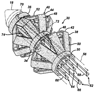
10018) FIGURE 1 is perspective view of an single lumen access deployable ring
for
Intralumenal anastomosis installed upon an applier being inserted
laparoscopically to
an anastomosis target site on each of two portions of a patient's small
intestine.
100191 FIGURE 2 is the applier of FIG. 1 after actuation of the single lumen
access
deployable ring to appose the two portions of small intestine.
100201 FIGURE 3 is a detail view of the unactuated single lumen access
deployable
ring and distal tip and catches of the applier.
100211 FIGURE 4 is a perspective detail view of a partially-actuated ring
device and
the catches and distal tip of the applier of FIG. 2 .
100221 FIGURE 5 is a side elevation detail view of the partially-actuated ring
device
and distal portion of the applier of FIG. 2 cutaway along the longitudinal
axis.
100231 FIGURE 6 is a perspective detail view of a fully actuated ring device
and
distal portion of the applier of FIG. 2.
100241 FIGURE 7 is a side elevation view of the fully actuated and deployed
ring
device of FIG. 6.
Detailed Description of the Invention
100251 Turning to the Drawings, wherein like numerals denote like components
throughout the several views, FIG. 1 depicts an applier 10 having an elongate
implement portion 12 dimensionally sized for insertion through a cannula of a
trocar
or laparoscopic port to tissues walls 14, 16 to anastomose two lumens. A
distal
introducer tip 18 of the applier 10 pierces through an opening 20 at an
anastomosis
site 22 to position an actuating portion 24 that holds a ring device 30 for
single lumen
anastomosis.
-6-
CA 02483719 2004-09-29
100261 The ring device 30 has three primary rings, depicted as a proximal ring
32, a
center ring 34, and a distal ring 36, that are cylindrically aligned with one
another.
The proximal ring 32 is longitudinally attached to the center ring 34 by
proximal arms
38, which in turn is longitudinally attached to the distal ring 36 by distal
arms 40.
Each proximal and distal arm 38, 40 is bisected respectively by a hinged joint
42, 44
defining an inner arm segment 46, 48 also hingedly attaching to the center
ring 34 and
an outer arm segment 50, 52 also hingedly attached to the respective proximal
or
distal ring 32, 36. In its unactuated state as depicted in FIG. 1, the device
30 is
cylindrical. The relative lengths of the inner arm segments 46, 48 to outer
arm
segments 50, 52 may be selected to provide a desired angular contact to tissue
walls
14, 16. In the illustrative version, the relationship resembles a cantilevered
contact
with the inner arm segments 46, 48 actuating to an approximately parallel
relationship
to the tissue walls 14, 16.
100271 A handle portion 54 is proximally connected to a shaft 56 of the
implement
portion 12. The shaft 56 may be rigid or flexible, with the latter being
desirable for
Intralumenal insertion, such as through the esophagus. The handle includes
controls
for longitudinally positioning the rings 32-36 of the ring device 30. In the
illustrative
version, this includes a center ring slide control 58 and a distal ring slide
control 60.
Although a manually positioned and actuated applier 10 is depicted for
clarity, it
should be appreciated that a remotely positioned and actuated applier may be
used
consistent with aspects of the invention, for instance to allow placement in a
more
controlled manner, to avoid disturbing an imaging modality, or for other
reasons. The
handle 54 may further include controls for a distal tip illumination
capability so that
actuation of the distal arms 40 in the distal lumen may be proximally viewed
from an
endoscope. It will be appreciated that the terms "proximal" and "distal" are
used
herein with reference to a clinician gripping the handle portion 54 of the
applier 10.
100281 In FIG. 2, two slide controls 58, 62 have been withdrawn proximally,
bringing
both the center and distal rings 34, 36 into locking proximity of the proximal
ring 32,
which is held in place by resting against the shaft 56. In response thereto,
the
proximal and distal arms 38, 40 hinge outwardly from the longitudinal axis of
the
device 30, creating a hollow rivet or hourglass shape for apposing tissue
walls 14, 16.
The center ring 34 sits at a tissue junction between lumens and the distal and
proximal
-7-
CA 02483719 2004-09-29
rings 32, 36 come to rest in respective lumens. By latching rings 32-38 one to
another
when actuated, the device 30 is held in the actuated position with bent arms
38, 40
apposing tissue. The proximal arms 38 may be staggered, as depicted, from
distal
arms 40 to create a tortuous path for the compressed tissue. Alternatively,
the arms
38, 40 may be aligned to directly mate to each other.
[00291 It should be appreciated that in the illustrative version, the proximal
ring 36 is
stationary with respect to the applier 10. In some applications, a third
control may be
incorporated so that each of the three rings may be positioned independently
from the
rest, further enhancing the ability to actuate either the distal or the
proximal arms 40,
38. As another alternative, the center ring 34 may be stationary with respect
to the
applier 10, with controls effective to move the proximal and distal rings 32,
36
inwardly to the center ring 34.
100301 In FIG. 3, the unactuated ring device 30 is shown with the distal
introducer tip
18 of the applier 10. The ring device 10 may be comprised of a single piece of
molded
or stamped material. For instance, the ring device 10 may be advantageously
formed
from a stamped piece of sheet metal that is wound around a mandrel and tack
welded
into a cylindrical shape. Cuts define the arms 38, 40 and creases define the
hinged
portions. Similar manufacturing economies may be achieved by molding the ring
device 30 from a polymeric material. Furthermore, the device 10 may be formed
entirely or partially of a biofragmentable or absorbable material to assist in
the
eventual passing of the device 10, leaving a patent anastomosis. The ring
device 10
may advantageously include radiopaque markers in the arms to allow diagnostic
imaging to confirm placement of the device 10 and/or to confirm passing. It
should be
appreciated that the afore-described methods of manufacture are believed to
yield
economical and therapeutic advantages; however, other techniques for
fabrication and
assembly may be employed.
[00311 Also depicted in FIG. 3, a center ring actuating member 62 and a distal
ring
actuating member 64 are shown that move within the shaft 56 in response to the
center and distal ring slide controls 58, 60. In the illustrative version,
each actuating
member 62, 64 is formed from a rigid polymer or sheet metal to have two
parallel
elongate prongs 66, 68 springedly outwardly biased or urged outwardly by other
-8-
CA 02483719 2004-09-29
portions of the applier 10 to present distally and laterally presented catches
70 to the
inner surface of their respective rings for engagement. Proximal to each catch
70 is a
releasing ramp 72 that causes the catch 70 to move inwardly as the releasing
ramp 72
contacts the next more proximal ring at or near full actuation. Thus, the ring
device 30
is disengaged from the actuating portion 24 of the applier 10 and may be
deployed.
100321 In FIGS. 4-5, the actuating members 62, 64 are depicted as having moved
proximally to an intermediate locking position. The shaft 56 (shown in
phantom) has
restrained the proximal ring 32 while center ring actuating member 62 has
drawn back
the center ring 34 such that the proximal arms 38 have partially actuated.
Similarly,
the distal ring actuating member 64 has drawn back the distal ring 36 such
that the
relative distance between the distal and center 36, 34 is sufficient to also
partially
actuate the distal arms 40. A locking mechanism, depicted as proximally
directed
locking hook 74, is connected to the distal ring 36 and is depicted as
transitioning past
the center ring 34 at this intermediate actuating position. It may be desired
in some
applications for there to be sufficient interference or latching at
intermediate points
during actuation for the ring device 30 to remain in a partially actuated
position.
100331 In FIGS. 6-7, the ring device 30 has fully actuated. In FIG. 6, the
actuating
members 62, 64 have caused the locking hook 74 to lock the distal ring 36 to
the
proximal ring 32. It should be appreciated that this simple latching mechanism
is
illustrative and for clarity. A distally presented hook from the proximal ring
32 for
instance may intermediately latch to the center ring 34 when the proximal arms
38 are
partially actuated and latch to the distal ring 36 when the proximal arms 38
are fully
actuated. In FIG. 7, the applier 10 has been withdrawn from the ring device
30. An
advantage of having the locking hook exposed in the proximal lumen is
convenient
access for confirming latching and for reversing the closing of the device 30
in
instances where a leak is detected after actuation (e.g., from the opening 20
out
between the tissue walls 14, 16).
(00341 In use, a ring device 30 is received upon an actuating portion 24 of an
implement portion 12 of an applier 10. Specifically, the proximal ring 32 of
the device
30 rests against the shaft 56, a center ring actuating member 62 engages the
center
ring 34 of the device 30, and a distal ring actuating member 64 engages the
distal ring
-9-
CA 02483719 2004-09-29
36 of the device 30. A clinician manipulates the handle 54 to insert the
implement
portion 12 through the cannula of a trocar, laparoscopic port, or through a
lumen such
as the esophagus to the anastomosis site 22. The tissue walls 14, 16 are
proximately
placed and the introducer tip 18 of the implement portion 12 passes through
the
opening 20 formed in these walls 14, 16. The introducer tip may include a
piercing
shape and/or electromagnetically or thermally enhanced cutting features to
assist in
forming the opening 20. Once the distal arms 40 of the device 30 are in the
distal
lumen, the distal ring slide control 60 may be proximally moved to actuate the
distal
arms into a partially actuated ring shape, latching the locking hook 74 to the
center
ring 34. The distal tissue wall 16 thus held may be drawn back proximally if
necessary such that the proximal arms 38 reside within the first lumen.
Drawing back
the center ring slide control 58 thus partially actuates the proximal arms 38.
If the
positioning is correct, the slide controls 58, 60 may be fully slid, latching
the locking
hook 74 to the proximal ring and causing the proximal and distal arms 38, 40
to be
fully actuated and disengaging the catches 70 that hold the applier 10 to the
ring
device 30. Then, the distal tip 18 of the applier is withdrawn from the ring
device 30
leaving it deployed to form the anastomotic attachment. Over time, the tissue
walls
14, 16 permanently heal together and the ring device 30 may be passed out of
the
digestive tract, especially if biofragmentable.
[00351 While the present invention has been illustrated by description of
several
embodiments and while the illustrative embodiments have been described in
considerable detail, it is not the intention of the applicant to restrict or
in any way
limit the scope of the appended claims to such detail. Additional advantages
and
modifications may readily appear to those skilled in the art.
[0036) For example, aspects of the invention have application to surgical
procedures
performed endoscopically and laparoscopically, as well as an open procedure.
Use
herein of one of these or similar terms should not be construed to limit the
present
invention for use in only one category of surgical procedure.
[0037[ For another example, although bariatric procedures for bypassing
portions of a
gastrointestinal tract are depicted, it should be appreciated that other
surgical
-10-
CA 02483719 2004-09-29
procedures may benefit by an anastomotic ring device having aspects described
herein, such as for the bile duct and vascular bypasses.
100381 As an additional example, instead of a center ring 34, the proximal
arms 38
may attach to the distal arms 40 in an accordion-like fashion with the
proximal ring
32 locking to the distal ring 36. Thus, the center portion of the device 30 at
the tissue
junction is capable of dilating, thereby further stabilizing the lumens to be
anastomosed and preventing tissue slippage. This dilation may be effected
either by
the proximal and distal rings 32, 36 forcing a center portion to dilate with a
wedging
action or by making the inner arm segments 46-48 shorter than the outer arm
segments 50-52.
[00391 As yet a further example, the rings 32, 34, 36 present an internally
projecting
contour that may be engaged by the catches 70 of the applier. Other
engagements may
be incorporated, such as a frangible adhesion between actuating members and
one or
more rings. In addition, a distal introducer tip may act as an anvil that may
be
withdrawn proximally to longitudinally compress the device, with features that
may
be radially withdrawn to thereafter allow the distal introducer tip to be
removed from
the ring device for deployment.
[00401 As yet another example, pads on the inner arm segments may be included
to
control the pressure profile on the tissue. Corners may be softened or
smoothed to
avoid any adverse effects of a traumatic contact to tissue.
100411 What is claimed is:
-11-