Note: Descriptions are shown in the official language in which they were submitted.
CA 02483779 2004-10-26
WO 03/092807 PCT/US03/10437
METHOD AND APPARATUS FOR SELECTING AN OPTIMAL ELECTRODE
CONFIGURATION
FIELD OF THE INVENTION
The present invention relates generally to an implantable electrical
stimulation
and/or sensing lead, and more particularly, the present invention relates to a
method and
apparatus for improved stimulation and sensing of a medical electrical lead
having a
multiple electrode array.
~o
BACKGROUND OF THE INVENTION
A wide assortment of implantable medical devices (IMDs) are presently known
and in commercial use. Such devices include cardiac pacemakers, cardiac
defibrillators,
cardioverters, neurostimulators, and other devices for delivering electrical
signals to
15 excitable tissue and/or receiving signals from the tissue. Devices such as
pacemakers,
whether implantable or temporary external type devices, are part of a system
for delivering
an electrical therapy or monitoring a patient condition. In addition to the
pacemaker
device, which typically has some form of pulse generator, a pacing system
includes one or
more leads carrying electrodes for delivering generated stimulation pulses to
the heart and
20 for sensing cardiac signals.
Pacemakers treat heart conditions in which the heart beats at a rate that is
considered to be too slow, commonly referred to as bradycardia, by sensing
cardiac signals
and delivering appropriately timed electrical stimulation pulses to the atria
and/or
ventricles as needed to cause the myocardium to contract. Pacemakers may sense
intrinsic
25 cardiac signals that occur when the myocardium depolarizes naturally,
causing a normal
myocardial contraction or heart beat. A sensed signal associated with
ventricular
contraction is referred to as an R-wave, and a sensed signal associated with
atrial
contraction is a P-wave. When an intrinsic R-wave or P-wave is not sensed by
the
pacemaker, a stimulation pacing pulse is delivered, eliciting an evoked
response which
30 causes the myocardium to contract, thus maintaining a desired heart rate.
Pacemakers operate in either a unipolar or bipolar mode, and pace the atria
and/or
the ventricles of the heart. Unipolar pacing requires a lead having only one
distal electrode
CA 02483779 2004-10-26
WO 03/092807 PCT/US03/10437
2
for positioning in the heart, and utilizes the case, or housing of the
implanted device as the
other electrode for the pacing and sensing operations. For bipolar pacing and
sensing, the
lead typically has two electrodes, a tip electrode disposed at the distal end
of the lead, and
a ring electrode spaced somewhat back from the distal end. Each electrode is
electrically
coupled to a conductive cable or coil, which carnes the stimulating current or
sensed
cardiac signals between the electrodes and the implanted device via a
connector.
Combination devices are available for treating both fast and slow cardiac
arrhythmias by delivering electrical shock therapy for cardioverting or
defibrillating the
heart in addition to cardiac pacing therapies. Such a device, commonly known
as an
implantable cardioverter defibrillator or "ICD", uses coil electrodes for
delivering high-
voltage shock therapies. An implantable cardiac lead used in combination with
an ICD
may be a tripolar or quadrapolar lead equipped with a tip electrode and a ring
electrode for
pacing and sensing functions and one or two coil electrodes for shock
therapies.
In order to achieve stimulation or sensing in the right side of the heart, a
lead may
be positioned against the endocardium by advancing the lead through the vena
cava into
the right atrium for right atrial applications, or further advancing the lead
into the right
ventricle for right ventricular applications. In order to achieve stimulation
or sensing in
the left heart chambers, a lead, often referred to as a "coronary sinus lead,"
may be
positioned within the vasculature of the left side of the heart via the
coronary sinus and
great cardiac vein. This endovascular lead placement is sometimes referred to
as
"epicardial" placement since electrodes on a coronary sinus lead will sense or
stimulate
epicardial heart tissue.
In order to work reliably, cardiac leads need to be positioned and secured at
a
targeted cardiac tissue site in a stable manner. Unacceptable pacing or
sensing thresholds
measured during an implant procedure may require lead repositioning. Shifting
or
dislodgement of the lead over time may result in changing thresholds,
sometimes requiring
programming adjustments in order to maintain an appropriate level of therapy.
At the
same time, increased pacing thresholds decrease the useful life of the battery
in the
implantable device, requiring earlier device replacement. Poor or inaccurate
sensing of
CA 02483779 2004-10-26
WO 03/092807 PCT/US03/10437
naturally occurnng heart signals may result in inappropriate withholding or
delivery of
then apy.
To address these problems, an electrode may be passively secured in a desired
endocardial position by the use of tines located at the distal end of a lead.
The tines
engage with the endocardial trabeculae, holding the distal lead end in place.
Alternatively,
an electrode may be actively secured by the use of a rotatable fixation helix.
The helix
exits the distal end of the lead and can be screwed into the body tissue. The
helix itself
may serve as an electrode or it may serve exclusively as an anchoring
mechanism to locate
an electrode mounted on the lead adjacent to a targeted tissue site. The
fixation helix may
be coupled to a drive shaft that is further connected to a coiled conductor
that extends
through the lead body as generally described in U.S. Pat. No. 4,106,512 issued
to Bisping
et al. A physician rotates the coiled conductor at a proximal end to cause
rotation of the
ftxation helix via the drive shaft. As the helix is rotated in one direction,
the helix is
secured in the cardiac tissue. Rotation of the fixation helix in the opposite
direction
removes the helix from the tissue to allow for repositioning of the lead at
another location.
These fixation methods, however, are not entirely appropriate in left heart
stimulation and sensing applications when the lead is positioned
endovascularly. A helical
coil would puncture a cardiac vein. Tines would make lead re-positioning
difftcult
because retraction of a tined lead within a narrow vein could potentially
damage the valves
within the vein. Tissue encapsulation of various passive and active fixation
devices is
normally encouraged to further stabilize an endocardial lead position. Tissue
encapsulation is undesirable in stabilizing an endovascular lead, however,
since such
tissue ingrowth may obstruct blood flow. Methods for stabilizing an
endovascular lead
must allow for unimpeded blood flow. One method for stabilizing an
endovascular lead is
disclosed in U.S. Patent No. 6,161,029, issued to Spreigl, et al., and
includes an expanded
stmt that is lodged against the blood vessel wall to inhibit movement of the
stmt and a
distal electrode support. The expanded stmt lumen is aligned with the
electrode support
lumen for allowing blood to flow through the aligned electrode support lumen
and
expanded stmt lumen.
Another problem encountered in left heart stimulation is that conventional
circumferential tip or ring electrodes on a coronary sinus lead will direct
current in the
CA 02483779 2004-10-26
WO 03/092807 PCT/US03/10437
4
direction of the adjacent epicardium but also in directions away from the
targeted tissue,
which may reduce stimulation efficiency. Stray current may also cause
undesired
extraneous stimulation, such as phrenic nerve stimulation or atrial
stimulation during
ventricular pacing. A coronary sinus lead would preferably direct current only
in the
direction of the targeted myocardium. Correctly positioning an endovascular
lead having
an electrode on only one side, however, would be difficult and time consuming.
Lead failure sometimes occurs when a conductor becomes fractured or the
insulation between electrodes andlor conductors fails. A unipolar lead failure
generally
requires a surgical procedure to replace the failed lead. In the case of a
bipolar lead, a
l0 bipolar stimulation or sensing configuration may be reprogrammed to
unipolar if one
electrode on the lead remains functional. However, the remaining functional
electrode
may be positioned at a different location relative to the targeted cardiac
tissue and may not
provide as effective or efficient sensing or stimulation as the bipolar pair.
Furthermore, in
some patients, unipolar sensing does not provide an acceptable signal-to-noise
ratio.
15 For effective cardiac pacing, a delivered stimulation pulse must be of
adequate
energy to cause depolarization of the myocardium, referred to as "capture."
The lowest
pulse energy that successfully captures the heart is referred to as the pacing
threshold. In
order to verify that a pacing pulse has captured the heart, modern pacemakers
are equipped
with automatic capture detection algorithms. Capture may be verified by
various
20 methodologies known in the art such as sensing for an evoked R-wave or P-
wave after
delivery of a pacing pulse, sensing for the absence of an intrinsic R-wave or
P-wave
during the refractory period after a pacing pulse, or detecting a conducted
depolarization
in an adjacent heart chamber. Various capture verification methods are
described in U.S.
Pat. No. 5,601,615 issued to Markowitz et al., U.S. Pat. No. 5,324,310 issued
to
25 Greeninger et al., and U.S. Pat. No. 5,61,012 issued to Stroebel, each of
which patents
are incorporated herein by reference in their entirety. If capture is not
verified, the pacing
pulse energy may be automatically increased.
An electrode configuration used for pacing and evoked response sensing for
capture detection may utilize a bipolar lead on which a tip electrode provides
unipolar
30 pacing and the tip and ring electrode pair provide bipolar sensing of the
evoked response.
A limitation of using the same electrode for pacing and evoked response
sensing is that the
CA 02483779 2004-10-26
WO 03/092807 PCT/US03/10437
pacing pulse and ensuing after-potential and electrode-tissue polarization
artifact mask the
evoked response until they dissipate, after which the evoked response, if any,
has typically
passed the sensing electrodes. Therefore, it is desirable to use an electrode
pair that does
not include the pacing electrode for sensing an evoked response. To overcome
the
problems of after-potential and the electrode-tissue polarization artifact,
capture
verification methods have been proposed which involves sensing for a conducted
depolarization at a site away from the pacing electrode. For example, sensing
a
ventricular depolarization after an atrial pacing pulse has been delivered is
evidence that
the atrium was captured and the evoked depolarization was conducted to the
ventricle.
For accurate evoked response detection, however, it is desirable to sense the
evoked response using a bipolar sensing electrode pair in the vicinity of the
stimulated
cardiac tissue site. Unipolar sensing or sensing in other areas of the heart
could lead to
erroneous evoked response detection due to noise or other myopotentials being
sensed as
an evoked response. Furthermore, sensing for an evoked response in another
area of the
heart may not be possible in patients having conduction disorders.
What is needed, therefore, is an improved lead design that allows accurate
targeting of excitable tissue in both endovascular and endocardial
applications. A lead
having an electrode arrangement that allows for reliable pacing and evoked
response
sensing for the purpose of capture verification is also desirable. Such a lead
must be
stabilized in a way that, when used endovascularly, does not cause undue
vessel damage
during fixation or repositioning and allows for unimpeded blood flow.
Furthermore, an
improved lead design should provide for alternative stimulation or sensing
configurations
without compromising effectiveness and efficiency of therapy delivery in case
one
electrode fails.
SUMMARY OF THE INVENTION
The present invention is directed to implantable electrical lead that includes
an elongated
lead body that extends between a proximal lead end and distal lead end, and a
plurality of
electrodes located along the distal lead end. An insulating material is
positioned between
each of the plurality of electrodes to electrically isolate each of the
plurality of electrodes,
and a plurality of insulated electrical conductors are each connected to a
respective
CA 02483779 2004-10-26
WO 03/092807 PCT/US03/10437
6
electrode of the plurality of electrodes. A microprocessor performs a
threshold search
corresponding to combinations of one or more electrodes of the plurality of
electrodes to
determine an optimal threshold, and selects the electrodes of the plurality of
electrodes
corresponding to the optimal threshold.
BRIEF DESCRIPTION OF THE DRAWINGS
Features and advantages of the present invention will be readily appreciated
as the
invention becomes better understood by reference to the following detailed
description
considered in connection with the accompanying drawings, in which like
reference
numerals designate like parts throughout:
FIG. 1 is a plan view of an implantable electrical lead having a tip electrode
array
and a ring electrode array;
FIG. 2 is a perspective view of a distal end of the lead shown in FIG. 1;
FIG. 3 is a plan view illustrating alternative arrangements of electrodes
within an
electrode array according to the present invention;
FIG. 4 is a side, cut-away view of the distal lead end shown in FIG. 2;
FIG. 5 is a side, cut-away view of a distal lead end of an implantable
electrical lead
having a helical expansion member for expanding a tip electrode array;
FIG. 6 is a side cut-away view of the distal lead end shown in FIG. 5 showing
a tip
electrode array in a fully expanded position;
FIG. 7 is a side, cut-away view of a distal lead end having an alternative
expansion
member for expanding a tip electrode array according to the present invention;
FIG. 8 is an illustration showing the lead of FIG. 1 implanted within the
coronary
vessels of a patient's heart via the coronary sinus and in communication with
an
implantable cardioverter defibrillator, according to a preferred embodiment of
the present
invention;
FIG. 9 is a functional, block diagram of the implantable cardioverter
defibrillator
(ICD) shown in FIG. 8; and
FIG. 10 is a flow chart of a method for using the lead shown in FIG. 8 in
conjunction with the implantable cardioverter defibrillator (ICD) of FIG. 9.
CA 02483779 2004-10-26
WO 03/092807 PCT/US03/10437
DETAILED DESCRIPTION OF THE INVENTION
In the following detailed description, references are made to illustrative
embodiments of medical leads adapted to be located in the heart or cardiac
blood vessels
in which aspects of the present invention may be implemented. It is understood
that the
invention may be practiced in other body implantable leads positioned for
sensing or
stimulating excitable tissue.
FIG. 1 is a plan view of a multipolar cardiac lead in accordance with an
embodiment of the present invention. As illustrated in FIG. 1, a lead 10
according to the
present invention includes an elongated lead body 12 having a connector
assembly 16 at a
LO proximal end adapted for connecting to an implantable device, such as an
ICD, and an
electrode head assembly 68 at a distal end 14 for carrying one or more
electrodes. Lead
is shown having, at or near distal end 14, a tip electrode array 20, a ring
electrode array
30, a ring electrode 40, and a defibrillation coil electrode 50. The tip
electrode array 20
and the ring electrode array 30 each include multiple electrodes, for example
three
electrodes, separated by insulating material. Electrodes within the tip
electrode anay 20
andlor ring electrode array 30 and/or ring electrode 40 may be utilized to
sense cardiac
signals andlor deliver pacing pulses to a patient's heart. The defibrillation
coil electrode
50 is used for delivery of a defibrillation shock as a result of a detected
tachycardia or
fibrillation condition.
The lead body 12 takes the form of an extruded tube of biocompatible plastic
such
as silicone rubber. The lead body 12 includes multiple lumens for carrying
multiple
I
insulated conductors from the connector assembly 16 to the corresponding
electrodes
arrays 20 and 30 and electrodes 40 and 50 located at or near the distal lead
end 14. The
mufti-lumen lead body 12 may correspond generally to that disclosed in U. S.
Pat. No.
5,584,873 issued to Shoberg et al., incorporated herein by reference in its
entirety. Two of
the insulated conductors carried by lead body 12 may be stranded or cabled
conductors,
each electrically coupled to one of the ring electrode 40 and the
defibrillation coil 50. The
cabled conductors may correspond generally to the conductors disclosed in U.S.
Pat. No.
5,246,014, issued to Williams et al., incorporated herein by reference in its
entirety. A
third and fourth conductor are preferably mufti-filar coiled conductors, for
example of the
type described in U.S. Pat. No. 4,922,607 issued to Doan et al., incorporated
herein by
CA 02483779 2004-10-26
WO 03/092807 PCT/US03/10437
8
reference in its entirety. Each filer of the mufti-filer coiled conductors is
coupled to an
individual electrode within the tip electrode array 20 or the ring electrode
array 30. The
filers are electrically insulated from each other for example by
polytetrafluoroethylene
(PTFE) or ethyl tetrafluoroethylene (ETFE) tubing.
The connector assembly 16 includes multiple connector extensions 22, 32, and
52
arising from a trifurcated connector sleeve 18, typically formed of silicone
rubber. The
connector extensions 22, 32 and 52 couple the lead 10 to an implantable
medical device
such as an implantable cardioverter defibrillator (ICD).
Connector extension 22 is shown as a tri-polar connector including three
connector
rings 24. Connector extension 22 houses a mufti-filer coiled conductor of
which each filer
is electrically coupled at a proximal end to one of the connector rings 24 and
at a distal
end to one of the three electrodes included in tip electrode array 30. A
stylet 60 may be
advanced within an inner lumen of the coiled conductor carried by connector
extension 22
toward the distal end of the lead 10 to aid in lead placement during an
implant procedure.
l5 Connector extension 32 is shown as a quadrapolar connector including three
connector rings 34 and a fourth connector ring 36. The three connector rings
34 are
electrically coupled to individual filers within a mufti-filer coiled
conductor extending to
the ring electrode array 30. The distal end of each filer is coupled to one of
three
electrodes included in ring array 30. The fourth connector ring 36 is coupled
to an
ZO insulated cabled conductor that extends to ring electrode 40.
Connector extension 52 carnes a single connector pin 54 that is electrically
coupled to an insulated cable extending the length of the lead body 12 and
electrically
coupled to the defibrillation coil electrode 50. While the lead 10 depicted in
FIG. 1 is a
mufti-polar pacing and defibrillation lead, aspects included in the invention
may be
25 practiced in any unipolar, bipolar, or mufti-polar lead by providing at
least one tip or ring
electrode array. One or more electrode arrays may be provided alone or with
any
combination of conventional tip, ring or coil electrodes.
FIG. 2 is an enlarged, perspective view of the electrode head assembly 68
located
at the distal lead end 14 shown in FIG. 1. The tubular electrode head assembly
68 is
30 preferably fabricated from a relatively rigid biocompatible polymer, such
as polyurethane.
As illustrated in FIG. 2, tip electrode array 20, mounted on the tip of the
electrode head
CA 02483779 2004-10-26
WO 03/092807 PCT/US03/10437
assembly 68, includes three approximately equally sized electrodes 25, 27 and
29 arranged
circumferentially with respect to the electrode assembly 68. The tip electrode
array 20
could alternatively comprise two or more electrodes of approximately equal or
unequal
sizes. The electrodes 25, 27 and 29 are preferably platinum iridium
electrodes, but may be
manufactured from any acceptable, medical-grade, conductive biomaterial. A
layer of
insulating material 64, such as ceramic, is arranged radially with respect to
the electrode
head assembly 68, between each of the electrodes 25, 27 and 29 such that the
electrodes
25, 27 and 29 within the array 20 are electrically insulated from each other.
The insulator 64 optionally provides a center port 56. When the electrode
array 20
is used as a tip electrode, as shown in FIG. 1, the port 56 may be used to
hold a
pharmaceutical agent. The pharmaceutical agent, which may be an anti-
inflammatory,
antibiotic, or other agent, may be added as a powdered form to a polymer
adhesive that is
injected into port 56 such that the agent elutes from the polymer over time
after
implantation. In one embodiment, the port 56 holds a steroid powder added to
medical
grade silicone adhesive, which when released after implantation will minimize
the
inflammatory tissue response around the electrode array 20. Various
embodiments for
providing a drug dispenser in an electrical medical lead that may be used in
conjunction
with the present invention are disclosed in U.S. Pat. No. 4,711,251 issued to
Stokes,
incorporated herein by reference in its entirety.
In the same way, the ring electrode array 30 includes three, approximately
equally-
sized, circumferentially arranged electrodes separated from each other by a
layer of
insulating material 66. FIG. 3 illustrates alternative arrangements of
electrodes within an
electrode array. In the alternative tip electrode 120, three electrodes 125,
127, and 129 are
arranged circumferentially around the electrode head assembly 68 but staggered
along its
length such that electrode 125 is located at the distal lead tip, electrode
129 is located
slightly proximal to electrode 125, and electrode 127 is slightly proximal to
electrode 129.
This staggered arrangement could equally be applied to a ring electrode array.
The ring electrode array 130 shown in FIG. 3 includes three ring electrodes
135,
137, and 139, each encircling electrode head assembly 68 and spaced at close
intervals
longitudinally with respect to each other along the electrode head assembly
68. This
longitudinally-spaced ring arrangement could also be applied to a tip
electrode array. It is
CA 02483779 2004-10-26
WO 03/092807 PCT/US03/10437
recognized that numerous variations of electrode array arrangements may exist
in which
two or more electrodes are arranged in close proximity to each other.
FIG. 4 is a side cut-away view of the tubular electrode head assembly 68 of
the
lead 10 shown in FIG. 2. Electrodes 27 and 29 included in tip array 20 are
visible in this
view, and electrodes 37 and 39 of ring array 30 are visible in this view.
Electrodes 27 and
29 included in tip array 20 are each provided with connection tabs 82 to allow
electrical
coupling, for example by laser welding, to individual filars 84 included in
the multi-filar
coiled conductor 80. Multi-filar conductor 80 is connected at a proximal end
to connector
rings 24 (FIG. 1). Insulation material 64 is shown between electrodes 27 and
29.
~0 Electrodes 37 and 39 included in ring array 30 are each provided with
connection
tabs 92 to allow electrical coupling to individual filars 94 included in the
multi-filar coiled
conductor 90. Mufti-filar conductor 90 is connected at its proximal end to
connector rings
34 (FIG. 1). Ring electrode 40 is shown coupled to cabled conductor 70, which
is further
coupled at its proximal end to connector ring 36. By providing separate,
insulated
conductors to each of the insulated electrodes 25, 27, 29 of tip array 20 and
35, 37 and 39
of ring array 30, the electrodes 25, 27, 29, 35, 37 and 39 may be selected
individually or in
any combination for pacing and/or sensing functions.
An alternative embodiment of the lead 10 is shown by the side cut-away view of
FIG. 5. In this embodiment, the tip electrode array 20 is expandable. The
electrodes 27
and 29 within array 20 are mounted on flexible electrode extensions 87 and 89,
respectively. An expansion member for expanding the flexible electrode
extensions 87
and 89 takes the form of a sonically-shaped helix 100. The helix 100 may
function
exclusively as an expansion member, in which case the helix may be formed fiom
any
relatively rigid biocompatible polymer, such as urethane, or a biocompatible
metal. The
tip of helix 100 may be blunted to prevent unintentional tissue damage. In
other
embodiments, the helix 100 may also serve as an additional electrode for
cardiac pacing
and/or sensing. When used as an electrode, the helix 100 is formed from a
conductive
biocompatible metal such as platinum iridium alloy. The helix 100 may also
seine as an
active fixation device for anchoring the lead 10 in a desired position for
additional
stability. In this case, the helix 100 has a sharpened tip for securing the
helix 100 in tissue.
CA 02483779 2004-10-26
WO 03/092807 PCT/US03/10437
11
Reference is made to U.S. Patent No. 4,217,913 issued to butcher, incorporated
herein by
reference in its entirety.
The helix 100 is shown in FIG. 5 to be mounted on a drive shaft 102 that is
further
connected to a rotatable coil 110. The coil 110 extends the length of the lead
body 12 and
may be coupled to a connector pin provided on one of the connector extensions
of
connector assembly 16. During a lead implant or explant procedure, a physician
may
rotate such a connector pin relative to the connector assembly 16 causing
advancement or
retraction of the helix 100 in a manner generally described in U.S. Pat. No.
4,106,512 to
Bisping et al., incorporated herein by reference in its entirety. Rotation of
the connector
pin rotates the drive shaft 102 via the coil 110. As the drive shaft 102 is
rotated, the helix
100 is actuated by a guide 106 such that the helix 100 is advanced toward the
lead end. A
drive shaft seal 104 is optionally provided to prevent the ingress of body
fluids into the
lumen of lead 10.
In FIG. 6, the tip electrode array 20 is shown in a fully expanded position.
The
helix 100 is in an advanced position such that the widest portion of the
conical helix 100
has caused the flexible electrode extensions 87 and 89, each carrying one of
the electrodes
27 and 29 included in tip array 20, to bend outward.
Expansion of the tip array 20 in this way provides a passive fixation
mechanism
for stabilizing the lead position. When used as an endocardial electrode, the
expanded
electrode array 20 may engage with the endocardial trabeculae, holding the
distal lead end
in place. If the initial lead position does not result in acceptable pacing or
sensing
thresholds, the helix 100 may be retracted, contracting the tip array 20, to
allow easy
removal and lead repositioning. This reversible fixation mechanism is
particularly useful
when the lead 10 is used as an endovascular lead. Contraction of the tip array
20 allows
easy retraction of the lead within a narrow vein without undue damage to
vessel walls or
vein valves. Furthermore, the expanded electrode array provides stable lead
positioning
within a blood vessel without blocking the flow of blood or puncturing the
blood vessel
walls.
Another advantage of expanding the tip electrode array 20 relates to the
benefit of
increasing the inter-electrode distance when the tip array 20 is used for
pacing and evoked
response sensing. If, for example, one electrode of tip array 20 is used for
pacing in a
CA 02483779 2004-10-26
WO 03/092807 PCT/US03/10437
12
unipolar configuration with a device housing or in a bipolar configuration
with any of ring
electrode array 30 or ring electrode 40, the remaining two electrodes within
tip array 20
are available for sensing an evoked response in the same vicinity of the
delivered pacing
pulse. Sensing for an evoked response at the site of stimulation delivery
enables accurate
capture detection since other myopotentials, which may be present at more
remote sensing
sites, are less likely to interfere with evoked response sensing. By using
electrodes
different than the electrode used for pacing, problems associated with post-
pace
polarization artifacts can be avoided. The increased inter-electrode distance
in an
expanded tip array further enhances the ability to sense the evoked response
using
electrodes within the same array because the post-pace polarization artifact
will diminish
as the distance from the pacing electrode increases.
An alternative embodiment of an expansion member is shown in FIG. 7. In this
embodiment, the expansion member takes the form of a grooved cone 150, which
is
preferably fabricated from a biocompatible, relatively rigid polymer such as
polyurethane.
The cone 150 is mounted on a drive shaft 152 having a screw-like head 154 with
a slot
156. A stylet 158 having a screw driver-like blade 160 mounted on its distal
end may be
advanced within a lumen of the lead body 12. The blade 160 may be inserted
into slot 156
and, upon rotation of the stylet 158 at its proximal end, cause rotation of
the drive shaft
152. When the drive shaft 152 is rotated, the cone 150 is actuated by the
guide 106 and is
advanced toward the distal lead tip to cause expansion of the tip electrode
array 20
mounted on flexible electrode extensions 87 and 89.
In one embodiment, the expansion member may be coated with a substrate or
solvent carrying a pharmaceutical agent, such as an anti-inflammatory drug.
The
expansion member may be dip-coated in a solvent, such as acetone, in which a
steroid has
been dissolved. The steroid will elute from the coating over time after
implantation and
prevent a hyper-inflammatory response at the implant site. A method for
treating an
electrode with a steroid solution, which may be adapted for use in the present
invention for
treating the expansion member, is generally described in U.S. Pat. No.
5,987,746 issued to
Williams, incorporated herein by reference in its entirety.
In FIG. 8, the lead 10 is shown as a part of a cardiac stimulation system
including
an ICD 410 coupled to a patient's heart 450 by way of lead 10. The ICD 410 is
encased in
CA 02483779 2004-10-26
WO 03/092807 PCT/US03/10437
13
a housing 411 and provided with a connector block 412 to accommodate
connection of
lead 10 to the ICD 410. The heart 450 is shown with a partially open view
revealing the
coronary sinus 430. The lead 10 is advanced within the vasculature of the left
side of the
heart via the coronary sinus and great cardiac vein. A tip electrode array 20
is disposed in
a vascular lumen 440 adjacent the left ventricle 460. The tip electrode array
20 is shown in
an expanded position at a desired cardiac implantation site. A blunted
expansion cone 150
has been advanced in order to expand the electrodes within array 20 against
the walls of
lumen 440 so as to provide better electrode contact with the epicardial tissue
and to
stabilize the position of the lead 10 as previously described in conjunction
with FIG. 7.
0 The coronary sinus lead 10 is also equipped with a ring electrode array 30,
a ring electrode
40 and a defibrillation coil electrode 50. The coronary sinus lead 10 is shown
connected
to the ICD 410 via the trifurcated connector assembly 16, which accommodates
connection of ICD circuitry to the conductors within lead body 12 and their
respective
electrodes.
l 5 A functional schematic diagram of the ICD 410 is shown in FIG. 9. This
diagram
should be talcen as exemplary of one type of device within a body implantable
system that
includes a lead having one or more electrode arrays in accordance with the
present
invention. The disclosed embodiment shown in FIG. 9 is a microprocessor-
controlled
device, but the methods of the present invention may also be practiced in
other types of
ZO devices such as those employing dedicated digital circuitry.
With regard to the electrode system illustrated in FIG. 8, the ICD 410 is
provided
with a number of connection terminals for achieving electrical connection to
the lead 10
via the connector assembly 16 and the respective electrodes via their
associated
conductors. The connection terminal 311 provides electrical connection to the
housing
25 411 for use as the indifferent electrode during unipolar stimulation or
sensing. The
connection terminal 350 provides electrical connection to the defibrillation
coil electrode
50. The connection terminals 311 and 350 are coupled to the high voltage
output circuit
234 to facilitate the delivery of high energy shocking pulses to the heart
using the
defibrillation coil electrode 50 and housing 411.
30 The connection terminals 325, 327 and 329 provide electrical connection to
the
electrodes 25, 27 and 29, respectively, within tip electrode array 20. The
connection
CA 02483779 2004-10-26
WO 03/092807 PCT/US03/10437
14
terminals 335, 337 and 339 provide electrical connection to the electrodes 35,
37 and 39,
respectively, within ring electrode array 30. The comiection terminal 340
provides
electrical connection to the ring electrode 40. The connection terminals 325,
327, 329,
335, 337, 339, and 340 are further coupled to a switch matrix 208.
Switch matrix 208 is used to select which of the available electrodes are
coupled to
a ventricular sense amplifier (AMP) 200 for sensing ventricular signals.
Selection of the
electrodes is controlled by the microprocessor 224 via data/address bus 218.
The selected
electrode configuration may be varied according to the various sensing,
pacing,
cardioversion and defibrillation functions of the ICD 410.
The ventricular sense amplifier 200 preferably takes the form of an automatic
gain
controlled amplifier with adjustable sensing thresholds. The general operation
of the
ventricular sense amplifier 200 may correspond to that disclosed in U.S. Pat.
No.
5,117,824 issued to Keimel et al., incorporated herein by reference in its
entirety.
Whenever a signal received by the ventricular sense amplifier 200 exceeds a
ventricular
sensing threshold, a signal is generated on the R-out signal line 202.
Switch matrix 208 is also used to select which of the available electrodes are
coupled to a wide band amplifier 210 for use in digital signal analysis.
Signals from the
electrodes selected for coupling to bandpass amplifier 210 are provided to
multiplexer
220, and thereafter converted to multi-bit digital signals by A/D converter
222, for storage
in random access memory 226 under control of direct memory access circuit 228.
Microprocessor 224 may employ digital signal analysis techniques to
characterize the
digitized signals stored in random access memory 226 to recognize and classify
the
patient's heart rhythm employing any of the numerous signal processing
methodologies
known in the art.
The telemetry circuit 330 receives downlink telemetry from and sends uplink
telemetry to an external programmer, as is conventional in implantable anti-
arrhythmia
devices, by means of an antenna 332. Data to be uplinked to the programmer and
control
signals for the telemetry circuit are provided by microprocessor 224 via
address/data bus
218. Received telemetry is provided to microprocessor 224 via multiplexer 220.
Numerous types of telemetry systems known for use in implantable devices may
be used.
CA 02483779 2004-10-26
WO 03/092807 PCT/US03/10437
The remainder of the circuitry illustrated in FIG. 9 is an exemplary
embodiment of
circuitry dedicated to providing cardiac pacing, cardioversion and
defibrillation therapies.
The pacer timing and control circuitry 212 includes programmable digital
counters, which
control the basic time intervals associated with various pacing modes or anti-
tachycardia
pacing therapies delivered in the ventricle. Pacer circuitry 212 also
determines the
amplitude of the cardiac pacing pulses under the control of microprocessor
224.
During pacing, escape interval counters within pacer timing and control
circuitry
212 are reset upon sensing of R-waves as indicated by signals on R-out signal
line 202.
10 The durations of the escape intervals are determined by microprocessor 224
via
data/address bus 218. The value of the count present in the escape interval
counters when
reset by sensed R-waves can be used to measure R-R intervals for detecting the
occurrence
of a variety of arrhythmias. In accordance with the selected mode of pacing,
if the
ventricular escape interval expires pacing pulses are generated by ventricular
pacer output
15 circuit 216. The pacer output circuit 216 is coupled to the desired pacing
electrodes via
switch matrix 208 along signal line 217. The escape interval counters are
reset upon
generation of pacing pulses, and thereby control the basic timing of cardiac
pacing
functions, including anti-tachycardia pacing. When a pacing pulse is
delivered, a signal is
generated by pacer timing and control 212 on blanking signal line (V BLAND)
211 to
prevent saturation of the sense amplifier 200 during the pacing pulse.
Thus, complete programmability of the electrodes used in pacing and/or sensing
is
possible via switch matrix 208. Any of the electrodes included in tip array
20, ring
electrode array 30 and ring electrode 40 may be selected individually or in
any
combination as the anode for unipolar pacing with the ICD housing 411 serving
as the
cathode. For bipolar or multi-polar electrode configurations, the electrodes
within tip
array 20, ring array 30 and ring electrode 40 may be selected in any
combination. For
example, one or more of the electrodes within an array may be selected to
serve as an
anode with any or all of the remaining electrodes in the same array selected
as the cathode.
Alternatively, electrodes may be selected from one array 20 or 30 to serve as
the anode
and from the other array to serve as the cathode. Electrodes within arrays 20
or 30 may
also be selected to function with ring electrode 30 in a bipolar
configuration.
CA 02483779 2004-10-26
WO 03/092807 PCT/US03/10437
16
The ICD 410 is preferably equipped with a capture detection algorithm executed
under the control of microprocessor 224. Following delivery of a pacing pulse
by
ventricular pacer output circuit 216, a desired pair of electrodes may be
selected via switch
matrix 208 to sense for the evoked response. If an evoked response is not
detected, the
pacing pulse amplitude may be adjusted by pacer circuitry 212 under the
control of
microprocessor 224. Exemplary circuitry for detecting an evoked response is
described in
previously incorporated U.S. Pat. No. 5,601,615 issued to Markowitz et al.,
U.S. Pat. No.
5,324,310 issued to Greeninger et al., and U.S. Pat. No. 5,861,012 issued to
Stroebel.
Pacer timing and control circuitry 212 is coupled to lead recognition circuit
250 for
determining availability of pacing or sensing paths. The lead recognition
circuit 250 may
include impedance measuring circuitry such that valid lead pathways may be
identified
when a measured impedance between electrodes falls within an acceptable range.
Lead
recognition circuit 250 is coupled to possible electrode configurations via
switch matrix
208 along signal line 252. A lead recognition apparatus and method that may be
used in
IGD 410 is generally described in U.S. Pat. No. 5,534, 018 issued to
Wahlstrand et al.,
incorporated herein by reference in its entirety.
The microprocessor 224 includes associated ROM in which stored programs
controlling the operation of the microprocessor 224 reside. A portion of the
memory 226
may be configured as a number of recirculating buffers capable of holding a
series of
measured intervals for analysis by the microprocessor 224 for predicting or
diagnosing an
arrhythmia.
In response to the detection of tachycardia, anti-tachycardia pacing therapy
can be
delivered by loading a regimen from microcontroller 224 into the pacer timing
and control
circuitry 212 according to the type of tachycardia detected. In the event that
higher
voltage cardioversion or defibrillation pulses are required, microprocessor
224 activates
the cardioversion and defibrillation control circuitry 230 to initiate
charging of the high
voltage capacitors 246 and 248 via charging circuit 236 under the control of
high voltage
charging control line 240. The voltage on the high voltage capacitors is
monitored via a
voltage capacitor (VCAP) line 244, which is passed through the multiplexer
220. When
the voltage reaches a predetermined value set by microprocessor 224, a logic
signal is
generated on the capacitor full (CAP FULL) line 254, terminating charging. The
CA 02483779 2004-10-26
WO 03/092807 PCT/US03/10437
17
defibrillation or cardioversion pulse is delivered to the heart under the
control of the pacer
timing and control circuitry 212 by an output circuit 234 via a control bus
238. The output
circuit 234 determines the electrodes used for delivering the cardioversion or
defibrillation
pulse and the pulse wave shape.
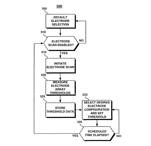
The flow chart shown in FIG. 10 is an overview of one method for using the
lead
in conjunction with the ICD 410. Although a single chamber, left-ventricular
device is
depicted in FIGS. 8 and 9, a lead having one or more electrode arrays could be
used with
atrial or ventricular single chamber devices, with dual chamber devices or
multichamber
devices. These devices may be any of implantable or temporary pacemakers, ICDs
or
10 cardiac monitoring systems. Other than cardiac stimulation or monitoring
systems, the
lead 10 and the method 500 of FIG. 10 to be described may also be used in
implantable or
temporary neurostimulators or other medical devices used for stimulating
and/or sensing
excitable tissue.
In regard to the implantable system illustrated in FIG. 8 and the ICD 410
shown in
FIG. 9, the method 500 shown in FIG. 10 is preferably performed under the
control of
microprocessor 224. Method S00 allows the microprocessor 224 to automatically
determine which of the electrodes included in an electrode array provide the
optimal
stimulation or sensing configuration by performing an electrode scan. During
the electrode
scan, the pacing and/or sensing thresholds of the available electrode
combinations is
measured. Additionally, electrode lead impedance may be measured. The optimal
stimulation configuration is determined as the electrode or combination of
electrodes
resulting in the lowest pacing threshold that successfully captures the
targeted tissue
without depolarizing non-targeted tissue. For example, in the embodiment shown
in FIG.
8, left ventricular capture is desired without atrial capture or phrenic nerve
stimulation.
An optimal sensing configuration may be identified as the electrode
configuration
resulting in the highest signal amplitude or signal-to-noise ratio. For the
left ventricular
application of FIG. 8, the optimal sensing configuration would provide the
highest R-wave
amplitude or the greatest R-wave signal-to-noise ratio.
When the method 500 begins at step 505, the electrode configuration selected
is a
default configuration. Typically, the default configuration is the
simultaneous selection of
all the electrodes included in an electrode array. This default configuration
will be used
CA 02483779 2004-10-26
WO 03/092807 PCT/US03/10437
18
for designated pacing or sensing functions until a more optimal configuration
is identified.
For example, electrodes 25, 27 and 29 may be selected simultaneously to serve
as the
anode during unipolar pacing as the default pacing configuration. A default
sensing
configuration may be set as the bipolar combination of the simultaneously
selected tip
array electrodes 25, 27 and 29 paired with the simultaneously selected ring
array
electrodes 35, 37 and 39.
At decision step 510, the microprocessor 224 determines if an electrode scan
is
enabled. The electrode scan feature is preferably enabled or disabled by a
physician using
an external programmer in telemetric communication with the ICD 410. If the
electrode
scan is disabled, the electrode selection remains in the default
configuration.
Alternatively, a physician may manually program an electrode configuration to
override
the default selection.
If the electrode scan is enabled, a scan is initiated at step 515. An
electrode scan
may be initiated by any of a number of triggering events. Upon implantation of
the lead
10 and ICD 410, a detection of valid electrode pathways by lead recognition
circuitry 250
may trigger the initiation of the electrode scan at step 515. Other triggering
events for an
electrode scan may include detection of a lead failure or a change in lead
status.
Reference is made to previously incorporated U.S. Pat. No. 5,534, 018 and to
U.S. Pat.
No. 6,317,633 issued to Jorgenson et al., incorporated herein by reference in
its entirety.
A scan may also be triggered manually, on a scheduled or periodic basis, or in
response to
a loss of capture.
At step 520, the microprocessor 224 performs a threshold search on each
electrode
within an array individually and in any number of desired combinations. A
threshold
search may be performed according to methods known in the art. For example, a
threshold search may be performed by successively reducing the pacing pulse
amplitude
until capture is lost. For an exemplary threshold search algorithm, reference
is made to
U.S. Pat. No. 3,757,792 issued to Mulier, incorporated herein by reference in
its entirety.
In regard to the electrode configuration shown in FIG. 8, elechodes 25, 27 and
29
in tip electrode array 20 and electrodes 35, 37, and 39 in ring electrode
array 30 may be
selected in any unipolar, bipolar or multipolar configuration. For each
configuration
selected, the left ventricular pacing threshold and/or the R-wave sensing
threshold is
CA 02483779 2004-10-26
WO 03/092807 PCT/US03/10437
19
measured and stored in memory at step 525. After measuring and storing the
thresholds
for all desired electrode configurations, the configuration yielding the
optimal threshold is
selected via switch matrix 208 at step 530 to operate as the designated
configuration for
the associated pacing or sensing function. The pacing pulse energy and/or the
sensing
threshold may also be set at step 530 according to the stored threshold for
the selected
electrode configuration.
An electrode scan may be performed automatically as described or semi-
automatically under the supervision of a clinician such that observation of
any extraneous
stimulation or undersensing or oversensing may be made. Final selection of the
optimal
electrode configuration may then be made manually to eliminate electrode
configurations
producing extraneous stimulation or inaccurate sensing.
At decision step 535, the microprocessor 224 determines if a preset amount of
time, for example 24 hours, has elapsed. Once this time is elapsed, an
electrode scan may
be automatically repeated. Threshold changes may occur over time as electrodes
become
encapsulated by frbrotic scar tissue or with changes in a patient's
physiologic condition,
the use of drugs, or changes in disease state. By repeating the electrode scan
periodically,
the optimal electrode configuration and appropriate pacing energy or sensing
threshold
settings may be updated in response to such changes.
The present invention is realized in an implantable medical lead possessing
one or
more electrode arrays, each comprising multiple electrodes that are
electrically insulated
from each other. The electrodes within an array are preferably arranged
circumferentially
in relation to the lead body and may be located substantially in the area
normally occupied
by a conventional tip or ring electrode.
A lead provided by the present invention includes a lead body extending
between a
proximal lead end and distal lead end for carrying multiple, insulated
conductors. The
conductors are each electrically coupled to an associated electrode at or near
the distal lead
end and to coimectors at the proximal lead end for establishing connection to
an
implantable medical device. The lead may be equipped with a tip electrode
array and/or
one or more ring electrode arrays comprising two or more, preferably three,
electrodes
each. The electrodes within an array may be spaced from each other around the
circumference of the lead and/or along its length. The electrodes within an
array are
CA 02483779 2004-10-26
WO 03/092807 PCT/US03/10437
electrically insulated from each other by non-conductive material, such as a
ceramic,
layered between each electrode in the array.
In one embodiment, a tip electrode array may be expandable in order to improve
the contact of one or more electrodes with a targeted cardiac tissue site.
Expanding the tip
array advantageously increases the spacing between electrodes to improve
sensing and
stimulation performance. Moreover, expansion of the tip array against the
walls of a
blood vessel stabilizes the lead position. If used as an endovascular lead,
blood will easily
flow between the expanded electrodes. A tip array may be expanded by advancing
an
expansion member toward the distal lead end. The expansion member is
preferably
10 conical such that as it is advanced through an electrode head assembly
carrying the tip
array, the widening circumference of the expansion member causes radial
expansion of the
electrodes in the array.
In one embodiment the expansion member may be a fixation helix mounted on a
drive shaft that is coupled to a rotatable coil extending to the proximal lead
end. Rotation
15 of the proximal end of the coil causes rotation of the drive shaft,
advancing the cone-
shaped helix. The helix may be used as an active fixation device to further
stabilize lead
position. Alternatively, a sonically-shaped expansion member may be mounted on
a drive
shaft having a screw-like head. A stylet equipped with a screwdriver-like
blade may be
used to engage the shaft head and, when rotated, cause advancement of the
expansion
20 member.
The lead provided by the present invention may be used with a cardiac pacing
device or ICD equipped with a microprocessor-based control system for
controlling device
functions, a pulse generator for generating electrical impulses to be applied
to the heart,
and sense amplifiers for sensing cardiac signals. The device is preferably
equipped with a
switch matrix for selectively connecting one or more of the electrodes within
an array in
varying combinations for associated sensing and pacing functions. For example,
one
electrode within an array may be used for pacing and the other two electrodes
within an
array may be used for sensing the evoked response. Such a configuration
advantageously
overcomes the problem of polarization artifacts normally encountered when
sensing for an
evoked response using the same pair of electrodes as used for pacing. The
pacing device
or ICD is also equipped with a memory for storing cardiac data and, in
particular, data
CA 02483779 2004-10-26
WO 03/092807 PCT/US03/10437
21
relating to the pacing threshold or sensing threshold associated with various
electrodes
within an array.
In operation, the cardiac pacing device or ICD performs an electrode scan to
determine which electrode or combination of electrodes within an array
provides the
lowest pacing threshold. Once the electrode configuration providing the lowest
pacing
threshold is identified, the control system of the device automatically
selects this
configuration as the pacing electrode configuration via the switch matrix.
Alternatively or
additionally, if an electrode array is to be used for sensing, a sensing
threshold search may
be performed in which the electrodes) providing the highest signal amplitude
or greatest
signal-to-noise ratio may be determined and selected as the sensing electrode
configuration via the switch matrix.
According to the present invention, when the lead is placed endovascularly for
left
heart applications, the electrodes) within an array that are in closest
contact with the heart
tissue may be selected for stimulation and/or sensing. Stray current is
minimized. If
phrenic nerve stimulation or undesired atrial pacing occurs after implantation
of a
coronary sinus lead for left ventricular pacing, an alternative electrode
within a given
electrode array may be selected that still provides an acceptable pacing
threshold at the
targeted ventricular tissue site without extraneous stimulation.
According to the present invention, an electrode pair is selected for sensing
an
evolved response that is in the same vicinity of the paced tissue site but
does not include
the pacing electrode. In addition, battery longevity of the stimulation device
may be
improved by minimizing the surface area used to stimulate a targeted tissue
site. A
smaller electrode surface area associated with selecting one or two electrodes
within an
array increases the pacing impedance resulting in less current drawn from the
battery.
Furthermore, the electrode selection is "fme-tuned" by selecting only the
electrodes)
within an array that provide the lowest pacing threshold, eliminating stray
current and
further extending the useful life of the device. Device performance may be
also be
improved by the ability to select an optimal sensing electrode configuration
such that
accurate sensing of cardiac signals is achieved.
The lead provided by the present invention may be stabilized by an expandable
tip
array and still be readily deployed and repositioned when used as an
endovascular lead.
CA 02483779 2004-10-26
WO 03/092807 PCT/US03/10437
22
Stabilizing the lead position over time may ensure stable pacing and/or
sensing thresholds.
By providing a lead with a reversible fixation device, the lead is easily
advanced or
retracted through a vascular pathway so that the surgical time required for
positioning the
lead may be reduced, with fewer complications encountered. If an electrode
should fail,
other electrodes within the same array may be used for targeting the same
tissue site.
Thus a medical lead that allows accurate targeting of excitable tissue has
been
described and with which extraneous stimulation may be avoided and improved
evoked
response sensing may be achieved. The lead is readily deployed, secured and
repositioned, if necessary, and provides alternative electrode configurations
should a lead
failure occur. A method for using the medical lead has also been described in
which
optimal electrode configurations may be automatically, or semi-automatically,
selected.
While the medical lead and associated method included in the present invention
have been
described according to specific embodiments in the above disclosure, these
embodiments
should be considered exemplary, rather than limiting, with regard to the
following claims.