Note: Descriptions are shown in the official language in which they were submitted.
CA 02484929 2004-10-28
WO 03/092493 PCT/US03/10052
1
METHOD AND APPARATUS TO DETECT AND MONITOR THE FREQUENCY
OF OBSTRUCTIVE SLEEP APNEA
BACKGROUND OF THE INVENTION
This invention relates generally to implantable medical devices, and more
particularly, to a method and apparatus to automatically detect and monitor
the frequency
of obstructive sleep apnea.
Although the function of sleep is not well understood, one consequence of an
inadequate quantity or poor quality of sleep is an inability to maintain
adequate
wakefulness. The amount of sleep an individual needs is thought to be
neurologically
determined and is generally stable over time. Among other factors, an
insufficient amount
of sleep (i. e., quantity of sleep) or a disruption of sleep continuity (i.
e., quality of sleep)
will result in increased daytime sleepiness. Increased sleepiness in a person
rnay cause a
plethora of problems to that person as well as others. Increased sleepiness is
a major cause
of accidents because people who are sleepy axe generally not fully aware of
their
surroundings. Additionally, because of this decreased awareness, a person who
does not
receive the adequate quantity and quality of sleep at night may also be prone
to decreased
efficiency at home and at work. A sleepy person may also require frequent naps
during
the day to recuperate, thereby reducing productivity in the office as well as
in the chores of
daily life. As a result, it is important for people generally to receive a
good night's rest.
However, many people have medical conditions that prevent them from receiving
a good
night's rest. One such condition is sleep apnea.
Sleep apnea is generally defined as the cessation of breathing during sleep.
One
type of a sleep apnea, obstructive sleep apnea ("OSA"), is caused by
repetitive upper
airway obstruction during sleep as a result of narrowing of the respiratory
passages.
Partial obstruction of the passageways may simply lead to hypopnea. Prolonged
obstruction of the passageways, however, may lead to nocturnal arousals.
OSA is generally characterized by a sleep-related withdrawal of upper airway
inspixatory muscle tone superimposed on a narrow, highly compliant pharynx. As
a result,
the pharynx may during sleep, leading to obstructive apnea.
CA 02484929 2004-10-28
WO 03/092493 PCT/US03/10052
2
The cause of OSA is thought to be a combination of anatomic characteristics of
the
upper airway and abnormalities in the neuromuscular control of the muscles in
the throat.
Sleep apnea is more common in individuals with large tonsils, palate, and
tongue, and with
a short thiclc neck. This anatomy may predispose the throat to easily
collapse. A badly
deviated nasal septum or other nasal obstruction can also worsen OSA because
it limits the
ability to breathe through the nose. Overweight individuals are also at high
risk for OSA.
Not all individuals with these anatomic features will have OSA, and OSA
occasionally
occurs in people with normal-appearing throats.
OSA may cause a variety of medical and other problems among patients. Cycles
of sleep, snoring, obstruction, arousal, and sleep may occur many times
throughout the
night. Although such nocturnal arousals may last only a few seconds, they
prevent a
person from reaching the deep stages of sleep, Which the body generally needs
to rest and
replenish its strength. As a result, patients with OSA may not receive a
restful sleep
because of multiple nocturnal arousals.
Furthermore, multiple arousals with sleep fragmentation are likely to cause
excessive daytime sleepiness and fatigue, cognitive impairment, depression,
headaches,
chest pain, and diminished sexual drive. OSA is generally associated with
cardiovascular
morbidity, including systemic hypertension, pulmonary hypertension, ischemic
heart
disease, stroke, and cardiac arrhythmias. OSA is also usually associated with
increased
mortality by negatively effecting the status, progression, and outcomes of
previously
existing conditions, such as congestive heart failure ("CHF").
OSA is a disorder which is generally underdiagnosed and undertreated. Because
OSA may worsen the effects of a previously existing condition, such as CHF,
treatment of
OSA may be beneficial to reduce its negative on the previously existing
condition. Once
OSA has been properly diagnosed, a variety of therapies rnay be available.
Common OSA
therapies include non-surgical methods, such as continuous positive airway
pressure
("CPAP"), as well as surgical methods, such as uvulopalatopharyngoplasty
("UPPP").
Effective therapy for OSA can often reverse or ameliorate the problems
associated with
OSA.
One method of diagnosis for OSA is nocturnal polysomnography. In nocturnal
polysomnography, multiple physiological parameters are measured while the
patient
CA 02484929 2004-10-28
WO 03/092493 PCT/US03/10052
3
sleeps in a laboratory. Typical parameters in a nocturnal polysomnography
include eye
movement observations (to determine whether a patient has reached REM sleep),
an
electroencephalogram (to determine arousals from sleep), chest wall monitors
(to
document respiratory movements), nasal and oral air-flow measurements, and an
electrocardiogram, among other parameters. A combination of these and other
factors are
used by doctors and other qualified sleep specialists to determine whether a
patient has
OSA. However, nocturnal polysomnography is generally expensive and time-
consuming.
Furthermore, many patients experience the symptoms of OSA (e.g., nocturnal
arousals,
snoring) while they are asleep, and therefore, never recognize that they may
have a
sleeping disorder. As a result, many patients with OSA may not seek proper
diagnosis or
treatment of their sleeping disorder from a doctor or other qualified sleep
specialist. Even
if a patient is diagnosed with OSA, frequent laboratory monitoring of the
patient is
generally not feasible due to the expense and time involved in a nocturnal
polysomnography. .
The technology explosion in the implantable medical devices industry has
resulted
in many new and innovative devices and methods for analyzing and improving the
health
of a patient. The class of implantable medical devices now includes
pacemakers,
implantable cardioverters, defibrillators, neural stimulators, and drug
administering
devices, among others. Today's state-of the-art implantable medical devices
are vastly
more sophisticated and complex than early ones, capable of performing
signiftcantly more
complex tasks. The therapeutic benefits of such devices have been well proven.
There are many implementations of implantable medical devices that provide
data
acquisition of important physiological data from a human body. Many
implantable
medical devices are used for cardiac monitoring and therapy. Often these
devices
comprise sensors that are placed in blood vessels and/or chambers of the
heart. Often
these devices are operatively coupled with implantable monitors and therapy
delivery
devices. For example, such cardiac systems include implantable heart monitors
and
therapy delivery devices, such as pacemakers, cardioverters, defibrillators,
heart pumps,
cardiomyostimulators, ischemia treatment devices, drug delivery devices, and
other heart
therapy devices. Most of these cardiac systems include electrodes for sensing
and gain
CA 02484929 2004-10-28
WO 03/092493 PCT/US03/10052
4
amplifiers for recording and/or driving sense event signals from the inter-
cardiac or
remote electrogram ("EGM").
Many patients who use implantable medical devices rnay be at rislc for OSA.
However, patients are generally Ieft with traditional forms of diagnosis for
OSA, such as
nocturnal polysomnography. As mentioned, nocturnal polysomnography may be an
expensive and time-consuming procedure. Furthermore, many patients may not
recognize
that they have symptoms relating to OSA, such that they would seek diagnosis
and
treatment for the disorder. Nocturnal polysomnography is generally an
infrequent
procedure that does not provide long term monitoring of the patient's
condition after he
has been diagnosed. The present invention is directed to overcoming, or at
least reducing
the effects of, one or more of the problems set forth above.
SUMMARY OF THE INVENTION
In one aspect of the present invention, an apparatus is provided for detecting
and
monitoring obstructive sleep apnea. The apparatus includes an intracardiac
impedance
sensor to measure intracardiac impedance, a movement sensor to measure an
amount of
movement of a patient, and a controller operatively coupled to said
inixacardiac impedance
sensor and said movement sensor, said controller adapted to receive at least
one of an
intracardiac impedance and the amount of movement of the patient and detect
obstructive
sleep apnea based upon said intracardiac impedance and said movement.
In another aspect of the present invention, a method is provided for detecting
and
monitoring obstructive sleep apnea. The method includes measuring an
intracardiac
impedance to detect a change in the intracardiac impedance, measuring an
amount of
movement of a patient, and determining the presence of obstructive sleep apnea
based
upon the change in the intracardiac impedance and the movement of the patient.
BRIEF DESCRIPTION OF THE DRAW1NGS
The invention rnay be understood by reference to the following description
taken
in conjunction with the accompanying drawings, in which like reference
numerals identify
like elements, and in which:
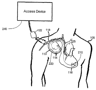
Figure 1 is a simplified diagram of an implementation of an implantable
medical
device, in accordance with one illustrative embodiment of the present
invention;
CA 02484929 2004-10-28
WO 03/092493 PCT/US03/10052
Figure 2 illustrates a simplified block diagram representation of an
implantable
medical system in accordance with one illustrative embodiment of the present
invention;
Figure 3 illustrates a more detailed block diagram representation of the
implantable
medical device of Figures I and 2, in accordance with one illustrative
embodiment of the
present invention; and
Figure 4 illustrates a more detailed block diagram representation of a
plurality of
sensors and its associated data interfaces of Figure 3, in accordance with one
illustrative
embodiment of the present invention.
While the invention is susceptible to various modifications and alternative
forms,
specific embodiments thereof have been shown by way of example in the drawings
and
are herein described in detail. It should be understood, however, that the
description
herein of specific embodiments is not intended to limit the invention to the
particular
forms disclosed, but on the contrary, the intention is to cover all
modifications,
equivalents, and alternatives falling within the spirit and scope of the
invention as defined
by the appended claims.
DETAILED DESCRIPTION OF SPECIFIC EMBODIMENTS
Illustrative embodiments of the invention are described below. In the interest
of
clarity, not all features of an actual implementation are described in this
specification. It
will of course be appreciated that in the development of any such actual
embodiment,
numerous implementation-specific decisions must be made to achieve the
developers'
specific goals, such as compliance with system-related and business-related
constraints,
which will vary from one implementation to another. Moreover, it will be
appreciated~that
such a development effort might be complex and time-consuming, but would
nevertheless
be a routine undertaking for those of ordinary skill in the art having the
benefit of this
disclosure.
There are many discrete processes involving the operation of implantable
medical
devices (e.g., pacemakers, cardio defibrillators, the Iike). The operation of
an implantable
medical device includes collecting, storing, and analyzing physiological data
relating to a
patient, andlor delivering therapy (e.g., cardiac therapy) to a portion of a
patient's body.
Often, these tasks are performed by an implantable medical system, which
includes an
implantable medical device. Based upon the analysis performed by the
implantable
CA 02484929 2004-10-28
WO 03/092493 PCT/US03/10052
6
medical system, one or more therapies may be delivered to a particular portion
of a
patient's body. One example of such a therapy is a cardiac therapy, which is
delivered to a
patient's heart.
Embodiments of the present invention may be utilized to detect and monitor the
common symptoms and conditions related to patients with Obstructive Sleep
Apnea
(OSA). It should be appreciated that the present invention may be included in
an
implantable device capable of collecting data other than data used to diagnose
or monitor
OSA. The data may be collected by sensors in the implantable device and may be
used by
doctors and other sleep experts to judge the severity of apneas and to
determine the
efficacy of apnea therapy, without the use of nocturnal polysomnography.
Tuxning now to Figure 1, one embodiment of implementing an implantable
medical device into a human body is illustrated. A sensor device 210 (e.g.,
devices
attached to leads 114) placed upon the heart 116 of the human body 105 is used
to acquire
and process physiological data. In one embodiment, the sensox device 210 may
also be a
therapy delivery device (described in greater detail below). An implantable
medical
device 220 collects and processes a plurality of data acquired from the human
body 105.
In one embodiment, the implantable medical device 220 may be a cardiac
pacemaker or an
implantable cardiovertor defibrillator ("ICD"). The data acquired by the
implantable
medical device 220 can be monitored by an external system, such as the access
device 240
comprising a programming head 122, which remotely communicates with the
implantable
medical device 220. The programming head 122 is utilized in accordance with
medical
device programming systems known to those skilled in the art having the
benefit of the
present disclosure, for facilitating two-way communication between the
implantable
medical device 220 and the access device 240.
In one embodiment, a plurality of access devices 240 can be employed to
collect a
plurality of data, including OSA data, processed by the implantable medical
device 220 in
accordance with embodiments of the present invention. 'The implantable medical
device
220 is housed within a hermetically sealed, biologically inert outer canister
or
housing 113, which may itself be conductive so as to serve as an electrode in
the
implantable medical device 220 pacing/sensing circuit. One ox more
sensors/leads,
collectively identified with reference numexal 114 in Figuxe 1, are
electrically coupled to
CA 02484929 2004-10-28
WO 03/092493 PCT/US03/10052
7
the implantable medical device 220 and extended into the patient's heart 116
via a
vein 118. Disposed generally near a distal end of the leads 114 are one or
more exposed
conductive electrodes (i.e., sensor device 210) for receiving electrical
cardiac signals or
delivering electrical pacing stimuli to the heart 116. The leads 114 may be
implanted with
their distal end situated in either the atrium or ventricle of the heart 116.
In an alternative
embodiment, the sensor device 210, or the leads 114 associated with the sensor
device
210, may be situated in a blood vessel on the heart 116, such as a vein 118.
Turning now to Figure 2, a system 200, in accordance with one embodiment of
the
present invention, is illustrated. The system 200 comprises a plurality of
sensor devices,
collectively identified with reference numeral 2I0 in Figure 2, an implantable
medical
device 220, an access device 240, and an interface 230 that provides a
communication link
between the implantable medical device 220 and the access device 240.
Embodiments of
the present invention provide a plurality of physiological data from the
sensor devices 2I0,
which are then processed and stored in the implantable medical device 220. In
one
embodiment, the sensor devices 210 may collect data that is used to detect and
monitor
OSA in a patient.
As mentioned, based upon physiological data and other fact~rs, the implantable
medical device 220 may deliver a therapy to a portion of the patient's body,
via the sensor
devices 210. The access device 240 can then be used to monitor and analyze the
organized data from the implantable medical device 220 via the interface 230
and view
results from delivered therapy. The access device 240 can be used to monitor
the
efficiency of the therapy delivered by the implantable medical device 220. The
access
device 240 can be used to determine, based upon data stored by the implantable
medical
device 220, whether a therapy delivered was of proper energy intensity.
Turning now to Figure 3, a more detailed block diagram depiction of one
embodiment of the implantable medical device 220 is illustrated. The
implantable medical
device 220 comprises a processor 310, a control logic 320, a memory unit 330,
a data
acquisition controller 340, a telemetry interface 350, and a plurality of data
interfaces 360,
370, 380, 390. The plurality of sensor devices 210 of Figure 2 provide various
physiological data to the implantable medical device 220. The processor 310
controls the
operation of the implantable medical device 220. The processor 310 utilizes
the control
CA 02484929 2004-10-28
WO 03/092493 PCT/US03/10052
8
logic 320 to perform a plurality of operations, including memory access and
storage
operations. The processor 310 communicates with the control logic 320 and the
data
acquisition controller 340 via a bus line 325. The control logic 320 sends
control signals
to the memory unit 330 for controlling and installing the memory unit 330, and
to the data
acquisition controller 340, which controls the acquisition of physiological
data and drives
output signals to the telemetry interface 350.
The telemetry interface 350 can facilitate real-time access of physiological
data
acquired by the data acquisition controller 340. Therefore, a physician can
view
physiological data on a real time basis by accessing the data acquisition
controller 340, via
the telemetry interface 350. The data acquisition controller 340 can prompt
the data
interfaces 360, 370, 380, 390 to retrieve physiological data from the sensor
device 210,
process such data, and deliver physiological data to the data acquisition
controller 340.
The data interfaces 360, 370, 380, 390 can perform a number of analog-to-
digital
conversions and time-interval conversions, known to those skilled in the art,
upon the
acquired physiological data. The data interfaces 360, 370, 380, 390 can
acquire,
condition, and process physiological data and forwaxd them to the data
acquisition
controller 340.
It should be appreciated that, in an alternate embodiment, the functionality
of the
data interfaces 360, 370, 380, 390 may be combined with the sensor devices
210, such that
information gathered by the sensor devices 210 may be readily utilized by the
implantable
medical device 220 without further processing by another device or interface.
It should
also be appreciated that, although the sensor devices 210 are separated from
the
implantable medical device 220 fox illustrative purposes in Figures 2 and 3,
the
implantable medical device 220 may further comprise the sensor devices 210.
Turning now to Figure 4, one embodiment of the sensor device 210 and the
implantable medical device 220 of Figures l, 2, and 3, in accordance with the
present
invention, is shown. In the illustrated embodiment of Figure 4, four sensors
are shown.
However, it should be appreciated that the implantable medical device 220 may
comprise
of more or less sensors than the illustrated embodiment of Figuxe 4, such that
OSA may be
properly diagnosed on a patient. It should also be appreciated that the
functionality of
CA 02484929 2004-10-28
WO 03/092493 PCT/US03/10052
9
each sensor described below may be combined into one or more sensors, such
that OSA
rnay be properly diagnosed on a patient.
The implantable medical device 220 comprises four sensors, which individually
and in combination may be used to detect OSA in a patient. An intracardiac
impedance
sensor 210-1 measures impedance between an intracardiac atrial electrode and
an
intracardiac ventricular electrode. An intrathoracic impedance sensor 210-2
measures
impedance across the thorax. In one embodiment, the intrathoracic impedance
sensor 210-
2 may include pacemaker sensors, which measure impedance between a pacemaker
and an
intracardiac electrode. The impedance between the pacemaker and the
intracardiac
electrode may be used to estimate in minute ventilation ("MV"). A movement
sensor 210-
3 detects movement in a patient during sleep. The movement sensor 210-3, in ~
one
embodiment, may be a piezo crystal or an accelerometer. An electrical sensor
210-4
detects cardiac depolarizations.
Using the four sensors 210-1, 210-2, 210-3, 210-4, a plurality of information
can
be gathered to properly diagnosis OSA on a patient. The information gathered
by the
sensors 210-1, 210-2, 210-3, and 210-4 is processed by an intracardiac data
interface 360,
an intrathoracic impedance data interface 370, a movement data interface 380,
and an
electrical data interface 390, respectively, before the information is
forwarded to the data
acquisition controller 340. Although sensors can be used individually to
diagnose OSA,
combinations of two or more sensors may form a basis for diagnosis of OSA. For
example, a large decrease in impedance between atrial and ventricular
electrodes (i. e., a
decrease in the intracardiac impedance sensor 210-1) occurring when a patient
is riot
exercising (i.e., a low reading from the movement sensor 210-3) may be a
factor towards
diagnosis of OSA. As a patient attempts to breath while his airway is
obstructed, negative
intrathoracic pressure may increase, which overfills the right side of the
heart. This
overfilling of the right side of the heart increases the diameter of the
atrium and the
ventricle. Although the volume of the heart does not change, the shape of the
heart
becomes shorter and wider. As a result, a drop in atrial-to-ventricular
impedance may be
observed during OSA because the wider blood pool will cause a reduced
impedance. The
wider blood pool may also be a result vigorous exercise from the patient.
Therefore, the
intracardiac impedance sensor 210-1 may be read in conjunction with the
movement
CA 02484929 2004-10-28
WO 03/092493 PCT/US03/10052
sensor 210-3. In one embodiment, a low reading from the movement sensor 210-3
indicates the patient is not exercising or in some other physical activity. A
decrease in the
intracardiac impedance sensor 210-1 and a low reading from the movement sensor
2I0-3
indicates a possibility that the patient has OSA.
Another possible indication that a patient has OSA may be provided by the
intrathoracic impedance sensor 210-2. In one embodiment, the intrathoracic
impedance
sensor 210-2 may measure impedance between an implantable device housing, such
as a
can electrode, and an endocardial electrode, such as an atrial electrode or a
ventricular
electrode. A decrease in intrathoracic impedance during attempted inspiration
may be a
factor in determining whether a patient has OSA. As a patient attempts to
breathe while
his airway is obstructed, the circumference of his thorax may expand. As the
patient's
thorax expands, the diaphragm pushes the, heart and lungs upward. As the lungs
are
pushed upward, the lungs do not change volume, but instead become shorter and
wider.
The shorter and wider shape of the lungs may reduce intrathoracic impedance.
In
addition, as a patient attempts to breath while his airway is obstructed,
negative
intrathoracic pressure may increase, which overfills the right side of the
heart. As the right
side of the heart overflows, a wider blood pathway forms, thereby reducing
intrathoracic
impedance. Furthermore, upward movement of the diaphragm pushes the heart
closer to
the implantable medical device 220, thereby reducing impedance. A decrease in
the
intrathoracic impedance sensor 210-2 indicates the possibility that the
patient has OSA.
Another possible indication that a patient has OSA comes from a rapid increase
in
impedance minute ventilation ("MV") followed by a slower decrease in impedance
MV.
A person who does not have sleep apnea breathes normally during rest. A
patient who has
OSA usually cannot breathe normally during rest because of an obstruction in
the airway.
While the patient's heart is attempting to pump blood into the lungs, the
obstruction is
preventing the patient from breathing. As a result, the patient's heart begins
to pump
faster to compensate for the lack of blood flow. After an arousal event, the
patient begins
to breathe again, but because the heart was pumping fast before the arousal
event,. the
patient goes through a hyperpneic phase, which is a period of abnormally rapid
or deep
breathing. Studies have shown that during the hyperpneic phase there may be an
immediate rise in MV followed by a more gradual drop in MV over a period of
time.
CA 02484929 2004-10-28
WO 03/092493 PCT/US03/10052
11
An arousal event each time an obstruction is relieved may cause brief periods
of
movement detectable by the movement sensor. As mentioned, in one embodiment,
the
movement sensox may be a piezo crystal or an accelerometer. During the
hyperpneic
phase following the release of an obstruction, there is usually a brief
arousal which can
cause body movement detectable by movement sensors. A brief period of movement
during sleep coinciding with the dramatic increase in respiration (i.e., a
rapid increase in
impedance MV followed by a slower decrease in impedance MV), as described
above,
may indicate the patient has OSA. An immediate rise in MV followed by a more
gradual
drop in MV, coupled with a brief of movement of sleep, indicates the
possibility that the
patient has OSA.
Yet another possible indication that a patient has OSA may be provided by the
electrical sensor 210-4. OSA can cause bradycardia during an obstruction.
Bradycardia is
an abnormally slow or unsteady heart rhythm (usually less than 60 beats per
minute) that
causes symptoms such as dizziness, fainting, fatigue, and shortness of breath.
Release of
the obstruction is generally accompanied by sinus tachycardia and possibly
increased
atrial-ventricular conduction. Sinus tachycardia is a fast heartbeat (usually
more than 150
beats per minute) because of rapid firing of the sinoatrial (i.e., sinus)
node. Both sinus
tachycardia and increased atrial-ventricular conduction can be detected by
intracardiac
electrodes and electrical sensing amplifiers or by subcutaneous electrodes and
electrical
sensing amplifiers.
Referring back to Figure 3, as the sensors 210-1, 210-2, 210-3, 210-4 collect
data,
the data interfaces 360, 370, 380, 390 process the data, in accordance with
conventional
practice, and forwards the data to the data acquisition controller 340. The
processor 310
then utilizes the control logic 320 to provide the data from the data
acquisition controller
to the memory unit 330. In addition, a doctor or another qualified sleep
professional may
use the telemetry interface 350 to facilitate real-time access to the data on
the data
acquisition controller. The doctor or another qualified sleep specialist may
analyze the
collected data to determine whether the patient has OSA.
The particular embodiments disclosed above are illustrative only, as the
invention
may be modified and practiced in different but equivalent manners apparent to
those
skilled in the art having the benefit of the teachings herein. Furthermore, no
limitations
CA 02484929 2004-10-28
WO 03/092493 PCT/US03/10052
12.
are intended to the details of construction or design herein shown, other than
as described
in the claims below. Tt is therefore evident that the particular embodiments
disclosed
above may be altered or modified and all such variations axe considered within
the scope
and spirit of the invention. Accordingly, the protection sought herein is as
set forth in the
claims below.