Note: Descriptions are shown in the official language in which they were submitted.
CA 02485107 2010-07-07
SURGICAL STAPLING APPARATUS HAVING
A WOUND CLOSURE MATERIAL APPLICATOR ASSEMBLY
BACKGROUND
1. Technical Field
The present disclosure relates to surgical staplers, and more particularly, to
a surgical
stapling apparatus for applying a plurality of surgical fasteners to body
tissue and having a
wound closure material applicator assembly for dispensing a quantity of wound
closure
material or components thereof, at least along a knife cut line and/or a
staple line.
2. Background of Related Art
Surgical procedures requiring cutting of tissue can result in bleeding at the
site of the
cut. Various techniques have been developed to control bleeding with varying
degrees of
success, such as, for example, suturing, applying clips to blood vessels, and
using surgical
fasteners, as well as electrocautery and other tissue healing techniques.
Surgical instruments using surgical fasteners entail grasping or clamping
tissue
between opposing jaw structure and then joining the tissue by employing the
surgical fasteners.
These instruments are well known in the art. In some instruments, a knife is
25
1
CA 02485107 2010-07-07
provided to cut the tissue, which has been joined by the fasteners. The
fasteners are typically in
the form of surgical staples however, two part polymeric fasteners can also be
utilized.
Instruments for this purpose can include two elongated or circular members,
which are
respectively used to capture or clamp tissue. Typically, one of the members
carries a cartridge,
which houses a plurality of staples arranged in at least two lateral rows
while the other member
carries an anvil, which defines a surface for forming the staple legs as the
staples are driven
from the cartridge. Where two part fasteners are used, one of the members
carries a cartridge
which houses one half of a fastener while the other member carries the second
part of the
fastener, e.g., the mating part, which halves are configured and adapted to be
held together
upon approximation. Typically, the stapling operation is effected by a driving
member, which
travels longitudinally through the cartridge carrying member, with the driving
member acting
upon pushers, which engage the staples for sequentially ejecting them from the
cartridge. A
knife can be provided which travels between the staple rows to longitudinally
cut (i.e., form a
knife cut line) and/or open the stapled tissue between the rows of staples.
Usually, but not
always, the knife is associated with or travels with the staple driving
member. Such
instruments are disclosed in U.S. Pat. Nos. 3,079,606 and 3,490,675.
A later stapler disclosed in U.S. Pat. No. 3,499,591, applies a double row of
staples on
each side of the incision or the knife cut line. This is accomplished by
providing a cartridge
assembly in which a cam member moves through an elongate guide path between
two sets of
staggered staple carrying grooves. Staple drive members are located within the
grooves and are
positioned in such a manner so as to be contacted by the longitudinally moving
cam to effect
ejection of the staples. Other examples of staplers are disclosed in U.S. Pat.
No. 4,429,695,
5,065,929, and 5,156,614.
2
CA 02485107 2004-11-02
WO 03/094743 PCT/US03/14699
Electrocautery devices are preferred in certain surgical procedures for
effecting
improved hemostasis by heating tissue and blood vessels using thermogenic
energy,
preferably radiofrequency energy, to cause coagulation or cauterization.
Monopolar devices
utilize one electrode associated with a cutting or cauterizing instrument and
a remote return
electrode, usually adhered externally to the patient. Bipolar instruments
utilize two electrodes
and the cauterizing current is generally limited to tissue between the two
electrodes of a tissue
treating portion (e.g., end effector) of an instrument.
Even though stapling apparatus and electrocauterizing apparatus are
respectively
generally well suited to control bleeding along the knife cut line, other
techniques are herein
envisioned for being used in conjunction with these techniques.
Therefore, it is an aspect of the invention to provide a surgical stapling
apparatus for
providing general hemostasis, tissue joining or welding, and also a wound
closure material,
for example, for providing additional hemostasis along a cut line formed by a
knife or other
cutting means and/or along a staple line of the surgical stapling apparatus to
reduce or prevent
bleeding along the cut line and/or staple line.
SUMMARY
This present disclosure relates to surgical stapling apparatus having a wound
closure
material applicator for applying a plurality of surgical fasteners to body
tissue and dispensing
a quantity of wound closure material or components thereof, along a staple
line and/or a knife
cut line.
According to one aspect of the present disclosure, a surgical stapling
apparatus for
enhancing one or more properties of body tissue that is or is to be repaired
or joined is
provided. The surgical stapling apparatus includes a staple anvil positioned
on a distal end of
the stapling apparatus and having a longitudinal knife track and a staple
cartridge positioned
adjacent a distal end of the stapling apparatus, the staple cartridge and
staple anvil being
3
CA 02485107 2004-11-02
WO 03/094743 PCT/US03/14699
juxtaposable relative to each other. The staple cartridge includes a plurality
of surgical
staples individually disposed within individual staple slots formed in rows in
the staple
cartridge and having a longitudinal knife slot.
The surgical stapling apparatus includes a driving member for firing the
surgical
staples from the individual staple slots and against the staple anvil, a knife
blade structure
including a knife blade receivable in and axially movable along the knife
track and knife slot,
and a wound closure material applicator assembly operatively associated with
the stapling
apparatus. The wound closure material applicator assembly includes a channel
with an
orifice, and a conduit in fluid communication with the channel, wherein axial
movement of
the knife blade structure through the knife track and knife slot axially
advances the knife
blade structure to permit the orifice to dispense wound closure material from
the orifice into
an area between the staple anvil and the staple cartridge.
It is envisioned that the applicator assembly further includes at least one
reservoir in
fluid communication with the conduit, the at least one reservoir containing a
wound closure
material therein. The driving member can include an actuation sled and the
knife blade
structure is part of the actuation sled. The knife blade structure preferably
includes a needle
having the orifice.
The wound closure material can be an astringent, a sulfate of aluminum, an
adhesive,
a hemostat and/or a sealant.
The reservoir can be compressible. Compression of the reservoir can cause the
wound
closure material to be dispensed from a needle of the applicator assembly.
The orifice of the needle can be oriented in at least one of a proximal,
distal,
downward and upward direction. Preferably, the needle has a tip and the
orifice is located at
the tip. The needle can include a plurality of orifices oriented in at least
one of a proximal,
distal, downward and upward direction.
4
CA 02485107 2004-11-02
WO 03/094743 PCT/US03/14699
The conduit is preferably extendable through at least a portion of the staple
cartridge.
The needle can be secured to the knife blade structure. As such, the needle
can be
adapted to dispense the wound closure material into at least an area near the
knife blade
and/or into at least an area behind the knife blade.
According to a further aspect of the present disclosure, a surgical stapling
apparatus
for enhancing one or more properties of body tissue that is or is to be
repaired or joined is
provided. The surgical stapling apparatus includes a staple anvil positioned
on a distal end of
the stapling apparatus and having a longitudinal knife track and a staple
cartridge positioned
adjacent a distal end of the stapling apparatus, the staple anvil and staple
cartridge being
juxtaposable relative to each other. The staple cartridge includes a working
surface, one or
more rows of individual staple slots formed in the working surface, a knife
slot formed along
a length of the working surface, and a plurality of surgical staples
individually disposed
within the individual staple slots.
The surgical stapling apparatus includes a driving member translatably
receivable in
the staple cartridge for firing the surgical staples from the individual
staple slots and against
the staple anvil, the driving member including an actuation sled having a
knife operatively
connected thereto, the actuation sled being configured and adapted to position
the knife to be
axially moveable within the track and the knife slot.
The surgical stapling apparatus further, includes a wound closure material
applicator
assembly including an applicator having an orifice and configured to dispense
a quantity of
wound closure material from the orifice as the knife moves along a length of
the knife slot.
The wound closure material applicator can include a needle having an orifice
and
secured to a portion of the actuation sled, wherein the needle directs the
dispensation of
wound closure material through the orifice, and a conduit in fluid
communication with the
needle for delivering the quantity of wound closure material to the needle.
The wound
5
CA 02485107 2004-11-02
WO 03/094743 PCT/US03/14699
closure material applicator assembly can further include a reservoir,
containing at least one
quantity of wound closure material, in fluid communication with the conduit.
The quantity of wound closure material can be an astringent, an adhesive, a
hemostat,
and a sealant.
The reservoir can be compressible. Compression of the reservoir causes the
wound
closure material to be dispensed from the needle of the applicator.
The needle has a tip with the orifice and the tip of the needle is oriented in
at least one
of a proximal, distal, downward and upward direction. The needle includes a
plurality of
orifices oriented in at least one of a proximal, distal, downward and upward
direction. The
orifice is adapted to spray a mist of the wound closure material near and/or
behind the knife.
According to a further aspect of the present disclosure, a surgical stapling
apparatus
for enhancing one or more properties of body tissue that is or is to be
repaired or joined,
wherein the surgical stapling apparatus includes a staple anvil positioned on
a distal end of
the stapling apparatus, a staple cartridge positioned adjacent a distal end of
the stapling
apparatus, the staple cartridge including a working surface defining a knife
slot formed along
a length thereof, a driving member translatably receivable in the staple
cartridge and
including an actuation sled having a knife structure operatively connected
thereto and
positioned within the knife slot, is provided.
The improvement includes a wound closure material applicator assembly
configured
to dispense a quantity of wound closure material as the knife structure moves
along a length
of the knife slot.
It is envisioned that the wound closure material applicator includes a needle
secured to
the actuation sled, wherein the needle directs the dispensation of wound
closure material, and
a conduit in fluid communication with the needle for delivering the quantity
of wound closure
material to the needle. It is further envisioned that the needle is secured to
the knife.
6
CA 02485107 2004-11-02
WO 03/094743 PCT/US03/14699
It is contemplated that the wound closure material applicator further includes
a
reservoir, containing the quantity of wound closure material, in fluid
communication with the
conduit.
The quantity of wound closure material is an astringent, an adhesive, a
hemostat
and/or a sealant.
The reservoir can be compressible. Accordingly, compression of the reservoir
causes
the wound closure material to be dispensed from the needle. The reservoir can
be in the form
of a syringe. The syringe can include two chambers each containing a different
wound
closure material. Alternatively, the syringe includes two chambers each
containing a
component of a wound closure material, wherein the wound closure material is
activated
upon combination of the two components of the wound closure material.
It is contemplated that the needle directs wound closure material onto, in
front of,
behind or to the sides of the knife. It is also contemplated that
advantageously, in
combination with any of the above aspects of the invention, the conduit can
have one or more
holes therein to dispense wound closure material onto tissue disposed between
the anvil and
the cartridge.
Further features of the surgical apparatus of the invention will become more
readily
apparent to those skilled in the art from the following detailed description
of the apparatus
taken in conjunction with the drawings.
7
CA 02485107 2004-11-02
WO 03/094743 PCT/US03/14699
BRIEF DESCRIPTION OF THE DRAWINGS
Various embodiments of the surgical stapling apparatus of the invention will
be
described hereinbelow with reference to the drawings wherein:
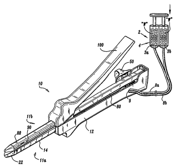
FIG. 1A is a perspective view of a surgical stapling apparatus having a wound
closure
material applicator assembly constructed in accordance with a preferred
embodiment, with
the clamping handle of the apparatus disposed in an upright open position;
FIG. lB is a perspective view of the surgical stapling apparatus illustrated
in FIG. 1A
with the clamping handle disposed in a closed position;
FIG. 2A is an exploded perspective view of the surgical stapling apparatus of
FIG.
1A;
FIG. 2B is a perspective view of a surgical stapling apparatus having a wound
closure
material applicator assembly constructed in accordance with another preferred
embodiment;
FIG. 2C is a perspective view of a surgical stapling apparatus having a wound
closure
material applicator assembly constructed in accordance with another preferred
embodiment;
FIG. 3 is a perspective view of a cartridge half-section of the surgical
stapling
apparatus of FIG. IA;
FIG. 4A is a top plan view of a retention channel of the surgical stapling
apparatus of
FIG. 1A;
FIG. 4B is a side elevational view of the retention channel shown in FIG. 4A;
FIG. 4C is a perspective view of the retention channel of FIGS. 4A and 4B with
the
disposable loading unit retained therein;
FIG. 5A is an enlarged perspective view, with parts separated, of the
disposable
loading unit and actuation assembly of the surgical stapling apparatus of FIG.
1A;
FIG. 5B is a cross-sectional view of the indicated area in FIG. 5A showing the
engagement of the cartridge lip and the retention channel;
8
CA 02485107 2004-11-02
WO 03/094743 PCT/US03/14699
FIG. 6A is a perspective view of the actuation sled of the disposable loading
unit
shown in FIG. 5A in a pre-formed condition;
FIG. 6B is a perspective view of the actuation sled shown in FIG. 6A in a
formed
condition with the knife blade and a dispensing needle of the wound closure
material
applicator assembly separated therefrom for illustrative purposes;
FIG. 6C is a perspective view of the formed actuation sled shown in FIG. 6B
with the
knife blade and the dispensing needle mounted to the blade support portion
thereof;
FIG. 6D is a perspective view of the dispensing needle and a portion of the
conduit in
an alternate embodiment;
FIG. 7 is a plan view of the preformed anvil plate which is mounted to the
anvil
support beam of the anvil half-section of the surgical stapling apparatus
shown in FIG. 1A;
FIG. 8 is a cross-sectional view of the preformed anvil plate taken along line
8--8 of
FIG. 7;
FIG. 9 is a front end view of the preformed anvil plate illustrated in FIGS. 7
and 8;
FIG. 10 is a perspective view of the anvil half-section of the surgical
stapling
apparatus of FIG. IA with an enlarged localized view of a distal portion
thereof illustrating
the connective engagement between the anvil plate and the anvil support beam;
FIG. 11 is an exploded perspective view of a lockout mechanism to prevent
reactuation of the apparatus;
FIG. 11A is an enlarged cross-sectional view of the T-shaped member of the
lockout
mechanism;
FIG. 11B is a perspective view of the needle and the shank according to an
alternate
embodiment of the present disclosure wherein the needle is forked with two
tines and each
tine is directed to an opposing side of the shank;
9
CA 02485107 2004-11-02
WO 03/094743 PCT/US03/14699
FIG. 12 is an enlarged perspective view of the actuation channel having an
edge for
engagement by the hook of the lockout mechanism;
FIGS. 13A and 13B are side views of the lockout mechanism illustrating its
movement from a non-engaged to an engaged position;
FIG. 14 is a side elevational view in cross-section of the surgical stapling
apparatus of
the present invention with the actuation sled supporting the adhesive
dispensing needle and
disposed in a pre-actuated proximal position;
FIG. 15 is a side elevational view in cross-section of the surgical stapling
apparatus of
the present invention with the actuation sled disposed in a partially advanced
position;
FIG. 16 is a side elevational view in cross-section of the surgical stapling
apparatus of
the subject application with the actuation sled advanced to the distal end of
the cartridge at
the conclusion of a staple firing procedure;
FIG. 17 is a side elevational view in cross-section of the surgical stapling
apparatus of
the subject application with the actuation sled advanced to the distal end of
the cartridge at
the conclusion of a staple firing procedure and a dispensing needle located
near the knife
blade;
FIG. 18A is a perspective view of an actuation sled including a wound closure
material applicator, in accordance with an alternative embodiment, operatively
connected
thereto;
FIG. 18B is a top plan view of the actuation sled and applicator of FIG. 18A;
FIG. 18C is a bottom plan view of the actuation sled and applicator of FIG.
18A;
FIG. 19A is a perspective view of an actuation sled including a wound closure
material applicator operatively connected thereto, in accordance with another
embodiment;
FIG. 19B is a top plan view of the actuation sled and applicator of FIG. 19A;
FIG. 19C is a bottom plan view of the actuation sled and applicator of FIG.
19A;
CA 02485107 2004-11-02
WO 03/094743 PCT/US03/14699
FIG. 20A is a perspective view of an actuation sled including a wound closure
material applicator operatively connected thereto, according to yet another
embodiment;
FIG. 20B is a top plan view of the actuation sled and applicator of FIG. 20A;
FIG. 20C is a bottom plan view of the actuation sled and applicator of FIG.
20A.
DETAILED DESCRIPTION OF PREFERRED EMBODIMENTS
Preferred embodiments of the presently disclosed surgical stapling apparatus
will now
be described with reference to the drawing figures wherein like reference
numerals identify
similar or identical elements. In the drawings and in the following
description, the term
"proximal", as is traditional, will refer to the end of the apparatus which is
closer to the
operator, while the term "distal" will refer to the end of the apparatus which
is further away
from the operator.
The present invention provides a surgical stapling apparatus having a wound
closure
material applicator assembly which applies at least one biological and/or
synthetic
biocompatible sealant, hemostat, adhesive, and combinations thereof
(individually or
collectively referred to herein as wound closure material), as well as
surgical fasteners or
staples, for example, for providing hemostasis, tissue joining or welding. The
application of a
wound closure material to the cut line and/or staple line can provide short,
i.e., temporary,
and long-term, i.e., permanent, hemostasis and sealing, and reduce or prevent
bleeding along
a knife cut line and/or staple line, while the stapling features provide short
and long-term
tissue strength and hemostasis.
Since knife cut line and staple line bleeding is reduced or prevented, the
surgical
stapling apparatus of the present invention makes it possible to expand the
applicable range of
specific staple sizes to include thinner or thicker staples used in highly
vascularized tissue.
11
CA 02485107 2004-11-02
WO 03/094743 PCT/US03/14699
For example, it is contemplated that relatively large-size staples could be
used with the
surgical stapling apparatus of the present invention to join thin, highly
vascularized tissue.
Referring now to the drawings wherein like reference numerals identify similar
structural elements, there is illustrated in FIGS. 1A and 1B a surgical
stapling apparatus in
accordance with a preferred embodiment and designated generally as reference
numeral 10.
Surgical stapling apparatus 10 includes a cartridge half-section 11a and an
anvil half-section
1 lb.
Referring to FIGS. 2A, 2B and 3, stapling apparatus 10 includes a body portion
12
defining a handle for grasping and supporting stapling apparatus 10. A
retaining channel 14
is mounted in an interior cavity 15 of body portion 12 adjacent the distal end
thereof.
Retaining channel 14 is dimensioned and configured to support a disposable
loading unit 20,
as illustrated in FIG. 4C.
As shown in FIG. 5A, disposable loading unit 20 includes a cartridge 22 having
tissue
contacting or working surface 21, a plurality of slots 22c which support a
corresponding
number of surgical staples 24, a plurality of staple pushers or ejectors 26
adapted and
configured to eject staples 24 from slots 22c when acted upon by a staple
driving force, and
an actuation sled 28 which is mounted to translate through cartridge 22 in a
longitudinal
direction to transmit a staple driving force to ejectors 26 and dispense a
quantity of wound
closure material to be a target surgical site.
As seen in the figures, particularly in FIGS. 2A, 3, and 5A, surgical stapling
apparatus
10 includes a wound closure material applicator assembly 2 operatively
associated with
surgical stapling apparatus 10. Wound closure material applicator assembly 2
includes a
compressible (or syringe-like, etc.) reservoir 4 (see FIG. 2A) in fluid
communication with a
needle 6 via a conduit 8. In use, wound closure material applicator 2 manually
or
automatically supplies a wound closure material "W", or a component thereof,
to a target
12
CA 02485107 2004-11-02
WO 03/094743 PCT/US03/14699
surgical site. Conduit 8 enters body portion 12 via an opening 9 and is
configured for
traversing approximately the entire length of body portion 12 and cartridge
22, during firing
of apparatus 10. Preferably, a distal portion of conduit 8 is supported by
actuation sled 28
and needle 6 is secured to, connected to, or otherwise mounted on a portion of
actuation sled
28, here on knife blade 36, in such a manner that the tip of needle 6 is
oriented in a proximal
direction. Conduit 8 preferably has sufficient slack to extend along the
entire path of knife
blade 36 for applying wound closure material "W" along the entire or
substantially the entire
length of a knife cut line formed by knife blade 36 (see FIG. 16).
Reservoir 4, in one embodiment, is compressible and configured for placement
between cartridge half-section 11a and anvil half-section 1 lb of apparatus
10. In this manner,
as lever handle 100 is moved towards body portion 12 (see FIG. 2B) reservoir 4
is
compressed. Compression of reservoir 4 causes wound closure material "W"
contained
therein to be urged through conduit 8 and dispensed from needle 6. Preferably,
wound
closure material "W" is dispensed during the staple firing procedure so that
wound closure
material "W" is dispensed along the length of the staple line and/or knife cut
line.
In another embodiment, as shown by FIG. 2C, wound closure material applicator
assembly 2 includes two reservoirs 3a, 3b, e.g., two syringe-type non-
compressible reservoirs,
each in fluid communication with dispensing needle 6 (not shown) via at least
one respective
conduit 8a, $b. First reservoir 3a stores one component of wound closure
material "W" and
second reservoir 3b stores a second component of wound closure material "W".
Preferably,
the first and second reservoirs 3a, 3b are identical for encasing an equal or
appropriate
volumetric amount of their respective component as compared to the other
reservoir to
maintain a predetermined desired ratio of the first component to the second
component,
which is typically a 1:1 ratio. Reservoirs 3a, 3b are preferably actuated
manually for
13
CA 02485107 2004-11-02
WO 03/094743 PCT/US03/14699
dispensing their respective component. Alternatively, depending on the
components and
situation, the two components can be joined and fed through a common conduit.
Preferably, wound closure material "W" is formed by the two components is
fibrin
glue or fibrin sealant, which acts as a hemostatic agent and as a tissue
adhesive. Fibrin
sealant is formed by the rapid polymerization, which occurs when a solution of
proteomic
clotting factors, such as fibrinogen, comes into contact with a solution of a
proteomic catalyst,
such as thrombin. This rapid polymerization typically commences within two
seconds after
the solutions initially contact one another, and it typically attains a soft
set within ten seconds
of contact. Because of the rapid polymerization upon intimate interaction of
fibrinogen and
thrombin, it is important and preferable to maintain these two blood proteins
separate until
applied at the application site. Accordingly, it is preferred that wound
closure material
applicator assembly 2 supplies each blood protein separately from the other
blood protein by
using a separate conduit for each protein.
It is envisioned that wound closure material "W" can include one or a
combination of
adhesives, hemostats, sealants. Surgical biocompatible wound closure materials
which can be
employed in or applied the surgical instruments, especially surgical staplers,
include
adhesives whose function is to attach or hold organs, tissues or structures,
sealants to prevent
fluid leakage, and hemostats to halt or prevent bleeding. Examples of
adhesives which can be
employed include protein derived, aldehyde-based adhesive materials, for
example, the
commercially available albumin/glutaraldehyde materials sold under the trade
designation
BioGlueTm by Cryolife, Inc., and cyanoacrylate-based materials sold under the
trade
designations IndermilTm and Derma Bond by Tyco Healthcare Group, LP and
Ethicon
Endosurgery, Inc., respectively. Examples of sealants, which can be employed,
include fibrin
sealants and collagen-based and synthetic polymer-based tissue sealants.
Examples of
commercially available sealants are synthetic polyethylene glycol-based,
hydrogel materials
14
CA 02485107 2004-11-02
WO 03/094743 PCT/US03/14699
'sold under the trade designation CoSealTM by Cohesion Technologies and Baxter
International, Inc. Examples of hemostat materials, which can be employed,
include fibrin-
based, collagen-based, oxidized regenerated cellulose-based and gelatin-based
topical
hemostats. Examples of commercially available hemostat materials are
fibrinogen-thrombin
combination materials under sold the trade designations CoStasisTM by Tyco
Healthcare
Group, LP, and TisseelTM sold by Baxter International, Inc. Hemostats herein
include
astringents, e.g., aluminum sulfate, and coagulants.
Cartridge 22 is preferably fabricated from liquid crystal polymer material,
such as
liquid crystal polymer resin, commercially available from Plasticsnet.com
under the
trademark Xydar, although other materials are contemplated. Cartridge 22 has a
lip 23 which
engages the retention channel 14 to prevent inward rotation of cartridge 22
(see FIG. 5B).
As best seen in FIG. 6A, actuation sled 28 is preferably monolithically formed
from a
single piece of sheet metal which is folded into the desired structural
configuration shown in
FIGS. 6B and 6C. In this configuration, actuation sled (staple actuator) 28
defines a base
portion 30, two upstanding cam wedges 32 and 34, and an upstanding shank 35
which
supports the knife blade 36 and the distal portion of conduit 8. Conduit 8 and
knife blade 36
are preferably spot welded to shank 35, although other known fastening
methods, e.g.,
clamping, may be employed. As illustrated in FIG. 6B, a weldment port 37 and a
winglet 39
are provided to facilitate the proper alignment and cohesion of knife blade 36
to shank 35
during fabrication. Actuation sled 28 can also be non-monolithically formed.
Needle 6 is
preferably formed to have a generally semi-circular configuration (shown in
FIGS. 6B, 6C)
where it has an arc, or radius of curvature, that is about 180 .
Alternatively, in another
embodiment, needle 6 is formed to have an arc, or radius of curvature, that is
about 270 (see
FIG. 6D).
CA 02485107 2004-11-02
WO 03/094743 PCT/US03/14699
Cam wedges 32 and 34 are staggered with respect to one another so that one
leads the
other throughout the sled's translation through cartridge 22. Longitudinal
slots 22a and 22b
accommodate the longitudinal translation of cam wedges 32 and 34, while a
slot, or knife slot
22d (see FIGS. 5A and 5B), i.e., knife track, accommodates the longitudinal
translation of
shank 35.
Base portion 30 of actuation sled 28 has a transverse slot 40 defined therein
which is
dimensioned and configured to releasably retain an upturned flange 42 formed
at the distal
end of elongated actuation channel 44 (FIG. 5A). When disposable loading unit
20 is placed
into retaining channel 14 and actuation sled 28 is disposed in its proximal-
most position,
flange 42 releasably engages slot 40. Thus, movement of actuation channel 44
moves
actuation sled 28. After a stapling operation, when disposable loading unit 20
is removed
from retaining channel 14, flange 42 is easily disengaged from slot 40.
With continued reference to FIG. 5A, actuation channel 44 is defined by a base
portion 45 and two parallel upstanding beams 46 and 48 of elongate
configuration. The distal
ends of beams 46 and 48 are staggered to match the staggered orientation of
cam wedges 32
and 34, respectively. The proximal end of each beam projects rearwardly to
engage a
mounting block 49 that is associated with a firing knob 50. A pair of slots 52
(only one of
which is shown) is formed in mounting block 49 for receiving the proximal end
of each of the
upstanding beams 46, 48 of actuation channel 44 and slots 52 are provided with
detents 54 for
engaging apertures 56 in the beam ends to lockingly retain beams 46, 48 in
mounting block
49. In use, longitudinal movement of firing knob 50 causes corresponding
longitudinal
translation of actuation channel 44 and actuation sled 28.
Referring to FIGS. 2A and 4C, retention channel 14 includes a base portion 60
and
two upstanding parallel walls 62 and 64. Numerical indicia are imprinted on
the walls 62, 64
of retention channel 14 to indicate the length of the staple line. Retention
structures in the
16
CA 02485107 2004-11-02
WO 03/094743 PCT/US03/14699
form of retention notches 66a, 66b are provided at the distal end of each of
walls 62, 64 to
engage corresponding structures in the form of protuberances 67 provided on
disposable
loading unit 20. Similarly, slots 68a and 68b are provided at the distal end
of each of walls
62, 64 for engaging corresponding detents, such as detent 69 provided on
disposable loading
unit 20. These structures inhibit lateral, longitudinal and perpendicular
shifting of cartridge
22 (and disposable loading unit 20) within retaining channel 14. Ramped
engagement slots
70a and 70b are also defined in the opposed walls of retention channel 14 for
interacting with
a pair of opposed protuberances 72a and 72b of disposable loading unit 20
(FIG. 5A) to guide
disposable loading unit 20 into retention channel 14 when loaded into the
surgical stapling
apparatus 10.
Referring again to FIG. 2A, surgical stapling apparatus 10 further includes an
elongate
anvil support beam 80 which has a generally U-shaped cross-sectional
configuration.
Proximal end portion 82 of support beam 80 has a notched area 84 for engaging
a pair of
corresponding detents 86 (only one of which is shown), which extend into
cavity 15 of body
portion 12 adjacent the proximal end thereof. Detents 86 are engaged when
cartridge half-
section l la and anvil half-section 1lb are mated with one another. Distal end
portion 88 of
anvil support beam 80 is configured to support a preformed anvil plate 90
against which
staples 24 are driven and formed during a stapling procedure.
Referring to FIGS. 7 and 8, anvil plate 90 can be formed from a unitary piece
of metal
and cold formed and stamped to define a plurality of staple forming recesses
or cups 91.
Each staple forming recess 91 corresponds to a particular staple housed within
cartridge 22.
Anvil plate 90, as shown in FIG. 2A, is provided with two opposed tangs 92a
and 92b which
extend inwardly to engage complementary engagement slots 93b (only one is
shown) in anvil
support beam 80 during fabrication and assembly (see FIG. 10). The cross-
sectional
configuration of anvil plate 90 is dimensioned to complement the cross-
sectional geometry of
17
CA 02485107 2004-11-02
WO 03/094743 PCT/US03/14699
support beam 80 (see FIG. 9). More particularly, a cavity, or knife track 97,
which extends
along the length of anvil plate 90, corresponds to a similar channel formed in
support beam
80. These areas accommodate shank 35 (see FIGS. 6A-6C), and knife blade 36 and
needle 6
as they translate distally to form an incision in stapled body tissue during a
stapling operation.
A pair of rectangular apertures 95a and 95b are formed in anvil plate 90
adjacent the
proximal end thereof for receiving a pair of correspondingly positioned
flanges or projections
96a and 96b which project upwardly away from the tissue contacting surface
(see FIGS. 2 and
4C). The interaction between aperture 95a, 95b and flanges 96a, 96b ensures
that cartridge 22
and anvil plate 90 are properly aligned with one another during a stapling
procedure. Flanges
96a, 96b are spaced proximally of tissue stop portion 61 of retention channel
14. Portion 61
and distal edge 13 of body portion 12, best seen in FIG. 3, cooperate to
prevent tissue from
extending proximally.
Referring again to FIGS. 2A and 2B, anvil half-section 1 lb of stapling
apparatus 10
further includes clamping handle 100 which is used to securely clamp tissue
between the
staple forming surface of anvil plate 90 and tissue contacting surface 21 of
cartridge 22 (see
FIG. 5A). Clamping handle 100 is pivotably mounted to anvil support beam 80
about a
transverse pivot pin, which is not shown in the drawings. A pair of clamping
hooks 102a and
102b depend from clamping handle 100 for interacting with U-shaped clamping
beam 104
supported within the internal cavity defined in body portion 12.
When apparatus 10 is assembled prior to use, notched area 84 at proximal end
82 of
anvil support beam 80 is engaged with cooperating detents 86 in inner cavity
15 of body
portion 12. Thereupon, anvil half-section 1lb is mated with cartridge half-
section 11 a, and
clamping handle 100 is disposed in the upright unclamped position shown in
FIG. 2B.
Subsequently, when body tissue is disposed between the staple forming surface
of anvil plate
90 and tissue contacting surface 21 of cartridge 22 (see FIG. 5A), anvil half-
section 1 lb is
18
CA 02485107 2004-11-02
WO 03/094743 PCT/US03/14699
pivoted towards cartridge half-section l la, about the detents in body portion
12, such that the
distal ends of clamping hooks 102a and 102b are positioned immediately
adjacent the
proximal end of the base of U-shaped clamping beam 104. Concomitantly, flanges
96a and
96b engage apertures 95a and 95b in anvil plate 90 to ensure proper alignment
of the anvil
and the cartridge.
Then, to securely clamp the captured body tissue, clamping handle 100 is
pivoted
from the position illustrated in FIG. 1A to that which is shown in FIG. 1B. At
such a time,
clamping hooks 102a and 102b engage the base of clamping beam 104, locking
surgical
stapling apparatus 10 in a clamped condition. During clamping, the captured
body tissue
exerts a counter-force against the tissue contacting surface of cartridge 22
and the fastener
forming surface of the anvil plate 90, urging the two structures apart. To
overcome these
forces and prevent the proximal portion 82 of anvil support beam 80 from
bending, bearing
surfaces are defined within retention channel 14 to support the compressive
forces generated
during clamping. In particular, as illustrated in FIG. 4A, opposed bearing
shelves 110a and
110b are stamp formed in opposed walls 62 and 64 of retention channel 14. The
bearing
shelves are positioned to abut the medial section of anvil support beam 80
proximate the
clamping handle pivot point.
It may also be desirable to provide a locking mechanism to prevent reactuation
of the
apparatus after it has been actuated. For example, a locking member 120 shown
in FIG. 11
can be positioned in retaining channel 114. Locking member 120 is biased to an
upward
engagement position and each end extends through a window 141, 143 in channel
114. A T-
shaped member 124 is positioned between cam wedges 132, 134 to bias the hook
portion 122
out of engagement with actuation channel 144. Head portion 126 of T-shaped
member 124
(FIG. 1 1A) is initially retained in the cartridge by a pair of detents in the
cartridge which
extend into the knife slot. When the apparatus is actuated, head portion 126
of T-shaped
19
CA 02485107 2004-11-02
WO 03/094743 PCT/US03/14699
member 124 is in the knife slot. Needle 6 preferably is formed to have an
angle of about 90
with respect to the bottom surface of actuation channel 144 (FIG. 11). Further
still, it is
contemplated that needle 6 could be disposed to each of the sides of the
shank, as shown in
FIG. 11B. Preferably, needle 6 has a forked configuration with a pair of tines
6a, 6b where
each tine 6a, 6b is directed towards an opposing side of shank 35.
A second pair of detents (not shown) at the distal end of the knife slot
engages head
portion 126 of T-shaped member 124 to hold it at the distal end of cartridge
122 when cam
wedges 132, 134 are advanced to the distal position. When actuation channel
144 is retracted
from the post-actuated position to the pre-actuated position, T-shaped member
124 remains
forward allowing hook portion 122 to return to the upward position and extend
through the
window 141 in retaining channel 114 to engage edge 143 (see FIGS. 12 and 13A)
of actuation
channel 144 to prevent advancement of the actuation channel.
FIGS. 13A and 13B illustrate movement of the locking member 120 from an
initial
non-engaged position (FIG. 13A) out of engagement with actuation channel 144
to an
engaged position (FIG. 13B) in engagement with actuation channel 144 to
prevent distal
movement thereof.
Referring now to FIGS. 14-16, there is illustrated, in sequential order, a
staple firing
operation in which a plurality of staples are ejected from cartridge 22 and
driven against the
working or staple forming surface of anvil plate 90 while knife blade 36 cuts
the tissue
forming a knife cut line and needle 6 applies an adhesive into, on, or over
the knife cut line,
and, preferably also over one or more staple lines, especially where staple
legs penetrate the
tissue. In operation, prior to firing surgical stapling apparatus 10,
actuation sled 28 is in the
proximal-most position shown in FIG. 14. At such a time, knife blade 36 and
the distal
portion of conduit 8 are enclosed or protected in protective housing 25 formed
adjacent the
proximal end of disposable loading unit 20.
CA 02485107 2004-11-02
WO 03/094743 PCT/US03/14699
To fire the apparatus, firing knob 50 (see FIG. 2A) is moved in a distal
direction.
Accordingly, as illustrated in FIG. 15, actuation channel 44 drives actuation
sled 28 distally
into and through cartridge 22. During its distal translation, the angled
leading surfaces of cam
wedges 32 and 34 sequentially contact ejectors 26, urging them in a direction
transverse to the
direction of movement of actuation sled 28. As a result, ejectors 26 push
staples 24 from
their individual slots 22a, driving each staple into a respective staple
forming cup 91 in anvil
plate 90.
Sequential firing of the staples continues until actuation sled 28 is advanced
to the
distal end of cartridge 22, at which time, all of the staples once housed
within cartridge 22
will have been ejected (see FIG. 16) and the knife cut line formed by knife
blade 36 and
preferably adjacent or all worked portions of the tissue have been supplied
with wound
closure material by wound closure material applicator assembly 2 including
particularly
needle 6. Thereafter, firing knob 50 is retracted to its original position,
the cartridge and anvil
sections are separated, and the spent disposable loading unit 20 is removed
from retaining
channel 14. Subsequently, a new, fully loaded disposable loading unit 20 can
be positioned in
retaining channel 14 such that slot 40 of actuation sled 28 engages flange 42
of actuation
channel 44 to enable re-use of surgical stapling apparatus 10. Further,
reservoir 4 may be
replaced or refilled prior to re-use of surgical stapling apparatus 10.
With reference to FIG. 17 there is shown a side elevational view in cross-
section of
surgical stapling apparatus 10 with actuation sled 28 advanced to the distal
end of cartridge
22 at the conclusion of a staple firing procedure and, in a variation of the
embodiment, where
dispensing needle 6 is shown located on the same side as and over knife blade
36 for
dispensing adhesive on and/or in front of knife blade 36 during the staple
firing procedure.
Needle 6 in FIG. 17 can have orifices along its bottom surface to facilitate
dispensing of
wound closure material on, in front of, or near knife blade 36.
21
CA 02485107 2004-11-02
WO 03/094743 PCT/US03/14699
Turning now to FIGS. 18A-18C, an alternative embodiment of a portion of a
wound
closure material applicator assembly, generally designated as 100, is shown
operatively
connected to actuation sled 28. Wound closure material applicator assembly 100
includes a
conduit 108 for transmitting wound closure material "W" from reservoir 4 (see
FIG. 2A) and
a needle 106, having a substantially inverted "J-shape", connected to a distal
end of conduit
108. Needle 106 includes a hook portion 106a configured and dimensioned to
hook over
upstanding shank 35 such that a distal end of hook portion 106a includes an
orifice that is
oriented substantially downwardly. In an alternative embodiment, conduit 108
can itself be
adapted, e.g. with an orifice, to perform the function of needle 106.
Turning now to FIGS. 19A-19C, a further alternative embodiment of a portion of
a
wound closure material applicator assembly, generally designated as 110, is
shown
operatively connected to actuation sled 28. Wound closure material applicator
110 includes a
conduit 118 for transmitting wound closure material "W" from a source, e.g.,
reservoir 4 (see
FIG. 2A), and a needle 116 connected to a distal end of conduit 118. Needle
116 includes a
manifold head portion 116a having two substantially "U-shaped" tips 116b, 116c
with
orifices (not shown). Head portion 116a is preferably oriented such that tip
116b hooks over
upstanding shank 35 of actuation sled 28 and tip 1 16c extends laterally from
upstanding
shank 35 in a direction substantially opposite to tip 116b. In this manner,
wound closure
material "W" can be dispensed on either side of upstanding shank 35.
Turning now to FIGS. 20A-20C, a still further alternative embodiment of a
portion of
wound closure material applicator assembly, generally designated as 120, is
shown
operatively connected to actuation sled 28. Wound closure applicator 120
includes a conduit
128 for transmitting wound closure material "W" from reservoir 4 (see FIG. 2A)
and a needle
126 fluidly connected to a distal end of conduit 128. Needle 126 includes a
manifold head
126a having three "U-shaped" tips 126b, 126c, and 126d with orifices (not
shown). Head
22
CA 02485107 2004-11-02
WO 03/094743 PCT/US03/14699
portion 126a is preferably oriented such that tips 126b, 126c hook over
upstanding shank 35
of actuation sled 28, and tip 126d extends laterally from upstanding shank 35
in a direction
opposite tips 126b, 126c. In this manner, wound closure material "W" can be
more widely
dispersed on either side of upstanding shank 35. Alternatively, lip 126c can
be extended to,
or over, or beyond the distal edge of knife blade 36.
While wound closure material applicators having single (FIGS. 1-18C), double
(FIGS.
19A-19C) and triple (FIGS. 20A-20C) distribution tips are shown and described,
it is
understood and within the scope of the present disclosure that any number of
distribution tips
and configurations can be provided for distributing wound closure material "W"
along the
to staple line and/or the knife cut line.
While the distribution tips of FIGS. 18A-20C, have been shown as being
oriented in a
substantially downward direction, it is envisioned that the distribution tips
can be oriented in
a proximal, distal downward or upward direction or any combination thereof.
It is envisioned that each conduit 108, 118 and 128 shown herein can include
at least
one, preferably a plurality, of apertures 108a, 118a, and 128a, respectively,
formed along a
length thereof. Preferably, apertures 108a, 118a, and 128a are formed along
the upper side
and along the lateral sides thereof. In this manner, when wound closure
material "W" is
urged through conduits 108, 118, and 128, a quantity of wound closure material
"W" is
dispensed from apertures 108a, 118a, 128a along staple line and/or knife cut
line.
Although FIGS. 18A-20C show a conduit disposed to the side of shank 35 of
actuation sled 28, it is preferred that the conduit be disposed along the
proximal or rear edge
of the shank, sled, or like structure. The conduit and/or needle can be
secured, connected to,
or mounted permanently or removably on or to a sled, knife blade, or blade
carrier structure in
any suitable manner. Relative conduit and needle lengths and configurations
can be modified
to suit the application.
23
CA 02485107 2004-11-02
WO 03/094743 PCT/US03/14699
In any or all of the embodiments, or combinations thereof disclosed herein, at
least a
portion of the conduit can have openings or orifices along its side surface
and/or its top
surface and/or, especially, along its bottom surface such that wound closure
material can be
dispensed directly into and about the knife cut line, for example, as the
conduit connected, for
example, to actuation sled 28, with or without a needle, is moved axially
along the cartridge
and/or anvil.
It is to be understood that if openings are provided in a conduit, depending
on the
wound closure material and use, the openings may have to be temporarily
previously sealed
until it is desired to dispense the wound closure material through the
conduit. This would
apply, e.g. if the wound closure material were in the conduit or the needle,
or applicator, such
that its orifice(s) would need to be sealed. The seal can be burst by the
fluid pressure applied
by compression of reservoir 4 or other suitable, e.g. hydraulic or
pressurized, system.
It is to be understood that the dispensing of wound closure material "W" can
be as a
fluid spray of any suitable volume, including a mist, applied temporarily,
continuously, or
continually. Particulate material, e.g. a fine powder is contemplated to be a
fluid within the
scope of this disclosure.
It is provided that a number of different wound closure materials "W" can be
dispensed by wound closure material applicator assembly 2 or a combination of
the number
of different wound closure materials "W". The wound closure material dispensed
by wound
closure material applicator assembly 2 can, for example, be an astringent,
such as a sulfate of
aluminum, which causes small blood vessels to close and helps the blood to
coagulate. It is
provided that wound closure material "W" can be an astringent provided in the
material
commercially available under the trade designation No Nix Styptic Pencils from
RequaTM,
Inc.
24
CA 02485107 2004-11-02
WO 03/094743 PCT/US03/14699
Although the subject apparatus has been described with respect to preferred
embodiments, it will be readily apparent, to those having ordinary skill in
the art to which it
appertains, that changes and modifications may be made thereto without
departing from the
spirit or scope of the subject apparatus.