Note: Descriptions are shown in the official language in which they were submitted.
CA 02485204 2004-11-05
WO 2004/019790 PCT/US2003/024763
DEVICE AND METHOD FOR
TREATMENT OF A VASCULAR DEFECT
BACKGROUND OF THE INVENTION
Many minimally invasive or noninvasive interventional medical devices
and procedures have been used to treat defects in the vasculature which are
not easily
reached by surgical procedures. Such medical devices which are adapted for
implantation in body lumens in order to support weakened or occluded vessel
walls and
allow fluid flow are well known and commercially available. One such device is
a
vascular stmt, for example. Stems may be employed to prop up vessel walls and
maintain openings in vessels in the coronary system, the brain, the urinary,
biliary,
esophageal, tracheal and bronchial tracts, and so forth.
However, in some situations, it is desirable to block fluid flow. For
example, one serious defect in the vascular system is an aneurysm which is an
area of a
weakened vessel wall that causes a bulge or bubble to protrude from the
adjacent vessel..
If untreated, an aneurysm may continue expanding until it bursts, causing
hemorrhage. It
is therefore often desirable to bloclc fluid flow to the aneurysm.
Devices used for the treatment of such defects may be referred to as vaso-
occlusive devices and are commonly deployed to the aneurysm site through the
use of a
catheter device. Vaso-occlusive devices can have a variety of configurations,
and are
generally formed of one or more elements that have a deployed configuration
for
blocking blood flow which is different from their configuration during
delivery to the
site.
Probably the most widely used method of treating aneurysms
endovascularly is coil embolization. However, while this method is very
effective for
aneurysms having a smaller neclc size, it is not as easily used for wide-
necked or giant
aneurysms because it is more difficult to fill the aneurysm sac adequately
and/or to
maintain the stability of the coils inside the sac.
Devices for bridging the necks of wide-necked or narrow-necked
aneurysms are found, for example, in US 5935145, US 6063070, US 6036720, US
6063104 and US 6139564. These devices may also be used to stabilize the
placement of
vaso-occlusive devices such as helically wound coils, i.e. coil embolization
methods, in
the aneurysm or may be used to, at least partially, close the aneurysm neck.
The
aneurysm neck bridge or retainer assemblies described in the patents above may
be
CA 02485204 2004-11-05
WO 2004/019790 PCT/US2003/024763
delivered to the aneurysm in a variety of different ways, but preferably are
attached to an
electrolytically severable joint for their deployment. After deployment of the
neck
bridge or retainer, the aneurysm is at least partially filled with a vaso-
occlusive device
such as a helically wound coil. The vaso-occlusive devices may also be
delivered to the
aneurysm using a number of different methods such as by a core wire which is
linked to
the coils by an electrolytically severable joint or a mechanically severable
joint. The
vaso-occlusive devices may also be simply pushed into the aneurysm. The
success of
such devices as those described above may depend on several factors, however,
including whether or not the device can migrate out of the aneurysm through
the neck of
the aneurysm.
Another example of a vaso-occlusive device applicable to the treatment of
an aneurysm is a covered stent or a stmt-graft. Some covered stems have a
limited
usefulness due to the stiffness of the device, and synthetic grafts themselves
have a
tendency to occlude when employed in small blood vessels. Arteries where there
is an
aneurysm typically have a lot of branching, and when employing a covered
stent, there is
a further risk of occluding the small branch vessels arising from the parent
artery rather
than simply blocking the neck of the aneurysm as desired.
Thus, it would be beneficial to have a vaso-occlusive device that can be
delivered to an aneurysm or other body vessel in a primary unexpanded
configuration,
wherein such device can be deployed and released to assume a secondary,
expanded
configuration which occludes the neclc of the aneurysm, and which can be
anchored at
the site of the aneurysm so that it does not migrate from the site.
SUMMARY OF THE INVENTION
The present invention relates to an improved device and method for the
treatment of large, wide-necked aneurysms.
In one aspect, the present invention relates to a device for the treatment of
a defect in a vessel of a patient wherein the defect is in the form of a sac
and the sac
further has a neck portion. The device includes at least one sheet for
occluding the
defect from the vessel, and at least one securement member. At least one of
the sheet or
the securement member is within the sac and at least one of the sheet or the
securement
member is within the vessel, but both are not in the sac or in the vessel.
2
CA 02485204 2004-11-05
WO 2004/019790 PCT/US2003/024763
In one embodiment the securement member is within the sac and includes
a plurality of struts and the sheet is in the vessel and substantially
occludes the defect
from the vessel.
In another aspect, the present invention relates to a device for the
occlusion of a vascular defect which includes a two-leaf or two-sheet
structure including
a first sheet and a second sheet which may be formed from a material which is
the same
as or different from the first sheet. At least one sheet functions as a
securement member,
and at least one sheet functions as an occluding member. The first sheet
and/or the
second sheet may act to occlude the vascular defect and the first and/or
second sheet
may also act to anchor and stabilize the vaso-occlusive device at the neck of
the vascular
defect. The first sheet and the second sheet may be formed of the same
material, or may
be formed of different materials. The first sheet or the second sheet may be
replaced by
an alternative securement member according to the present invention.
The first sheet may include a first surface which interfaces with the
vasculature and a second opposing non-interfacing surface and the second sheet
may also
include a first surface which interfaces with the vasculature and a second
opposing non-
interfacing surface. It may be desirable to coat, imbed, or mix in the
material from
which the sheet is formed, a third material which promotes integration of the
device with
the vasculature and/or which promotes healing of the aneurysm. The interfacing
surface
may be desirably coated, for example, with such a material.
The first sheet and the second sheet further have a first unexpended
configuration for delivery of the vaso-occlusive device through the
vasculature to the site
of the aneurysm, and a second expanded configuration in which the first sheet
occludes
the vascular defect from the parent vessel, and the second sheet anchors the
first sheet
inside of, and at the neck of the vascular defect. Desirably, the vaso-
occlusive device is
delivered to the site of the vascular defect through the use of a catheter
delivery device.
A retractable sheath may also be employed.
Alternatively, the second sheet may occlude the vascular defect while the
first sheet functions as an anchor.
The vaso-occlusive device may be further positioned inside the vascular
defect and at the neck of the vascular defect through the use of any means
known to
those of skill in the art such as a pusher wire. The vaso-occlusive device may
be
detachably connected using severable junctions, for example, to the pusher
wire using
any detachable connection known in the art. Severable junctions can be severed
using a
CA 02485204 2004-11-05
WO 2004/019790 PCT/US2003/024763
number of different mechanisms including, but not limited to, electrolytic
corrosion,
mechanical actuation, hydraulic pressure, thermal processes, electromagnetic
energy, and
so forth.
The first sheet or second sheet may be optionally replaced with another
anchoring system such as struts. In this embodiment, it is particularly
desirable to
include a biocompatible material on the interfacing surface of the first sheet
to promote
integration with the vasculature and/or healing of the vascular defect.
The first and second sheet may be connected prior to delivery through the
patient's vasculature, or they may be connected in situ.
In any embodiments, the sheet may further include a material which
promotes integration of the device with the vasculature such as a
biocompatible
adhesive, a material which promotes healing, a material which promotes
fibrosis, a
material which promotes endothelialization, a material which promotes tissue
growth, or
some mixture thereof.
The present invention further relates to a method of treating a vascular
defect including the steps of deploying the vaso-occlusive device to the site
of the
vascular defect, inserting the first sheet or other anchoring means through
the neck of the
vascular defect, deploying the first sheet, and deploying the second sheet or
other
anchoring means on the opposing side of the neck of the vascular defect.
Suitably, both
the first sheet and the second sheet are deployed through the use of a
catheter delivery
device. A retractable sheath may optionally be employed. If the first sheet or
the second
sheet are replaced with another anchoring means, then the anchoring means is
also
suitably deployed through the use of a catheter delivery device. A retractable
sheath may
be optionally employed. Deployment may be carried out with one device, or a
combination of devices if the sheets are connected in situ, for example.
The device may be employed in minimally invasive, interventional
procedures for the treatment of a vascular defect where it is desirable to
block the flow of
fluid, if not completely then to a substantial degree, into the defective area
of the vessel.
These and other aspects and advantages of the invention will become
apparent from the following detailed description and the accompanying
drawings, which
illustrate by way of example the features of the invention.
4
CA 02485204 2004-11-05
WO 2004/019790 PCT/US2003/024763
BRIEF DESCRIPTION OF THE DRAWINGS
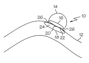
Fig. 1 illustrates a two-leaf vaso-occlusive device according to the present
invention in a deployed state at the site of a vascular defect.
Fig. 2 illustrates a device according to the present invention in an
unexpanded state in combination with a catheter delivery device.
Fig. 3 illustrates an embodiment of a vaso-occlusive device according to
the present invention in a deployed state at the site of a vascular defect.
Fig. 4 illustrates an alternative embodiment of a device according to the
present invention in an unexpanded state inside a catheter delivery device.
Fig. 5 illustrates the same device as in Fig. 4 in a partially deployed state
at the site of a vascular defect.
Fig. 6 illustrates the device of Figs. 4 and S in a fully deployed and
expanded state.
Fig. 7 illustrates an alternative embodiment of the device according to the
present invention in an unexpanded state within a catheter delivery device.
Fig. 8 illustrates the same device as in Fig. 7 in a deployed, expanded
state at the site of a vascular defect.
Fig. 9 illustrates a device similar to that shown in Figs. 7 and 8 being
deployed to a terminal vascular defect inside a catheter delivery device.
Fig. 10 illustrates the same device as in Fig. 9 in a partially deployed
state.
Fig. 11 illustrates the same device as in Figs. 9 and 10 in a fully deployed,
expanded state.
DETAILED DESCRIPTIONS OF THE PREFERRED EMBODIMENTS
While this invention may be embodied in many different forms, there are
described in detail herein specific embodiments of the invention. This
description is an
exemplification of the principles of the invention and is not intended to
limit the
invention to the particular embodiments illustrated.
Turning now to the figures, Fig. 1 shows generally at 10, a two-leaf or
two-sheet vaso-occlusive device, according to the present invention,
hereinafter referred
to as sheets. The vaso-occlusive device is shown positioned inside a blood
vessel 12 at
the site of a vascular defect, in this case, a side-wall aneurysm 14. Device
10 is shown
having a first sheet 16 in an expanded state inside of the aneurysm 14 and a
second sheet
5
CA 02485204 2004-11-05
WO 2004/019790 PCT/US2003/024763
18 connected to the first sheet 16 by a connector 20. First sheet 16 has a
vessel interface
surface 22 and an opposing, non-interface surface and second sheet 18 has a
vessel
interface surface 24 and an opposing non-interface surface. The first sheet 16
may be
connected to the second sheet 18 prior to deployment in the vessel and may
thus be
deployed in a single catheter device, or first sheet 16 and second sheet 18
may be
deployed separately, and connected inside the vessel.
Further, either interface surface 22 of sheet 16, interface surface 24 of
sheet 18, or both, may optionally be coated with, embedded with, or the
material itself
mixed with, a biocompatible material to promote integration of the device with
the
vessel, or to promote healing, or the like. This may include biocompatible
materials
which promote adhesion, fibrosis, tissue growth, endothelialization or cell
growth, and so
on and so forth.
Examples of biocompatible polymeric materials include, but are not
limited to, proteins such as collagen, fibrin, fibronectin, antibodies,
cytokines, growth
factors, enzymes, and so forth; polysaccharides such as heparin, chondroitin;
biologically
originated crosslinked gelatins; hyaluronic acid; poly(a-hydroxy acids); RNA;
DNA;
polyesters and polyorthoesters such as polyglycolides, polylactides and
polylactide-co-
glycolides; polylactones including polycaprolactones; polydioxanones;
polyamino acids
such as polylysine; polycyanoacrylates; poly(phosphazines);
poly(phosphoesters);
polyesteramides; polyacetals; polyketals; polycarbonates and
polyorthocarbonates
including trimethylene carbonates; degradable polyethylenes; polyalkylene
oxalates;
polyalkylene succinates; chitin; chitosan; oxidized cellulose;
polyhydroxyalkanoates
including polyhydroxybutyrates, polyhydroxyvalerates and copolymers thereof;
polymers and copolymers of polyethylene oxide; acrylic terminate polyethylene
oxide;
polyamides; polyethylenes; polyacrylonitriles; polyphosphazenes;
polyanhydrides
formed from dicarboxylic acid monomers including unsaturated polyanhydrides,
poly(amide anhydrides), poly(amide-ester) anhydrides, aliphatic-aromatic
homopolyanhydrides, aromatic polyanhydrides, polyester anhydrides), fatty acid
based
polyanhydrides, and so forth; other biocompatible or naturally occurring
polymeric
materials; and so forth; copolymers and terpolymers thereof; fragments of
biologically
active materials; and mixtures thereof. Hereinafter, the term copolymer shall
be used to
refer to any polymer having two or more monomers.
6
CA 02485204 2004-11-05
WO 2004/019790 PCT/US2003/024763
Some biocompatible polymers are also considered bioabsorbable such as
polylactides, polyglycolides, polylactide-co-glycolides, polyanhydrides, poly-
p-
dioxanones, trimethylene carbonates, polycaprolactones, polyhydroxyalkanoates,
and so
forth.
Biocompatible polymers which are not biodegradable which find utility
herein include, but are not limited to, polyacrylates; ethylene-vinyl
acetates; cellulose
and cellulose derivatives including cellulose acetate butyrate and cellulose
acetate
propionate; acyl substituted cellulose acetates and derivatives thereof; non-
erodible
polyolefins; polystyrenes; polyvinyl chlorides; polyvinyl fluorides; polyvinyl
(imidazoles); chlorosulphonated polyolefins; polyethylene oxides; polyethylene
glycols;
polyvinyl pyrrolidones; polyurethanes; polysiloxanes; copolymers and
terpolymers
thereof; and mixtures thereof.
Some examples of various polymers described above are found in US
4891225 and US 4906474 (polyanhydrides), US 4767628 (polylactides, polylactide-
co-
glycolic acid), US 4530840 (polylactides, polyglycolides, and copolymers
thereof), US
5234520 (biodegradable polymers), and so forth. Each of these patents is
incorporated
by reference herein in its entirety.
Some of these biocompatible polymers are described in US 6413536
which is also incorporated by reference herein in its entirety.
See also commonly assigned US 6335029 which is incorporated by
reference herein in its entirety.
One of ordinary skill in the art would understand that such biodegradable
polymers are by far too numerous to list here. Thus, this list is not
exhaustive and is
intended for illustrative purposes only.
Suitable non-polymeric materials include, for example, hormones and
antineoplastic agents.
Examples of other biocompatible materials which promote integration
with the vasculature of the patient include, for example, processed human or
animal
tissue including, for example, cells or cell fragments, engineered vascular
tissue, matrix
material from bladder, stomach, liver, genetic material of a natural or
synthetic origin,
and so forth.
Figs. 2-3 illustrate deployment of a vaso-occlusive device 10 according to
the present invention having a two-sheet structure into a side wall aneurysm
14. As
shown in Fig. 2, vaso-occlusive device 10 is delivered through vessel 12 in a
collapsed
7
CA 02485204 2004-11-05
WO 2004/019790 PCT/US2003/024763
configuration inside the shaft 17 of a catheter delivery device 15. The
catheter delivery
device 15 is used to position vaso-occlusive device 10 such that sheet 16 and
sheet 18 of
vaso-occlusive device 10 are approximately centered at the neck 26 of aneurysm
14.
Sheet 16 and sheet 18 of vaso-occlusive device 10 are shown in their
unexpended state
inside the catheter delivery device 15 in Fig. 2. In this embodiment, the
second sheet, in
its unexpended state, is in a rolled form. The sheets may also be folded, for
example. A
pusher wire 28 for pushing the first sheet 16 from the catheter and into the
aneurysm and
the second sheet 18 from the catheter is also shown.
Fig. 3 illustrates the vaso-occlusive device 10 of Fig. 2 after deployment
and expanded at the aneurysm 14. At this point, the catheter delivery device
15 has been
pulled back such that sheet 16 and sheet 18 are deployed. As catheter delivery
device 15
is pulled back, sheet 16 is first deployed, and as catheter delivery device is
pulled back
further, sheet 18 is then released. Optionally, a retractable sheath may be
employed.
Fig. 3 shows both sheet 16 and sheet 18 in their fully deployed, expanded
state. - In this
embodiment sheet 18 is shown in the form of a rectangular sheet which is
convex to the
vessel wall 34 on which the vascular defect 14 is located. Sheet 18 is also
convex to the
aneurysm neck 26. The interfacing surface 24 of sheet 18 may include a
biocompatible
material for promoting integration of the device with the patient's
vasculature or more
rapid healing of the aneurysm. A portion of the opposing non-interfacing
surface 30 is
clearly shown in Fig. 3. Further, the interfacing surface 22 of sheet 16 may
also
optionally include a biocompatible material for promoting integration of the
device with
the patient's vasculature or more rapid healing of the aneurysm.
The first sheet, in this embodiment, is functioning primarily as a
securement member to lceep the device in position, while the second sheet is
functioning
primarily as the occlusion member. In other embodiments, the shape of the
first sheet
may be designed to occlude the defect, and the second sheet designed to
function as a
securement member, or both sheets may be shaped to function as both occluding
members and as securement members.
Figs. 4-6 illustrate deployment of a vaso-occlusive device 10 according to
the present invention in which the vaso-occlusive device 10 is employed in a
terminal
aneurysm 14. As shown in Fig. 4, vaso-occlusive device having a first sheet 16
and a
second sheet 18 is delivered through vessel 12 via a catheter delivery system
1 S to the
site of the aneurysm 14. Fig. 4 illustrates the vaso-occlusive device in which
sheet 16
and sheet 18 are both inside catheter shaft 17 in an unexpended state. Sheet
16 and sheet
8
CA 02485204 2004-11-05
WO 2004/019790 PCT/US2003/024763
18 are attached via connector 20. In this example, the sheets are in a folded
rather than a
rolled configuration in their unexpanded states. A pusher wire 28 is shown
removably
attached to vaso-occlusive device 10. Removable detachment may be accomplished
through a variety of means, including, for example, severable junctions such
as those
severable by electrolytic corrosion, mechanical actuation, hydraulic pressure,
thermal
processes, electromagnetic energy, and so forth. This list is intended for
illustrative
purposes only, and is not exhaustive of what attachment systems may be
employed in the
present invention. One of ordinary skill in the art knows such attachment
systems.
Fig. 5 illustrates the same vaso-occlusive device 10 as shown in Fig. 4 in
a now partially deployed state in which first sheet 16, shown in an umbrella
form, has
been deployed inside aneurysm 14 and has been positioned at neck 26 of
aneurysm 14.
Pusher wire 28 is employed to push sheet 16 into the aneurysm, and may then be
used to
pull sheet 16 baclc until it is in contact with neck 26. Sheet 18 is still in
an undeployed,
unexpanded state inside of catheter shaft 17. Sheet 18 is connected to the
first sheet 16
by a connector 20.
The operator may then continue to use pusher wire 28 to push sheet 18
outside of catheter shaft 17 while catheter deliver device 15 is then pulled
back (not
shown).
Fig. 6 illustrates vaso-occlusive device 10 in a fully deployed state
wherein both sheet 16 and sheet 18 are in position at neck 26 of aneurysm 14
and sheet
18 has also been deployed.
It is important to note that the sheets do not have to be in any particular
shape or configuration so long as the shape of at least one of the sheets
provides
adequate occlusion of the vascular defect such that a substantial amount of
the blood
flow is blocked and so long as at least one of the sheets provides adequate
securement of
the device at the neck of the vascular defect. Some examples of shapes
include, but are
not limited to, umbrella like structures, parabolic structures, spheres,
discs, rectangular
structures or semicircular partial cylinders which bend convexly toward the
vascular
defect, and the like. Furthermore, the sheet may be in the form of a rectangle
which
forms a semi-folded convex structure when deployed. The convex side is toward
the
neck of the vascular defect.
Fig. 7 illustrates an alternative embodiment in which sheet 16 has been
replaced with struts 19 for anchoring the vaso-occlusive device in position at
the site of
the vascular defect 14. In this depiction, the vaso-occlusive device 10 is
shown in an
9
CA 02485204 2004-11-05
WO 2004/019790 PCT/US2003/024763
unexpanded state inside the shaft 17 of catheter delivery device 15. Struts 19
are
collapsed over sheet 18 which in this embodiment is shown in a rolled
configuration in
its unexpanded configuration. Pusher wire 28 can be seen detachably connected
at 21
using a severable junction which can be severed using a number of different
mechanisms
including, but not limited to, electrolytic corrosion, mechanical actuation,
hydraulic
pressure, thermal processes, electromagnetic energy, and so forth. It is thus
at this
junction 21 that the vaso-occlusive device 10 is eventually detached from
pusher wire 28
which is disposed inside catheter shaft 17 of catheter delivery device 15. A
retractable
sheath may be optionally employed. Other methods of detachment not described
herein,
but known in the art, may also be employed in detaching the device of the
present
invention. Severable junctions which may be employed in the present invention
are
described, for example, in US 5122136, US 5354295, US 5540680, US 5855578, US
5895385, US 5925037, US 5944714, US 5947963, US 5976126, US 6010498, US
6066133 and US 6083220, each of which is incorporated by reference herein in
its
15. entirety.
Fig. 8 illustrates the same device as in Fig. 7 in a deployed, expanded
state at the site of the vascular defect 14. In this embodiment, the
interfacing surface 22
of sheet 18 has a coating of a biocompatible material for promoting
integration of the
device with the vasculature. Sheet 18 is shown convex to the neck 26 of
vascular defect
14 and to vessel wall 34 on which the vascular defect is found and thus the
interfacing
surface 22 is in close contact with the vessel wall 34 and the neck 26 of
vascular defect
14. The non-interfacing surface 30 can be clearly seen in this embodiment. The
device
has been detached from pusher wire 28.
Fig. 9 illustrates an alternative embodiment in which sheet 16 again has
been replaced by anchoring struts 19. This particular device is being employed
at the
site of a terminal aneurysm rather than a side wall aneurysm as in Figs. 7 and
8. Again,
in Fig. 9, the device is shown in a collapsed configuration inside of the
shaft 17 of a
catheter delivery device 1 S. A retractable sheath may be optionally employed.
Struts 19
are shown at the distal end 40 of catheter 15 so that the struts are pushed
into the
vascular defect 14 first and are deployed first as well.
Fig. 10 shows the struts 19 deployed inside the vascular defect 14 while
sheet 18 is still collapsed inside the shaft 17 of catheter delivery device
15. In both Figs.
9 and 10, the vaso-occlusive device 10 is shown connected to pusher wire 28 at
21.
CA 02485204 2004-11-05
WO 2004/019790 PCT/US2003/024763
In Fig. 1 l, the sheet has now been deployed, the catheter delivery device
has been pulled baclc, the pusher wire detached and the device is anchored at
the neck 26
of aneurysm 14. In this embodiment, sheet 18 is not shown convex to the vessel
wall 34
and aneurysm neclc 26 as the embodiment shown in Fig. 8.
The sheets may be constructed from any of a variety of materials
including, but not limited to, polymeric material. Biocompatible,
bioresorbable and
biodegradable materials are suitable. Of course, materials may have any
combination of
those properties or all of those properties, as well.
Examples of useful polymeric materials include both synthetic and natural
materials. Further, the materials may be biocompatible and/or biodegradable
materials.
Examples of useful polymer materials include, but are not limited to,
polyolefins
including polyethylene and polypropylene, polyesters such as
polyethyleneterephthalate(PET) and polybutylene terephthalate (PBT),
polyurethanes,
acrylics, polypeptides, polyethers, polyamides, fluoropolymers such as
expanded
polytetrafluoroethylene, and so on and so forth.
Swellable polymeric materials find utility herein. Such materials include
those which are lcnown to expand and become lubricious in aqueous fluids
including, for
example, a class of materials referred to generally as hydrogels may also be
employed in
the manufacture of the device according to the present invention. Such
materials include
hydrophilic, macroporous, polymeric, hydrogel foam material. Examples of such
materials include, but are not limited, polyvinylpyrrolindone, polyethylene
oxide and its
copolymers with polypropylene oxide, polyacrylic acids, polyvinyl alcohols,
hyaluronic
acid, heparin, chondroitin sulfate, pectinic acid, carboxyl-derivatized
polysaccharides,
polyhydroxy ethyl methacrylate, polyacrylamide, hydrolyzed polyacrylonitriles,
polymethacrylic acid, polyethylene amines, polysaccharides, and copolymers and
combinations thereof, and so forth.
One particular example of a swellable material includes a swellable foam
matrix formed as a macroporous solid is described in US 5750585 which is
incorporated
by reference herein in its entirety. This material includes a foam stabilizing
agent and a
polymer or copolymer of a free radical polymerizable hydrophilic olefin
monomer cross-
linked with up to about 10% by weight of a multiolefm-functional cross-linking
agent.
Naturally based materials or those which are biologically derived which
find utility herein include, but are not limited to, collagen foams, harvested
vascular
material, films constructed from processed tissues, and so forth.
11
CA 02485204 2004-11-05
WO 2004/019790 PCT/US2003/024763
Suitable bioresorbable materials include, but are not limited to,
degradable hydrogels, lactides/glycolides or PHAs. More specific examples of
suitable
bioresorable materials include, but are not limited to, collagen,
polycaprolactone,
poly(glycolic acid), poly(3-hydroxybutric acid), poly(dl-lactic acid), poly(1-
lactic acid),
poly(dl-lactide/glycolide) 50:50, poly(hydroxyvalerate), poly(hydroxyvalerate-
hydroxybutyrate), or other PHAs. Such materials are described in US 5056211
and US
6251116, both of which are incorporated by reference herein in their entirety.
Non resorbable polymers and elastomers such as silicones, polyolefins,
fluoropolymers, or polyurethanes might also be used.
Shape memory materials are suitable for use in formation of the vaso-
occlusive device of the present invention. Shape memory materials may be
polymeric or
metallic. Shape memory materials have the ability to remember their original
shape,
either after mechanical deformation, or by cooling and heating. Such materials
are said
to undergo a structural phase transformation. Typically, shape memory polymers
(SMPs) are found to be segregated linear block co-polymers having a hard
segment and a
soft segment wherein the hard segment is crystalline, with a defined melting
point, and
the soft segment is amorphous, with a defined glass transition temperature.
However,
the hard segment may be amorphous and have a glass transition temperature
rather than a
melting point, and the soft segment may be crystalline and have a melting
point rather
than a glass transition temperature. The melting point or glass transition
temperature of
the soft segment is substantially less than the melting point or glass
transition
temperature of the hard segment. Some examples of shape memory polymers
include,
but are not limited to, those formed from polyethers, polyacrylates,
polyamides,
polysiloxanes, polyurethanes, polyether amides, polyurethane/ureas, polyether
esters,
urethane/butadiene copolymers, polynorbornenes, and mixtures thereof. See, for
example, US 5506300, US 5145935, US 5665822, and US 6388043 each of which is
incorporated by reference herein in its entirety. Degradable shape memory
polymers
may also be employed.
Shape memory metals suitable for use herein include the alloys of TiNi
(NITINOL~), CuZnAI, and FeNiAI, for example. These materials undergo a
structure
phase transformation referred to as a martensitic transformation.
In some situations, where a shape memory metal is employed, for
example, it may be appropriate to employ a metal mesh construction having
appropriate
geometrical features and cross patterns to provide adequate flexibility. Such
metal
12
CA 02485204 2004-11-05
WO 2004/019790 PCT/US2003/024763
meshes may be constructed from Nitinol~, for example, which is a super elastic
nickel
titanium alloy. Furthermore, with such a configuration, stainless steel, may
also be used.
This type of configuration may be more appropriate for an embodiment in which
the first
sheet is employed as a securement member for the second sheet, which functions
as an
occluding member for the vascular defect. The first sheet may then be
appropriately
constructed of the metal mesh configuration.
It is also possible to employ metals for other configurations. When using
a metal substructure, it may be desirable to coat it with a biocompatible,
polymeric,
biodegradable, or bioabsorbable material. Furthermore, the coating may have
all of
those characteristics. When the device is comprised of metal or includes metal
components the metal must be sufficiently flexible to provide the desired
degree of
flexibility in the vessels it is used in. As noted above, the geometric
pattern of the metal
within the device may be important to obtaining preferred results and may be a
sinusoidal or circular metal substructure.
Compressed foams may also be employed in the present invention
because they have the ability to return to their original shape. Both open and
closed cell
foams may be employed. Materials satisfactory for use in compressed foams
include,
but are not limited to medical grade silicones and polyurethanes. As described
above,
natural materials such as collagens, may also be employed to make a compressed
foam
material.
Copolymers, and crosslinkable versions of the above described materials
may also be suitable for use herein. And, of course, mixtures of the various
materials
described above may also be employed in the manufacture of the device
according to the
present invention.
Each sheet may be constricted of the same material, or they may be
constructed of different materials or blends of materials.
The sheets may be of a uniform thickness, or the thickness of the sheet
may be varied over the surface of the sheet. For example, the sheets may be
formed such
that they are thinner at the edges.
If the first sheet is replaced by a securement member such as one having a
plurality of struts, the struts may be formed from a metal or metal alloy as
well.
As described above, it is desirable to incorporate either into the sheet
material itself, or on the surface of the sheet, a biocompatible material
which promotes
13
CA 02485204 2004-11-05
WO 2004/019790 PCT/US2003/024763
integration with the vasculature or healing such as biocompatible adhesives,
polymeric
materials, tissue, cells, genetic material, and so forth.
The desirable compound or drug may be added to the sheet or sheets
using a variety of methods including coating the sheet(s), embedding the
compounds or
drugs into the material from which the sheets) is constructed, mixing the
compounds or
drugs in the material prior to formation of the sheet(s), and so forth.
A biocompatible adhesive may be added on the surface which is capable
of forming a bond at the aneurysm neclc, either on the inside of the aneurysm,
if the
device is delivered and deployed inside the aneurysm or vascular defect, or to
the outside
of the aneurysm neck, if it is delivered and deployed inside the parent vessel
but outside
of the aneurysm. Such biocompatible adhesives are described in US 6368586
incorporated by reference herein in its entirety.
As noted above, such compounds or drugs may promote a variety of
activities in the body, including, for example, tissue growth or
endothelialization. In the
latter instance, the some or all of the surfaces of the sheet, in particular
the surface which
interfaces with the vasculature, may be lined or coated with endothelial
cells. These cells
may be cells extracted from the patient the device is being placed in or from
a tissue
culture of such cells from another patient.
Other useful compounds include the polysaccharides such as heparin, for
example, which can be beneficially used alone or in combination with hydrogels
or
hydrophilic compounds, for example.
Anticoagulants compound may be extremely useful as a coating on
devices inserted into the vessels of the cardiovascular system. Compounds such
as
TaxolO may be a useful compound for coating or embedding within materials of a
device of the invention.
Other useful materials which may be incorporated into the device include,
but are not limited to, antiplatelet agents, calcium agonists,
antiinflammatory
compounds, antiproleferative drugs, hypolipidemic agents, and angiogenic
factors. The
device may be comprised such that all or any of these compounds are coated or
embedded on the surface of the material, or mixed in the material.
The material from which the vaso-occlusive device is formed or the vaso-
occlusive device itself may be modified, or provided with other additives as
well, to
malce the vaso-occlusive device visible by conventional imaging techniques.
For
example, the device may be rendered visible using fluoroscopic techniques,
rendered
14
CA 02485204 2004-11-05
WO 2004/019790 PCT/US2003/024763
MRI visible, or both. This can be accomplished through the use of markers such
as wire
windings, marker bands, rivets, plugs, and so forth, or the radiopaque or MRI
visible
materials may be incorporated into the material from which the vaso-occlusive
device is
formed. Any suitable radiopaque or MRI visible material may be employed.
Suitable materials for providing radiopacity to the device include but are
not limited to, platinum, rhodium, palladium, rhenium, iridium, tantalum,
tungsten, gold,
silver, alloys of these metals, as well as polymeric materials with barium,
for example.
Radiopacity is desirable for visualization of the device for purposes of
positioning the
device at the site of the defect and to position the device inside the defect
and for proper
anchoring of the device.
The above lists of materials are intended for illustrative purposes only and
are by no means exhaustive. Ther a is a vast array of materials which may be
employed
in the device of the present invention for a variety of purposes. One of
ordinary skill in
the art knows of such materials.
The invention is also directed to the vaso-occlusive device of the present
invention in combination with a catheter delivery device. Various
constructions of
catheter delivery devices are known in the art and as such any suitable
construction may
be employed herein. A retractable sheath may be optionally employed.
The invention is further directed to a method of occluding a vascular
defect having an opening. The method comprises the steps of:
a) deploying a first sheet having an unexpanded configuration and an
expanded configuration, as discussed above, through the neck of a vascular
defect and
into the vascular defect;
b) expanding the first sheet in the vascular defect;
c) deploying a second sheet having an expanded configuration and an
unexpanded configuration, as discussed above, on the outside of the vascular
defect, the
second sheet being attached to the first sheet; and
d) expanding the second sheet.
The sheets may be connected prior to delivery to the site, or they may be
connected in situ.
The first sheet or the second sheet may be replaced with an alternative
securement structure. In one embodiment, the securement structure includes a
plurality
of struts.
CA 02485204 2004-11-05
WO 2004/019790 PCT/US2003/024763
The invention may be used to close and substantially occlude an opening
of an aneurysm from a parent blood vessel.
The above disclosure is intended for illustrative purposes only and is not
exhaustive. The embodiments described therein will suggest many variations and
alternatives to one of ordinary skill in this art. All these alternatives and
variations are
intended to be included within the scope of the attached claims. Those
familiar with the
art may recognize other equivalents to the specific embodiments described
herein which
equivalents are also intended to be encompassed by the claims attached hereto.
16