Note: Descriptions are shown in the official language in which they were submitted.
CA 02485488 2004-11-16
WO 03/096889 PCT/IB03/01741
SPECIFICATION
CORRECTION OF BAROMETRIC PRESSURE BASED ON REMOTE SOURCES OF INFORMATION
FIELD OF THE INVENTION
The present invention relates generally to barometric pressure correction in
implantable biosensors, and more particularly to barometric pressure
correction for
implantable pressure sensors based on remote sources of information, including
remote
databases and web sites.
BACKGROUND OF INVENTION
Devices are known that may be implanted within a patient's body for
monitoring one or more physiological conditions and/or to provide therapeutic
functions.
For example, sensors or transducers may be located deep within the body for
monitoring
a variety of properties, such as temperature, pressure, strain, fluid flow,
chemical
properties, electrical properties, magnetic properties, and the like. In
addition, devices
may be implanted that perform one or more therapeutic functions, such as drug
delivery,
defibrillation, electrical stimulation, and the like.
Often it is desirable to communicate with such devices once they are
implanted within a patient using an external controller, for example, to
obtain data, and/or
to activate or otherwise control the implant. An implant may include wire
leads from the
implant to an exterior surface of the patient, thereby allowing an external
controller or
other device to be directly coupled to the implant. Alternatively, the implant
may be
remotely controlled, e.g., using an external induction device. For example, an
external
radio frequency (RF) transmitter may be used to communicate with the implant.
RF
energy, however, may only penetrate a few millimeters into a body, because of
the body's
dielectric nature, and therefore may not be able to communicate effectively
with an
implant that is located deep within the body. In'addition, although an RF
transmitter may
be able to induce a current within an implant, the implant's receiving
antenna, generally a
low impedance coil, may generate a voltage that is too low to provide a
reliable switching
mechanism.
-1-
CA 02485488 2011-11-02
72501-155
In a further alternative, electromagnetic energy may be used to control
an implant, since a body generally does not attenuate magnetic fields. The
presence
of external magnetic fields encountered by the patient during normal activity,
however, may expose the patient to the risk of false positives, i.e.,
accidental
activation or deactivation of the implant. Furthermore, external
electromagnetic
systems may be cumbersome and may not be able to effectively transfer coded
information to an implant.
Notably, implantable biosensors that measure pressure deep within
anatomical structures such as blood vessels or the brain, can only communicate
the
absolute pressure associated with the immediate anatomical environment. These
devices are not capable of communicating gauge pressure because they are
confined and sealed away from the ambient pressure external the body. In most
cases, it is gauge pressure and not absolute pressure that is sought to be
known,
since the body regulates its activities based on the ambient pressure. Gauge
pressure may be determined by correlating the absolute pressure with the
ambient
pressure. For example, Miesel et al. (U.S. Patent No. 6,248,080), describes
using a
barometer to determine gauge pressure based on a correlation of absolute
pressure
and ambient pressure. The Miesel system, however, requires a barometer to
determine the ambient pressure.
SUMMARY OF THE INVENTION
According to one aspect of the present invention, there is provided a
system for measuring pressure in a body, comprising: an implant device
configured
for measuring absolute pressure in a body, the implant device further
configured to
communicate measured absolute pressure information outside of the body using
telemetric signals; and an external monitor configured to: receive the
telemetric
signals from the implant device, receive geographic position data representing
the
position of the external monitor, transmit the geometric position data to a
remote
2
CA 02485488 2011-11-02
72501-155
source comprising real-time barometric pressure information for a plurality of
geographic locations, receive barometric pressure information from the remote
source associated with the geographic position data, and derive gauge pressure
from
the received absolute pressure information and the barometric pressure
information.
According to another aspect of the present invention, there is provided
a system for measuring pressure in a body, comprising: an implant device
including a
pressure sensor configured for measuring absolute pressure in a body, the
implant
device further configured to communicate measured absolute pressure
information
outside of the body using telemetric signals; and an external monitor
configured to:
receive the telemetric signals from the implant device, receive geographic
position
data representing the position of the external monitor, transmit received
absolute
pressure information and the geographic position data to a remote source
comprising
real time barometric pressure information for a plurality of geographic
locations, and
receive gauge pressure information from the remote source, the gauge pressure
information derived from the absolute pressure information and barometric
pressure
information.
According to still another aspect of the present invention, there is
provided a system for measuring pressure in a body, comprising: an external
monitor
configured to receive barometric pressure information from a remote source,
the
barometric pressure information associated with a geographic location of the
external
monitor, wherein the remote source comprises real-time barometric pressure
information for a plurality of geographic locations; and an implant device
including a
pressure sensor and configured to receive barometric pressure information from
the
external monitor, measure absolute pressure in a body, and derive gauge
pressure
from the received absolute pressure information and barometric pressure
information.
According to yet another aspect of the present invention, there is
provided a system for measuring pressure in a body, comprising: an implant
device
for measuring intro-body absolute pressure, the implant device comprising a
pressure
sensor, and a transducer coupled to the pressure sensor for acquiring absolute
2a
CA 02485488 2011-11-02
72501-155
pressure information from the pressure sensor, the transducer configured to
transmit
acoustic signals comprising absolute pressure information acquired from the
pressure
sensor; an external monitor configured to receive acoustic signals from the
implant
device and to receive real time barometric pressure information from a remote
source, the remote source comprising real-time barometric pressure information
for a
plurality of geographic locations; and a global positioning system (GPS)
signal
receiver coupled to the external monitor, the global positioning system (GPS)
signal
receiver configured to receive geographic position data representing the
position of
the external monitor, the external monitor further configured to receive
barometric
pressure information from the remote source associated with the geographic
position
data and derive gauge pressure based on absolute pressure information received
from the implant device and the real-time barometric pressure received from
the
remote source.
Some embodiments of the invention are generally directed to systems
for measuring pressure in a sealed or isolated system by converting or
correcting
data received from the sealed or otherwise isolated system using one or more
remote
databases. This generally involves a sensor placed within an isolated or
enclosed
system. Such enclosed systems can include anatomical structures such as blood
vessels within a human circulatory system or other anatomical locations. They
can
also include isolated systems associated with automobiles, such as braking
systems,
cooling systems, cylinders and combustion chambers of an internal combustion
engine, air intake systems, fuel systems including carburetors, electrical
systems, air
conditioning and heating systems, etc. The sensors can include those that are
capable of measuring pressure, temperature, electrical impedance, position,
strain,
pH, fluid flow, chemical properties, electrical properties, magnetic
properties and the
like. An external monitor is used to communicate with the isolated sensor and
obtain
data about the parameters that are monitored by the sensor. The communication
means can be wireless and can involve the transmission and reception of
2b
CA 02485488 2011-11-02
72501-155
any type of telemetric signal including acoustic, RF, microwave,
electromagnetic, light
(e.g. infrared), etc. The external monitor can include one or more transducers
to convert
the telemetric signal into an electric signal, which can be processed by a
microprocessor integrated into the external monitor. The external monitor can
also
include a GPS receiver to communicate geographic location data including
altitude data
to the microprocessor. The external monitor can communicate through various
means
known in the art with an external or remote database that includes real-time
data, such as
real-time temperature or barometric pressure data associated with numerous
geographic
locations. The remote database can be associated with a web site such as Yahoo
weather, weather.com, AWS.com, etc. The external monitor can use specific
information
obtained from the remote database to correct data received from the sensor. It
can also
use the real-time data to calibrate a measurement device, such as a barometer,
which can
be an integrated component of the external monitor or a stand-alone device in
communication with the external monitor.
In one embodiment, there is disclosed a system for measuring
pressure in a body. The system includes an implant device configured for
measuring
absolute pressure in a body. The implant is also configured to communicate any
measured absolute pressure information outside of the body using telemetric
signals. The
system also includes an external monitor that is configured to receive
telemetric signals
from the implant device. It is also configured to receive barometric pressure
information
from a remote source. The barometric pressure information can be associated
with the
geographic location of the body. The external monitor is also configured to
derive gauge
pressure from the received absolute pressure information and barometric
pressure
information. The remote source with which the external monitor is configured
to
communicate can be associated with a web site that includes weather
information, such as
barometric pressure information for numerous locations around the world. The
system
- can also include a global position system (GPS) signal receiver, which can
be coupled
either to the implant device or to the external monitor. Thus, both or either
the implant
device or the external monitor can be configured to receive geographic
position
information from the GPS signal receiver. The external monitor can be
configured to
communicate this position information to the remote source, and to request and
receive
barometric pressure information that corresponds with the geographic position.
-3-
CA 02485488 2004-11-16
WO 03/096889 PCT/IB03/01741
Other objects and features of the present invention will become apparent
from consideration of the following description taken in conjunction with the
accompanying drawings.
BRIEF DESCRIPTION OF THE DRAWINGS:
The invention is herein described, by way of example only, with reference
to the accompanying drawings, wherein:
FIGS. IA-1C are schematic drawings, showing exemplary embodiments of
an implant, in accordance with the present invention.
FIG. 2 is a schematic of an exemplary circuit for use as an acoustic switch,
in accordance with the present invention.
FIG. 3 is a cross-sectional view of a patient's body, showing a system for
communicating with an implant, in accordance with the present invention.
FIG. 4 is a schematic of an external monitor for communicating with an
implant, such as that shown in FIG. 3, in accordance with the present
invention.
FIG. 5 is a schematic of another exemplary embodiment of an implant, in
accordance with the present invention.
FIG. 6 is a perspective view of an exemplary embodiment of a pressure
sensing implant, in accordance with the present invention.
FIG. 7 is a schematic layout of the implant of FIG. 6.
FIG. 8A is a top view of an energy exchanger that may be provided in an
implant, such as that shown in FIGS. 6 and 7, in accordance with the present
invention.
FIG. 8B is a cross-sectional view of the energy exchanger of FIG. 8A,
taken along line B-B.
FIG. 9 is a schematic of an exemplary embodiment of a rectifier for use
with an implant, such as that shown in FIG. 7.
FIG. 10 is a schematic of another exemplary embodiment of a rectifier for
use with an implant, such as that shown in FIG. 7.
FIG. 11 is a schematic of an exemplary embodiment of a transmission
circuit for use with an implant, such as that shown in FIG. 7.
FIG. 12 is a schematic of another exemplary embodiment of a transmission
circuit for use with an implant, such as that shown in FIG. 7.
-4-
CA 02485488 2004-11-16
WO 03/096889 PCT/IB03/01741
FIG. 13A is a top view of an another embodiment of an implant, in
accordance with the present invention.
FIG. 13B is a side view of the implant of FIG. 13A.
FIG. 14 is a cross-sectional view of a patient's body, showing an external
device communicating with an implant located within the patient's body.
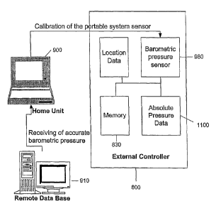
FIGS. 15A and 15B are diagrams of a barometric pressure correcting
system in communication with a database having barometric pressure data
according to
one embodiment.
FIG. 16 is a block diagram depicting the flow of information in a
barometric pressure correcting system according to one embodiment.
FIG. 17 is a block diagram depicting the flow of information in a
barometric pressure correcting system according to another embodiment.
FIG. 18A is a block diagram depicting the flow pressure calibration system
according to another embodiment.
FIG. 18B is a block diagram depicting the flow pressure calibration system
according to another embodiment.
FIG. 19 is a flow chart of some embodiments of the present invention.
FIG. 20 is a flow chart of other embodiments of the present invention.
FIG. 21 is a diagram of a system for delivering barometric pressure
information to a medical device.
DESCRIPTION OF THE PREFERRED EMBODIMENTS:
Turning to the drawings, various embodiments of biosensor implants and
external controllers (also referred to as external monitors) configured to
communicate
with biosensor implants are first shown and described. FIGS. IA-1C
schematically show
several exemplary embodiments of an implant 110, 210, 310, in accordance with
the
present invention. Generally, the implant 110, 210, 310 includes an electrical
circuit 112,
212, 312 configured for performing one or more functions or commands when the
implant 110, 210, 310 is activated, as described further below. In addition,
the
implant 110, 210, 310 includes an energy storage device 114 and optionally may
include
a switch 116 coupled to the electrical circuit 112, 212, 312 and the energy
storage
device 114. The switch 116 may be activated upon acoustic excitation 100 from
an
-5-
CA 02485488 2011-11-02
72501-155
external acoustic energy source (not shown) to allow current flow from the
energy storage
device 114 to the electrical circuit 112, 212, 312.
In one embodiment, the switch 116 includes an acoustic transducer 118,
such as that disclosed in PCT Publication No. WO 99/34,453, published July 8,
1999, or
in U.S. Patent Application 20020045921, published April 18, 2002. In addition,
the
switch 116 also includes a switch circuit 120, such as switch circuit 400
shown in FIG. 2,
although alternatively other switches, such as a miniature electromechanical
switch and
the like (not shown) may be provided. In a further alternative, the acoustic
transducer 118 may be coupled to the electrical circuit 112, 212, 312 and/or
the energy
storage device 114, and the switch circuit 120 may be eliminated.
The energy storage device 114 may be any of a variety of known devices,
such as an energy exchanger, a battery and/or a capacitor (not shown).
Preferably, the
energy storage device 114 is capable of storing electrical energy
substantially indefinitely
for as long as the acoustic switch 116 remains open, i.e., when the implant
110, 210, 310
is in a "sleep" mode. In addition, the energy storage device 114 may be
capable of being
charged from an external source, e.g., inductively using acoustic telemetry,
as will be
appreciated by those skilled in the art. In an exemplary embodiment, the
energy storage
device 114 includes both a capacitor and a primary, non-rechargeable battery.
Alternatively, the energy storage device 114 may include a secondary,
rechargeable
battery and/or capacitor that may be energized before activation or use of the
implant 110,
210, 310.
The implant 110, 210, 310 may be surgically or minimally invasively
inserted within a human body in order to carry out a variety of monitoring
and/or
therapeutic functions. For example, the electrical circuit 112, 212, 312 may
include a
control circuit 122, 222, 322, a biosensor 124, 224, an actuator 226, 326,
and/or a
transmitter 128, as explained in U.S. Patent No. 6,628,989. The
implant 210, 310 may be configured for providing one or more therapeutic
functions, for
example, to activate and/or control a therapeutic device implanted within a
patient's body,
such as an atrial defibrillator or pacemaker, a pain relief stimulator, a
neuro-stimulator, a
drug delivery device, and/or a light source used for photodynamic therapy.
Alternatively,
the implant may be used to monitor a radiation dose including ionizing,
magnetic and/or
acoustic radiation, to monitor flow in a bypass graft, to produce cell
oxygenation and
membrane electroporation, and the like. In addition or alternatively, the
implant 110 may
-6-
CA 02485488 2004-11-16
WO 03/096889 PCT/IB03/01741
be used to measure one or more physiological parameters within the patient's
body, such
as pressure, temperature, electrical impedance, position, strain, pH, and the
like.
The implant may operate in one of two modes, a "sleep" or "passive"
mode when the implant remains dormant and not in use, i.e., when the acoustic
switch
116 is open, and an "active" mode, when the acoustic switch 116 is closed, and
electrical
energy is delivered from the energy storage device 114 to the electrical
circuit 112, 212,
312. Alternatively, the implant may operate continuously or intermittently.
Because the
acoustic switch 116 is open in the sleep mode, there is substantially no
energy
consumption from the energy storage device 114, and consequently, the implant
may
remain in the sleep mode virtually indefinitely, i.e., until activated. Thus,
an implant in
accordance with the present invention may be more energy efficient and,
therefore, may
require a relatively small energy storage device than implants that
continuously draw at
least a small amount of current in their "passive" mode.
Turning to FIG. 1A, an exemplary embodiment of an implant 110 is shown
in which the electrical circuit 112 includes a control circuit 122, a
biosensor 124 coupled
to the controller 122, and a transmitter 128 coupled to the control circuit
122. The
controller 122 may include circuitry for activating or controlling the
biosensor 124, for
receiving signals from the biosensor 124, and/or for processing the signals
into data, for
example, to be transmitted by the transmitter 128. Optionally, the electrical
circuit 112
may include memory (not shown) for storing the data. The transmitter 128 may
be any
device capable of transmitting data from the control circuit 122 to a remote
location
outside the body, such as an acoustic transmitter, a radio frequency
transmitter, and the
like. Preferably, the control circuit 122 is coupled to the acoustic
transducer 118 such
that the acoustic transducer 118 may be used as a transmitter 128, as well as
a receiver,
instead of providing a separate transmitter.
The biosensor 124 may include one or more sensors capable of measuring
physiological parameters, such as pressure, temperature, electrical impedance,
position,
strain, pH, fluid flow, electrochemical sensor, and the like. Thus, the
biosensor 124 may
generate a signal proportional to a physiological parameter that may be
processed and/or
relayed by the control circuit 122 to the transmitter 128, which, in turn, may
generate a
transmission signal to be received by a device outside the patient's body.
Data regarding
the physiological parameter(s) may be transmitted continuously or periodically
until the
-7-
CA 02485488 2004-11-16
WO 03/096889 PCT/IB03/01741
acoustic switch 116 is deactivated, or for a fixed predetermined time, as will
be
appreciated by those skilled in the art.
Turning to FIG. 1B, another exemplary embodiment of an implant 210 is
shown in which the electrical circuit 212 includes a control circuit 222 and
an
actuator 226. The actuator 226 may be coupled to a therapeutic device (not
shown)
provided in or otherwise coupled to the implant 210, such as a light source, a
nerve
stimulator, a defibrillator, an electrochemical oxidation/reduction electrode,
or a valve
communicating with an implanted drug reservoir (in the implant or otherwise
implanted
within the body in association with the implant).
When the switch 120 is closed, the control circuit 222 may activate the
actuator 226 using a pre-programmed protocol, e.g., to complete a
predetermined
therapeutic procedure, whereupon the switch 120 may automatically open, or the
controller 222 may follow a continuous or looped protocol until the switch 120
is
deactivated. Alternatively, the acoustic transducer 118 may be coupled to the
control
circuit 222 for communicating a new or unique set of commands to the control
circuit
222. For example, a particular course of treatment for a patient having the
implant 210
may be determined, such as a flow rate and duration of drug delivery, drug
activation,
drug production, or an energy level and duration of electrical stimulation.
Acoustic
signals including commands specifying this course of treatment may be
transmitted from
an external controller (not shown), as described below, to the acoustic switch
116, e.g.,
along with or subsequent to the activation signal 100. The control circuit 222
may
interpret these commands and control the actuator 226 accordingly to complete
the course
of treatment.
Turning to FIG. 1 C, yet another exemplary embodiment of an implant 310
is shown in which the electrical circuit 312 includes a control circuit 322, a
biosensor 324, and an actuator 326, all of which may be coupled to one
another. This
embodiment may operate similarly to the embodiments described above, e.g., to
obtain
data regarding one or more physiological parameters and/or to control a
therapeutic
device. In addition, once activated, the control circuit 322 may control the
actuator 326 in
response to data obtained from the biosensor 324 to control or adjust
automatically a
course of treatment being provided by a device connected to the actuator 326.
For
example, the actuator 326 may be coupled to an insulin pump (not shown),
and,the
biosensor 324 may measure glucose levels within the patient's body. The
control circuit
-8-
CA 02485488 2004-11-16
WO 03/096889 PCT/IB03/01741
322 may control the actuator to open or close a valve on the insulin pump to
adjust a rate
of insulin delivery based upon glucose levels measured by the biosensor 324 in
order to
maintain the patient's glucose within a desired range.
Turning to FIG. 2, an exemplary embodiment of a switch 400 is shown
that may be incorporated into an implant in accordance with the present
invention. The
switch 400 includes a piezoelectric transducer, or other acoustic transducer
(not shown,
but generally connected to the switch 400 at locations piezo + and piezo -), a
plurality of
MOSFET transistors (Q1-Q4) and resistors (Rl-R4), and switch Si. A "load"
maybe
coupled to the switch 400, such as one of the electrical circuits described
above. In the
switch's "sleep" mode, all of the MOSFET transistors (Q1 -Q4) are in an off
state. To
maintain the off state, the gates of the transistors are biased by pull-up and
pull-down
resistors. The gates of N-channel transistors (Q 1, Q3 & Q4) are biased to
ground and the
gate of P-channel transistor Q2 is biased to +3V. During this quiescent stage,
switch Si
is closed and no current flows through the circuit. Therefore, although an
energy storage
device (not shown, but coupled between the hot post, labeled with an exemplary
voltage
of +3V, and ground) is connected to the switch 400, no current is being drawn
therefrom
since all of the transistors are quiescent.
When the acoustic transducer of the implant detects an external acoustic
signal, e.g., having a particular frequency, such as the transducer's resonant
frequency,
the voltage on the transistor Q1 will exceed the transistor threshold voltage
of about one
half of a volt. Transistor QI is thereby switched on and current flows through
transistor
Q1 and pull-up resistor R2. As a result of the current flow through transistor
Q1, the
voltage on the drain of transistor Qi and the gate of transistor Q2 drops from
+3V
substantially to zero (ground). This drop in voltage switches on the P-channel
transistor
Q2, which begins to conduct current through transistor Q2 and pull-down
resistor R3.
As a result of the current flowing through transistor Q2, the voltage on the
drain of transistor Q2 and the gates of transistors Q3 and Q4 increases from
substantially
zero to +3V. The increase in voltage switches on transistors Q3 and Q4. As a
result,
transistor Q3 begins to conduct current through resistor R4 and main switching
transistor
Q4 begins to conduct current through the "load," thereby switching on the
electrical
circuit.
As a result of the current flowing through transistor Q3, the gate of
transistor Q2 is connected to ground through transistor Q3, irrespective of
whether or not
-9-
CA 02485488 2004-11-16
WO 03/096889 PCT/IB03/01741
transistor Q 1 is conducting. At this stage, the transistors (Q2, Q3 & Q4) are
latched to the
conducting state, even if the piezoelectric voltage on transistor Q1 is
subsequently
reduced to zero and transistor Q1 ceases to conduct. Thus, main switching
transistor Q4
will remain on until switch Si is opened.
In order to deactivate or open the switch 400, switch S I must be opened,
for example, while there is no acoustic excitation of the piezoelectric
transducer. If this
occurs, the gate of transistor Q2 increases to +3V due to pull-up resistor R2.
Transistor
Q2 then switches off, thereby, in turn, switching off transistors Q3 and Q4.
At this stage,
the switch 400 returns to its sleep mode, even if switch S 1 is again closed.
The switch
400 will only return to its active mode upon receiving a new acoustic
activation signal
from the piezoelectric transducer.
It should be apparent to one of ordinary skill in the art that the above-
mentioned electrical circuit is not the only possible implementation of a
switch for use
with the present invention. For example, the switching operation my be
performed using
a CMOS circuit, which may draw less current when switched on, an
electromechanical
switch, and the like.
Turning to FIGS. 3 and 4, a system 410 is shown for communicating with
an implant 412, such as one of those described above. Generally, the system
410 includes
an external communications device or controller 414, and may include a charger
416, one
or more implants 412 (only one shown for simplicity), and an external
recorder,
computer, or other electronic device 434.
With particular reference to FIG. 4, the external controller 414 may
include a processor or other electrical circuit 418 for controlling its
operation, and an
energy source 420, e.g., a nonrechargeable or a rechargeable battery, coupled
to the
processor 418 and/or other components of the controller 414, such as a power
amplifier or
an oscillator (not shown). In addition, the controller 414 may include one or
more
acoustic transducers 422 that are configured for converting between electrical
energy and
acoustic energy, similar to those described above. As shown, a single acoustic
transducer 422 is provided that may communicate using acoustic telemetry,
i.e., capable
both of converting electrical energy to acoustic energy to transmit acoustic
signals, and
converting acoustic energy to electrical energy to receive acoustic signals,
as explained
further below. Alternatively, separate and/or multiple acoustic transducers
may be
provided for transmitting and receiving acoustic signals.
-10-
CA 02485488 2004-11-16
WO 03/096889 PCT/IB03/01741
In one embodiment, the controller 414 also includes memory 424 coupled
to the processor 418, e.g., for storing data provided to the controller 414,
as explained
further below. The memory 424 may be a temporary buffer that holds data before
transfer to another device, or non-volatile memory capable of storing the data
substantially indefinitely, e.g., until extracted by the processor 418 or
other electronic
device. For example, the memory 424 may be a memory card or an eprom (not
shown)
built into the controller 414 or otherwise coupled to the processor 418. The
controller 414 may also include an interface 426, such as a lead or connector,
or a
transmitter and/or receiver, that may communicate with the external electronic
device, as
explained further below.
Preferably, the controller 414 is carried by a patch 415 that may be secured
to a patient, e.g., to the patient's skin 92. For example, the patch 415 may
include one or
more layers of substantially flexible material to which the controller 414
and/or its
individual components are attached. The patch 415 may include a single
flexible
membrane (not shown) to which the controller 414 is bonded or otherwise
attached, e.g.,
using a substantially permanent adhesive, which may facilitate the patch 415
conforming
to a patient's anatomy. Alternatively, the controller 414 may be secured
between layers
of material, e.g., within a pouch or other compartment (not shown) within the
patch 415.
For example, the patch 415 may include a pair of membranes (not shown)
defining the
pouch or compartment. The space within which the controller 414 is disposed
may be
filled with material to acoustically couple the acoustic transducer(s)
(formed, for
example, from PZT, composite PZT, Quartz, PVDF, and/or other piezoelectric
material)
of the controller 414 to an outer surface of the patch 415. Alternatively, the
acoustic
transducer(s) may be exposed, e.g., in a window formed in a wall of the patch
415.
The patch 415 may be formed from a flexible piezoelectric material, such
as PVDF or a PVDF copolymer. Such polymers may allow the patch 415 to produce
ultrasonic waves, as well as allowing the controller 414 to be secured to the
patient's
skin 92. Thus, the wall of the patch 415 itself may provide an acoustic
transducer for the
controller 414, i.e., for transmitting acoustic energy to and/or receiving
acoustic energy
from the implant 412.
The patch 415 may then be secured to the patient's skin 92 using a
material, such as a layer of adhesive (not shown), substantially permanently
affixed or
otherwise provided on a surface of the patch. The adhesive may be hydrogel,
silicon,
-11-
CA 02485488 2004-11-16
WO 03/096889 PCT/IB03/01741
polyurethane, polyethylene, polypropylene, fluorocarbon polymer, and the like.
Alternatively, a separate adhesive may be applied to the patch 415 and/or to
the patient's
skin 92 before applying the patch 415 in order to secure the controller 414 to
the patient's
skin 92. Such an adhesive may enhance acoustically coupling of the acoustic
transducer(s) of the controller 414 to the patient's skin 92, and consequently
to the
implant 412 within the patient's body 94. Optionally, additional wetting
material,
including water, silicone oil, silicone gel, hydrogel, and the like, and/or
other acoustically
conductive material may be provided between the patch 415 or the acoustic
transducer 422, and the patient's skin 92, e.g., to provide substantial
continuity and
minimize reflection or other losses and/or to secure the patch 415 to the
patient.
Alternatively, the controller 414 may be carried by a belt (not shown) that
may be secured around the patient, e.g., such that the acoustic transducer 422
is secured
against the patient's skin. The belt may carry other components of the system
410, e.g.,
an external power supply for the controller 414. For example, a battery pack
(not shown)
may be carried by the belt that may be coupled to the controller 414 for
providing
electrical energy for its operation.
The patch 415 may be relatively light and compact, for example, having a
maximum surface dimension (e.g., width or height) not more than about ten to
two
hundred millimeters (10-200 mm), a thickness not more than about five to one
hundred
millimeters (5-100 mm), and a weight not more than about twenty to four
hundred grams
(20-400 g), such that the controller 414 may be inconspicuously attached to
the patient.
Thus, the patient may be able to resume normal physical activity, without
substantial
impairment from the controller. Yet, the internal energy source of the
controller 414 may
be sufficiently large to communicate with the implant 412 for an extended
period of time,
e.g., for hours or days, without requiring recharging or continuous coupling
to a separate
energy source.
The system 410 may be used to control, energize, and/or otherwise
communicate with the implant 412. For example, the controller 414 may be used
to
activate the implant 412. One or more external acoustic energy waves or
signals 430 may
be transmitted from the controller 414 into the patient's body 94, e.g.,
generally towards
the location of the implant 412 until the signal is received by the acoustic
transducer (not
shown in FIGS. 3 and 4) of the implant 412. Upon excitation by the acoustic
wave(s)
430, the acoustic transducer produces an electrical output that is used to
close, open, or
-12-
CA 02485488 2004-11-16
WO 03/096889 PCT/IB03/01741
otherwise activate the switch (also not shown in FIGS. 3 and 4) of the implant
412.
Preferably, in order to achieve reliable switching, the acoustic transducer of
the
implant 412 is configured to generate a voltage of at least several tenths of
a volt upon
excitation that may be used as an activation signal to close the switch, as
described above.
As a safety measure against false positives (e.g., erroneous activation or
deactivation), the controller 414 may be configured to direct its acoustic
transducer 422 to
transmit an initiation signal followed by a confirmation signal. When the
acoustic
transducer of the implant 412 receives these signals, the electrical circuit
may monitor the
signals for a proper sequence of signals, thereby ensuring that the acoustic
switch of the
implant 412 only closes upon receiving the proper initiation and confirmation
signals.
For example, the acoustic switch may only acknowledge an activation signal
that includes
a first pulse followed by a second pulse separated by a predetermined delay.
Use of a
confirmation signal may be particularly important for certain applications,
for example, to
prevent unintentional release of drugs by a drug delivery implant.
In addition to an activation signal, the controller 414 may transmit a
second acoustic signal that may be the same as or different than the acoustic
wave(s) used
to activate the acoustic switch of the implant 412. Thus, the switch may be
opened when
the acoustic transducer of the implant 412 receives this second acoustic
signal, e.g., by the
acoustic transducer generating a termination signal in response to the second
acoustic
signal, in order to return the implant 412 to its sleep mode.
For example, once activated, the switch may remain closed indefinitely,
e.g., until the energy storage device (not shown in FIGS. 3 and 4) of the
implant 412 is
completely depleted, falls below a predetermined threshold, or until a
termination signal
is received by the acoustic transducer of the implant 412 from the controller
414.
Alternatively, the acoustic switch of the implant 412 may include a timer (not
shown),
such that the switch remains closed only for a predetermined time, whereupon
the switch
may automatically open, returning the implant 412 to its sleep mode.
FIG. 5 shows an alternative embodiment of an implant 510 that does not
include an acoustic switch. Generally, the implant includes a sensor 512, one
or more
energy transducers 514, one or more energy storage devices 516, and a control
circuit
518, similar to the embodiments described above. The sensor 512 is preferably
a pressure
sensor for measuring intra-body pressure, such as an absolute variable
capacitance type
pressure sensor. In alternative embodiments, one or more other sensors may be
provided
-13-
CA 02485488 2004-11-16
WO 03/096889 PCT/IB03/01741
instead of or in addition to a pressure sensor 512. For example, the sensor
512 may
include one or more biosensors capable of measuring physiological parameters,
such as
temperature, electrical impedance, position, strain, pH, fluid flow, and the
like. An
external controller (not shown), such as that described above, may also be
used to
communicate with this implant.
Returning to FIG. 3, an external controller 414 in accordance with the
present invention preferably has only sufficient power to control its own
operation and to
communicate with the implant 412. Because of its limited energy requirements,
the
controller 414 maybe relatively small and portable, e.g., may be attached to
the patient,
while still allowing the patient to engage in normal physical activity. The
controller 414
may be used to communicate with the implant 412, e.g., periodically activating
or
deactivating the implant 412, and/or recording data generated and transmitted
by the
implant 412. Because it is located outside the patient's body, the controller
414 may be
more easily programmed or reprogrammed than the implant 412 itself, and/or may
be
repaired or replaced if necessary or desired.
In addition to the external controller 414, the system 410 may include one
or more electronic devices 434 that may be coupled to the controller 414 via
the
interface 426, such as a recorder, a computer, a personal digital assistant,
and/or a
wireless device, such as a cellular telephone. The electronic device 434 may
be directly
coupled to the controller 414, by a connector or lead (not shown) extending
from the
patch 415 within which the controller 414 is provided. Alternatively, the
controller 414
and/or patch 415 may include a wireless transmitter and/or receiver (not
shown), e.g., a
short-range RF transceiver, for communicating with the electronic device 434.
The electronic device 434 may be used to extract data from the
memory 424 of the controller 414, e.g., sensor data and the like, received
from the
implant 412. This data may be included in a patient database maintained by
health care
professionals monitoring the patient receiving the implant 412. In addition,
the electronic
device 434 may be used to program the controller 414, e.g., to program
commands,
timing sequences, and the like.
The system 410 may also include an external charger 418. For example,
the implant 412 may include a rechargeable energy storage device (not shown in
FIG. 3),
preferably one or more capacitors, that are coupled to the acoustic transducer
(also not
shown in FIG. 3). The charger 416 may include a probe 428, including an
acoustic
-14-
CA 02485488 2004-11-16
WO 03/096889 PCT/IB03/01741
transducer 430 for contacting a patient's skin 92. The charger 416 also
includes a source
of electrical energy 432, such as a radio frequency (RF) generator, that is
coupled to the
acoustic transducer 430. The charger 418 may also include electrical circuits
for
controlling its operation and buttons or other controls (not shown) for
activating and/or
deactivating the acoustic transducer 430.
The charger 418 may be used to charge or recharge the implant, e.g.,
periodically or before each activation. Because the charger 418 includes a
substantially
more powerful energy source than the controller 414, the charger 418 is
generally a
relatively bulky device compared to the controller 414, in particular due to
the energy
generator, which may be stationary or of limited mobility. In addition, the
charger 418
may be used to recharge the controller 414 periodically, e.g., by a direct or
wireless
coupling. Alternatively, the controller 414 and patch 415 may be disposable,
e.g., after its
energy has been depleted, and replaced with another.
For purposes of comparison, an exemplary charger 416 may need to
generate about ten kiloPascals (10 kPa) of acoustic energy for about twenty
seconds (20
sec.) in order to fully charge the implant 412. In contrast, an exemplary
controller 414
may be limited to outputting relatively smaller bursts of acoustic energy for
communicating with, but not charging, the implant 412. Such acoustic signals
may have
a duration of as little as about one millisecond (1 ms), as opposed to the
significantly
longer charging signals generated by the charger 416.
The transducer 422 of the controller 414 may consume about one Watt
(1 W) of power to produce a 1 kPa acoustic signal for about one millisecond.
If the
controller 414 communicates with the implant 412 on an hourly basis, the
energy
source 420 of the controller 418 may only need sufficient capacity to provide
0.024 Watt
seconds per day (0.024 W.sec./day). Because of this low energy requirement,
the energy
source 420, and, consequently, the controller 418, may be relatively compact
and
portable, as compared to the charger 416. Thus, the energy source 420 may be
self-
contained within the controller 418, i.e., carried by the patch 415.
Alternatively, a
portable energy source, e.g., an external battery pack (not shown) may be
provided for
supplying electrical energy to the controller 418 that may be carried by the
patient, e.g.,
on a belt (not shown).
In an alternative embodiment, the controller and charger may be provided
as a single device (not shown), e.g., including one or more acoustic
transducers and/or
-15-
CA 02485488 2004-11-16
WO 03/096889 PCT/IB03/01741
one or more processors for performing the functions of both devices, as
described above.
In this embodiment, the implant 412 may operate in a "half-duplex" mode, a
quasi-
continuous mode, or in a "full-duplex" mode, as described in the applications
incorporated above.
FIGS. 6 and 7 show another embodiment of an implant 10, in accordance
with the present invention. Generally, the implant 10 includes a sensor 12,
one or more
energy transducers 14, one or more energy storage devices 16, and a controller
18.
The sensor 12 is preferably a pressure sensor for measuring infra-body
pressure. The sensor 12 may measure pressure within a range as low as a few
millibars
gauge (e.g., pressure ranges experienced within the cranium or within the
pulmonary
artery) and up to about 400 millibars gauge (e.g., blood pressure ranges
experienced
during systole). In addition, because the barometric pressure may vary by
location, i.e.,
altitude, the absolute pressure range capacity of the sensor is preferably
between about
650 and 1450 millibars absolute.
The sensor 12 can be an absolute variable capacitance type pressure
sensor. Alternatively, a piezoresistive pressure sensor may be used, although
the energy
consumption of this type of sensor may be substantially higher than a variable
capacitance pressure sensor. For example, a typical piezoresistive sensor may
have a
bridge resistance of about five kiloohms (5 ku). Assuming that one volt (1 V)
is
sufficient to allow pressure sampling, a current of at least about 0.2
milliAmperes (mA)
would be required to operate the sensor. This may be about one hundred times
more than
the current required to obtain pressure samples using a variable capacitance
pressure
sensor.
Some reduction in power consumption of piezoresistive pressure sensors
may be obtained by reducing the sampling rate of the sensor or otherwise
reducing the
duty cycle of the implant. Alternatively, to reduce power consumption, a
sample-and-
hold circuit (not shown) may be provided for capturing voltages, and an analog-
to-digital
converter (also not shown) may be provided for converting the voltages when
desired.
Thus, the current may be on for relatively short times during each sampling
cycle.
Preferably, a silicon MEMS-based pressure sensor is used, because of its
relative small size, e.g., smaller than about four millimeters (4 mm) maximum
footprint,
e.g., not more than about four millimeters (4 mm) width by four millimeters (4
mm)
length. Preferably, the sensor is no larger than about 0.8 mm width by about
2.1 mm
-16-
CA 02485488 2004-11-16
WO 03/096889 PCT/IB03/01741
length by about 0.3 mm thickness. Silicon is a particularly useful material
for the
sensor 12, as it generally does not suffer from creep and fatigue, and
therefore may result
in a substantially stable sensor. MEMS-based sensors are presently preferred
because
they may be manufactured in large volume at relatively low cost compared to
other
sensors. Other materials that may be used include titanium, as is used for the
Chronicle TM
device manufactured by Medtronic, Inc. Preferably, the sensor 12 is made from
biocompatible materials, although the sensor 12 may be coated, if necessary or
desired,
with a biocompatible and/or chemically resistive coating (not shown), as will
be
appreciated by those skilled in the art.
In alternative embodiments, one or more other sensors may be provided
instead of or in addition to a pressure sensor. For example, the sensor 12 may
include one
or more biosensors capable of measuring physiological parameters, such as
temperature,
electrical impedance, position, strain, pH, fluid flow, and the like. U.S.
Patent
Nos. 4,793,825 issued to Benjamin et al. and 5,833,603 issued to Kovacs et al.
disclose
additional exemplary embodiments of biosensors that may be provided. The
disclosure of
these references and others cited therein are expressly incorporated herein by
reference.
The sensor 12 may generate a signal proportional to a physiological parameter
that may
be processed and/or relayed by the controller 18 to the energy transducer 14,
as described
further below. Alternatively, the sensor 12 may be configured to monitor a
radiation dose
including ionizing, magnetic and/or acoustic radiation, to monitor flow in a
bypass graft,
to produce cell oxygenation and membrane electroporation, and the like.
In further alternatives, a device for providing one or more therapeutic
functions (not shown) may be provided in addition to or instead of the sensor
12. For
example, the device may be used to activate and/or control a therapeutic
device implanted
within a patient's body, such as an atrial defibrillator, a pain relief
stimulator, a neuro-
stimulator, a drug delivery device, and/or a light source used for
photodynamic therapy.
Turning to FIGS. 8A and 8B, the energy transducer 14 is preferably an
acoustic transducer for converting energy between electrical energy and
acoustic energy.
As explained further below, the acoustic transducer 14 is configured for
converting
acoustic energy from a source external to the implant into electrical energy
and/or for
transmitting an acoustic signal including sensor data to a location external
to the implant.
In one embodiment, the energy transducer 14 is configured to operate
alternatively as
either an energy exchanger or an acoustic transmitter, or simultaneously as an
energy
-17-
CA 02485488 2004-11-16
WO 03/096889 PCT/IB03/01741
exchanger and an acoustic transmitter. Alternatively, multiple energy
transducers (not
shown) may be provided, e.g., one or more converting acoustic energy striking
the energy
exchanger into electrical energy, and one or more transmitting acoustic
signals to a
location external to the implant 10. In a further alternative, multiple energy
transducers
(not shown) may be provided for increasing the electrical energy produced for
a given
acoustic energy transmitted to the implant 10.
The energy transducer 14 generally includes a substrate 20 including one
or more cavities 22 therein, such as the.array of cavities 22 shown in FIG.
8A. The
cavities 22 may extend completely through the substrate 20 or only partially
into the
substrate 20. The cavities 22 are preferably substantially round in cross-
section, although
oval or other elongate slotted cavities (not shown) may be provided, which may
increase
sensitivity and/or efficiency as compared to a substantially round cavity. The
cavities 22
may have a cross-section of about 0.5-2.5 millimeters, and preferably between
about 1.0
and 1.3 millimeters (mm). For elliptical or other elongate cavities (not
shown), the
cavities preferably have a width of 0.2-2.5 millimeters and a length of 1.0-25
millimeters.
The substrate 20 may be formed from a relatively high modulus polymer, such as
poly
ether ether ketone (PEEK), silicon, and/or a printed circuit board, e.g., of
FR4, Rogers, a
ceramic, or Kapton.
A substantially flexible piezoelectric layer 24 is attached to the
substrate 20 across cavities 22. The piezoelectric layer 24 generally includes
a polymer
layer 28, preferably a fluorocarbon polymer, such as poly vinylidene fluoride
(PVDF).
The polymer layer 28 may have a thickness of between about three and two
hundred fifty
micrometers (3-250 gm), and preferably about thirty micrometers (30 gm) or
less. A first
conductive layer 30 is provided on an external surface of the polymer membrane
28 and a
second conductive layer 32 provided on an internal surface of the polymer
membrane 28.
The second conductive layer 32 may be coupled to a conductive region 36
provided on a
wall of the cavities 22. A pad 34 is provided on a lower surface of the
substrate 20 for
coupling the second conductive layer 32 to a printed circuit board (not
shown), as
described further below.
To manufacture the energy transducer 14, a substantially flexible polymer
layer 28, such as a PVDF membrane, is provided. Because PVDF is generally
chemically
inert, the polymer layer 28 may need to be activated, e.g., using an etching
process. For
example, a sodium napthalene solution may be used to chemically attack the
PVDF to
-18-
CA 02485488 2004-11-16
WO 03/096889 PCT/IB03/01741
cleave the carbon-fluorine bonds and/or other solutions to cleave the carbon-
hydrogen
bonds and/or carbon-carbon bonds in the material. Alternatively, a gas phase
plasma
treatment, e.g., using an oxygen, air, Helium, and/or Argon plasma, may be
used.
A substantially planar substrate 20 is provided, and one or more
cavities 22 are formed in a surface of the substrate 20, for example, by
mechanical
drilling, laser drilling, or punching. Alternatively, the cavities 22 may be
etched into the
substrate 20, e.g., using VLSI/micro-machining technology or any other
suitable
technology.
A thin layer of adhesive (not shown) may be applied over the substrate 20,
such as an epoxy or acrylic-based adhesive. Preferably, a relatively low
viscosity (e.g.,
less than one thousand centi-poise) adhesive is used that may be atomized over
the
substrate 20. More preferably, the adhesive is light-activated, thereby
facilitating
positioning of the piezoelectric layer 24 over the substrate 20 before the
adhesive is
cured. The piezoelectric layer 24 is applied against the adhesive over the
substrate 20.
Alternatively, individual piezoelectric layers (not shown) may be bonded or
otherwise
attached over one or more individual cavities 22. The cavities 22 may be
filled with a
gas, such as air, to a predetermined pressure, e.g., ambient pressure or a
predetermined
vacuum, that may be selected to provide a desired sensitivity and ruggedness
for the
energy transducer 14.
The assembled substrate 20 and piezoelectric layer 24 may be placed in a
pressure chamber, and a predetermined pressure applied against the
piezoelectric
layer 24. This may cause the piezoelectric layer 24 to press against the
substrate 20, e.g.,
to facilitate spreading the adhesive more evenly between the substrate 20 and
the
piezoelectric layer 24. In addition, the predetermined pressure preferably
causes the
piezoelectric layer 24 to at least partially enter the cavities 22, thereby
creating
depressions in the piezoelectric layer 24 corresponding to the cavities 22, as
best seen in
FIG. 8B. Optionally, the pressure chamber may be heated to a predetermined
temperature
to facilitate creating the depressions and/or cure the adhesive. In addition
or alternatively,
the adhesive may then be cured, e.g., by exposing the assembled substrate 20
and
piezoelectric layer 24 to visible or ultraviolet light, pressure, and/or heat
for a
predetermined time.
Thus, the piezoelectric layer 24 may include depressions, which may be
useful for enhancing the efficiency and/or sensitivity of the energy
transducer 12. For
-19-
CA 02485488 2004-11-16
WO 03/096889 PCT/IB03/01741
example, the depressions may enhance the conversion of an acoustic pressure
wave
striking the piezoelectric layer 24 into mechanical strain, resulting in an
increased yield of
electrical energy for a given pressure amplitude. The depressions may also be
used to
customize the natural resonant frequency of the piezoelectric layer 24. The
depth of the
depressions may be between about one and two hundred micrometers (1-200 gm),
and
preferably between about twenty and one hundred micrometers (20-100 m),
although
depths greater than this may also increase efficiency as compared to a planar
piezoelectric
layer 24 without depressions. To ensure that these depths are consistently
reproducible,
the depth of the depressions may be measured, for example, using a non-contact
optical
profiler.
Both surfaces of the polymer layer 28 may be coated with conductive
layers 30, 32, preferably metallization layers, at any stage of manufacturing.
For
example, the conductive layers 30, 32 may be applied either before or after
the
piezoelectric layer 24 has been bonded to the substrate 20. Because the
current
encountered during use of the energy transducer 14 is relatively low (e.g.,
about thirty
microamperes (30 A) or less, and preferably about five microamperes (5 pA) or
less), a
thickness of the conductive layers 30, 32 may be relatively thin, e.g.,
fifteen micrometers
(15 gm) or less, and more preferably about two hundred nanometers (200 nm) or
less.
The thickness of the conductive layers 30, 32 may be substantially equal to or
different
from one another. For example, the first or outer conductive layer 30 may be
substantially thicker than the second or inner conductive layer 32 to protect
the energy
transducer 14 from environments to which it is exposed, such as those
encountered within
a human body. The conductive layers 30, 32 may be formed from biocompatible
and/or
metallic materials, including one or more of gold, platinum, titanium,
tantalum,
palladium, vanadium, copper, nickel, silver, and the like.
The conductive layers 30, 32 may be coated on the surfaces of the polymer
layer 28 using any known method, such as depositing an electro-less nickel,
gold, or
copper base layer, followed by depositing a galvanic coating, including any of
the
materials listed above. The conductive layers 30, 32 may be deposited using
physical
vapor deposition, chemical vapor deposition, sputtering, and/or other gas
phase coating
processes known to those skilled in the art. The conductive layers 30, 32 may
be applied
as single layers or as multiple layers of one or more materials in order to
optimize the
layers' electrical, mechanical, and/or chemical properties. Exemplary methods
for
-20-
CA 02485488 2004-11-16
WO 03/096889 PCT/IB03/01741
making the piezoelectric layer 24 may be found in "Handbook of Physical Vapor
Deposition (PVD) Processing," Donald M. Mattox (ISBN: 0-8155-1422-0 Noyes
publications, 1998) and "Handbook of Deposition Technologies for Films and
Coatings,"
Rointan F. Bunshah (ed.), (Noyes Publications; ISBN: 0815513372 2nd edition
1994.)
The method described above may be used to make individual energy
transducers or alternatively to make a plurality of energy transducers. For
example, a
plurality of energy transducers may be made as a single panel, and, after the
metallization
process, the panel may be separated into individual energy transducers. The
separation
may be accomplished using known dicing systems and methods, for example, using
a
dicing machine known to those in the microelectronics industry for dicing
silicon wafers,
a knife cutter, a milling machine, or a laser, e.g., a diode laser, a
neodymium YAG laser,
a CO2 laser, or an excimer laser. Upon separation of the individual energy
transducers,
the electrical impedance of each of the energy transducers may be measured to
confirm
their integrity and proper operation. Additional information on acoustic
transducers or
energy exchangers appropriate for use with implants in accordance with the
present
invention maybe found in U.S. Patent No. 6,140,740.
In an alternative embodiment, the substrate 20 may be formed from
silicon, with or without electronics. The cavities 22 may be formed therein,
the
piezoelectric layer 24 may be attached to the substrate 20, and the surfaces
metalized,
generally as described above. In order to avoid large capacitances, an
insulating oxide or
other ring (not shown) may be provided around the cavities 22. The bottom of
the
cavities 22 may be sealed using an adhesive, e.g., an underfill adhesive used
during the
flip-chip process.
Returning to FIGS. 6 and 7, the energy storage device 16, preferably one
or more capacitors, is coupled to the energy transducer 14. In an exemplary
embodiment,
the capacitor may be a tantalum or ceramic capacitor, e.g., a 10.0 .F
tantalum capacitor,
such as model No. TACL106KO06R, sold by AVX. Alternatively, the energy storage
device 16 may be a battery or other known device, preferably capable of
storing electrical
energy substantially indefinitely. In addition, the energy storage device 16
may be
capable of being charged from an external source, e.g., using acoustic energy,
as
described further below. In an alternative embodiment, the energy storage
device 16 may
include both a capacitor and a primary, non-rechargeable battery (not shown).
Alternatively, the energy storage device 16 may include a secondary,
rechargeable battery
-21-
CA 02485488 2004-11-16
WO 03/096889 PCT/IB03/01741
and/or capacitor that may be energized before activation or use of the implant
10. For
example, the energy storage device 16 may include a first relatively fast-
charging
capacitor and a second relatively slow-charging capacitor (not shown).
Turning to FIG. 7, the controller 18 may be an Application Specific
Integrated Circuit (ASIC) and/or a plurality of discrete electronic
components. The
controller 18 generally interfaces between the sensor 12, the energy
transducer 14, and/or
other active or passive components of the implant 10. The controller 18 is
also coupled to
the energy storage device 16 for receiving electrical energy to operate the
controller 18
and/or other components of the implant 10. The controller 18 generally
includes a
rectifier 40, reset and threshold circuitry 42, signal detect circuitry 44,
transmission
circuitry 46, a clock oscillator 48, an analog-to-digital converter 50, and
power
management and control logic circuitry 52. In addition, the controller 18 may
include a
voltage reference circuit, e.g., a bandgap reference, a Zener device, or a
buried Zener
device.
The rectifier 40 is coupled to the energy transducer 14 for converting
electrical energy generated by the energy transducer 14 into a form suitable
for powering
components of the implant 10. For example, the rectifier 40 may be configured
for
converting incoming alternating current (AC) voltage from the energy
transducer 14 into
direct current (DC) voltage for storage by the energy storage device 16 and/or
for
powering the controller 18 and other components of the implant 10. The
rectification
may be performed by diodes arranged in a configuration suitable for the
requirements of
the mode of operation, preferably resulting in a passive circuit that draws
substantially no
current.
FIG. 9 shows a first embodiment of a full-bridge rectifier 40' that may be
provided. The energy transducer 14 and energy storage device 16 may be
connected to
the rectifier 40' such that AC current generated by the energy transducer 14
is converted
into DC current for charging the energy storage device 16. The full-bridge
configuration
of the rectifier 40' may yield relatively high current and power efficiency
that may be
suitable for "full-duplex" operation of the energy transducer 14, i.e., where
the energy
transducer 14 simultaneously converts external acoustic energy into electrical
energy and
transmits an acoustic signal.
FIG. 10 shows a second embodiment of a voltage-doubler rectifier 40' that
may be used. The configuration of this rectifier 40" may yield less current
than the
-22-
CA 02485488 2004-11-16
WO 03/096889 PCT/IB03/01741
rectifier 40' shown in FIG. 9, although it may generate a relatively higher
voltage for a
given acoustic excitation of the energy transducer 14. This rectifier 40" may
be better
suited for "half-duplex" operation, i.e., where the energizing and
transmitting functions of
the energy transducer 14 are temporally distinct. This embodiment may also
only require
two diodes to operate and may keep one side of the energy transducer 14
substantially
grounded, thereby simplifying construction of the implant 10.
Alternatively, other rectification circuits (not shown) may be used,
including Schottky diodes, voltage triplers or other multiplier circuits, and
the like. In
addition, the rectifier 40 may include an overvoltage protector (not shown),
which may
prevent the energy storage device 16 from overcharging, e.g., to unsafe
levels. For
example, the overvoltage protector may include a Zener diode, or a transistor
that opens
at a predetermined threshold voltage.
Returning to FIG. 7, the reset and threshold circuitry 42 is coupled to the
energy storage device 16 for monitoring for particular events. For example,
the reset and
threshold circuitry 42 may reset the controller 18 as the energy storage
device 16 is
recharging. This "power-on" reset function may occur when the capacitor
voltage of the
energy storage device 16 reaches a predetermined charging voltage, e.g. 3.8 V.
In
addition, during operation of the implant 10, the reset and threshold
circuitry 42 may
automatically turn the controller 18 and/or other components of the implant 10
off when
the capacitor voltage of the energy storage device 16 drops below a
predetermined shut-
down voltage, e.g., 1.5 V.
The reset circuitry 42 preferably monitors the voltage of the energy storage
device 18 in a substantially passive manner. For example, the reset circuitry
42 may
include a field-effect transistor (FET) that is switched on when its gate
voltage exceeds a
predetermined threshold. Thus, the reset circuitry 42 may be passive, i.e.,
drawing
substantially no current from the energy storage device 16.
The signal detect circuitry 44 generally is coupled to the energy
transducer 16 for monitoring when the energy transducer 16 is receiving
acoustic signals
from a source external to the implant 10. Preferably, the signal detect
circuitry 44 is a
passive FET circuit, thereby drawing substantially no current. The signal
detect
circuitry 44 may also include a smoothing capacitor (not shown) and/or logic
for reducing
the sensitivity of the signal detect circuitry 44 to spurious transient
signals. The signal
detect circuitry 44 may provide a communication channel into the implant 10,
e.g., to pass
-23-
CA 02485488 2004-11-16
WO 03/096889 PCT/IB03/01741
commands and/or information in the acoustic excitation signals received by the
energy
transducer 16 for use by the controller 18. In addition, the signal detect
circuitry 44 may
pass commands or other signals to controller 18, e.g., that acoustic
excitation signals have
been discontinued, and/or that the implant 10 should become operative. For
example,
when the implant 10 is configured for operation in half-duplex mode, the
signal detect
circuitry 44 may monitor for termination of an energizing transmission for
charging the
energy storage device 16, whereupon the controller 18 may begin sampling
and/or
transmitting sensor data.
The transmission circuitry 46 is coupled to the energy transducer 14, and is
generally responsible for preparing signals for transmission from the implant
10 to a
location exterior to the implant 10. The signals are preferably digital
electrical signals,
which may be generated, for example, by grounding one pin of the energy
transducer 14
and alternately connecting the other pin between ground and a predetermined
voltage.
Alternatively, the signals may be generated by alternately grounding the first
pin and
connecting the second pin to the predetermined voltage, and then grounding the
second
pin and connecting the first pin to the predetermined voltage. In a further
alternative, the
signal may be processed or modulated, e.g., using spread spectrum, direct
sequence
mixing, CDMA, or other technologies, as will be appreciated by those skilled
in the art.
FIG. 11 shows an exemplary embodiment .of a transmission circuit 46' that
may be used for transmitting such digital signals. The energy transducer 14 is
coupled to
ground and between a pair of transistors 471' and 472'. The gates of the
transistors 471'
and 472' may be coupled to the control logic circuitry 52 (shown in FIG. 7)
for receiving
signals for transmission, such as sensor data signals from the sensor 12 (also
shown in
FIG. 7). Alternatively, the gates may be coupled directly to the analog-to-
digital
converter 50 (also shown in FIG. 7) or to the sensor 12. The incoming sensor
data signals
may alternatively couple the energy transducer 14 between ground and +V,
thereby
converting the sensor data signals into acoustic energy, which may be
transmitted to a
location exterior to the implant 10.
FIG. 12 shows another embodiment of a transmission circuit 46" that may
be provided for full-duplex operation, i.e., for simultaneously receiving an
energizing
signal and transmitting a data signal. For example, the energy transducer 14
may receive
an energizing signal at a first frequency fl, while the transmission circuit
switches the
transistor 49 on and off at a second frequency f2, e.g., using sensor data
signals. This
-24-
CA 02485488 2004-11-16
WO 03/096889 PCT/IB03/01741
periodic switching induces a current in the energy transducer 14 at
frequencies f1 +/- f2
and possibly others. This current causes the energy transducer 14 to transmit
acoustic
signals at the new frequencies, which may be correlated back to the sensor
data by a
receiver exterior to the implant 10. In a further alternative, the
transmission circuitry 46
may include analog circuitry for generating analog signals that may be
transmitted by the
energy transducer 14.
In an alternative embodiment (not shown), a full-bridge transmission
circuit may be used for the transmission circuit. Using this circuit, pins of
the energy
transducer may be coupled alternately to ground and +V. For example, a first
pin may be
coupled to ground and a second pin coupled to +V, and then the first pin may
be coupled
to +V and the second pin coupled to ground. This circuit may generate signals
at about
twice the amplitude of the other embodiments described above.
Returning to FIG. 7, the clock oscillator 48 may provide timing and/or
clocking signals for the controller 18 and/or the various components of the
implant 10.
For example, the clock oscillator 48 may generate signals at fixed frequencies
between
about twenty and sixty kilohertz (20-60 kHz).
The analog-to-digital (A/D) converter 50 is coupled to the sensor 12, and
to the control logic circuitry 52 or directly to the transmission circuit 46.
The A/D
converter 50 may digitize the sensor output for further processing by the
controller 18
and/or for transmission by the energy transducer 14, using one of a variety of
known
digitization systems. For a variable capacitance pressure sensor, a switched-
capacitor
sigma-delta converter may be provided. Alternatively, for piezo-resistive or
strain-gauge
sensors, a track and hold amplifier followed by a successive approximation
converter may
be provided.
The A/D converter 50 may also include a calibrated voltage reference,
against which measurements may be performed. Preferably, this is a bandgap
reference,
based upon the properties of silicon transistors. Alternatively, other
reference circuits,
such as Zener or buried Zener diode references, may be used.
The power management and control logic circuitry 52 may include several
subsystems, such as a power management unit, a reception decoder, a
transmission
encoder, a state machine, and/or a diagnostic unit (not shown), which may be
discrete
hardware components and/or software modules. For example, an ASIC-compatible
microprocessor, such as a Coo1RISC processor available from Xemics, may be
used for
-25-
CA 02485488 2004-11-16
WO 03/096889 PCT/IB03/01741
the power management and control logic circuitry 52. The power management unit
may
be provided for switching current on and off and/or for biasing voltages of
the various
components of the controller 18, particularly for any analog subcircuits, on
demand.
Thus, power may be supplied only to those portions or components currently in
need of
power, in order to conserve resources of the implant 10. The reception decoder
is
coupled to the signal detect circuitry 44 for decoding signals extracted by
the signal detect
circuitry 44 into commands to be passed to other components of the implant 10.
These
commands may include initialization, identification, control of system
parameters,
requests for sensor data or other information, and the like.
The transmission encoder is coupled to the transmission circuitry 46 and
generally latches digital information supplied by the A/D converter 50 and
prepares it for
serial transmission by the transmission circuitry 46. The information may
include an
acknowledgement symbol, an identification code (e.g., a model, a serial
number, or other
identifier identifying the implant 10), internal status information (such as
capacitor
voltage), and/or measurements obtained by the sensor 12. Data may be sent
using an
asynchronous serial protocol, including, for example, a start bit, one or more
synchronization bits, eight bits of data, a parity bit, and/or a stop bit. The
data
transmission rate and bit structure are preferably constructed so as to avoid
data
corruption due to reflections and reverberations of the acoustic signal within
a body. For
example, each bit of information may be made up of sixteen oscillations of the
acoustic
wave in order to ensure fidelity of the transmission. In addition, there may
be
predetermined delays between sequential transmissions, e.g., to minimize
interference
and/or to allow reverberations to die out.
The state machine controls the operational mode of the control logic
circuitry 52. For example, it may determined the current mode (e.g., idle,
decode,
sample, transmit, and the like), and may contain logic for switching from one
mode to
another.
The diagnostic unit may include circuits used during manufacturing and/or
calibration of the implant 10. This unit may not be operational after system
integration,
but may be awakened periodically by external command, e.g., to conduct in-vivo
system
diagnostics.
Turning to FIG. 6, to manufacture an implant 10, in accordance with the
present invention, the various components may be assembled onto a double-sided
printed
-26-
CA 02485488 2004-11-16
WO 03/096889 PCT/IB03/01741
circuit board (PCB) 11. The PCB 11 is preferably made from FR4 or other
materials
commonly used in the semiconductor industry, such as polyamide, Rogers, a
ceramic, or
TeflonTM. The PCB 11 may have a thickness of between about ten and one
thousand
micrometers (10-1000 gm), and preferably about 0.25 millimeter (mm) or less.
The
sensor 12 and controller 18 may be flip chip bonded or wire bonded to one side
of the
PCB 11, e.g. using anistropic glue, a conductive adhesive, a nonconductive
adhesive, or
solder bumps. The active sensing area of the sensor 12 may be exposed through
an
opening 13 in the PCB 11, since the sensing area may be disposed on the same
side as the
electrical pads (not shown).
Alternatively, a single-sided PCB may be used, which may result in an
implant that has a smaller thickness, but which may be longer or wider to
accommodate
the circuits printed thereon. A longer, thinner implant may be useful for
implantation in
particular locations within a patient's body, as will be appreciated by those
skilled in the
art. In a further alternative, a single-sided or double-sided flexible PCB may
be used,
e.g., having a thickness of about twenty five micrometer (25 m). After
assembly, the
PCB may be folded, rolled, or otherwise arranged to minimize its volume.
To protect the sensor 12 and/or to prevent drift, the sensor 12 may be
covered with a protective coating, e.g., a moisture barrier (not shown).
Preferably, the
sensor 12 is coated with a relatively soft material, such as silicone (e.g.,
NuSil
MED4161). This coating may substantially minimize the stiffness or stress that
maybe
imposed upon the sensor 12, which may otherwise affect its sensitivity and
stability.
Other protective and/or moisture barrier layers may then be applied over this
coating,
such as a relatively thin metal layer and/or Parylene C, without significantly
affecting
performance of the sensor 12. After the sensor 12 is assembled and coated, it
may be
calibrated, for example, by trimming the controller 18, e.g., by fuse blowing,
and/or by
soldering or otherwise bonding trim resistors 17 to the print side of the PCB
11.
The energy storage device 16, preferably a capacitor, may be attached to
an edge of the PCB 11, e.g., bonded using epoxy or other adhesive. Conductive
glue may
be used for electrical contacts. The energy transducer 14 is attached to the
print side of
the PCB 111, e.g., by bonding with conductive glue. Additional mechanical
fixation may
be achieved, if desired, using an additional adhesive, such as an epoxy,
around and/or
under the energy transducer 14. Alternatively, the energy transducer 14 maybe
bonded
using a conductive epoxy for electrical pad areas, and a structural epoxy for
areas away
-27-
CA 02485488 2004-11-16
WO 03/096889 PCT/IB03/01741
from the pads. When the energy transducer 14 is attached to the PCB 11, the
active area
15 of the energy transducer 14 is disposed away from the PCB 11 and/or
otherwise
exposed to transmit and/or receive acoustic energy, as described further
below.
Preferably, a panel of implants are assembled, e.g., by attaching the
components for multiple implants onto a single PCB. To calibrate the panel (or
individual implants) following assembly, the panel may be inserted into a
testing and
diagnostic chamber (not shown). The chamber may be thermostatically controlled
to
ensure substantially constant temperature. In addition, pressure within the
chamber may
also be controlled within pressure ranges defined by the implants'
specifications, e.g.,
pressure ranges to which the implants may be subjected during use. Preferably,
the
chamber includes a "bed of nails" or similar fixture (also not shown) that
provides contact
between desired electrical pads on the PCB and the conductive "nails." The
nails are
coupled to external diagnostic electronics that may perform diagnostics and
calibration,
e.g., via trimming, as required. Thus, the diagnostic electronics may
communicate and/or
control the implants on the panel via the nails. The testing generally
includes calibration
of the pressure sensors' sensitivity and offset, e.g., based upon comparison
of
measurements of the implants to a calibrated pressure sensor, and/or
calibration of the
frequency of the internal oscillator.
Once the panel has been assembled and/or calibrated, the panel may be
separated into individual implants. For example, the panel may be diced using
a milling
machine, a dicing machine such as that used for dicing silicon wafers, a
laser, or a knife-
based cutter. If desired, an intermediate moisture barrier, such as Parylene
C, may be
applied to any or all of the components, e.g., the pressure sensor, the
controller, etc., to
provide additional protection for the covered components.
After separation, each implant 10 is generally placed within a box or other
casing (not shown). The casing may protect the implant 10 from penetration of
moisture
or other body fluids, which may cause corrosion of the electrical pads or
traces and/or
may cause drift. The casing may also provide mechanical protection and/or may
provide
connection points from which to attach the implant 10, e.g., to other devices
that may also
be implanted within a patient. The casing may be provided from titanium, gold,
platinum,
tantalum, stainless steel, or other metal. Alternatively, other biocompatible
materials may
be used, e.g., a polymer, such as a fluorocarbon, polyamide, PEEK, preferably
covered
with a metallization layer to improve the polymer's performance and/or to
enhance its
-28-
CA 02485488 2004-11-16
WO 03/096889 PCT/IB03/01741
moisture resistance. The casing may also include a connector or other
attachment fixture
that may facilitate connecting the implant to other devices implanted within a
patient's
body, e.g., for receiving a suture that extends from a stent-graft or other
implanted device.
Preferably, the casing is a five-sided box, and the implant 10 is disposed
within the box such that the active areas of the sensor 12 and the energy
transducer 14 are
exposed through the open side. The implant 10 may be sealed within the box.
For
example, after assembly, a lid (not shown) may be attached to the sixth side,
e.g., by
welding, soldering, brazing, gluing, and the like. The lid may include
openings
corresponding to the active areas of the sensor 12 and/or the energy
transducer 14, the
perimeters of which may be sealed. Alternatively, a six sided casing may be
used, having
one side made of a relatively thin foil, e.g., only a few microns thick. In a
further
alternative, a six-sided compartment may be used, with one or more walls or
one or more
regions of walls being thinner than the others. The interior of the casing may
be filled
with a non-ionic solution, e.g., silicone oil, silicone gel, or other low
modulus material,
for coupling the pressure sensor and the energy transducer to the foil or thin-
walled
regions. U. S. Patent No. 4,407,296 issued to Anderson, discloses a casing
that may be
appropriate for use with an implant constructed in accordance with the
invention.
With the implant 10 within the casing, it may placed in a vacuum oven,
e.g., at a temperature of about eighty degrees Celsius (80 C) for outgassing,
followed by
plasma treatment for surface activation. The implant 10 may be attached to the
casing
using an adhesive, such as an epoxy, or silicone. The outer surface of the
assembled
casing and implant may be covered with a layer of Parylene C for improving
corrosion
resistance, a polymer to improve biocompatibility, and/or a metal deposition
layer to
provide a final moisture barrier. Preferably, a metal coating may be applied,
which may
electrically ground the casing with the energy transducer 14, and then a final
coating of
Parylene C or other corrosion resistance coating may be applied.
Turning to FIGS. 13A and 13B, in an alternative embodiment, an
implant 53 may be assembled using wire bonding rather than the flip-chip
process
described above. Similar to the previous embodiment, the implant 53 generally
includes
a sensor 55, one or more energy transducers 54, one or more energy storage
devices 56,
and a controller 58, which may include any of the subsystems or components
described
above. The implant 53 may be mounted within a casing (not shown), which maybe
formed from Titanium or other material, similar to the previous embodiment. In
the
-29-
CA 02485488 2004-11-16
WO 03/096889 PCT/IB03/01741
exemplary embodiment shown, the overall dimensions of the implant 53 may be
not more
than about 5.75 mm long, 2.1 mm wide, and 0.95 mm deep. The casing may have a
width
about 0.1 mm wider than the widest component, e.g., the controller 58, and a
depth of
about 1.3 mm. Of course, these dimensions are only exemplary and may be varied
to
accommodate different size components or to facilitate implantation within
predetermined locations within a patient's body.
During assembly, the sensor 55, the energy storage device(s) 56, and the
controller 58 may be attached to the casing, e.g., to a bottom panel 69 (shown
in phantom
in FIG. 13B). After fabricating the energy transducer(s) 54, e.g., using the
methods
described above, the energy transducer(s) 54 may be attached to the controller
58, e.g., to
an upper surface, as shown. The energy storage device(s) 56, e.g., one or more
capacitors, may be coated, e.g., to electrically isolate the positive terminal
and/or other
portions of the energy storage device(s) 56.
Wires 59 may be bonded to provide any required electrical connections
between the components, e.g., between the sensor 55, the energy exchanger(s)
54, the
energy transducer(s) 56, and/or the controller 58. For example, the components
may
include one or more electrical contacts 61 to which ends of respective wires
59 may be
soldered or otherwise bonded using known methods. The wires 59 may be bonded
before
testing the controller 58, e.g., in order to test operation of the entire
implant 53.
Alternatively, the wires 59 may be bonded after testing the controller 58
and/or other
components individually or at intermediate stages of testing. For example,
testing,
calibration, and/or trimming the controller 58 may be completed using a probe
card (not
shown) that may be coupled to leads on the controller 58, e.g., similar to the
bed of nails
described above. During or after testing, trim resistor(s) 117 may be attached
to the
bottom 69 of the casing and/or electrically coupled to the controller 58 or
other
component. The trim resistor(s) 57 may be electrically isolated from the other
components.
The subassembly may be cleaned and/or coated, similar to the previous
embodiment. Tor example, the entire subassembly may be coated with Parylene or
other
moisture barrier. The sensor may be coated, for example, with silicone
(NuSil), which
may still expose an active area of the sensor, e.g., a membrane of a pressure
sensor, to
body conditions. Ground connections may be made, e.g., for the trim resistors
57 and/or
other components. The casing may then be at least partially filled with
potting
-30-
CA 02485488 2004-11-16
WO 03/096889 PCT/IB03/01741
compound, e.g., using a mold to protect the active area of the sensor 55.
Preferably, the
potting compound is filled to line 62 (shown in phantom in FIG. 13B), thereby
covering
all of the components, except the active area of the sensor 55 and/or the
active area of the
energy transducer(s) 54.
A lid, membrane, or other seal (not shown) may be attached to the casing
to protect the implant 53 from an exterior of the casing, while still coupling
the active
areas of the sensor 55 and/or the energy transducer 54 to the exterior,
similar to the
previous embodiment. The space within the casing above the potting compound 62
may
be filled with a fluid to acoustically couple and/or otherwise couple the
active areas to the
lid, membrane, or other seal. The lid may be attached first to the energy
transducer 54
and then may be secured across an open end of the casing and/or the lid may be
welded to
the casing open end using a laser, electron beam plasma, magnetic welding, or
any other
welding method. The welding may be performed in a gas environment, preferably
an
inert gas (e.g., helium or argon), or while the parts are immersed within a
fluid.
Alternatively a thin membrane may be chemically etched or diffusion bonded to
the lid.
Wire bonding may have advantages over the flip-chip process described
above. For example, wire bonding may eliminate need for the PCB 11, and may
allow
the pressure sensor or other sensor to be mounted face up within the casing,
which may
simplify assembly. In addition, wire bonding may allow the implant 53 to be
narrower in
width and/or shorter in length than the previous embodiment. Because of the
elimination
of the PCB 11, the implant 53 may be easier, less expensive, and/or faster to
assemble.
Turning to FIG. 14, during operation of an implant in accordance with the
present invention, such as the implant 10, e.g., upon implantation within a
patient's
body 90, the implant 10 may be configured to operate in a "half-duplex" mode.
In this
mode, an external transducer 70 located outside the patient's body 90 may be
used to
control, charge, and/or communicate with the implant 10. The external
transducer 70
includes a probe 72 having one or more energy transducers 74, e.g., similar to
the energy
transducer of the implant 10, for converting energy between acoustic energy
and
electrical energy. The external transducer 70 also generally includes control
circuitry 76,
memory for storing data 78, and a transmitting/receiving (T/R) switch 80,
which may be
separate from, but coupled to, the probe 72, or may be within the probe (not
shown). The
T/R switch 80 may toggle the energy transducer 74 to operate in one of two
modes, an
energizing mode for charging or activating the implant 10, and a receiving
mode for
-31-
CA 02485488 2004-11-16
WO 03/096889 PCT/IB03/01741
receiving data from the implant 10. As described below, the external
transducer 70 may
automatically switch between these two modes one or multiple times during use.
First, the probe 72 may be coupled to the patient, e.g., placed against the
patient's skin 92, and the energy transducer 74 operated in the energizing
mode,
transmitting acoustic energy from its energy transducer to the implant 10
through the
patient's body 90. The acoustic energy from this energizing transmission
passes through
the patient's body 90, at least some of the energy striking the active area 15
of the energy
transducer 14 of the implant 10. The energy transducer 14 converts the
acoustic energy
into electrical energy, e.g., which may be used to charge the energy storage
device (not
shown) or otherwise operate the implant 10, and/or to receive commands from
the
external transducer 70, as explained further below.
Initially, the external transducer 70 may be operated in a diagnostic mode.
For example, the external transducer 70 may transmit a broadband signal or a
scanning
signal, i.e., scanning through a range of frequencies, and wait for the
implant 10 to
respond. The implant 10 may transmit at different frequencies in response to
the
diagnostic signal, and the external transducer 70 may determine the optimal
frequency for
communicating with the implant based upon the responses. For example, the
external
transducer 70 may repeatedly charge the implant 10 using different frequency
signals and
measure the length of time that the implant 10 is capable of sampling and
transmitting
data signals at each frequency to determine the optimal frequency.
Alternatively, when
the implant 10 detects the signal, it may transmit a response, the response
being at an
optimal frequency that should be used to communicate with the implant 10.
Once the external transducer 70 has determined the optimal frequency for
communicating with the implant 10 (or the external transducer 70 may already
know the
proper frequency to use), the external transducer 70 may then begin its
operation in
energizing mode, transmitting acoustic energy from its energy transducer 74
through the
patient's body 90 to the implant 10, which is stored in the energy storage
device. The
energy storage device may continue to store energy until a predetermined
voltage is
achieved, e.g., about eight Volts (8 V), and then the controller (not shown)
may
automatically disconnect the energy storage device from the energy transducer
14.
Alternatively, the energy storage device may continue to store energy until a
stop
command is transmitted by the external transducer 70.
-32-
CA 02485488 2004-11-16
WO 03/096889 PCT/IB03/01741
After a predetermined time, e.g., between about five and sixty seconds (5-
60 sec.), the external transducer 70 may automatically cease the energizing
transmission.
At the end of the energizing transmission, the external transducer 70 may send
an
identification code, e.g., a predetermined pulse sequence, identifying a
specific implant.
In addition, the external transducer 70 may send a stop command, an activation
command, a sampling rate instruction, or one or more other instructions. The
external
transducer 70 may then automatically switch to receiving mode and await data
transmission from the implant 10 matching the identification code.
Alternatively, the
external transducer 70 may be switched manually to its receiving mode.
The controller of the implant 10 may detect the end of the energizing
transmission and the identification code. The controller may confirm that the
identification code matches the implant 10, and automatically activate the
implant 10.
Alternatively, the controller may acquire an activation command or other
instructions
from the external transducer 70, such as a sampling rate and the like, and
activate in
accordance with the instructions.
For example, once activated, the implant 10 may draw electrical energy
from the energy storage device, and begin to sample data using the sensor 12.
The
controller may receive signals, e.g., raw pressure readings, from the sensor
12, digitize
and/or otherwise process the signals, and transmit sensor data using the
energy
transducer 14. For example, the A/D converter may convert the raw pressure
readings
into digital data signals, which may be further processed by the controller in
preparation
for data transmission. The energy transducer 14 may convert the processed
digital data
signals from the controller into acoustic energy that may be transmitted
through the
patient's body 90 to the external transducer 70.
The implant 10 may continue to sample data and transmit the data signals
until the voltage of the energy storage device 16 falls below a predetermined
threshold,
e.g., below a level at which the pressure sensor may not continue to operate
effectively,
such as 1.5 volts. For example, using a 4.7 F tantalum capacitor for the
energy storage
device 16, the implant 10 may operate for between about two and six seconds (2-
6 sec.).
After the voltage falls below the predetermined threshold, the controller may
automatically discontinue operation of the implant 10 and return to a passive
state until
energized and activated by the external transducer. The controller may also
include
additional information in the data transmission, e.g., an initial confirmation
of instructions
-33-
CA 02485488 2004-11-16
WO 03/096889 PCT/IB03/01741
received from the external transducer, an identification code identifying the
implant 10,
and/or a stop notice when the signal transmission is being discontinued.
Thus, the external transducer 70 and one or more implants within the
patient may operate in a cooperative manner. The external transducer 70 may
energize
one or more implants with an energizing transmission and/or may send
instructions to
individual or multiple implants. Thus, the external transducer 70 may
selectively activate
and receive data from one or more implants. The activated implant(s) may
acquire data,
transmit data signals to the external transducer 70 as acoustic energy, and
then
automatically return to their passive mode awaiting further instructions. The
external
transducer 70 may receive data from the one or more implants, which may be
stored in
memory 78 of the external transducer 70 or transferred to other equipment for
use by
medical personnel and the like.
In an alternative embodiment, the energy storage device may include a
first relatively fast-charging capacitor and a second relatively slow-charging
capacitor
(not shown). For example, the first capacitor, which may be a relatively low-
value
capacitor, may be coupled to the energy transducer 14 initially, and, once the
first
capacitor is charged, the second capacitor, which may be a much higher value
capacitor,
may then be coupled to the energy transducer 14. In addition, once the first
capacitor is
charged, the controller may automatically transmit a signal to the external
transducer,
thereby opening a communication channel with the external transducer, e.g.,
identifying
the implant 10, identifying its optimal communication frequency, and the like.
For example, the first capacitor may charge in about fifty to two hundred
milliseconds (50-200 ms), thereby allowing the implant to respond promptly
upon
detecting a signal from an external transducer, e.g., within about fifty to
two hundred
milliseconds (50-200 ms). The charge retained by the first capacitor, however,
may only
allow the implant 10 to transmit a short reply, e.g., an identification code
or other one or
two word acknowledgement, in response to an interrogation from the external
transducer.
The second capacitor may retain a more substantial charge, e.g., that may be
used to
operate the implant 10 for more extended periods of time, similar to the
embodiment
described above.
In a further alternative embodiment, the external transducer 70 and
implant 10 may operate in a quasi-continuous state, i.e., alternating between
energizing/charging modes and transmitting/receiving modes. For example, the
external
-34-
CA 02485488 2004-11-16
WO 03/096889 PCT/IB03/01741
transducer 70 may transmit an energizing transmission, e.g., for between about
one and
one hundred milliseconds (1-100 msec.), to charge the energy storage device
with
sufficient energy to operate the implant 10 for a predetermined time, e.g.,
several
milliseconds. The external transducer 70 may then switch to receiving mode,
and the
implant 10 may become activated, as described above, and sample and transmit
data.
After the predetermined time, the implant 10 may automatically switch back to
charging
mode and wait for another energizing transmission from the external transducer
70. After
receiving the data transmission from the implant 10, the external transducer
70 may
switch back to the energizing mode and transmit another energizing
transmission to
recharge the implant 10. Thus, the process of "interrogating," i.e.,
requesting data from
the implant 10, and transmitting sensor data may be repeated substantially
indefinitely, as
desired. For example, the external transducer 70 and implant 10 may operate at
a
predetermined duty cycle, e.g., at a rate of about fifteen to thirty Hertz (15-
30 Hz),
depending upon how much information is needed. This mode of operation may
allow a
smaller capacitor or other energy storage device to be used, while still
allowing
substantially continuous monitoring with no specific duration limit.
This quasi-continuous mode may also be implemented by the implant 10
in a hybrid mode. The external transducer 70 may transmit an energizing signal
whenever the operation of the implant 10 allows. For example, when the implant
10 is
obtaining and/or processing data or between bits being transmitted by the
implant 10, the
energy transducer 14 may be available to receive additional energy from the
external
transducer. These additional energizing signals may be used to "top off' the
charge on
the energy storage device, thereby substantially extending the length of time
that the
implant 10 may operate.
In a further alternative embodiment (not shown), the implant may be
operated in full-duplex mode. To facilitate this mode, the energy transducer
is generally
configured to transmit at a different frequency than the data signal
transmissions of the
implant. This may be achieved by providing one or more separate energy
transmitters
and receivers in the external transducer. Alternatively, the external
transducer may
include a single energy transducer and a circuit for separating the data
transmission
frequency, similar to the transmission circuit shown in FIG. 12 and described
above.
Thus, the external transducer and the implant may both be configured for
filtering and/or
otherwise separating the two transmissions from one another. Full-duplex mode
may
-35-
CA 02485488 2004-11-16
WO 03/096889 PCT/IB03/01741
allow the implant truly to operate continuously. Because the energy transducer
of the
implant may receive energy substantially continuously from the external
transducer via
the energizing transmission, the implant may sample and transmit data
substantially
indefinitely, if desired, or until a stop command is transmitted from the
external
transducer.
Although full-duplex mode allows continuous operation of the implant, the
half-duplex mode also has advantages over the full-duplex mode. First, because
of its
higher efficiency, i.e., only activating components as they are needed, half-
duplex mode
may reduce the amount of energy consumed by the implant 10, allowing the
implant 10 to
operate at higher voltages, although for relatively short periods of time.
Second,
simultaneous energizing and transmitting in full-duplex mode may cause
interference
between the energizing and data signal transmissions. In particular, because
the
energizing transmission is much stronger than the data signal transmission,
the energizing
transmission may create background noise for the signal transmission. In half-
duplex
mode, the energizing and data signal transmissions are separated in time,
increasing the
fidelity and detection of the signal transmission. Finally, half-duplex mode
may allow a
single energy transducer to be used as both an energy exchanger and as a
transmitter,
simplifying construction of the implant and possibly reducing the amount of
acoustic
energy needed.
Having described various embodiments of an implantable biosensor and
systems for communicating with implantable biosensors, barometric pressure
correction
for implantable devices in accordance with a further aspect of the invention
will now be
described. The systems described herein can be used with any implantable or
external
device that can benefit from barometric pressure data or barometric pressure
correction.
But in addition to implantable biosensors and implantable patient monitors,
the systems
disclosed herein can also be used in conjunction with other implantable and
external
devices, such as pacemakers, ventricular assist blood pumps, implantable and
external
drug delivery pumps such as insulin pumps, infusion pumps, artificial hearts,
lung
machines, drug infusion and drug release devices activated with telemetric
signals,
defibrillators, neurostimulating devices, aortic assistant balloons, intra
ocular shunts for
controlling intra ocular pressure, intra cranial shunts, incontinence control
devices,
contrast media automatic injectors, impotence devices, etc.
-36-
CA 02485488 2004-11-16
WO 03/096889 PCT/IB03/01741
FIG. 15A shows a system for measuring pressure in a body by converting
absolute pressure data acquired from an implantable biosensor to gauge
pressure data.
The system includes an implantable biosensor 700. The implantable biosensor
700 can be
of any type, including those shown and described in FIGS. 1A-14. The
implantable
biosensor 700 is configured to obtain absolute pressure data within an
anatomical
structure, such as a blood vessel. The implantable biosensor 700 can include a
pressure
sensor and an internal controller coupled to the pressure sensor for acquiring
absolute
pressure data from the pressure sensor. The implantable biosensor 700 can also
include
an acoustic transducer for converting energy between electrical energy and
acoustic
energy. The acoustic transducer of the biosensor 700 can be configured to
convert
acoustic energy from an external monitor 800 into electrical energy to power
the
biosensor 700. The acoustic transducer of the biosensor 700 can include an
acoustic
transmitter for transmitting an acoustic signal to the external monitor 800.
More
particularly, it can be configured to transmit an acoustic signal comprising
absolute
pressure data to the external monitor 800. In addition, the acoustic
transducer can include
an energy exchanger coupled to an energy storage device. The energy storage
device can
be configured for storing electrical energy converted by the acoustic
transducer from
acoustic energy. In one embodiment, the biosensor 700 can include a processor
for
calculating gauge pressure based on the absolute pressure that it monitors and
measures
and barometric pressure information received from the external monitor 800.
The
biosensor 700 can transmit the pressure data, whether it be absolute pressure
or gauge
pressure to the external monitor using a telemetric signal.
The system shown in FIG. 15A can also include an external monitor 800, a
GPS satellite communications system 920, a computer system or network 900, and
a
database 910 in communication with or stored in a memory of the computer
system 900.
The database 910 can include real-time barometric pressure data for numerous
geographic
locations throughout the world. The database 910 can alternatively be
associated with a
remote computer system, which can be accessed by the computer system 900, or
directly
by the external monitor 800 through a telecommunications link achieved through
a
wireless transmitter 850 or connector or lead (not shown) extending from the
external
monitor 800.
The external monitor 800 can be like any one of those external
communications devices shown and described in FIGS. 1A-14 particularly FIGS.
3, 4,
-37-
CA 02485488 2004-11-16
WO 03/096889 PCT/IB03/01741
and 14. The external monitor 800 can be configured to receive absolute
pressure data
from the biosensor 700, real-time barometric pressure data from the database
910, or
gauge pressure data calculated by the computer system 900 or other source. In
addition,
it can be configured to receive barometric pressure data from a barometer 885
(shown in
FIG. 15B), through a lead or connector, or through wireless transmission of
data through
the wireless transmitter 850. Alternatively, the barometer 885 can be
integrated into the
external monitor 800 (not shown).
Preferably, the external monitor 800 includes a central processing unit or
microprocessor 810. The microprocessor 810 can be configured to control an
acoustic
transducer 840, with which it is in communication. The external monitor 800
can include
an energy source 855 for powering the external monitor 800, and particularly
the acoustic
transducer 840. The acoustic transducer 840 can be configured to convert
acoustic
signals received from the biosensor 700, which can represent, inter alia,
absolute pressure
data, into electrical signals. The acoustic transducer can transmit the
electrical signals to
the microprocessor 810 for processing and storage within a memory 830. Like
the
memory 424 described in relation to FIG. 4, the memory 830 may be a temporary
buffer
that holds data before transfer to another device, or non-volatile memory
capable of
storing the data substantially indefinitely, e.g., until extracted by the
microprocessor 810
or other electronic device. The memory can be configured to store geographic
location
data, altitude data, temporal data, and pressure data.
The microprocessor 810 can also include a computer program, such as an
Internet browser, which is configured for interfacing with the computer system
900, a
global communications network, or an outside electronic device (not shown).
The
microprocessor can also include a computer program 870 configured to calculate
gauge
pressure data by subtracting barometric pressure data from absolute pressure
data based
on the following equation:
Pgauge = Pabs - Pbaro
where
Pgauge = gauge pressure
Pabs = absolute pressure
Pbaro = barometric pressure
-38-
CA 02485488 2004-11-16
WO 03/096889 PCT/IB03/01741
The computer program 870 can also be configured to factor in altitude, which
changes
barometric pressure by about 1 mbar per every eight meters in altitude based
on the
following equation:
P(z) = P(sea level)-
where P(z) = pressure at height z
P(sea level) = seal level pressure (1013 millibars)
z = height in meters
H scale height (i.e., RT/g)
Alternatively, the computer program 870 can be associated with a processor
located in the
biosensor 700. The external monitor 800 can also include an interface 820,
which can
include a keyboard or keypad. The interface 820 can also include a display for
displaying
pressure data, including absolute pressure data received from the biosensor
700,
barometric pressure data received from the computer system 900, database 910,
or
barometer 885, or gauge pressure data.
The external monitor'800 can also include a GPS receiver 860 and an
altimeter 880. Both the GPS receiver 860 and altimeter 880 can be coupled to
or
otherwise in communication with the microprocessor 810. The microprocessor 810
can
be configured to process geographic location data and altitude data received
from the
GPS receiver 860 and altimeter 880. Both the geographic location data and
altitude data
can be stored in the memory 830. Alternatively, the GPS receiver 860 and
satellite
system 920 can be used to provide altitude data in addition to or in
conjunction with
geographic position data, in which case the altimeter 880 is unnecessary or
can be
integrated for redundancy.
The computer system 900 can be a personal computer, a local area
network, a wide area network, or any other system, including one that includes
an Internet
connection. The database 910 can be one stored within a memory directly
associated
with the computer system 900 and constantly updated through manual input or
automated
retrieval of information through satellite or telecommunications links with
external
sources. The computer system 900 in turn can include a memory programmed with
the
location and altitude data associated with its temporary or permanent location
and
altitude. This data can be modified and the computer reprogrammed with new
data
whenever the computer system 900 is moved to a new location. Alternatively,
the
database 910 can be a remote database accessed by the computer system 900
through a
-39-
CA 02485488 2004-11-16
WO 03/096889 PCT/IB03/01741
global communications network. The external monitor 800 can be connected to
the
computer system 900 through a wireless communications link achieved through
the
wireless transmitter 850, or through a lead or connector (not shown). Wireless
communication between the components of the system, such as the external
monitor 800
and computer system 900, may be accomplished or assisted using devices which
conform
to the BLUETOOTH standard, a 2.4 GHz wireless technology employed to transport
data
between cellular phones, notebook PCS, and other handheld or portable
electronic gear at
speeds of up to 1 megabit per second. Other suitable wireless communication
standards
and methods now existing or later developed are contemplated in the present
invention.
In another alternative, if the database 910 is a remote database, the external
monitor 800 can communicate with the remote database 910 through a
telecommunications link 925 (shown in FIG. 15B) of its own rather than through
the
computer system 900. This can be achieved by the use of a mobile telephone or
other
electronic device (not shown) configured for wireless Internet access. The
mobile
telephone or other electronic device can be in communication with the external
monitor
800 through the wireless transmitter 850 or a lead or connector (not shown).
Alternatively, the link with the remote database 910 can be achieved through
telecommunications hardware and software (not shown) built into the external
monitor 800.
Now turning to FIG. 16, the external monitor 800 can receive absolute
pressure data 1100 from the implantable biosensor 700 and can store it in
memory 830.
The external monitor 800 can obtain location/altitude data 1000 from a number
of sources
as described in more detail below, and can store that data in memory as well.
The
location and altitude data 1000 and the absolute pressure data 1100 can all be
transmitted
through a download to a computer system 900, which can be a home unit. The
computer
system 900 can be used to obtain real-time barometric pressure data from the
remote
database 910. The remote database 910, can be associated with an Internet
weather web
site, such as Yahoo weather, Weather.com, or AWS.com, or it can be any
database that
includes real-time barometric pressure data for numerous locations throughout
the world.
The real-time barometric pressure data that the computer system 900 retrieves
from the
remote database 910 can correspond with the location of the external monitor
800 as
represented by the location data 1000. The computer system 900 can include an
algorithm to calculate gauge pressure based on the following two equations:
-40-
CA 02485488 2004-11-16
WO 03/096889 PCT/IB03/01741
Pgauge = Pabs - Pbaro and P(z) = P(sea level)`.
After calculating the gauge pressure including the altitude factor, the
computer
system 900 can transmit the gauge pressure data to the external monitor 800,
where the
data can be displayed on the interface 820, or it can be stored for subsequent
retrieval in
memory 830. The computer system 900 can display the resulting gauge pressure
data on
its computer screen simultaneously with or in place of displaying it on the
interface 820.
The data can also be stored for subsequent retrieval in a memory of the
computer
system 900. Alternatively, the computer system 900 can transmit real-time
barometric
pressure data corresponding to the geographic location associated with the
location data
1000 to the external monitor 800, which can then calculate gauge pressure by
using the
computer program 870, which can use an algorithm based on the same two
equations
shown above. The gauge pressure data can then be displayed on the interface
820, and it
can also be stored indefinitely in the memory 830 for subsequent recall. In
addition, the
computer system 900 can be preprogrammed with predetermined location and
altitude
data. The external monitor 800 can then be used to call upon the computer
system 900 to
obtain real-time barometric pressure data from outside sources corresponding
with the
predetermined location associated with the predetermined location data.
Alternatively, as shown in FIG. 17, the external monitor 800 can
communicate directly with a remote database 910 having barometric pressure
data.
Again, the remote database 910 can be associated with an Internet weather web
site, such
as Yahoo weather, Weather.com, or AWS.com, or it can be any database that
includes
real-time barometric pressure data for numerous locations throughout the
world. To
communicate with the remote database 910, the external monitor 800 can be
coupled to a
mobile telephone or other electronic device configured for wireless Internet
access or
access to the remote database 910. The external monitor 800 can also include
the
requisite hardware and software for wireless Internet access or wireless
communication
with the remote database 910, thus obviating the need for a mobile telephone
or other
external electronic device. Once in communication with the remote database
910, the
external connector can send its location data 1000 to the database 910 and can
request
barometric pressure data for a location corresponding with the location
represented by the
location data 1000. The barometric pressure data is then received into the
memory 830,
whereupon the computer program 870 calculates gauge pressure data based upon
the
same two equations shown above . The resulting gauge pressure data can be
displayed on
-41-
CA 02485488 2004-11-16
WO 03/096889 PCT/IB03/01741
the interface 820 and can be stored indefinitely in the memory 830 for
subsequent recall.
In addition, the location data 1000, absolute pressure data 1100, barometric
pressure data,
gauge pressure data, and altitude data can all be stored in the memory 830
separately, and
they can be transmitted, either through a telecommunications link or a lead or
connector,
to a home unit 930 for permanent storage and subsequent recall. Alternatively,
the home
unit 930 can transmit the data to a database 950 for permanent storage and
subsequent
recall. The home unit 930 can be located at a facility controlled and/or
monitored by, for
example, a healthcare organization or doctor's office. Thus, the system
enables rapid
communication of pressure data, as well as any physiological parameters
monitored by
the biosensor 700, can be rapidly and automatically relayed to a doctor's
office, which
can store the information or use it for rapid deployment of emergency
healthcare services
to the patient.
The location data 1000, can be determined in a number of ways. In one
embodiment, the GPS receiver 860 can obtain location data 1000 from a GPS
satellite
communications system 920. The GPS receiver can transmit the data to the
microprocessor 810, which can process the data and store it in the memory 830.
The GPS
receiver 860 and GPS satellite communications system 920 can also determine
altitude
along with geographic location for a packet of position data corresponding
with
geographic position and altitude position.
Alternatively, the location data can be determined by the use of a zip code
or telephone number along with altitude data acquired by an altimeter 840. The
microprocessor 810 can include a database of zip codes and telephone number
area codes
and prefixes. The zip code or telephone number can be entered through a keypad
associated with the interface 820 of the external monitor 800. The information
can be
transmitted to the microprocessor 810, which can process the information and
obtain
location data 1000 and then store it in the memory 830. Alternatively, the
external
monitor 800 can obtain location data 1000 by communicating-by any of the means
described above-with a remote database (not shown) of zip codes or telephone
numbers
with associated location data. It can send a request for location data
associated with the
zip code or telephone number entered into the keypad, and it can receive the
corresponding location data 1000, which it can then process and store in the
memory 830.
In yet another alternative, either of the computer systems 900 or 930 can
include a
database of zip codes or telephone numbers with associated location data. The
external
-42-
CA 02485488 2004-11-16
WO 03/096889 PCT/IB03/01741
monitor 800 can transmit the zip code or telephone number to the computer
system 900 or
930 and receive the corresponding location data 1000. In yet another
alternative, a
mobile telephone network system can be used to obtain location data. The
external
monitor 800 can be coupled to a mobile telephone (not shown), and can use the
network
associated with that mobile phone to obtain location data 1000. In another
alternative,
location data can be obtained by using a local network location system. With
this
embodiment, precise gauge pressure data can be calculated using the altitude
data from
the altimeter 840 factored into the geographic position data obtained using
phone
numbers, zip codes, mobile telephone network systems, or local network
location system.
FIG. 18A shows another embodiment of the invention. In this
embodiment, the external monitor 800 includes a barometer 980. Alternatively,
the
barometer 980 can be an external device that is coupled to the external
monitor 800 with a
lead or connector or is in close-range wireless communication with the
external monitor
800. The barometer 980 can be calibrated using a remote database 910 having
real-time
barometric pressure data for numerous locations throughout the world. The
external
monitor 800 can obtain accurate barometric pressure data in any of the ways
described
with respect to FIGS. 16 and 17, which can include the use of a home unit 900.
For example, in one embodiment as shown in FIG. 18B, the system can
include a computer system 900 that is preprogrammed with geographic location
and
altitude data, which corresponds with its home location. This data can be
modified
whenever the computer system 900 is moved to a new location. The external
monitor 800
can call upon the computer system 900 to retrieve real-time barometric
pressure data from
a remote database 910, such as any of those described above. The computer
system 900
can then transmit that data to the microprocessor 810 of the external monitor
800, which
can in turn transmit the data to the barometer for processing and automated
calibration of
the barometer. Alternatively (not shown), the microprocessor 810 can store
barometric
pressure data received from the barometer 980 in the memory 830. Using a
calibration
algorithm, the microprocessor 810 can use the data received from the remote
database
910 to correct the data from the barometer 980. The location and altitude data
can be
obtained in the same ways as described with respect to FIGS. 16-18B.
FIG. 19 shows a preferred method for correcting absolute pressure data
received from an implantable biosensor to account for ambient (e.g.,
barometric)
pressure. The method utilizes an external monitor 800, such as that described
above,
-43-
CA 02485488 2004-11-16
WO 03/096889 PCT/IB03/01741
which is capable of performing the steps involved in the method. The method
includes
the first step of using the external monitor 800 to receive a signal 1500 from
a
biosensor 700. The signal can be any telemetric signal such as acoustic, RF,
electromagnetic, microwave, light (e.g., infrared) or any other form of
telemetric signal.
The signal can represent absolute pressure data. The next step can be to
convert the
signal into absolute pressure data 1510, which can be stored 1520 in a memory
830. This
step can be accomplished with a transducer, such as the acoustic transducer
840 described
above, for converting the telemetric signal into an electric signal, optical
signal, or any
other signal that can be transmitted to a microprocessor 810 of the external
monitor 800.
The microprocessor 810 can then process and store 1520 the absolute pressure
data in a
memory 830.
In parallel fashion, the external monitor 800 can receive position data 1600
from various sources. The position data is then used to obtain real-time
barometric
pressure data corresponding with the geographic location represented by the
position data.
The steps of obtaining the appropriate real-time barometric pressure data from
a remote
microprocessor are performed at steps 1530, 1610, 1620, and 1630. In one
embodiment,
the external monitor 800 includes a GPS receiver 860. The GPS receiver can
receive
GPS signals 1540 from GPS satellites 920, and it can process geographic
position and
altitude data 1550, thus determining the location and altitude of the external
monitor 800
and consequently the biosensor, which is in close proximity to the external
monitor 800.
Altitude data be obtained 1595 from an altimeter in addition to or in place of
the GPS
system, the altimeter being integrated with or otherwise coupled to the
external monitor
800. Alternatively, the geographic position of the external monitor 800 can be
determined using a local network location system 1560. In yet another
alternative
embodiment, the position of the external monitor 800 can be determined by
using a
mobile telephone network location system 1580. Alternatively, the position of
the
external monitor 800 can be determined by entering a phone number 1570 or a
zip code
1590 into an interface 820 keypad located on the external monitor 800. The
microprocessor 810 of the external monitor 800 can either process the
appropriate
position data associated with the phone number or zip code by searching an
internal
database of phone numbers and/or zip codes with corresponding geographic
position data.
Alternatively, upon receiving position data 1600, the microprocessor 810 can
store it
1520 in a retrievable database, or it can send the position data to a remote
-44-
CA 02485488 2004-11-16
WO 03/096889 PCT/IB03/01741
microprocessor 1530 for use in retrieving relevant barometric pressure data
1610. If the
geographic position data represents a telephone number or zip code rather than
geographic position data, the remote microprocessor 810 can search its own
database of
phone numbers and zip codes for corresponding geographic location data.
Alternatively,
it can search an online database of telephone numbers and zip codes for
corresponding
geographic position data. The geographic position data is used by the remote
microprocessor to select barometric pressure data corresponding with the
geographic
location represented by the geographic position data. Thus, the remote
microprocessor
searches a database of real-time barometric pressure data 1620, retrieves the
appropriate
data 1610, and sends it to the microprocessor 810 of the external monitor
1630, which
consequently receives that data. The microprocessor 810 of the external
monitor 800 can
include an algorithm for calculating gauge pressure from absolute pressure
using
barometric pressure and altitude data. The calculation involves the following
equations:
Pgauge = Pabs - Pbaro and P(z) = P(sea level)-
Thus, the microprocessor 810 recalls the absolute pressure value corresponding
with the
absolute pressure data and subtracts the barometric pressure value associated
with the
real-time barometric pressure data received 1630 from the remote
microprocessor, to
calculate gauge pressure 1640, which can be corrected for altitude based on
the second
equation. The final step is to display the gauge pressure 1650 on the
interface 820 of the
external monitor 800.
In an alternative embodiment (not shown) the gauge pressure calculation
can be performed by an algorithm associated with the remote microprocessor
rather than
the microprocessor of the external monitor 800. In addition, the gauge
pressure can be
displayed 1650 on a monitor or other interface of an outside electronic device
or
computer. The electronic device or computer can either be coupled to the
external
monitor 800 through leads or connectors, or can otherwise communicate with the
external
monitor 800 through short-range telemetry, such as RF, microwave, acoustic,
electromagnetic, light (e.g., infrared), etc.
FIG. 20 shows another embodiment of a method of correcting absolute
pressure data received from an implantable biosensor, such as the biosensor
700
described above, to account for ambient (e.g., barometric) pressure. The
method again
utilizes an external monitor, such as the external monitor 800 described
above, which is
capable of performing the steps involved in the method. The method also
utilizes a
-45-
CA 02485488 2004-11-16
WO 03/096889 PCT/IB03/01741
barometer 885. The barometer 885 can be integrated into the external monitor
800 and
directly communicating with the microprocessor 810 of the external monitor
800.
Alternatively, the barometer 885 can be a stand-alone device that is coupled
to the
external monitor 800 with leads or connectors or in communication with the
external
monitor 800 through short-range telemetry, such as RF, microwave, acoustic,
electromagnetic, light (e.g., infrared), etc therewith.
The method includes the first step of using the external monitor 800 to
receive a signal 2000 from a biosensor 700. The signal can be any telemetric
signal such
as acoustic, RF, electromagnetic, microwave, light (e.g., infrared) or any
other form of
telemetric signal. The signal can represent absolute pressure data. The next
step can be
to convert the signal into absolute pressure data 2010, which can be stored
2020 in a
memory 830. This step can be accomplished with a transducer, such as the
acoustic
transducer 840 described above, for converting the telemetric signal into an
electric
signal, optical signal, or any other signal that can be transmitted to the
microprocessor 810 of the external monitor 800. The microprocessor 810 can
then
process and store 2020 the absolute pressure data in a memory 830.
In parallel fashion, the external monitor 800 can receive position data 2100
from various sources. The position data can include altitude data as well. The
position
data is then used to obtain real-time barometric pressure data corresponding
with the
geographic location represented by the position data. The steps of obtaining
the
appropriate real-time barometric pressure data from a remote microprocessor
are
performed at steps 2030, 2110, 2120, and 2130. In one embodiment, the external
monitor
800 includes a GPS receiver 860. The GPS receiver 860 can receive GPS signals
2040
from GPS satellites 920, and it can process geographic position data
(including altitude
data) 2050, thus determining the location of the external monitor 800 and
consequently
the biosensor 700, which is in close proximity to the external monitor 800.
Alternatively,
the geographic position of the external monitor 800 can be determined using a
local
network location system 2060. In yet another alternative embodiment, the
position of the
external monitor 800 can be determined by using a mobile telephone network
location
system 2080. Alternatively, the position of the external monitor 800 can be
determined
by entering a phone number 2070 or a zip code 2090 into an interface 820
keypad located
on the external monitor 800. The microprocessor 810 of the external monitor
800 can
either process the appropriate position data associated with the phone number
or zip code
-46-
CA 02485488 2004-11-16
WO 03/096889 PCT/IB03/01741
by searching an internal database of phone numbers and/or zip codes with
corresponding
geographic position data. Alternatively, upon receiving position data 2100,
the
microprocessor 810 can store it 2020 in a retrievable database or memory 830,
or it can
send the position data to a remote microprocessor 2030 for use in retrieving
relevant
barometric pressure data 2110. If the geographic position data represents a
telephone
number or zip code rather than geographic position data, the remote
microprocessor 810
can search its own database of phone numbers and zip codes for corresponding
geographic location data. Alternatively, it can search an online database of
telephone
numbers and zip codes for corresponding geographic position data. The
geographic
position data is used by the remote microprocessor to select barometric
pressure data
corresponding with the geographic location represented by the geographic
position data.
Thus, the remote microprocessor searches a database of real-time barometric
pressure
data 2120, retrieves the appropriate data 2110, and sends it 2130 to the
microprocessor
810 of the external monitor 800, which consequently receives that data. The
real-time
barometric pressure data is then used to calibrate 2140 the barometer 885. The
calibration can be performed manually or automatically. In the case of
automated
calibration, the real-time barometric pressure data can be transmitted to the
barometer,
where a calibration algorithm associated with the barometer 885 microprocessor
performs
the calibration.
In another embodiment (not shown), the microprocessor 810 of the
external monitor 800 can save and recall the barometric pressure data received
from the
barometer 885 and can calibrate that data based on the real-time barometric
pressure data
received from the remote microprocessor at step 2130. Thus, the calibration is
performed
by the microprocessor of the external monitor 800 rather than the barometer.
In addition, in the same manner as described with respect to the
embodiment shown in FIG. 19, the microprocessor 810 of the external monitor
800 can
include an algorithm 870 for calculating gauge pressure from absolute pressure
using
barometric pressure. The calculation involves subtracting the barometric
pressure value
from the absolute pressure value. Thus, the microprocessor 810 recalls the
absolute
pressure value corresponding with the absolute pressure data and subtracts the
barometric
pressure value obtained by the calibrated barometer 885, to calculate gauge
pressure
2150. The final step is to display the gauge pressure 2160 on the interface
820 display on
the external monitor 800 or the barometer 885.
-47-
CA 02485488 2004-11-16
WO 03/096889 PCT/IB03/01741
In an alternative embodiment (not shown) the gauge pressure calculation
can be performed by an algorithm associated with the remote microprocessor
rather than
the microprocessor 810 of the external monitor 800. In addition, the gauge
pressure can
be displayed 2160 on a monitor or other interface of an outside electronic
device or
computer. The electronic device or computer can either be coupled to the
external
monitor 800 through leads or connectors, or can otherwise communicate with the
external
monitor 800 through short-range telemetry, such as RF, microwave, acoustic,
electromagnetic, light (e.g., infrared), etc.
As shown in FIG. 21, in which like numbers have been used to show like
components with respect to the previous figures, the systems disclosed and
described
herein can also be used to provide barometric pressure information to any
medical
apparatus such as pacemakers, ventricular assist blood pumps, implantable and
external
drug delivery pumps such as insulin pumps, infusion pumps, artificial hearts,
lung
machines, drug infusion and drug release devices activated with telemetric
signals,
defibrillators, neurostimulating devices, aortic assistant balloons, intra
ocular shunts for
controlling intra ocular pressure, intra cranial shunts, incontinence control
devices,
contrast media automatic injectors, impotence devices, etc. A medical device
3000 is
shown, which is modified to communicate with the external monitor 800, a
computer
system 900 or a remote source 910. The medical device 3000, for example an
external
insulin pump, which delivers insulin to the patient through an infusion set
3250 inserted
under the skin of the patient. The device 3000 can include an interface 820, a
microprocessor 810 for controlling the pump, a GPS receiver 860 to communicate
with a
GPS satellite system 920 for determining the location of the pump, a memory
830, and
antenna 850 for wireless communication with outside and/or remote sources. The
device 3000 can include built in telecommunications hardware and software in a
telecommunications module 3100 for communicating with a computer system 900 or
a
remote source of information 910, such as a web site. The computer system 900
or the
remote source 910 can include real-time barometric pressure information for a
plurality of
geographic locations. Alternatively, the device 3000 can communicate with an
external
monitor 800, which can relay information from a computer system 900 or a
remote
source 910 to the device 3000.
-48-