Note: Descriptions are shown in the official language in which they were submitted.
CA 02485506 2004-11-09
WO 03/097834 PCT/GB03/02102
1
A METHOD FOR IN VITRO MOLECULAR EVOLUTION
OF PROTEIN FUNCTION
The present invention relates to a method for in vitro molecular evolution of
protein function, in particular by shuffling of single-stranded DNA
segments obtained using a nuclease.
Protein function can be modified and improved in vitro by a variety of
methods, including site directed mutagenesis (Alber et al., Nature, 5;
330(6143):41-46, 1987) combinatorial cloning (Huse et at., Science,
246:1275-1281, 1989; Marks et al., Biotechnology, 10: 779-783, 1992) and
random mutagenesis combined with appropriate selection systems (Barbas
et al., PNAS. USA, 89: 4457-4461, 1992).
The method of random mutagenesis together with selection has been used in
a number of cases to improve protein function and two different strategies
exist. Firstly, randomisation of the entire gene sequence in combination
with the selection of a variant (mutant) protein with the desired
characteristics, followed by a new round of random mutagenesis and
selection. This method can then be repeated until a protein variant is found
which is considered optimal (Schier R. et al., J. Mol. Biol. 1996 263 (4):
551-567). Here, the traditional route to introduce mutations is by error
prone PCR (Leung et at., Technique, 1: 11-15, 1989) with a mutation rate of
approximately 0.7%. Secondly, defined regions of the gene can be
mutagenised with degenerate primers, which allows for mutation rates up to
100% (Griffiths et al., EMBO. J, 13: 3245-3260, 1994; Yang et al., J. Mol.
CA 02485506 2004-11-09
WO 03/097834 PCT/GB03/02102
2
Biol. 254: 392-403, 1995). The higher the mutation rate used, the more
limited the region of the gene that can be subjected to mutations.
Random mutation has been used extensively in the field of antibody
engineering. In vivo formed antibody genes can be cloned in vitro (Larrick
et al., Biochem. Biophys. Res. Commun. 160: 1250-1256, 1989) and
random combinations of the genes encoding the variable heavy and light
genes can be subjected to selection (Marks et al., Biotechnology, 10: 779-
783, 1992). Functional antibody fragments selected can be further
improved using random mutagenesis and additional rounds of selections
(Schier R. et al., J. Mol. Biol. 1996 263 (4): 551-567).
The strategy of random mutagenesis is followed by selection. Variants with
interesting characteristics can be selected and the mutated DNA regions
from different variants, each with interesting characteristics, are combined
into one coding sequence (Yang et al., J. Mol. Biol. 254: 392-403, 1995).
This is a multi-step sequential process, and potential. synergistic effects of
different mutations in different regions can be lost, since they are not
subjected to selection in combination. Thus, these two strategies do not
include simultaneous mutagenesis of defined regions and selection of a
combination of these regions.
Another process involves combinatorial pairing of genes which can be used
to improve e.g. antibody affinity (Marks et al., Biotechnology, 10: 779-783,
1992). Here, the three CDR-regions in each variable gene are fixed and this
technology does not allow for shuffling of individual gene segments in the
gene for the variable domain, for example, including the CDR regions,
between clones.
CA 02485506 2004-11-09
WO 03/097834 PCT/GB03/02102
3
The concept of DNA shuffling (Stemmer, Nature 370: 389-391, 1994)
utilises random fragmentation of DNA and assembly of fragments into a
functional coding sequence. In this process, it is possible to introduce
chemically synthesised DNA sequences and in this way target variation to
defined places in the gene which DNA sequence is known (Crameri et al.,
Biotechniques, 18: 194-196, 1995). Stemmer and coworkers developed this
in vitro method, which resembles the normal evolution process of protein in
nature. The DNA shuffling generates diversity by recombination,
combining useful mutations from individual genes. It has been used
successfully for artificial evolution of different proteins, e.g. enzymes and
cytokines (Chang et al. Nature Biotech 17, 793-797, 1999; Zhang et al.
Proc. Natl. Acad. Sci. USA 94, 4504-4509,1997; Christians et al. Nature
Biotech. 17, 259-264, 1999). The genes are randomly fragmented using
DNase I and then reassembled by recombination with each other. The
starting material can be either a single gene (first randomly mutated using
error-prone PCR) or naturally occurring homologous sequences (so-called
family shuffling). DNase I hydrolyses DNA preferentially at sites adjacent
to pyrimidine nucleotides, therefore it is a suitable choice for random
fragmentation of DNA. However, the activity is dependent on Mg or Mn
ions, Mg ions restrict the fragment size to 50bp, while the Mn ions will give
fragment sizes less than 50bp. Therefore, in order to have all possible sizes
for recombination the gene in question needs to be treated at least twice
with DNase I in the presence of either of the two different ions, followed by
removal of these very same ions.
In theory, it is possible to shuffle DNA between any clones. However, if
the resulting shuffled gene is to be functional with respect to expression and
activity, the clones to be shuffled have preferably to be related or even
identical, with the exception of a low level of random mutations. DNA
CA 02485506 2008-05-21
.. v uJNY7834 PCTIGB031U2IU2
4
shuffling between genetically different clones will generally produce non-
functional genes. However, it has been proven by the methodology of
ITCHY that interspecies fusion libraries can be created between fragments
of the E. coil and human, glycinazuide ribonucleotide transformylase genes,
which have only 50% identity on the DNA level (Ostermeier et al., Nat
Biotechnol 17, 1205-9, 1999).
A successful recombination of two. different genes requires formation of
hetero-duplex molecules. In some cases the family shuffling almost only
form homo-duplexes resulting in a low frequency of recombination. This
problem has been addressed by using DNase I-digested single-stranded
DNA (Kikuchi et al. Gene 243,133-137 2000).
Single-stranded DNA can be obtained using methods known in the art. For
example, biotinylated primers may be used- in the PCR reactions in
combination with e.g, Dynabeads (Dynal, Norway) or AffiniTip
Streptavidin Capture Micro-columns (Geuosys Biotechnologies Inc., The
Woodlands, USA). Alternatively, single-stranded DNA can be obtained by
utilising bacteriophage that are able to pack single-stranded DNA (Viruses
and Related Entities in Modem Microbiology, Principles and Applications
pp.171-192, Ed. E.A. Birge, Win. C. Brown Publishersl992; Sambrook et
at. Molecular Cloning, A laboratory manual 2"4 edition. Cold Spring Labor
Laboratory Press, 1989). In addition, asymmetric PCR methods may be
used (see Example 1).
Selection of enzymes with altered and improved properties is often based on
the actual function of the enzyme, For example, increased thermostability
of an enzyme can be selected for by incubating transformed colonies at
temperatures that cause inactivation of wild type enzyme. In addition,
CA 02485506 2004-11-09
WO 03/097834 PCT/GB03/02102
improved (3-glucosidase activity can be identified by using PNPG as the
substrate (Arrizubieta et al J Biol Chem Jun 27, 2000).
Selection of functional proteins from molecular libraries has been
5 revolutionised by the development of the phage display technology
(Parmley et al., Gene, 73: 305-391 1988; McCafferty et al., Nature, 348:
552-554, 1990; Barbas et al., PNAS. USA, 88: 7978-7982, 1991). Here, the
phenotype (protein) is directly linked to its corresponding genotype (DNA)
and this allows for direct cloning of the genetic material, which can then be
subjected to further modifications in order to improve protein function.
Phage display has been used to clone functional binders from a variety of
molecular libraries with up to 1011 transformants in size (Griffiths et al.,
EMBO. J. 13: 3245-3260, 1994). Thus, phage display can be used to
directly clone functional binders from molecular libraries, and can also be
used to improve further the clones originally selected. Other types of
viruses that have been used for surface expression of protein libraries and
selections thereof are baculovirus (Boublik et al Biotechnol 13:1079-1084.
1995; Mottershead et al Biochem Biophys Res Corn 238:717-722, 1997;
Grabherr et al Biotechniques 22:730-735, 1997) and retrovirus (Buchholz et
al Nature Biotechnol 16:951-954, 1998).
Selection of functional proteins from molecular libraries can also be
perfonned by cell surface display. Also here, the phenotype is directly
linked to its corresponding genotype. Bacterial cell surface display has been
used for e.g. screening of improved variants of carboxymethyl cellulase
(CMCase) (Kim et al Appl Environ Microbiol 66:788-93, 2000). Other cells
that can be used for this purpose are yeast cells (Boder and Wittrup Nat.
Biotechnol 15:553-557, 1997), COS cells (Higuchi et al J Immunol Meth
CA 02485506 2004-11-09
WO 03/097834 PCT/GB03/02102
6
202:193-204, 1997) and insect cells (Granzerio et al J Immunol Meth
203:131-139, 1997; Ernst et al Nucleic Acids Res 26:1718-1723, 1998).
Random combination of DNA from different mutated clones in combination
with selection of desired function is a more efficient way to search through
sequence space as compared to sequential selection and combination of
selected clones.
The present invention seeks to provide improved methods for in vitro
protein evolution. In particular, the invention aims to provide more
efficient recombination and shuffling methods, which will give rise to more
altered molecules and thereby improve the probability of finding molecules
with desirable properties.
According to a first aspect of the present invention, there is provided a
method for generating a polynucleotide sequence or population of
sequences from parent single-stranded (ss) polynucleotide sequences
encoding one or more protein motifs, comprising the steps of
a) providing a first population of single-stranded polynucleotide
molecules and a second population of single-stranded
polynucleotide molecules, the first and second populations
together constituting plus and minus strands of parent
polynucleotide sequences;
b) carrying out a reaction for digesting the first and second
populations of single-stranded polynucleotide molecules with an
exonuclease to generate corresponding populations of single-
stranded polynucleotide fragments;
CA 02485506 2004-11-09
WO 03/097834 PCT/GB03/02102
7
c) contacting said fragments generated from the plus strands with
fragments generated from the minus strands and optionally,
adding primer sequences that anneal to the 3'and 5'ends of at
least one of the parent polynucleotides under annealing
conditions;
d) amplifying the fragments that anneal to each other to generate at
least one polynucleotide sequence encoding one or more protein
motifs having altered characteristics as compared to the one or
more protein motifs encoded by said parent polynucleotides.
wherein, in step (b), at least one parameter of the reaction used for
digestion
of the first population of single-stranded polynucleotide molecules is
different from the equivalent parameter(s) used in the reaction for digestion
of the second population of single-stranded polynucleotide molecules.
Thus, the invention provides a method for generating a variant
polynucleotide sequence or population of variants from parent single-
stranded polynucleotide sequences.
The use of different parameters of the reaction used for digestion of the
first
and second populations of single-stranded polynucleotide molecules
provides the advantage of increased variability in the variant
polynucleotides produced by the method of the invention.
Preferably, the polynucleotide molecules of step (a) are DNA molecules.
By `corresponding populations of single-stranded polynucleotide fragments'
we mean the population'of fragments produced by digestion of the first and
CA 02485506 2004-11-09
WO 03/097834 PCT/GB03/02102
8
second populations of single-stranded polynucleotide molecules with an
exonuclease.
By `equivalent parameter' we mean the same parameter used in the reaction
for digestion of the other population of single-stranded polynucleotide
molecules. For example, the exonuclease used for digestion of the first
population of single-stranded polynucleotide molecules may differ from the
exonuclease used for digestion of the second population of single-stranded
polynucleotide molecules.
By `exonuclease' we mean a polypeptide, e.g. enzyme or fragment thereof,
having exonucleolytic activity. Preferably, the exonucleolytic activity of
the polypeptide is greater than the endonucleolytic activity of the
polypeptide. More preferably, the polypeptide has exonucleolytic activity
but is substantially free of endonucleolytic activity.
Advantageously, the parameter of the digestion reaction which differs is
selected from exonuclease type, exonuclease concentration, reaction
volume, duration of the digestion reaction, temperature of the reaction
mixture, pH of the reaction mixture, length of parent single-stranded
polynucleotide sequences, amount of single-stranded polynucleotide
molecules and buffer composition of the reaction mixture.
In a preferred embodiment of the method of the first aspect of the invention,
the exonuclease used for digestion of the first population of single-stranded
polynucleotide molecules is different from the exonuclease used for
digestion of the second population of single-stranded polynucleotide
molecules. Preferably, the exonuclease used for digestion of the first
population of single-stranded polynucleotide molecules is a 3' exonuclease
CA 02485506 2004-11-09
WO 03/097834 PCT/GB03/02102
9
(i.e. which preferentially or exclusively removes nucleotides from 3'
terminus of ss polynucleotides) and the exonuclease used for digestion of
the second population of single-stranded polynucleotide molecules is a 5'
exonuclease (i.e. which preferentially or exclusively removes nucleotides
from 5' terminus of ss polynucleotides).
In a further embodiment of the method of the first aspect of the invention,
the exonuclease concentration used for digestion of the first population of
single-stranded polynucleotide molecules is different from the exonuclease
concentration used for digestion of the second population of single-stranded
polynucleotide molecules.
In a further embodiment of the method of the first aspect of the invention,
the reaction volume used for digestion of the first population of single-
stranded polynucleotide molecules is different from the reaction volume
used for digestion of the second population of single-stranded
polynucleotide molecules.
In a further embodiment of the method of the first aspect of the invention,
the duration of the digestion reaction used for digestion of the first
population of single-stranded polynucleotide molecules is different from the
duration of the digestion reaction used for digestion of the second
population of single-stranded polynucleotide molecules.
In a further embodiment of the method of the first aspect of the invention,
the temperature of the reaction mixture used for digestion of the first
population of single-stranded polynucleotide molecules is different from the
temperature of the reaction mixture used for digestion of the second
population of single-stranded polynucleotide molecules.
CA 02485506 2004-11-09
WO 03/097834 PCT/GB03/02102
In a further embodiment of the method of the first aspect of the invention,
the pH of the reaction mixture used for digestion of the first population of
single-stranded polynucleotide molecules is different from the pH of the
5 reaction mixture used for digestion of the second population of single-
stranded polynucleotide molecules.
In a further embodiment of the method of the first aspect of the invention,
the length of the polynucleotides in the first population of single-stranded
10 polynucleotide molecules is different from the length of the
polynucleotides
in the second population of single-stranded polynucleotide molecules.
In a further embodiment of the method of the first aspect of the invention,
the buffer composition of the reaction mixture used for digestion of the first
population of single-stranded polynucleotide molecules is different from the
buffer composition of the reaction mixture used for digestion of the second
population of single-stranded polynucleotide molecules.
In a further embodiment of the method of the first aspect of the invention,
the amount of single-stranded polynucleotide molecules in the first
population of single-stranded polynucleotide molecules is different from the
amount of single-stranded polynucleotide molecules in the second
population of single-stranded polynucleotide molecules.
In a further embodiment of the method of the first aspect of the invention,
the first population of single-stranded polynucleotide molecules constitutes
the plus strands of parent polynucleotide sequences and the second
population of single-stranded polynucleotide molecules constitutes the
minus strands of parent polynucleotide sequences.
CA 02485506 2004-11-09
WO 03/097834 PCT/GB03/02102
11
Conveniently, step c) further comprises adding primer sequences that anneal
to the 3'andlor 5'ends of at least one of the parent polynucleotides under
annealing conditions.
Thus, the invention provides a method of combining polynucleotide
fragments to generate a polynucleotide sequence or population of sequences
of desired characteristics, which method comprises the steps of:
a) digesting a linear parent single-stranded polynucleotide
encoding one or more protein motifs with a nuclease other than DNase I to
generate a population of single-stranded fragments of varying lengths;
b) assembling a polynucleotide sequence from the sequences
derived from step (a).
Preferably the method further comprises the step of (c) expressing the
resulting protein encoded by the assembled polynucleotide sequence and d)
screening the protein for desired characteristics.
By controlling the parameters of the exonuclease digestion reaction, the size
of the polynucleotide fragments may be controlled. Determining the lengths
of the polynucleotide fragments in this way avoids the necessity of having
to provide a further step such as purifying the fragments of desired length
from a gel.
In order to generate a polynucleotide sequence of desired characteristics the
parent polynucleotides encoding one or more protein motifs may be
subjected to mutagenesis to create a plurality of differently mutated
CA 02485506 2004-11-09
WO 03/097834 PCT/GB03/02102
12
derivatives thereof. Likewise, a parent polynucleotide may be obtained
already encoding a plurality of variant protein motifs of unknown sequence.
Random mutation can be accomplished by any conventional method as
described above, but a suitable method is error-prone PCR.
It is preferable to use PCR technology to assemble the single-stranded
polynucleotide fragments into a double-stranded (ds) polynucleotide
sequence.
The polynucleotide sequence is preferably DNA although RNA may be
used. For simplicity the term polynucleotide will now be used in the
following text in relation to DNA but it will be appreciated that the present
invention is applicable to both RNA and DNA.
Preferably, any exonuclease that digests polynucleotide from the 5' prime
end to the 3' prime, from the 3' to the 5' end or from both the 3' and the 5'
ends may be used. Examples of suitable exonucleases which may be used
in accordance with the present invention include BAL 31, exonuclease I,
exonuclease V, exonuclease VII, exonuclease T7 gene 6, bacteriophage
lambda exonuclease and exonuclease Rec Jf.
Using BAL 31 nuclease in the DNA shuffling process of the invention
provides a fast, easy and controllable system. This enzyme can give all
sizes of gene fragments and the activity of the enzyme can be easily
controlled by stopping the digestion at various time points. BAL 31 is
predominately a 3' prime exonuclease that removes mononucleotides from
both 3' termini of the two strands of a linear DNA. BAL 31 is also an
endonuclease; thus the single-stranded DNA generated by the 3' prime
CA 02485506 2004-11-09
WO 03/097834 PCT/GB03/02102
13
exonuclease activity is degraded by the endonuclease. The 3' prime
exonuclease activity of the enzyme works about 20-fold more efficiently
than the endonuclease. The enzyme concentrations are therefore important
for the obtained DNA fragments. High concentration of enzyme favours
blunt-ended DNA whereas at low concentrations the single-stranded DNA
termini may be very long. BAL 31 consists of two kinetically distinct forms
of the enzyme, a fast (F) and a slow (S) form. The S form is a proteolytic
degradation product of the F form. Furtherinore, BAL 31 works
asynchronously, generating a population of DNA molecules whose termini
have been resected to various extents and whose single-stranded tails vary
in length. Both forms also act on ssDNA in an exonucleolytic fashion in a
highly processive manner. The direction of attack is from the 5' end, in
contrast to the mode of digestion of duplex DNA. It has been suggested that
the nuclease molecules initially are non-productively bound away from the
5'ends and undergo facilitated diffusion to yield productive (terminally
bound) enzyme-substrate complexes (Lu T and Gray jr. HB Biochimica et
Biophysica Acta 1995, vol. 1251, p125-138). The enzyme uses Ca2+ as a co-
factor which can be bound in complex with EGTA (Ethylene Glycol bis ((3-
amino ethyl Ether) N,N,N',N'-tetra acetic acid). Linear DNA sequences are
digested with BAL31 and the reaction stopped at different time points by
the addition of EGTA.
The individual digested fragments are purified, mixed and reassembled with
PCR technology. The assembled (reconstituted) gene may then be cloned
into an expression vector for expressing the protein. The protein may then
be analysed for improved characteristics.
CA 02485506 2004-11-09
WO 03/097834 PCT/GB03/02102
14
The method of the present invention provides several advantages over
known shuffling techniques, including increased rates of recombination,
increased variability and control of fragment size.
The method of the present invention produces a set of progressively
shortened DNA fragments for each time point a DNA sample is taken from
the BAL31 treatment. The DNA samples may be collected and pooled
together or, optionally, individual samples may be chosen and used in the
method. Thus the present invention allows a selection of what DNA
samples are to be used in the recombination system and thereby offers a
further degree of control.
The method of the present invention may be carried out on any
polynucleotide which codes for a particular product, for example any
protein having binding or catalytic properties e.g. antibodies or parts of
antibodies, enzymes or receptors. Furthermore, any polynucleotide that has
a function that may be altered, such as catalytic RNA, may be shuffled in
accordance with the present invention. It is preferable that the parent
polynucleotide encoding one or more protein motif is at least 12 nucleotides
in length, more preferably at least 20 nucleotides in length, even more
preferably more than 50 nucleotides in length. Polynucleotides being at
least 100 nucleotides in length or even at least 200 nucleotides in length
may be used. Where parent polynucleotides are used that encode large
proteins such as enzymes or antibodies, these may be many hundreds or
thousands of bases in length. The present invention may be carried out on
any size of parent polynucleotide.
The present invention also provides polynucleotide sequences generated by
the method described above having desired characteristics. These sequences
CA 02485506 2004-11-09
WO 03/097834 PCT/GB03/02102
may be used for generating gene therapy vectors and replication-defective
gene therapy constructs or vaccination vectors for DNA-based vaccinations.
In addition, the polynucleotide sequences may be used as research tools.
5 The present invention also provides a polynucleotide library of sequences
generated by the method described above from-which a polynucleotide may
be selected which encodes a protein having the desired characteristics. It is
preferable that the polynucleotide library is a DNA or cDNA library.
10 The present inventions also provides proteins such as enzymes, antibodies,
and receptors having characteristics different to that of the wild type
produced by the method described above. These proteins may be used
individually or within a pharmaceutically acceptable carrier as vaccines or
medicaments for therapy, for example, as immunogens, antigens or
15 otherwise in obtaining specific antibodies. They may also be used as
research tools.
The desired characteristics of a polynucleotide generated by the present
invention or a protein encoded by a polynucleotide generated by the present
invention may be any variation or alteration in the normal activity of the
wild type (parent) polynucleotide or the polypeptide, protein or protein
motifs it encodes. For example, it may be desirable to reduce or increase
the catalytic activity of an enzyme, or improve or reduce the binding
specificity of an antibody. Furthermore, if the protein or polynucleotide is
an iinmunogen, it may be desirable to reduce or increase its ability to obtain
specific antibodies against it.
The parent polynucleotide preferably encodes one or more protein motifs.
These are defined as regions or elements of polynucleotide sequence that
CA 02485506 2004-11-09
WO 03/097834 PCT/GB03/02102
16
encode a polypeptide (i.e. amino acid) sequence which has, or potentially
has, characteristic protein function. For example, a protein motif may
define a portion of a whole protein, such as an epitope, a cleavage site or a
catalytic site etc. However, within the scope of the present invention, an
expressed protein motif does not have to display activity, or be "correctly"
folded.
Several searchable databases of protein motifs and potential protein motifs
are available, such as MOTIF, PROSITE, SMART and BLOCKS
(www.blocks.fhcrc.org).
It may be desirable to modify a protein so as to alter the conformation of
certain epitopes, thereby improving its antigenicity and/or reducing cross-
reactivity. For example, should such a protein be used as an antigen, the
modification may reduce any cross-reaction of raised antibodies with
similar proteins.
Although the term "enzyme" is used, this is to be interpreted as also
including any polypeptide having enzyme-like activity, i.e. a catalytic
function. For example, polypeptides being part of an enzyme may still
possess catalytic function. Furthermore, proteins such as interferons and
cytokines are included. Likewise, the term "antibody" should be construed
as covering any binding substance having a binding domain with the
required specificity. This includes antibody fragments, derivatives,
functional equivalents and homologues of antibodies, including synthetic
molecules and molecules whose shape mimics that of an antibody enabling
it to bind an antigen or epitope. Examples of antibody fragments, capable of
binding an antigen or other binding partner are Fab fragment consisting of
the VL, VH, Cl and CH 1 domains, the Fd fragment consisting of the VH
CA 02485506 2004-11-09
WO 03/097834 PCT/GB03/02102
17
and CH1 domains; the Fv fragment consisting of the VL and VH domains
of a single arm of an antibody; the dAb fragment which consists of a VH
domain; isolated CDR regions and F(ab')2 fragments, a bivalent fragment
including two Fab fragments linked by a disulphide bridge at the hinge
region. Single chain Fv fragments are also included.
In order to obtain expression of the generated polynucleotide sequence, the
sequence may be incorporated in a vector having control sequences
operably linked to the polynucleotide sequence to control its expression.
The vectors may include other sequences such as promoters or enhancers to
drive the expression of the inserted polynucleotide sequence, further
polynucleotide sequences so that the protein encoded for by the
polynucleotide is produced as a fusion and/or nucleic acid encoding
secretion signals so that the protein produced in the host cell is secreted
from the cell. The protein encoded for by the polynucleotide sequence can
then be obtained by transforming the vectors into host cells in which the
vector is functional, culturing the host cells so that the protein is produced
and recovering the protein from the host cells or the surrounding medium.
Prokaryotic and eukaryotic cells are used for this purpose in the art,
including strains of E. coli, yeast, and eukaryotic cells such as COS or CHO
cells. The choice of host cell can be used to control the properties of the
protein expressed in those cells, e.g. controlling where the protein is
deposited in the host cells or affecting properties such as its glycosylation.
The protein encoded by the polynucleotide sequence may be expressed by
methods well known in the art. Conveniently, expression may be achieved
by growing a host cell in culture, containing such a vector, under
appropriate conditions which cause or allow expression of the protein.
CA 02485506 2004-11-09
WO 03/097834 PCT/GB03/02102
18
Systems for cloning and expression of a protein in a variety of different host
cells are well known. Suitable host cells include bacteria, eukaryotic cells
such as mammalian and yeast, and baculovirus systems. Also, utilising the
retrovirus system for cloning and expression is a good alternative, since this
virus can be used together with a number of cell types. Mammalian cell
lines available in the art for expression of a heterologous polypeptide
include Chinese hamster ovary cells, HeLa cells, baby hamster kidney cells,
COS cells and many others. A common, preferred bacterial host is E. coli.
Suitable vectors can be chosen or constructed, containing appropriate
regulatory sequences, including promoter sequences, terminator fragments,
polyadenylation sequences, enhancer sequences, marker genes and other
sequences as appropriate. Vectors may be plasmids, viral e.g. phage, or
phagemid, as appropriate. For farther details see, for example, Molecular
Cloning: a Laboratory Manual: 3rd edition, Sambrook and Russell, 2001,
Cold Spring Harbor Laboratory Press. Many known techniques and
protocols for manipulation of polynucleotide sequences, for example in
preparation of polynucleotide constructs, mutagenesis, sequencing,
introduction of DNA into cells and gene expression, and analysis of
proteins, are described in detail in Current Protocols in Molecular Biology,
Ausubel et al. eds., John Wiley & Sons, 1992.
The system can be used for the creation of DNA libraries comprising
variable sequences which can be screened for the desired protein function in
a number of ways. Enzyme function can be screened for with methods
specific for the actual enzyme function e.g. CMCase activity, (3-glucosidase
activity and also thermostability. Furthermore, phage display and cell
surface display may be used for screening for enzyme function (Crameri A.
et al., Nature 1998 15; 391 (6664):288-291; Zhang J. H. et al., PNAS. USA
CA 02485506 2004-11-09
WO 03/097834 PCT/GB03/02102
19
1997 94 (9): 4504-4509; Warren M.S. et al., Biochemistry 1996, 9; 35(27):
8855-8862; Kim et al., Appl Environ Microbiol 66:788-93, 2000) as well as
for altered binding properties of e.g. antibodies (Griffith et al., EMBO J.
113: 3245-3260, 1994).
A protein provided by the present invention may be used in screening for
molecules which affect or modulate its activity or function. Such molecules
may be useful in a therapeutic (possibly including prophylactic) context.
10. The present invention also provides vectors comprising polynucleotide
sequences generated by the method described above.
The present inventions also provides compositions comprising either
polynucleotide sequences, vectors comprising the polynucleotide sequences
or proteins generated by the method described above and a
pharmaceutically acceptable carrier or a carrier suitable for research
purposes.
The present invention further provides a method comprising, following the
identification of the polynucleotide or polypeptide having desired
characteristics by the method described above, the manufacture of that
polypeptide or polynucleotide in whole or in part, optionally in conjunction
with additional polypeptides or polynucleotides.
Thus, a further aspect of the invention provides a method for making a
polypeptide having desired properties, the method comprising the following
steps:
CA 02485506 2004-11-09
WO 03/097834 PCT/GB03/02102
(a) generating variant forms of a parent polynucleotide using a
method according to the first aspect of the invention;
(b) expressing the variant polynucleotides produced in step (a) to
produce variant polypeptides;
5 (c) screening the variant polypeptides for desired properties; and
(d) selecting a polypeptide having desired properties from the
variant polypeptides.
The invention further provides a polypeptide obtained by the above method.
Following the identification of a polynucleotide or polypeptide having
desired characteristics, these can then be manufactured to provide greater
numbers by well-known techniques such as PCR, cloning and expression
within a host cell.
The resulting polypeptides or polynucleotides may be used in the
preparation of industrial enzymes, e.g. laundry detergent enzymes where an
increased activity is preferred at lower temperatures. Alternatively, the
manufactured polynucleotide or polypeptide may be used as a research tool,
i.e. antibodies may be used in immunoassays, and polynucleotides may be
used as hybridization probes or primers. Alternatively, the resulting
polypeptides or polynucleotides may be used in the preparation of
medicaments for diagnostic use, pharmaceutical use, therapy etc. as
discussed as follows.
The polypeptides or polynucleotides generated by the methods of the
invention and identified as having desirable characteristics can be
formulated in pharmaceutical compositions. These compositions may
comprise, in addition to one of the above substances, a pharmaceutically
CA 02485506 2004-11-09
WO 03/097834 PCT/GB03/02102
21
acceptable excipient, carrier, buffer, stabilizer or other materials well
known
to those skilled in the art. Such materials should be non-toxic and should
not interfere with the efficacy of the active ingredient. The precise nature
of
the carrier or other material may depend on the route of administration,
e.g. oral, intravenous, cutaneous or subcutaneous, nasal, intramuscular,
intraperitoneal routes.
Pharmaceutical compositions for oral administration may be in tablet,
capsule, powder or liquid form. A tablet may include a solid carrier such as
gelatin or an adjuvant. Liquid pharmaceutical compositions generally
include a liquid carrier such as water, petroleum, animal or vegetable oils,
mineral oil or synthetic oil. Physiological saline solution, dextrose or other
saccharide solution or glycols such as ethylene glycol, propylene glycol or
polyethylene glycol may be included.
For intravenous, cutaneous or subcutaneous injection, or injection at the site
of affliction, the active ingredient will be in the form of a parenterally
acceptable aqueous solution which is pyrogen-free and has suitable pH,
isotonicity and stability. Those of relevant skill in the art are well able to
prepare suitable solutions using, for example, isotonic vehicles such as
Sodium Chloride Injection, Ringer's Injection, Lactated Ringer's Injection.
Preservatives, stabilizers, buffers, antioxidants and/or other additives may
be included, as required.
Thus, the invention further provides a polypeptide produced by the methods
of the invention for use in medicine and the use of provides a polypeptide
produced by the methods of the invention in the preparation of a
medicament for use in the treatment, therapy and/or diagnosis of a disease.
CA 02485506 2004-11-09
WO 03/097834 PCT/GB03/02102
22
Whether it is a polypeptide, e.g. an antibody or fragment thereof, an
enzyme, a polynucleotide or nucleic acid molecule, identified following
generation by the present invention that is to be given to an individual,
administration is preferably in a "prophylactically effective amount" or a
"therapeutically effective amount" (as the case may be, although
prophylaxis may be considered therapy), this being sufficient to show
benefit to the individual. The actual amount administered, and rate and
time-course of administration, will depend on the nature and severity of
what is being treated. Prescription of treatment, e.g. decisions on dosage
etc, is within the responsibility of general practitioners and other medical
doctors, and typically takes account of the disorder to be treated, the
condition of the individual patient, the site of delivery, the method of
administration and other factors known to practitioners. Examples of the
techniques and protocols mentioned above can be found in Remington's
Pharmaceutical Sciences, 16th edition, Osol, A. (ed), 1980.
Alternatively, targeting therapies may be used to deliver the active agent
more specifically to certain types of cell, by the use of targeting systems
such as antibody or cell specific ligands. Targeting may be desirable for a
variety of reasons; for example if the agent is unacceptably toxic, or if it
would otherwise require too high a dosage, or if it would not otherwise be
able to enter the target cells.
Instead of administering these agents directly, they could be produced in the
target cells by expression from an encoding gene introduced into the cells,
e.g. in a viral vector (a variant of the VDEPT technique i.e. the activating
agent, e.g. an enzyme, is produced in a vector by expression from encoding
DNA in a viral vector). The vector could be targeted to the specific cells to
CA 02485506 2004-11-09
WO 03/097834 PCT/GB03/02102
23
be treated, or it could contain regulatory elements which are switched on
more or less selectively by the target cells.
Alternatively, the agent could be administered in a precursor form, for
conversion to the active form by an activating agent produced in, or targeted
to, the cells to be treated. This type of approach is sometimes known as
ADEPT or VDEPT; the former involving targeting the activating agent to
the cells by conjugation to a cell-specific antibody, while the latter
involves
producing the activating agent, e.g. an enzyme, in a vector by expression
from encoding DNA in a viral vector (see for example, EP-A-415731 and
WO 90/07936).
A composition may be administered alone or in combination with other
treatments, either simultaneously or sequentially dependent upon the
condition to be treated.
As a further alternative, the polynucleotide identified as having desirable
characteristics following generation by the method of the present invention
could be used in a method of gene therapy, to treat a patient who is unable
to synthesize the active polypeptide encoded by the polynucleotide or
unable to synthesize it at the normal level, thereby providing the effect
provided by the corresponding wild-type protein.
Vectors such as viral vectors have been used in the prior art to introduce
polynucleotides into a wide variety of different target cells. Typically the
vectors are exposed to the target cells so that transfection can take place in
a
sufficient proportion of the cells to provide a useful therapeutic or
prophylactic effect from the expression of the desired polypeptide. The
transfected nucleic acid may be permanently incorporated into the genome
CA 02485506 2004-11-09
WO 03/097834 PCT/GB03/02102
24
of each of the targeted tumour cells, providing long lasting effect, or
alternatively the treatment may have to be repeated periodically.
A variety of vectors, both viral vectors and plasmid vectors, are known in
the art, see US Patent No. 5,252,479 and WO 93/07282. In particular, a
number of viruses have been used as gene transfer vectors, including
papovaviruses, such as SV40, vaccinia virus, herpes viruses, including HSV
and EBV, and retroviruses. Many gene therapy protocols in the prior art
have used disabled murine retroviruses.
As an alternative to the use of viral vectors other known methods of
introducing nucleic acid into cells includes electroporation, calcium
phosphate co-precipitation, mechanical techniques such as microinjection,
transfer mediated by liposomes and direct DNA uptake and receptor-
mediated DNA transfer.
As mentioned above, the aim of gene therapy using nucleic acid encoding a
polypeptide, or an active portion thereof, is to increase the amount of the
expression product of the nucleic acid in cells in which the level of the wild-
type polypeptide is absent or present only at reduced levels. Such treatment
may be therapeutic in the treatment of cells which are already cancerous or
prophylactic in the treatment of individuals known through screening to
have a susceptibility allele and hence a predisposition to, for example,
cancer.
The present invention also provides a kit for generating a polynucleotide
sequence or population of sequences of desired characteristics comprising
reagents for ssDNA preparation, an exonuclease and components for
CA 02485506 2004-11-09
WO 03/097834 PCT/GB03/02102
carrying out a PCR technique, for example, thermostable DNA
(nucleotides) and a stopping device, for example, EGTA.
As outlined above the present invention conveniently provides for the
5 creation of mutated enzyme gene sequences and their random combination
to functional enzymes having desirable characteristics. As an example of
this aspect of the invention, the enzyme genes are mutated by error prone
PCR which results in a mutation rate of approximately 0.7%. The resulting
pool of mutated enzyme genes are then digested with an exonuclease,
10 e.g. BAL3 1, and the reaction inhibited by the addition of EGTA or by heat
inactivation at different time points, resulting in a set of DNA fragments of
different sizes. These may then be subjected to PCR based reassembly as
described above. The resulting reassembled DNA fragments are then cloned
and a gene library constructed. Clones may then be selected from this
15 library and sequenced.
A further application of this technology is the generation of a population of
variable DNA sequences which can be used for further selections and
analyses. Besides encoding larger proteins, e.g. antibody fragments and
20 enzymes, the DNA may encode peptides where the molecules functional
characteristics can be used for the design of different selection systems.
Selection of recombined DNA sequences encoding peptides has previously
been described (Fisch et al., PNAS. USA 1996 Jul 23; 93 (15): 7761-7766).
In addition, the variable DNA population can be used to produce a
25 population of RNA molecules with e.g. catalytic activities. Vaish et al.,
(PNAS. USA 1998 Mar 3; 95 (5): 2158-2162) demonstrated the design of
functional systems for the selection of catalytic RNA and Eckstein F (Ciba
Found. Symp. 1997; 209; 207-212) has outlined the applications of catalytic
RNA by the specific introduction of catalytic RNA in cells. The system may
CA 02485506 2004-11-09
WO 03/097834 PCT/GB03/02102
26
be used to further search through the sequence space in the selection of
functional peptides/molecules with catalytic activities based on recombined
DNA sequences.
Aspects and embodiments of the present invention will now be illustrated,
by way of example, with reference to the accompanying figures. Further
aspects and embodiments will be apparent to those skilled in the art.
Figure 1 shows the principle of the method from template molecule to
improved molecule.
Figure 2 shows the principle steps in preparation of single-stranded DNA
using biotin.
Figure 3 shows the principle steps in the preparation of single-stranded
DNA using phage.
Figure 4 shows the principle steps generating single-stranded DNA
fragments using exonuclease treatment.
Figure 5 shows the principle steps for assembly of single-stranded DNA
fragments using PCR.
Figure 6 shows the % of recombinants formed having one cross-over
following digestion of dsDNA with 20 U/ml BAL31 for varying periods of
time.
CA 02485506 2004-11-09
WO 03/097834 PCT/GB03/02102
27
Figure 7 shows the % of recombinants formed having two cross-overs
following digestion of dsDNA with 20 U/ml BAL31 for varying periods of
time.
Figure 8 shows the % of recombinants formed having one cross-over
following digestion of ssDNA with 1.25 U/ml BAL31 for varying periods
of time.
Figure 9 shows the % of recombinants formed having two cross-overs
following digestion of ssDNA with 1.25 U/ml BAL31 for varying periods
of time.
Figure 10 shows the % of recombinants formed having one cross-over
following digestion of ssDNA with 11 U/ml BAL31 for varying periods of
time.
Figure 11 shows the % of recombinants formed having two cross-overs
following digestion of ssDNA with 11 U/ml BAL31 for varying periods of
time.
Figure 12 shows an agarose electrophoresis gel image of fragments
generated following digestion of 300 ng ssDNA with BAL31 for 50 minutes
(lane 1), 30 minutes (lane 2) and 10 minutes (lane 3). Untreated ssDNA is
shown in lane 4. Molecular weight markers are shown in lane 5.
Figure 13 shows the corresponding gel chromatograms for lane 4 in
Figure 12.
CA 02485506 2004-11-09
WO 03/097834 PCT/GB03/02102
28
Figure 14 shows the corresponding gel chromatograms for lane 3 in
Figure 12.
Figure 15 shows the corresponding gel chromatograms for lane 2 in
Figure 12.
Figure 16 shows an agarose electrophoresis gel image of fragments
generated following digestion of 300 ng DNA with exonuclease VII for 10
minutes (lane 3), 20 minutes (lane 4) and 30 minutes (lane 5). Untreated
ssDNA is shown in lane 2. Molecular weight markers are shown in lane 1.
Figure 17 shows an agarose electrophoresis gel image of fragments
generated following digestion of 300 ng DNA with exonuclease Rec Jf
(9 U/mg ss/DNA) for 10 minutes (lane 2), 20 minutes (lane 3) and 30
minutes (lane 4). Untreated ssDNA is shown in lane 1. Molecular weight
markers are shown in lane 5.
Figure 18 shows an agarose electrophoresis gel image of fragments
generated following digestion of 300 ng DNA with exonuclease Rec Jf
(36 U/mg ss/DNA) for 10 minutes (lane 3), 20 minutes (lane 4) and 30
minutes (lane 5). Untreated ssDNA is shown in lane 2. Molecular weight
markers are shown in lanes 1 and 6.
Figure 19 shows an agarose electrophoresis gel image of fragments
generated following digestion of 300 ng DNA with DNase I (0.15 U
enzyme/mg DNA). Lane samples were as follows:
Lane 1: Molecular weight markers
Lane 2: Untreated ssDNA in Mg buffer
CA 02485506 2004-11-09
WO 03/097834 PCT/GB03/02102
29
Lane 3: ssDNA fragmented with DNase I in Mg buffer
Lane 4: Untreated ssDNA in Mn buffer
Lane 5: ssDNA fragmented with DNase I in Mn buffer
Lane 6: (empty)
Lane 7: (empty)
Figure 20 shows an agarose electrophoresis gel image of fragments
generated following digestion of 300 ng DNA with DNase I. Lane samples
were as follows:
Lane 1: Molecular weight markers
Lane 2: Untreated dsDNA in Mg buffer
Lane 3: Untreated dsDNA in Mg buffer
Lane 4: Untreated ssDNA (forward strand) in Mg buffer
Lane 5: Untreated ssDNA (forward strand) in Mg buffer
Lane 6: Untreated ssDNA (reverse strand) in Mg buffer
Lane 7: Untreated ssDNA (reverse strand) in Mg buffer
Lane 8: dsDNA fragmented with DNase I (0.24 U enzyme/ g
DNA) in Mg buffer
Lane 9: dsDNA fragmented with DNase I (1.3 U enzyme/ g
DNA) in Mg buffer
Lane 10: ssDNA (forward strand) fragmented with DNase I (0.24
U enzyme/ g DNA) in Mg buffer
Lane 11: ssDNA (forward strand) fragmented with DNase I (1.3
U enzyme/ g DNA) in Mg buffer
Lane 12: ssDNA (reverse strand) fragmented with DNase I (0.24
U enzyme/ g DNA) in Mg buffer
Lane 13: ssDNA (reverse strand) fragmented with DNase I (1.3
U enzyme/ g DNA) in Mg buffer
CA 02485506 2004-11-09
WO 03/097834 PCT/GB03/02102
Figure 21 shows the corresponding gel chromatograms for lane 6 in
Figure 20.
5 Figure 21 shows the corresponding gel chromatograms for lane 6 in
Figure 20.
Figure 22 shows the corresponding gel chromatograms for lane 12 in
Figure 20.
Figure 23 shows the corresponding gel chromatograms for lane 13 in
Figure 20.
Figure 24 shows an agarose electrophoresis gel image of fragments
generated following digestion of 300 ng DNA with Mung bean nuclease.
Lane samples were as follows:
Lane 1: Untreated ssDNA in Mg buffer
Lane 2: ssDNA fragmented with Mung bean nuclease for 10
minutes
Lane 3: Molecular weight markers
Figure 25 shows the effect of duration of fragmentation on frequency of
recombination of tet-resistance genes following fragmentation of single-
stranded DNA with (a) BAL 31, (b) Exo I, (c) T7gene6 and (d) Exo V
combined with Exo I.
Figure 26 shows the percentage of multiple cross-overs generated after
treatment with different Exonucleases. Frequency of recombination was
CA 02485506 2004-11-09
WO 03/097834 PCT/GB03/02102
31
evaluated for a) Exol treated ssDNA, b) Exol (10 min) treated ssDNA
combined with ExoVII treated DNA, c) Exol (10 min) treated ssDNA
combined with ExoV treated ssDNA, and d) Exol (10 min) treated ssDNA
combined with ExoV and ExoVII treated ssDNA.
Figure 27 shows a comparison of the number of recombinations observed
following fragmentation of ssDNA with an exonuclease and fragmentation
of dsDNA with an endonuclease.
CA 02485506 2004-11-09
WO 03/097834 PCT/GB03/02102
32
EXAMPLES
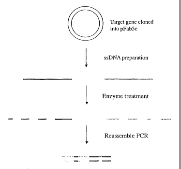
The DNA shuffling procedure can be illustrated by the steps shown in
Figures 1 to 5. The gene encoding the protein of interest (X) in the plasmid
pFab5chis is used in this example. Random mutations are introduced by
error prone PCR. Single-stranded DNA is prepared. This can be carried out
by either biotinylated primers or by the use of phage being able to pack
single-stranded DNA, as discussed above. The coding and the non-coding
ssDNA strands are prepared in different reactions (A and B). The ssDNA
strands from either reactions are subjected to separate enzymatic treatment
using e.g. BAL 31. By mixing the two pools of single-stranded DNA
fragments in equimolar amounts the gene can be reassembled in a shuffled
nature and in many versions by the use of two subsequent PCR reactions,
where the first reaction contains no primers. After cloning this library of
reassembled genes in pY, selections can be performed to achieve the
improved molecule of interest.
A more detailed description of examples of the present invention is given
below.
Example 1
Reagents
AmpliTaq polymerase was purchased from Perkin-Elmer Corp., dNTPs
from Boehringer Mannheim Biochemica (Mannheim, Germany), and
BAL31 Nuclease from New England Biolabs Inc. (Beverly, USA). All
restriction enzymes were purchased from New England Biolabs Inc.
CA 02485506 2004-11-09
WO 03/097834 PCT/GB03/02102
33
(Beverly, USA). Ethidium bromide was purchased from Bio-Rad
Laboratories (Bio-Rad Laboratories, Hercules, CA, USA). T4 DNA Ligase
was purchased from New England Biolabs Inc. (Beverly, USA). EDTA and
EGTA was purchased from Kebo Lab (Sweden).
All primers were designed in the laboratory and obtained from Life
Technologies (Taby, Sweden) and SGS-DNA (Koping, Sweden).
PCR
All Polymerase Chain Reactions (PCR) were carried out in a automatic
thermocycler (Perkin-Elmer Cetus 480, Norwalk, CT,USA). PCR
techniques for the amplification of nucleic acid are described in US Patent
No. 4,683,195. References for the general use of PCR techniques include
Mullis et al., Cold Spring Harbor Symp. Quant. Biol., 51:263, (1987),
Ehrlich (ed), PCR technology, Stockton Press, NY, 1989, Ehrlich -et al.,
Science, 252:1643-1650, (1991), "PCR protocols; A Guide to Methods and
Applications", Eds. Innis et al., Academic Press, New York, (1990).
Sequencing
All constructs have been sequenced by the use of BigDye Terminator Cycle
Sequencing kit (Perkin-Elmer, Elmervill, CA, USA). The sequencing was
performed on a ABI Prism 377 DNA Sequencer.
Agarose electrophoresis
Agarose electrophoresis of DNA was performed with 2% agarose gels
(AGAROSE (FMC Bioproducts, Rockland, ME, USA)) with 0.25 g/ml
CA 02485506 2008-05-21
WO 03/097833 PCP/G1103l02102
34
etlidium bromide in Tris-acetate buffer (TAE-buffer 0.04M Tris-acetate,
0.001M EDTA). Samples for electrophoresis were mixed with a sterile
filtrated loading buffer composed of 25% Ficoll nd Bromphenolic blue and
loaded into wells in a the 2% agarose gel. The electrophoresis was run at 90
V for 45 minutes unless otherwise stated in Tris=-acetate buffer with 0.25
l rglml ethidium bromide. Bands of appropriate size were gel-purified using
the Qiagtzick Gel Extraction Kit (Qiagen (3mbH, Tilden, Germany) when
needed. As molecular weight standard, DNA molecular weight marker 1 kb
ladder (Gibco BRL) was used. The DNA-concentration of the gel extracted
products were estimated using a spectrophotometer.
Bacterial Strains
The E'scherichia coli-strain TOPIOF' was used as a bacterial host for
1.5 transformations. Chemically competent cells of this strain were produced
basically as described Hanahan, D. 1993. Studies on transformation of
Escherichia. coli with plasmids. J. Mol. Biol. 166: 557-580.
Electrocompetent cells of this bacterial strain were produced (Dower, W.J.,
J. F. Miller, and C.W. Ragsdale. 1988: High efficiency transformation of
E. coli by high voltage eleetroporation. Nucleic Acids Res. 16:6127).
Plcxsmids
All genetic manipulations were performed in pFab5chis according to
Molecular cloning; a laboratory manual (Second Edition, Cold Spring
Harbor Laboratory Press, 1989). This vector is designed to harbour any
scFv gene inserted between Sfil and Notl sites. The SfiI site is located in
the p,173 leader and the Notl site is located just after the VL region, such
CA 02485506 2004-11-09
WO 03/097834 PCT/GB03/02102
that VH-linker-VL is inserted. In this case, an antibody directed to CD40
was used.
Primers
5
Two biotinylated primers surrounding the antibody gene of pFab5chis were
designed with the following sequences including designated unique
restriction sites:
10 1736 SfiI forward primer:
5'-ATT ACT CGC GGC CCA GCC GGC CAT GGC CCA CAG GTC
AAG CTC GA
15 and 1735 NotI reversed primer:
5'-TTA GAG CCT GCG GCC GCC TTG TCA TCG TCG TCC TT
Two non-biotinylated primers surrounding the antibody gene of pFab5chis
20 were designed with the following sequences including designated unique
restriction sites:
1664 Sfil forward primer:
25 5'-ATT ACT CGC GGC CCA GCC GGC CAT GGC CCA CAG GTC
AAG CTC GA
and 1635 Notl reversed primer:
CA 02485506 2004-11-09
WO 03/097834 PCT/GB03/02102
36
5'-TTA GAG CCT GCG GCC GCC TTG TCA TCG TCG TCC TT
Standard PCR
Standard PCR reactions were run at 25 cycles consisting of following
profile: denaturation (94 C, 1 minute), primer annealing (55 C, 1 minute)
and extension (72 C, 3 minutes). Each PCR reaction contained 10 mM
Tris-HC1, pH 8.3, 50 mM KCl, 1.5 mM MgC12, 200 RM dNTP, 1 RM
forward primer, 1 RM reverse primer, 1.25 U AmpliTaq thermostable
DNA polymerase (Perkin-Elmer Corp.), and 50 ng template in a final
volume of 100 Rl.
Error Prone PCR
The error prone PCR reactions were carried out in a 10 x buffer containing
500 mM NaCl, 100 mM Tris-HCI, pH 8.8, 5mM MgC12 100 jig gelatine
(according to Kuipers et al., Nucleic Acids Res. 1991, Aug 25;19 (16):4558
but with MgCl2 concentration increased from 2 mM to 5 mM).
For each 100 RI reaction the following was mixed:
dATP 5 mM 5 l
dGTP 5 mM 5 l
dTTP 10 mM 10 RI
dCTP 10 mM 10 Rl
20 RM 3' primer 1.5 l
20 RM 5'-primer 1.5 Rl
I Ox Kuipers buffer 10 Rd
sterile rap H2O 46.3 RI
CA 02485506 2008-05-21
WO 031097834 PCT/GB03/02101
37
The template in pFab5chis vector was added at an amount of 50 ng. 10 Al
of 10 mM MuCl2 was added and the tube was checked that no precipitation
of MnOz occurred. At last 5 Units of Taq enzyme was added. The error
prone PCR was run at the following temperatures for 25 cycles without a
hot stmt: 94 C l', 45 C 1', 72 C 1' , + 72 C f'or 7 minutes. The resulting
product was an error proned insert over the proteihi of approximately 750
bp. This insert was purified with Gibco PCR purification kit, before further
treatment.
Generation of single-stranded DNA by biotinylated primer s
The fragment of interest was amplified by two separate PCR. reactions.
These reactions can be standard PCR as described above or error prone PCR
also as described above. The primers should be designed so that in one
reaction the forward primer is biotinylated and in the other reaction the
reverse primer is biot ].ylated. For example, PCR reactions with A) primers
1736 and 1635 and B) primers 1664 and 1735, with the above mentioned
profile was perfonned for' 25 cycles with pFab5chis-antibody as template.
This yielded MR -products of approximately 750 bp where in A the upper
strand was biotinylated and in B the lower strand was biotinylated.
The .non-biot'uiylated strands were retrieved by purification using a solid
matrix coated with streptavidin e.g. Dynabeads The magnetic beads are
washed and equilibrated with PBS/1% BSA and B& W buffer containing 5
m11I Tris pH 7.5, 1 M NaCI4 and 0.5 mM EGTA. 100 }dl of each PCR
product is mixed with 100 41 beads dissolved in 2 x 13&W buffer and
incubated at room temperature for 15 minutes with rotation. Unbound PCR
products are removed by careful washing twice with B&W. The non-
CA 02485506 2004-11-09
WO 03/097834 PCT/GB03/02102
38
biotinylated strand of the captured DNA is eluted by alkaline denaturation
by letting the DNA incubate with 25 gl 0.1 M NaOH for 10 minutes in
room temperature. The solution is separated from the beads and neutralised
with 7.5 gl 0.33 M HCl and 2.5 gl 1 M Tris pH 8.
Generation of single-stranded DNA using phage
The fragment of interest was cloned into bacteriophage M13 vectors
Ml3mpl8 and M13mp19 using PstI/HindIII restriction enzymes. The
bacteriophage were propagated using Escherichia coli-strain TOPIOF'
according to conventional methods. Single-stranded DNA for the upper
strand was prepared from bacteriophage vector M13mp18 and single-
stranded DNA for the lower strand was prepared from bacteriophage vector
M 13mp 19. Briefly, 1.5 ml of an infected bacterial culture was centrifuged at
12 OOOg for 5 minutes at 4 C. The supernatant was precipitated with 200 l
20% PEG8000/2.5 M NaCl. The pelleted bacteriophage was resuspended in
100 l TE. 50 l phenol equilibrated with Tris-CI (pH 8.0) was added and
the sample was vortexed. After centrifugation at 12 OOOg for 1 minute at RT
the upper phase, containing the DNA, was transferred and precipitated with
ethanol. The DNA pellet was dissolved in 50 l TE (pH 8.0) and stored at -
20 C. (Sambrook et al. Molecular Cloning, A laboratory manual 2nd edition.
Cold Spring Habor Laboratory Press. 1989, chapter 4). Single-stranded
DNA prepared from phage is circular and must be opened prior to BAL31
treatment. This can be performed with an endonuclease able to cleave
single-stranded DNA.
CA 02485506 2004-11-09
WO 03/097834 PCT/GB03/02102
39
Generation of single-stranded DNA using asymmetric PCR
PCR products are purified using a spin column to remove excess primers
from the previous PCR. 150 ng of the purified product is used as template in
a linear amplification carried out in 100 l of 1 xGeneAmp 10 x PCR
buffer containing 1.5 mM MgC12 (Applied Biosystems), 200 gM of each
dNTP (New England BioLabs), 1,25 U AmpliTaq DNA Polymerase
(Applied Biosystems) and 1.0 gM of a single primer. PCR cycle conditions
are: denaturation at 94 C for 1 minute, 35 cycles of 94 C for 30 seconds,
55 C for 30 seconds, 72 C for 1 minute followed by extension at 72 C for
7 minutes.
Asymmetric PCR products are size separated from double stranded template
on a 1 % agarose gel and purified using Qiaquick Gel Extraction Kit
(Qiagen).
Generation of single-stranded fragmented DNA using BAL 31
The ssDNA strands (containing upper and lower strands, respectively) were
subjected to separate enzymatic treatment using e.g. BAL 31. Each
digestion reaction contained 0.02 g/ l ssDNA, 600 mM NaCl, 20 mM
Tris-HC1, 12- mM CaCl2, mM MgC12, 1 mM EDTA pH 8.0 and BAL 31
at various enzyme concentrations ranging from 0.1 - 5 U/ml. The reactions
were incubated at 30 C and fractions of digested ssDNA were collected
sequentially at 10, 30, 60 and 120 seconds or longer. The reactions were
stopped by addition of EDTA and heat treatment at 65 C for 10 minutes.
The ssDNA fragments were purified by phenol/chloroform extraction and
ethanol precipitated. The ssDNA are resuspended in 10 mM Tris pH 8Ø
CA 02485506 2008-05-21
WO 03/097834 PCT/GB03/02102
The digestion pattern was evaluated by 1 % agarose gel electrophoresis.
Purification of digestion produced fiagrnents:
5 Digested DNA fragments were purified by phenol/chloroform/
isoamylalcohol extraction. 50 I of buffered phenol 'was added to each tube
of 100 j.d sample together with 50 1 of a mixture of chloroform and
isoamylalcohol (24:1). The tubes were vortexed for 30 seconds and then
centrifuged for 1 minute in a microfiige at 14000 r.p.m. The upper phase
10 was then collected and mixed with 2.5 volumes of 99.5% Ethanol (1/10 was
3M Sodium Acetate, pH 5.2). The DNA was precipitated for 1 hour in -80
C. The DNA was then pelleted by centrifugation for 30 minutes in a
microfuge at 14.000 r.p.m. The pellet was washed once wih.70% ethanol
and then re-dissolved in 10 I of sterile water.
Analysis of digestion produced purified fragments on agarose gel
5 pI of the dissolved pellet from each time point and from the blank were
mixed with 2.5 lit of loading buffer (25% Ficoll and Bromphenolic blue)
and loaded into wells in a 2% agarose gel. The electrophoresis of the
different time points were performed as above.
Reassembly off di length fragments
Reassembly of the ssDNA fragments is achieved by two sequential PCR
reactions. The first PCR reaction should contain 10 mM Tris-HCI, pH 8.3,
50 mg KCI, 1.5 mM MgCl2, 200 pM dNTP, 0.3 U Taq polymerase and 2
.d BAL31 treated sample, all in a final volume of 25 tit, and subjected to 5
cycles with the following profile: 94 C for I minute, 50 C for I minute
CA 02485506 2004-11-09
WO 03/097834 PCT/GB03/02102
41
and 72 C for 2 minutes + 72 C for 5 minutes. The second PCR reaction
should contain 10 mM Tris-HCI, pH 8.3, 50 mM KCI, 1.5 MM M902, 200
gM dNTP, 0.6 U Taq polymerase, 1 gM forward primer, 1 M reverse
primer, and 5 l sample from the first PCR reaction, all in a final volume of
50 l, and subjected to 15 cycles with the following profile: 94 C for 1
minute, 55 C for 1 minute and 72 C for 2 minutes + 72 C for 7 minutes.
The resulting products can be evaluated by agarose gel electrophoresis.
Restriction digestion of reassembled fragment and plasmid with Sf1 and
Notf
The reassembled fragment and the plasmid pFab5chis were first cleaved
with SfiI by using NEB buffer 2 including BSA and 11 U enzyme/ g DNA.
The reaction was carried out for 4 h at 50 C. After this the DNA was
cleaved with Notl by adding conversion buffer and 6 U enzyme/ g DNA.
This reaction was carried out for 37 C overnight.
Gel purification of restriction digested vector and restriction digested
reassembled fragment
The cleavage reactions were analysed on a 1% agarose gel. The restriction
digested insert showed a cleavage product of about 750 bp. This
corresponds well with the expected size. The band of the cleaved insert and
plasmid was cut out and gel-extracted as previously described.
Ligation of reassembled restriction digested fragment with restriction
digested pFab5chis
Purified cleaved pFab5chis was ligated with purified reassembled restriction
CA 02485506 2004-11-09
WO 03/097834 PCT/GB03/02102
42
digested fragment at 12 C water bath for 16 hours. 50 l of the vector was
mixed with 50 l of the insert and 15 l of lOx buffer (supplied with the
enzyme), 7.5 l ligase (5 U/ l) and sterile water to a final volume of 150 l.
A ligation of restriction digested pFab5chis without any insert was also
performed in the same manner.
Transformation of chemically competent E coli TOPIOF' with the ligated
reassembled insert and pFabSchis
The ligation reactions were purified by phenol/chloroform extraction as
described above. The upper phase from the extraction was collected and
mixed with 2.5 volumes of 99.5% Ethanol (1/10 was 3M Sodium Acetate,
pH 5.2). The DNA was precipitated for 1 hour in -80 C. The DNA was
then pelleted by centrifugation for 30 minutes in a microfuge at 14.000
r.p.m. The pellet was washed once with 70% ethanol and then re-dissolved
in 10 l of sterile water. 5 l of each ligation was separately mixed with 95
l chemically competent E coli TOP1OF incubated on ice for 1 hour and
then transformed (Sambrook et al. Molecular Cloning, A laboratory manual
2d edition. Cold Spring Habor Laboratory Press, 1989). After one hour's
growth the bacteria from the two transformations were spread onto
ampicillin containing agar plates (100 g/ml). The plates were grown
upside-down in a 37 C incubator for 14 hours.
CA 02485506 2008-05-21
WO 03/097834 PCT/G1303/021.02
43
Example 2 -- Recombination frequencies; comparison of dsDNA and
ssDNA
In further comparable experiments, three scFv antibody fragments were
used in a recombination experiments, either as dsDNA or as ssDNA.
dsDNA
The three scFv genes were each amplified in PCR using forward and
reverse primers and standard PCR. procedure. The size of the bands was
confirmed with agarose electrophoresis and the rest of the amplified PCR
products were purified using Concert PCR purification kit (Gibco). The
dsDNA from the three scFv were mixed in equimolar amounts and treated
with BAL3 1. Each digestion reaction contained dsDNA at a concentration
of 0.02 g/ 1 reaction volume, 600 ruM NaCI, 20 mM Tris-HCI, 12 mM
CaC12i 12 mM MgCI2,1 mM EDTA pH 8.0 and BAL31 at various enzyme
concentrations (using 4, 20 or 100 U enzyme/ml reaction volume). The
reactions were incubated at 30 C and fractions of digested dsDNA were
collected sequentially at 10, 30, and 50 minutes. The reactions were
stopped with EDTA and heat treatment (alternatively, an EDTA-free heat
inactivation protocol may be used; see below) and purified using
phenol/chloroform extraction and ethanol precipitation. The dsDNA
samples were resuspended in 10 mM Tris pH 8Ø
Keeping each time point separate, the samples were subjected to reassembly
PCR (for Ibis reassembly. 60 ng DNA is used) and amplification PCR
according to the protocol, and cloned in pGEM (Product No A362A,
Promega, Madison, USA). Eighteen clones from each time point were
CA 02485506 2004-11-09
WO 03/097834 PCT/GB03/02102
44
sequenced and the number and frequency of recombinations were
determined.
Heat inactivation of exonuclease digestions
A protocol to stop the BAL31 reaction without using EDTA has been
established. This heat inactivation protocol avoids using phenol/chloroform
extraction, which is hazardous to health and also causes loss of material.
In brief, the sample is incubated for 10 minutes at 95 C and then put directly
in ice, to stop the enzymatic reaction. After this the sample can be directly
precipitated using ethanol.
ssDNA
The three scFv genes were each amplified in two PCR reactions using
primer pairs forward/reverse-biotin and forward-biotin/reverse using
standard PCR procedure. The size of the bands were confirmed with
agarose electrophoresis and the rest of the amplified PCR products were
purified using Concert PCR purification kit (Gibco). Single-stranded DNA
was obtained using magnetic beads according to the protocol, achieving
three sense strands and three antisense strands. The sense strands and the
antisense strands, respectively, from the three scFv were mixed in
equimolar amounts and treated with BAL31 according to the protocol
(using 1.25 or 11 U enzyme/ml reaction volume and ssDNA at a
concentration of 0.015 g/ l reaction volume) and samples were taken out
at 0 (i.e. undigested), 10, 30 and 50 minutes. The reactions were stopped
with EDTA and heat treatment and purified using phenol/chloroform
extraction and ethanol precipitation. Keeping each time point separate, but
CA 02485506 2004-11-09
WO 03/097834 PCT/GB03/02102
mixing sense and antisense strands, the samples were subjected to
reassembly PCR (for this reassembly 60 ng DNA is used) and amplification
PCR according to the protocol, and cloned in pGEM. Eighteen clones from
each time point were sequenced and the number and frequency of
5 recombinations were determined.
Results
The highest frequency of recombination using dsDNA was achieved using
10 20 U enzyme/ml reaction volume (containing 0.02 g/ l DNA) and treating
for 10 minutes. This gave 39% of the clones with one cross-over (Figure 6)
and 17% of the clones with two cross-overs (Figure 7). Using 4 U
enzyme/ml gave no cross-overs independent of time for fragmentation and
100 U enzyme/ml resulted in complete fragmentation into very small
15 fragments, as indicated by the failure to regain the full-length gene
during
reassembly.
The results from the experiments using ssDNA are shown in Figures 8 to
10. Figure 8 shows 1.25 U/ml BAL31 and clones with one cross-over,
20 Figure 9 shows 1.25 U/ml BAL31 and clones with two cross-overs. Figure
10 shows 11 U/ml BAL31 and clones with one cross-over, and Figure 11
shows 11 U/ml BAL3 land clones with two cross-overs.
The highest frequency of recombination giving one cross-over using ssDNA
25 was achieved using 11 U enzyme/ml and treating for 10 minutes (Figure
10). 59% of the clones had one cross over. The highest frequency of
recombination giving two cross-overs using ssDNA was achieved using
1.25 U enzyme/ml and treating for 30 minutes (Figure 9). 20% of the clones
had two cross overs.
CA 02485506 2004-11-09
WO 03/097834 PCT/GB03/02102
46
Conclusions and comments
These data clearly show that a higher frequency of recombination is
achieved using ssDNA. The three scFv used have the same framework
sequences, indicating that the number of cross overs reported may be higher
due to cross overs in regions where no sequence difference will result.
These experiments using ssDNA were carried out in a non-optimal fashion
for showing maximum recombination, since all strands from all three
molecules were mixed. Mixing the sense strand from one scFv with the
antisense strand from another scFv would produce higher frequencies of
cross overs, see Example 3 below. Also, each time point was here kept
separate and it would be logical to estimate the frequency of cross overs to
increase if different time points, i.e. different fragments sizes, are mixed.
Example 3 - Recombination frequencies; homology dependence using
ssDNA
To investigate the homology required to achieve cross-over we set up
experiments to recombine four scFv (designated SMUC159, CT17, AE11
and MO 152) making up three pairs with different homologies, as follows:
SMUC159-CT17 92%
SMUC159 - AE11 70%
SMUC 159 - MO 152 60%
The four scFv genes were each amplified in two PCR reactions using primer
pairs forward/reverse-biotin and forward-biotin/reverse using standard PCR
CA 02485506 2004-11-09
WO 03/097834 PCT/GB03/02102
47
procedure. The size of the bands were confirmed with agarose
electrophoresis and the rest of the amplified PCR products were purified
using Concert PCR purification kit (Gibco). Single-stranded DNA was
obtained using magnetic beads according to the protocol, achieving four
sense strands and four antisense strands. Each strand was treated with
BAL31 according to the protocol (using 4.2 or 12.5 U enzyme/ml) and
samples were taken out at 0, 10, 30 and 50 minutes, or 0, 15, 30, 45 and 60
minutes. The reactions were stopped with EDTA and heat treatment and
purified using phenol/chloroform extraction and ethanol precipitation.
Keeping each time point separate, but mixing sense and antisense strands
forming the pairs as indicates above, the samples were subjected to
reassembly PCR and amplification PCR according to the protocol, and
cloned in pGEM. Fifteen clones from each time point were sequenced and
the number and frequency of recombination were determined.
Results
Cross overs were identified in all combinations of scFv, indicating that as
low as 60% homology is enough to achieve recombination.
Example 4 - Improved control of fragment size using exonucleases
(A) Exonucleases
We use exonucleases, e.g. BAL31, exonuclease I, exonuclease V,
exonuclease VII, exonuclease T7 gene 6, bacteriophage lambda
exonuclease, and exonuclease Rec Jf for fragmentation in the methods of the
present invention. These enzymes cleave off one nucleotide at a time, either
CA 02485506 2004-11-09
WO 03/097834 PCT/GB03/02102
48
from the 5' end or from the 3'end or from both ends. The reaction can be
stopped using EDTA or heat inactivation (see above), depending on the
enzyme used. This means that fragments of all possible sizes, differing with
only one nucleotide, can be obtained.
The following examples demonstrate how exonuclease digestion allows the
creation of fragments of various and controllable sizes depending on the
conditions used.
BAL 31
Single-stranded DNA was digested with BAL31 according to the protocol
in Example 1, with an enzyme concentration of 4.2 U/ml reaction volume
and ssDNA concentration of 0.008 g/ l reaction volume.
In a typical experiment, about 300 ng DNA is isolated at each time point of
BAL31 treatment. Figure 12 shows an agarose electrophoresis gel image of
such an experiment with untreated ssDNA in lane 4 and ssDNA treated for
10 minutes in lane 3, for 30 minutes in lane 2 and for 50 minutes in lane 1.
Lane 5 is the molecular weight (MW) standard.
Figures 13 to 15 shows the corresponding gel chromatograms of the lanes,
respectively. Figure 13 is the untreated material and the multiple peaks
refer to different conformations of the ssDNA. Figure 14 corresponds to
lane 3 and material treated for 10 minutes. The material was heat treated to
stop the enzymatic reaction, and thus resolving the different conformations,
and one peak of a distinct size is shown. Figure 15 corresponds to lane 2
and material treated for 30 minutes.
CA 02485506 2004-11-09
WO 03/097834 PCT/GB03/02102
49
Here it is clear that the peak corresponding to larger fragments is decreasing
and a peak of smaller DNA fragments has appeared.
Exonuclease VII
Single-stranded DNA was digested with exonuclease VII using an enzyme
concentration of 7.7 U/ml reaction volume and ssDNA concentration of
0.008 g/ l reaction volume. The reaction buffer comprised 67 mM
potassium phosphate (pH 7.9), 10 mM mercaptoethanol, 6.7 mM MgC12 and
8.3 mM EDTA.
The reaction was allowed to proceed at 37 C for 10, 20 and 30 minutes,
before being stopped by heat inactivation (95 C for 5 minutes).
In Figure 16 the fragmentation pattern using exonuclease VII is shown.
Lane 1 is MW standard, lane 2 is untreated ssDNA, lane 3 is ssDNA
fragmented with exonuclease VII for 10 minutes, lane 4 is ssDNA
fragmented with exonuclease VII for 20 minutes, and lane 5 is ssDNA
fragmented with exonuclease VII for 30 minutes. This shows that the
fragment sizes are decreased by time.
Exonuclease Rec Jf
Single-stranded DNA was digested with exonuclease Rec Jf using an
enzyme concentration of either 2.5 U/ml reaction volume or 10 U/ml
reaction volume and ssDNA at a concentration of 0.007 g/ l reaction
volume, corresponding to 0.36 U enzyme/ g DNA and 1.4 U enzyme/ g
DNA, respectively. The reaction buffer comprised 50 mM NaCl, 10 mM
Tris.HC1, 10 mM MgC12 and 1 mM dithiothreitol, at pH 7.9
CA 02485506 2004-11-09
WO 03/097834 PCT/GB03/02102
The reaction was allowed to proceed at 37 C for 10, 20 and 30 minutes,
before being stopped by heat inactivation (95 C for 5 minutes).
5 In Figure 17 the fragmentation pattern using exonuclease Rec Jf at
036 U/microgram ssDNA is shown. Lane 1 untreated ssDNA, lane 2 is
ssDNA fragmented with exonuclease Rec Jf for 10 minutes, lane 3 is
ssDNA fragmented with exonuclease Rec Jf for 20 minutes, and lane 4 is
ssDNA fragmented with exonuclease Rec Jf for 30 minutes. This shows that
10 the fragment sizes are decreased by time. In Figure 18 the enzyme
concentration is increased 4 times (1.4 U/microgram ssDNA) and the
fragmentation pattern is shown from 0 to 30 minutes, showing a higher
degree of fragmentation as compared to Figure 17. This shows that both
time and enzyme concentration can be used to control the fragmentation.
15 (B) Endonucleases
Conventional DNA shuffling methods typically use DNase I for
fragmentation (for example, see Stemmer, 1994, Nature 370:389-391).
DNase I cleaves DNA in an endonucleolytic fashion at sites adjacent to
20 pyrimidines. Consequently, not all possible fragment sizes can be obtained.
Moreover, using magnesium in the reaction buffer, a homologous mix of
mono- and oligomers is obtained. Hence, different methods such as gel
agarose electrophoresis purification or gel filtration need to be used in
order
25 to isolate fragments of different sizes. Often fragments of small size or a
mix of small and larger fragments are desired to optimise recombination.
However, these purification methods introduce single-stranded nicks in the
double-stranded PCR products. Fragments of a particular size purified on a
gel would thus consist of dsDNA with a large number of single-stranded
CA 02485506 2004-11-09
WO 03/097834 PCT/GB03/02102
51
nicks, which would give rise to many smaller fragments upon denaturation.
This means that many of the single-stranded fragments generated upon
denaturation would be too short to function as primers during the annealing,
resulting in a great loss of product.
Using manganese in the reaction buffer creates fragments of sizes smaller
than 50 bp and no gel purification is needed. However, here you are
restricted to use only small fragments and these can not be mixed with
larger fragments, something that would probably increase the recombination
frequency.
The problems associated with the use of endonucleases are demonstrated in
the following experiments:
DNase I
DNA was digested for 5 minutes with DNase I at a concentration of
0.15 U/ g DNA.
Magnesium and manganese buffers were compared when fragmenting with
DNase I and the result is shown in Figure 19. Lane 1 is MW standard, lane
2 is untreated ssDNA in Mg buffer, lane 3 is ssDNA fragmented with
DNase I in Mg buffer according to Stemmer (1994) Nature 370:389-391,
lane 4 is untreated ssDNA in Mn buffer and lane 5 is ssDNA fragmented
with DNase I in Mn buffer according to Kikuchi et al. (2000) Gene
243:133-137. It is clear from Figure 19 that, when using Mg buffer and
conditions according to the Stemmer and Kikuchi papers, no fragmentation
occurs. Moreover, when using Mn buffer and conditions according to the
CA 02485506 2004-11-09
WO 03/097834 PCT/GB03/02102
52
Stemmer and Kikuchi papers, all material is totally fragmented within only
a few minutes.
In an attempt to obtain fragments of different sizes we decided to use Mg
buffer and increase the enzyme concentration. Figure 20 shows an agarose
electrophoresis gel image of such an experiment using DNase I. Lane 1 is
the MW standard. Lane 6 is untreated ssDNA. Lane 12 is ssDNA treated
according to the Stemmer and Kikuchi papers, using 0.15 U
enzyme/microgram DNA and lane 13 is the same material treated with 1 U
enzyme/microgram DNA (i.e. six times more enzyme).
Figures 21 to 23 shows the corresponding chromatograms. The untreated
ssDNA has been heat-treated, therefore only one peak appears in Figure 21
(indicated by arrow). In figure 22, it is apparent that using the amount of
DNase I according to the Stemmer and Kikuchi papers the peak for
untreated ssDNA is somewhat decreased (indicated by arrow) but no
distinct peak is visible for the fragmented DNA, only a smear. Using 6
times more enzyme the untreated ssDNA is totally abolished (Figure 23)
and neither here is any visible peak of the fragments.
Mung bean nuclease
Single-stranded DNA was digested with Mung bean nuclease (Product No
M0250S, New England Biolabs) using an enzyme concentration of either
0.375 U/ml reaction volume and ssDNA at a concentration of 0.007 g/ 1
reaction volume. The reaction buffer comprised 50 mM sodium acetate,
mM NaCL, 1 mM ZnSO4, at pH 5Ø
CA 02485506 2004-11-09
WO 03/097834 PCT/GB03/02102
53
The reaction was allowed to proceed at 25 C for 10 minutes, before being
stopped by heat inactivation (95 C for 5 minutes).
Figure 24 shows fragmentation using another endonuclease, Mung bean
nuclease. Lane 1 is the untreated ssDNA, lane 2 is the same material treated
for 10 minutes. Lane 3 is the MW standard.
Results indicate that all DNA was totally fragmented after only 10 minutes
digestion with Mung bean nuclease (see lane 2), despite using the enzyme at
a concentration lower than that recommended by the manufacturer.
Conclusions and continents
The above examples show how the fragment sizes can be controlled using
exonucleases and altering the reaction conditions, i.e. time, reaction volume,
enzyme concentration. The different peaks are visualised using gel image
chromatograms.
In contrast, using endonucleases, such as DNase I, gives a reaction which is
hard to control. Using conditions as referred in the literature, either using
Mg or Mn containing buffers, typically gives a situation when either
everything or nothing is fragmented, see especially Figure 20. An
experiment using another endonuclease (Mung bean nuclease) confirms
these observations.
CA 02485506 2004-11-09
WO 03/097834 PCT/GB03/02102
54
Example 5 - Digestion of sub-populations of single-stranded DNA
starting material with different exonucleases
In further experiments, the single-stranded DNA starting material was split
into two populations, which were then digested using different
exonucleases.
Materials and methods
Plasmids
A tetracycline-deleted variant of plasmid pBR322 was constructed by
cleavage with Sall and BamHI (Roche, Basel, Switzerland) Klenow
treatment (Amersham Biosciences AB, Uppsala, Sweden) and blunt-end
ligation (New England Biolabs, MA, USA). The resulting plasmid was
checked for tetracycline sensitivity and is called pBR322dtet.
PBR322stopl and pBR322stop3 were created by PCR amplification of the
tetracycline gene of pBR322 using specific primers (Table 1). Each mutated
tetracycline gene was cloned into pBR322.
Table 1
Primer sequences
pBR322 Nhel forward stop:
5'-CACTATGGCGTGCTGCTAGCGCTATATGCGTTGATGCAATTT
CT ATGAGCACCCGTTCT-3'
pBR322 Eagl reversed:
5'- CGTAGCCCAGCGCGTCGGCCGCCATGCCGGCGATAATG-3'
CA 02485506 2004-11-09
WO 03/097834 PCT/GB03/02102
pBR322 Hindlll forward:
5'- CAGCTTATCATCGATAAGCTTTAATGCGGTAGTTTAT-3'
5 pBR322 Sall reversed stop:
5'-TCTCAAGGGCATCGGTCGACGCTCTCCCTTATGCGACTCCTGC
ATTAGGAATCAGCCCAGTAGTA-3'
PCR
Unless otherwise noted PCR reactions contained 4 M of each primer, 160
M dNTP (Roche, Basel, Switzerland), Ix AmpliTaq reaction buffer, 2.5 U
AmpliTaq thermostable DNA polymerase (Applied Biosystems, CA, USA).
FIND PCR 1: 5 or 25 cycles of 94 C 30 s, 50 C 45 s, 72 C 1 minute and
then 72 C for 7 minutes, no external primers were included.
FIND PCR 2: 15, 25 or 50 cycles of 94 C 30 s, 55 C 45s, 72 C 1 minute
and then 72 C for 7 minutes with external primers included.
Single-stranded DNA preparation
The gene of interest, i.e. tet-r, was amplified using specific primers, one of
the primer was biotinylated. SsDNA from sense and antisense strains was
purified using streptavidin-magnetic beads (purchased from either Dynal
AS, Oslo, Norway or Miltenyi Biotec, Bergisch Gladbach, Germany)
according to manufacturer's recommendations. The ssDNA hereby obtained
was further purified either by ethanol precipitation or by using recochip
(TaKaRa, Shiga, Japan) according to manufacturers recommendations.
CA 02485506 2004-11-09
WO 03/097834 PCT/GB03/02102
56
FIND experinients
The FIND experiments were initiated by digesting DNA with an
exonuclease. The DNA was single-stranded (prepared as above) and
originated from the tetracycline resistance gene (pBR322stopl or
pBR322stop3, 945 bp). The exonucleases were BAL31 (0.08-1 U/ g DNA,
New England Biolabs, MA, USA), exonuclease I (100 U/ g DNA, New
England Biolabs, MA, USA), T7 gene 6 exonuclease (320 U/ g DNA,
USB, Cleveland Ohio, USA) and exonuclease V (12.5 U/ g DNA, USB,
Cleveland Ohio, USA). The time for digestion was in the range 2-90
minutes. The digestion reactions were stopped by adding EDTA to a final
concentration of 20 mM and/or heat inactivation at 65 or 95 C for 10
minutes. When EDTA was used to stop the DNA fragmentation the DNA
was further purified by phenol/chloroform extraction and ethanol
precipitation. The fragments were recombined in a FIND PCR1 reaction for
5 or 25 cycles and the material was amplified in a FIND PCR 2 reaction for
15, 25 or 50 cycles. Finally the full length genes were cloned into
pBR322dtet by the use of HindIII and EagI (New England Biolabs, MA,
USA) for functionality evaluation or into pGEM (Promega, Madison, WI,
USA) for sequencing.
Evaluation of functionality of tetracycline clones
The clones introduced into pBR322dtet were transformed into chemical
competent TG1 E. coli and plated on LB agar plates containing 1 gg/ml
ampicillin. One to two hundred clones were then moved to LB agar plates
containing 50 gg/ml tetracycline and the frequency of tetracycline resistant
clones could be calculated.
CA 02485506 2004-11-09
WO 03/097834 PCT/GB03/02102
57
Results
As shown above, higher frequencies of recombination using the FIND
procedure of the present invention could be achieved using ssDNA in the
fragmentation. The exonuclease BAL 31 is predominately a 3' exonuclease
that removes mononucleotides from both 3' termini of the two strands of a
linear double stranded DNA. However BAL 31 can also degrade the single-
stranded DNA ends generated by the 3' exonuclease activity on the double
stranded DNA. The activity of BAL 31 on ssDNA is removal of
mononucleotides from the 5' termini only. Using BAL 31 for
fragmentation of ssDNA and then reassembly to full length genes will
theoretically result in one cross-over per gene. Experiments were therefore
performed to test different exonucleases for the fragmentation of ssDNA.
Exonuclease I has 3' activity only whereas BAL 31, T7gene6 and RecJ
exonucleases have 5' activity only. Exonuclease V and Exonuclease VII
have activity from both ends (5' and 3'). In order to show that these
exonucleases can be used in the fragmentation step in a FIND experiment
and yield functionally recombined genes a model system based on
tetracycline resistance genes was used.
It was found that BAL 31 (Figure 25a) as well as Exonuclease I (Figure
25b) and T7 gene 6 exonuclease (Figure 25c) all worked well in the FIND
procedure and a dependency on fragmentation time of recombination
frequency was observed.
If only one exonuclease, which digests ssDNA from only one end, is used
only one cross-over can in theory be achieved. However, it was found that
further cross-overs can be the obtained if DNA fragments from treatment by
CA 02485506 2004-11-09
WO 03/097834 PCT/GB03/02102
58
different exonucleases were combined. Exonuclease V and Exonuclease VII
treatment will result in small fragments without the 5' and 3' ends. These
ends are necessary in order to amplify the recombined material in the last
PCR reaction. These DNA fragments can therefore be combined with DNA
digested from 5' or 3' ends. The result from such a combination can be seen
in Figure 25d where ssDNA treated with Exonuclease I for 10 minutes was
combined with ssDNA treated with Exonuclease V for 40 and 50 minutes.
Functional clones of up to 40 % were obtained, a result that should be
compared to the maximum 25 % achieved in the same system using only
one enzyme (Figure 25a-c). Fragments have also combined from
Exonuclease I and Exonuclease VII treatment, and fragments from T7 gene
6 exonuclease and Exonuclease VII treatment, at different time points, and
functionally recombined clones could be obtained (data not shown).
Example 6 - Digestion of sub-populations of single-stranded DNA
starting material with different exonucleases (further experiments)
In a separate set of experiments, multiple cross-overs were produced by
combining fragments obtained by digesting single-stranded DNA with
Exonuclease I with fragment obtained by digesting single-stranded DNA
Exonuclease V and/or Exonuclease VII.
Exonuclease I (Exo I) has only 3' activity whereas Exonuclease V (ExoV)
and Exonuclease VII (ExoVII) have activity from both ends (5' and 3').
Hence, ExoV and ExoVII treatment will result in small fragments without
the 5' and 3' ends. These ends are necessary in order to amplify the
recombined material in the last PCR reaction. These DNA fragments can
therefore be combined with DNA digested from 5' or 3' ends only.
CA 02485506 2004-11-09
WO 03/097834 PCT/GB03/02102
59
An equimolar mixture of the three scFv genes was used in fragmentation
with Exol, ExoV and ExoVII. Different combination of fragmented
material was mixed and PCR reassembled into full length genes and finally
sequenced (Figure 26a-d). Exol behaved similar to BAL 31 with up to 40%
clones with one recombination (Figure 26a). The combination of Exol with
ExoV or ExoVIl increased number of crossover up to five recombinations
(Figure 26 a and c, respectively). ExoVII seems to be more efficient than
ExoV, the ExoI/ExoVII combination generated recombinations in over 75%
of the clones. Finally, a combination of fragmented DNA from all three
enzymes was mixed which generated recombinations (I to 4) in almost all
clones, only 7% of them were wild type genes. Furthermore, longer
fragmentation times were most efficient (Figure 26d). In this particular case
the scFv genes consist of large areas of complete homology (the frame work
regions), meaning that the number of recombinations identified (up to 4) are
the ones that are possible to identify, i.e. the actual number of
recombinations might be much higher.
CA 02485506 2004-11-09
WO 03/097834 PCT/GB03/02102
Example 7 - Comparison of the effects of digestion of ss DNA with an
exonuclease with the effects of digestion of ds DNA with an
endonuclease
5 The experiments in Figure 27 were carried out by recombining genes
coding for three different scFv's using either ssDNA and exonuclease
treatment or dsDNA and endonuclease treatment.
Endonuclease digestion of dsDNA was performed using DNase I, as
10 described in Stemmer et al., 1994, Nature 370:389-91. Exonuclease
digestion of ssDNA was performed using Exol, ExoV and ExoVI1, with
equimolar mixes of fragments from Exol (10 minutes reaction time), ExoV
(30 minutes reaction time) and ExoVII (30 minutes reaction time) being
used in recombination reactions.
The number of recombinations were evaluated by the use of sequencing.
The result shows that ssDNA/exonuclease digestion yields fewer wild type
clones and more recombinations.
CA 02485506 2008-05-21
SEQUENCE LISTING
<110> Alligator Bioscience AB
<120> A Method for In Vitro Molecular Evolution of Protein Function
<130> ALLBA/P28382PC
<140> PCT/GB03/02102
<141> 16 May 2003
<150> GB 0211369.4
<151> 17 May 2002
<150> US 10/321195
<151> 17 December 2002
<160> 8
<170> SeqWin99
<210> 1
<211> 44
<212> DNA
<213> Artificial Sequence
<220>
<223> Oligonucleotide 1736 Sfil forward primer
<400> 1
attactcgcg gcccagccgg ccatggccca caggtcaagc tcga 44
<210> 2
<211> 35
<212> DNA
<213> Artificial Sequence
<220>
<223> Oligonucleotide 1735 reverse primer
<400> 2
ttagagcctg cggccgcctt gtcatcgtcg tcctt 35
<210> 3
<211> 44
<212> DNA
<213> Artificial Sequence
<220>
<223> Oligonucleotide 1664 SfiI forward primer
<400> 3
attactcgcg gcccagccgg ccatggccca caggtcaagc tcga 44
<210> 4
<211> 35
<212> DNA
<213> Artificial Sequence
<220>
<223> Oligonucleotide 1664 NotI reverse primer
1 of 2
CA 02485506 2008-05-21
<400> 4
ttagagcctg cggccgcctt gtcatcgtcg tcctt 35
<210> 5
<211> 59
<212> DNA
<213> Artificial Sequence
<220>
<223> Oligonucleotide pBR322 NheI forward stop primer
<400> 5
cactatggcg tgctgctagc gctatatgcg ttgatgcaat ttctatgagc acccgttct 59
<210> 6
<211> 38
<212> DNA
<213> Artificial Sequence
<220>
<223> Oligonucleotide pBR322 EagI reverse primer
<400> 6
cgtagcccag cgcgtcggcc gccatgccgg cgataatg 38
<210> 7
<211> 37
<212> DNA
<213> Artificial Sequence
<220>
<223> Oligonucleotide pBR322 Hindlll forward primer
<400> 7
cagcttatca tcgataagct ttaatgcggt agtttat 37
<210> 8
<211> 65
<212> DNA
<213> Artificial Sequence
<220>
<223> Oligonucleotide pBR322 SalI reverse stop primer
<400> 8
tctcaagggc atcggtcgac gctctccctt atgcgactcc tgcattagga atcagcccag 60
tagta 65
2 of 2