Note: Descriptions are shown in the official language in which they were submitted.
CA 02485576 2004-11-09
WO 03/096264 PCT/US03/13674
TOMOGRAPHIC RECONSTRUCTION OF SMALL OBJECTS USING
A PRIORI KNOWLEDGE
Field of the Invention
The present invention relates to three-dimensional (3D) imaging systems in
general, and, more particularly to three-dimensional (3D) imaging systems
using a priori
knowledge about an object of interest that is undergoing image reconstruction
using
computed tomography.
Background of the Invention
Image reconstruction algorithms in use today compute the typically three-
dimensional (3D) structure of an object from its one or two-dimensional
projections
without using additional information about the object. Known reconstruction
algorithms in
the tomography field, and especially the optical tomography field, are
deficient in the use
of a priori knowledge for enhancing object reconstruction.
Summary of the Invention
In one embodiment, the present invention provides a method for three-
dimensional
(3D) reconstruction of an object of interest, such as a cell, including
adjusting a current set
of projection images according to a priori knowledge to produce adjusted
projection
images, for example, based on simple probability masks or Bayesian analysis of
multiple
similar objects in the same sample. A reconstruction algorithm is used on the
adjusted
projection images to generate a 3D image. The 3D image is further adjusted
according to
the a priori knowledge to generate an adjusted 3D image. Criteria for process
completion
are applied to determine whether the adjusted 3D image is adequate. Otherwise,
a set of
CA 02485576 2011-12-01
77501-24
pseudo projections are computationally created at the same projection angles
as the
current set of projection images and then compared to the current set of
projection
images to produce a more realistic set of new projections, wherein the new
projections are input again to the reconstruction algorithm and the steps of
the
method are repeated until the adequacy criteria are met.
According to one aspect of the present invention, there is provided a
method for tomographic three-dimensional (3D) reconstruction of a sample
including
at least one object of interest, the method comprising the steps of: (a)
obtaining a
current set of projection images from an optical tomography system; (b)
adjusting the
current set of projection images of the sample according to a priori knowledge
to
produce adjusted projection images by removing unallowable features and
conforming the current set of projection images to allowable features
according to the
a priori knowledge wherein the a priori knowledge includes a priori knowledge
selected from the group consisting of cell preparation chemistry, cell
features, cell
boundaries, contrast agents having known distributions in contrast, a measured
modulation transfer function of the projection system, Bayesian analysis of
multiple
similar objects in the sample, and errors flowing from a 3D image
reconstruction
algorithm; (c) using a reconstruction algorithm on the adjusted projection
images to
generate a 3D image; (d) further adjusting the 3D image according to the a
priori
knowledge to generate an adjusted 3D image; (e) applying criteria for process
completion to determine whether the adjusted 3D image meets a selected level
of
confidence values; and (f) if the adjusted 3D image does not meet the selected
level
of confidence values, then computationally creating a set of pseudo
projections,
where the set of pseudo projections is created computationally by generating
artificial
projections through the 3D data volume at the same projection angles as the
current
set of projection images and comparing the current set of projection images
with the
pseudo projection images to produce a set of new projections, wherein the new
projections are input again at step (a) as a current set of projection images
and steps
(a) through (e) are repeated until the adequacy criteria are met.
2
CA 02485576 2011-12-01
77501-24
According to another aspect of the present invention, there is provided
a system for tomographic three-dimensional (3D) reconstruction of an object of
interest in a sample, comprising: (a) an optical tomography system for
generating a
current set of projection images from the sample; (b) means, coupled to
receive the
current set of projection images, for adjusting the current set of projection
images of
the sample according to a priori knowledge to produce adjusted projection
images by
removing unallowable features and conforming the current set of projection
images to
allowable features according to the a priori knowledge wherein the a priori
knowledge
includes a priori knowledge selected from the group consisting of cell
preparation
chemistry, cell features, cell boundaries, contrast agents having known
distributions
in contrast, a measured modulation transfer function of the projection system,
Bayesian analysis of multiple similar objects in the sample, and errors
flowing from a
3D image reconstruction algorithm; (c) means, coupled to receive the adjusted
projection images, for using a reconstruction algorithm on the adjusted
projection
images to generate a 3D image; (d) means, coupled to receive the 3D image, for
further adjusting the 3D image according to the a priori knowledge to generate
an
adjusted 3D image; (e) means, coupled to receive the adjusted 3D image, for
applying criteria for process completion to determine whether the adjusted 3D
image
meets a selected level of confidence values; and (f) means, coupled to receive
the
adjusted 3D image if it does not meet the selected level of confidence values,
for
otherwise computationally creating a set of pseudo projections, where the set
of
pseudo projections is created computationally by generating artificial
projections
through the 3D data volume at the same projection angles as the current set of
projection images and comparing the current set of projection images with the
pseudo projection images to generate a set of new projections, wherein the new
projections are input again to the means for adjusting a current set of
projection
images as a current set of projection images.
2a
CA 02485576 2011-12-01
77501-24
Brief Description of the Drawings
FIG. 1 shows an illustration of an idealized biological cell that may serve
as a probability mask and may be improved through Bayesian analysis of similar
cells
in the sample in accordance with the teachings of the present invention.
FIG. 2 is an illustration of a flow diagram showing the use of a priori
knowledge and iterative processing as contemplated by an embodiment of the
present invention.
FIG. 3 schematically shows an example of various forms of statistical
information that may be utilized in an image reconstruction process to provide
a
measure of confidence for each voxel in a 3D image as contemplated by an
embodiment of the present invention.
FIG. 4 schematically shows a block diagram of an example of a system
for tomographic reconstruction of small objects as contemplated by an
embodiment
of the present invention.
Detailed Description of the Preferred Embodiments
This invention describes the advantageous use of a priori knowledge
about an object of interest that is undergoing image reconstruction using
computed
tomography. In most cases, there exists certain information about an observed
object that can be utilized in the image reconstruction to compute more
accurate or
more realistic 3D
2b
CA 02485576 2004-11-09
WO 03/096264 PCT/US03/13674
reconstructions. Such a priori knowledge serves to constrain the
reconstruction within the
bounds of allowable features (i.e., what "can be") and unallowable features
(i.e., what
"cannot be"). In a noisy image, knowing a set of unallowable features (i.e.,
what "cannot
be") can significantly improve the accuracy, and as an additional consequence,
the speed
and efficiency, of the computed image reconstruction. The example used to
illustrate the
principals of image reconstruction using a priori knowledge is the biological
cell, but
those skilled in the art will recognize that these principals may be applied
generally to any
object undergoing image reconstruction where certain information concerning
the shape
and structure of the object is known.
Referring now to FIG. 1, there shown is an illustration of an idealized
biological
cell. There exists certain information in the form of knowledge about a cell1
that is useful
in a reconstruction process as contemplated by one example of the method of
the
invention. For instance, biologists already know that a typical cell 1
consists of an external
bounding membrane 2 (e.g., the cytoplasmic membrane), an internal bounding
membrane
3 (e.g., the nuclear membrane), and these two membrane surfaces may typically
be
smoothly continuous and roughly concentric. The two bounding membranes define
three
compartments: the nuclear compartment 5 inside the nuclear membrane, the
cytoplasmic
compartment 4 outside the nuclear membrane but inside the cytoplasmic membrane
and
the exterior space 6 outside the cytoplasmic membrane. The exterior space 6
has no
biological structure unless it is contiguous with another cell.
Additionally, in applying one example of the method of the invention, one may
advantageously assume that the two separate membrane surfaces are indeed
continuous
and that there is no useful information in the exterior space where contrast
values in that
3
CA 02485576 2004-11-09
WO 03/096264 PCT/US03/13674
exterior space might be set to either extreme of the contrast range depending
on the nature
of the imaging system. In an optical tomography system, the exterior space 6
may be
assigned a gray value at either end of the contrast distribution; in practice,
exterior space 6
is substantially transparent in the image formation system. This a priori
knowledge is
useful for improving the reconstructed data set in an imperfect projection
data set. The a
priori knowledge may typically be in the form of a probability mask that is
applied to each
projection image and likewise to each subsequent pseudo projection image. For
example,
the probability mask may be binary as a first simple approximation in the
elimination of
background noise outside the cytoplasmic membrane and may be warped to
optimally fit
the projection image.
In operation, a tomographic imaging system designed to reconstruct the 3D
picture
of a cell from its two-dimensional (2D) projections may use a priori knowledge
about the
cell in the computed image reconstruction of that cell. One example of such a
tomographic system is described, for example, in U.S. application number
09/927,151 of
Alan C. Nelson, filed 08/10/2001, entitled "APPARATUS AND METHOD FOR
IMAGING SMALL OBJECTS IN A FLOW STREAM USING OPTICAL
TOMOGRAPHY," (hereinafter called the FOT design), incorporated herein by this
reference. In the aforesaid FOT design, cell motion is accomplished in a flow
stream,
wherein cells in suspension move with constant velocity along the single flow
axis of a
capillary tube.
Another example of such a tomographic system is described, for example, in U.
S.
application number 10/126026 of Alan C. Nelson, filed 04/19/2002, entitled
"VARIABLE-MOTION OPTICAL TOMOGRAPHY OF SMALL OBJECTS," (hereinafter
4
CA 02485576 2004-11-09
WO 03/096264 PCT/US03/13674
called the VOT design), incorporated herein by this reference. In the
aforesaid VOT
design, cell motion is accomplished in a variable motion system.
Each radial 2D projection of the cell comprises an independent shadowgram
through the cell and will contain noise. A priori information about noise in
this case
typically includes the fact that noise introduces an uncertainty in both the
location of a
contrast element and the actual density value of that contrast element. A
priori
information may also include data representative of the fact that, because of
noise, certain
unallowable features will be present in the projection. In some cases, for
example, edges
that should be continuous, such as membrane edges, may appear fragmented and
discontinuous. There may appear to be structure, due to light scatter and
diffraction, in the
exterior space where no structure should exist. Within the cell itself, there
may appear to
be contrast values at either extreme of the contrast range and as such are
highly
improbable, etc. Unallowable features may be identified using many one-
dimensional
(1D) and 2D image processing techniques including masking, thresholding,
histogramming, mathematical morphology, template matching, adaptive
processing,
statistical and other methods available to those skilled in the art.
Therefore, it is possible
to adjust the projection image to better represent allowable features and
remove
unallowable features before the image is sent to a reconstruction algorithm.
Once
unallowable features are identified using a priori knowledge, the unallowable
features
may be removed from the image using standard image processing techniques.
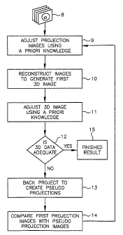
Referring now to FIG. 2, there shown is an illustration of a flow diagram
showing
the use of a priori knowledge and iterative processing for generating a 3D
image as
contemplated by an embodiment of the present invention. Unprocessed projection
images
5
CA 02485576 2004-11-09
WO 03/096264 PCT/US03/13674
8 are adjusted according to a priori knowledge at step 9, and then input to
the
reconstruction algorithm to generate the first 3D image at step 10. This in
turn is further
adjusted by removing unallowable features and conforming the image to
allowable
features according to a priori knowledge at step 11. Criteria for process
completion are
applied to determine whether the current reconstruction is adequate at step
12. Criteria
for process completion may be any useful imaging related criteria, as, for
example, a
selected level of confidence values assigned to voxels or pixels as discussed
below.
Otherwise, a set of pseudo projections is created computationally by
generating artificial
projections through the 3D data volume at the same projection angles as the
current set of
projection images, and the current set of projection images is compared to the
computed
pseudo projection images to produce a more optimal set of new projections at
step 14.
These new projections are adjusted again to conform with the a priori
knowledge then
input again to the reconstruction algorithm at step 9 to generate a subsequent
3D image.
The process continues until the adequacy criteria are met producing a finished
3D image
result at step 15.
Because the input 2D projection images 8 are adjusted at step 9 according to
the a
priori knowledge before computing the 3D reconstruction at step 10, the 3D
reconstructed
image will be more realistic and more likely to represent the true structure
of the cell as
compared to other reconstruction methods. However, once the reconstruction is
complete,
the 3D image will again contain noise in the form of uncertainty in contrast
value and its
location. As in the case of the 2p projection images, noise can create the
appearance of
discontinuities in surfaces that should be continuous and result in assigned
contrast values
that lie outside the range of probability. Therefore, the reconstructed 3D
image may
6
CA 02485576 2004-11-09
WO 03/096264 PCT/US03/13674
advantageously be further adjusted at step 11 to conform to the a priori
knowledge. These
adjustments are typically image processing techniques applied in 3D to assess
features
such as surfaces, volumes and textures. Having adjusted the 3D image, pseudo
2D
projections are generated at step 13 at the same projection angles as the
original actual
projections and the pairs of pseudo versus actual projection images are now
compared and
adjusted, then re-input to the 3D reconstruction algorithm at step 14.
Clearly, steps 9-14
comprise an iterative process that may be repeated through several cycles, but
in practice,
substantial improvement will be achieved after the second 3D reconstruction
using the
first 3D reconstruction to generate the first set of pseudo projections which
when
compared and adjusted against the original actual projections are input to the
second 3D
reconstruction.
For speed and/or ease of computations a priori knowledge applied to the 2D
projection images 8 adjusted at step 9 may comprise a first subset of all
available a priori
knowledge. Similarly, the a priori knowledge applied to the 3D image may
comprise a
second subset of all available a priori knowledge. The first and second
subsets may
comprise some or all of the same a priori knowledge depending upon the
application.
Referring now to FIG. 3, there shown are examples of a plurality of forms of
statistical information that may advantageously be utilized in an image
reconstruction
process to provide a measure of confidence for each pixel in a given
projection image 21
and each voxel in a 3D reconstructed image 22 as contemplated by an embodiment
of the
present invention. Because populations of similar types of cells would
typically be
analyzed in the optical tomography system, Bayesian analysis 16 may be used to
improve
the a priori knowledge based on the accumulated information in a sample
consisting of
7
CA 02485576 2004-11-09
WO 03/096264 PCT/US03/13674
many similar cells. In addition to the a priori knowledge 17, other sources of
statistical
variation come from the modulation transfer function (MTF) of the imaging
system itself
18, the chemistry of preparing and staining cells 19 and the propagation of
errors through
the image reconstruction algorithm 20.
Referring now to FIG. 4, there shown schematically is a block diagram of an
example of a system for tomographic reconstruction of small objects. The
system includes
a bank of a priori knowledge 24, coupled by a first communication link 25 to a
computer
26. The computer 26 is, in turn, coupled by a second communication link 27 to
a
projection system 28. The projection system 28 receives sample objects 30
through a
conventional conduit 29. In one example, the projection system 29 may comprise
Nelson's FOT design or VOT design. The bank of a priori knowledge 24 may be
stored
in any convenient memory format and includes the a priori knowledge discussed
above
with reference to FIG. 3. The computer 26 may advantageously comprise, for
example,
software programs for carrying out Bayesian analysis 16, confidence level
analysis for
each pixel, confidence level analysis for each voxel and image adjustments.
The
tomographic reconstruction process using a priori knowledge 17 readily lends
itself to
statistical analysis. The a priori knowledge may advantageously be further
adjusted
through a Bayesian process 16 whereby the probability of the a priori
knowledge
approaching truth is improved through the analysis of multiple similar cells
from the same
sample having been subjected to the 3D tomographic process. Additionally,
knowledge of
the imaging system modulation transfer functions 18, which may be directly
measured
using conventional techniques, will set certain expectation distributions in
contrast values
and spatial localization that are independent of the cell.
8
CA 02485576 2004-11-09
WO 03/096264 PCT/US03/13674
As employed in one example embodiment of the invention, a priori knowledge
comprises, for example, the chemistry of preparing the cell and using contrast
agents 19
that will further result in certain known distributions in contrast. And
finally, the 3D
image reconstruction algorithm propagates and creates errors in a known and/or
testable
manner 20. Generally, these probability distributions, except those imposed by
the image
reconstruction algorithm, will combine multiplicatively into the projection
images and
provide a means to assess the confidence level of a particular pixel in the
context of
surrounding pixels. In the end, each pixel in a projection image, and
adjustments thereto,
are assigned confidence levels with regard to the gray value, location and
context 21.
Likewise, in the final 3D reconstructed image each voxel is assigned a
confidence level
with regard to the gray value, location and context 22.
linage Reconstruction.
The most common and easily implemented reconstruction algorithms, known as
filtered backprojection methods, are derived from a similar paradigm in
computerized x-
ray tomography (CT) using cone beam and fan beam geometry. (See the following
references, for example, Kak, AC and Slaney, M, Principles of Computerized
Tomo graphic Imaging, IEEE Press, New York, 1988, and Herman, G, Image
Reconstruction from Projections: The Fundamentals of Computerized Tomography,
Academic Press, New York, 1980.) These methods are based on theorems for Radon
transforms with modifications that reflect the particular geometry of the
source/detector
configuration and the ray paths in the irradiating beam. However, in the case
of clinical x-
ray CT, for slice-by-slice acquisition, the human subject is usually held
motionless while
the x-ray source and detector arrays may move along an arc around the patient
to collect
9
CA 02485576 2004-11-09
WO 03/096264 PCT/US03/13674
data from multiple projection angles within a given slice. Then the human
subject is
repositioned along the z-axis and another slice of data is collected, etc.
Alternatively, in
the more modem clinical helical CT, the patient may be continuously translated
in the z-
direction while the source-detector assembly rotates continuously to provide
helical
projection data, which is then interpolated to provide projections orthogonal
to the patient
z-axis. In flow or variable-motion optical tomography, the subject (a cell) is
moved
relative to the stationary sources and detector arrays wherein the plurality
of
source/detector systems acquire data in synchrony with specific gated time
points along
the cell velocity vector in a fashion that generates multiple projection angle
data within a
given slice or volume. For slice-by-slice scanning using a fan beam geometry,
the
reconstruction algorithm will compute a 2D image of a plane perpendicular to
the axis of
motion, and the serial stacking of multiple slices will generate the 3D
picture of the
subject where contrast is a function of the variations in the x-ray
attenuation coefficient or
optical absorption coefficient within the subject for CT or optical
tomography,
respectively. For volumetric cone beam scanning, the reconstruction algorithm
computes a
3D image of a volume within the cell or other object directly from planar
transmission or
emission optical projections, where the contrast is a function of the optical
density and/or
tagged probe density distribution, respectively, within the imaged object.
It may be desirable for either the transmission data to produce the cell
density
reconstruction or for the emission data to reconstruct the labeled probe
distribution, or
both, to employ image reconstruction algorithms other than filtered
backprojection. The
general class known as iterative reconstruction algorithms is more efficacious
in some
instances, especially for emission tomography or when it is possible, as in
the instance of
CA 02485576 2004-11-09
WO 03/096264 PCT/US03/13674
the current invention where the axial symmetry and tricompartmental nature of
the object
are known, to incorporate a priori information into the reconstruction
algorithm to
improve the quality of the reconstruction (See, for example, Gilbert, P,
"Iterative Methods
for the Three-dimensional Reconstruction of an Object from Projections,"
Journal of
Theoretical Biology 36:105-17, 1972, and other references noted hereinabove).
The invention has been described herein in considerable detail in order to
comply
with the Patent Statutes and to provide those skilled in the art with the
information needed
to apply the novel principles of the present invention, and to construct and
use such
exemplary and specialized components as are required. However, it is to be
understood
io that the invention may be carried out by specifically different equipment,
and devices and
reconstruction algorithms, and that various modifications, both as to the
equipment details
and operating procedures, may be accomplished without departing from the true
spirit and
scope of the present invention.
What is claimed is:
11