Note: Descriptions are shown in the official language in which they were submitted.
CA 02485602 2004-11-10
WO 03/095986 PCT/1B03/01821
10 SYSTEM AND METHODS FOR RAPID
AND AUTOMATED SCREENING OF CELLS
Field of The Invention
The present invention relates generally to automated cell screening in drug
discovery and, more particularly, concerns a system for performing such
screening, including
an automated microscope, a fast autofocus device, and a digital imaging
system; as well as
processes implemented in software through which relevant cellular material is
segmented and
quantified with minimal user interaction.
Background of The Invention
New drug candidates are discovered by testing compounds against targets, a
process termed screening. Traditionally, screening was a relatively slow
process, with major
pharmaceutical companies able to screen hundreds or a few thousands of
compounds per
week. This was acceptable, because the available compounds and biological
targets were
quite limited in number.
Recent advances in compound synthesis (e.g. combinatorial chemistry) and in
the identification of biological targets (from genomics, proteomics and other
disciplines) have
led to a change in the nature of screening. There are many more compounds and
the number
of targets is also projected to grow rapidly. The extent of the growth can be
appreciated if
one considers that current drugs target about 450 of the estimated 50,000
potential gene
products, each of which is a possible target. This is to say nothing of the
targets that will be
made available from the study of gene products (proteins). Therefore, the
number of tests
that could be done has become very large and will continue to grow.
Pharmaceutical
screening departments are implementing technologies which promise to increase
the rate of
CONFIRMATION COPY
CA 02485602 2004-11-10
WO 03/095986 PCT/1B03/01821
2
testing. Their logic is that the more tests conducted per unit of time, the
more often a new
drug candidate will be discovered.
Screening at high rates is termed "high throughput screening" (HTS), and may
be defined as the process of making thousands or many thousands of tests per
day. HTS
requires instruments and robotics optimized for high throughput, and systems
for this purpose
have been disclosed (e.g. US published patent application No. 2001/0028510 to
Ramm et al.).
Most commonly, the instruments and robotics used for HTS do not
accommodate tissues. Rather, they are applied to compounds and isolated
targets. A
compound of interest (referred to ,as the compound) is tested against a target
(another
compound, receptor molecule, protein or other), using label incorporation or
some other
property to reflect molecular interactions between the compound and its
target. High
throughput testing of compounds against targets is termed "primary screening."
Given that
primary screening makes many thousands of tests per day, and that a proportion
of those tests
yields compounds worthy of further investigation ("hits", usually less than
0.5% of the
screen), hits generated by primary screening are accumulating at an
unprecedented rate.
These hits must be evaluated in post-primary screening stages, to characterize
the efficacy,
toxicity and specificity of the hit compounds. With these factors
characterized, a small
number of the best-qualified hits ("leads") can be moved into very costly and
time-
consuming pre-clinical and clinical trials.
Unfortunately, post-primary testing is more complex and much slower than
primary testing. It is not enough to simply detect molecular interactions
between compounds
and isolated target molecules. Rather, compounds must be tested for
interaction with tissues.
Therefore, the accumulation of hits is now a major bottleneck within the drug
discovery
pipeline and there is a need for post-primary tests which can verify leads at
rates higher than
possible in the past.
The bottleneck can be mitigated if post-primary tests are efficient in
demonstrating interactions of compounds with biology. One promising path is to
perform
post-primary assays upon cells. Cells can provide a more biologically relevant
test than is
obtained from a simple compound mixture. At the same time, cell assays are
less costly,
much quicker to conduct and more socially acceptable than assays conducted in
complex
organisms (e.g. rodents). It is projected that the importance of cell-based
assays will continue
to grow, as cellular models for ogranismic response continue to develop and
improve.
A potential problem with cell assays is the relatively low level of throughput
that most evidence. For example, a "metabolic rate" method is disclosed by
Dawes (1972),
CA 02485602 2004-11-10
WO 03/095986 PCT/1B03/01821
3
and a "pooled quantity" method described in Freshney (1987). These types of
low throughput
techniques are typical of those used to analyze cell populations without the
use of imaging or
other high throughput methods of detection.
To achieve higher rates of throughput, image-based measurements may be
made upon cell populations (e.g. Malay et al., 1989; Schroeder and Neagle,
1996; Ramm,
1999), and may be combined with various methods for automating and optimizing
the
processes of handling, imaging, and analyzing the cellular samples. In these
disclosures, the
entity of measurement is a population of cells within each of a plurality of
wells in a
microwell plate. Cellular or subcellular detail is not resolved.
Detection of cell population responses may be contrasted with a requirement
for detection of effects occurring within discrete cells in a population. In
this case, cellular or
subcellular resolution is required and a number of systems and methods for
microscopic cell
screening have been developed. As with population screens, the key is to
construct systems
and methods which automate and optimize the processes of handling, imaging,
and analyzing
the cellular samples. With the present invention, automated cell screens can
be conducted
with single cell and subcellular resolution.
Image Cytometty
"Cytometry" is the measurement of features from discrete cells. "Image
cytometry" is the use of imaging systems to perform cytometric measurements.
Cytometric
measurements may or may not require subcellular detail. If discrete cells are
imaged at low
resolution, each cell occupies a small number of image pixels and is treated
as a homogenous
measurement point (e.g. Miraglia et al., 1999). We refer to these as "point
cell assays."
Cellular anatomy can also be resolved at higher resolution, with parts of
cells each occupying
numbers of pixels. The level of subcellular resolution ranges from the
visualization of only
the largest structures (e.g. Galbraith et al., 1991), to the resolving of
subcellular organelles
(most of the material dealt with in this body of art). Common classes of
cytometric
measurement include:
Morphometry - the size, shape, and texture of cells, nuclei and organelles.
For
example:
= Neurite outgrowth is used as an index of neural development or
regeneration
(Masseroli et al., 1993; Siklos et al, 1993; Malgrange et al, 1994; Mezin et
al, 1994;
Turner et al, 1994; de Medinaceli et al, 1995; Pauwels et al, 1995;
Ventimiglia et al,
CA 02485602 2004-11-10
WO 03/095986 PCT/1B03/01821
4
1995; Stahlhut et al, 1997; Isaacs et al, 1998; Bilsland et al, 1999; Pollack
et al, 1999;
Ronn et al, 2000).
= Changes in nuclear size, shape and chromatin distribution can be
correlated with
progression through the cell cycle. (e.g. De Le Torre and Navarrete, 1974;
Sawicki, et
al., 1974; Giroud, 1982), or with classification of proliferative tendencies
(e.g.
Crissman et al., 1990; Martin et al., 1984; Smith et al., 1989; Souchier et
al., 1995).
Morphometry is commonly implemented upon diagnostic imaging cytometers.
These are automated devices, which incorporate dedicated components and
software methods
for clinical screening (e.g. as disclosed in Lee et al., 1992; Wied et al.,
1987; US 5,281,517;
5,287,272; 5,627,908; 5,741,648; 5,978,498; 6,271,036; 6,252,979).
Functional analysis - It is common to measure the amount of a substance or
comparative amounts of a substance or substances within subcellular
compartments, and to
use that measurement as an index of cellular function.
=
Ion channels Changes in cellular electrical potential reflect the operation
of ion
channels.
Intracellular label localization can be used as an alternative to
electrophysiology, to investigate the operation of ion channels (e.g. review
in Taylor
et al., 2001; Omalley, 1994).).
= Translocation (movement of proteins between subcellular compartments)
Proteins
are localized in two types of subcellular compartments. They may be embedded
in or
associated with membranes (e.g. receptors decorating a cell membrane), or they
may
be in an aqueous phase (in nucleoplasm or cytoplasm). Many cellular functions
are
associated with protein transitions between these compartments. Functional
imaging
can be used to examine localization to specific intracellular receptor
compartments
(e.g. Luby-Phelps et al., 1985) or trafficking of receptors between cellular
compartments. For example, Georget et al. (1998) and Trapman and Brinlcmann
(1993) disclose the analysis of receptor localization using imaging
quantification of
the nuclear/cytoplasmic ratio. A fluor labels the receptor, and movement of
the fluor
reflects alteration in the location of receptor molecules between nucleus and
cytoplasm.
= Localization (amount of protein within a cellular or subcellular
compartment)
Abundance of any (e.g. structural) proteins in subcellular compartments (e.g.
nucleus
CA 02485602 2004-11-10
WO 03/095986 PCT/1B03/01821
and cytoplasm) can be used as an index of function (e.g. of proliferative
tendency as
in Kawamoto et al., 1997).
Cytometric systems for morphometry and functional analysis may be built
around image analyzers of the type marketed by many commercial entities. Some
such
5 systems are designed for application in research labs (research systems),
and require frequent
operator interaction to perform their function. Therefore, these systems
investigate a small
number of specimens in a given time period. An example of such a system is the
MCID
image analyzer from Imaging Research Inc. Other such systems are designed for
application
in industrial drug discovery (industrial systems) or cell diagnostics
(diagnostic systems), and
they function without frequent operator interaction (automated), and
investigate a relatively
large number of specimens in a given period (termed "high throughput").
Examples of
industrial high throughput systems are the AutoLead Cell Analyzer from Imaging
Research
Inc. and the ArrayScan II from Cellomics Inc. An example of a cell diagnostic
system is the
LSC from CompuCyte Inc.
Numerous publications generated with research systems describe methods for
making morphometric and functional measurements upon cells. Widely known
examples of
such measurements include ratios of size or label intensity between nucleus
and cytoplasm, or
the relative intensity of fluorescence (as generated by standard fluorescence
methods or
spatially dependent methods such as fluorescence resonance energy transfer),
emitted at
multiple wavelengths.
Research systems have a theoretical application to diagnosis and screening, in
that they can be programmed and Operated to implement any cell detection
method (e.g.
Serra, 1982 is often cited). Most industrial and diagnostic systems use known
image
processing methods which have also been implemented on research systems to
enhance the
detection of cells in images.
However, research systems lack the automation and throughput which would
make them useful for industrial drug discovery or clinical diagnosis. Most
commonly, an
operator must interact with the system on a frequent basis. For example, Bacus
(U.S.5,018,209) discloses one such operator-assisted diagnostic system, which
is useful with
small numbers of samples, but which would not be useful in a high throughput
environment.
Methods Employed in Cytometric Imaging Systems
Presegmentation
CA 02485602 2004-11-10
WO 03/095986 PCT/1B03/01821
6
It is common to preprocess images to enhance the detectability of features.
For example, certain convolution filters such as the Prewitt (O'Gorman et al.,
1985) and
Hueckel (Hueckel, 1971) can sometimes better demonstrate a cell periphery than
unfiltered
images. Such methods improve the accuracy of subsequent segmentation and can
result in a
reduced requirement for operator editing of segmented pixels.
Other widely known corrections are applied to correct inhomegenities within
the collection optics and illumination field, and to correct local (e.g. as
disclosed in US
5,072,382) or global (as commonly applied in many commercial imaging systems)
background variations. In this respect, it is common to acquire an image of a
blank field,
process the image in some way to remove high frequency intensity variations,
calculate a
deviation from a reference pixel value at each location in the processed
image, and save the
matrix of deviation factors as a correction matrix (e.g. as reduced to
practice in the MCID
system from Imaging Research). The correction matrix is used to improve the
homogeneity
of the background in subsequent images.
Segmentation
Before a measurement may be made, relevant image features must be
discriminated from background. This discrimination is performed using widely
known
methods for image segmentation (reduced to practice in many commercial
products, e.g. the
Tracing
The simplest manual segmentation method is for the human operator to trace
CA 02485602 2004-11-10
WO 03/095986 PCT/1B03/01821
7
Thresholding
The simplest automated segmentation method, intensity thresholding, takes a
grayscale or color image as input, histograms the intensity frequencies, and
outputs a binary
image based on a single discriminating value (the threshold). Simple intensity
or color
thresholding is rarely adequate for industrial applications in that only some
of the segmented
pixels are valid and the segmented image needs operator editing. For example,
Takamatsu et
al. (1986) report that simple intensity thresholding resulted in lower
precision for cell
detection than was attained by flow cytometry. There are many problems,
including cell and
background intensities that vary from location to location in a single image
or set of images.
Target Regions
Once image pixels are segmented as being of possible relevance, they must be
classified as fitting within features of interest (termed regions or targets).
The point is to
group pixels to distinct regions according to criteria of homogeneity.
Homogeneity criteria
are based on some parameter (e.g. distance separating detected pixels), which
can be derived
in a variety of known ways. Among techniques for region extraction, the least
complex
method involves manual or semi-automated extraction. In this process people
confirm or
identify the assignment of segmented pixels to regions.
"Region growing" is the process of amalgamating separated segmented pixels
into regions. There are many criteria that can be used for region growing
(e.g. Chassery and
Garbay, 1984; Garbay 1986; Ong et al., 1993; Smeulders et al. 1979). For
example,
geometric features (e.g. distance from another region, size, shape, texture,
frequency
distribution, fractal dimensions, local curvature) or statistical features
(e.g. variance, mode,
skewness, kurtosis, entropy) could be used as part of the classification of
pixels to regions.
Region growing can also be based on morphological techniques. For example,
Seniuk et al.,
1991 and US 5,978,498 disclose the' use of morphology in a series of steps
using intensity-
based masks to discriminate nuclear and cytoplasmic compartments, followed by
erosion (to
extract a clean nucleus) and dilation (to extract a clean cytoplasmic area).
Grown regions can then be passed to various higher level processes. For
example, complex pixel statistics (e.g. multiscale wavelet maxima as disclosed
in US
6,307,957) can be applied to make measurements upon regions. Similarly,
knowledge based
methods for cellular classification take regions as input and make decisions
as their output.
These systems can incorporate expert systems and/or neural nets (e.g. US
5,287,272; Refenes
et al., 1990; Stotzka et al., 1995).
CA 02485602 2004-11-10
WO 03/095986 PCT/1B03/01821
8
Cell Screening Systems
Research systems which use assemblages of known methods for measuring
probe level within cells are widely disclosed (e.g. Macaulay and Palcic, 1990;
Mize et al.,
1988; Thompson et al., 1990; Zoli et al. 1990). Similarly, industrial cell
screening systems
implement known methods for presegmentation, segmentation, and target
classification (e.g.
as in the ArrayScan system from Cellomics and the InCell system from Amersham
Biosciences). What distinguishes research and industrial systems from each
other is that the
industrial system will function with minimal operator interaction
(automatically) and will
provide higher rates of throughput. Research applications can be accomplished
on almost
any image analysis system. Automation and throughput can only be achieved
within a
system integrating specialized software and hardware.
As an example, a widely applied principle is that of marking a readily
detected
subcellular component, in order to improve subsequent detection of cell
locations and of
subcellular components adjacent to the marked component. Commonly, the marked
component is a nucleus (e.g. as disclosed in Benveniste et al., 1989; Lockett
et al., 1991;
Anderson et al., 1992; Santisteban et al., 1992). In an industrial application
(e.g. as disclosed
in US 5,989,835 and as supplied with the ArrayScan II from Cellomics, Inc.),
cytoplasm
around a marked nucleus can be defined (automatically) by an annulus so as to
minimize
intrusion of one cell cytoplasm upon another (the cytoplasm of which lies
beyond the
annulus). The same annulus method can be implemented on a research system, but
without
automation of the microscope system and software so as to operate with minimal
user
interaction and high throughput. Specifically, Seniuk et al. (1991) disclose a
method for
marking cell nuclei with a DNA-specific fluorescent probe, and then creating
an annulus at a
distance from the nucleus (in this case, 1 pim distance was used) for image-
based
measurements of cytoplasmic probe content.
Marking of cellular components and use of these components to localize other
components are known methods. However, the assemblage of known methods into
systems
and methods usable in industrial cell screening systems constitutes novelty to
the extent that
these systems and methods yield better automation and throughput than is
available in the
prior art. The difficulty of creating such an automated and high throughput
system is not to
be underestimated, and is demonstrated by the very small number of such
systems which
have been disclosed or reduced to practice (e.g. Proffit et al., 1996; Ramm et
al., 2001, 2002;
US 5,989,835; US 6,103,479).
CA 02485602 2004-11-10
WO 03/095986 PCT/1B03/01821
9
The present invention provides a system and process which achieve
improvements in the following areas:
= Presegmentation and segmentation Known methods for image processing are
implemented in such a way that automated segmentation is achieved (e.g. as
disclosed
in Ramm et al., published U.S. patent application 2001/0028510).
= Measurement Sets of known measurements (pixel counting, etc.) are
implemented as
methods which demonstrate aspects of biology in a reliable fashion (e.g. as
disclosed
in Ramm et al., 2001/0028510).
= Optics, mechanicals and electronics Components for automated positioning,
focusing, imaging and processing of a multiplicity of samples are integrated
as
systems within which the segmentation and measurement methods may be mounted.
Components and methods are adapted into systems which yield more highly
automated and
more rapid cell screening.
In accordance with one aspect of the invention a library is provided of assay
processing procedures that are structured into methods that perform automated
analyses with
minimal user interaction. Members of the library are:
= Nonlinear suppression of high intensity peaks
= Adaptive noise smoothing (Gaussian)
= Adaptive noise smoothing and feature enhancement by nonlinear diffusion
filtering
= Thresholding by optimal histogram bipartition
= Seeded region growing
= Texture transform
= Morphological refinement of detected features
= Quantification by local contrast
= Distributional feature analyses
= Frequency domain detection of granular details
= Demarcation mapping
= Background correction
= Sieving
Disclosed methods include neurite assays, granular translocation assays,
nuclear translocation
assays, and membrane ruffling assays.
CA 02485602 2012-10-11
30310-71
9a
According to one aspect of the present invention, there is provided an
optomechanical system for automated analysis of cellular elaboration,
comprising: an
electronic camera; an optical subsystem providing a focused image for the
camera; a
positioning subsystem positioning specimens in a plurality of containers at a
location
within the range of the optical subsystem; a computer controlling the camera
and the
subsystems, the computer running a computer program including: a set of
selectable
sub programs arranged to achieve at least a subgroup of analytic processes;
nonlinear suppression of high intensity peaks; adaptive noise smoothing;
adaptive
noise smoothing and feature enhancement by nonlinear diffusion filtering;
thresholding by optimal histogram bipartition; seeded region growing; texture
transform; morphological refinement of detected features; quantification by
local
contrast; distributional feature analyses; frequency domain detection of
granular
details; demarcation mapping; background correction; and sieving, a set of
selectable
automated control processes comprising: automated control process for
analyzing
elaboration of neuritis; automated control process for analyzing granular
material
within cells; control process for analyzing characteristics and distribution
of the
granular material associated with translocation of substances between granular
and
nongranular subcellular compartments; automated control process for analyzing
translocation of material between the nongranular subcellular compartments;
and
automated control process for analyzing translocation of material between
nuclear
and cytoplasmic subcellular compartments; automated control process for
analyzing
compartmentalization of material within morphologically distinct elaborations
of a cell,
and wherein each automated control process involves execution of two or more
analytic processes from the set of selectable sub programs in a predetermined
sequence to manipulate digital images.
According to another aspect of the present invention, there is provided
a method for extracting information from digital images of cellular material
comprising
CA 02485602 2012-10-11
30310-71
9b
the steps: providing a set of selectable sub programs arranged to achieve at
least a
subgroup of analytic processes: nonlinear suppression of high intensity peaks;
adaptive noise smoothing; adaptive noise smoothing and feature enhancement by
nonlinear diffusion filtering; thresholding by optimal histogram bipartition;
seeded
region growing; texture transform; morphological refinement of detected
features;
quantification by local contrast; distributional feature analyses; frequency
domain
detection of granular details; demarcation mapping; background correction; and
sieving; providing a set of automated control processes comprising: automated
control process for analyzing elaboration of neuritis; automated control
process for
analyzing granular material within cells; control process for analyzing
characteristics
and distribution of the granular material associated with translocation of
substances
between granular and nongranular subcellular compartments; automated control
process for analyzing translocation of material between nongranular
subcellular
compartments; and automated control process for analyzing translocation of
material
between nuclear and cytoplasmic subcellular compartments; automated control
process for analyzing compartmentalization of material within morphologically
distinct
elaborations of a cell, and selecting one or more automated control processes
to be
applied on a set of digital images of cellular material, wherein each
automated control
process involves execution of two or more analytic processes from the set of
selectable sub programs in a predetermined sequence to manipulate digital
images.
According to still another aspect of the present invention, there is
provided a computer readable medium having stored thereon a set of
instructions for
execution by computer, that when executed implement the method as described in
the paragraph above.
CA 02485602 2004-11-10
WO 03/095986 PCT/1B03/01821
In accordance with another aspect of the present invention, the methods are
integrated within an automated opto-mechanical system that positions specimens
located in a
plurality of containers, focuses, and interfaces to laboratory automation
equipment.
In accordance with a further aspect, the invention includes an electronic
Brief Description of the Drawings
The foregoing brief description, as well as further objects, features and
advantages of the present invention will be understood more completely from
the following
10
detailed description of presently preferred, but nonetheless illustrative,
embodiments in
accordance with the present invention, with reference being had to the
accompanying
drawings, in which:
Figure 1 is a schematic block diagram illustrating the optical, mechanical and
electrical components of the system of the present invention;
Figure 2 is a schematic block diagram illustrating the fast autofocus device;
Figure 3 is a flow chart, showing the general procedure for neurite analysis;
Figure 4 is a flow chart showing the image preprocessing procedures used
within the method for automated neurite analysis;
Figure 5a shows an unstained cell image, as imaged using differential
interference contrast microscopy, and an energy texture transform of the image
preprocessing
procedures yields the image in figure 5b, in which neurites are enhanced and
more easily
detected by an automated system;
Figure 6 is a flow chart illustrating the binarization procedure of the
neurite
analysis method;
Figure 7a shows an original image (acquired using fluorescence microscopy),
and figure 7b shows a binary neurite image in which both neurites and cell
bodies have been
binarized accurately and automatically by the binarization procedures of the
present method;
Figure 8 is a flow chart illustrating the cell and neurite classification
procedure
of the present method;
Figure 9 is a flow chart illustrating the demarcation mapping procedure of the
present method;
Figure 10 illustrates zones of influence within which neurites and details of
neurite geometry are assigned, during the automated demarcation mapping
procedure for
localizing specific neurites and their geometrical properties to cells of
origin;
CA 02485602 2004-11-10
WO 03/095986 PCT/1B03/01821
11
Figure 11, on the left, shows a flow chart for the granule segmentation of the
analysis of granular translocation assays;
Figure 11, on the right, shows a flow chart for the cytoplasm segmentation of
the analysis of granular translocation assays;
Figure 12 is a flow chart illustrating the image preprocessing of cell body
segmentation of the method for analysis of granular translocation assays;
Figure 13 is a flow chart illustrating the binarization, seeded region
growing,
morphological refinement and sieving procedures of the cell body segmentation
of the
method for analysis of granular translocation assays;
Figure 14 is a flow chart illustrating the granular segmentation procedures of
the method for analysis of granular translocation assays;
Figure 15 is a flow chart illustrating the quantification procedures of the
method for analysis of granular translocation assays;
Figure 16 illustrates data from the frequency domain analysis method of
quantification, demonstrating that frequency domain discrimination of granular
alterations in
treated cells is a viable alternative to other methods such as measuring area
of granular
material;
Figure 17 is a flow chart illustrating the process for analysis of nuclear
translocation;
Figure 18 is a flow chart illustrating the preprocessing stage of the nuclear
segmentation used for analysis of nuclear translocation assays;
Figure 19 is a flow chart illustrating the binarization, seeded region growing
and morphological refinement processes of the nuclear segmentation of the
method for
analysis of nuclear translocation;
Figure 20 is a flow chart illustrating the preprocessing of the cytoplasmic
segmentation used for analysis of nuclear translocation assays;
Figure 21 is a flow chart illustrating the binarization, seeded region
growing,
morphological refinement and sieving processes of the cytoplasmic segmentation
used in the
method for analysis of nuclear translocation assays;
Figure 22 is a flow chart illustrating the quantification procedure used in
the
method for analysis of nuclear translocation assays;
Figure 23 is a flow chart illustrating the analysis of ruffle translocation;
Figure 24 is a flow chart illustrating the preprocessing stage of the nuclear
segmentation used in the method for analysis of ruffle translocation assays;
CA 02485602 2004-11-10
WO 03/095986 PCT/1B03/01821
12
Figure 25 is a flow chart illustrating the binarization, seeded region
growing,
morphological refinement and sieving processes of the nuclear segmentation
used in the
method for analysis of ruffle translocation;
Figure 26 is a flow chart illustrating the preprocessing stage of the
cytoplasmic
segmentation used for analysis of ruffle translocation assays;
Figure 27 is a flow chart illustrating the binarization, seeded region
growing,
morphological refinement and sieving processes of the ruffle segmentation used
in the
method for analysis of ruffle translocation assays; and
Figure 28 is a flow chart illustrating the quantification procedure used for
analysis of ruffle translocation assays.
Detailed Description of the Preferred Embodiments
The denotations and abbreviations used in this description are defined in
Table
1.
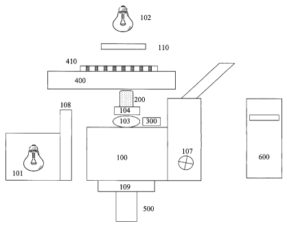
Turning now to the details of the drawings, figure 1 is a schematic block
diagram illustrating the optical, meehanical and electrical components of the
system of the
present invention. Inverted microscope stand 100 is equipped with fluorescence
epi-
illuminator 101 and tungsten halogen transilluminator 102. Mounted on
objective turret 103
is fast motor drive 104, preferably of the piezoelectric kind. Motor 104 moves
objective 200
in the Z-dimension (vertically) so as to reach the best focus position. The
best focus position
is defined by confocal autofocus device 300 as monitored by digital computer
600.
Microscope Z-focus drive 107 may also be used to move objective 200 in the Z-
dimension,
when software autofocus is selected. Filter changer 108 is positioned so as to
present filters
in the illumination path of illuminator 101, thereby selecting narrow band
excitation
illumination. Optionally, filter changer 109 may be mounted in the emission
path of
microscope 100, so as to select narrow band emission optics under computer
control. Shutter
110 transmits light from illuminator 102, under computer control. Motorized
stage 400
carries multiwell plate 410 so as to present each of the plurality of wells to
objective 200.
CCD camera 500 is mounted so as VO acquire images of cells in plate 410.
Digital computer
600 controls the components (filter changers 108/109, shutter 110, focus
components 104,
300, 107, stage 400, camera 500) and contains software to perform analyses.
The microscope 100 is, preferably, an inverted stand equipped with
epifluorescence optics and with a transmitted light illumination path. The
motorized and
computer-controlled stage 400 is mounted on the microscope, so as to move
specimen
CA 02485602 2004-11-10
WO 03/095986 PCT/1B03/01821
13
containers over the microscope optics. Preferably, the stage 400 is equipped
with a holder for
multi-well plates 410, and this holder is so constructed as to allow plate
insertion and removal
by standard laboratory robots such as the Twister 2 from Zymarc Industries.
Digital camera
500, preferably a cooled and low-noise CCD camera, is mounted on the
microscope so as to
acquire specimen images. System control and image storage are performed by
digital
computer 600.
TABLE 1
List of Denotations and Abbreviations
U(p) Grayscale intensity of the grayscale image U at the
location of pixel p
V Symbol of linear differential vector operator "nabla"
(Feynman 1964)
VU Gaussian gradient of image U (e.g. as explained in Jahne
1999, p.24.1)
AND, OR etc. Logical operations on binary images
A EXCP B Composite logical operation defined as A XOR (A AND B). This
operation has the meaning of exclusion from image A the common part
of images A and B
Mean[ U I A] Mean gray level value of the pixels within the subset A
of the image U
Std[ U I A] Standard deviation of the pixels within the subset A of
the image U
N(A) Number of elements in subset A (e.g. number of pixels within set of
pixels A)
CSS Cross-section size
MMS Minimal morphological size
NDF Nonlinear Diffusion Filtering
SGMD Scalar "Gradient Modulus"-driven Diffusion
AEED Anisotropic edge enhancing diffusion
ACED Anisotropic coherence enhancing diffusion
SPED Scalar peak enhancing diffusion
13 Diffusivity tensor
OHB Optimal Histogram Bipartition
SRG Seeded Region Growing
Figure 2 is a schematic block diagram illustrating the fast autofocus device.
Light emitted from a laser diode 100 passes through a transparent window 101',
so calculated
CA 02485602 2004-11-10
WO 03/095986 PCT/1B03/01821
14
as to compensate for aberrations introduced by beam splitter 103'. This
compensation is
arrived at by tilting the window to introduce compensating aberrations. Should
beamsplitter
103' be of a type that does not introduce aberrations (e.g. as in the case of
a very thin
beamsplitter), no correction from glass window 101' is required.
Leaving window 101', the laser beam then passes through an aperture 102'
which limits the width of the beam so that it later fills the back lens of
microscope objective
200. So as to operate with objectives with a back lens of 15-20 mm in
diameter, the aperture
is constructed with a diameter of 2.4 mm.
Beamsplitter 103' functions as a laser intensity limiting device. It is so
constructed as to reflect >95% of the incident laser beam toward the side onto
absorbing
surface 104'. Preferentially, this absorbance is of a high order (close to
100%) so as to
minimize retroreflections which could degrade measurement sensitivity by being
incident to
other components. The lateral reflection from beamsplitter 103' is so
calculated as to diverge
broadly as it proceeds towards absorbing surface 104' and there is minimal
intrusion of
focused reflections back towards detector 600'.
The system is designed so as to be efficient in the use of the remaining small
proportion of the laser beam. The low power of the laser beam and the
efficiency of the
device allows the autofocus to be certified within a relatively non-
restrictive category (Class
1). Were a larger proportion of the laser beam to be required for sensitive
operation, the
certification category would be more restrictive and both the cost and
complexity of the
device would be much greater.
Another light path is transmitted through beam splitter 103' so as to pass to
mirror 105', which is of high flatness (X/4) to maintain focus of the final
beam, and of high
reflectivity to maximize efficiency in the near infra-red and infra-red
wavelengths that the
laser emits. The mirror coating is of gold which has the property of
efficiently reflecting the
relevant wavelengths.
Light from mirror 105' is reflected to a positive lens 106 of such a focal
length
that it collimates the light and best fills the aperture of photodetector pin
hole 500'.
Preferably, lens 106' is diffraction limited with respect to the operating
wavelength X.
The collimated beam then passes to another mirror 107' which includes a filter
108'. An example of such a mirror is a high quality dichroic assembly with a
flatness of X/2,
and with the property of transmitting wavelengths below 750 nm, and reflecting
wavelengths
above 750 nm. Mirror 107' is tilted at such an angle that it most efficiently
reflects the
CA 02485602 2004-11-10
WO 03/095986 PCT/1B03/01821
desired wavelengths towards the back lens of objective 200. In a preferred
embodiment, the
back surface of mirror 107' is anti-reflection coated so as to minimize
unwanted reflections.
Light is transmitted through microscope objective 200 to the bottom surface of
a specimen container 300'. Objective 200 is moved in the vertical dimension
relative to
5
container 300', so as to sweep the laser beam through a detection volume which
is thick
enough to span a distance greater than the bottom surface of container 300'
and which
includes part of the contents of well 310.
Reflections from the interfaces between the transparent surfaces of container
300' and air (bottom surface 301) and fluid (inner surface 302) are collected
by objective 200
10 and
sent to filter/mirror 107'/108'. Mirror 107'/108' passes the laser
wavelength
preferentially and blocks other emissions from container 300' and specimen
medium 303.
The reflected light passes back through lens 106', mirror 105', and beam
splitter 103', which
directs part of the light back to photodetector 600'.
Photodetector 600' monitors the beam as objective 200 is moved to address
15
sample volume 310. The amount of light produced by specular reflection can be
calculated
as:
= (N_N)2/(N N)2
Where N is the index of reflection of a first medium through which light
passes, and N' is the index of reflection of a second medium through which
light passes. The
value of I is maximized when the refractive indices of N and N' are different.
Thus, a first
transition 303 from air to the bottom of specimen container 310 will generate
a larger
reflection than a transition 302 from the material of the specimen container
to a watery
contained fluid. A software algorithm in computer 600 monitors the shape of
the waveform
produced by the photodetector in real time, and locates transition 302.
In operation, the positional auto focus of the present invention transmits a
laser
beam through the microscope objective and into the specimen container 300'. A
rapid focus
drive, which can be a piezo actuator, moves the microscope objective 200 in
the z-plane
(depth) relative to the plate bottom 301, establishing a sampling volume. At
each point in the
sampling volume, a retroreflection is transmitted to the confocal
photodetector 600'. The
photodetector monitors the reflection intensities, converting them to voltages
which can be
transmitted to the digital computer. Software in the computer calculates a
best focus position
on the basis of intensity characteristics arising as the illumination beam
transits through
CA 02485602 2004-11-10
WO 03/095986 PCT/1B03/01821
16
surfaces of the specimen container. Components and construction of the device
are similar to
widely known embodiments of confocal optical paths (as disclosed in US
4,881,808, US
6,130,745, W092/15034, W095/22058, W098/44375, W000/37984). Some of these
systems also detect a focus plane corresponding to a substrate upon which
cells lie, and then
establish a cell focus at some fixed distance beyond the substrate.
It is a feature of the autofocus of the present invention that it integrates a
software autofocus algorithm so that it may be used with cells which lie at
positions that are
not fixed with respect to a surface of the container (e.g. within a range of 5-
15 um above).
The method involves these steps: a) use the best focus position achieved by
the positional
autofocus as a reference; b) move into the specimen container a fixed
distance; c) take a
number of images at intervals in the z-plane, and calculate a best focus from
these images
(Fig. 3). One skilled in the art will recognize that a software autofocus is
slow when used
alone, because it must take a large number of images. However, the use of the
present
hardware to come to a position defined by the specimen container, and then
initiating a
limited set of image acquisitions at a point referenced to that container
allows the system of
the present invention to function more rapidly than a software autofocus used
alone.
It is a feature of the system of the present invention that it can also be
used to
focus thick specimens. For example, transient expression of green fluorescent
protein (GFP)
in dopaminergic neurons has been observed following injection of dopamine
transporter
promoter-GFP constructs into one-cell embryos of the zebrafish. These embryos
are raised to
adulthood to establish homozygous stocks of transgenic fish. Then, embryos of
the transgenic
line can be studied in a screening mode, by placing the embryos in microwell
plates and
administering compounds. These embryos are thicker than the depth of focus of
a standard
microscope objective. The system of the present invention accommodates
specimens that
extend beyond a single plane of focus. The method involves these steps: a) use
the best focus
position achieved by the positional autofocus as a reference; b) move into the
specimen
container a fixed distance; c) acquire a set of images in the z-plane,
spanning a distance large
enough to encompass the specimen; d) combine the images into a single image
that best
shows the entire thickness of the specimen using known image combination
algorithms.
In another aspect, the same focus drive system can be used to create a stack
of
fluorescent Z-plane images from which a single best-focused image is
calculated, using
known methods for digital deconvolution. In this case, image deconvolution
using known
algorithms is substituted for image combination, as described above.
CA 02485602 2004-11-10
WO 03/095986 PCT/1B03/01821
17
Figure 3 is a flow chart, showing the general procedure for neurite analysis
as
further detailed in figures 4-9. Original image 110' is subjected to a set of
procedures which
include image preprocessing, binarization, seeded region growing,
morphological refinement,
cell and neurite classification, and demarcation mapping.
Figure 4 is a flow chart showing the image preprocessing procedures used
within the method for automated neurite analysis. Original image 110' is sent
to decision
point 111. If image 110' is fluorescently labeled it proceeds directly to
nonlinear suppression
114 (Process 1 --this process and all other numbered processes are described
below in further
details). If original image 110' is unlabeled, it is subjected to texture
transform 112 (Process
6) to create image 113, which is then subjected to nonlinear suppression 114
(Process 1).
Image 115 is output from suppression 114.
Image 115 is subjected to adaptive noise smoothing 116 (Process 2) and
output as preprocessed neurite image 117.
Figure 5a shows an unstained cell image, as imaged using differential
interference contrast microscopy, and an energy texture transform yields the
image in figure
5b, in which neurites are enhanced relative to other image components. It is
the object of this
figure to show that the energy texture transform of the present method yields
an image in
which neurites are more easily segmented by automated procedures.
Figure 6 is a flow chart illustrating the binarization procedure of the
neurite
analysis method. At 120, image 117 is input. At 121, preprocessed neurite
image 117 is
binarized by histogram bipartition (Process 4). Binary image 122 is output. At
123, image
122 serves as a seed for a SRG procedure (Process 5). Region image 124 is
output. At 125,
region image 124 is subjected to morphological image refinement (Process 7) to
remove
small holes and smooth boundaries. Binary neurite image 126 is output, as
shown in Fig. 7.
Figure 7a shows an original image (acquired using fluorescence microscopy),
and figure 7b shows a binary neurite image 126 in which both neurites and cell
bodies have
been binarized accurately by the present method. It is the object of this
figure to show that
the binarization process of the present method leads to accurate segmentation
of neurites and
cell bodies.
Figure 8 is a flow chart illustrating the cell and neurite classification
procedure
of the present method. At 127, image 126 is input. At 128, image 126 is sieved
by a multi-
criterion process (Process 13). Sieve 128 removes objects with shape and area
which are not
characteristic of neurites or cells. Sieve 128 outputs image 129 containing
both cells and
CA 02485602 2004-11-10
WO 03/095986 PCT/1B03/01821
18
neurites. At 130, image 129 is subjected to a morphological opening process.
Precursor
image 131 is output.
At 132, a sieve by size (Process 13) is applied to image 131. The output of
sieve 132 is binary cell image 133, which contains only objects which are
larger than a
minimal cell size.
At 134, precursor image 131 is logically excluded from cell and neurite image
129. This results in image 135 containing only neurites. At 136, image 135 is
sieved by a
multicriterion process including size, shape and proximity (Process 13), to
create binary
neurite image 137. In image 137, only objects with neurite shape and size and
which are
proximal to cell bodies (as demonstrated in image 133) are present.
Figure 9 is a flow chart illustrating the demarcation mapping procedure of the
present method. At 138, binary neurite image 137 is skeletonized to create
skeletonized
neurite image 139.
At 140, a tessellation ,procedure is applied to binary cell image 133 to
create
tessellated cell image 141 consisting of zones of influence of cell bodies
(see Fig. 10). These
zones of influence are geometrically defined areas around each cell, within
which neurites
can be assigned to cells of origin.
At 142, neurites and details of neurite geometry (end points, branch points,
attachment points and so forth) are determined in skeletonized neurite image
139. Using cell
image 133 and tessellated image 141, neurites and details of neurites may be
assigned to cells
of origins.
Figure 10 illustrates zones of influence within which neurites and details of
neurite geometry are assigned. "C" labels denote cell bodies. "N" labels
define neurite
skeletons. "Z" labels denote boundaries of influence zones. "d" labels denote
details of
neurite geometry. It is the object of this figure to show that the demarcation
mapping of the
present method is effective in both creating zones around each cell, and in
localizing the
origins of neurites and their geometric features. Within the zone of each
cell, the neurites and
their features that are shown may be related to the cell of origin for that
zone.
Figure 11, on the left, shows a flow chart for the granule segmentation of the
analysis of granular translocation assays. Original image 210 is subjected to
a set of
procedures which include image preprocessing, binarization, and
quantification.
Figure 11, on the right, shows a flow chart for the cytoplasm segmentation of
the analysis of granular translocation assays. Original image 200 is subjected
to a set of
CA 02485602 2004-11-10
WO 03/095986 PCT/1B03/01821
19
procedures which include image preprocessing, binarization, seeded region
growing,
morphological refinement, sieving and quantification.
Figure 12 is a flow chart illustrating the image preprocessing of cell body
segmentation of the method for analysis of granular translocation assays.
Original image 200
is subjected to nonlinear suppression 220 (Process 1). Output image 201 is
then sent to
decision point 221. If output image 201 is noisy, it is subjected to adaptive
noise smoothing
222 (Process 2) or nonlinear diffusion filtering 223 (Process 3) to produce
image 202.
Preferably, filtering 223 is achieved by iterations of SGMD and AEED
processing. If image
201 is not noisy, it proceeds directly to process 224. At 224, a decision is
made whether
image 201 or 202 should be subjected to background correction 225 (Process
12).
Preprocessed cell image 203 is produced.
Figure 13 is a flow chart illustrating the binarization, seeded region
growing,
morphological refinement and sieving procedures of the cell body segmentation
of the
method for analysis of granular translocation assays. At 225', preprocessed
cell mage 203 is
input. At 226, image 203 is binarized by OHB (Process 4) to yield binary seed
image 204.
At 227, image 204 is subjected to SRG (Process 5) to yield region image 205.
At 228, region
image 205 is subjected to morphological refinement (Process 7) and refined
precursor cell
image 206 is output. At 229, refined cell image 206 is subjected to a sieve by
size (Process
13) which generates binary cell image 207. Cell image 207 does not contain
objects smaller
than the minimal cell size.
Figure 14 is a flow chart illustrating the granular segmentation procedures of
the method for analysis of granular translocation assays. Binary cell image
207 is subjected
to nonlinear diffusion filtering 230 '(Process 3) to generate output image
208. Preferably,
diffusion filtering is by SPED processing. Enhanced intensity peaks in image
208 correspond
to vesicles and are detected as local maxima at 231, to generate binary
granule image 209.
Figure 15 is a flow chart illustrating the quantification procedures of the
method for analysis of granular translocation assays. At 232, binary granule
image 209 and
binary cell image 207 are used to locate cytoplasmic and vesicular (granules
within
cytoplasm) components in original image 200. From the located components of
image 200,
any form of intensity or spatially-based analysis may be conducted.
Preferably,
quantification by local contrast (Process 8) and/or distributional feature
analysis (Process 9)
and/or frequency domain analysis (Process 10, Fig. 16) is performed at
quantification 233.
Figure 16 illustrates a frequency domain analysis (Process 10) of
quantification 233, demonstrating discrimination of granular alterations in
treated cells.
CA 02485602 2004-11-10
WO 03/095986 PCT/1B03/01821
Differences in intracellular granular material are detected from the Fourier
spectra of cell
images. The energy spectrum of control cells is depicted by dots (lower
curve), while the
spectra of cells treated with three doses of a drug and containing granules of
increasing
quantity and size are depicted by circles, squares and crosses,
correspondingly. This figure
5
shows that biologically relevant effects may be discriminated by the spatial
domain analysis
of the present method.
Figure 17 is a flow chart illustrating the process for analysis of nuclear
translocation. Original image 300 is an image which best demonstrates the
nuclei as a
geometrical positioning aid. Original image 301 is an image which best shows
the labeled
10
molecule of interest, with fluorescence intensity corresponding to the local
concentration of
the labeled molecule. Preferably, differential visualization of nuclei and non-
nuclear cell
compartments in image 300 and image 301 is accomplished by different
conditions of
excitation and emission filtering on the microscope.
Image 300 (Figure 17 left) is subjected to a set of procedures which segment
15
nuclei. These procedures include image preprocessing, binarization, seeded
region growing,
morphological refinement, sieving and quantification.
Image 301 (Figure 17 right) is subjected to a set of procedures which segment
cytoplasm. These procedures include image preprocessing, binarization, seeded
region
growing, morphological refinement, sieving and quantification.
20
Figure 18 is a flow chart illustrating the preprocessing stage of the nuclear
segmentation used for analysis of nuclear translocation assays. Original image
300 is input at
330. At 331, image 300 is subjected to nonlinear suppression (Process 1) and
image 302 is
output. Image 302 is sent to decision point 332. If image 302 is noisy, it is
subjected to
adaptive noise smoothing 333 (Process 2) or nonlinear diffusion filtering 334
(Process 3).
Preferably, filtering 334 is achieved by iterations of SGMD and AEED
processing. Image
303 is output. Image 303 is sent to decision point 335. If background
correction is desirable,
image 303 is subjected to background correction 336 (Process 12). Preprocessed
nuclear
image 304 is produced.
Figure 19 shows the binarization, seeded region growing, morphological
refinement and sieving processes of the nuclear segmentation used for analysis
of nuclear
translocation assays. At 337, image 3,04 is input. At 338, image 304 is
subjected to a process
in which nuclear image pixels darker than the most probable pixel value are
set to the most
probable pixel value. Image 305 is output. At 339, image 305 is binarized by
OHB (Process
4) and image 306 is output. At 340, image 306 is subjected to SRG (Process 5)
to yield
CA 02485602 2004-11-10
WO 03/095986 PCT/1B03/01821
21
region image 307. At 341, region image 307 is used as a mask to define pixels
for a second
iteration of OHB (Process 4) performed on image 305. Binary image 308 is
output, and
provides a more precise definition of nuclear boundaries than does region
image 307. At 342
image 308 is subjected to morphological refinement (Process 7). Image 309 is
output. At
343, image 309 is sieved (Process 13). Sieve 343 removes objects smaller than
a minimum
nuclear size, said objects being confusable with nuclei if not removed. Binary
nuclear image
310 is output.
Figure 20 is a flow chart illustrating the preprocessing of the cytoplasmic
segmentation used for analysis of nuclear translocation assays. At 344,
cytoplasmic image
301 is input. Image 301 is subjected to nonlinear suppression at 345 (Process
1). Image 311
is output. Image 311 is sent to decision point 346. If image 311 is noisy, it
is subjected to
adaptive noise smoothing 347 (Process 2) or nonlinear diffusion filtering 348
(Process 3).
Preferably, filtering 348 is achieved by iterations of SGMD and AEED
processing. Image
312 is output. Image 312 is sent to decision point 349. If background
correction is desirable,
image 312 is subjected to background correction 350 (Process 12). Preprocessed
cytoplasmic
image 313 is produced.
Figure 21 is a flow chart illustrating the binarization, seeded region
growing,
morphological refinement and sieving processes of the cytoplasmic segmentation
used in the
method for analysis of nuclear translocation assays. At 351, image 313 is
input. At 352,
image 313 is subjected to a process in which nuclear image pixels darker than
the most
probable pixel value are set to the most probable pixel value. Image 314 is
output. At 353,
image 314 is binarized by OHB (Process 4) and seed image 315 is output. At
354, image 315
is subjected to SRG (Process 5) to yield region image 316. At 355, region
image 316 is used
as a mask to define pixels for a second iteration of OHB (Process 4) performed
on image 314.
Binary image 317 is output, and provides a more precise definition of nuclear
boundaries (for
nuclear exclusion) than does region image 316. At 356 image 317 is subjected
to
morphological refinement (Process 7). Image 318 is output. At 357, image 318
is sieved
(Process 13). Sieve 357 removes objects smaller than a minimum cell size, said
objects being
confusable with cells if not removed. Binary cytoplasm image 319 is output.
Figure 22 is a flow chart illustrating the quantification procedure used in
the
method for analysis of nuclear translocation assays. In one aspect,
quantification uses
segmented nuclei as an origin. Intensity data are then read from original
cytoplasm image
301, at fixed locations defined by proximity to nuclei (e.g. a collar starting
at 2 pixels from
the nucleus and extending to 6 pixels from the nucleus).
CA 02485602 2004-11-10
WO 03/095986 PCT/1B03/01821
22
At 358 binary nuclear image 310 is input. Preferably, at 359, image 310 is
subjected to a morphological dilation operation (as disclosed in Russ 1999, p.
460 and Parker
1997, p. 68) to generate dilated binary nuclear image 320. Preferably, the
dilation is
performed with a circular structural element (as disclosed in Parker 1997, p.
73). Image 320
is composed of both the nuclear component of binary nuclear image 310, and a
pen-nuclear
component created by the dilation process.
At 360, image 310 is excluded from image 320 to leave image 321, containing
just the pen-nuclear component.
At 361, image 310 sefves as a mask for identifying nuclear pixels in cytoplasm
image 301, and image 321 serves as a mask for identifying pen-nuclear pixels
in cytoplasm
image 301.
Preferably, at 362, translocation is quantified from a ratio of pen-nuclear
label
intensity and nuclear label intensity (Process 8). In another preferable
aspect, at 363,
quantification includes distributional feature analysis (Process 9) of ratios
362.
In another aspect, at 364, binary cytoplasm image 319 is used to identify
cytoplasmic pixels in cytoplasm image 301, and cytoplasmic pixel intensities
are calculated
from these identified pixels.
At 364, binary nuclear image 310 serves as a mask for
identifying nuclear regions within cytoplasmic image 301, and nuclear pixel
intensities are
calculated from these identified pixels. Preferably, at 365, translocation is
quantified from a
ratio of cytoplasmic label intensity inside the nucleus and in an area that
includes as much as
possible of the cytoplasm of that cell (Process 8). In another aspect,
quantification can
include distributional feature analysis 366 (Process 9) of ratios 365.
CA 02485602 2004-11-10
WO 03/095986 PCT/1B03/01821
23
Figure 23 is a flow chart illustrating the analysis of ruffle translocation.
Original image 400 is an image which best demonstrates nuclei as a geometrical
positioning
aid. Original image 401 is an image which best shows the labeled molecule of
interest, with
fluorescence intensity corresponding to the local concentration of the labeled
molecule.
Preferably, differential visualization of nuclei and non-nuclear cell
compartments in image
400 and image 401 is accomplished by different conditions of excitation and
emission
filtering on the microscope.
Image 400 (Figure 23 left) is subjected to a set of procedures which segment
nuclei. These procedures include image preprocessing, binarization, seeded
region growing,
morphological refinement and sieving.
Image 401 (Figure 23 right) is subjected to a set of procedures which segment
cytoplasm including ruffles. These procedures include image preprocessing,
binarization,
seeded region growing, morphological refinement and sieving.
Figure 24 is a flow chart illustrating the preprocessing stage of the nuclear
segmentation used in the method for analysis of ruffle translocation assays.
Original image
400 is input at 430. At 431, image 400 is subjected to nonlinear suppression
(Process 1) and
image 402 is output. Image 402 is sent to decision point 432. If image 402 is
noisy, it is
subjected to adaptive noise smoothing 433 (Process 2) or nonlinear diffusion
filtering 434
(Process 3). Preferably, filtering 434 is achieved by iterations of SGMD and
AEED
processing. Image 403 is output. Image 403 is sent to decision point 435. If
background
correction is desirable, image 403 is subjected to background correction 436
(Process 12).
Preprocessed nuclear image 404 is produced.
Figure 25 is a flow chart illustrating the binarization, seeded region
growing,
morphological refinement and sieving processes of the nuclear segmentation
used in the
method for analysis of ruffle translocation. At 437, image 404 is input. At
438, image 404 is
subjected to a process in which nuclear image pixels darker than the most
probable pixel
value are set to the most probable pixel value. Image 405 is output. At 439,
image 405 is
binarized by OHB (Process 4) and image 406 is output. At 440, image 406 is
subjected to
SRG (Process 5) to yield region image 407. At 441, region image 407 is used as
a mask to
define pixels for a second iteration of OHB (Process 4) performed on image
405. Binary
image 408 is output, and provides a more precise definition of nuclear
boundaries than does
region image 407. At 442 image 408 is subjected to morphological refinement
(Process 7).
Image 409 is output. At 443, image 409 is sieved (Process 13). Sieve 443
removes objects
CA 02485602 2004-11-10
WO 03/095986 PCT/1B03/01821
24
smaller than a minimum nuclear size, said objects being confusable with nuclei
if not
removed. Binary nuclear image 410 is output.
Figure 26 is a flow chart illustrating the preprocessing stage of the
cytoplasmic
segmentation used for analysis of ruffle translocation assays. At 444,
cytoplasmic image 401
is input. At 445, image 401 is subjected to background correction (Process 12)
which has the
additional advantageous property that it emphasizes details of a size
characteristic of ruffles.
Image 411 is output. Image 411 is subjected to nonlinear diffusion filtering
at 446 (Process
3).
Preferably, filtering 446 is achieved by iterations of SGMD and AEED
processing.
Preprocessed ruffle image 412 is produced.
Figure 27 is a flow chart illustrating the binarization, seeded region
growing,
morphological refinement and sieving processes of the ruffle segmentation used
in the
method for analysis of ruffle translocation assays. At 447, preprocessed
ruffle image 412 is
input. At 448, image 412 is subjected to a process in which ruffle pixels
darker than the most
probable pixel value are set to the most probable pixel value. Image 413 is
output. At 449,
image 413 is binarized by OHB (Process 4) and image 414 is output. At 450,
image 414 is
subjected to SRG (Process 5) to yield region image 415. At 451, region image
415 is used as
a mask to define pixels for a second iteration of OHB (Process 4) performed on
image 413.
Binary image 416 is output, and provides a more precise definition of ruffles
than does region
image 415. At 452 image 416 is subjected to morphological refinement (Process
7). Image
417 is output. At 453, image 417 is sieved (Process 13) by size to remove
objects confusable
with ruffles. Binary ruffle image 418 is output.
Preferably, at 454 binary nuclear image 410 is logically excluded from binary
ruffle image 418 to create refined binary ruffle image 419 in which ruffles
cannot be
localized over nuclei.
Figure 28 is a flow chart illustrating the quantification procedure used for
analysis of ruffle translocation assays. At 455, binary ruffle image 418 or
refined binary
ruffle image 419 serves as a mask for calculation of ruffle intensity from
cytoplasmic image
401. Preferably, at 456, quantification is achieved by local contrast (Process
8). In another
aspect, quantification can include distributional feature analysis 457
(Process 9) based upon
ruffle size and proximity.
CA 02485602 2004-11-10
WO 03/095986 PCT/1B03/01821
Functions Used in the Methods of the Present Invention
The algorithmic steps of the methods are so devised as to best suit the
characteristics of commonly used cell assays. The methods are constructed for
each specific
assay by integrating functions from the library described, below. While the
general nature of
5 the functions used in the methods of the present invention are given
below, it is to be
understood that any of these functions may be parameterized to optimally
enhance, select, or
otherwise affect features in images.
10 Process 1) Nonlinear suppression of high intensity peaks
Artifacts arising from high intensity peaks can introduce undesirable
variability of feature gray level statistics, and perturb adaptive threshold
and region growing
procedures. The peak suppression method is a variant of the known technique of
histogram
correction. As implemented in the present invention, the process takes the gay
level
15 reference image as input, and applies nonlinear suppression to the
pixels with highest gray
level values and an identity transform to the pixels within the rest of
dynamic range. The
output image exhibits a reduction in intensity variation over brighter
objects, but not over less
bright objects. This has the advantage that it improves the performance of
subsequent image
processing as described below.
Process 2) Adaptive noise smoothing
An adaptive noise smoothing procedure can be beneficial in improving feature
detectability (and obvious to one skilled, e.g. as disclosed in Morrison et
al., 1995). In a
preferred aspect of the invention, a procedure is used which increases the
image signal-to-
noise ratio without compromising fine feature details. Original and Gaussian-
smoothed
images (U and Ua, respectively) are combined as shown in expression 2.1:
R = W=U + (1 ¨ W).1Ja, (2.1)
where (R) is the result image and W = W(FaUl) is a weight function dependant
upon the
modulus of the Gaussian gradient FaUl of the original image, and a is the
standard deviation
of the Gaussian function used for smoothing.
Use of weight function W has the advantage that the pixels in result image R
display values close to those of original image U in areas of high gradient
magnitudes and to
CA 02485602 2004-11-10
WO 03/095986 PCT/1B03/01821
26
those of smoothed image II, in areas with low gradient magnitudes. The areas
of low
gradient magnitude tend to contain a greater proportion of the image noise
which is thereby
reduced in relative amplitude.
Adaptive noise smoothing has the additional desirable property that noise in
Process 3) Adaptive Noise Smoothing And Feature Enhancement By Nonlinear
Nonlinear diffusion filtering (NDF) methods are members of the family of
scale space techniques for image filtering. NDF methods (e.g. as disclosed in
Weickert 1997)
are useful where it is desirable to remove noise (defined as spatial
modulations of high
frequency) and preserve features with lower spatial frequency.
The present invention applies NDF methods to remove image noise, while
relevant image features are enhanced in a fashion dependent upon their shape
and size. An
image is processed by iterative application of a nonlinear diffusion operator.
The exact
nature of the NDF operator is varied according to the desired feature
characteristics, and a
U(+1) = () dU
a
dU = V (An) (0
V U )dt = = = -- = u = at
(n)
a bYx(n)bYY (n) ax ay =(ft)
ay
where U=U(x,y) is the coordinate-dependent image intensity, An) is diffusivity
tensor (with
In preferred aspects, the present invention incorporates one or more of three
known methods for NDF, as disclosed in Weickert 1997.
30 = Scalar "Gradient Modulus"-driven Diffusion (SGMD)
CA 02485602 2004-11-10
WO 03/095986 PCT/1B03/01821
27
= Anisotropic Edge Enhancing Diffusion (AEED)
= Anisotropic Coherence Enhancing Diffusion (ACED)
The input image is the gray scale reference image. 3.1 is applied with a
diffusivity tensor as specified in SGMD, AEED, or ACED. This process may be
iterated any
number of times. The selection of SGMD, AEED, or ACED is performed on the
basis of the
morphology of the features being accentuated or suppressed.
In a preferred aspect, SGMD and/or AEED are used with features in which
edge preservation is important. ACED is used with fiber-like details. If both
fiber and edge
preservation are required, all three methods may be used.
If isolated intensity peaks must be preserved, the present invention applies
an
additional transformation which we term Scalar Peak Enhancing Diffusion
(SPED).
It is an advantage of the SPED process of the present invention that NDF may
be optimized for isolated intensity peaks, for example those associated with
granular material
inside of cells. In performing a SPED iteration, the output of a SGMD
iteration is convolved
with a peak-shaped mask, in which pixel gray level values decay exponentially
with distance
from the mask center. The size of mask is pre-set to match the characteristic
size of the peak-
like image details which are to be accentuated. This procedure is iterated for
some pre-set
number of iterations and emphasizes sharp intensity peaks while suppressing
noise.
Process 4) Thresholding by optimal histogram bipartition
Preferably, the invention applies an optimal histogram bipartition (OHB) step
for segmentation. It is a feature of the OHB method that it accommodates the
broad dynamic
range present in biological images.
The input of the OHB procedure is a grayscale image, optionally processed
using steps 1-3 above. The output i a binary image, in which segmented pixels
correspond
to cellular features of interest.
Various OHB methods are known (e.g. Parker 1997, Paulus 1995) and there is
a potential for bias in threshold selection arising from use of one or another
of the OHB
methods. Therefore, it is an advantage of the present invention that it
calculates a threshold
using some property (e.g. the mean of all four, the mean of the middle two
values sorted in
ascending order, the smallest or largest) of several thresholds calculated by
multiple OHB
methods. This statistical threshold value is less likely to suffer from bias
introduced by any
one of the OHB methods.
CA 02485602 2004-11-10
WO 03/095986 PCT/1B03/01821
28
In a preferred aspect, four OHB methods are used to generate a threshold
value:
= The gray level value maximizing the entropy measure for binarization
(Paulus 1995,
pp. 278-281).
= The gray level value maximizing the mean square separation measure for
binarization
(Paulus 1995, pp. 278-281).
= The gray level value minimizing the Shannon measure of the image taken as
a fuzzy
set (Parker 1997, p.125).
= The gray level value minimizing the Yager measure of the image taken as a
fuzzy set
(Parker 1997, p.125).
Process 5) Seeded region growing
The input to region growing is a grayscale reference image, and a binary
image, which is created from the reference image by a process such as is
described in Step 4.
It is a disadvantage of the initial binary image that the binary pixels which
represent features
of interest in the reference image do not correspond exactly to those features
in the reference
image. Therefore, region growing is used so that a final binary image can
better represent
features in the reference image. It is an aspect of the present invention that
a seeded region
growing method uses the initial binary image as its seed image. A tunable
iterative procedure
(e.g. as described in Russ, 1991, pp.87-89) is then used to add binary pixels
to regions.
Tuning is defined as using the statistical properties of the growing objects,
their vicinities and
the background to select candidate pixels, with one embodiment shown in
equation 5.1. The
statistical parameters are recalculated iteratively, and the procedure is
continued until optimal
assignment of pixels to regions of interest is obtained.
TN= max( Mean[UIBN] + kB=Std[UIBN] , Mean[131Bckid + kBek=Std[UlBckN]) ),
(5.1)
where TN is the threshold used for the current iteration, Mean[UIBN] and
Std[UIBN] are mean
and standard deviation calculated by the ensemble of boundary pixels,
Mean[UlBckN] and
Std[UlBckN] are mean and standard deviation calculated by the ensemble of
background
pixels (i.e. pixels not included in the object ON or in the boundary BN), and
kB and kBck are
controlling coefficients with values close to unity.
At the N-th iteration of the region growing process, a candidate boundary
pixel
p (pcBN), adjacent to the growing Set of pixels ON (at first iteration, 01
coincides with the
CA 02485602 2004-11-10
WO 03/095986 PCT/1B03/01821
29
seed image) is included in the growing set of pixels ON+j for the next
iteration, if and only if
the corresponding gray value U(p) on the reference image U exceeds the
threshold value of
TN. This threshold is calculated from global statistics of the image U as in
Eq. X.
The iterative process continues until there is no candidate pixel (as defined
by
U(p) > TN) adjacent to the growing set of pixels.
Process 6) Texture transform
It is an advantage of the invention that segmentation and analysis of
unstained
or vitally stained specimens is possible. Such specimens are acquired using
differential
interference contrast (DIC), brightfield, or other forms of nonfluorescence
microscopy.
These methods are most useful in imaging living cells which are intolerant of
fluorescence or
other staining procedures.
In a key aspect, the present invention localizes intensity undulations of
defined
textural types, to enhance the detectability of features. The texture
transform procedures are
based upon gray level co-occurrence statistics (e.g. as disclosed in Parker
1997, p. 155).
These procedures take as their input gray level reference images and create as
their output
gray level processed images in which features of appropriate texture are
brighter than other
features (are enhanced). Said enhanced features can then be segmented using
procedures
similar to those used for fluorescent images. Thus, it is a key advantage of
the texture
transform that a similar set of segmentation procedures may be used to analyze
fluorescent
and nonfluorescent materials.
In a preferred aspect, an "energy" texture transform (as described in Parker
1997, p. 160) is used. This transform is parameterized by the value of minimal
morphological
scale (MMS) of the specimen. The MMS is user-defined as a minimal size for
meaningful
image detail.
While texture transforms are preferred methods for enhancing nonfluorescent
images prior to segmentation, it is to be appreciated that other transforms
could be used. The
key aspect is an enhancement in which an intensity increase in the output
image is dependent
upon structural characteristics of features in a reference image.
Process 7) Morphological refinement of detected features
Fine projections, various sizes of holes or other discontinuities in feature
boundaries can cause an undesirable variability in segmented shapes. In turn,
this could lead
to degraded performance of quantification algorithms. For example,
skeletonization
CA 02485602 2004-11-10
WO 03/095986 PCT/1B03/01821
algorithms function poorly with jagged object edges. It is a feature of the
present invention
that morphological smoothing and sieve-by-size controlled hole filling are
used prior to
quantification. The value of MMS serves as a threshold size for a smoothing
procedure. In a
preferred aspect, this procedure removes all image details of size less than
the MMS, thereby
5 removing roughness.
Process 8) Quantification by local contrast
Generally, features are defined by their intensity relative to the intensity
of
surrounding cellular material. The local contrast between a feature and its
local surround is
10 defined in eq. 8.1.
Local contrast = Mean [U I Feature] / Mean [U I Feature surround] (8.1)
The contrast value may be calculated directly from the reference image, or
from locations
15 defined on a processed image and transferred to a reference image.
Process 9) Distributional feature analyses
The distribution of a feature upon some measured characteristic can reflect
underlying biology. It is common to see frequency histograms of feature size
or intensity
20 used to reflect underlying biology.
The present invention uses mixed feature distributions as indices of changes
in
a cell sample. The feature distribution is modeled by a probability density
distribution
function (PDDF). Then, hypotheses are tested against some predetermined model
of what the
frequency distribution should be. A unimodal distribution would result if, for
example, cell
25 granules were distributed about a single characteristic size. A bimodal
distribution would
result if cell granules are so altered by treatment that a population of
larger or smaller
granules appears (as with the Transfluor assay from Norak). In this case, a
judgment that a
particular treatment is effective may be made on the basis of extent to which
an observed
PDDF is bimodal.
30 In the specific case of a bimodal distribution of feature x, the
mixed PDDF
13,,u(x) is expressed in terms of discrete PDDFs of its two components as
shown in
expression 9.1:
Pn.(x) all(x)+ (1¨ a)P2(x), 0 a <1 (9.1)
CA 02485602 2004-11-10
WO 03/095986 PCT/1B03/01821
31
where both partial PDDFs Pi(x), P2(x) have finite averages and dispersions pi
and o (i=1,2).
In the bimodal representation of the mixed PDDF (9.1), a is a weighting
parameter for a
bimodal model. The two weighting factors a and (1-a) reflect the relative
amounts of
contribution of the partial PDDFs Pi(x), P2(x) to the mixed PDDF P,nix(x).
The mean and the dispersion of the mixed PDDF shown in (9.1) are:
,uõ,, = + (1- a),u2 (9.2a)
crmix2 acr12
+ (1 - a)Cr2 2 + a (1 - -u2) (9.2b)
where ,umix, and andx are the mean and standard deviation of the mixed sample,
respectively.
An experimental estimate (Z. of the weighting parameter a may be calculated
from a sample
according to expressions (9.2a) and/or (9.2b), as shown in 9.3a and 9.3b.
a =17"1"- 172 (9.3a)
- T/2
(1 - B -C) V(1- B - C)2 - 4(A -C)
&-1,2 = ______________________ ,where
2
2
m1r
A= (9.3b)
-P2)2
2
a,
B = ________
(Pi - 112)2
2
a2
c=
(Pi¨ P2)2
Where estimates ,u, õu2 , ,o-2 õuõõ1õ5--õ,ix are means and standard deviations
of the partial
samples and mixed sample. To define the partial samples, the mixed sample must
be split.
This is achieved by a threshold bipartition operation. The bipartition
threshold t may be
defined by any known method (e.g. the OHB method of Process 4).
In a preferred aspect, separation of the samples is expressed as a normalized
distance between the means of the two populations, calculated as in expression
9.4.
SS = j_22
(9.4)
where SS is sample separation.
In another preferred aspect, the proportion of the mixed distribution
contributed by each partial distribution is Ei as shown above.
SS and EZ are preferred parameters for distribution feature analysis of the
present method.
CA 02485602 2004-11-10
WO 03/095986 PCT/1B03/01821
32
10) Frequency domain detection of granular details
Granular structures (e.g. vesicles) within the cell body can increase or
decrease in size and intensity in ways that reflect biology. Therefore, it is
a feature of the
present invention that granular structure analyses may be made by analyzing
the image
energy spectrum. The energy spectrum is described by an analytical expression
which
evaluates both granular and nongranular features.
The general form of an energy spectrum is shown in eq. 10.1.
E6c9 = <F60, OF* 69, VP to (10.1)
where E(p) is the energy spectrum, F(p, ilt) is Fourier transform of the
original image
expressed in polar coordinates, <...> v, denotes averaging by an angular
coordinate, and p and
1/1 are radial and angular coordinates in Fourier space, correspondingly.
Using known methods (e.g. Granlund et al., 1995), granules are treated as a
set
of scattered intensity peaks of approximately the same width. In a preferred
aspect, the
intensity profile of a granule is modeled by a Gaussian function (Eq. 10.2)
F¨Fo 12
,4(i) oc f (a)e 2 ,
(10.2)
=
where a is the effective average radius of a granule, Fo is granule's
location. f(a) is a
proportionality multiplier which relates the granule's brightness to its size
(f(a)-.a3). With
bright granules (e.g. fluorescence), proportionality multiplier f(a) improves
size
measurements because a granule's brightness is proportional to its volume.
The energy spectrum of granules of the same size is defined as the square of
modulus of Fourier transform of the Gaussian function (Eq. 10.3):
f\,,,-(apY
-'-'granules õ
It is known (Granlund et al., 1995), that nongranular features yield power
terms in an energy
spectrum as shown in Eq. 10.4:
Enongranular(P) 13-3 (10.4)
A model expression (Eq. 10.5) for the energy spectrum is therefore taken in
the form of
weighted sum of contributions of the two main components ¨ nongranules and
granules:
CA 02485602 2004-11-10
WO 03/095986 PCT/1B03/01821
33
E(p) = Enongranulars (p) + Egranules (0) = A1p-3 + A2 f (a)e-(aPY , (10.5)
where AI, A2 are> O.
The discrimination between biological conditions is made on the basis of the
two fitted parameters (obtained from Eq. 10.5) ¨ a (an estimated mean granule
radius) and
ratio (A2 / Ad, which reflects the contribution of the granular component to
the power
spectrum.
The analysis proceeds through energy spectrum construction and then
quantification.
Energy spectrum construction
The Fourier spectrum of granules is produced by known methods (as
described in Press 1992, p.689). This spectrum is then reduced to the discrete
one-
dimensional frequency dependence after averaging by an angle coordinate, and
discretization
of radial distance in Fourier space. = This procedure implements conversion
(10.1), defined
for the discrete set of values of radial distance pi (j=1,...Np), where Np is
the number of
discrete values of radial distance. As a result of this operation, the average
spectrum intensity
<E>1 is calculated for each value of pi, producing the discrete representation
of spectrum
{pj ,<E>j}.
Quantification (spectrum fitting)
Known methods of nonlinear fitting (Press 1992, p.683, p.408 ) are used to
obtain three fitting parameters from the energy spectrum ¨ a (effective
average radius of
granule) and amplitudes A2 and Al from model expression (10.5). In a preferred
aspect, the
values of a and ratio (A2 /A1) are used for image quantification.
11) Demarcation mapping
Demarcation mapping is a procedure used to perform geometric analyses on
segmented images. The present invention uses demarcation mapping to localize
geometric
areas around neurite origins (Fig. 10). Most typically, each cell has a
demarcated region
around it, output by the demarcation mapping process.
As one aspect of demarcation mapping, a segmented neurite image (as output
from processes described below) is skeletonized (e.g. as disclosed in Russ
1991, pp. 483-
485). In the skeletonized image, neurites, neurite end points, neurite branch
points, and the
CA 02485602 2004-11-10
WO 03/095986 PCT/1B03/01821
34
cells of origin for each neurite on a corresponding cell image (attachment
points) may be
found.
= A pixel of a binary skeleton is considered to be a branch point if and
only if there are
more than two non-zero pixels in its 3x3 neighborhood.
= A pixel of a binary skeleton is considered to be a endpoint if and only
if there is only one
non-zero pixel in it's 3x3 neighborhood.
= A pixel of a binary skeleton is assumed to be an attachment point of
neurite to cell if it is
an end point and is proximal to the cell.
= A neurite is considered as originating in a specific cell body if that
neurite lies within the
demarcated region of a cell in the corresponding cell tessellation image.
As a second aspect of demarcation mapping, a cell tessellation image is
created. Tessellation is the result of unconditional region growing or binary
dilation of any
segmented targets which serve as seeds (Parker 1997, p.69). In the present
case, the targets
are most typically cell bodies.
Therefore, demarcation mapping has two input images. A segmented neurite
image is input to skeletonization. A segmented cell image is input to
tessellation. A
skeletonized neurite image and a tessellated cell image are intermediate
outputs. The final
outputs are measurements of neurite geometry, taken from the skeletonized
image, and
localization of neurite origins to specific cells, taken from the tessellated
cell image.
12) Background correction
Background correction removes spatial nonuniformity in illumination or
emission intensity from an original image. The preferred method is to process
an image to
create a highly smoothed image in which specimen detail is absent but low
frequency
background components remain. The highly smoothed image is subtracted from or
divided
into the original image.
Various procedures for smoothing images will be apparent to one skilled. For
example, Gaussian smoothing, grayscale opening, pair-wise filtering (opening
followed by
closing or closing followed by opening), or alternating sequential filtering
(Jahne 1999,
p.627-680) have all been used in this type of operation.
It is to be appreciated that a smoothing operation or other method of
background correction may also be used to optimally select features of a given
size, while de-
emphasizing features which are bigger.
CA 02485602 2004-11-10
WO 03/095986 PCT/1B03/01821
13) Sieving
Sieving is a process by which a binary image is filtered to remove segmented
targets which have geometry that does not correspond to features of interest.
For example,
images are sieved by size and only features which fall within a specified size
range are left in
5 the sieved image. Many other types of sieve depend upon geometric
properties of features.
For example, images could be sieved by shape descriptors (as disclosed in Russ
1999, p. 553-
555). It is a feature of the present invention that sieving is applied using
single (e.g. size) or
multiple criteria. As an example of multi-criteria sieves, the method of the
present invention
sieves two images according to different criteria (e.g. round in the first
image and elongated
10 in the second), and then performs a further pairwise sieving step. In
pairwise sieving, only
those features which meet another criterion (e.g. elongated objects proximal
to round objects)
are retained.
Method for Neurite Assays
15 Neurite material is structurally complex and images contain many
potentially
confusable features. It is a feature of the present method that it performs
automated and
accurate detection of neurites within a broad variety of specimens, including
fluorescently
labeled and unlabeled specimens.
In one aspect, the method uses an energy texture transform to improve
20 subsequent segmentation in unstained images.
In another aspect, the method improves detectability of neurites and cell
bodies by employing processes of nonlinear diffusion filtering, optimal
histogram bipartition,
seeded region growing, sieving, and morphological image refinement.
In another aspect, the method demarcates zones of influence for cell bodies,
25 using a tessellation procedure. From these zones, neurite structures may
be related to their
cell bodies of origin. It is a feature of the present invention that a broad
variety of neurite
structures may be identified and related to cell bodies of origin.
Details of procedures for neurite analysis are best shown in Figures 3-10.
30 Method for Granular Translocation Assays
The present invention performs analyses of granular material as commonly
observed in nuclear translocation assays such as the Transfluor assay from
Norak Inc. In
these assays, cytoplasmic granules of pre-defined size must be segmented and
analyzed,
while granular artifacts outside cytoplasm must be ignored. It is a feature of
the present
CA 02485602 2004-11-10
WO 03/095986 PCT/1B03/01821
36
method that it detects even weakly labeled cytoplasmic material within which
granules may
then be localized.
In one aspect, the method improves detectability of granules and cytoplasm by
employing processes of nonlinear suppression of high intensity peaks,
nonlinear diffusion
filtering or adaptive noise smoothing, optimal histogram bipartition, seeded
region growing,
and morphological image refinement.
In a preferred aspect the method uses distributional feature analysis to
report
alterations in granular intensity or geometric properties.
Details of procedures for granular translocation assays are shown in Figures.
11-17.
Method for Nuclear Translocation Assays
Nuclear translocation' is commonly quantified by a change in the relative
intensity of fluorescent label contained in nuclei and cytoplasm. Typically,
two images are
acquired. One image best demonstrates the nuclei as a geometrical positioning
aid and/or to
show viability or other cell functional aspects. A second image best shows
cytoplasm, with
fluorescence intensity corresponding to the local concentration of the labeled
molecule of
interest.
In one aspect of the present invention, translocation is quantified from cell
images processed to best show nuclear and cytoplasmic areas for making
measurements.
Preferably, processing to show nuclei includes nonlinear suppression of high
intensity peaks,
noise suppression by nonlinear diffusion filtering, background correction,
optimal histogram
bipartition, and morphological refinement. Preferably, processing to show
cytoplasm
includes nonlinear suppression of high intensity peaks, noise suppression by
adaptive noise
smoothing or nonlinear diffusion filtering, background correction, optimal
histogram
bipartition, and morphological refinement.
In one aspectõ distributional feature analysis may be used to quantify
translocation. In this case, the relative contributions of darker and brighter
nuclei and/or
cytoplasm may be distinguished from a bimodal character of the nuclear or
cytoplasmic
intensity histograms.
Any of the intensity parameters calculated from the intensity quantification
process may be subjected to distributional analyses. For example, the nuclear-
cytoplasmic
ratio, the nuclear intensity, and the cytoplasmic intensity may all be used.
CA 02485602 2011-09-27
31798-3
37
The method for analysis of nuclear translocation assays is shown in
Figures 18-23.
Method for Membrane Ruffling Assays
Some translocation events are characterized by a regionalized
distribution of label within non-punctuated regions of cytoplasm, which are
morphologically distinct or ridge-shaped elaborations, here referred to as
"ruffles".
Ruffles are defined as intensity-discriminated features of a specified cross-
sectional
size. The method is similar to that used for nuclear translocation assays,
with
detailed refinements to better detect ruffle objects. It is a feature of the
functions of
the present invention that they are integrated into a method that provides
automated
discrimination of membrane ruffles (Figs. 24-28).
Although preferred embodiments of the invention have been disclosed
for illustrative purposes, those skilled in the art will appreciate that many
additions,
modifications and substitutions are possible, and the claims are not to be
limited by
the preferred or exemplified embodiments.
CA 02485602 2004-11-10
WO 03/095986 PCT/1B03/01821
38
Materials Cited
Anderson, C.M., Georgiou, G.N., Morrison, I.E.G., Stevenson, G.W. and Cherry,
R.J.
Tracking of cell surface receptors by fluorescence digital imaging microscopy
using a
charge-coupled device camera, Journal of Cell Science 101:415-425 (1992).
Benveniste, M., Schlessinger, J. and Kam, Z. Characterization of
internalization and
endosome formation of epidermal growth factor in transfected NI1H-3T3 cells by
computerized image-intensifed three-dimensional fluorescence microscopy, The
Journal of Cell Biology 109:2105-2115 (1989).
Chassery, J.M. and Garbay, C. An interative segmentation method based on a
contextual
color and shape criterion, IEEE PAMI 6:794 (1984).
Conway, B.R., Minor, L.K., Xu, J.Z., Gunnet, J.W., DeBiasio, R., D'Andrea,
M.R., Rubin,
R., DeBiasio, R., Giuliano, K., Zhou, L. and Demarest, K.T. Quantification of
G-
protein coupled receptor internalization using G-protein coupled receptor-
green
fluorescent protein conjugates with the ArrayScan high-content screening
system.
Journal of Biomolecular Screening 4:75-86 (1999).
Crissman, J.D., Visscher, D.W. and Kubus, J. Image cytophotometric DNA
analysis of
atypical hyperplasias and intraductal carcinomas of the breast, Archives of
Pathology
and Laboratory Medicine 114:1249-1253 (1990).
Dawes, E. A. Quantitative Problems in Biochemistry, Baltimore: Williams and
Wilkins,
pp.293-311, 1972.
De Le Torre, C. and Navarrete, M. H. Experimental Cell Research 88: 171-174,
1974.
Deligdisch et al., Cancer 72:3253-3257, 1993.
Feynman R. et al, The Feynman Lectures on Physics, Addison-Wesley Publishing
Co., Inc.,
Reading, Massachusetts, Palo Alto, London, 1964, p. 35.
Freshney, R. I. Quantitation and Experimental Design, in Culture of Animal
Cells, a Manual
of Basic Technique, 2nd ed., New York: Alan R. Liss, pp. 227-256, 1987.
Fu K. S. and Mui, J. K. A Survey on Image Segmentation, Pattern Recognition
13:3-16
(1981).
Galbraith, W. Wagner, M.C.E., Chao, J., Abaza, M., Ernst, L.A., Nederlof,
M.A., Hartsock,
R.J., Taylor, D.L. and Waggoner, A.S. Imaging cytometry by multiparameter
fluorescence, Cytometry 12:579-596 (1991).
Garbay, C. Image structure representation and processing: a discussion of some
segmentation methods in cytology, IEEE Transactions PAMI 2:140 (1986).
CA 02485602 2004-11-10
WO 03/095986
PCT/1B03/01821
39
Georget, V., Terouanne, B., Lumbroso, S., Nicolas, J.-C. and Sultan, S.
Trafficking of
Androgen Receptor Mutants Fused to Green Fluorescent Protein: A New
Investigation of Partial Androgen Insensitivity Syndrome, The Journal of
Clinical
Endocrinology & Metabolism 83:3597-3603 (1998).
Gibson, D. and Gaydecki, P.A. The application of local gray level histograms
to arganelle
classification in histological images, Gibson, D. and Gaydeicki, P.A.
Computers in
Biology and Medicine 26:329-337 (1996).
Gil, J., Marchevsky, A.M. and Sialge, D.A. Applications of computerized
interactive
morphometry in pathology: I. Tracings and generation of graphics standards,
Laboratory Investigation 54:222-227 (1986).
Giroud, F. Biology of the Cell 44:177-188 (1982)
Granlund G.H., Knutsson H., Signal Processing For Computer Vision, Kluwer
Academic
Publishers, 1995, pp 174-176.
Hueckel, M.H. An operator which locates edges in digitized pictures, Journal
of the
Association of Computing Machines 1:113 (1971).
Ishido, T., Itabashi, M., Ochiai, A., Hirota, T., Yokota, T. and Saito, D.
Morphometric
analysis of colorectal dysplasia with image processing, Archives of Pathology
and
Laboratory Medicine 118:619-623 (1994).
Kawamoto, H., Koizumi, H. and Uchikoshi, T. Expression of the G2-M checkpoint
regulators cyclin B1 and cdc2 in nonmalignant and malignant human breast
lesions,
American Journal of Pathology 150:15-23 (1997).
Lee et al., A processing strategy for automated Papanicolaou smear screening,
Analytical and
Quantitatiave Cytology and Histology 14:415-425 (1992).
Lockett, S.J., Jacobsen, K., O'Rand, M., Kaufman, D.G., Corcoran, M.,
Simonsen, M.G.,
Taylor, H. and Herman, B. Automated image-based cytometry with fluorescence-
stained specimens, Biotechniques 10:514-519 (1991).
Luby-Phelps, K., Lanni, F. and Taylor, D.L. Behavior of a fluorescent analogue
of
calmodulin in living 3T3 cells, The Journal of Cell Biology 101:1245-1256
(1985).
Macaulay, C. and Palcic, B. A comparison of some quick and simple threshold
selection
methods for stained cells, Analytical and Quantitative Cytology and Histology,
3:134
(1988).
Macaulay, C. and Palcic, B. An edge relocation segmentation algorithm,
Analytical and
Quantitative Cytology and Histology, 6:394 (1990).
CA 02485602 2004-11-10
WO 03/095986 PCT/1B03/01821
Malay, F.-E., Vittoz, M., Urwyler, A., Koshikawa, K., Schleinkofer, L. and De
Weck, A.L.
A dual microtiter plate (192 sample) luminometer employing computer-aided
single-
photon imaging applicable to cellular luminescence and luminescence
immunoassay,
Journal of Immunological Methods 122:91-96 (1989).
5
Martin, H., Voss, K., Hufnagel, P. and Frolich, K. Automated image analysis of
gliomas: An
objective and reproducible method for tumor grading, Acta Neuropathologica
63:160-
169 (1984).
Miraglia, S., Swartzman, E.A., Mellentin-Michelotti, J., Evangelista, L.,
Smith, C., Gunawan,
I., Lohman, K., Goldberg, E.M., Manian, B. and Pau-Miau, Y. Homogeneous cell-
10
bead-based assays for high-throughput screening using fluorometric microvolume
assay technology. Journal of Biomolecular Screening 4:193-204 (1999).
Mize, R.R., Holdefer, R.N. and Nabors, L.B. Quantitative immunocytochemistry
using an
image analyzer. I. Hardware evaluation, image processing, and data analysis,
Journal
of Neuroscience Methods 26:1-24 (1988).
15
Morrison, I.E.G., Anderson, C.M., Georgiou, G.N., Stevenson, G.V.W. and
Cherry, R.J.
Analysis of receptor clustering on cell surfaces by imaging fluorescent
particles,
Biophysical Journal 67:1280-1290 (1994).
O'Gorman, L., Sanderson, A.C. and Preston, K. Jr. A system for automated liver
tissue
image analysis: methods and results, IEEE Transactions BME 9:696 (1985).
20
Oldmixon, E.H., Butler, J.P. and Hoppin, F.G. Semi-automated measurement of
true chord
length distributions and moments by video microscopy and image analysis,
Journal of
Microscopy 175:60-69 (1994).
Omalley, D.M. Calcium permeability of the neuronal nuclear envelope:
evaluation using
confocal volumes and intracellular perfusion, Journal of Neuroscience 14:5741-
5758
25 (1994).
Ong, S.H., Giam, S.T., Jayasooriah, Sinniah, R. Adaptive window-based tracking
for the
detection of membrane structures in kidney electron micrographs, Machine
Vision
and Applications 6:215 (1993).
Parker, J.R., Algorithms for Image Processing and Computer Vision, John Wiley
& Sons,
30 1997
Paulus, D.W.R., Hornegger J., Pattern Recognition and Image Processing in C++,
Vieweg
1995
Press W. H. et all, Numerical Recipes in C, Cambridge University Press, 1992
Proffit et al., Cytometry 24:204-213, 1996
CA 02485602 2004-11-10
WO 03/095986 PCT/1B03/01821
41
Ramm, P. Imaging systems in assay screening, Drug Discovery Today 4:401-410
(1999).
Ramm, P., Soltys, B., Cholewinski, A., Nadon, R., Alexandrov, Y., Cybuch, J.,
Donders, P.,
Kennedy, A. and Bula, W. Automated screening of neurite outgrowth, Paper
presented at the 2001 Annual Meeting of the Society for Biomolecular
Screening,
September 2001.
Ramm, P., Soltys, B., Cholewinski, A., Nadon, R., Alexandrov, Y., Cybuch, J.,
Donders, P.,
Kennedy, A. and Bula, W. Automated screening of neurite outgrowth, Submitted
to
Journal of Biomolecular Screening, January 2002.
Refenes, A.N., JaM, N. and Alsulaiman, M.M. An integrated neural network
system for
histological image understanding, Proceedings of the SPIE Machine Vision
Systems
Integration in Industry 1386:62 (1990).
Russ, J.C., Computer Assisted Microscopy, Plenum Press, 1991, pp. 87-89.
Russ, J.C., Image Processing Handbook, CRC Press LLC, 1999
Santisteban, M.-S., Montmasson, M.-P., Giroud, F., Ronot, X. and Brugal, G.
Fluorescence
image cytometry of nuclear DNA content versus chromatin pattern: A comparative
study of ten fluorochromes, The Journal of Histochemistry and Cytochemistry
40:1789-1797 (1992).
Sawicki, W., Rowinski, J. and Swenson, R. Journal of Cell Physiology 84:423-
428, (1974).
Serra, J. Image Analysis and Mathematical Morphology, Acadmic Press, 1982.
Smeulders, A.W.M., Veldstra, L.L., Ploem, J.S. and Cornelisse, C.J. Texture
analysis of
cervical cell nuclei by segmentation of chromatin patterns, Journal of
Histochemistry
and Cytochemistry 1:199 (1979).
Smith, T.G., Marks, W.B., Lange, G.D., Sheriff, W.H. and Neale, E.A. A fractal
analysis of
cell images, Journal of Neuroscience Methods 27:173-180 (1989).
Schroeder, K.S. and Neagle, B.D. FLIPR:; A new instrument for accurate, high
throughput
optical screening, Journal of Biomolecular Screening 1:75-84 (1996).
Seniuk, N.A., Tatton, W.G., Cannon, P.D., Garber, A.T. and Dixon, G.H. First
expression of
protamine message in trout testis, Annals of the New York Academy of Sciences
637:277-288 (1991).
Souchier, C., Ffrench, M., Benchaib, M., Catallo, R. and Bryon, P.A. Methods
for cell
proliferation analysis by fluorescent image cytometry, Cytometry 20:203-209
(1995).
Stotzka, R., Manner, R., Bartels, RH. and Thompson, D. A hybrid neural and
statistical
classifier system for histopathologic grading of prostatic lesions, Analytical
and
Quantitative Histology and Cytology 17:204-218 (1995).
CA 02485602 2004-11-10
WO 03/095986 PCT/1B03/01821
42
Takamatsu, T. et al., Acta Histochem. Cytochem. 19: 61-71, 1986
Taylor, D. L., Woo, E.S. and Giuliano, K.A. Real-time molecular and cellular
analysis: the
new frontier of drug discovery, Current Opinion in Biotechnology 12:75-81
(2001).
Thompson, D., Bartels, H.G., Haddad, J.W. and Bartels, P.H. Scene segmentation
in a
machine vision system for histopathology, SPIE Proceedings New Technologies in
Cytometry and Molecular Biology 1206:40 (1990).
Wied, G.L. et al. Expert systems as classifiers in diagnostic cytopathology,
IEEE/Ninth
Annual Conference of the Engineering in Medicine and Biology Society, pp.1915-
1917 (1987).
Zoli, M., Zini, I., Agnati, L.F., Guidolin, D., Ferraguti, F. and Fuxe, K.
Neurochemistry
International 16:383-418 (1990).
Wolberg, W.H., Street, W.N. and Mangasarian, O.L. Breast cytology diagnosis
with digital
image analysis, Analytical and Quantitative Cytology and Histology 15:396-404
(1993).