Note: Descriptions are shown in the official language in which they were submitted.
CA 02485697 2004-11-10
WO 03/105067 PCT/US03/14706
SYSTEMS AND METHODS FOR ANALYZING TARGET CONTRAST FEATURES
IN IMAGES OF BIOLOGICAL SAMPLES
Background of the Invention
[0001] The present invention relates to systems and
methods for analyzing textural features in biological
samples. More specifically, the present invention relates
to systems and methods for analyzing target contrast
features in digital images of biological samples.
[0002] Analysis of textural features in biological
specimens is desirable in a wide range of applications. It
is often useful to have a quantitative measurement of the
occurrence of a defined structural element in a biological
sample, as well as a quantitative comparison between two
samples of the occurrence of a feature.
[0003] There is a current need in drug discovery and
development, as well as general biological research, to
quickly and accurately image and analyze textural features
in a large numbers of biological samples. This need has
largely arisen in the pharmaceutical industry where it is
common to test chemical compounds for activity against a
' variety of biochemical targets (e. g., receptors, enzymes,
and nucleic acids).
[0004] Many current techniques for determining structure
and texture in biological specimens require significant
manual intervention or complex, time-intensive computation.
[0005] Accordingly, given the need for imaging large
numbers of samples which frequently results in a large
amount of data, it would be desirable to provide rapid
methods for analyzing images of samples shortly after their
acquisition. It would also be desirable to complete
analysis of images quickly enough so as to not slow down
data acquisition.
CA 02485697 2004-11-10
WO 03/105067 PCT/US03/14706
[0006] It would further be desirable to provide systems
and methods for rapid analysis of target contrast features
in images of biological samples.
Summarv of the Invention
[0007] The present invention relates to systems and
methods for rapidly identifying contrast features of
specific size and contrast in digital images of biological
samples that may have varying backgrounds. The present
invention may efficiently search an image for object seed
points in a target contrast feature, make decisions in the
analysis procedure as to whether to further consider a
local region or to move to the next region, and if a region
is evaluated, to use selected portions in calculating
parameters to qualify the contrast feature as a grain.
[0008] The present invention may analyze two
dimensional, three dimensional, or other suitable multi-
dimensional images. Images may be acquired, for example,
by using fluorescence imaging, fluorescence polarization
imaging, dark field imaging, bright field transmission
imaging, phase contrast imaging, differential interference
contrast imaging, or any other suitable imaging technique
or image acquisition system after which analysis by the
present invention may ensue.
[0009] Contrast features analyzed by the present
invention may be comprised of small clusters of pixels that
are either higher or lower in intensity than the pixels
that surround them. In some embodiments, parameters may be
tuned to locate contrast features whose characteristic size
or intensity level correspond to features having
significance in a given application. Contrast features
that meet size or intensity requirements may be classified
2
CA 02485697 2004-11-10
WO 03/105067 PCT/US03/14706
as grains (textural features).
[0010] For example, contrast features may be analyzed
using the systems and methods of the present invention to
determine cell surface receptor internalization, nuclear
chromatin condensation, localization to intracellular
compartments that produce a punctate staining pattern
(e. g., mitochondria or golgi), localization to any vesicle,
pit, lysosome or endosome either within a cell or on the
cell surface, any endocytosis, exocytosis or degranulation
event whereby matter is internalized or released from a
cell via vesicular structures, or any other suitable
determination from an image of a biological sample.
[0011] In some embodiments, the present invention may
calculate the number of grains found in a region of
interest, the number of grains found in an image, the
number of grains found in a biological sample, the number
of grains per unit area, the ratio of grain intensity to
image intensity, the average grain intensity, the area
fraction occupied by grains, any combination thereof, or
any other suitable computation.
Brief Description of the Drawings
[0012] Further features of the invention, its nature and
various advantages will be more apparent from the following
detailed description of the preferred embodiments, taken in
conjunction with the accompanying drawings, in which like
reference characters refer to like parts throughout, and in
which:
[0013] FIG. 1 illustrates a system for imaging a
biological sample and analyzing contrast features of the
image in accordance with various embodiments of the present
invention;
3
CA 02485697 2004-11-10
WO 03/105067 PCT/US03/14706
[0014] FIG. 2 illustrates a flow diagram for analyzing
contrast features of images of biological samples in
accordance with various embodiments of the present
invention;
[0015] FIGS. 3-4 illustrate a more detailed flow diagram
of a method for analyzing contrast features of images of
biological samples in accordance with various embodiments
of the present invention;
[0016] FIG. 5 illustrates an image of a biological
sample with contrast features, where regions of interest
within the image have been identified and subdivided to
form an array of grid boxes in accordance with various
embodiments of the present invention;
[0017] FIG. 6 illustrates examining a grid box to locate
a pixel to determine if it is within the region of interest
in accordance with various embodiments of the present
invention;
[0018] FIG. 7 illustrates centering a test box around a
first located pixel to search for a second pixel in
accordance with various embodiments of the present
invention;
[0019] FIG. 8 illustrates determining the average
intensity of the pixels in a test region and the average
intensity of perimeter pixels located on the perimeter of
the test region in accordance with various embodiments of
the present invention;
[0020] FIG. 9A illustrates a fluorescence image of a
cell in accordance with various embodiments of the present
invention;
[0021] FIG. 9B illustrates the identification of a cell
nucleus in accordance with various embodiments of the
present invention;
[0022] FIG. 9C illustrates a box around the nucleus of a
4
CA 02485697 2004-11-10
WO 03/105067 PCT/US03/14706
cell and smaller boxes containing contrast features
qualifying as grains in accordance with various embodiments
of the present invention; and
[0023] FIG. 9D illustrates a control cell in accordance
with various embodiments of the present invention.
Detailed Description of the Invention
[0024] The present invention is now described in more
detail in conjunction with FIGS. 1-9.
[0025] FIG. 1 illustrates system 100, which may be used
for imaging biological samples and analyzing contrast
features in the images. As shown, system 100 may include
biological sample 110, image acquisition system 120,
processor 130, user interface 140, and communication links
150.
[0026] Sample 110 may be cells, tissue, or any other
suitable biological sample that may be imaged by image
acquisition system 120 to determine contrast features or
grains. For example, biological sample 110 may be located
in a micro-titre well plate or other suitable device. In
some embodiments, biological sample 110 may contain
fluorescent markers, nuclear markers (e. g., Hoechst 33342,
propidium iodide, etc.), or any other suitable markers that
may facilitate the acquisition of an image of biological
sample 110 by image acquisition system 120. Additionally,
markers may be used to identify portions (e. g., cell
nuclei, etc.) of biological sample 110 which may be of
interest when sample 110 is imaged by image acquisition
system 120.
[0027] Digital images may be acquired from samples
(e. g., cellular samples) using image acquisition system
120. Preferably, images may be acquired with a multi-
5
CA 02485697 2004-11-10
WO 03/105067 PCT/US03/14706
channel (e. g., red, green, blue, etc.) fluorescence imaging
system. Alternatively, digital images of the sample may be
acquired by bright field transmission imaging, dark field
imaging, phase contrast imaging, differential interference
contrast imaging, magnetic resonance imaging, fluorescence
polarization imaging, or by any other suitable imaging
technique. The digital images acquired with image
acquisition system 120 may be two-dimensional, three-
dimensional, or any other suitable multi-dimensional images
of biological sample 110.
[0028] In system 100, processor 130 may be an integrated
circuit, microprocessor, personal computer, laptop
computer, handheld computer, personal digital assistant
(PDA), computer terminal, server, minicomputer, mainframe
computer, a combination of such devices, or any other
suitable device. Processor 130 may be used to identify
contrast features in at least one image of biological
sample 110 acquired by image acquisition system 120.
[0029] In some embodiments, processor 130 may utilize
thresholding and filtering techniques to identify regions
of interest (e.g., cell nuclei, etc.) in an image of
biological sample 110. Dilation, erosion, or other
suitable data operations may be performed by processor 130
to define a region of interest of the imaged sample. In
some embodiments, processor 130 may determine grains in an
image, as well as determine the characteristics of the
grains (e. g., total number of grains in an image, total
number of grains in a sample, number of grains per unit of
area, sum of intensity of grains to total intensity of an
image, etc.).
[0030] Processor 130 may have a communications interface
to send or receive data from image acquisition system 120
or user interface 140 over communication links 150.
6
CA 02485697 2004-11-10
WO 03/105067 PCT/US03/14706
[0031] User interface 140 may be hardware, software, a
combination thereof, or any other suitable device. For
example, user interface 140 may be a mouse, keyboard,
computer, touch screen, or any other suitable device. User
interface 140 may allow a user to control image acquisition
system 120. In some embodiments, user interface 140 may
allow a user to locate regions of interest in a image,
manipulate the size of an area of interest, or any other
suitable function.
[0032] Communication links 150 may be wire links,
wireless links, coaxial cable links, telephone line links,
satellite links, lightwave links, microwave links, optical
links, a combination thereof, or any other suitable links
for communicating data between image acquisition system
120, processor 130, or user interface 140.
[0033] FIG. 2 illustrates a flow diagram for method 200
for analyzing target contrast features of digital images of
biological samples. Method 200 is discussed below and is
followed by a description of method 300 (illustrated in
FIGS. 3-4). Method 300 incorporates additional features
and may, for example, be used to analyze a digital image
containing multiple cells, where each cell is to be
identified and analyzed for grain content. The example in
method 300 represents a typical application and is the
preferred embodiment of the present invention.
[0034] Turning to FIG. 2, after acquisition of an image
of a biological sample with an apparatus as illustrated in
Fig. 1, method 200 may commence to analyze contrast
features contained in the image. Method 200 may be
implemented on system 100 of FIG. 1, on a stand-alone
computer or network of computers, or on any other suitable
equipment. Method 200 may be run automatically with or
without user input.
7
CA 02485697 2004-11-10
WO 03/105067 PCT/US03/14706
[0035] Method 200 may be used to analyze target contrast
features in biological specimens, such as receptor
internalization, nuclear chromatin, vesicular structures,
cellular organelles, tissue regions, subcellular regions,
any combination thereof, or any other suitable features.
[0036] At step 210 (FIG. 2), the image may be
partitioned into search areas. The search areas may be,
for example, an array of grid boxes. The search areas are
preferably, but need not be, similar in size to the target
contrast features. As defined herein, regions or areas or
features are said to be "similar" in size to each other
when they have linear dimensions that are equal to each
other within ~Oo, ~50, ~100, ~200, ~500, ~900, or within a
factor of two in size; any suitable amount in the range of
-90o to +1000 may be used. Regions, areas, or features may
have linear dimensions that are above or below these
ranges, and method 200 may still be performed.
[0037] At step 220, all pixels in each search area are
in turn examined to identify a first pixel in each search
area based on a brightness criterion (e. g., intensity
level). The first pixels may be the brightest pixel or
alternatively, the first pixel may be the dimmest pixel in
each search area. In some embodiments, a user may set or
adjust the brightness criterion (e.g., intensity level) for
the first pixel by manipulating a user interface (e. g.,
user interface 140 of FIG. 1). Alternatively, apparatus
such as system 100 of FIG. 1 may automatically set or
adjust the brightness criterion.
[0038] At step 230, a test region, again similar in size
to the target contrast feature, is centered around each of
the first pixels. A test region may, for example, be a
box, circle, or other suitably shaped region. In some
embodiments of the invention, it may be sufficient that the
8
CA 02485697 2004-11-10
WO 03/105067 PCT/US03/14706
first pixel is located within the test region, rather than
being centered in the test region.
[0039] The shape of the test region may be chosen based
on a balance of the criteria to best conform to the target
contrast features and to expedite processing times. For
example, the test region is often a box, which facilitates
faster processing times by allowing contiguous elements in
computer memory space to be sequentially accessed. This
box shape has been found suitable for analyzing endosomes
or other vesicles within a cell.
[0040] The test regions are used in calculating an
intensity localization value (V) at step 240. This may be
accomplished by comparing an intensity (value) of a
plurality of pixels located within the test region, defined
as interior pixels with intensity IC, to an intensity
(value) of a plurality of pixels located at the perimeter
of the test region, defined as perimeter pixels with
intensity Ib, or by using any other suitable technique. 'The
intensity of a plurality of pixels as defined herein
represents the average intensity of the pixels, the summed
intensity of the pixels, the median intensity of the
pixels, or any other suitable method for calculating the
collective pixel intensity. Not every pixel in the test
region is required to calculate the intensity localization
value .
[0041] The interior pixels examined and used to
calculate the intensity level within the test region may be
in any suitable pixel pattern (e. g., cross, box, circle,
etc.) and need not be contiguous. The interior pixels may
contain any fraction of the pixels in the test region
ranging from a single pixel, 100 of the pixels, 250 of the
pixels, 500 of the pixels, 750 of the pixels, or up to 1000
of the pixels in the test region.
9
CA 02485697 2004-11-10
WO 03/105067 PCT/US03/14706
[0042] The perimeter pixels examined and used to
Calculate the perimeter pixel intensity may include a
plurality of pixels located at the perimeter of the test
region. The perimeter pixels may touch the perimeter of
the test region and be located inside, outside, or both
inside and outside the perimeter of the test region. The
perimeter pixels may be in a pattern that is a single pixel
wide or have a nominal width that is 10%, 25%, 50%, 750, or
up to 100% of the linear dimension of the test region. The
perimeter pixels may start adjacent to the edge of the test
region protruding outward or inward or may overlap the test
region perimeter. The perimeter pixels may include or
touch the test box perimeter, or may be offset from the
test box perimeter ~0%, ~100, ~250, ~500, ~750, ~1000 or
any suitable fraction of the linear dimension of the test
region in the range of 0 to 1000. In some embodiments, the
perimeter pixels may overlap the interior pixels. The
perimeter pixels examined and used to calculate the
perimeter pixel intensity may be in any suitable pattern
(e.g., four corners, four sides, box) and need not be
contiguous. Not every pixel adjacent to the test region
perimeter is required in calculating the average intensity
of the perimeter pixels.
[0043] The intensity localization value may be
calculated in any suitable manner that represents the
comparison of the intensity of the interior pixels to the
intensity of the perimeter pixels but that differ with
regard to normalization for specific background intensity
levels.
[0044] In low background intensity conditions, the
intensity localization value may be computed by dividing
the intensity of the interior pixels by the intensity of
the perimeter pixels (V = I~/Ib) . In high background
CA 02485697 2004-11-10
WO 03/105067 PCT/US03/14706
intensity conditions, the intensity localization value may
be computed by taking the difference between the intensity
of the interior pixels and the intensity of the perimeter
pixels, and dividing the result by the intensity of the
, perimeter pixels (V = (I~ - Ib) /Ib) . V may in some
embodiments equal the difference between I~ and Ib.
[0045] Moreover, the test region need not be perfectly
centered around the first pixel. If desired, the test
region may be centrally located around the first pixel so
that the center of the test region is at a distance [X]
from the first pixel. While the test region may be
centrally located around the first pixel, method 200 may
still be performed if the test region is not centrally
located around the first pixel. Method 200 may be
performed while the first pixel is located within the test
region. If the largest distance from the center of the
test region to the perimeter of the test region is R, the
test region may be considered to be centrally located
around the first pixel so long as the first pixel is within
the test region and X has a value of 0 (i.e., the center of
the test region is perfectly aligned with the first pixel),
a value of 5% of R, a value of 10 0 of R, a value of 20 0 of
R, a value of 50% of R, a value of 100% of R, or any
suitable value within in the range of 0 to 100% of R.
[0046] When an image of a biological sample has been
analyzed with method 200, several calculated parameters may
be examined. An image averaged or median value of the
intensity localization value, a distribution of intensity
localization values within the image, a maximum spread and
standard deviation of intensity localization values, or any
other suitable calculated parameter based on the intensity
localization values may report the biological activity of
the imaged specimen.
11
CA 02485697 2004-11-10
WO 03/105067 PCT/US03/14706
[0047] The typical application of this algorithm to a
biological image may require identification of unique
regions of interest for processing based on separate sample
characteristics. In addition, ensuring that only well
separated features are considered, and qualification of
identified contrast features by the calculated intensity
localization value may increase the speed of the algorithm
and improve the accuracy of the data.
[0048] FIGS. 3-4 illustrate method 300, which
incorporates these additional features and is the preferred
embodiment of the invention.
[0049] As shown in step 302 of FIG. 3, a first region of
interest (e. g., first region of interest R1 as illustrated
in FIGS. 5-8) in an image of a sample may be identified.
For example, nuclear markers in a cellular sample (e. g.,
biological sample 110 illustrated in FIG. 1) may be used to
identify the nucleus as a first region of interest.
[0050] In some embodiments, a user may manipulate a user
interface (e.g., user interface 140 illustrated in FIG. 1)
to select a first region of interest in an image. In other
embodiments, a user may manipulate a user interface to
select parameters which may characterize a region of
interest. For example, if individual cells are to be
identified by their nuclei, the user may set parameters for
nuclear threshold, nuclear size and a dilation factor to
define a region (e.g., region R1 as illustrated in FIGS. 5-
8) of each cell to analyze for grain content. In some
embodiments, creating a first region of interest may not be
necessary in which case method 300 would consider the
entire image as the first region of interest.
[0051] Continuing with the present example, at step 304,
a Cartesian bounding-box or any other suitable device may
be placed around the first region of interest (e. g.,
12
CA 02485697 2004-11-10
WO 03/105067 PCT/US03/14706
dilated nucleus) determined at step 302 to create a second
region of interest (e.g., region of interest R2 as
illustrated in FIG. 5). Creating the second region of
interest from the first region of interest is an optional
step and need not be performed if the first region of
interest adequately defines a suitable portion of the
sample for analysis.
[0052] At step 306, the second region of interest may be
subdivided into search areas; the search areas in the
present example consist of an array of grid boxes (Gig),
each with linear dimension Lg. In some embodiments, the
linear dimension is preferably, but need not be, similar in
size to the target contrast features. Each search area
(grid box) may contain a plurality of pixels. The value of
linear dimension may be set or adjusted by the user by
manipulating a user interface (e.g., user interface 140 of
FIG. 1), or set or adjusted by the apparatus of FIG. 1, or
a combination thereof.
[0053] As illustrated in FIG. 5, a second region of
interest, as illustrated in the form of a bounding box R~,
may surround a first region of interest R1. As shown, the
second region of interest R2 may be subdivided into search
areas (grid boxesGoo, Glo, etc. ) with linear dimension Lg.
[0054] Turning again to FIG. 3, analysis of the portion
of the image in a first search area (e.g., grid box Goo
illustrated in FIG. 5) may begin at step 308. Steps 310-
332, which relate to the analysis of the image, may be
repeated for each of the search areas. As shown at step
310, a plurality of pixels within the search area may be
examined to identify the first pixel. In some embodiments,
the first pixel may be the brightest pixel, or
alternatively, the dimmest pixel. For example, factors
such as the type of imaging acquisition system used, the
13
CA 02485697 2004-11-10
WO 03/105067 PCT/US03/14706
biological sample, the markers located within the
biological sample, a combination thereof, or any other
suitable factors may determine whether the search should be
for the brightest or the dimmest pixel. Similar to step
220 described above and illustrated in FIG. 2, a user may
set or adjust a brightness criterion (e. g., intensity
level) for the first pixel at step 310 by manipulating a
user interface (e.g., user interface 140 of FIG. 1). In
some embodiments, a system such as the apparatus shown in
FIG. 1 may be used to determine or adjust a brightness
criterion for the first pixel.
[0055] In some embodiments, it may be preferable in
method 300 that the first pixel intersect the region of
interest R1. Test 312 may determine whether the first pixel
(e. g., Pl illustrated in FIG. 6) is located within the first
region of interest (e.g., R1 illustrated in FIG. 6). If it
is preferred that the first pixel intersect the first
region of interest but found not to do so, the present
search area may be discarded and the next search area
determined at step 314, after which step 310 may be
repeated. If there is no preference that the first pixel
intersect the first region of interest, then step 312 may
be skipped.
[0056] For example, as shown in FIG. 6, grid box Goo may
be searched for the brightest pixel (P1). P1 is represented
as being located within the contrast feature F1. In some
embodiments, if it is preferred that P1 intersect region of
interest R1 ( i . a . , P1 is within region R1) , Pl would be
skipped by the locating process, and the next grid box
3 0 ( a . g . , Go,, ) would be examined .
[0057] When a valid first pixel (e. g., brightest pixel,
dimmest pixel, etC.) is determined, a test region (e. g.,
test region T, illustrated as a box in FIG. 7) or other
14
CA 02485697 2004-11-10
WO 03/105067 PCT/US03/14706
suitable device may be preferably centered on the first
pixel as described in step 316 of FIG. 3. The
characteristics of the test region of step 316 may be
similar to the test region of step 230 described above.
Again, the test region need not be centered on the first
pixel for method 300 to work. The test region may have a
linear dimension (Lt) similar in size to the contrast
features of interest.
[0058] The test region may contain the first pixel and a
plurality of interior pixels which may be located within
the test region. In addition, the test region may have a
plurality of perimeter pixels located at the perimeter of
the test region as described above in connection with
method 200.
[0059] At step 318, a plurality of pixels of the test
region may be examined to determine a second pixel. For
example, the second pixel may be determined by locating the
brightest or the dimmest pixel in a similar manner as to
how the first pixel was located at step 310.
[0060] Test 320 may be used to determine if the first
and second pixels are the same pixel. If they are not the
same pixel, the analysis of the current search area may be
discarded and method 300 may continue by locating the next
search area at step 314. If the first and second pixels
are the same pixel, method 300 may continue by performing
an intensity localization measurement on the test region,
as illustrated in steps 322-330 of FIG. 4.
[0061] For example, FIG. 7 illustrates a pixel, P2,
located within grid box Gol of the test region and within
contrast feature Fa, that intersects region of interest R1.
A test region (T),with linear dimension Lt, may be centered
around Pz. Test region T may be searched for the brightest
(or dimmest) pixel, Q2. If the pixels are the same (i.e.,
CA 02485697 2004-11-10
WO 03/105067 PCT/US03/14706
Pz = ~2z). the analysis (method 300 illustrated in FIGS. 3-4)
may continue. Otherwise, the next grid box (search area)
in the second region of interest, Rz, may be examined.
[0062] As illustrated in FIG. 4, step 322 may determine
the intensity of a sample of the interior pixels of the
test region; this intensity is denoted I~ and is described
above with method 200. The interior pixels sampled may be
adjacent to the first pixel. The pixels included in the
sample may be selected to be within a predetermined
distance of the first pixel. In the present example, the
interior pixels included in calculating I~ may be within a
sampling box (e.g., sampling box C illustrated in FIG. 8)
preferably centered on the second pixel with linear
dimension LC. The sampling box need not be centered on the
second pixel in order for the sampling to be effective.
The sampling box dimension L~ may be less than the linear
dimension of the test region. In some embodiments,
sampling box side LC may be one half the dimension of the
test region. Thus, the sampled region may include at least
one of the interior pixels, but may exclude the perimeter
pixels (e.g., the perimeter pixels that may be located at
or adjacent to boundary B illustrated in FIG. 8) of the
test region. In some embodiments, averaging the interior
pixels located adjacent to the first pixel may ensure that
the measurement is not skewed by anomalously valued single
pixels. In some embodiments, the pixels included in
calculating I~ may preferably intersect R1.
[0063] For example, FIG. 8 illustrates a sample area of
pixels in region C surrounding the first pixel where I~ may
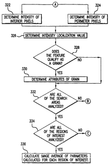
be determined by step 322 (FIG. 3).
[0064] The intensity of the perimeter pixels may be
measured at step 324 of FIG. 4; this intensity is denoted Ib
and is described above with method 200. The pixels
16
CA 02485697 2004-11-10
WO 03/105067 PCT/US03/14706
included in calculating Ib may be pixels located within a
predetermined distance Lb of the test region (e. g.,
perimeter pixel pattern B illustrated in FIG. 8). In some
embodiments, the pixels used to calculate Ib may be required
to intersect R1.
[0065] FIG. 8 also illustrates the perimeter pixel
pattern B, whose pixels may be sampled to determine the
intensity Ib. The portion of the pixels located within
region R1, as shown in FIG. 8, may be used for the samples
for I~ and Ib. In some embodiments, step 324 of FIG. 4 may
be performed simultaneously with step 322.
[0066] Turning again to FIG. 4, an intensity
localization value,V, may be calculated at step 326. In
some embodiments, the intensity localization value may be
computed in any suitable manner that represents the
comparison of the intensity of the interior pixels, I~, to
the intensity of the perimeter pixels, Ib, but that differ
with regard to normalization for specific background
intensity levels as described above in connection with
method 200.
[0067] Test 328 may determine if the test region
contains a feature that qualifies as a grain by determining
if the computed intensity localisation value is greater
than or less than a predetermined threshold value; that is,
if the absolute value of the intensity localization value
is greater than a predetermined threshold value. In some
embodiments, a user may set or adjust the threshold value
by manipulating a user interface (e.g., user interface 140
of system 100 of FIG. 1) or the value may be set or
adjusted by the apparatus (e.g., system 100 of FIG. 1).
Factors such as the background intensity level of the
image, the imaging system or technique used, the type of
sample imaged, or the criteria for an object of interest to
17
CA 02485697 2004-11-10
WO 03/105067 PCT/US03/14706
be identified may determine what threshold level is
desirable.
[0068] If the feature qualifies as a grain, it may be
included in the calculations for a series of properties
(e.g., number of grains per region of interest, number of
grains per image, number of grains per area, etc.) for the
region of interest at step 330. In some embodiments, test
328 may be forgone such that each of the identified second
pixels qualify as grain features for which suitable
attributes may be calculated.
[0069] Test 332 may determine whether all of the search
areas of the region of interest have been analyzed. If
all of the search areas have not been analyzed, step 314
(illustrated in FIG. 3) may retrieve the next search area
of the region of interest.
[0070] If all of the search areas for a region of
interest have been analyzed, a series of parameters may be
calculated from the number, intensity, or distribution of
grain features. For example, the number of grains per
image, the number of grains per region of interest, the
number of grains per biological sample, or the average
intensity localization value may be calculated. In
addition, the total or fractional area in a region of
interest or the entire image occupied by qualified grains
may be calculated.
[0071] An area of a grain in a test region may be
defined by the area of the interior pixels, the total area
of the test region, the area of the perimeter pixels, or
the sum of the interior and perimeter pixels. If the area
of grains are being calculated in a second region of
interest, the pixels (e. g., interior pixels, perimeter
pixels, test region, etc.) used to calculate the area may
be required to intersect the first region of interest.
18
CA 02485697 2004-11-10
WO 03/105067 PCT/US03/14706
[0072] An intensity of the qualified grains may also be
considered. For example, the total or average intensity of
all grains may be Calculated for the entire image or for a
region of interest. The intensity of a grain may be
defined as the summed or averaged pixel intensities of the
interior pixels of the test region, the entire test region,
the perimeter pixels of the test region, or the combined
interior and perimeter pixels of the test region. In
addition, the ratio of all grain intensities of the entire
image to the intensity of the entire image may be
calculated. In some embodiments, the ratio of all grain
intensities in a region of interest to the intensity of the
entire region of interest may be calculated. If the
intensity of grains are being calculated in a separately
defined region of interest, the pixels (e. g. interior
pixels, perimeter pixels, test region) used to calculate
the intensity may be required to intersect the region of
interest.
[0073] Test 334 may determine whether all regions of
interest have be selected and analyzed. If all regions of
interest in an image have been analyzed, step 336 may
calculate an image average of parameters (e.g., number of
grains, area fraction, ratio of total intensity of grains
to intensity of image, etC.) that may be calculated for
each region of interest, along with associated standard
deviations. The values for all grains in each analyzed
region of interest may be pooled or averaged, either
directly or with a suitable weighting factor, to create
values for the entire image.
[0074] In some embodiments, step 336 may also calculate
information for the digital image, including the total
number of grains in the digital image, the area of at least
one grain in the digital image, the total number of grains
19
CA 02485697 2004-11-10
WO 03/105067 PCT/US03/14706
per unit area in the digital image, the intensity of grains
in the digital image, the ratio of the intensity of grains
in the image to the total intensity of the digital image,
any combination thereof, or any other suitable calculation.
[0075] If other regions of interest exist in an image,
step 338 (illustrated in FIG. 3) may locate the next region
of interest. Upon locating the next region, method 300
may begin again starting at step 304.
[0076] In some embodiments, grain analysis may be
performed on a three-dimensional image or other suitable
mufti-dimensional image. Steps may be added to the above-
described method 300 (illustrated in FIGS. 3-4). For
example, a three-dimensional image may be divided into
suitable sections in which the steps of method 300 may be
performed on an individual section. Analysis of each
section may be performed until there are no more sections
of the three-dimensional image.
[0077] Method 300, as described above, may be used to
analyze biological samples (e.g., cells located in wells of
a microtiter plate). Many cells may exist within an
individual well, and each cell may be imaged and analyzed.
It may be desirable to locate grains within the nucleus of
a cell. For example, a cell may be marked with a
fluorescent marker that causes the nucleus to fluoresce red
during imaging. FIG. 9A illustrates red and green channels
of a fluorescence image of a cell sample 910. Nucleus 912
of the cell may fluoresce red, while GFP-labeled protein
may fluoresce green to show punctate pattern 914.
[0078] FIG. 9B illustrates the identification of nucleus
922 in cell sample 920. Identification of nucleus 922 may
be a seed point to identify grains. FIG. 9C shows cell
sample 930 which may contain analyzed region 932. As
shown, box 934 may represent the region of interest in the
CA 02485697 2004-11-10
WO 03/105067 PCT/US03/14706
image, and smaller boxes 936 may identify the features
qualifying as grains. For illustrative purposes, FIG. 9D
shows an image of cell 940 without staining or markers.
[0079] The present invention should be applicable to
characterizing granularity in a variety of biological
samples including but not limited to: cell surface
receptor internalization, nuclear chromatin condensation,
localization to intracellular compartments that produce a
punctate staining pattern (e. g. mitochondria or golgi),
localization to any vesicle, pit, lysosome or endosome
either within a cell or on the cell surface, or any
endocytosis, exocytosis or degranulation event whereby
matter is internalized or released from a cell via
vesicular structures. Further, the granularity related to
the clustering of a unique tissue type or marker in a
larger imaged tissue sample may also be calculated. For
example, distributions of plaques, tumors, or chemical
moieties that can be histochemically or otherwise marked
and imaged may be characterized by granularity analysis.
[0080] It will be understood that the foregoing is only
illustrative of the principles of the invention, and that
various modifications can be made by those skilled in the
art without departing from the scope and spirit of the
invention.
21