Note: Descriptions are shown in the official language in which they were submitted.
CA 02485799 2011-07-18
78391-6
PLACEMENT STRUCTURE FOR FACILITATING PLACEMENT OF AN
IMPLANTABLE DEVICE PROXIMATE TO NEURAL / MUSCULAR TISSUE
FOR AFFECTING AND/OR SENSING NEURAL / MUSCULAR TISSUE
Background of the Invention
[0001] The present invention relates to systems for monitoring and/or
affecting parameters of a patient's body for the purpose of medical diagnosis
and/or treatment. More particularly, systems in accordance with the
invention are characterized by a plurality of devices, preferably battery-
powered, configured for implanting within a patient's body, each device being
configured to sense a body parameter, e.g., temperature, 02 content,
physical position, etc., and/or to affect a parameter, e.g., via nerve
stimulation.
[0002] Applicants' commonly assigned U.S. Patent Application No.
09/030,106 entitled "Battery Powered Patient Implantable Device", now
U.S. Patent No. 6,185,452, describes devices configured for
implantation within a patient's body, i.e., beneath a patient's
skin, for performing various functions including: (1) stimulation of body
tissue, (2) sensing of body parameters, and (3) communicating between
implanted devices and devices external to a patient's body.
-1-
CA 02485799 2004-10-25
A328B-USA
Summary of the Invention
[0003] The present invention is directed to a system for monitoring
and/or
affecting parameters of a patient's body and more particularly to such a
system comprised of a system control unit (SCU) and one or more devices
implanted in the patient's body, i.e., within the envelope defined by the
patient's skin. Each said implanted device is configured to be monitored
and/or controlled by the SCU via a wireless communication channel.
[0004] In accordance with the invention, the SCU comprises a
programmable
unit capable of (1) transmitting commands to at least some of a plurality of
implanted devices and (2) receiving data signals from at least some of those
implanted devices. In accordance with a preferred embodiment, the system
operates in a closed loop fashion whereby the commands transmitted by the
SCU are dependent, in part, on the content of the data signals received by
the SCU.
[0005] In accordance with a preferred embodiment, each implanted
device is
configured similarly to the devices described in Applicants' commonly
assigned U.S. Patent Application No. 09/030,106, now U.S. Patent No.
6,185,452, and typically comprises a sealed housing suitable for injection
into the patient's body. Each housing preferably contains a power source
having a capacity of at least 1 microwatt-hour, preferably a rechargeable
battery, and power consuming circuitry preferably including a data signal
transmitter and receiver and sensor/stimulator circuitry for driving an
input/output transducer.
[0006] In accordance with a significant aspect of the preferred
embodiment,
a preferred SCU is also implemented as a device capable of being injected
into the patient's body. Wireless communication between the SCU and the
other implanted devices can be implemented in various ways, e.g., via a
modulated sound signal, AC magnetic field, RF signal, or electrical
conduction.
-2-
CA 02485799 2004-10-25
A328B-USA
[0007] In accordance with a further aspect of the invention, the SCU
is
remotely programmable, e.g., via wireless means, to interact with the
implanted devices according to a treatment regimen. In accordance with a
preferred embodiment, the SCU is preferably powered via an internal power
source, e.g., a rechargeable battery. Accordingly, an SCU combined with
one or more battery-powered implantable devices, such as those described
in the commonly assigned U.S. Patent No. 6,185,452, form a self-sufficient
system for treating a patient.
[0008] In accordance with a preferred embodiment, the SCU and other
implanted devices are implemented substantially identically, being comprised
of a sealed housing configured to be injected into the patient's body. Each
housing contains sensor/stimulator circuitry for driving an input/output
transducer, e.g., an electrode, to enable it to additionally operate as a
sensor
and/or stimulator.
[0009] Alternatively, the SCU could be implemented as an implantable
but
non-injectable housing which would permit it to be physically larger enabling
it to accommodate larger, higher capacity components, e.g., a battery,
microcontroller, etc. As a further alternative, the SCU could be implemented
in a housing configured for carrying on the patient's body outside of the skin
defined envelope, e.g., in a wrist band.
[0010] In accordance with the invention, the commands transmitted by
the
SCU can be used to remotely configure the operation of the other implanted
devices and/or to interrogate the status of those devices. For example,
various operating parameters, e.g., the pulse frequency, pulse width, trigger
delays, etc., of each implanted device can be controlled or specified in one
or more commands addressably transmitted to the device. Similarly, the
sensitivity of the sensor circuitry and/or the interrogation of a sensed
parameter, e.g., battery status, can be remotely specified by the SCU.
-3-
i========.=========
CA 02485799 2011-07-18
78391-6
[0011] In accordance with a significant feature of the
preferred embodiment, the SCU and/or each implantable device
includes a programmable memory for storing a set of default
parameters. In the event of power loss, SCU failure, or any
other catastrophic occurrence, all devices default to the safe
harbor default parameters. The default parameters can be
programmed differently depending upon the condition being
treated. In accordance with a further feature, the system
includes a switch, preferably actuatable by an external
DC magnetic field, for resetting the system to its default
parameters.
[0012] In an exemplary use of a system in accordance with
the present invention, a patient with nerve damage can have a
damaged nerve "replaced" by an implanted SCU and one or more
implanted sensors and stimulators, each of which contains its
own internal power source. In this exemplary system, the
SCU would monitor a first implanted sensor for a signal
originating from the patient's brain and responsively transit
command signals to one or more stimulators implanted past the
point of nerve damage. Furthermore, the SCU could monitor
additional sensors to determine variations in body parameters
and, in a closed loop manner, react to control the command
signals to achieve the desired treatment regimen.
[0013] In accordance with one aspect of the invention, there
is provided a placement structure for facilitating placement of
an implantable device having at least two electrodes proximate
to neural/muscular tissue, said implantable device selected
-4-
CA 02485799 2012-12-19
- 78391-6
from the group consisting of: microstimulators, microsensors and
microtransponders,
said placement structure comprising: a holder having a hollow cavity that
essentially
conforms to the size and shape of the implantable device such that the
implantable
device may be snapped into the cavity and is held by the elasticity of the
holder; at
least one set of elastic wings for capturing neural/muscular tissue; and
wherein said
placement structure is primarily formed from a biocompatible plastic.
In accordance with another aspect of the present invention, there is
provided a method for forming a placement structure for facilitating placement
of an
implantable device having at least two electrodes proximate to neural/muscular
tissue,
said implantable device selected from the group consisting of:
microstimulators,
microsensors and microtransponders, said method comprising the steps of:
forming a
holder having a hollow cavity that essentially conforms to the size and shape
of the
implantable device such that the implantable device may be snapped into the
cavity
and is held by the elasticity of the holder; forming at least one set of
elastic wings for
capturing neural/muscular tissue integral to said holder; and wherein said
holder and
said wings which comprise said placement structure are primarily formed from a
biocompatible plastic.
In accordance with another aspect of the present invention, there is
provided a use of the placement structure as described above for facilitating
placement of an implantable device.
[0014] The novel features of the invention are set forth with
particularity in the
appended claims. The invention will be best understood from the following
description
when read in conjunction with the accompanying drawings.
-5-
CA 02485799 2004-10-25
A328B-USA
Brief Description of the Drawings
[0015] FIG. 1 is a simplified block diagram of the system of the
present
invention comprised of implanted devices, e.g., microstimulators,
microsensors and microtransponders, under control of an implanted system
control unit (SCU).
[0016] FIG. 2 comprises a block diagram of the system of FIG. 1
showing the
functional elements that form the system control unit and implanted
microstimulators, microsensors and microtransponders.
[0017] FIG. 3A comprises a block diagram of an exemplary implanted
device,
as shown in the commonly assigned U.S. Patent No. 6,185,452, including a
battery for powering the device for a period of time in excess of one hour in
response to a command from the system control unit.
[0018] FIG. 3B comprises a simplified block diagram of controller
circuitry
that can be substituted for the controller circuitry of FIG. 3A, thus
permitting
a single device to be configured as a system control unit and/or a
microstimulator and/or a microsensor and/or a microtransponder.
[0019] FIG. 4 is a simplified diagram showing the basic format of
data
messages for commanding/interrogating the implanted microstimulators,
microsensors and microtransponders which form a portion of the present
invention.
[0020] FIG. 5 shows an exemplary flow chart of the use of the present
system in an open loop mode for controlling/monitoring a plurality of
implanted devices, e.g., microstimulators, microsensors.
[0021] FIG. 6 shows a flow chart of the optional use of a translation
table for
communicating with microstimulators and/or microsensors via
microtransponders.
[0022] FIG. 7 shows a simplified flow chart of the use of closed loop
control
of a microstimulator by altering commands from the system control unit in
response to status data received from a microsensor.
-6-
CA 02485799 2004-10-25
A328B-U SA
[0023] FIG. 8 shows an exemplary injury, i.e., a damaged nerve, and
the
placement of a plurality of implanted devices, i.e., microstimulators,
microsensors and a microtransponder under control of the system control
unit for "replacing" the damaged nerve.
[0024] FIG. 9 shows a simplified flow chart of the control of the
implanted
devices of FIG. 8 by the system control unit.
[0025] FIG. 10A shows a side view of a battery-powered implanted
device,
e.g., a microstimulator, made in accordance with the present invention.
[0026] FIG. 10B shows a side view of another implantable battery-
powered
device, one employing an internal coupling capacitor, made in accordance
with the invention.
[0027] FIGS. 10C and 10D show two side cutaway views of the presently
preferred embodiment of an implantable ceramic tube suitable for housing
the system control unit and/or microstimulators and/or microsensors and/or
microtransponders.
[0028] FIG. 11 illustrates an exemplary battery suitable for powering
the
implantable devices which comprise the components of the present
invention.
[0029] FIG. 12 shows an exemplary housing suitable for an implantable
SCU
having a battery enclosed within that has a capacity of at least 1 watt-hour.
[0030] FIG. 13 is an alternative embodiment of the housing of FIGS.
10A-10D.
[0031] FIG. 14 is a cross-sectional view of the housing of FIG. 13
taken
along line 14-14.
[0032] FIG. 15 is an alternative embodiment of the housing of FIGS.
10A-10D.
[0033] FIG. 16 is a cross-sectional view of the housing of FIG. 15
taken
along line 16-16.
-7-
CA 02485799 2004-10-25
A328B-USA
[0034] FIG. 17 is an alternative embodiment of the housing of FIGS.
10A-10D.
[0035] FIG. 18 is a cross-sectional view of the housing of FIG. 17
taken
along line 18-18.
[0036] FIG. 19 is an alternative embodiment of the housing of FIGS.
10A-10D.
[0037] FIG. 20 is a cross-sectional view of the housing of FIG. 19
taken
along line 20-20.
[0038] FIG. 21 is an alternative embodiment of the housing of FIGS.
10A-10D.
[0039] FIG. 22 is a cross-sectional view of the housing of FIG. 21
taken
along line 22-22.
[0040] FIG. 23 is an alternative embodiment of the housing of FIGS.
10A-10D.
[0041] FIG. 24 is a cross-sectional view of the housing of FIG. 23
taken
along line 24-24.
[0042] FIG. 25 is a perspective view of an exemplary placement
structure of
the present invention which is formed for holding one of the aforementioned
implantable device in close proximity to a nerve, muscle tissue, or the like.
[0043] FIG. 26 is a perspective view of the placement structure of
FIG. 25
having one of the aforementioned placement devices held within a hollow
cavity within its holder portion.
[0044] FIG. 27 is a perspective view of the placement structure of
FIGS. 25
and 26 showing its wings capturing neural / muscular tissue.
[0045] FIG. 28 is an end view of the placement structure of FIGS. 25
and 26.
[0046] FIG. 29 is an end view of the placement structure of FIGS. 25
and 26
having hooks at the ends of its wings for providing additional means for
retaining the placement structure in close proximity to the neural / muscular
tissue.
-8-
_________________ --
CA 02485799 2004-10-25
,
A328B-USA
[0047] FIG. 30 is an exemplary laparoscopic device suitable for
implanting
the placement structure of the present invention which in turn is holding one
of the aforementioned implantable devices in close proximity to neural /
muscular tissue.
[0048] FIG. 31 is a cross sectional view of that shown in FIG. 30
along the
line 31-31 wherein the wings of the placement structure have been folded
inward toward the implantable device before insertion, e.g., via its tip, into
the hollow portion of the laparoscopic device.
[0049] FIG. 32 is a cross sectional view of that shown in FIG. 27
along the
line 32-32 showing the wings of the placement structure holding neural /
muscular tissue and the resulting stimulation/sensing vectors.
[0050] FIG. 33 is an alternative embodiment of the placement
structure of
FIG. 25 wherein inner portions of the wings and the cavity include conductive
layers (preferably a plurality of conductive paths) to provide additional
electrical coupling between the electrodes of the implantable device axially
along the neural / muscular tissue.
[0051] FIG. 34 is a next alternative embodiment of the placement
structure of
FIG. 25 wherein inner portions of the wings and the cavity include conductive
layers (preferably a plurality of conductive paths) to provide additional
electrical coupling between the electrodes of the implantable device
transversely across the neural / muscular tissue using a pair of wings.
[0052] FIG. 35 is an alternative embodiment of the placement
structure of
FIG. 25 and the implantable medical device of FIGS. 10A-10D wherein the
implantable medical device additionally includes a plurality of stimulator /
sensor circuitry portions that are coupled via a plurality of electrode
connectors and a plurality of conductive paths to inner portions of the wings
and the cavity of the placement structure to provide stimulation to or sensing
from displaced portions of the neural / muscular tissue.
-9-
_ _______________ - -
CA 02485799 2004-10-25
A328B-USA
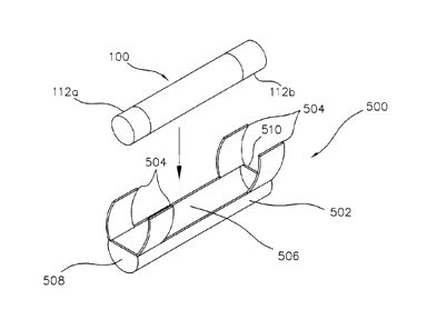
[0053] FIG. 36 shows an alternative implementation of that which is
functionally described in relation to FIG. 35. However, in this implementation
a single, essentially U-shaped, structure having elastic wings is integrally
formed which encompasses the functionality of the implantable medical
device of FIGS. 10A-10D contained within the placement structure.
[0054] FIG. 37 shows a next alternative implementation of that which
is
functionally described in relation to FIGS. 35 and 36 to the extent that it
too
is an integral device but it has its elastic wings 504 formed from a silicon
rubber impregnated cloth that is permanently attached to the functional
equivalent of the implantable medical device which was described in
reference to FIGS. 10A-10D.
-10-.
CA 02485799 2004-10-25
A328B-USA
Detailed Description of the Preferred Embodiments
[0055] The present invention is directed to a system for monitoring
and/or
affecting parameters of a patient's body and more particularly to such a
system comprised of a system control unit (SCU) and one or more devices
implanted in a patient's body, i.e., within the envelope defined by the
patient's skin. Each such implantable device is configured to be monitored
and/or controlled by the SCU via a wireless communication channel.
[0056] In accordance with the invention, the SCU comprises a
programmable
unit capable of (1) transmitting commands to at least some of a plurality of
implanted devices and (2) receiving data signals from at least some of those
implanted devices. In accordance with a preferred embodiment, the system
operates in closed loop fashion whereby the commands transmitted by the
SCU are dependent, in part, on the content of the data signals received by
the SCU.
[0057] In accordance with a preferred embodiment, each implanted
device is
configured similarly to the devices described in Applicants' commonly
assigned U.S. Patent Application No. 09/030,106, now U.S. Patent No.
6,185,452, and typically comprises a sealed housing suitable for injection
into the patient's body. Each housing preferably contains a power source
having a capacity of at least 1 microwatt-hour, preferably a rechargeable
battery, and power consuming circuitry preferably including a data signal
transmitter and receiver and sensor/stimulator circuitry for driving an
input/output transducer.
[0058] FIG. 1 (essentially corresponding to FIG. 2 of the commonly
assigned
U.S. Patent No. 6,185,452) and FIG. 2 show an exemplary system 300
made of implanted devices 100, preferably battery powered, under control of
a system control unit (SCU) 302, preferably also implanted beneath a
patient's skin 12. As described in the commonly assigned U.S. Patent No.
6,185,452, potential implanted devices 100 (see also the block diagram
-11-
CA 02485799 2004-10-25
A328B-USA
shown in FIG. 3A) include stimulators , e.g., 100a, sensors, e.g., 100c, and
transponders, e.g., 100d. Such stimulators, e.g., 100a, can be remotely
programmed to output a sequence of drive pulses to body tissue proximate
to its implanted location via attached electrodes. The sensors, e.g., 100c,
can be remotely programmed to sense one or more physiological or
biological parameters in the implanted environment of the device, e.g.,
temperature, glucose level, 02 content, etc. Transponders, e.g., 100d, are
devices which can be used to extend the interbody communication range
between stimulators and sensors and other devices, e.g., a clinician's
programmer 172 and a patient control unit 174. Preferably, these
stimulators, sensors and transponders are contained in sealed elongate
housing having an axial dimension of less than 60 mm and a lateral
dimension of less than 6 mm. Accordingly, such stimulators, sensors and
transponders are respectively referred to as microstimulators, microsensors,
and microtransponders. Such microstimulators and microsensors can thus
be positioned beneath the skin within a patient's body using a hypodermic
type insertion tool 176.
[0059] As described in the commonly assigned U.S. Patent No.
6,185,452,
microstimulators and microsensors are remotely programmed and
interrogated via a wireless communication channel, e.g., modulated AC
magnetic, sound (i.e., ultrasonic), RF or electric fields, typically
originating
from control devices external to the patient's body, e.g., a clinician's
programmer 172 or patient control unit 174. Typically, the clinician's
programmer 172 is used to program a single continuous or one time pulse
sequence into each microstimulator and/or measure a biological parameter
from one or more microsensors. Similarly, the patient control unit 174
typically communicates with the implanted devices 100, e.g., microsensors
100c, to monitor biological parameters. In order to distinguish each
implanted device over the communication channel, each implanted device is
-12-
CA 02485799 2004-10-25
A328B-USA
manufactured with an identification code (ID) 303 specified in address
storage circuitry 108 (see FIG. 3A) as described in the commonly assigned
U.S. Patent No. 6,185,452.
[0060] By using one or more such implantable devices in conjunction
with the
SCU 302 of the present invention, the capabilities of such implanted devices
can be further expanded. For example, in an open loop mode (described
below in reference to FIG. 5), the SCU 302 can be programmed to
periodically initiate tasks, e.g., perform real time tasking, such as
transmitting
commands to microstimulators according to a prescribed treatment regimen
or periodically monitor biological parameters to determine a patient's status
or the effectiveness of a treatment regimen. Alternatively, in a closed loop
mode (described below in reference to FIGS. 7-9), the SCU 302 periodically
interrogates one or more microsensors and accordingly adjust the
commands transmitted to one or more microstimulators.
[0061] FIG. 2 shows the system 300 of the present invention comprised
of
(1) one or more implantable devices 100 operable to sense and/or stimulate
a patient's body parameter in accordance with one or more controllable
operating parameters and (2) the SCU 302. The SCU 302 is primarily
comprised of (1) a housing 206, preferably sealed and configured for
implantation beneath the skin of the patient's body as described in the
commonly assigned U.S. Patent No. 6,185,452 in reference to the implanted
devices 100, (2) a signal transmitter 304 in the housing 206 for transmitting
command signals, (3) a signal receiver 306 in the housing 206 for receiving
status signals, and (4) a programmable controller 308, e.g., a microcontroller
or state machine, in the housing 206 responsive to received status signals
for producing command signals for transmission by the signal transmitter 304
to other implantable devices 100. The sequence of operations of the
programmable controller 308 is determined by an instruction list, i.e., a
program, stored in program storage 310, coupled to the programmable
-13-
---
CA 02485799 2004-10-25
A328B-USA
controller 308. While the program storage 310 can be a nonvolatile memory
device, e.g., ROM, manufactured with a program corresponding to a
prescribed treatment regimen, it is preferable that at least a portion of the
program storage 310 be an alterable form of memory, e.g., RAM, EEPROM,
etc., whose contents can be remotely altered as described further below.
However, it is additionally preferable that a portion of the program storage
310 be nonvolatile so that a default program is always present. The rate at
which the program contained within the program storage 310 is executed is
determined by clock 312, preferably a real time clock that permits tasks to be
scheduled at specified times of day.
[0062] The signal transmitter 304 and signal receiver 306 preferably
communicate with implanted devices 100 using sound means, i.e.,
mechanical vibrations, using a transducer having a carrier frequency
modulated by a command data signal. In a preferred embodiment, a carrier
frequency of 100 KHz is used which corresponds to a frequency that freely
passes through a typical body's fluids and tissues. However, such sound
means that operate at any frequency, e.g., greater than 1 Hz, are also
considered to be within the scope of the present invention. Alternatively, the
signal transmitter 304 and signal receiver 306 can communicate using
modulated AC magnetic, RF, or electric fields.
[0063] The clinician's programmer 172 and/or the patient control unit
174
and/or other external control devices can also communicate with the
implanted devices 100, as described in the commonly assigned U.S. Patent
No. 6,185,452, preferably using a modulated AC magnetic field.
Alternatively, such external devices can communicate with the SCU 302 via
a transceiver 314 coupled to the programmable controller 308. Since, in a
preferred operating mode, the signal transmitter 304 and signal receiver 306
operate using sound means, a separate transceiver 314 which operates
using magnetic means is used for communication with external devices.
-14-
CA 02485799 2004-10-25
A328B-USA
However, a single transmitter 304/receiver 306 can be used in place of
transceiver 314 if a common communication means is used.
[0064] FIG. 3A comprises a block diagram of an exemplary implanted
device
100 (as shown in FIG. 2 of the commonly assigned U.S. Patent No.
6,185,452) which includes a battery 104, preferably rechargeable, for
powering the device for a period of time in excess of one hour and
responsive to command signals from a remote device, e.g., the SCU 302.
As described in the commonly assigned U.S. Patent No. 6,185,452, the
implantable device 100 is preferably configurable to alternatively operate as
a microstimulator and/or microsensor and/or microtransponder due to the
commonality of most of the circuitry contained within. Such circuitry can be
further expanded to permit a common block of circuitry to also perform the
functions required for the SCU 302. Accordingly, FIG. 3B shows an
alternative implementation of the controller circuitry 106 of FIG. 3A that is
suitable for implementing a microstimulator and/or a microsensor and/or a
microtransponder and/or the SCU 302. In this implementation, configuration
data storage 132 can be alternatively used as the program storage 310 when
the implantable device 100 is used as the SCU 302. In this implementation,
XMTR 168 corresponds to the signal transmitter 304 and a RCVR 114b
corresponds to the signal receiver 306 (preferably operable using sound
means via transducer 138) and the RCVR 114a and XMTR 146 correspond
to the transceiver 314 (preferably operable using magnetic means via coil
116).
[0065] In a preferred embodiment, the contents of the program storage
310,
i.e., the software that controls the operation of the programmable controller
308, can be remotely downloaded, e.g., from the clinician's programmer 172
using data modulated onto an AC magnetic field. In this embodiment, it is
preferable that the contents of the program storage 310 for each SCU 302
be protected from an inadvertent change. Accordingly, the contents of the
-15-
CA 02485799 2004-10-25
A328B-USA
address storage circuitry 108, i.e., the ID 303, is preferably used as a
security code to confirm that the new program storage contents are destined
for the SCU 302 receiving the data. This feature is significant if multiple
patient's could be physically located, e.g., in adjoining beds, within the
communication range of the clinician's programmer 172.
[0066] In a further aspect of the present invention, it is preferable
that the
SCU 302 be operable for an extended period of time, e.g., in excess of one
hour, from an internal power supply 316. While a primary battery, i.e., a
nonrechargeable battery, is suitable for this function, it is preferable that
the
power supply 316 include a rechargeable battery, e.g., battery 104 as
described in the commonly assigned U.S. Patent No. 6,185,452, that can be
recharged via an AC magnetic field produced external to the patient's body.
Accordingly, the power supply 102 of FIG. 3A (described in detail in the
commonly assigned U.S. Patent No. 6,185,452) is the preferred power
supply 316 for the SCU 302 as well.
[0067] The battery-powered devices 100 of the commonly assigned U.S.
Patent No. 6,185,642 are preferably configurable to operate in a plurality of
operation modes, e.g., via a communicated command signal. In a first
operation mode, device 100 is remotely configured to be a microstimulator,
e.g., 100a and 100b. In this embodiment, controller 130 commands
stimulation circuitry 110 to generate a sequence of drive pulses through
electrodes 112 to stimulate tissue, e.g., a nerve, proximate to the implanted
location of the microstimulator, e.g., 100a or 100b. In operation, a
programmable pulse generator 178 and voltage multiplier 180 are configured
with parameters (see Table I) corresponding to a desired pulse sequence
and specifying how much to multiply the battery voltage (e.g., by summing
charged capacitors or similarly charged battery portions) to generate a
desired compliance voltage V. A first FET 182 is periodically energized to
store charge into capacitor 183 (in a first direction at a low current flow
rate
-16-
CA 02485799 2004-10-25
A328B-USA
through the body tissue) and a second FET 184 is periodically energized to
discharge capacitor 183 in an opposing direction at a higher current flow rate
which stimulates a nearby nerve. Alternatively, electrodes can be selected
that will form an equivalent capacitor within the body tissue.
Current: continuous current charging of storage
capacitor
Charging currents: 1, 3, 10, 30, 100, 250, 500 pa
Current Range: 0.8 to 40 ma in nominally 3.2% steps
Compliance Voltage: selectable, 3-24 volts in 3 volt steps
Pulse Frequency(PPS): 1 to 5000 PPS in nominally 30% steps
Pulse Width: 5 to 2000 ps in nominally 10% steps
Burst On Time (BON): 1 ms to 24 hours in nominally 20%
steps
Burst Off Time (BOF): 1 ms to 24 hours in nominally 20%
steps
Triggered Delay to BON: either selected BOF or pulse width
Burst Repeat Interval: 1 ms to 24 hours in nominally 20%
steps
Ramp On Time: 0.1 to 100 seconds (1, 2, 5, 10 steps)
Ramp Off Time: 0.1 to 100 seconds (1, 2, 5, 10 steps)
Table I - Stimulation Parameters
[0068] In a next operation mode, the battery-powered implantable
device 100
can be configured to operate as a microsensor, e.g., 100c, that can sense
one or more physiological or biological parameters in the implanted
environment of the device. In accordance with a preferred mode of
operation, the system control unit 302 periodically requests the sensed data
from each microsensor 100c using its ID stored in address storage circuitry
108, and responsively sends command signals to microstimulators, e.g.,
100a and 100b, adjusted accordingly to the sensed data. For example,
-17-
CA 02485799 2004-10-25
A3286-USA
sensor circuitry 188 can be coupled to the electrodes 112 to sense or
otherwise used to measure a biological parameter, e.g., temperature,
glucose level, or 02 content and provided the sensed data to controller
circuitry 106. Preferably, the sensor circuitry includes a programmable
bandpass filter and an analog to digital (ND) converter that can sense and
accordingly convert the voltage levels across the electrodes 112 into a
digital
quantity. Alternatively, the sensor circuitry can include one or more sense
amplifiers to determine if the measured voltage exceeds a threshold voltage
value or is within a specified voltage range. Furthermore, the sensor
circuitry
188 can be configurable to include integration circuitry to further process
the
sensed voltage. The operation modes of the sensor circuitry 188 is remotely
programmable via the devices communication interface as shown below in
Table II.
Input voltage range: 5 pv to 1 V
Bandpass filter rolloff: 24 dB
Low frequency cutoff choices: 3, 10, 30, 100, 300, 1000 Hz
High frequency cutoff choices: 3, 10, 30, 100, 300, 1000 Hz
Integrator frequency choices: 1 PPS to 100 PPS
Amplitude threshold
for detection choices: 4 bits of resolution
Table II - Sensincr Parameters
[0069] Additionally, the sensing capabilities of a microsensor
include the
capability to monitor the battery status via path 124 from the charging
circuit
122 and can additionally include using the ultrasonic transducer 138 or the
coil 116 to respectively measure the magnetic or ultrasonic signal
magnitudes (or transit durations) of signals transmitted between a pair of
implanted devices and thus determine the relative locations of these devices.
-18-
_ _ ___________________________________________________ -
CA 02485799 2004-10-25
A3286-USA
This information can be used to determine the amount of body movement,
e.g., the amount that an elbow or finger is bent, and thus form a portion of a
closed loop motion control system.
[0070] In another operation mode, the battery-powered implantable
device
100 can be configured to operate as a microtransponder, e.g., 100d. In this
operation mode, the microtransponder receives (via the aforementioned
receiver means, e.g., AC magnetic, sonic, RF or electric) a first command
signal from the SCU 302 and retransmits this signal (preferably after
reformatting) to other implanted devices (e.g., microstimulators,
microsensors, and/or microtransponders) using the aforementioned
transmitter means (e.g., magnetic, sonic, RF or electric). While a
microtransponder may receive one mode of command signal, e.g., magnetic,
it may retransmit the signal in another mode, e.g., ultrasonic. For example,
clinician's programmer 172 may emit a modulated magnetic signal using a
magnetic emitter 190 to program/command the implanted devices 100.
However, the magnitude of the emitted signal may not be sufficient to be
successfully received by all of the implanted devices 100. As such, a
microtransponder 100d may receive the modulated magnetic signal and
retransmit it (preferably after reformatting) as a modulated ultrasonic signal
which can pass through the body with fewer restrictions. In another
exemplary use, the patient control unit 174 may need to monitor a
microsensor 100c in a patient's foot. Despite the efficiency of ultrasonic
communication in a patient's body, an ultrasonic signal could still be
insufficient to pass from a patient's foot to a patient's wrist (the typical
location of the patient control unit 174). As such, a microtransponder 100d
could be implanted in the patient's torso to improve the communication link.
[0071] FIG. 4 shows the basic format of an exemplary message 192 for
communicating with the aforementioned battery-powered devices 100, all of
which are preconfigured with an address (ID), preferably unique to that
-19-
CA 02485799 2004-10-25
A328B-USA
device, in their address storage circuitry 108 to operate in one or more of
the
following modes (1) for nerve stimulation, i.e., as a microstimulator, (2) for
biological parameter monitoring, i.e., as a microsensor, and/or (3) for
retransmitting received signals after reformatting to other implanted devices,
i.e., as a microtransponder. The command message 192 is primarily
comprised of a (1) start portion 194 (one or more bits to signify the start of
the message and to synchronize the bit timing between transmitters and
receivers), (2) a mode portion 196 (designating the operating mode, e.g.,
microstimulator, microsensor, microtransporider, or group mode), (3) an
address (ID) portion 198 (corresponding to either the ID in address storage
circuitry 108 or a programmed group ID), (4) a data field portion 200
(containing command data for the prescribed operation), (5) an error
checking portion 202 (for ensuring the validity of the message 192, e.g., by
use of a parity bit), and (6) a stop portion 204 (for designating the end of
the
message 192). The basic definition of these fields are shown below in Table
III. Using these definitions, each device can be separately configured,
controlled and/or sensed as part of a system for controlling one or more
neural pathways within a patient's body.
-20-
CA 02485799 2004-10-25
A328B-USA
MODE ADDRESS (ID)
00 = Stimulator 8 bit identification
address
01 = Sensor 8 bit identification
address
02 = Transponder 4 bit identification
address
03 = Group 4 bit group identification
address
DATA FIELD PORTION
Program/Stimulate = select operating mode
Parameter /
Preconfiguration
Select = select programmable parameter in
program mode or preconfigured stimulation
or sensing parameter in other modes
Parameter Value = program value
Table III - Message Data Fields
[0072] Additionally, each device 100 can be programmed with a group
ID
(e.g., a 4 bit value) which is stored in its configuration data storage 132.
When a device 100, e.g., a microstimulator, receives a group ID message
that matches its stored group ID, it responds as if the message was directed
to its ID within its address storage circuitry 108. Accordingly, a plurality
of
microstimulators, e.g., 100a and 100b, can be commanded with a single
message. This mode is of particular use when precise timing is desired
among the stimulation of a group of nerves.
[0073 The following describes exemplary commands, corresponding to
the
command message 192 of FIG. 4, which demonstrate some of the remote
control/sensing capabilities of the system of devices which comprise the
present invention:
[0074] Write Command - Set a microstimulator/microsensor specified in
the
address field 198 to the designated parameter value.
[0075] Group Write Command - Set the microstimulators / microsensors
within the group specified in the address field 198 to the designated
parameter value.
-21-
CA 02485799 2004-10-25
A328B-USA
[0076] Stimulate Command - Enable a sequence of drive pulses from the
microstimulator specified in the address field 198 according to previously
programmed and/or default values.
[0077] Group Stimulate Command - Enable a sequence of drive pulses
from
the microstimulators within the group specified in the address field 198
according to previously programmed and/or default values.
[0078] Unit Off Command - Disable the output of the microstimulator
specified in the address field 198.
[0079] Group Stimulate Command - Disable the output of the
microstimulators within the group specified in the address field 198.
[0080] Read Command - Cause the microsensor designated in the address
field 198 to read the previously programmed and/or default sensor value
according to previously programmed and/or default values.
[0081] Read Battery Status Command - Cause the microsensor designated
in the address field 198 to return its battery status.
[0082] Define Group Command - Cause the microstimulator / microsensor
designated in the address field 198 to be assigned to the group defined in
the microstimulator data field 200.
[0083] Set Telemetry Mode Command - Configure the microtransponder
designated in the address field 198 as to its input mode (e.g., AC magnetic,
sonic, etc.), output mode (e.g., AC magnetic, sonic, etc.), message length,
etc.
[0084] Status Reply Command - Return the requested status/sensor data
to
the requesting unit, e.g., the SCU.
[0085] Download Program Command - Download program / safe harbor
routines to the device, e.g., SCU, microstimulator, etc., specified in the
address field 198.
[0086] FIG. 5 shows a block diagram of an exemplary open loop control
program, i.e., a task scheduler 320, for controlling/monitoring a body
-22-
CA 02485799 2004-10-25
A328B-USA
function/parameter. In this process, the programmable controller 308 is
responsive to the clock 312 (preferably crystal controlled to thus permit real
time scheduling) in determining when to perform any of a plurality of tasks.
In this exemplary flow chart, the programmable controller 308 first
determines in block 322 if it is now at a time designated as TEVENT1 (or at
least within a sampling error of that time), e.g., at 1:00 AM. If so, the
programmable controller 308 transmits a designated command to
microstimulator A (STA) in block 324. In this example, the control program
continues where commands are sent to a plurality of stimulators and
concludes in block 326 where a designated command is sent to
microstimulator X (STx). Such a subprocess, e.g., a subroutine, is typically
used when multiple portions of body tissue require stimulation, e.g.,
stimulating a plurality of muscle groups in a paralyzed limb to avoid atrophy.
The task scheduler 320 continues through multiple time event detection
blocks until in block 328 it determines whether the time TEVENTM has arrived.
If so, the process continues at block 330 where, in this case, a single
command is sent to microstimulator M (STm). Similarly, in block 332 the task
scheduler 320 determines when it is the scheduled time, i.e., TEVENTO, to
execute a status request from microsensor A (SEA). If so, a subprocess,
e.g., a subroutine, commences at block 334 where a command is sent to
microsensor A (SEA) to request sensor data and/or specify sensing criteria.
Microsensor A (SEA) does not instantaneously respond. Accordingly, the
programmable controller 308 waits for a response in block 336. In block
338, the returned sensor status data from microsensor A (SEA) is stored in a
portion of the memory, e.g., a volatile portion of the program storage 310, of
the programmable controller 308. The task scheduler 320 can be a
programmed sequence, i.e., defined in software stored in the program
storage 310, or, alternatively, a predefined function controlled by a table of
parameters similarly stored in the program storage 310. A similar process
-23-
CA 02485799 2004-10-25
A328B-USA
can be used where the SCU 302 periodically interrogates each implantable
device 100 to determine its battery status.
[0087] FIG. 6 shows an exemplary use of an optional translation table
340
for communicating between the SCU 302 and microstimulators, e.g., 100a,
and/or microsensors, e.g., 100c, via microtransponders, e.g., 100d. A
microtransponder, e.g., 100d, is used when the communication range of the
SCU 302 is insufficient to reliably communicate with other implanted devices
100. In this case, the SCU 302 instead directs a data message, i.e., a data
packet, to an intermediary microtransponder, e.g., 100d, which retransmits
the data packet to a destination device 100. In an exemplary
implementation, the translation table 340 contains pairs of corresponding
entries, i.e., first entries 342 corresponding to destination addresses and
second entries 344 corresponding to the intermediary microtransponder
addresses. When the SCU 302 determines, e.g., according to a timed event
designated in the program storage 310, that a command is to be sent to a
designated destination device (see block 346), the SCU 302 searches the
first entries 342 of the translation table 340, for the destination device
address, e.g., STm. The SCU 302 then fetches the corresponding second
table entry 344 in block 348 and transmits the command to that address in
block 350. When the second table entry 344 is identical to its corresponding
first table entry 342, the SCU 302 transmits commands directly to the
implanted device 100. However, when the second table entry 344, e.g., TN,
is different from the first table entry 342, e.g., STm, the SCU 302 transmits
commands via an intermediary microtransponder, e.g., 100d. The use of the
translation table 340 is optional since the intermediary addresses can,
instead, be programmed directly into a control program contained in the
program storage 310. However, it is preferable to use such a translation
table 340 in that communications can be redirected on the fly by just
reprogramming the translation table 340 to take advantage of implanted
-24-
CA 02485799 2004-10-25
A328B-USA
transponders as required, e.g., if communications should degrade and
become unreliable. The translation table 340 is preferably contained in
programmable memory, e.g., RAM or EPROM, and can be a portion of the
program storage 310. While the translation table 340 can be remotely
programmed, e.g., via a modulated signal from the clinician's programmer
172, it is also envisioned that the SCU 302 can reprogram the translation
table 340 if the communications degrade.
[0088] FIG. 7 is an exemplary block diagram showing the use of the
system
of the present invention to perform closed loop control of a body function. In
block 352, the SCU 302 requests status from microsensor A (SEA). The
SCU 302, in block 354, then determines whether a current command given
to a microstimulator is satisfactory and, if necessary, determines a new
command and transmits the new command to the microstimulator A in block
356. For example, if microsensor A (SEA) is reading a voltage corresponding
to a pressure generated by the stimulation of a muscle, the SCU 302 could
transmit a command to microstimulator A (STA) to adjust the sequence of
drive pulses, e.g., in magnitude, duty cycle, etc., and accordingly change the
voltage sensed by microsensor A (SEA). Accordingly, closed loop, i.e.,
feedback, control is accomplished. The characteristics of the feedback
(position, integral, derivative (PID)) control are preferably program
controlled
by the SCU 302 according to the control program contained in program
storage 310.
[0089] FIG. 8 shows an exemplary injury treatable by embodiments of
the
present system 300. In this exemplary injury, the neural pathway has been
damaged, e.g., severed, just above the patient's left elbow. The goal of this
exemplary system is to bypass the damaged neural pathway to permit the
patient to regain control of the left hand. An SCU 302 is implanted within the
patient's torso to control a plurality of stimulators, ST1-ST5, implanted
proximate to the muscles respectively controlling the patient's thumb and
-25-
CA 02485799 2004-10-25
A328B-USA
fingers. Additionally, microsensor 1 (SE1) is implanted proximate to an
undamaged nerve portion where it can sense a signal generated from the
patient's brain when the patient wants hand closure. Optional microsensor 2
(SE2) is implanted in a portion of the patient's hand where it can sense a
signal corresponding to stimulation/motion of the patient's pinky finger and
microsensor 3 (SE3) is implanted and configured to measure a signal
corresponding to grip pressure generated when the fingers of the patient's
hand are closed. Additionally, an optional microtransponder (Ti) is shown
which can be used to improve the communication between the SCU 302 and
the implanted devices.
[0090] FIG. 9 shows an exemplary flow chart for the operation of the
SCU
302 in association with the implanted devices in the exemplary system of
FIG. 8. In block 360, the SCU 302 interrogates microsensor 1 (SE1) to
determine if the patient is requesting actuation of his fingers. If not, a
command is transmitted in block 362 to all of the stimulators (ST1-ST5) to
open the patient's hand, i.e., to de-energize the muscles which close the
patient's fingers. If microsensor 1 (SE1) senses a signal to actuate the
patient's fingers, the SCU 302 determines in block 364 whether the
stimulators ST1-ST5 are currently energized, i.e., generating a sequence of
drive pulses. If not, the SCU 302 executes instructions to energize the
stimulators. In a first optional path 366, each of the stimulators are
simultaneously (subject to formatting and transmission delays) commanded
to energize in block 366a. However, the command signal given to each one
specifies a different start delay time (using the BON parameter).
Accordingly, there is a stagger between the actuation/closing of each finger.
[0091] In a second optional path 368, the microstimulators are
consecutively
energized by a delay A. Thus, microstimulator 1 (STi) is energized in block
368a, a delay is executed within the SCU 302 in block 368b, and so on for all
of the microstimulators. Accordingly, paths 366 and 368 perform essentially
-26-
CA 02485799 2004-10-25
A328B-USA
the same function. However, in path 366 the interdevice timing is performed
by the clocks within each implanted device 100 while in path 368, the SCU
302 is responsible for providing the interdevice timing.
[0092] In path 370, the SCU 302 actuates a first microstimulator
(ST1) in
block 370a and waits in block 370b for its corresponding muscle to be
actuated, as determined by microsensor 2 (SE2), before actuating the
remaining stimulators (ST2-ST5) in block 370c. This implementation could
provide more coordinated movement in some situations.
[0093] Once the stimulators have been energized, as determined in
block
364, closed loop grip pressure control is performed in blocks 372a and 372b
by periodically reading the status of microsensor 3 (SE3) and adjusting the
commands given to the stimulators (ST1-ST5) accordingly. Consequently,
this exemplary system has enabled the patient to regain control of his hand
including coordinated motion and grip pressure control of the patient's
fingers.
[0094] Referring again to FIG. 3A, a magnetic sensor 186 is shown. In
the
commonly assigned U.S. Patent No. 6,185,452, it was shown that such a
sensor 186 could be used to disable the operation of an implanted device
100, e.g., to stop the operation of such devices in an emergency situation, in
response to a DC magnetic field, preferably from an externally positioned
safety magnet 187. A further implementation is disclosed herein. The
magnetic sensor 186 can be implemented using various devices. Exemplary
of such devices are devices manufactured by Nonvolatile Electronics, Inc.
(e.g., their AA, AB, AC, AD, or AG series), Hall effect sensors, and
subminiature reed switches. Such miniature devices are configurable to be
placed within the housing of the disclosed SCU 302 and implantable devices
100. While essentially passive magnetic sensors, e.g., reed switches, are
possible, the remaining devices include active circuitry that consumes power
during detection of the DC magnetic field. Accordingly, it is preferred that
-27-
CA 02485799 2004-10-25
A328B-USA
controller circuitry 302 periodically, e.g., once a second, provide power to
the
magnetic sensor 186 and sample the sensor's output signal 374 during that
sampling period.
[0095] In a preferred implementation of the SCU 302, the programmable
controller 308 is a microcontroller operating under software control wherein
the software is located within the program storage 310. The SCU 302
preferably includes an input 376, e.g., a non maskable interrupt (NMI), which
causes a safe harbor subroutine 378, preferably located within the program
storage 310, to be executed. Additionally, failure or potential failure modes,
e.g., low voltage or over temperature conditions, can be used to cause the
safe harbor subroutine 378 to be executed. Typically, such a subroutine
could cause a sequence of commands to be transmitted to set each
microstimulator into a safe condition for the particular patient
configuration,
typically disabling each microstimulator. Alternatively, the safe harbor
condition could be to set certain stimulators to generate a prescribed
sequence of drive pulses. Preferably, the safe harbor subroutine 378 can be
downloaded from an external device, e.g., the clinician's programmer 172,
into the program storage 310, a nonvolatile storage device. Additionally, it
is
preferable that, should the programmable contents of the program storage
be lost, e.g., from a power failure, a default safe harbor subroutine be used
instead. This default subroutine is preferably stored in nonvolatile storage
that is not user programmable, e.g., ROM, that is otherwise a portion of the
program storage 310. This default subroutine is preferably general purpose
and typically is limited to commands that turn off all potential stimulators.
[0096] Alternatively, such programmable safe harbor subroutines 378
can
exist in the implanted stimulators 100. Accordingly, a safe harbor subroutine
could be individually programmed into each microstimulator that is
customized for the environment of that individual microstimulator and a safe
harbor subroutine for the SCU 302 could then be designated that disables
-28.
CA 02485799 2004-10-25
A328B-USA
the SCU 302, i.e., causes the SCU 302 to not issue subsequent commands
to other implanted devices 100.
[0097] FIG. 10A shows a side view of a microstimulator 100 made in
accordance with the present invention which includes battery 104 for
powering the circuitry within. The battery 104 conveniently fits within a
sealed elongate housing 206 (preferably hermetically sealed) which encases
the microstimulator 100. In a preferred device 100, the axial dimension 208
is less than 60 mm and the lateral dimension 207 is less than 6 mm.
[0098] For the embodiment shown in FIG. 10A, the battery 104 is
preferably
housed within its own battery case 209, with the battery terminals comprising
an integral part of its case 209 (much like a conventional AA battery). Thus,
the sides and left end of the battery 104 (as oriented in FIG. 10A) may
comprise one battery terminal 210, e.g., the negative battery terminal, and
the right end of the battery 104 may comprise the other battery terminal,
e.g.,
the positive battery terminal used as the output terminal 128.
Advantageously, because such a battery case 209 is conductive, it may
serve as an electrical conductor for connecting an appropriate circuit node
for the circuitry within the microstimulator 100 from one side of the battery
to
the other. More particularly, for the configuration shown in FIG. 10A, the
battery terminal 210 may serve as a ground point or node for all of the
circuitry housed within the device housing 206. Hence, stem 212 from the
electrode 112a on the left end of the microstimulator 100, which from an
electrical circuit point of view is simply connected to circuit ground, may
simply contact the left end of the battery 104. Then, this same circuit ground
connection is made available near or on the rim of the battery 104 on its
right
side, near one or more IC chips 216 (preferably implementing the device's
power consuming circuitry, e.g., the controller 106 and stimulation circuitry
110) on the right side of battery 104 within the right end of the housing 206.
By using the conductive case 209 of the battery 104 in this manner, there is
-29-
CA 02485799 2004-10-25
A328B-USA
no need to try to pass or fit a separate wire or other conductor around the
battery 104 to electrically connect the circuitry on the right of the device
100
with the electrode 112a on the left side of the device 100.
[0099] FIG. 10B shows a battery powered microstimulator 100' that is
substantially the same as the device 100 shown in FIG. 10A except that the
microstimulator 100' includes internal coupling capacitor 183 (used to
prevent DC current flow through the body tissue). The internal coupling
capacitor 183 is used for the embodiment shown in FIG. 10B because both
of the microstimulator electrodes 112a and 112b used by the microstimulator
100' are made from the same material, iridium. In contrast, the electrodes
112a and 112b for the microstimulator 100 shown in FIG. 10A are made
from different materials, and in particular from iridium (electrode 112b) and
tantalum (electrode 112a), and such materials inherently provide a
substantial capacitance between them, thereby preventing DC current flow.
See, e.g., col. 11, lines 26-33, of U.S. Pat. No. 5,324,316.
[0100] FIGS. IOC and 10D show two side cutaway views of the presently
preferred construction of the sealed housing 206, the battery 104 and the
circuitry (implemented on one or more IC chips 216 to implement electronic
portions of the SCU 302) contained within. In this presently preferred
construction, the housing 206 is comprised of an insulating ceramic tube 260
brazed onto a first end cap forming electrode 112a via a braze 262. At the
other end of the ceramic tube 260 is a metal ring 264 that is also brazed onto
the ceramic tube 260. The circuitry within, i.e., a capacitor 183 (used when
in a microstimulator mode), battery 104, IC chips 216, and a spring 266 is
attached to an opposing second end cap forming electrode 112b. A drop of
conductive epoxy is used to glue the capacitor 183 to the end cap 112a and
is held in position by spring 266 as the glue takes hold. Preferably, the IC
chips 216 are mounted on a circuit board 268 over which half circular
longitudinal ferrite plates 270 are attached. The coil 116 is wrapped around
-30-
CA 02485799 2004-10-25
A328B-USA
the ferrite plates 270 and attached to IC chips 216. A getter 272, mounted
surrounding the spring 266, is preferably used to increase the hermeticity of
the SCU 302 by absorbing water introduced therein. An exemplary getter
272 absorbs 70 times its volume in water. While holding the circuitry and the
end cap 112b together, one can laser weld the end cap 112b to the ring 264.
Additionally, a platinum, iridium, or platinum-iridium disk or plate 274 is
preferably welded to the end caps of the SCU 302 to minimize the
impedance of the connection to the body tissue.
[0101] An exemplary battery 104 is described more fully below in
connection
with the description of FIG. 11. Preferably, the battery 104 is made from
appropriate materials so as to provide a power capacity of at least
1 microwatt-hour, preferably constructed from a battery having an energy
density of about 240 mW-Hr/cm3. A Li-I battery advantageously provides
such an energy density. Alternatively, an Li-I-Sn battery provides an energy
density up to 360 mW-Hr/cm3. Any of these batteries, or other batteries
providing a power capacity of at least 1 microwatt-hour may be used with
implanted devices of the present invention.
[0102] The battery voltage V of an exemplary battery is nominally 3.6
volts,
which is more than adequate for operating the CMOS circuits preferably
used to implement the IC chip(s) 216, and/or other electronic circuitry,
within
the SCU 302. The battery voltage V, in general, is preferably not allowed to
discharge below about 2.55 volts, or permanent damage may result.
Similarly, the battery 104 should preferably not be charged to a level above
about 4.2 volts, or else permanent damage may result. Hence, a charging
circuit 122 (discussed in the commonly assigned U.S. Patent No. 6,185,452)
is used to avoid any potentially damaging discharge or overcharge.
[0103] The battery 104 may take many forms, any of which may be used
so
long as the battery can be made to fit within the small volume available. As
previously discussed, the battery 104 may be either a primary battery or a
-31-
CA 02485799 2004-10-25
A328B-USA
rechargeable battery. A primary battery offers the advantage of a longer life
for a given energy output but presents the disadvantage of not being
rechargeable (which means once its energy has been used up, the
implanted device no longer functions). However, for many applications, such
as one-time-only muscle rehabilitation regimens applied to damaged or
weakened muscle tissue, the SCU 302 and/or devices 100 need only be
used for a short time (after which they can be explanted and discarded, or
simply left implanted as benign medical devices). For other applications, a
rechargeable battery is clearly the preferred type of energy choice, as the
tissue stimulation provided by the microstimulator is of a recurring nature.
[0104] The considerations relating to using a rechargeable battery as
the
battery 104 of the implantable device 100 are presented, inter alia, in the
book, Rechargeable Batteries, Applications Handbook, EDN Series for
Design Engineers, Technical Marketing Staff of Gates Energy Products, Inc.
(Butterworth-Heinemann 1992). The basic considerations for any
rechargeable battery relate to high energy density and long cycle life.
Lithium based batteries, while historically used primarily as a
nonrechargeable battery, have in recent years appeared commercially as
rechargeable batteries. Lithium-based batteries typically offer an energy
density of from 240 mW-Hr/cm3 to 360 mW-Hr/cm3. In general, the higher
the energy density the better, but any battery construction exhibiting an
energy density resulting in a power capacity greater than 1 microwatt-hour is
suitable for the present invention.
[0105] One of the more difficult hurdles facing the use of a battery
104 within
the SCU 302 relates to the relatively small size or volume inside the housing
206 within which the battery must be inserted. A typical SCU 302 made in
accordance with the present invention is no larger than about 60 mm long
and 8 mm in diameter, preferably no larger than 60 mm long and 6 mm in
diameter, and includes even smaller embodiments, e.g., 15 mm long with an
-32-
CA 02485799 2004-10-25
A328B-USA
O.D. of 2.2 mm (resulting in an I.D. of about 2 mm). When one considers
that only about% to 1/2 of the available volume within the device housing 206
is available for the battery, one begins to appreciate more fully how little
volume, and thus how little battery storage capacity, is available for the SCU
302.
[0106] FIG. 11 shows an exemplary battery 104 typical of those
disclosed in
the commonly assigned U.S. Patent No. 6,185,452. Specifically, a parallel-
connected cylindrical electrode embodiment is shown where each cylindrical
electrode includes a gap or slit 242; with cylindrical electrodes 222 and 224
on each side of the gap 242 forming a common connection point for tabs
244 and 246 which serve as the electrical terminals for the battery. The
electrodes 222 and 224 are separated by a suitable separator layer 248.
The gap 242 minimizes the flow of eddy currents in the electrodes. For this
embodiment, there are four concentric cylindrical electrodes 222, the outer
one (largest diameter) of which may function as the battery case 234, and
three concentric electrodes 224 interleaved between the electrodes 222, with
six concentric cylindrical separator layers 248 separating each electrode 222
or 224 from the adjacent electrodes.
[0107] Accordingly, a preferred embodiment of the present invention
is
comprised of an implanted SCU 302 and a plurality of implanted devices
100, each of which contains its own rechargeable battery 104. As such, a
patient is essentially independent of any external apparatus between battery
chargings (which generally occur no more often than once an hour).
However, for some treatment regimen, it may be adequate to use a power
supply analogous to that described in U.S. Patent No. 5,324,316 that only
provides power while an external AC magnetic field is being provided, e.g.,
from charger 118. Additionally, it may be desired, e.g., from a cost
standpoint, to implement the SCU 302 as an external device, e.g., within a
-33-
CA 02485799 2004-10-25
A328B-USA
watch-shaped housing that can be attached to a patient's wrist in a similar
manner to the patient control unit 174.
[0108] The power consumption of the SCU 302 is primarily dependent
upon
the circuitry implementation, preferably CMOS, the circuitry complexity and
the clock speed. For a simple system, a CMOS implemented state machine
will be sufficient to provide the required capabilities of the programmable
controller 308. However, for more complex systems, e.g., a system where
an SCU 302 controls a large number of implanted devices 100 in a closed
loop manner, a microcontroller may be required. As the complexity of such
microcontrollers increases (along with its transistor count), so does its
power
consumption. Accordingly, a larger battery having a capacity of 1 watt-hour
is preferred. While a primary battery is possible, it is preferable that a
rechargeable battery be used. Such larger batteries will require a larger
volume and accordingly, cannot be placed in the injectable housing
described above. However, a surgically implantable device within a larger
sealed housing, e.g., having at least one dimension in excess of 1 inch, will
serve this purpose when used in place of the previously discussed injectable
housing 206. FIG. 12 shows an exemplary implantable housing 380 suitable
for such a device.
[0109] While embodiments with a circular cross section are presently
preferred, embodiments with a non-circular cross section are also
envisioned. As will be discussed further below, non-circular cross sections
are selected from the group consisting of rectangular, triangular, oval,
hexagonal, octagonal and polygon shaped. Non-circular cross sections
allow additional manufacturing alternatives. Additionally, while it is not
believed that devices with circular cross sections will migrate significantly
after implantation, it is believed that devices with non-circular cross
sections
will migrate even less and thus may allow a more precise and stable
implantation near nerve or muscle tissue and thus may present additional
-34-
CA 02485799 2004-10-25
A328B-USA
benefits, e.g., higher sensing sensitivity or lower stimulation power and thus
longer battery life between chargings. Alternative non-circular embodiments
of the housing 206 of microstimulator 100, contemplated by the present
invention, are shown in FIGS. 13-24. More specifically, FIG. 13 shows a
schematic representation of housing 206', having a square cross-section
(see FIG. 14) without expressly showing the inclusion of the internal
elements of the microstimulator 100. It is to be understood that operation of
the microstimulator 100, including the electrode structure for contact with
body tissue, is configured and functions in accordance with the invention
described herein independent of the shape of the housing 206, and thus
need not be repeated for each housing shape embodiment. The lengthwise
dimension 456 may be greater than 60 mm, e.g., in the range of about 60
mm to 70 mm, and the lateral dimension 458 may be greater than 6 mm,
e.g., in the range of about 6 mm to 7 mm. The lengthwise dimension 456
and the lateral dimension 458 are preferably selected from the following
dimensional groupings: a) lengthwise dimension 456 being less than 60 mm
and lateral dimension 458 being greater than or equal to 6 mm; b) lengthwise
dimension 456 being greater than 60 mm and lateral dimension 458 being
less than or equal to 6 mm; and c) lengthwise dimension 456 being less than
or equal to 60 mm and lateral dimension 458 being less than or equal to 6
mm.
[0110] With reference to FIG. 15 and the cross-sectional view of FIG.
16,
there is shown yet another housing embodiment 206". The housing 206" is
rectangular in cross-section having a lengthwise dimension 260 which may
be greater than 60 mm and preferably is in the range of 60 mm to 70 mm. A
lateral dimension 462 may be greater than 6 mm and preferably is in the
range of 6 mm to 7 mm. The lengthwise dimension 460 and the major
lateral dimension 462 are preferably selected from the following dimensional
groupings: d) lengthwise dimension 460 being less than 60 mm and major
-35-
CA 02485799 2004-10-25
_
A328B-USA
lateral dimension 462 being greater than or equal to 6 mm; e) lengthwise
dimension 460 being greater than 60 mm and major lateral dimension 462
being less than or equal to 6 mm; and f) lengthwise dimension 460 being
less than or equal to 60 mm and major lateral dimension 462 being less than
or equal to 6 mm. Similarly, minor lateral dimension 464 may be less than or
greater than 6 mm and preferably is in the range of 6 mm to 7 mm.
[0111] With reference to FIG. 17 and the cross-sectional view of FIG.
18,
there is shown still yet another housing embodiment 206m. The housing
206" is triangular in cross-section having a lengthwise dimension 466 which
may be greater than 60 mm and preferably is in the range of 60 mm to
70 mm and a lateral dimension 468 which may be greater than 6 mm and
preferably in the range of 6 mm to 7 mm. The lengthwise dimension 466
and the lateral dimension 468 are preferably selected from the following
dimensional groupings: g) lengthwise dimension 466 being less than 60 mm
and lateral dimension 468 being greater than or equal to 6 mm; h) lengthwise
dimension 466 being greater than 60 mm and lateral dimension 468 being
less than or equal to 6 mm; and i) lengthwise dimension 466 being less than
or equal to 60 mm and lateral dimension 468 being less than or equal to
6 mm.
[0112] With reference to FIG. 19 and the cross-sectional view of FIG.
20,
there is shown a still further housing embodiment 206". The housing 206"
is oval in cross-section having a lengthwise dimension 470 which may be
greater than 60 mm and preferably is in the range of 60 mm to 70 mm and a
major lateral dimension 472 which may be greater than 6 mm and preferably
is in the range of about 6 mm to 7 mm and minor lateral dimension 474 of
about 6 mm and preferably is in the range of about 6 mm to 7 mm. The
lengthwise dimension 470 and the major lateral dimension 472 are
preferably selected from the following dimensional groupings: j) lengthwise
dimension 470 being less than 60 mm and major lateral dimension 472
-36-
_ _________________
CA 02485799 2004-10-25
A328B-USA
being greater than or equal to 6 mm; k) lengthwise dimension 470 being
greater than 60 mm and major lateral dimension 472 being less than or
equal to 6 mm; and I) lengthwise dimension 470 being less than or equal to
60 mm and major lateral dimension 472 being less than or equal to 6 mm.
[0113] With reference to FIG. 21 and the cross-sectional view of FIG.
22,
there is shown a further housing embodiment 206 ........................ . The
housing 206 is
hexagonal in cross-section having a lengthwise dimension 476 which may be
greater than 60 mm and preferably is in the range of 60 mm to 70 mm, a
major lateral dimension 478 which may be greater than 6 mm and preferably
is in the range of 6 mm to 7 mm and a minor lateral dimension 480 of about
6 mm and preferably is in the range of about 6 mm to 7 mm. The lengthwise
dimension 476 and the major lateral dimension 478 are preferably selected
from the following dimensional groupings: m) lengthwise dimension 476
being less than 60 mm and major lateral dimension 478 being greater than
or equal to 6 mm; n) lengthwise dimension 476 being greater than 60 mm
and major lateral dimension 478 being less than or equal to 6 mm; and
o) lengthwise dimension 476 being less than or equal to 60 mm and major
lateral dimension 478 being less than or equal to 6 mm.
[0114] With reference to FIG. 23 and the cross-sectional view of FIG.
24,
there is shown a still further housing embodiment 206 .................. . The
housing
206" is octagonal in cross-section having a lengthwise dimension 482
which may be greater than 60 mm and preferably is in the range of 60 mm to
70 mm, a major lateral dimension 484 which may be greater than 6 mm and
preferably is in the range of 6 mm to 7 mm, and a minor lateral dimension
486 of about 6 mm and preferably is in the range of about 6 mm to 7 mm.
The lengthwise dimension 482 and the major lateral dimension 484 are
preferably selected from the following dimensional groupings: p) lengthwise
dimension 482 being less than 60 mm and major lateral dimension 484
being greater than or equal to 6 mm; q) lengthwise dimension 482 being
-37-
_
CA 02485799 2004-10-25
A328B-USA
greater than 60 mm and major lateral dimension 484 being less than or
equal to 6 mm; and r) lengthwise dimension 482 being less than or equal to
60 mm and major lateral dimension 484 being less than or equal to 6 mm.
[0115] Preferably, as identified in FIGS. 14, 16, 18, 20, 22, and 24,
the
housing wall thickness T (290) is in the range of about 1 mm to 4 mm.
Moreover, although the cross-sectional views of the housings of FIGS. 14,
16, 18, 20, 22, and 24 appear to have sharp corners, it is to be understood
that rounded corners are also contemplated by the invention. As can be
appreciated, rounded corners for the housing, facilitate manufacture of the
housing as well as the implantation of the microstimulator 100. The
dimensional groupings for the housing as presented above provide
significant flexibility in configuring the microstimulator 100 to house
alternative arrangements of the microstimulator's internal and external
electrical and/or mechanical parts.
[0116] While various implantable devices have been shown and
described
having cylindrical and non-cylindrical cross sections, it is to be understood
that other polygon shaped cross sections that have not been specifically
mentioned are also considered to be within the scope of the present
invention. For example, various polygon shaped cross sections have been
specifically shown, i.e., triangular (3 sided), rectangular (4 sided),
hexagonal
(6 sided), and octagonal (8 sided) shapes have been already shown and
described but other polygon shaped cross sections are also considered to be
within the scope of the present invention, e.g., pentagonal (5 sided), 7
sided,
and 9 or more sided polygons. Additionally, while not expressly discussed
so far, it is to be recognized by one of ordinary skill in the art that the
inner
cross sectional shape of the insertion tool 176 is preferably altered to
accommodate devices with non-cylindrical cross sections, e.g., a square
shape for a square shaped device, triangular shaped for a triangular shaped
device, etc.
-38-
CA 02485799 2004-10-25
A328B-USA
[0117] FIGS. 25-34 are directed to a placement structure 500 that is
useful
for placing and retaining one of the aforementioned implantable devices 100
in close proximity to a nerve, muscle tissue, or the like, i.e., neural /
muscular
tissue. For the purposes of this application neural / muscular tissue is
understood to signify tissue that passes or responds to neural signals which
includes nerve fibers or muscle tissue or any combination thereof. This
structure 500 may present additional benefits, e.g., higher sensing
sensitivity
or lower stimulation power and thus longer battery life between chargings.
The placement structure 500 is preferably comprised of two main portions:
(1) a holder 502 for holding and retaining the implantable device 100 within
and (2) one or more sets (e.g., pairs) of wings 504 for capturing neural /
muscular tissue. Preferably, the placement structure 500 is primarily formed
from of a biocompatible plastic, e.g., silastic, that is elastic and is also
an
electrical insulator. In an exemplary embodiment, the holder 502 is
essentially semi-circular in cross section and has a hollow cavity 506 having
end plates 508 and 510 that essentially conforms to the size and shape of
implantable device 100 such that the implantable device 100 may be
snapped into the cavity 506 and is held by the elasticity of the holder 502
(see FIGS. 25 and 26 which show the insertion of the implantable device
100 into the cavity 506 of the holder 502 of the placement structure 500. It
should be noted that while the exemplary capture device 500 is shown for
holding an implantable device 100 having a circular cross section, it should
be readily apparent to one of ordinary skill in the art that this exemplary
structure is readily alterable to accommodate devices having non-circular
cross sections as well.
[0118] With the implantable device 100 within the cavity 506, the
placement
structure 500 may be placed in contact, e.g., snapped around, with neural /
muscular 512 tissue using the elasticity of the wings 504 to capture/grab the
neural / muscular tissue 512 (see FIG. 27, also see the cross sectional view
-39-
CA 02485799 2011-07-18
78391-6
of FIG. 32). As noted in FIGS. 28 and 29, preferred embodiments include
structures that rely upon the elasticity of the wings 504 to capture/grab the
neural / muscular tissue (see FIG. 28) as well as structures that include hook
elements 514 that further supplement the elasticity of the wings 504 for
capturing/grabbing the neural / muscular tissue 512.
[0119] While a cut-down procedure may be used, it is preferred that
implantable device 100 within the placement structure 500 be inserted with
a hypodermic type insertion tool, e.g., an adapted laparoscopic device 516
(see FIG. 30 and U.S. Patent No. 6,582,441). In preparation
for implantation, the wings 504 of the placement
structure 500 are preferably folded inward in proximity to the implantable
device 100 within holder 502 and the combination is inserted within the
laparoscopic device 516 (see FIG. 31). The laparoscopic device 516 is then
inserted as is known in the art into the patient until the tip 518 of
laparoscopic device 516 approaches the desired insertion point of the neural
/ muscular tissue. Upon reaching its desired insertion point, the placement
structure 500 is ejected from the laparoscopic device 516 (or conversely and
equivalently, the laparoscopic device 516 is withdrawn while the placement
structure 500 is held at the desired insertion point) and the wings 504
elastically extend to their nominal position (see FIG. 28) where they are
suitable for capturing the neural / muscular tissue 512.
[0120] In a first preferred embodiment 500' (see FIG. 27), the
electrodes 112
of the implantable device 100 directly make contact with the neural /
muscular tissue 512 at electrode/tissue contact points 520 and 522 (for the
exemplary two electrode implantable device 100). Accordingly, the initial
depolarization (or sensing) associated with the implantable device 100
extends axially along the neural / muscular tissue 512.
[0121] In a second preferred embodiment 500" (see FIG. 33), the wings
504
and a portion of the cavity 506 include conductive layers 524, 526 (preferably
-40-
CA 02485799 2004-10-25
A328B-USA
comprised of a plurality of discrete conductive paths, e.g., comb shaped,
slotted, or formed of serpentine paths, to reduce eddy currents and heat
build up associated with the receipt of RF fields during charging).
Accordingly (again referring to FIG. 32), the conductive layer 524 now
additionally makes contact with at contact surfaces 528 and 530 (in addition
to contact point 520) and thus there are now three contact point areas
associated with each electrode 112 and thus current flow within the neural /
muscular tissue 512 may be increased without increasing the compliance
voltage since there will now be a lower resistance between the electrodes
112 and the neural / muscular tissue 512.
[0122] In a third preferred embodiment 500" (see FIG. 34), the
initial
depolarization (or sensing) is applied transversely to the neural / muscular
tissue 512 through a single pair of wings 504. In this embodiment, the distal
end 532 of the capture device 500" is a boot type structure 534 that is
suitable for capturing distal electrode 112b of the implantable device 100.
Within the boot type structure 534, a conductive layer 536 (preferably a
plurality of paths, e.g., slotted, to reduce eddy circuits, as previously
described) electrically connect the distal electrode 112b of the implantable
device 100 along pathway 538 to first proximal wing 504' at the proximal end
540 of the capture device 500". Preferably, wing 504' is longer/wider than
the proximal electrode 112a so that electrical pathway 538 and its associated
conductive layers 536 and 542 do not make contact with the proximal
electrode 112a. Conductive layer 546 extends from within the cavity 506 at
the proximal end 540 to the inner surface of second proximal wing 504".
Accordingly, once inserted, the distal electrode 112b is electrically coupled
to
first proximal wing 504' and the proximal electrode 112a is electrically
coupled to the second proximal wing 504". Once the placement structure
500" is used to capture the neural / muscular tissue 512, stimulation vectors
548 and 550 are applied transversely to the tissue 512 (see FIG. 32).
-41-
CA 02485799 2004-10-25
A328B-USA
Alternatively, the electrical pathways associated with second proximal wing
504" may be omitted, in which case only stimulation vector 550 is present.
(Note, the polarity of the stimulation vector is only shown for exemplary
purposes and may be reversed as needed. Furthermore, the use of the term
stimulation vector is equally applicable to describe the vector for sensing a
neural / muscular signal, i.e., a sensor or stimulation/sensor vector.)
[0123] In the third preferred embodiment 500", the implantable device
100 is
inserted into the capture device 500" by first inserting the distal end, i.e.,
electrode 112b, of the implantable device 100 into the boot type structure
534 of the placement structure 5001" and then pressing the proximal end,
i.e., electrode 112a, of the implantable device 100 into the proximal end 540
of the placement structure 500". This differs from the other two
embodiments where both ends of the implantable device 100 are preferably
inserted concurrently into the placement structure.
[0124] Notably, in the third preferred embodiment, there is only one
set of
wings, i.e., first and second proximal wings 504' and 504". Accordingly,
during implantation, only a single pair of wings need to capture the neural /
muscular tissue 512 and thus implantation is simplified.
[0125] FIG. 35 is an alternative embodiment 500" of the placement
structure
of FIG. 25 and the implantable medical device of FIGS. 10A-10D wherein
the implantable medical device 100" additionally includes a plurality of
stimulator / sensor circuitry portions 560 (e.g., 560a-560n) that are coupled
to inner portions of the wings 504 via electrode connectors 562, 564 on the
outer surface of the implantable medical device 100" and the cavity of the
placement structure 500" includes a plurality of conductive paths to provide
electrical coupling between the electrode connectors 562, 564 of the
implantable device 100" to electrodes 567, 569 within the wings 504 for
coupling to displaced portions of the neural / muscular tissue. In this
embodiment, the implantable medical device 100" includes a plurality of
-42-
CA 02485799 2004-10-25
A328B-U SA
stimulator / sensor circuitry portions 560 each of which includes the
capabilities of the aforementioned stimulator circuitry 110 and/or sensor
circuitry 188 described in reference to FIG. 3A. Accordingly, when used with
a plurality of stimulator circuitry portions 560, each portion may be
stimulated
with different current intensities and/or timing and thereby steer the
stimulation pulses to a desired portion (foci) of the neural / muscular
tissue.
Alternatively or additionally, a plurality of sensor circuitry portions 560
may
be used to sense neural / muscular responses from different portions of the
neural / muscular tissue, e.g., to sense evoked responses or discrete neural /
muscular signals.
[0126] To facilitate use of these functions, the implantable medical
device
100" may include a plurality of electrode connectors (preferably semicircular
rings) 562, 564 which are coupled to the stimulator / sensor circuitry
portions
560. Lower portions of these rings 562, 564 are respectively coupled to the
placement structure 500' when the implantable medical device 100" is
located within the placement structure 500" to contact electrical pathways
566, 568. Upper portions of these rings / electrodes 562, 564 may make
direct contact with the neural / muscular tissue after implantation. These
functions may be further facilitated by the placement of electrodes 567, 569
within the wings 504 that have displaced locations within the wings and, in
operation, are distributed around the neural / muscular tissue. Preferably,
upper portions of the electrical pathways that would otherwise contact the
neural / muscular tissue are coated with an insulation layer 570 (not shown)
with the exception of the portions corresponding to electrodes 567, 569 to
allow the electrodes 567, 569 to perform current steering.
[0127] FIG. 36 shows an alternative implementation of that which was
functionally described in relation to FIG. 35. However, in this implementation
a single, essentially U-shaped, structure 600 having elastic wings 504 is
integrally formed which encompasses the functionality of the implantable
-43- ,
-
CA 02485799 2004-10-25
A328B-USA
medical device 100" contained within the placement structure 500". In this
single integral structure 600, a plurality of electrodes 602, 604, 606 (e.g.,
602a-602n, 604a-604n, 606a-606n) are distributed (and preferably
individually driven by circuitry portions 560 contained within the U-shaped
structure 600 along with other circuitry as described in reference to FIG. 3A)
within the inner U-shaped cavity 608 of structure 600.
[0128] FIG. 37 shows a next alternative implementation of an integral
device
650 similar to that shown in FIG. 36 to the extent that it too is an integral
device but in this case it has its elastic wings 504 formed from a silicon
rubber impregnated cloth that is permanently attached to the functional
equivalent of the implantable medical device 100" described in reference to
FIG. 35. In most other aspects, this embodiment is functionally equivalent to
that which has been previously described.
[0129] While the invention herein disclosed has been described by
means of
specific embodiments and applications thereof, numerous modifications and
variations could be made thereto by those skilled in the art without departing
from the scope of the invention. For example, while not expressly shown, the
hook portions shown and described in reference to FIG. 29 are equally
applicable to the embodiments of FIGS. 36 and 37. It is therefore to be
understood that within the scope of the claims, the invention may be practiced
otherwise than as specifically described herein.
-44-