Note: Descriptions are shown in the official language in which they were submitted.
CA 02487917 2004-11-29
WO 03/103501 PCT/US03/14572
A TRANSDUCER WITH MULTIPLE RESONANT
FREQUENCIES FOR AN IMAGING CATHETER
FIELD OF THE INVENTION
The present invention relates generally to the field of medical ultrasonic
catheters, and
in particular, to a transducer element having multiple resonant frequencies
for use in ultrasonic
imaging medical catheters.
BACKGROUND OF THE INVENTION
In recent years, the use of ultrasound systems for medical diagnostics has
continued to
grow. Ultrasonic systems are used in a vast array of medical fields and
diagnostic areas. As
the desire to use ultrasonic imaging systems has grown, so has the level of
sophistication of
those systems.
To assist physicians and staff in performing diagnostic and therapeutic
procedures, a
number of ultrasonic imaging systems have been designed for use with
catheters. In general,
these systems comprise a single transducer element, frequently made of
piezoelectric material,
attached to the distal portion of an imaging catheter. The imaging catheter is
inserted into the
patient so that the transducer is positioned to image a desired region of the
patient's anatomy.
Such catheters typically operate by sending an electrical signal or excitation
pulse to the
transducer. The transducer then converts the electrical energy into mechanical
energy, which
propagates into a patient's surrounding body tissues as ultrasonic waves. The
frequency of the
emitted ultrasonic waves are a function of the resonant frequency of the
transducer element and
the frequency content of the excitation pulse. The ultrasonic waves are
reflected back to the
transducer as reflected signals or echoes, which the transducer converts into
an electrical
signal, which is used to produce an image of the patient's anatomy.
By operating with a transducer having only one resonant frequency, however,
the
focusing capability of the imaging catheters is limited. The frequency of
emitted sound waves
is a function of the resonant frequency and bandwidth of the transducer
element and the
frequency content of the excitation pulse, and can only be altered by varying
the excitation
pulse frequency. As a result, the ability of the single resonant frequency
transducer element to
be focused at different depths into the surrounding tissue is limited.
Other catheter systems attempt to solve the focusing problem by switching out
the
catheter or imaging core during operation so that the replacement catheter
would contain a
transducer element with a different frequency. However, this method of
catheter replacement
CA 02487917 2004-11-29
WO 03/103501 PCT/US03/14572
is very time-consuming which necessarily makes the imaging procedure longer
than is
necessary.
Therefore, there exists a present need to provide a multiple resonant
frequency
transducer for an imaging catheter system, which is capable of providing high
quality
ultrasound images at different depths without having to switch out the
catheter or the imaging
core.
SUMMARY OF THE INVENTION
A first, separate aspect of the invention involves an ultrasonic imaging
catheter
assembly including a catheter body configured to be inserted and guided
through the vascular
system of a living being, the catheter body having a distal end, a proximal
end, and a lumen
extending through its longitudinal axis. The ultrasonic imaging catheter
assembly further
includes a rotatable imaging core adapted to pass through the lumen, the
imaging core
including a flexible drive-shaft and a single transducer element that is
capable of oscillation at
a plurality of natural resonant frequencies.
A second, separate aspect of the invention involves an ultrasonic imaging
catheter
assembly including a transducer having a plurality of natural resonant
frequencies including a
lower resonant frequency and a higher resonant frequency. During use in the
vascular system
of a living body, the transducer can be switched between the lower resonant
frequency and the
higher resonant frequency, wherein the transducer element is to be oscillated
at the lower
resonant frequency to optimize depth of penetration and wherein the transducer
element is to
be oscillated at the higher resonant frequency to optimize resolution.
Switching between
resonant frequencies is accomplished using, for example, an external
instrument console and,
consequently, does not require removal of the catheter body or imaging core
from the vascular
system of the living body.
A third, separate aspect of the invention involves an ultrasonic imaging
catheter
assembly including a transducer having a plurality of natural resonant
frequencies including a
lower resonant frequency, a middle frequency and a higher resonant frequency,
wherein for
example, the lower frequency is about 7.5 MHz, the middle frequency is about
10 MHz and the
higher frequency is about 30 MHz. During use within the vascular system of a
living being,
the different frequencies can be used for different purposes. For instance,
the lower frequency
may be adapted for imaging within arteries, the middle frequency may be
adapted for guiding
the catheter into place and the higher frequency may be adapted for imaging
within the heart.
A fourth, separate aspect of the invention involves an ultrasonic imaging
catheter
assembly including a generally disk-shaped cylindrical transducer formed from
piezoelectric
2
CA 02487917 2004-11-29
WO 03/103501 PCT/US03/14572
ceramic materials, piezocomposite materials, piezoelectric plastics, barium
titanates, lead
zirconate titanates, lead metaniobates, or polyvinylidenefluorides. The
transducer is attached
to the drive-shaft by a transducer housing, wherein the transducer may be
mounted in a cut-
away portion of the transducer housing such that it slopes at an angle with
respect to the central
axis of the drive-shaft to reduce internal reflections inside the catheter.
The invention may include any one of these separate aspects individually, or
any
combination of these separate aspects.
Other features and advantages of the invention will be evident from reading
the
following detailed description, which is intended to illustrate, but not
limit, the invention.
BRIEF DESCRIPTION OF THE DRAWINGS
The drawings illustrate the design and utility of preferred embodiments of the
invention, in which similar elements are referred to by common reference
numerals.
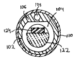
FIG. 1 is a cut-away partial side view of an ultrasound catheter assembly.
FIG. lA is a cross-sectional view of the ultrasound catheter assembly of FIG.
1 taken
along line 1 A-1 A.
DESCRIPTION OF THE PREFERRED EMBODIMENT
The improved transducer element having multiple resonant frequencies may be
used,
for example, in an ultrasound imaging system. Mechanical scanning ultrasound
imaging
catheter systems typically employ a single transducer mounted inside a
rotating housing. In
one example, the transducer transmits and receives ultrasonic waves while the
transducer
housing rotates about a fixed axis in an acoustic window located at a distal
tip of the catheter.
The rotational motion of the transducer housing is accomplished by a flexible
drive-shaft that
extends through an axially disposed lumen of the catheter, wherein the drive-
shaft has one end
connected to the transducer housing. Once the distal end of the catheter is
positioned, for
example, in a patient's vascular system, a cross-sectional image of the tissue
surrounding the
distal catheter tip is produced by using imaging and control circuitry that
are electrically
coupled to the transducer via an electrical conductor extending through the
drive shaft.
FIGS. 1 and lA illustrate an example embodiment of a flexible ultrasound
catheter 100.
Ultrasound catheter 100 is adapted to be positioned within the vascular system
by standard,
well-known catheter procedures by guiding the flexible catheter 100 through
various blood
vessels along a circuitous path, beginning, for example, by percutaneous
introduction through a
perforation of the femoral artery.
CA 02487917 2004-11-29
WO 03/103501 PCT/US03/14572
The catheter includes an elongate tubular member 102, which forms an axially
disposed
lumen 104. The inner dimensions of lumen 104 are sufficient to allow an
imaging core 118 to
be slidably disposed therein. The imaging core includes a flexible drive-shaft
120 connected to
a transducer housing 122 having a generally disk-shaped transducer 124 mounted
therein. The
imaging core 118 is capable of translation along its central axis. In
addition, imaging core 118
is capable of rotation about its central axis at speeds in excess of 1800 rpm.
Further disclosure
concerning rotatable, motor-driven imaging cores can be found in United States
Patent No.
6,004,269, which is incorporated herein by reference.
A dome-shaped acoustic imaging window 112 is attached to a distal end of the
elongate
tubular element 102, thereby forming an enclosed tip of the catheter 100.
Alternatively, the
shape of the acoustic imaging window 112, the transducer 124, or any other
component may be
virtually any shape or combination of shapes. A cover tube 110 formed of a
suitable material,
such as a heat shrinkable nylon, urethane, polyethylene or other plastic, is
disposed around
tubular element 102, wherein cover tube 110 provides both structural integrity
to the catheter
100, as well as a smooth outer surface for ease in axial movement in a
patient's body passage
with minimal friction.
Preferably, the acoustic imaging window 112 has its proximal end open and its
distal
end rounded and is attached to a distal outer circumferential portion of the
tubular element 102
to form an enclosed catheter tip 114, with respective ends of the cover tube
110 and acoustic
imaging window 112 bonded together at a common joint 116. The outer diameter
of the
proximal end of window 112 is substantially equal to that of the installed
cover tube 110, so
that a smooth outer surface is provided at joint 116. As best seen in FIG. 1,
optional upper
portion 106 of the elongate tubular member 102 forms a smaller lumen 108,
which can be used
for other catheter functions such as, by way of non-limiting examples, housing
pullwires, drug
delivery, balloon angioplasty, laser ablation, or for housing a stiffening
member to help prevent
the collapsing of the catheter 100. Of course, the catheter may have any
number of lumens of
any configuration. The catheter could have a balloon or a plurality of
balloons, if desired. The
catheter could also have more than one window, wires embedded in the catheter
walls, multiple
transducers, or other features known in the field. For example, the catheter
could use another
transducer in addition to the multiple resonant frequency transducer.
Optionally, the catheter
could even use a plurality of multiple resonant frequency transducers.
With further reference to the particular example shown in FIG. 1, the
transducer
housing 122 has a longitudinally disposed cut-away portion 113, which slopes
at a slight angle,
alpha, with respect to the central axis 126 of drive-shaft 120. The transducer
124 is mounted in
the cut-away portion 113 of the transducer housing 122 such that its active
surface 119 also
4
CA 02487917 2004-11-29
WO 03/103501 PCT/US03/14572
slopes at angle alpha with respect to central axis 126 of drive-shaft 120.
This tilting of
transducer 124 helps to minimize internal reflections inside of catheter tip
114.
Although the preferred transducer 124 of the present invention is disk-shaped,
it may,
alternatively, be any other shape. In use, it converts electrical energy
pulses into mechanical
energy, which propagates out from the face of the transducer 124 in the form
of ultrasonic
waves. The frequencies of these ultrasonic waves are dependent upon the
excitation
frequencies and the natural resonant frequencies of the transducer 124. The
natural resonant
frequencies of the transducer 124 are a product of the shape and thickness of
the transducer 123
and the transducer material.
Transducer 124 is formed in a known manner from materials capable of
transforming
pressure distortions on its surface into electrical voltages and vice versa.
Such materials
include, but are not limited to piezoelectric ceramic materials,
piezocomposite materials,
piezoelectric plastics, barium titanates, lead zirconate titanates, lead
metaniobates and
polyvinylidenefluorides.
As discussed above, the frequency at which the transducer 124 emits ultrasonic
waves
is a function of the resonant frequencies of the transducer 124 and the
frequency of the
excitation pulse sent to the transducer 124. When the ultrasonic waves impinge
on an object,
the ultrasonic waves are reflected back to the transducer 124, which converts
the mechanical
energy back into an electrical signal. The electrical signal from the
transducer 124 is
transmitted from the distal end of the catheter 100 to the catheter system's
imaging equipment
by a transmission line.
By using an imaging catheter 100 with a transducer 124 having multiple
resonant
frequencies, a user is capable of producing images having varying
characteristics depending
upon which frequency is utilized. Two important imaging characteristics are
depth of field and
resolution. Depth of field permits greater penetration during operation, which
can be useful for
imaging the in the heart, for example. High resolution is important for close-
up imaging within
a vessel such as a coronary artery. However, good depth of field comes at the
expense of lower
resolution and vice versa. Preferably, the improved transducer 124 is capable
of operating at
low, middle and high resonant frequencies in order to take advantage of the
differing imaging
characteristics of each frequency.
As an example, the multiple resonant frequency catheter 100 of the present
invention
can be configured to have three resonant frequencies at 30 MHz, lOMHz and 7.5
MHz. In a
single procedure, all three frequencies can be employed by a user. As an
example, suppose an
imaging procedure entails first imaging a coronary artery, then guiding the
catheter 100 into
the right atrium and then imaging the left atrium. To image the coronary
artery, the transducer
CA 02487917 2004-11-29
WO 03/103501 PCT/US03/14572
124 should be operated at the 30MHz frequency since the higher frequency
yields good close-
up resolution.
After imaging the coronary artery, the catheter must be guided into the right
atrium.
Since guiding the catheter into place requires both depth of field and
resolution, the middle
range frequency of l OMHz is preferred for this maneuver. Upon guiding the
catheter into
place in the right atrium, the left atrium is to be imaged. Imaging the atrium
at a distance will
require considerable depth of f eld and the lower 7.SMHz resonant frequency
will, therefore, be
preferable at this stage. In this manner, all three resonant frequencies can
be employed in a
typical imaging procedure. However, this procedure is merely one example of a
myriad of
imaging procedures that require a transducer to operate at multiple
frequencies.
Because the multiple resonant frequency transducer 124 is capable of
oscillation at
three resonant frequencies, the catheter and/or imaging core does not have to
be switched out
during operation. Switching catheters out is a timely procedure, which
necessarily makes the
imaging procedure longer than is necessary. Instead, a catheter can use a
single transducer 124
having multiple resonant frequencies, without having to sacrifice depth of
field or resolution.
Switching between resonant frequencies may be accomplished, for example, using
an external
instrument console. Any number of typical instrument console expedients can be
used to
accomplish this task including a button, dial, switch, voice command, mouse,
track ball, or
pointing device.
Any one or more of the features depicted in FIGS. 1 and lA, or described in
the
accompanying text, may be interchanged with that of another figure to form
still other
embodiments.
While preferred embodiments and methods have been shown and described, it will
be
apparent to one of ordinary skill in the art that numerous alterations may be
made without
departing from the spirit or scope of the invention. Therefore, the invention
is not limited
except in accordance with the following claims.
6