Note: Descriptions are shown in the official language in which they were submitted.
CA 02488382 2004-12-03
WO 03/104427 PCT/US03/18239
METHODS AND COMPOSITIONS FOR DETECTING CANCERS
Cross-Reference to Related Applications
This application claims the benefit of priority of U.S. Provisional
Application No.
60/386,653 filed June 5, 2002, the specification of which is incorporated by
reference herein in
its entirety.
Funding
Work described herein was supported by National Institutes of Health Grant
ROlCA
67409. The United States Government has certain rights in the invention.
Background
In 2001, over 1.2 million new cases of hurrian c ancer will be diagnosed and
over 0.5
million people will die from cancer (American C ancer Society a stimate).
Despite this, more
people than ever are living with and surviving cancer. In 1997, for example,
approximately 8.9
million living Americans had a history of cancer (National Cancer Institute
estimate). People
are more likely to survive cancer if the disease is diagnosed at an early
stage of development,
since treatment at that time is more likely to be successful. Early detection
depends upon
availability of high-quality methods. Such methods are also useful for
determining patient
prognosis, selecting therapy, monitoring response to therapy and selecting
patients for additional
therapy. Consequently, there is a need for cancer diagnostic methods that are
specific, accurate,
minimally invasive, technically simple and inexpensive.
Colorectal cancer (cancer of the colon or rectum) is one particularly
important type of
human cancer. Colorectal cancer is the second most common cause of cancer
mortality in adult
Americans (Landis, et al., 1999, CA Cancer J Clin, 49:8-31). Approximately 40%
of individuals
with colorectal cancer die. In 2001, it is estimated that there will be
135,400 new cases of
colorectal cancer (98,200 cases of colon and 37,200 cases of rectal cancer)
and 56,700 deaths
(48,000 colon cancer and 8,800 rectal cancer deaths) from the disease
(American Cancer
Society). As with other cancers, these rates can be decreased by improved
methods for
diagnosis. Although methods for detecting colon cancer exist, the methods are
not ideal.
Digital rectal exams (i.e., manual probing of rectum by a physician), for
example, although
-1-
CA 02488382 2004-12-03
WO 03/104427 PCT/US03/18239
relatively inexpensive, are unpleasant and can be inaccurate. Fecal occult
blood testing (i.e.,
detection of blood in stool) is nonspecific because blood in the stool has
multiple causes.
Colonoscopy and sigmoidoscopy (i.e., direct examination of the colon with a
flexible viewing
instrument) are both uncomfortable for the patient and expensive. Double-
contrast barium
enema (i.e., taking X-rays of barium-filled colon) is also an expensive
procedure, usually
performed by a radiologist.
Other cancers such as breast cancer, thyroid cancer and stomach cancer, cause
significant
public health problem as well. For example, thyroid cancer is the most common
endocrine
malignancy. In the United States, there are approximately 14,000 new patients
and 1,100 deaths
per year (Shah et al., 1995, CA Cancer J Clin 45:352-68). Because of the
disadvantages of
existing methods for detecting and treating cancer, new methods and tools in
cancer diagnosis
and cancer therapy are needed.
Summary'of the Invention
In accordance with the present invention, new diagnostic tools and methods for
detecting
cancer (e.g., colon cancer, breast cancer, thyroid cancer, or stomach cancer)
are provided. In
certain aspects, the invention is based in part on the discovery of a novel
polynucleotide
sequence encoding a novel sodium/solute symporter-like protein (SLCSA8).
Applicants
previously referred to the SLCSA8 gene as the "Huil" gene.
In one embodiment, the invention provides an isolated polypeptide comprising
an amino
acid sequence selected from the group consisting of a) an amino acid sequence
at least 95%
identical to SEQ )D NO: 1; and b) an amino acid sequence encoded by a nucleic
acid that
hybridizes under high stringency conditions to a nucleic acid of any one of
SEQ ID NOs: 3 or 4,
wherein said polypeptide is a cell surface protein. The subject polypeptide
comprises a
transmembrane domain as set forth in any one of SEQ B3 NOs: 19-31. The present
invention
contemplates the subject polypeptide as a sodium symporter.
In another embodiment, the invention provides an isolated antibody or fragment
thereof,
which is specifically immunoreactive with an epitope of a SCLSA8 protein
sequence as set forth
in SEQ II? NO: 1. The antibody of the invention can be selected from the group
consisting of: a
polyclonal antibody, a monoclonal antibody, an Fab fragment and a single chain
antibody.
Optionally, the antibody is labeled with a detectable label.
-2-
CA 02488382 2004-12-03
WO 03/104427 PCT/US03/18239
In another embodiment, the invention provides an isolated SCLSA8 nucleic acid
selected
from the group consisting of: a) a nucleic acid comprising the nucleotide
sequence of SEQ ZD
NO: 2, or a complement thereof; b) a nucleic acid molecule that encodes a
polypeptide
comprising the amino acid sequence at least 95% identical to the amino acid
sequence of SEQ
>I7 NO: 7; and c) a nucleic acid molecule that hybridizes under stringent
conditions to SEQ ID
NO: 2 . O ptionally, t he n ucleic a cid o f t he i nvention further c
omprises a v ector n ucleic a cid
sequence. In certain embodiments, the invention provides a kit comprising the
SLCSAB nucleic
acid probes or primers and instructions for use.
In another embodiment, the invention provides a host cell which contains the
subject
SCLSAB nucleic acid of the invention. In another embodiment, the invention
provides a method
for producing the subject polypeptide, comprising culturing the host cell
under conditions in
which the subj ect nucleic acid molecule is expressed.
In another embodiment, the invention provides a method for detecting the
presence of
the subject SCLSAB polypeptide in a sample, comprising: a) contacting the
sample with an
antibody which selectively binds to the polypeptide of claim 1; and b)
determining whether the
antibody binds to the polypeptide in the sample.
In another embodiment, the invention provides a kit for detecting a human
SCLSA8
polypeptide comprising: (i) an antibody of claim 2; and (ii) a detectable
label for detecting said
antibody.
In another embodiment, the invention provides a method for detecting the
presence of
the SCLSA8 nucleic acid in a sample, comprising: a) contacting the sample with
an SCLSAB
probe or primer; and b) determining whether the probe or primer binds to a
SCLSAB nucleic
acid in the sample.
In another embodiment, the invention provides a method for identifying a
compound
which binds to the SCLSA8 polypeptide, comprising: a) contacting the p
olypeptide, or a cell
expressing the SCLSA8 polypeptide, with a test compound; andb) determining
whether the
polypeptide binds to the test compound.
In another embodiment, the invention provides a method for modulating the
activity of
the SCLSA8 polypeptide, comprising contacting the polypeptide or a cell
expressing the
-3-
CA 02488382 2004-12-03
WO 03/104427 PCT/US03/18239
polypeptide with a compound which binds to the polypeptide in a sufficient
concentration to
modulate the activity of the polypeptide.
In another embodiment, the invention provides a method of inhibiting aberrant
activity of
a SLCSAB-expressing cell, comprising contacting the cell with a compound that
modulates the
activity or expression of the polypeptide, in an amount which is effective to
reduce or inhibit the
aberrant activity of the cell.
In certain embodiments, compounds used in the methods of the invention are
selected
from the group consisting of a peptide, a phosphopeptide, a small organic
molecule, an antibody,
and a peptidomimetic. Cells in the methods of the invention can be found in
the colon, kidney,
lung, esophagus, small bowel, stomach, thyroid, uterus, and breast.
In another embodiment, the invention provides a method of treating or
preventing a
disorder characterized by aberrant activity of a SLCSAB-expressing cell, in a
subject, comprising
administering to the subject an effective amount of a compound that modulates
the activity or
expression of the SLCSAB polypeptide, such that the aberrant activity of the
SLCSAB-
expressing cell is reduced or inhibited.
In another embodiment, the invention provides a transgenic mouse having
germline and
somatic cells comprising a chromosomally incorporated transgene that disrupts
the genomic
SLCSA8 gene and inhibits expression of said gene, wherein said disruption
comprises insertion
of a selectable marker sequence resulting in said transgenic mouse exhibiting
increased
susceptibility t o t he formation o f t umors a s c ompaxed t o t he w ildtype
mouse. T he t ransgenic
mouse can be homozygous r heterozygous for the disruption.
In another embodiment, the invention provides a transgenic mouse having
germline and
somatic cells in which at least one allele of a genomic SLCSAB gene is
disrupted by a
chromosomally incorporated transgene, which transgene inhibits the expression
of the genomic
SLCSA8 gene, wherein (i) the genomic SLCSA8 gene encodes a SLGSA8 protein; and
(ii) the
disruption comprises insertion of a selectable marker sequence, which replaces
all or a portion of
the genomic SLCSAS gene or is inserted into the coding sequence of the genomic
SLCSA8
gene; and (iii) the transgenic mouse has increased susceptibility to the
development of
neoplasms.
-4-
CA 02488382 2004-12-03
WO 03/104427 PCT/US03/18239
In another embodiment, the invention provides isolated mammalian cells
comprising a
diploid genome including a chromosomally incorporated transgene, which
transgene disrupts the
genomic SLCSA8 gene and inhibits expression of said gene. Optionally, the
cells are mouse
cells.
In another embodiment, the invention provides a method for generating a mouse
and
mouse embryonic stem cells having a functionally disrupted endogenous SLCSA8
gene,
comprising the steps of: (i) constructing a transgene construct including (a)
a recombination
region having all or a portion of the endogenous SLCSAB g ene, which r
ecombination region
directs recombination of the transgene w ith the a ndogenous SLCSA8 g ene; and
(b) a marker
sequence which provides a detectable signal for identifying the presence of
the transgene in a
cell; (ii) transfernng the transgene into embryonic stem cells of a mouse;
(iii) selecting
embryonic stem cells having a correctly targeted homologous recombination
between the
transgene and the SLCSA8 gene; (iv) transfernng said cells identified in step
(iii) into a mouse
blastocyst and implanting the resulting chimeric blastocyst into a female
mouse; and
(v) selecting offspring harboring an endogenous SLCSAB gene allele comprising
the correctly
targeted recombination.
In another embodiment, the invention provides a method of evaluating the
carcinogenic
potential of an agent comprising: (i) contacting the transgenic mouse of claim
16A with a test
agent; and (ii) comparing the number of transformed cells in a sample from the
treated mouse
with the number of transformed cells in a sample from an untreated transgenic
mouse or
transgenic mouse treated with a control agent, wherein the difference in the
number of
transformed cells in the treated mouse, relative to the number of transformed
cells in the absence
of treatment or treatment with a control agent, indicates the carcinogenic
potential of the test
compound.
In another embodiment, the invention provides a method of evaluating an anti-
proliferative activity of a test compound, comprising: (i) providing a
transgenic mouse of claim
16A having germline and somatic cells in which the expression of the SLCSA8
gene is inhibited
by said chromosomally incorporated transgene, or a sample of cells derived
therefrom; (ii)
contacting the transgenic mouse or the sample of cells with a test agent; and
(iii) determining the
number of transformed cells in a specimen from the transgenic mouse or in the
sample of cells,
wherein a s tatistically s ignificant d ecrease i n t he n umber o f t
ransformed c ells, r elative t o t he
-5-
CA 02488382 2004-12-03
WO 03/104427 PCT/US03/18239
number of transformed cells in the absence of the test agent, indicates the
test compound is a
potential anti-proliferative agent.
In certain aspects, the present invention is based, at least in part, on
Applicants'
discovery of a particular human genomic DNA region in which the cytosines
within CpG
dinucleotides are methylated in tissues from human cancers and unmethylated in
normal human
tissues. The region is referred to hereinafter as the "SLCSAB-methylation
target region" is
encompassed by base pairs 82200 to 83267 of GenBank entry AC063951, and is
located in the
promoter andlor exon 1 of the SLCSA8 gene. The present methods are also based,
at least in
part, on Applicants' discovery that the levels of SLCSA8 transcript in tissues
from human
cancers are lower than the levels of SLCSA8 transcript in normal tissues.
In one embodiment, the method comprises assaying for the presence of
differentially
methylated SLCSA8 nucleotide sequences (e.g., in the SLCSA8 methylation target
region) in a
tissue sample or a bodily fluid sample from a subject. Preferred bodily fluids
include blood,
serum, plasma, a blood-derived fraction, stool, colonic effluent or urine. In
one embodiment,
the method involves restriction enzyrne/methylation-sensitive PCR. In another
embodiment, the
method comprises reacting DNA from the sample with a chemical compound that
converts non-
methylated cytosine bases (also called "conversion-sensitive" cytosines), but
not methylated
cytosine bases, to a different nucleotide base. In a preferred embodiment, the
chemical
compound is sodium bisulfate, which converts unxnethylated cytosine bases to
uracil. The
compound-converted DNA is then amplified using a methylation-sensitive
polymerise chain
reaction (MSP) employing primers that amplify the compound-converted DNA
template if
cytosine bases within CpG dinucleotides of the DNA from the sample are
methylated.
Production of a PCR product indicates that the subject has cancer or
precancerous adenomas.
Other methods for assaying for the presence of methylated DNA are known in the
art.
In another embodiment, the method comprises assaying for decreased levels of
an
SLCSA8 transcript in the sample. A sequence of the SLCSA8 transcript (SEQ m
NO: 3) is
shown in Figure 2. The SLCSA8 transcript is encoded by 15 exons within the
present genomic
contig. In another aspect the method comprises assaying for decreased levels
of a protein
encoded by the SLCSA8 transcript in the sample.
In another embodiment, the present invention provides a detection method for
prognosis
of a cancer (e.g., colon cancer, breast cancer, thyroid cancer, or stomach
cancer) in a subject
-6-
CA 02488382 2004-12-03
WO 03/104427 PCT/US03/18239
known to have or suspected of having cancer. Such method comprises assaying
for the presence
of methylated SLCSA8 DNA (e.g., in the SLCSAB methylation target region) in a
tissue sample
or bodily fluid from the subject. In certain cases, it is expected that
detection of methylated
SLCSA8 DNA in a blood fraction is indicative of an advanced state of cancer
(e.g., colon
cancer). In other cased, detection of methylated SLCSA8 DNA in a tissue or
stool derived
sample or sample from other bodily fluids may be indicative of a cancer that
will respond to
therapeutic agents that demethylate DNA or reactivate expression of the SLCSA8
gene.
In another embodiment, the present invention provides a method for monitoring
over
time the status of cancer (e.g., colon cancer, breast cancer, thyroid cancer,
or stomach cancer) in
a subject. The method comprises assaying for the presence of methylated SLCSA8
DNA (e.g.,
in the SLCSA8 methylation target region) in a tissue sample or bodily fluid
taken from the
subject at a first time and in a corresponding tissue sample or bodily fluid
taken from the subject
at a second time. Absence of methylated SLCSA8 DNA from the tissue sample or
bodily fluid
taken at the first time and presence of methylated SLCSAB DNA in the tissue
sample or bodily
fluid taken at the second time indicates that the cancer is progressing.
Presence of methylated
SLCSA8 DNA in the tissue sample or bodily fluid taken at the first time and
absence of
methylated SLCSA8 DNA from the tissue sample or bodily fluid taken at the
second time
indicates that the cancer is regressing.
In another embodiment, the present invention provides a method for evaluating
therapy
in a subject having cancer or suspected of having cancer (e.g., colon cancer,
breast cancer,
thyroid cancer, or stomach cancer). The method comprises assaying for the
presence of
methylated SLCSAB DNA (e.g., in the SLCSA8 methylation target region) in a
tissue sample or
bodily fluid taken from the subject prior to therapy and a corresponding
bodily fluid taken from
the subject during or following therapy. Loss of or a decrease in the levels
of methylated
SLCSA8 DNA in the sample taken after or during therapy as compared to the
levels of
methylated SLCSAB DNA in the sample taken before therapy is indicative of a
positive effect of
the therapy on cancer regression in the treated subj ect.
The present invention also relates to oligonucleotide primer sequences for use
in assays
(e.g., methylation-sensitive PCR assays or HpaII assays) designed to detect
the methylation
status of the SLCSA8 gene. The present invention also relates to antibodies
and to
oligonucleotides or oligomers for detecting the presence the SLCSA8 protein or
the SLCSA8
'----°--~~~'~ -espectively, in samples obtained from a subject.
_7_
CA 02488382 2004-12-03
WO 03/104427 PCT/US03/18239
The present invention also provides a method of inhibiting or reducing growth
of cancer
cells (e.g., colon cancer, breast cancer, thyroid cancer, or stomach cancer).
The method
comprises increasing the levels of the protein encoded by SLCSA8 in cancer
cells. In one
embodiment, the cells are contacted with the SLCSA8 protein or a biologically
active equivalent
or fragment thereof under conditions permitting uptake of the protein or
fragment. In another
embodiment, the cells are contacted with a nucleic acid encoding the SLCSA8
protein and
comprising a promoter active in the cancer cell, wherein the promoter is
operably linked to the
region encoding the SLCSA8 protein, under conditions permitting the uptake of
the nucleic acid
by the cancer cell. In another embodiment, the method comprises demethylating
the methylated
SLCSAB DNA, or otherwise reactivating the silenced SLCSAB promoter.
In one embodiment, the application provides isolated or recombinant SLCSA8
nucleotide
sequences that are at least 80%, 85%, 90%, 95%, 98%, 99% or identical to the
nucleotide
sequence of any one of SEQ )D NOs: 2-4 and 21, fragments of said sequences
that are 10, 15,
20, 25, S0, 100, or 150 base pairs in length wherein the SLCSA8 nucleotide
sequences are
differentially methylated in an SLCSAB-associated disease cell.
In another embodiment, the application provides a method for detecting colon
cancer,
comprising: a) obtaining a sample from a patient; and b) assaying said sample
for the
presence of methylation of nucleotide sequences within at least two genes
selected from the
group consisting of: SLCSAB, HLTF, p16, and hMLHl; wherein methylation of
nucleotide
sequences within the two genes is indicative of colon cancer. In such methods,
the sample is a
bodily fluid selected from the group consisting of blood, serum, plasma, a
blood-derived
fraction, stool, urine, and a colonic effluent. For example, the bodily fluid
is obtained from a
subject suspected of having or is known to have colon cancer.
In another a mbodiment, t he application p rovides a k it f or d etecting c
olon c ancer i n a
subject, comprising primers for detecting methylation of nucleotide sequence
within at least two
genes selected from the group consisting of SLCSAB, HLTF, p16, and hMLHl,
wherein the
primers for detecting methylation of SLCSA8 nucleotide sequence are selected
from SEQ )D
NOs: 5-11; wherein the primers for detecting methylation of HLTF nucleotide
sequence are
selected from 5'-TGGGGTTTCGTGGTTTTTTCGCGC-3', 5'-
CCGCGAATCCAATCAAACGTCGACG-3', 5'-
ATTTTTGGGGTTTTGTGGTTTTTTTGTGT-3',
"'T'~' n r''~'''~GAA.ATCCAATCAAACATCAACA-3',
_g_
CA 02488382 2004-12-03
WO 03/104427 PCT/US03/18239
GCACGACTA.AAA.~°~ATAAATCGCCGCG-3',
AAACACACAACTAAAAAATAAATCACCACA-3', 5'-
TAAAACCTCGTAACTTTCCCGCGCG-3', 5'-GTCGCGAGTTTAGTTAGACGTCGAC-3',
5'-TCCTAAA.ACCTCATAACTTTCCCACACA-3', and 5'-
AGTTGTTGTGAGTTTAGTTAGATGTTGAT-3', wherein the primers for detecting
methylation of hMLH1 nucleotide sequence are selected from
5'AACGAATTAATAGGAAGAGCGGATAGCG-3', 5'-
CGTCCCTCCCTAA.AACGACTACTACCC-3', 5'-
CGTTTTTTTTTGAAGCGGTTATTGTTTGT-3', and 5'-
AACGAACCAATAAAAAAAACAAACAACG-3'. Tthe kit may further comprise a
compound to convert a template DNA. Optioanally the compound is bisulfate.
Brief Description of The Drawings
Figure 1 shows the complete sequence of the Genomic clone AC063951 (SEQ ID NO:
2), with nucleotides 82200-83267 underlined on pages 35 of Figure 1. This
region (nucleotides
82200-83267 of AC063951, SEQ ID NO: 12, see Figure 4) encompasses the promoter
andlor
exon 1 of the SLCSA8 gene, and is herein referred to as the "SLCSA8
methylation target
region."
Figure 2 shows the nucleotide sequence of the SLCSAB mRNA transcript (SEQ ID
NO:
3). The SLCSA8 transcript is encoded by 15 exons within the present genornic
contig.
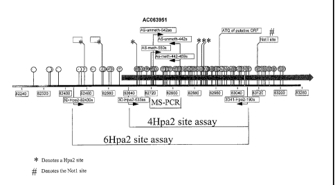
Figure 3 shows a diagram of the SLCSAB methylation target region. CpG sites
are
shown with circles and stems. The numerical coordinates are those of genornic
clone
AC063951. Lollipops designate CpG sites that are potential acceptors of
aberrant methylation.
Asterisks designate sites recognized by the HpaII restriction enzyme. Shown
are the positions of
PCR primers that amplify regions crossing 6 HpaII sites, or regions crossing 4
HpaII sites. Also
shown is the position of PCR primers designed for a methyl-specific PCR (MS-
PCR) assays.
Also shown in the gray bar is the 5' end of exon 1 of the SLCSAB transcript
which overlaps with
the methylation sites detected in both MS-PCR and HpaII based assays. Lastly
indicated is a
NotI site corresponding to methylation site 2D41 detected in Restriction
Landmark Genome
Scanning assay as methylated in colon cancer cell lines, though not in primary
tumors.
_g_
CA 02488382 2004-12-03
WO 03/104427 PCT/US03/18239
Figure 4 provides the sequence of AC063951 between nucleotides 82200-83267
(SEQ
DJ NO: 12), and designates every CpG site with a gray lollipop, and shows the
HpaII sites in the
assay as dark lollipops, and also shows the location of the PCR primers used
in the assay. In this
figure, the base pairs have been renumbered sequentially from 1-1068, with
nucleotide 82200
being renumbered as nucleotide 1.
Figure 5 shows the correlation between HpaII a ssays (over 4 HpaII sites and 6
HpaII
sites) and silencing of expression of the SLCSA8 transcript.
Figure 6 shows the results of the HpaII assays (over 4 HpaII sites and 6 HpaII
sites) in
actual colon cancer tumors and normal control colon tissues.
Figure 7 shows the results of assay for methylation at 61 CpG sites enumerated
in Figure
4 with site 1 corresponding to basepair 466 in Figure 4 and site 61
corresponding to basepair
1010. The bold arrows correspond to 4 of the HpaII sites at respectively
basepairs 466, 691,
709, and 716 in Figure 4. Methylation was assayed by sequencing DNA from
samples
following sodium bisulfite treatment of DNA that converts cytosine to uracil
but leaves methyl-
cytosine unchanged. Bases that are methylated are coded black, unmethylated
bases are coded
dark gray, and samples with both methylated and unmethylated bases are coded
light gray.
Figure 8 shows the wild-type sequence of the anti-sense strand of AC063951
between
bases 82200-83267 (SEQ ID NO: 13). Note that the sequence is the reverse
complement of that
shown in Figure 4, and therefore base number 1 on this diagram corresponds to
basepair 83267
in AC063951, and to basepair 1068 in Figure 4. Indicated on this diagram is
the position of the
MS-PCRl primers (AS-meth) and the UMS-PCR1 primers (AS-unmethy). The methyl
specific
MS-PCRI primers amplify a CpG sites numbered 6, 7, 8 and 15, 16, 17, 18
respectively in
Figure 7. The UMS-PCRl primers interrogate CpG sites 7, 8 and 15, 16, 17, 18
respectively.
Figure 9 shows a region within SEQ ID NO: 13 shown in Figure 8 (nucleotides
300-600,
SEQ ID NO: 14), and the sequences of the antisense strand that are amplified
by the methyl-
specific and unmethyl-specific PCR primers.
Figure 10 shows the bisulfate converted sequence of a uniformly methylated
SLCSA8
antisense s trand ( SEQ ID N O: 15), b ut n of t he w ild-type s equence o f t
he S LCSAB a ntisense
strand (corresponding to Figure 8). Indicated again are the position of the
methylation specific
PCR primers for the MS-PCRl assay.
-10-
CA 02488382 2004-12-03
WO 03/104427 PCT/US03/18239
Figure 11 shows the bisulfite converted sequence of a uniformly unmethylated
SLCSAB
antisense s trand ( SEQ ID N O: 16), b ut n of t he w ild-type s equence o f t
he S LCSAB a ntisense
strand shown in Figure 8. Indicated are the position of the unmethylation
specific PCR primers
for the UMS-PCRl assay.
Figure 12 provides the bisulfate converted sequence of the unmethylated SLCSAB
sense
strand of nucleotides 82200-83267 of AC063951, renumbered such that basepair
82200 is
designated as nucleotide 1 (SEQ ID NO: 17).
Figure 13 provides the bisulfate converted sequence of a uniformly methylated
SLCSA8
sense strand of nucleotides 82200-83267 (SEQ ID NO: 18).
Figure 14 shows the tabular results of MS-PCRl assay performed on 31 colon
cancer
cell lines that do or do not express the SLCSA8 transcript.
Figure 15 shows the tabular results of MS-PCRl assay performed on 63 matched
sets of
primary colon cancer tumor tissue and accompanying normal colon tissue.
Figure 16 shows the results of testing 12 normal colon tissues from
individuals without
colon cancer.
Figure 17 shows the tabular results of the MS-PCRl assay of 28 premalignant
colon
adenomas, 68% of which are detected.
Figure 18 shows the amino acid sequence (SEQ ID NO: 1) of the SLCSA8 protein.
Figure 19 shows RT-PCR detection of the SLCSA8 transcript in normal colon and
in a
minority subset of colon cancer cell lines.
Figure 20 shows RT-PCR detection of SLCSA8 transcript in colon cancer cell
lines that
have been treated with the DNA-demethylating agent 5-azacytidine. S-
azacytidine reactivates
expression of the SLCSAB gene in 6 of 8 colon cancer cell lines.
Figure 21 demonstrates detection of methylation of the SLCSA8 locus by showing
resistance o f the 1 ocus t o H pall d igestion. T he 4 H pall assay ( as d
escribed i n t he i nvention
disclosure) is based on PCR amplification of a portion of the SLCSA8 locus.
Lanes labeled U
show control amplification of undigested SLCSA8 DNA. Lanes labeled M show
amplification
""' T ' '' ~,t has first been cut with the restriction enzyme Msp 1.
-11-
CA 02488382 2004-12-03
WO 03/104427 PCT/US03/18239
Figure 22 demonstrates detection of SLCSA8 DNA methylation in primary colon
cancer
tumors but not in matched normal tissue from the same patients. Samples
labeled T represent
colon cancer tumor tissue; whereas samples labeled N represent the matched
normal tissue.
Figures 23A-23B show the identification of SLCSA8. (A) Shown is the genomic
structure of the SLCSA8 gene. Black boxes represent exons, and arrows the
start codon and
stop codons respectively. (B) The nucleotide sequence of the SLCSA8 coding
region (SEQ m
NO: 4).
Figures 24A-24F show SLCSA8 expression. (A) Shown is RT-PCR analysis
demonstrating SLGSAB transcript expression in three normal colon mucosa
samples (Nl, N2,
N3), but absence of SLCSA8 transcript in most colon cancer cell lines
(remaining samples). (B)
Shown is RT-PCR analysis demonstrating reactivation of SLCSA8 expression in
cell lines
treated with 5-azacytidine (+) compared to untreated (-) controls. (C)
Methylation specific PCR
(MS-PCR) assay for methylated (M) or unmethylated (U) SLC5A8 exon 1 sequences
detects
exclusively methylated templates in SLCSA8 silenced cell lines. (D) MS-PCR
detects only
unmethylated SLCSA8 templates in SLCSA8 expressing cell lines. (E) MS-PCR
detection of
methylated SLCSA8 templates in colon cancer tumors (T) antecedent to SLCSA8
methylated cell
lines (V425, V670). Matched normal colon tissue (I~ shows only unmethylated
templates.
Unmethylated templates in tumor tissue presumptively arise from contaminating
non-malignant
cells. (F) MS-PCR analysis of colon cancer tumors (T) and matched normal (I~
colon tissues.
Methyl specific bands are seen in each of the tumor samples, but none of the
normal controls.
Figures 25A-25B show real time MS-PCR analysis of SLCSAB methylation. Plotted
are
1000 times the ratio of measured SLCSAS methylated product to the control
MYODI derived
product. (A) Detection of SLCSA8 methylation in primary colon cancer tissues.
Column 1
displays values for normal colon tissues harvested from non-cancer resections
(daxk diamonds).
Column 2 displays values for normal colon tissues harvested from colon cancer
resections (dark
diamonds). Column 3 displays values for colon cancer tissues divided into
unmethylated
samples falling within the normal tissue range (dark diamonds at the bottom),
versus methylated
samples showing values greater than the normal tissue range (light diamonds at
the top).
Adjacent bars indicate population means. (B) Real time MS-PCR analysis of
SLCSA8
methylation in aberrant crypt foci. Column 1 displays values for 24 normal
colon tissues
harvested from colon resections from 11 individuals (dark diamonds). Column 2
displays values
r--' ~ ~'~~~-rant crypt foci harvested from the same 11 individuals'
resections. Dark diamonds (at
-12-
CA 02488382 2004-12-03
WO 03/104427 PCT/US03/18239
the bottom) indicate unmethylated samples within the normal range, and light
diamonds (at the
top) indicate methylated samples falling within the range previously
demonstrated by
methylated cancers. Adj acent bars indicate the mean value for each group.
Figure 26 shows real time MS-PCR analysis of SLCSA8 methylation in DNA
precipitated from the serum of colon cancer patients. Plotted are 1000 times
the ratio of
measured SLCSA8 methylated product to the control MYODI derived product.
Column 1
displays absence of detectable SLCSA8 methylation in serum of 13 individuals
whose colon
cancer tumors assayed as unmethylated by MS-PCR (dark diamonds at the bottom).
Column 2
displays values of SLCSA8 methylation in the serum of 10 individuals whose
colon cancer
tumors assayed as methylated by MS-PCR. Dark diamonds (at the bottom) indicate
6 sera
without detectable methylation, and light diamonds (at the top) indicate 4
sera in which SLCSAB
methylation was detectable.
Figures 27A-27B show SLCSA8 suppression of colon cancer colony formation.
Shown
are the number of 6418 resistant colonies arising from transfection with a
SLCSA8 expression
vector (SLCSA8) or a control empty expression vector (pcDNA) in SLCSA8
unmethylated and
expressing V364, V457, and V9M cells (panel A) as compared to SLCSA8
methylated and
deficient FET, V400, and RKO cells (panel B).
Figure 27 shows the cloning of SLCSA8 transcript. Black bars indicate
representative
ESTs. The lighter gray bar indicates sequence generated from an image clone.
The dark gray
bar indicates open reading frame encoding SLCSAB protein.
Figure 28 shows the protein alignments of SLCSAB, the closest marine homologue
of
SLCSA8, the human sodium iodide symporter SLCSAS, and the human sodium
dependent
multivitamin transporter SLCSA6.
Figures 30A-30B show methylation in SLCSA8 exon 1. (A) Diagrammatic
representation of the CpG island in SLCSA8 exon 1. Balloons represent CpG
dinucleotides.
Coordinates represent nucleotide positions numbered as per GenBank entry
AC063951.
Positions of the ATG and NotI site are indicated. Arrows cover the regions
interrogated by
primers for MS-PCR. (B) Diagrammatic summary of methylation status of the 62
CpG sites in
SLC5A8 exon 1 as determined by sequencing of bisulfite converted genomic DNA.
Each site is
sequentially represented by one shaded block. Black represents sites that are
fully methylated.
,~ represents sites that are fully unmethylated. And lighter gray represents
sites that
-13-
CA 02488382 2004-12-03
WO 03/104427 PCT/US03/18239
are partially methylated. Samples include 9 SLCSA8 silenced cell lines (Off
samples), 6
SLCSA8 expressing normal colonic mucosa (On samples designated I~, and 3
SLC5A8
expressing cell lines (On samples designated V). Arrows indicate sites that
are interrogated by
MS-PCR primers and bracket a differentially methylated region that is
unmethylated in SLCSA8
expressing samples and is methylated in SLCSA8 silenced samples.
Figure 30 shows methylation events in primary colon cancers. Shown is analysis
of 64
primary colon cancers for aberrant methylation at 4 genomic loci, SLCSAB,
HLTF, hMLHI, and
p16. Black bars represent positive assays for methylation in tumor tissue, and
gray bars
represent detection only of unmethylated alleles.
Figure 31 shows suppression of xenograft growth in 4 of 5 SLCSAB expressing
V400
transfected clones (square symbols, gray lines) as compared with control pools
of V400 cells
transfected with an empty expression vector (triangular symbols, black lines).
Detailed Description of the Invention
I. Definitions
For convenience, certain terms employed in the specification, examples, and
appended
claims are collected here. Unless defined otherwise, all technical and
scientific terms used
herein have the same meaning as commonly understood by one of ordinary skill
in the art to
which this invention belongs.
The articles "a" and "an" are used herein to refer to one or to more than one
(i.e., to at
least one) of the grammatical object of the article, unless the context
clearly indicates otherwise.
By way of example, "an element" means one element or more than one element.
The terms "adenoma", "colon adenoma," and "polyp" are used herein to describe
any
precancerous neoplasia of the colon.
The term "blood-derived fraction" herein refers to a component or components
of whole
blood. Whole blood comprises a liquid portion (i.e., plasma) and a solid
portion (i.e., blood
cells). The liquid and solid portions of blood are each comprised of multiple
components; e.g.,
different proteins in plasma or different cell types in the solid portion. One
of these components
or a mixture of any of these components is a blood-derived fraction as long as
such fraction is
m;~~;n~ ~ne or more components found in whole blood.
-14-
CA 02488382 2004-12-03
WO 03/104427 PCT/US03/18239
"Cells," "host cells" or "recombinant host cells" are terms used
interchangeably herein.
It is understood that such terms refer not only to the particular subject cell
but to the progeny or
potential progeny of such a cell. Because certain modifications may occur in
succeeding
generations due to either mutation or environmental influences, such progeny
may not, in fact,
be identical to the parent cell, but are still included within the scope of
the term as used herein.
A "chimeric polypeptide" or "fusion polypeptide" is a fusion of a first amino
acid
sequence with a second amino acid sequence where the first and second amino
acid sequences
are not naturally present in a single polypeptide chain.
The term "colon" as used herein is intended to encompass the right colon
(including the
cecum), the transverse colon, the left colon, and the rectum.
The terms "colorectal cancer" and "colon cancer" are used interchangeably
herein to
refer to any cancerous neoplasia of the colon (including the rectum, as
defined above).
The terms "compound", "test compound," and "agent" are used herein
interchangeably
and are meant to include, but are not limited to, peptides, nucleic acids,
carbohydrates, small
organic molecules, natural product extract libraries, and any other molecules
(including, but not
limited to, chemicals, metals, and organometallic compounds).
The term "compound-converted DNA" herein refers to DNA that has been treated
or
reacted with a chemical compound that converts unmethylated C bases in DNA to
a different
nucleotide base. For example, one such compound is sodium bisulfite, which
converts
unmethylated C to U. If DNA that contains conversion-sensitive cytosine is
treated with sodium
bisulfate, t he c ompound-converted D NA w ill c ontain U i n p lace o f C . I
f t he D NA w hich i s
treated with sodium bisulfate contains only methylcytosine, the compound-
converted DNA will
not contain uracil in place of the methylcytosine.
The term "de-methylating agent" as used herein refers agents that restore
activity and/or
gene expression of target genes silenced by methylation upon treatment with
the agent.
Examples of such agents include without limitation 5-azacytidine and 5-aza-2'-
deoxycytidine.
The term "detection" is used herein to refer to any process of observing a
marker, in a
biological sample, whether or not the marker is actually detected. In other
words, the act of
probing a sample for a marker is a "detection" even if the marker is
determined to be not present
-15-
CA 02488382 2004-12-03
WO 03/104427 PCT/US03/18239
or b elow t he 1 evel o f s ensitivity. D etection m ay be a q uantitative, s
emi-quantitative o r n on-
quantitative observation.
The term "differentially methylated SLCSA8 nucleotide sequence" refers to a
region of
the SLCSA8 nucleotide sequence that is found to be methylated in a SLCSAB-
associated cancer
such as a region of the SLCSA8 nucleotide sequence that is found to be
methylated in cancer
tissues or cell lines, but not methylated in the normal tissues or cell lines.
For example, Figure 3
delineates certain SLCSA8 regions that are differentially methylated, such as
SEQ )D NOs: 11-
13.
"Expression vector" refers to a replicable DNA construct used to express DNA
which
encodes the desired protein and which includes a transcriptional unit
comprising an assembly of
(1) genetic elements) having a regulatory role in gene expression, for
example, promoters,
operators, or enhancers, operatively linked to (2) a DNA sequence encoding a
desired protein (in
this case, a SLCSA8 protein) which is transcribed into mRNA and translated
into protein, and
(3) appropriate transcription and translation initiation and termination
sequences. The choice of
promoter and other regulatory elements generally varies according to the
intended host cell. In
general, expression vectors of utility in recombinant DNA techniques are often
in the form of
"plasmids" which refer to circular double stranded DNA loops which, in their
vector form are
not bound to the chromosome. In the present specification, "plasmid" and
"vector" are used
interchangeably as the plasmid is the most commonly used form of vector.
However, the
invention is intended to include such other forms of expression vectors which
serve equivalent
functions and which become known in the art subsequently hereto.
In the expression vectors, regulatory elements controlling transcription or
translation can
be generally derived from marrunalian, microbial, viral or insect genes. The
ability to replicate
in a host, usually conferred by an origin of replication, and a selection gene
to facilitate
recognition of transformants may additionally be incorporated. Vectors derived
from viruses,
such as retroviruses, adenoviruses, and the like, may be employed.
As used herein, the phrase "gene expression" or "protein expression" includes
any
information pertaining to the amount of gene transcript or protein present in
a sample, as well as
information about the rate at which genes or proteins are produced or are
accumulating or being
degraded (e.g., reporter gene data, data from nuclear runoff experiments,
pulse-chase data etc.).
Certain kinds of data might be viewed as relating to both gene and protein
expression. For
-16-
CA 02488382 2004-12-03
WO 03/104427 PCT/US03/18239
example, protein levels i n a cell are reflective of the level of protein as
well as the level of
transcription, and such data is intended to be included by the phrase "gene or
protein expression
information." Such information may be given in the form of amounts per cell,
amounts relative
to a control gene or protein, in unitless measures, etc.; the term
"information" is not to be limited
to any particular means of representation and is intended to mean any
representation that
provides relevant information. The term "expression levels" refers to a
quantity reflected in or
derivable from the gene or protein expression data, whether the data is
directed to gene transcript
accumulation or protein accumulation or protein synthesis rates, etc.
The terms "healthy", "normal," and "non-neoplastic" are used interchangeably
herein to
refer to a subject or particular cell or tissue that is devoid (at least to
the limit of detection) of a
disease condition, such as a neoplasia (e.g., cancer), that is associated with
SLCSA8 such as for
example n eoplasia a ssociated w ith s ilencing o f S LCSA8 g ene a xpression
d ue t o m ethylation.
These terms are often used herein in reference to tissues and cells of the
colon. Thus, for the
purposes of this application, a patient with severe heart disease but lacking
a SLCSAB silencing-
associated disease would be termed "healthy."
"Homology" or "identity" or "similarity" refers to sequence similarity between
two
peptides or between two nucleic acid molecules. Homology and identity can each
be determined
by comparing a position in each sequence which may be aligned for purposes of
comparison.
When an equivalent position in the compared sequences is occupied by the same
base or amino
acid, then the molecules are identical at that position; when the equivalent
site occupied by the
same or a similar amino acid residue (e.g., similar in steric and/or
electronic nature), then the
molecules can be referred to as homologous (similar) at that position.
Expression as a
percentage of homology/similarity or identity refers to a function of the
number of identical or
similar amino acids at positions shared by the compared sequences. A sequence
which is
"unrelated" or "non-homologous" shares less than 40% identity, preferably less
than 25%
identity with a sequence of the present invention. In comparing two sequences,
the absence of
residues (amino acids or nucleic acids) or presence of extra residues also
decreases the identity
and homology/similarity.
The term "homology" describes a mathematically based comparison of sequence
similarities which is used to identify genes or proteins with similar
functions or motifs. The
nucleic acid and protein sequences of the present invention may be used as a
"query sequence"
+~ ~ °-~~ ~ a s eaxch a gainst p ublic d atabases t o, f or a xample, i
dentify o ther family m embers,
-17-
CA 02488382 2004-12-03
WO 03/104427 PCT/US03/18239
related sequences or homologs. Such searches can be performed using the NBLAST
and
XBLAST p rograms ( version 2 .0) o f A ltschul, a t al. ( 1990) JMoI. B iol.
215:403-10. B LAST
nucleotide searches can be performed with the NBLAST program, score=100,
wordlength=12 to
obtain nucleotide sequences homologous to nucleic acid molecules of the
invention. BLAST
protein searches can be performed with the XBLAST program, score=50,
wordlength=3 to
obtain amino acid sequences homologous to protein molecules of the invention.
To obtain
gapped aligiunents f or c omparison p urposes, G apped BLAST c an b a a
tilized a s d escribed i n
Altschul et al., (1997) Nucleic Acids Res. 25(17):3389-3402. When utilizing
BLAST and
Gapped B LAST programs, the default parameters of the r espective programs
(e.g., XBLAST
and BLAST) can be used. See http://www.ncbi.nlm.nih.gov.
As used herein, "identity" means the percentage of identical nucleotide or
amino acid
residues at corresponding positions in two or more sequences when the
sequences are aligned to
maximize sequence matching, i.e., taking into account gaps and insertions.
Identity can be
readily calculated by known methods, including but not limited to those
described in
(Computational Molecular Biology, Lesk, A. M., ed., Oxford University Press,
New York, 1988;
Biocomputing: Informatics and Genome Projects, Smith, D. W., ed., Academic
Press, New
York, 1993; Computer Analysis of Sequence Data, Part I, Griffin, A. M., and
Griffin, H. G.,
eds., Humana Press, New Jersey, 1994; Sequence Analysis in Molecular Biology,
von Heinje,
G., Academic Press, 1987; and Sequence Analysis Primer, Gribskov, M. and
Devereux, J., eds.,
M Stockton Press, New York, 1991; and Carillo, H., and Lipman, D., SIAM J.
Applied Math.,
48: 1073, 1988). Methods to determine identity are designed to give the
largest match between
the sequences tested. Moreover, methods to determine identity are codified in
publicly available
computer programs. Computer program methods to determine identity between two
sequences
include, but are not limited to, the GCG program package (Devereux, J., et
al., Nucleic Acids
Research 12(1): 387 (1984)), BLASTP, BLASTN, and FASTA (Altschul, S. F. et
al., J. Molec.
Biol. 215: 403-410 (1990) and Altschul et al. Nue. Acids Res. 25: 3389-3402
(1997)). The
BLAST X program is publicly available from NCBI and other sources (BLAST
Manual,
Altschul, S., et al., NCBI NLM NIH Bethesda, Md. 20894; Altschul, S., et al.,
.I. Mol. Biol. 215:
403-410 (1990)). The well known Smith Waterman algorithm may also be used to
determine
identity.
"SLCSA8-associated cancer" refers to cancer associated with reduced expression
or no
expression of the SLCSAB gene (previously referred to as the Huil gene), and
cancer associated
-ential methylation of SLCSAB DNA. Examples of SLCSAB-associated cancer
-18-
CA 02488382 2004-12-03
WO 03/104427 PCT/US03/18239
include, but are not limited to, colon cancer, breast cancer, thyroid cancer,
and stomach cancer.
As used herein, the SLCSAB-associated cancers includes both cancers and pre-
cancer adenomas.
"SLCSAB-associated proliferative disorder" refers to a disease that is
associated with
either reduced expression or over-expression of the SLCSA8 gene.
A "SLCSAB-associated protein" refers to a protein capable of interacting with
and/or
binding to a SLCSA8 polypeptide. Generally, the SLCSAB-associated protein may
interact
directly or indirectly with the SLCSA8 polypeptide.
"SLCSAB-methylation target regions" as used herein refer to those regions of
SLCSA8
that are found to be methylated. These regions include nucleotide regions that
may be either
constitutively or differentially methylated regions. For example, Figure 3
discloses a SLCSA8
region wherein certain sequences of this region are differentially methylated
regions.
"SLCSAB-nucleotide sequence" or "SLCSAB-nucleic acid sequence" as used herein
refers to the SLCSA8 nucleotide sequences as set forth in SEQ m NOs: 2-7 and
fragments
thereof.
"SLCSA8-silencing associated diseases" as used herein includes SLCSAB-
associated
cancer.
The term "including" is used herein to mean, and is used interchangeably with,
the
phrase "including but not limited to."
The term "isolated" as used in reference to nucleic acids or polypeptides
indicates a
nucleic acid or polypeptide, such as a SLCSA8 nucleic acid or polypeptide,
that is isolated from,
or otherwise substantially free of other proteins that are normally associated
with the nucleic
acid or polypeptide.
The term "methylation-sensitive PCR" (i.e., MSP) herein refers to a polymerise
chain
reaction in which amplification of the compound-converted template sequence is
performed.
Two sets of primers are designed for use in MSP. Each set of primers c
omprises a forward
primer and a reverse primer. One set of primers, called methylation-specific
primers, will
amplify the compound-converted template sequence if C bases in CpG
dinucleotides within the
template DNA (e.g., a SLCSA8 nucleic acid) are methylated. Another s et of
primers, c aped
"r,-"Pthvlation-specific primers, will amplify the compound-converted template
sequences if C
-19-
CA 02488382 2004-12-03
WO 03/104427 PCT/US03/18239
bases in CpG dinucleotides within the template DNA (e.g., a SLCSA8 nucleic
acid) are not
methylated.
The term "nucleic acid" refers to polynucleotides such as deoxyribonucleic
acid (DNA),
and, where appropriate, ribonucleic acid (RNA). The term should also be
understood to include,
as equivalents, analogs of either RNA or DNA made from nucleotide analogs,
and, as applicable
to the embodiment being described, single (sense or antisense) and double-
stranded
polynucleotides.
"Operably linked" when describing the relationship between two DNA regions
simply
means that they are functionally related to each other. For example, a
promoter or other
transcriptional regulatory sequence is operably linked to a coding sequence if
it controls the
transcription of the coding sequence.
The term "or" is used herein to mean, and is used interchangeably with, the
term
"and/or", unless context clearly indicates otherwise.
The terms "polypeptide" and "protein" are used interchangeably herein.
The term "recombinant" as used in reference to a nucleic acid indicates any
nucleic acid
that is positioned adjacent to one or more nucleic acid sequences that it is
not found adjacent to
in nature. A recombinant nucleic acid may be generated in vitro, for example
by using the
methods of molecular biology, or in vivo, for example by insertion of a
nucleic acid at a novel
chromosomal location by homologous or non-homologous recombination. The term
"recombinant" as used in reference to a polypeptide indicates any polypeptide
that is produced
by expression and translation of a recombinant nucleic acid.
A "sample" includes any material that is obtained or prepared for detection of
a
molecular marker or a change in a molecular marker such as the methylation
state, or any
material that is contacted with a detection reagent or detection device for
the purpose of
detecting a molecular marker or a change in the molecular marker.
A "subject" is any organism of interest, generally a mammalian subject, such
as a mouse,
and preferably a human subject.
The term "transgene" is used herein to describe genetic material which has
been or is
about to be artificially inserted into the genome of a mammal, particularly a
mammalian cell of a
aal. By "transgenic animal" is meant a non-human animal, usually a mammal
(e.g.,
-20-
CA 02488382 2004-12-03
WO 03/104427 PCT/US03/18239
mouse, rat, rabbit, hamster, etc.), having a non-endogenous nucleic acid
sequence present as an
extrachromosomal element in a portion of its cells or stably integrated into
its germ line DNA
(i.e., in the genomic sequence of most or all of its cells). Heterologous
nucleic acid is
introduced into the germ line of such transgenic animals by genetic
manipulation of, for
example, embryos or embryonic stem cells of the host animal.
II. Overview
In certain aspects, the invention relates, in part, to methods for determining
whether a
patient is likely or unlikely to have a cancer, for example, colon neoplasia.
A colon neoplasia is
any cancerous or precancerous growth located in, or derived from, the colon.
The colon is a
portion of the intestinal tract that is roughly three feet in length,
stretching from the end of the
small intestine to the rectum. Viewed in cross section, the colon consists of
four distinguishable
layers arranged in concentric rings surrounding an interior space, termed the
lumen, through
which digested materials pass. In order, moving outward from the lumen, the
layers are termed
the mucosa, the submucosa, the rnuscularis propria and the subserosa. The
mucosa includes the
epithelial layer (cells adj acent to the lumen), the basement membrane, the
lamina propria and the
muscularis mucosae. In general, the "wall" of the colon is intended to refer
to the submucosa
and the layers outside of the submucosa. The "lining" is the mucosa.
Precancerous colon neoplasias are referred to as adenomas or adenomatous
polyps.
Adenomas are typically small mushroom-like or wart-like growths on the lining
of the colon and
do not invade into the wall of the colon. Adenomas may be visualized through a
device such as
a colonoscope or flexible sigmoidoscope. Several studies have shown that
patients who iuidergo
screening for and removal of adenomas have a decreased rate of mortality from
colon cancer.
For this and other reasons, it is generally accepted that adenomas are an
obligate precursor for
the vast majority of colon cancers. When a colon neoplasia invades into the
basement
membrane of the colon, it is considered a colon cancer, as the term "colon
cancer" is used
herein. In describing colon cancers, this specification will generally follow
the so-called
"Dukes" colon cancer staging system. The characteristics that the describe a
cancer are
generally of greater significance than the particular teen used to describe a
recognizable stage.
The most widely used staging systems generally use at least one of the
following characteristics
for staging: the extent of tumor penetration into the colon wall, with greater
penetration
generally correlating with a more dangerous tumor; the extent of invasion of
the tumor through
t~, P ~ "t nn wall a nd i nto other n eighboring t issues, w ith greater i
nvasion generally correlating
-21-
CA 02488382 2004-12-03
WO 03/104427 PCT/US03/18239
with a more dangerous tumor; the extent of invasion of the tumor into the
regional lymph nodes,
with greater invasion generally correlating with a more dangerous tumor; and
the extent of
metastatic invasion into more distant tissues, such as the liver, with greater
metastatic invasion
generally correlating with a more dangerous disease state.
"Dukes A" and "Dukes B" colon cancers are neoplasias that have invaded into
the wall
of the colon but have not spread into other tissues. Dukes A colon cancers are
cancers that have
not invaded beyond the s ubmucosa. Dukes B colon cancers are subdivided into
two groups:
Dukes B 1 and Dukes B2. "Dukes B 1" colon cancers are neoplasias that have
invaded up to but
not t hrough t he m uscularis p ropria. D ukes B2 colon c ancers a re c ancers
that h ave b reached
completely through the muscularis propria. Over a five year period, patients
with Dukes A
cancer who receive surgical treatment (i.e., removal of the affected tissue)
have a greater than
90% survival rate. Over the same period, patients with Dukes B1 and Dukes B2
cancer
receiving surgical treatment have a survival rate of about 85% and 75%,
respectively. Dukes A,
B1 and B2 cancers are also referred to as T1, T2 and T3-T4 cancers,
respectively. "Dukes C"
colon cancers are cancers that have spread to the regional lymph nodes, such
as the lymph nodes
of the gut. Patients with Dukes C cancer who receive surgical treatment alone
have a 35%
survival rate over a five year period, but this survival rate is increased to
60% in patients that
receive chemotherapy. " Dukes D" colon c ancers are c ancers that have m
etastasized to other
organs. The liver is the most common organ in which metastatic colon cancer is
found. Patients
with Dukes D colon cancer have a survival rate of less than 5% over a five
year period,
regardless of the treatment regimen. In general, colon neoplasia develops
through one of at least
three different pathways, termed chromosomal instability, microsatellite
instability, and the CpG
island methylator phenotype (CIMP). Although there is some overlap, these
pathways tend to
present somewhat different biological behavior. By understanding the pathway
of tumor
development, the target genes involved, and the mechanisms underlying the
genetic instability, it
is possible to implement strategies to detect and treat the different types of
colon neoplasias.
In one aspect, this application is based at least in part, on the recognition
that certain
target genes may be silenced or inactivated by the differential methylation of
CpG islands in the
5' flanking or promoter regions of the target gene. CpG islands are clusters
of cytosine-
guanosine residues in a DNA sequence, that are prominently represented in the
5-flanking region
or promoter region of about half the genes in our genome. In particular, this
application is based
at least in part on the recognition that differential methylation of the
SLCSA8 nucleotide
-22-
CA 02488382 2004-12-03
WO 03/104427 PCT/US03/18239
sequence may be indicative of a cancer (e.g., colon cancer, breast cancer,
thyroid cancer, or
stomach cancer).
As noted above, early detection of colon neoplasia, coupled with appropriate
intervention, is important for increasing patient survival rates. Present
systems for screening for
colon neoplasia are deficient for a variety of reasons, including a lack of
specificity and/or
sensitivity (e.g., Fecal Occult Blood Test, flexible sigmoidoscopy) or a high
cost and intensive
use of medical resources (e.g., colonoscopy). Alternative systems for
detection of colon
neoplasia would be useful in a wide range of other clinical circumstances as
well. For example,
patients who receive surgical and/or pharmaceutical therapy for colon cancer
may experience a
relapse. It would be advantageous to have an alternative system for
determining whether such
patients have a recurrent or relapsed colon neoplasia. As a further example,
an alternative
diagnostic system would facilitate monitoring an increase, decrease or
persistence of colon
neoplasia in a patient known to have a colon neoplasia. A patient undergoing
chemotherapy
may be monitored to assess the effectiveness of the therapy.
In another aspect, the invention is also based, in part, on the discovery of a
novel
polynucleotide sequence encoding a novel sodium/solute symporter-like protein
(SLCSAB). In
particular, SLCSA8 is closely related to the human sodium iodide symporter
(SLCSAS) and the
human sodium-dependent multivitamin transporter (SLCSA6).
Cell surface receptors and transmembrane transporter systems facilitate
communication
between cells and their environment by direct exchange of chemicals between
the intracellular
and extracellular milieu. Distinct transporter systems (also called permeases,
porters,
transporters, carriers, and channel proteins) are specific for ions, small and
medium size solutes
and macromolecules. A major class of transporter proteins couple solute
transport to the
movement of other species (often cations, such as protons and sodium ions)
either in the same
direction (cotransporter or symporter) or in the opposite direction (counter
transporter or
antiporter). Sodium/solute symport is a widespread mechanism of solute
transport across
cytoplasmic membranes of prokaryotic and eukaryotic cells. Proteins that
catalyze
sodimn/solute symport have been grouped into eleven families based on their
degree of
sequence similarities, their solute and cation specificities, size,
topographical features, and
evolutionary relationships (see, e.g., Reizer et al., (1994) Bichemica et
Biphysica Acta,
1197:133-166). There axe mixed families of transporters whose members differ
in the choice of
'-'-~ ----~'=~~g ion or catalyze symport or antiport processes.
-23-
CA 02488382 2004-12-03
WO 03/104427 PCT/US03/18239
Human sodium iodide transporter (NIS, or SLCSAS) is a best characterized
member
among the sodiumlsolute symporter superfamily. NIS localizes at the
basolateral membrane and
catalyses the active transport of iodide from blood into the cells using the
inwardly directed
sodium gradient with a 2 sodium 1 iodide stoichiometry. The tissue
distribution of NIS includes
the thyroid, salivary glands, stomach, thymus, and breast. Lower levels of
expression of NIS are
detected in the prostate, ovary, adrenal gland, lung, and heart. By contrast,
the NIS gene has not
been detected in the colon, orbital fibroblasts, or nasophaxyngeal mucosa
(see, e.g., Filetti et al.,
1999, Eur J Endocrinol. 141:443-4.57). Abnormal NIS expression andlor iodide
transport
activity have been linked to many thyroid diseases including autoimmune
thyroid diseases,
thyroid nodular hyperplasia, thyroid adenoma, thyroid carcinoma, and
congenital
hypothyroidism, as well as non-thyroid diseases such as breast cancer and
stomach cancer
(Chung, 2002, J Nucl Med 43:1188-200).
Besides sequence homology to the human sodium iodide transporter, SLCSA8
transcript
was found by Applicants to be expressed in the normal colon mucosa, kidney,
lung, esophagus,
small bowel, stomach, thyroid, and uterus. In addition, Applicants found that
SLCSA8 may
function as a sodium iodide transporter, and that differential methylation of
SLCSA8 and/or
reduced expression of SLCSA8 are linked to diseases such as colon cancer,
breast cancer, and
stomach c ancer. A ccordingly, t he p resent i nvention r elates t o m ethods
a nd c ompositions f or
detecting and treating such SLCSAB associated cancers.
III. SLCSA8 polypeptides
In certain aspects, the invention provides a full-length SLCSA8 polypeptide
(SEQ ID
NO: 1) and functional variants thereof. Preferred functional variants of
SLCSAB polypeptides
are those that have tumor suppressor activity or sodium transporter activity.
In certain aspects,
the present invention includes biologically-active fragments of the SLCSAB
protein and fusion
proteins including at least a portion of the SLCSA8 protein. These include
proteins with
SLCSA8 activity that have amino acid substitutions or have sugars or other
molecules attached
to amino acid functional groups.
In certain embodiments, the present disclosure makes available isolated andlor
purified
forms of the SLCSAB polypeptides, which are isolated from, or otherwise
substantially free of,
other proteins which might normally be associated with the protein or a
particular complex
including the protein. In certain embodiments, variant polypeptides have an
amino acid
-24-
CA 02488382 2004-12-03
WO 03/104427 PCT/US03/18239
sequence that is at least 75% identical to an amino acid sequence as set forth
in SEQ ID NO: 1.
In other embodiments, the variant polypeptide has an amino acid sequence at
least 80%, 85%,
90%, 95%, 97%, 98%, 99% or 100% identical to an amino acid sequence as set
forth in SEQ ID
NO: 1.
In certain aspects, variant SLCSAB polypeptides are agonists or antagonists of
the
SLCSA8 polypeptide as set forth in SEQ ll~ N0: 1. Variants of these
polypeptides may have a
hyperactive or constitutive activity, or, act to prevent the tumor suppressor
activity or sodium
transporter activity of SLCSAB. For example, a truncated form lacking one or
more domain
may have a dominant negative effect.
In certain aspects, isolated peptidyl portions of the SLCSAB polypeptide can
be obtained
by screening polypeptides recombinantly produced from the corresponding
fragment of the
nucleic acid encoding the polypeptide as set forth in SEQ ID NO: 1. In
addition, fragments can
be chemically synthesized using techniques known in the art such as
conventional Mernfield
solid phase f Moc or t-Boc chemistry. The fragments can be produced
(recombinantly or by
chemical synthesis) and tested to identify those peptidyl fragments which can
function as either
agonists or antagonists of the SLCSA8 activity (e.g., tumor suppressor or
sodium solute
symporter).
The SLCSAB protein is a transmembrane protein, with portions of the protein
that are
positioned outside the cell (the extracellular portions) and portions of the
protein that are
positioned inside the cell (the intracellular portions). Sequences and
positions of the predicated
thirteen transmembrane domains (TMl- TM13) are listed below.
TM1 (residues 10-32): FVVWDYVVFAGMLVISAAIGIYY (SEQ ID NO: 19)
TM2 (residues 52-74): MTAVPVALSLTASFMSAVTVLGT (SEQ ID N0: 20)
TM3 (residues 84-106): IFSIFAFTYFFVWISAEVFLPV (SEQ ID NO: 21)
TM4 (residues 127-149): VRLCGTVLFIVQTILYTGIVIYA (SEQ )D NO: 22)
TMS (residues 164-186): GAVVATGVVCTFYCTLGGLI~AVI (SEQ >D NO: 23)
TM6 (residues 193-215): IGIMVAGFASVIIQAVVMQGGIS (SEQ ID NO: 24)
'"" ''7 (residues 240-259): HTFWTIIIGGTFTWTSIYGV (SEQ ID NO: 25)
-25-
CA 02488382 2004-12-03
WO 03/104427 PCT/US03/18239
TM8 (residues 280-302): LYINLVGLWAILTCSVFCGLALY (SEQ ID N0: 26)
TM9 (residues 337-359): LPGLFVACAYSGTLSTVSSSINA (SEQ ID N0: 27)
TM10 (residues 380-402): SLSWISQGMSVVYGALCIGMAAL (SEQ ID NO: 28)
TM11 (residues 412-434): AALSVFGMVGGPLMGLFALGILV (SEQ ID NO: 29)
TM12 (residues 441-463): GALVGLMAGFAISLWVGIGAQIY (SEQ ID NO: 30)
TM13 (residues 519-541): LSYLYFSTVGTLVTLLVGILVSL (SEQ ID NO: 31)
Thus, certain embodiments of the invention include SLCSA8 fragments comprising
a
transmembrane domain as set forth in any of SEQ ID NOs: 19-21. In other
embodiments, the
present invention includes SLCSA8 fragments comprising an intracellular domain
or an
extracellular portion of the SLCSAB protein.
In certain aspects, variant SLCSA8 polypeptides containing one or more fusion
domains.
Well known examples of such fusion domains include, for example,
polyhistidine, Glu-Glu,
glutathione S transferase (GST), thioredoxin, protein A, protein G, and an
immunoglobulin
heavy chain constant region (Fc), maltose binding protein (MBP), which are
particularly useful
for isolation of the fusion polypeptide by affnuty chromatography. For the
purpose of affinity
purification, relevant matrices for affinity chromatography, such as
glutathione-, amylase-, and
nickel- or cobalt- conjugated resins are used. Many of such matrices are
available in "kit" form,
such as the Pharmacia GST purification system and the QIAexpress~ system
(Qiagen) usefixl
with (HIS6) fusion partners. Another fusion domain well known in the art is
green fluorescent
protein (GFP). This fusion partner serves as a fluorescent "tag" which allows
the fusion
polypeptide of the invention to be identified by fluorescence microscopy or by
flow cytometry.
The GFP tag is useful when assessing subcellular localization of the fusion
SLCSAB
polypeptide. The GFP tag is also useful for isolating cells which express the
fusion SLCSA8
polypeptide by flow cytometric methods such as a fluorescence activated cell
sorting (FACS).
Fusion domains also include "epitope tags," which are usually short peptide
sequences for which
a specific antibody is available. Well known epitope tags for which specific
monoclonal
antibodies are readily available include FLAG, influenza virus haemagglutinin
(HA), and c-myc
tags. In some cases, the fusion domains have a protease cleavage site, such as
for Factor Xa or
Thrombin, which allow the relevant protease to partially digest the fusion
SLCSA8 polypeptide
-26-
CA 02488382 2004-12-03
WO 03/104427 PCT/US03/18239
and thereby liberate the recombinant polypeptide therefrom. The liberated
polypeptide can then
be isolated from the fusion partner by subsequent chromatographic separation.
Different elements of fusion proteins may be arranged in any manner that is
consistent
with the desired functionality. For example, a SLCSAB polypeptide may be
placed C-terminal
to a heterologous domain, or, alternatively, a heterologous domain may be
placed C-terminal to
a SLCSA8 polypeptide. The SLCSA8 and the heterologous domain need not be
adjacent in a
fusion protein, and additional domains or amino acid sequences may be included
C- or N-
terminal to either domain or between the domains.
It is also possible to modify the structure of the subject SLCSA8 polypeptides
for such
purposes as enhancing therapeutic or prophylactic efficacy, or stability
(e.g., ex vivo shelf life
and resistance to proteolytic degradation in vivo). Such modified
polypeptides, when designed
to retain at least one activity of the naturally occurnng form of the protein,
are considered
functional equivalents of the SLCSAB polypeptides described in more detail
herein. Such
modified polypeptides can be produced, for instance, by amino acid
substitution, deletion or
addition.
For instance, it is reasonable to expect, for example, that an isolated
replacement of a
leucine with an isoleucine or valine, an aspartate with a glutamate, a
threonine with a serine, or a
similar replacement of an amino acid with a structurally related amino acid
(i.e., conservative
mutations) will not have a major effect on the biological activity of the
resulting molecule.
Conservative replacements are those that take place within a family of amino
acids that are
related i n t heir s ide chains ( see, f or a xample, B iochemistry, 2 nd ed.,
E d. b y L. S fryer, W .H.
Freeman and Co., 1981). Whether a change in the amino acid sequence of a
polypeptide results
in a functional homolog can be readily determined by assessing the ability of
the variant
polypeptide to produce a response in c ells in a fashion similar to the wild-
type protein. F or
instance, such variant forms of a SLCSA8 polypeptide can be assessed, e.g.,
for their ability to
transport sodium solute or their ability to suppress tumor formation.
Polypeptides in which
more than one replacement has taken place can readily be tested in the same
manner.
This invention further contemplates a method of generating sets of
combinatorial
mutants of the SLCSA8 polypeptides, as well as truncation mutants, and is
especially useful for
identifying potential variant sequences (e.g., homologs) that are functional
in binding to a
SLCSA8 polypeptide. The purpose of screening such combinatorial libraries may
be to
-27-
CA 02488382 2004-12-03
WO 03/104427 PCT/US03/18239
generate, for example, SLCSA8 homologs which can act as either agonists or
antagonist, or
alternatively, which possess novel activities all together. Combinatorially-
derived homologs can
be generated which have a selective potency relative to a naturally occurring
SLCSA8
polypeptide. Such proteins, when expressed from recombinant DNA constructs,
can be used in
gene therapy protocols. Likewise, mutagenesis can give rise to variants which
have intracellular
half lives dramatically different than the corresponding wild-type protein.
For example, the
altered protein c an be rendered either more stable or less stable to
proteolytic degradation or
other cellular process which result in destruction of, or otherwise
inactivation of the SLCSA8
polypeptide of interest. Such variants, and the genes which encode them, can
be utilized to alter
SLCSAB levels by modulating the half life of the protein. For instance, a
short half life can give
rise to more transient biological effects and, when part of an inducible
expression system, can
allow tighter control of recombinant SLCSA8 levels within the cell. As above,
such proteins,
and particularly their recombinant nucleic acid constructs, can be used in
gene therapy protocols.
In similar fashion, SLCSAB homologs can be generated by the present
combinatorial approach
to act as antagonists, in that they are able to interfere with the ability of
the corresponding wild-
type protein to function.
In a representative embodiment of this method, the amino acid sequences for a
population of SLCSAB homologs axe aligned, preferably to promote the highest
homology
possible. Such a population of variants can include, for example, homologs
from one or more
species, o r h omologs from t he s ame s pecies b ut w hich d iffer d ue t o m
utation. A mino a rids
which appear at each position of the aligned sequences may be selected to
create a degenerate
set of combinatorial sequences. In a preferred embodiment, the combinatorial
library is
produced by way of a degenerate library of genes encoding a library of
polypeptides which each
include at least a portion of potential SLCSA8 sequences. For instance, a
mixture of synthetic
oligonucleotides can be enzyrnatically ligated into gene sequences such that
the degenerate set
of potential SLCSA8 nucleotide sequences are expressible as individual
polypeptides, or
alternatively, as a set of larger fusion proteins (e.g., for phage display).
There are many ways by which the library of potential homologs can be
generated from a
degenerate oligonucleotide sequence. Chemical synthesis of a degenerate gene
sequence can be
carried out in an automatic DNA synthesizer, and the synthetic genes then be
ligated into an
appropriate gene for expression. The purpose of a degenerate set of genes is
to provide, in one
mixture, all of the sequences encoding the desired set of potential SLCSAB
sequences. The
~f degenerate oligonucleotides is well known in the art (see for example,
Narang, SA
-28-
CA 02488382 2004-12-03
WO 03/104427 PCT/US03/18239
(1983) Tetrahedron 39:3; Itakura et al., (1981) Recombinant DNA, Proc. 3rd
Cleveland Sympos.
Macromolecules, ed. AG Walton, Amsterdam: Elsevier pp273-289; Itakura et al.,
(1984) Annu.
Rev. Biochem. 53:323; Itakura et al., (1984) Science 198:1056; Ike et al.,
(1983) Nucleic Acid
Res. 11:477). Such techniques have been employed in the directed evolution of
other proteins
(see, for example, Scott et al,, (1990) Science 249:386-390; Roberts et al.,
(1992) PNAS USA
89:2429-2433; Devlin et al., (1990) Science 249: 404-406; Cwirla et al.,
(1990) PNAS USA 87:
6378-6382; as well as U.S. Patent Nos: 5,223,409, 5,198,346, and 5,096,815).
Alternatively, other forms of mutagenesis can be utilized to generate a
combinatorial
library. For example, SLCSA8 variants (both agonist and antagonist forms) can
be generated
and isolated from a library by screening using, for example, alanine scanning
mutagenesis and
the like (Ruf et al., (1994) Biochemistry 33:1565-1572; Wang et al., (1994) J.
Biol. Chem.
269:3095-3099; Balint et al., (1993) Gene 137:109-118; Grodberg et al., (1993)
Eur. J.
Biochem. 218:597-601; Nagashima et al., (1993) J. Biol. Chem. 268:2888-2892;
Lowman et al.,
(1991) Biochemistry 30:10832-10838; and Cunningham et al., (1989) Science
244:1081-1085),
by linker scanning mutagenesis (Gustin et al., (1993) Virology 193:653-660;
Brown et al.,
(1992) Mol. Cell Biol. 12:2644-2652; McKnight et al., (1982) Science 232:316);
by saturation
mutagenesis (Meyers et al., (1986) Science 232:613); by PCR mutagenesis (Leung
et al., (1989)
Method Cell Mol Biol 1:11-19); or by random mutagenesis, including chemical
mutagenesis,
etc. (Miller et al., (1992) A Short Course in Bacterial Genetics, CSHL Press,
Cold Spring
Harbor, NY; and Greener et al., (1994) Strategies in Mol Biol 7:32-34). Linker
scanning
mutagenesis, particularly in a combinatorial setting, is an attractive method
for identifying
truncated (bioactive) forms of SLCSAB polypeptides.
A wide range of techniques are known in the art for screening gene products of
combinatorial libraries made by point mutations and truncations, and, for that
matter, for
screening cDNA libraries for gene products having a certain property. Such
techniques will be
generally adaptable for rapid screening of the gene libraries generated by the
combinatorial
mutagenesis of SLCSAB variants. The most widely used techniques for screening
large gene
libraries typically comprises cloning the gene library into replicable
expression vectors,
transforming appropriate cells with the resulting library of vectors, and
expressing the
combinatorial genes under conditions in which detection of a desired activity
facilitates
relatively easy isolation of the vector encoding the gene whose product was
detected. Each of
the illustrative assays described below are amenable to high through-put
analysis as necessary to
a numbers of degenerate sequences created by combinatorial mutagenesis
techniques.
-29-
CA 02488382 2004-12-03
WO 03/104427 PCT/US03/18239
In an illustrative embodiment of a screening assay, candidate combinatorial
gene
products of one of the subject proteins are displayed on the surface of a cell
or virus, and the
ability of particular cells or viral particles to bind a SLCSA8 polypeptide is
detected in a
"panning assay." For instance, a library of SLCSA8 variants can be cloned into
the gene for a
surface membrane protein of a bacterial cell (Ladner et al." WO 88/06630;
Fuchs et al., (1991)
Bio/Technology 9:1370-1371; and Goward et al., (1992) TIBS 18:136-140), and
the resulting
fusion protein detected by panning, e.g., using a fluorescently labeled
molecule which binds the
SLCSA8 polypeptide, to score for potentially functional homologs. Cells can be
visually
inspected and separated under a fluorescence microscope, or, where the
morphology of the cell
permits, separated by a fluorescence-activated cell sorter.
In similar fashion, the gene library can be expressed as a fusion protein on
the surface of
a viral particle. For instance, in the filamentous phage system, foreign
peptide sequences can be
expressed on the surface of infectious phage, thereby conferring two
significant benefits. First,
since these phage can be applied to affinity matrices at very high
concentrations, a large number
of phage can be screened at one time. Second, since each infectious phage
displays the
combinatorial g ene p roduct o n i is s urface, i f a p articular p hage i s r
ecovered from a n a ffmity
matrix in low yield, the phage can be amplified by another round of infection.
The group of
alinost identical E. coli filamentous phages M13, fd, and fl are most often
used in phage display
libraries, as either of the phage gIII or gVIII coat proteins can be used to
generate fusion proteins
without disrupting the ultimate packaging of the viral particle (Ladner et
al., PCT publication
WO 90/02909; Garrard et al., PCT publication WO 92/09690; Marks et al., (1992)
J. Biol.
Chem. 267:16007-16010; Griffiths et al., (1993) EMBO J. 12:725-734; Clackson
et al., (1991)
Nature 352:624-628; and Barbas et al., (1992) PNAS USA 89:4457-4461).
In certain embodiments, the invention also provides for reduction of the
subject SLCSA8
polypeptides to generate mimetics, e.g., peptide or non-peptide agents, which
are able to mimic
binding of the authentic protein to another cellular partner. Such mutagenic
techniques as
described above, as well as the thioredoxin system, are also particularly
useful for mapping the
determinants of a SLCSA8 polypeptide which participate in protein-protein
interactions
involved in, for example, binding of proteins involved in angiogenesis to each
other. To
illustrate, the critical residues of a SLCSA8 polypeptide which are involved
in molecular
recognition of a substrate protein can be determined and used to generate
SLCSAB polypeptide-
derived peptidomimetics which bind to the substrate protein, and by inhibiting
SLCSAB binding,
pit its biological activity. By employing, for example, scanning mutagenesis
to map
-30-
CA 02488382 2004-12-03
WO 03/104427 PCT/US03/18239
the amino acid residues of a SLCSAB polypeptide which are involved in binding
to another
polypeptide, peptidomimetic compounds can be generated which mimic those
residues involved
in binding. For instance, non-hydrolyzable peptide analogs of such residues
can be generated
using benzodiazepine (e.g., see Freidinger et al., in Peptides: Chemistry and
Biology, G.R.
Marshall ed., ESCOM Publisher: Leiden, Netherlands, 1988), azepine (e.g., see
Huffman et al.,
in Peptides: Chemistry and Biology, G.R. Marshall ed., ESCOM Publisher:
Leiden, Netherlands,
1988), substituted gamma lactam rings (Garvey et al., in Peptides: Chemistry
and Biology, G.R.
Marshall ed., ESCOM Publisher: Leiden, Netherlands, 1988), keto-methylene
pseudopeptides
(Ewenson et al., (1986) J. Med. Chem. 29:295; and Ewenson et al., in Peptides:
Structure and
Function (Proceedings of the 9th American Peptide Symposium) Pierce Chemical
Co. Rockland,
IL, 1985), b-turn dipeptide cores (Nagai et al., (1985) Tetrahedron Lett
26:647; and Sato et al.,
(1986) J Chem Soc Perkin Trans 1:1231), and b-aminoalcohols (Gordon et al.,
(1985) Biochem
Biophys Res Commun 126:419; and Dann et al., (1986) Biochem Biophys Res Commun
134:71).
In certain embodiments, the SLCSA8 polypeptides may further comprise post-
translational or non-amino acid elements, such as hydrophobic modifications
(e.g., polyethylene
glycols or lipids), poly- or mono-saccharide modifications, phosphates,
acetylations, etc. Effects
of such elements on the functionality of a SLCSAB polypeptide may be tested as
described
herein for other SLCSA8 variants.
In certain aspects, the present invention contemplates directly delivery of
SLCSAB
polypeptides into a cell. Methods of directly introducing a polypeptide into a
cell include, but
are not limited to, protein transduction and protein therapy. For example, a
protein transduction
domain (PTD) can be fused to a nucleic acid encoding a SLCSA8 protein, and the
fusion protein
is expressed and purified. Fusion proteins containing the PTD are permeable to
the cell
membrane, and thus cells can be directly contacted with a fusion protein
(Derossi et al. (1994)
Journal of Biological Chernistry~ 269: 10444-10450; Han et al. (2000)
Molecules arad Cells 6:
728-732; Hall et al. (1996) Current Biology 6: 580-587; Theodore et al. (1995)
Journal of
Neuroscience 15: 7158-7167).
Although some protein transduction based methods rely on fusion of a
polypeptide of
interest to a sequence which mediates introduction of the protein into a cell,
other protein
transduction methods do not require covalent linkage of a protein of interest
to a transduction
~t least two commercially available reagents exist that mediate protein
transduction
-31 -
CA 02488382 2004-12-03
WO 03/104427 PCT/US03/18239
without covalent modification of the protein (ChariotTM, produced by Active
Motif,
www.acovemotif.com and Bioporter~ Protein Delivery Reagent, produced by Gene
Therapy
Systems, www.genetherapysystems.com). Briefly, these p rotein transduction
reagents c an b a
used t o d eliver p roteins, p epodes a nd antibodies d irectly t o c ells i
ncluding m ammalian c ells.
Delivery of proteins directly to cells has a number of advantages. Firstly,
many current
techniques o f g ene d elivery are b ased o n d elivery of a n ucleic a cid s
equence w hich m ust b a
transcribed and/or translated by a cell before expression of the protein is
achieved. This results
in a time lag between delivery of the nucleic acid and expression of the
protein. Direct delivery
of a protein decreases this delay. Secondly, delivery of a protein often
results in transient
expression of the protein in a cell.
As outlined herein, protein transduction mediated by covalent attachment of a
PTD to a
protein can be used to deliver a protein to a cell. These methods require that
individual proteins
be covalently appended with PTD moieties. In contrast, methods such as
ChariotTM and
Bioporter~ facilitate transduction by forming a noncovalent interaction
between the reagent and
the protein. Without being bound by theory, these reagents are thought to
facilitate transit of the
cell membrane, and following internalization into a cell the reagent and
protein complex
disassociates so that the protein is free to function in the cell.
IV. SLCSAB nucleic acids
In certain aspects, the invention provides isolated and/or recombinant SLCSA8
nucleic
acids encoding SLCSA8 polypeptides, for example, SEQ m NOs: 3 and 4. The
SLCSA8
polynucleotides may be single-stranded or double stranded. Such nucleic acids
may be DNA or
RNA molecules. The SLCSA8 nucleic acids are useful as diagnostic or
therapeutic agents, such
as for example, these nucleic acid molecules encode the SLCSA8 protein, and
are useful in
assaying for the presence of SLCSA8 transcripts in cancer cells (e.g., colon
cancer cells, breast
cancer cells, thyroid cancer cells, or stomach cancer cells).
SLCSA8 nucleic acids of the invention are further understood to include
nucleic acids
that comprise variants of SEQ m NOs: 3 and 4. Variant nucleotide sequences
include
sequences t hat d iffer b y o ne o r m ore nucleotide s ubstitutions, a
dditions or d eletions, s uch a s
allelic variants; and will, therefore, include coding sequences that differ
from the nucleotide
sequence of the coding sequence designated in SEQ m NOs: 3 and 4. Optionally,
a SLCSA8
nucleic acid of the invention will genetically complement a partial or
complete SLCSA8 loss of
-32-
CA 02488382 2004-12-03
WO 03/104427 PCT/US03/18239
function phenotype. For example, a SLCSA8 nucleic acid of the invention may be
expressed in
a cell in which the endogenous SLCSA8 gene has been deleted, and the
introduced SLCSA8
nucleic acid will mitigate a phenotype resulting from the gene deletion.
The present invention is based, at least in part, on the observation that
SLCSA8
nucleotide sequences can be differentially methylated in certain SLCSA8-
associated cancer,
such as colon cancer, breast cancer, thyroid cmcer or stomach cancer.
Accordingly, certain
aspects of the present invention provide SLCSA8 nucleic acids having certain
regions that are
differentially methylated in SLCSAB-associated cancer, for example, SEQ m NOs:
12, 13, and
14, and fragments thereof. Detection of methylation in any one of such
differentially methylated
nucleic acid sequences would be indicative of a SLCSAB-associated cancer.
In certain embodiments, the application provides isolated or recombinant
SLCSA8
nucleic acid sequences that are at least 80%, 85%, 90%, 95%, 97%, 98%, 99% or
100% identical
to the SLCSAB nucleic acid sequences (e.g., SEQ m NOs: 3-4 and 12-14). One of
ordinary skill
in the art will appreciate that SLCSAB nucleic acid sequences complementary to
SEQ m NOs:
3-4 and 12-14, and variants of SEQ m NOs: 3-4 and 12-14 are also within the
scope of this
invention. In further embodiments, the SLCSAB nucleic acid sequences of the
invention can be
isolated, recombinant, and/or fused with a heterologous nucleotide sequence,
or in a DNA
library.
In other embodiments, SLCSA8 nucleic acid sequences also include nucleotide
sequences that hybridize under highly stringent conditions to the nucleotide
sequences
designated in SEQ m NOs: 3-4 and 12-14, or fragments thereof. As discussed
above, one of
ordinary skill in the art will understand readily that appropriate stringency
conditions which
promote DNA hybridization can be varied. One of ordinary skill in the art will
understand
readily that appropriate stringency conditions which promote DNA hybridization
can be varied.
For example, one could perform the hybridization at 6.0 x sodium
chloridelsodium citrate (SSC)
at about 45 °C, followed by a wash of 2.0 x SSC at 50 °C. For
example, the salt concentration
in the wash step can be selected from a low stringency of about 2.0 x SSC at
50 °C to a high
stringency of about 0.2 x SSC at 50 °C. In addition, the temperature in
the wash step can be
increased from low stringency conditions at room temperature, about 22
°C, to high stringency
conditions at about 65 °C. Both temperature and salt may be varied, or
temperature or salt
concentration may be held constant while the other variable is changed. In one
embodiment, the
- 33 -
CA 02488382 2004-12-03
WO 03/104427 PCT/US03/18239
invention provides nucleic acids which hybridize under low stringency
conditions of 6 x SSC at
room temperature followed by a wash at 2 x SSC at room temperature.
Isolated SLCSA8 nucleic acids which differ from the nucleic acids (e.g., SEQ
ID NOs:
3-4 and 12-14) due to degeneracy in the genetic code are also within the scope
of the invention.
For example, a number of amino acids are designated by more than one triplet.
Codons that
specify the same amino acid, or synonyms (for example, CAU and CAC are
synonyms for
histidine) may result in "silent" mutations which do not affect the amino acid
sequence of the
protein. However, it is expected that DNA sequence polymorphisms that do lead
to changes in
the amino acid sequences of the subject proteins will exist among mammalian
cells. One skilled
in the art will appreciate that these variations in one or more nucleotides
(up to about 3-5% of
the nucleotides) of the nucleic acids encoding a particular protein may exist
among individuals
of a given species due to natural allelic variation. Any and all such
nucleotide variations and
resulting amino acid polymorphisms are within the scope of this invention.
In certain embodiments, the recombinant SLCSA8 nucleic acid may be operably
linked
to one or more regulatory nucleotide sequences in an expression construct.
Regulatory
nucleotide sequences will generally be appropriate for a host cell used for
expression.
Numerous types of appropriate expression vectors and suitable regulatory
sequences are known
in the art for a variety of host cells. Typically, said one or more regulatory
nucleotide sequences
may include, but are not limited to, promoter sequences, leader or signal
sequences, ribosomal
binding sites, transcriptional start and termination sequences, translational
start and termination
sequences, and enhancer or activator sequences. Constitutive or inducible
promoters as known
in the art are contemplated by the invention. The promoters may be either
naturally occurring
promoters, or hybrid promoters that combine elements of more than one
promoter. An
expression construct may be present in a cell on an episome, such as a
plasmid, or the expression
construct may be inserted in a chromosome. In a preferred embodiment, the
expression vector
contains a selectable marker gene to allow the selection of transformed host
cells. Selectable
marker genes are well known in the art and will vary with the host cell used.
In c ertain aspects, the application provides methylated forms of SLCSA8
nucleic acid
sequences of SEQ ID NOs: 12-14 or fragments thereof, wherein the cytosine
bases of the CpG
islands present in said sequences are methylated. In other words, the SLCSA8
nucleic acid
sequences of the present invention may be either in the methylated status
(e.g., as seen in
-34-
CA 02488382 2004-12-03
WO 03/104427 PCT/US03/18239
SLCSAB-associated cancer tissues) or in the unmethylated status (e.g., as seen
in normal
tissues).
In certain embodiments, the present invention provides bisulfate-converted
SLCSAB
template DNA sequences, for example, SEQ ID NOs: 15-18, and fragments thereof.
Such
bisulfate-converted SLCSA8 template DNA can be used for detecting the
methylation status, for
example, by an MSP reaction or by direct sequencing. These bisulfate-converted
SLCSAB
sequences are also of use for designing primers for MS-PCR reactions that
specifically detect
methylated or unmethylated SLCSA8 templates following bisulfate conversion. In
yet other
embodiments, the bisulfate-converted SLCSA8 nucleotide sequences of the
invention also
include nucleotide sequences that will hybridize under highly stringent
conditions to any
nucleotide sequence selected from SEQ ID NOs: 15-18. In further aspects, the
application
provides methods for producing such bisulfate- converted nucleic acid
sequences, for example,
the application provides methods for treating a nucleotide sequence with a
bisulfate agent such
that the unmethylated cytosine bases are converted to a different nucleotide
base such as a uracil.
The present invention also provides primers which can be used in PCR to obtain
the
SLCSAB nucleic acids from cDNA The present invention also encompasses
oligonucleotides
that are useful as hybridization probes for detecting transcripts of the genes
which encode the
SLCSA8 protein Preferably, such oligonucleotides comprise at least 200
nucleotides. Such
hybridization probes have a sequence which is at least 90% complementary with
a contiguous
sequence contained within the sense strand or antisense strand of a double
stranded DNA
molecule which encodes the SLCSAB protein. Such hybridization probes bind to
the sense
strand or antisense under stringent conditions, preferably under highly
stringent conditions. The
probes are used in Northern assays to detect transcripts of SLCSAB homologous
genes and in
Southern assays to detect SLCSA8 homologous genes. The identity of probes
which are 200
nucleotides i n 1 ength a nd h ave full c omplementarity with a p onion o f t
he s ense o r a ntisense
strand of a double-stranded DNA molecule which encodes the SLCSA8 protein as
set forth in
SEQ ID NO: 1.
The various Sequence Identification Numbers that have been used in this
application are
summarized below in Table 1.
Table 1. Sequence Identification Numbers that have been used in this
application.
-35-
CA 02488382 2004-12-03
WO 03/104427 PCT/US03/18239
SEQ ID Description/ Name Corresponding Figure
NO
1 amino acid sequence of human SLCSA8Figure 18.
protein.
2 genomic clone AC063951. NucleotidesFigure 1.
82200-
83267 encompasses the promoter
and/or exon 1
of the SLCSA8 gene, and referred
to as the
"SLCSA8 methylation target region."
3 nucleotide sequence of the SLCSA8Figure 2.
mRNA
transcript.
4 nucleotide sequence of the SLCSA8Figure 23B.
coding
region.
3D41-Hpa2-1908 N/A.
6 3D41-Hpa2-633F N/A.
7 3D41-Hpa2-82430F N/A.
8 AS-unmeth-442s N/A.
9 AS-unmeth-542as N/A.
AS-meth-442-459s N/A.
11 AS-meth-S S Oas N/A.
12 nucleotides 82200-83267 of AC063951,Figure 4.
wild-
type, sense strand.
13 nucleotides 82200-83267 of AC063951,Figure 8.
wild-
type, antisense strand.
14 nucleotides 300-600 of SEQ ID Figure 9.
NO: 12, wild-
type, antisense strand.
nucleotides 82200-83267 of AC063951,Figure 10.
antisense strand, bisulfate-converted/methylated.
16 nucleotides 82200-83267 of AC063951,Figure 11.
antisense strand, bisulfite-
converted/unmethylated.
17 nucleotides 82200-83267 of AC063951,Figure 12.
sense
strand, bisulfite-converted/methylated.
18 nucleotides 82200-83267 of AC063951,Figure 13.
sense
strand, bisulfate-converted/unmethylated.
-36-
CA 02488382 2004-12-03
WO 03/104427 PCT/US03/18239
V. SLCSA8 Expression Vectors
In certain aspects, nucleic acids encoding SLCSAB polypeptides and variants
thereof
may be used to increase SLCSA8 expression in an organism or cell by direct
delivery of the
nucleic acid. A nucleic acid therapy c onstruct o f the present invention can
be delivered, f or
example, as an expression plasmid which, when transcribed in the cell,
produces RNA which
encodes a SLCSA8 polypeptide.
In another aspect of the invention, the subject nucleic acid is provided in an
expression
vector comprising a nucleotide sequence encoding a subject SLCSAB polypeptide
and operably
linked to at least one regulatory sequence. Regulatory sequences are art-
recognized and are
selected to direct expression of the SLCSA8 polypeptide. Accordingly, the term
regulatory
sequence includes promoters, enhancers, and other expression control elements.
Exemplary
regulatory sequences are described in Goeddel; Gene Expression Technology:
Methods in
Enzymology, Academic Press, San Diego, CA (1990). For instance, any of a wide
variety of
expression control sequences that control the expression of a DNA sequence
when operatively
linked to it may be used in these vectors to express DNA sequences encoding a
SLCSAB
polypeptide. Such useful expression control sequences, include, for example,
the early and late
promoters of SV40, tet promoter, adenovirus or cytomegalovirus immediate early
promoter, the ,
lac system, the trp system, the TAC or TRC system, T7 promoter whose
expression is directed
by T7 RNA polymerase, the major operator and promoter regions of phage lambda
, the control
regions for fd coat protein, the promoter for 3-phosphoglycerate kinase or
other glycolytic
enzymes, t he p romoters o f a cid p hosphatase, a . g., P ho5, t he p
romoters o f t he yeast a -mating
factors, the polyhedron promoter of the baculovirus system and other sequences
known to
control the expression of genes of prokaryotic or eukaryotic cells or their
viruses, and various
combinations thereof. It should be understood that the design of the
expression vector may
depend on such factors as the choice of the host cell to be transformed and/or
the type of protein
desired to be expressed. Moreover, the vector's copy number, the ability to
control that copy
number and the expression of any other protein encoded by the vector, such as
antibiotic
markers, should also be considered.
As will be apparent, the subject gene constructs can be used to cause
expression of the
subject SLCSA8 polypeptides in cells propagated in culture, e.g., to produce
proteins or
polypeptides, including fusion proteins or polypeptides, for purification.
- 37 -
CA 02488382 2004-12-03
WO 03/104427 PCT/US03/18239
This invention also pertains to a host cell transfected with a recombinant
gene including
a coding sequence for one or more of the subject SLCSA8 polypeptides. The host
cell may be
any prokaryotic or eukaryotic cell. For example, a polypeptide of the present
invention may be
expressed in bacterial cells such as E. coli, insect cells (e.g., using a
baculovirus expression
system), yeast, or mammalian cells. Other suitable host cells are known to
those skilled in the
art.
Accordingly, the present invention further pertains to methods of producing
the subject
SLCSA8 polypeptides. For example, a host cell transfected with an expression
vector encoding
a SLCSA8 polypeptide can be cultured under appropriate conditions to allow
expression of the
polypeptide to occur. The polypeptide may be secreted and isolated from a
mixture of cells and
medium containing the polypeptide. Alternatively, the polypeptide may be
retained
cytoplasmically or in a membrane fraction and the cells harvested, lysed and
the protein isolated.
A cell culture includes host cells, media and other byproducts. Suitable media
for cell culture
are well known in the art. The polypeptide can be isolated from cell culture
medium, host cells,
or both using techniques known in the art for purifying proteins, including
ion-exchange
chromatography, gel filtration chromatography, ultrafiltration,
electrophoresis, and
immunoaffmity purification with antibodies specific for particular epitopes of
the polypeptide.
In a preferred embodiment, the SLCSAB polypeptide is a fusion protein
containing a domain
which facilitates its purification, such as a SLCSA8-GST fusion protein,
SLC5A8-intein fusion
protein, SLCSAB-cellulose binding domain fusion protein, SLCSAB-polyhistidine
fusion
protein, etc.
A recombinant SLCSAB nucleic acid can be produced by ligating the cloned gene,
or a
portion thereof, into a vector suitable for expression in either prokaryotic
cells, eukaryotic cells
(yeast, avian, insect or mammalian), or both. Expression vehicles for
production of a
recombinant SLCSAB polypeptides include plasmids and other vectors. For
instance, suitable
vectors for the expression of a SLCSA8 polypeptide include plasmids of the
types: pBR322-
derived plasmids, pEMBL-derived plasmids, pEX-derived plasmids, pBTac-derived
plasmids
and pUC-derived plasmids for expression in prokaryotic cells, such as E. coli.
The preferred mammalian expression vectors contain both prokaryotic sequences
to
facilitate the propagation of the vector in bacteria, and one or more
eukaryotic transcription units
that are expressed in eukaryotic cells. The pcDNAI/amp, pcDNAI/neo, pRc/CMV,
pSV2gpt,
"CV'7nan ,~S~T2-dhfr, pTk2, pRSVneo, pMSG, pSVT7, pko-neo and pHyg derived
vectors axe
-38-
CA 02488382 2004-12-03
WO 03/104427 PCT/US03/18239
examples of mammalian expression vectors suitable for transfection of
eukaryotic cells. Some
of these vectors are modified with sequences from bacterial plasmids, such as
pBR322, to
facilitate replication and drug resistance selection in both prokaryotic and
eukaryotic cells.
Alternatively, d erivatives o f v iruses s uch a s t he b ovine p apilloma v
irus ( BPV-1), o r E pstein-
Barr virus (pHEBo, pREP-derived and p205) can be used for transient expression
of proteins in
eukaryotic cells. Examples of other viral (including retroviral) expression
systems can be found
below in the description of gene therapy delivery systems. The various methods
employed in
the preparation of the plasmids and transformation of host organisms are well
known in the art.
For other suitable expression systems for both prokaryotic and eukaryotic
cells, as well as
general recombinant procedures, see Molecular CloningA Laboratory Manual, 2nd
Ed., ed. by
Sambrook, Fritsch and Maniatis (Cold Spring Harbor Laboratory Press, 1989)
Chapters 16 and
17. In some instances, it may be desirable to express the recombinant SLCSA8
polypeptide by
the use of a baculovirus expression system. Examples of such baculovirus
expression systems
include pVL-derived vectors (such as pVL1392, pVL1393 and pVL941), pAcUW-
derived
vectors (such as pAcUWl), and pBlueBac-derived vectors (such as the 13-gal
containing
pBlueBac III).
In another embodiment, a fusion gene coding for a purification leader
sequence, such as
a poly-(His)lenterokinase cleavage site sequence at the N-terminus of the
desired portion of the
recombinant SLCSA8 protein, can allow purification of the expressed fusion
protein by affinity
chromatography using a Ni2+ metal resin. The purification leader sequence can
then be
subsequently removed by treatment with enterokinase to provide the purified
SLCSAB
polypeptide (e.g., see Hochuli et al., (1987) J. Chromatography 411:177; and
Janknecht et al.,
PNAS USA 88:8972).
Techniques for making fusion genes are well known. Essentially, the joining of
various
DNA fragments coding for different polypeptide sequences is performed in
accordance with
conventional techniques, employing blunt-ended or stagger-ended termini for
ligation,
restriction a nzyme d igestion t o p rovide f or a ppropriate t ermini,
filling-in o f c ohesive a nds a s
appropriate, alkaline phosphatase treatment to avoid undesirable joining, and
enzymatic ligation.
In another embodiment, the fusion gene can be synthesized by conventional
techniques
including automated DNA synthesizers. Alternatively, PCR amplification of gene
fragments
can be carried out using anchor primers which give rise to complementary
overhangs between
two consecutive gene fragments which can subsequently be annealed to generate
a chimeric
-39-
CA 02488382 2004-12-03
WO 03/104427 PCT/US03/18239
gene sequence (see, for example, Current Protocols in Molecular Biology, eds.
Ausubel et al.,
John Wiley & Sons: 1992).
VI. Antibodies
Another aspect of the invention pertains to an antibody reactive with a SLCSAB
polypeptide, preferably antibodies that are specifically reactive with SLCSA8
polypeptide. For
example, by using irnmunogens derived from a SLCSAB polypeptide, anti-
protein/anti-peptide
antisera or monoclonal antibodies can be made by standard protocols (see, for
example,
Antibodies: A Laboratory Manual ed. by Harlow and Lane (Cold Spring Harbor
Press: 1988)).
A mammal, such as a mouse, a hamster or rabbit can be immunized with an
immunogenic form
of the peptide (e.g., a SLCSAB polypeptide or an antigenic fragment which is
capable of
eliciting an antibody response, or a fusion protein). Techniques for confernng
immunogenicity
on a protein or peptide include conjugation to carriers or other techniques
well known in the art.
An immunogenic portion of a SLCSA8 polypeptide can be administered in the
presence of
adjuvant. T he p rogress of i mmunization c an b a m onitored b y d etection o
f antibody t iters i n
plasma or serum. Standard ELISA or other immunoassays can be used with the
immunogen as
antigen to assess the levels of antibodies. In a preferred embodiment, the
subject antibodies are
immunospecific for antigenic d eterminants o f a SLCSA8 p olypeptide a s s et
forth in S EQ ID
NO: 1.
In one embodiment, antibodies are specific for the SLCSA8 protein as encoded
by
nucleic acid sequences as set forth in SEQ ID NOs: 3 and 4. In other
embodiments, an antibody
is immunoreactive with one or more proteins having an amino acid sequence that
is at least 85%,
90%, 95%, 98%, 99%, 99.3%, 99.5%, 99.7% or 100% identical to an amino acid
sequence as set
forth in SEQ ID NO: 1.
In another embodiment, antibodies of the invention are specific for the
extracellular
portion of the SLCSAB protein. In a set of exemplary embodiments, an antibody
binds to an
extracellular portion of SEQ ID NO: 1. In another embodiment, antibodies of
the invention are
specific for the intracellular portion or the transmembrane portion of the
SLCSA8 protein. In a
further embodiment, antibodies of the invention are specific for the soluble
SLCSAB protein and
variants thereof.
Following immunization of an animal with an antigenic preparation of a SLCSAB
nolvnentide, anti-SLCSA8 antisera can be obtained and, if desired, polyclonal
anti-SLCSA8
-40-
CA 02488382 2004-12-03
WO 03/104427 PCT/US03/18239
antibodies can be isolated from the serum. To produce monoclonal antibodies,
antibody-
producing cells (lymphocytes) can be harvested from an immunized animal and
fused by
standard somatic cell fusion procedures with immortalizing cells such as
myeloma cells to yield
hybridoma cells. Such techniques are well known in the art, and include, for
example, the
hybridoma technique (originally developed by Kohler and Milstein, (1975)
Nature, 256: 495-
497), the human B cell hybridoma technique (Kozbar et al., (1983) Immunology
Today, 4: 72),
and the EBV-hybridoma techW que to produce human monoclonal antibodies (Cole
et al., (1985)
Monoclonal Antibodies and Cancer Therapy, Alan R. Liss, Inc. pp. 77-96).
Hybridoma cells
can be screened immunochemically for production of antibodies specifically
reactive with a
SLCSA8 polypeptide of the present invention and monoclonal antibodies isolated
from a culture
comprising such hybridorna cells. In one embodiment, anti-SLCSAB antibodies
specifically
react with the protein encoded by a nucleic acid having the sequence of SEQ ID
NO: 3 or 4.
The term "antibody" as used herein is intended to include fragments thereof
which are
also specifically reactive with a subject SLCSA8 polypeptide. Antibodies can
be fragmented
using conventional techniques and the fragments screened for utility in the
same manner as
described above for whole antibodies. For example, F(ab)2 fragments can be
generated by
treating antibody with pepsin. The resulting F(ab)2 fragment can be treated to
reduce disulfide
bridges to produce Fab fragments. The antibody of the present invention is
further intended to _".
include bispecifrc, single-chain, and chimeric and humasuzed molecules having
affinity for a
SLCSAB polypeptide conferred by at least one CDR region of the antibody. In
preferred
embodiments, the antibody further comprises a label attached thereto and able
to be detected
(e.g., the label can be a radioisotope, fluorescent compound, enzyme or enzyme
co-factor).
In certain preferred embodiments, an antibody of the invention is a monoclonal
antibody,
and in certain embodiments, the invention makes available methods for
generating novel
antibodies. For example, a method for generating a monoclonal antibody that
binds specifically
to a SLCSA8 polypeptide may comprise administering to a mouse an amount of an
immunogenic composition comprising the SLCSA8 polypeptide effective to
stimulate a
detectable immune response, obtaining antibody-producing cells (e.g., cells
from the spleen)
from the mouse and fusing the antibody-producing cells with myeloma cells to
obtain antibody-
producing hybridomas, and testing the antibody-producing hybridomas to
identify a hybridoma
that produces a monocolonal antibody that binds specifically to the SLCSA8
polypeptide. Once
obtained, a hybridoma can be propagated in a cell culture, optionally in
culture conditions where
-41 -
CA 02488382 2004-12-03
WO 03/104427 PCT/US03/18239
the hybridoma-derived cells produce the monoclonal antibody that binds
specifically to the
SLCSA8 polypeptide. The monoclonal antibody may be purified from the cell
culture.
Anti-SLCSA8 antibodies can be used, e.g., to detect SLCSAB polypeptides in
biological
samples and/or to monitor SLCSAB polypeptide levels in an individual. The
level of SLCSA8
polypeptide maybe measured in a variety of sample types such as, for example,
in cells , stools,
and/or in bodily fluid, such as in whole blood samples, blood serum, blood
plasma and urine.
The adjective "specifically reactive with" as used in reference to an antibody
is intended to
mean, as is generally understood in the art, that the antibody is sufficiently
selective between the
antigen of interest (e.g., a SLCSAB polypeptide) and other antigens that are
not of interest that
the a ntibody i s a seful f or, a t m inimum, d etecting t he p resence o f t
he a ntigen o f i merest i n a
particular type of biological sample. In certain methods employing the
antibody, a higher degree
of specificity in binding may be desirable. For example, an antibody for use
in detecting a low
abundance protein of interest in the presence of one or more very high
abundance protein that
are not of interest may perform better if it has a higher degree of
selectivity between the antigen
of interest and other cross-reactants. Monoclonal antibodies generally have a
greater tendency
(as compared to polyclonal antibodies) to discriminate effectively between the
desired antigens
and cross-reacting polypeptides. In addition, an antibody that is effective at
selectively
identifying an antigen of interest in one type of biological sample (e.g., a
stool sample) may not
be as effective for selectively identifying the same antigen in a different
type of biological
sample (e.g., a blood sample). Likewise, an antibody that is effective at
identifying an antigen
of interest in a purified protein preparation that is devoid of other
biological contaminants may
not be as effective at identifying an antigen of interest in a crude
biological sample, such as a
blood or urine sample. Accordingly, in preferred embodiments, the application
provides
antibodies that have demonstrated specificity for a SLCSA8 protein in a sample
type that is
likely to be the sample type of choice for use of the antibody. In a
particularly preferred
embodiment, the application provides antibodies that bind specifically to a
SLCSA8 polypeptide
in a protein preparation from blood (optionally serum or plasma) from a
patient that has a
SLCSA8 associated cancer or that bind specifically in a crude blood sample
(optionally a crude
serum or plasma sample).
One characteristic that influences the specificity of an antibody:antigen
interaction is the
affinity of the antibody for the antigen. Although the desired specificity may
be reached with a
range of different affinities, generally preferred antibodies will have an
affinity (a dissociation
E about 10-6, 10-~, 10-$, 10-9 or less.
-42-
CA 02488382 2004-12-03
WO 03/104427 PCT/US03/18239
In addition, the techniques used to screen antibodies in order to identify a
desirable
antibody may influence the properties of the antibody obtained. For example,
an antibody to be
used for certain therapeutic purposes will preferably be able to target a
particular cell type.
Accordingly, to obtain antibodies of this type, it may be desirable to screen
for antibodies that
bind to cells that express the antigen of interest (e.g., by fluorescence
activated cell sorting).
Likewise, if an antibody is to be used for binding an antigen in solution, it
may be desirable to
test solution binding. A variety of different techniques are available for
testing interaction
between antibodies and antigens to identify particularly desirable antibodies.
Such techniques
include ELISAs, surface plasmon resonance binding assays (e.g., the Biacore
binding assay,
Bia-core AB, Uppsala, Sweden), sandwich assays (e.g., the paramagnetic bead
system of IGEN
International, Inc., Gaithersburg, Maryland), western blots,
immunoprecipitation assays, and
immunohistochemistry.
In certain embodiment, antibodies of the invention may be useful as diagnostic
or
therapeutic agents for detecting or treating SLCSAB-associated diseases (e.g.,
cancers). The
diagnostic method comprises the steps of contacting a sample of test cells or
a protein extract
thereof with immunospecific anti-SLCSA8 antibodies and assaying for the
formation of a
complex between the antibodies and a protein in the sample. Formation of low
levels of
complex in the test cell as compared to the normal cells indicates that the
test cell is cancerous.
VII. Transgenic Animals
Another aspect of the invention features transgenic non-human animals which
express a
heterologous SLCSAB gene, e.g., having a sequence of SEQ ID NO: 3 or 4, or
fragments
thereof. In another aspect, the invention features transgenic non-human
animals which have had
one or both copies of the endogenous SLCSA8 genes disrupted in at least one of
the tissue or
cell-types of the animal. In one embodiment, the transgenic non-human animals
is a mammal
such as a mouse, rat, rabbit, goat, sheep, dog, cat, cow or non-human primate.
Without being
bound to theory, it is proposed that such an animal may display a phenomenon
associated with
reduced or increased chance of cancer development (e.g., colon cancer, breast
cancer, thyroid
cancer, or stomach cancer). Accordingly, such a transgenic animal may serve as
a useful animal
model to study the progression of cancer diseases.
The term "transgene" is used herein to describe genetic material that has been
or is about
to be artificially inserted into the genome of a mammalian cell, particularly
a mammalian cell of
-43-
CA 02488382 2004-12-03
WO 03/104427 PCT/US03/18239
a living animal. The transgene is used to transform a cell, meaning that a
permanent or transient
genetic change, preferably a permanent genetic change, is induced in a cell
following
incorporation of exogenous DNA. A permanent genetic change is generally
achieved by
introduction of the DNA into the genome of the cell. Vectors for stable
integration include
plasmids, retroviruses and other animal viruses, YACs, and the like. Of
interest are transgenic
mammals, e.g., cows, pigs, goats, horses, etc., and particularly rodents,
e.g., rats, mice, etc.
Preferably, the transgenic-animals are mice.
Transgenic animals comprise an exogenous nucleic acid sequence present as an
extrachromosomal element or stably integrated in all or a portion of its
cells, especially in germ
cells. Unless otherwise indicated, it will be assumed that a transgenic animal
comprises stable
changes to the germline sequence. During the initial construction of the
animal, "clumeras" or
"chimeric animals" are generated, in which only a subset of cells have the
altered genome.
Chimeras are primarily used for breeding purposes in order to generate the
desired transgenic
animal. Animals having a heterozygous alteration are generated by breeding of
chimeras. Male
and female heterozygotes are typically bred to generate homozygous animals.
The exogenous gene is usually either from a different species than the animal
host, or is
otherwise altered in its coding or non-coding sequence. The introduced gene
may be a wild-type
gene, naturally occurring polymorphism, or a genetically manipulated sequence,
for example
having deletions, substitutions or insertions in the coding or non-coding
regions. Where the
introduced gene is a coding sequence, it is usually operably linked to a
promoter, which may be
constitutive or inducible, and other regulatory sequences required for
expression in the host
animal.
In one aspect of the invention, a SLCSA8 transgene can encode the wild-type
form of the
protein, homologs thereof, as well as antisense constructs. A SLCSA8 transgene
can also
encode a soluble form of SLCSAB that has tumor suppressor activity or sodium
solute
transporter activity.
It may be desirable to express the heterologous S LCSA8 transgene
conditionally such
that either the timing or the level of SLCSA8 gene expression can be
regulated. Such
conditional expression can be provided using prokaryotic promoter sequences
which require
prokaryotic proteins to be simultaneous expressed in order to facilitate
expression of the
-44-
CA 02488382 2004-12-03
WO 03/104427 PCT/US03/18239
SLCSA8 transgene. Exemplary promoters and the corresponding trans-activating
prokaryotic
proteins are given in U.S. Patent No. 4,833,080.
Moreover, transgenic animals exhibiting tissue specific expression can be
generated, for
example, by inserting a tissue specific regulatory element, such as an
enhancer, into the
transgene. For example, the endogenous SLCSA8 gene promoter or a portion
thereof can be
replaced with another promoter andlor enhancer, e.g., a CMV or a Moloney
marine leukemia
virus (MLV) promoter and/or enhancer.
Transgenic animals containing an inducible SLC5A8 transgene can be generated
using
inducible regulatory elements (e.g., metallothionein promoter), which are well-
known in the art.
SLC5A8 transgene expression can then be initiated in these animals by
administering to the
animal a compound which induces gene expression (e.g., heavy metals). Another
preferred
iriducible system comprises a tetracycline-inducible transcriptional activator
(U.S. Patent Nos.
5,654,168 and 5,650,298).
The present invention provides transgenic animals that carry the transgene in
all their
cells, as well as animals that carry the transgene in some, but not all cells,
i.e., mosaic animals.
The transgene can be integrated as a single transgene or in tandem, e.g., head
to head tandems,
or head to tail or tail to tail or as multiple copies.
The successful expression of the transgene can be detected by any of several
means well
known to those skilled in the art. Non-limiting examples include Northern
blot, in situ
hybridization of mRNA analysis, Western blot analysis, immunohistochemistry,
and FAGS
analysis of protein expression.
In a further aspect, the invention features non-human animal cells containing
a SLC5A8
transgene, preferentially a human SLC5A8 transgene. For example, the animal
cell (e.g.,
somatic cell or germ cell (i.e., egg or sperm)) can be obtained from the
transgenic animal.
Transgenic somatic cells or cell lines can be used, for example, in drug
screening assays.
Transgenic germ cells, on the other hand, can be used in generating transgenic
progeny.
Although not necessary to the operability of the invention, the transgenic
animals
described herein may comprise alterations to endogenous genes in addition to,
or alternatively,
to the genetic alterations described above. For example, the host animals may
be either
"knockouts" or "knockins" for the SLCSA8 gene. Knockouts have a partial or
complete loss of
- 45 -
CA 02488382 2004-12-03
WO 03/104427 PCT/US03/18239
function in one or both alleles of an endogenous gene of interest. Knockins
have an introduced
transgene with altered genetic sequence and/or function from the endogenous
gene. The two
may be combined, for example, such that the naturally occurring gene is
disabled, and an altered
form introduced. For example, it may be desirable to knockout the host
animal's endogenous
SLCSA8 gene, while introducing an exogenous SLC5A8 gene (e.g., a human SLC5A8
gene).
In a knockout, preferably the target gene expression is undetectable or
insignificant. For
example, a knock-out of a SLC5A8 gene means that function of the SLC5A8 has
been
substantially decreased so that expression is not detectable or only present
at insignificant levels.
This may be achieved by a variety of mechanisms, including introduction of a
disruption of the
coding sequence, e.g., insertion of one or more stop codons, insertion of a
DNA fragment,
deletion of coding sequence, substitution of stop codons for coding sequence,
etc. In some
cases, the exogenous transgene sequences are ultimately deleted from the
genome, leaving a net
change to the native sequence. Different approaches may be used to achieve the
"knock-out." A
chromosomal deletion of all or part of the native gene may be induced,
including deletions of the
non-coding regions, particularly the promoter region, 3' regulatory sequences,
enhancers, or
deletions of gene that activate expression of APP genes. A functional knock-
out rnay also be
achieved by the introduction of an anti-sense construct that blocks expression
of the native genes
(for example, see Li and Cohen (1996) Cell 85:319-329). "Knock-outs" also
include conditional
knock-outs, for example, where alteration of the target gene occurs upon
exposure of the animal
to a substance that promotes target gene alteration, introduction of an a
nzyme that promotes
recombination at the target gene site (e.g., Cre in the Cre-lox system), or
other method for
directing the target gene alteration postnatally.
A "knockin" of a target gene means an alteration in a host cell genome that
results in
altered expression or function of a native target gene. Increased (including
ectopic) or decreased
expression may be achieved by introduction of an additional copy of the target
gene, or by
operatively inserting a regulatory sequence that provides for enhanced
expression of an
endogenous c opy of t he t arget gene. T hese c hanges m ay b a c onstitutive
o r conditional, i .e.,
dependent on the presence of an activator or repressor. The use of knockin
technology may be
combined with production of exogenous sequences to produce the transgenic
animals of the
invention.
DNA constructs for random integration need not include regions of homology to
mediate
°~~~m'~;~~+ion. Where homologous recombination is desired, the DNA
constructs will comprise
-46-
CA 02488382 2004-12-03
WO 03/104427 PCT/US03/18239
at least a portion of the target gene with the desired genetic modification,
and will include
regions of homology to the target locus. Conveniently, markers for positive
and negative
selection are included. Methods for generating cells having targeted gene
modifications through
homologous recombination are known in the art. For various techniques for
transfecting
mammalian cells, see Keown et al. (1990) Methods in Enzymology 185:527-537.
For embryonic stem (ES) cells, an ES cell line may be employed, or embryonic
cells may
be obtained freshly from a host, e.g., mouse, rat, or guinea pig. Such cells
are grown on an
appropriate fibroblast-feeder layer or grown in the presence of appropriate
growth factors, such
as leukemia inhibiting factor (LIF). When ES cells have been transformed, they
may be used to
produce transgenic animals. After transformation, the cells are plated onto a
feeder layer in an
appropriate medium. Cells containing the construct may be detected by
employing a selective
medium. After sufficient time for colonies to grow, they are picked and
analyzed for the
occurrence of homologous recombination or integration of the construct. Those
colonies that are
positive may then be used for embryo manipulation and blastocyst injection.
Blastocysts are
obtained from 4 to 6 week old superovulated females. The ES cells are
trypsinized, and the
modified oells are injected into the blastocoel of the blastocyst. After
injection, the blastocysts
are returned to each uterine horn of pseudopregnant females. Females are then
allowed to go to
term and the resulting litters screened for mutant cells having the construct.
By providing for a
different phenotype of the blastocyst and the ES cells, chimeric progeny can
be readily detected.
The chimeric animals are screened for the presence of the modified gene and
males and
females having the modification are mated to produce homozygous progeny. If
the gene
alterations cause lethality at some point in development, tissues or organs
can be maintained as
allogeneic or congenic grafts or transplants, or in in vitro culture.
The transgenic animals of the present invention may be an animal model for a
SLCSAB-
associated disease (e.g., cancer), and display cancer-related phenotypes
(e.g., colon cancer,
breast cancer, thyroid cancer, or stomach cancer), depending on different
alleles generated.
Accordingly, such transgenic animals can be used in in vivo assays to identify
cancer
therapeutics. In an exemplary embodiment, the assay comprises administering a
test compound
to a transgenic animal of the invention, and comparing a phenotypic change in
cancer
development in the animal relative to a transgenic animal which has not
received the test
compound.
-47-
CA 02488382 2004-12-03
WO 03/104427 PCT/US03/18239
To illustrate, the transgenic animals and cell lines are particularly useful
in screening
compounds that have potential as prophylactic or therapeutic treatments of
diseases such as may
involve aberrant expression, or loss, of the SLCSA8 gene. Screening for a
useful drug would
involve administering the candidate drug over a range of doses to the
transgenic animal, and
assaying a t v arious t ime p oints f or t he effects) of t he d rug o n t he
d isease o r d isorder b eing
evaluated. Alternatively, or additionally, the drug could be administered
prior to or
simultaneously with exposure to induction of the disease, if applicable.
In one embodiment, candidate compounds are screened by being administered to
the
transgenic animal, over a range of doses, and evaluating the animal's
physiological response to
the compounds) over time. Administration may be oral, or by suitable
injection, depending on
the chemical nature of the compound being evaluated. In some cases, it may be
appropriate to
administer the compound in conjunction with co-factors that would enhance the
efficacy of the
compound.
In screening cell lines derived from the subject transgenic animals for
compounds useful
in treating various disorders, the test compound is added to the cell culture
medium at the
appropriate time, and the cellular response to the compound is evaluated over
time using the
appropriate biochemical and/or histological assays. In some cases, it may be
appropriate to
apply the compound of interest to the culture medium in conjunction with co-
factors that would
enhance the efficacy of the compound.
In another aspect, the animals of this invention can be used as a source of
cells,
differentiated or precursor, which can be immortalized in cell culture. Cells
in which the normal
function of the SLCSAB protein is altered by a transgene may be isolated from
potentially any
tissue of the animal, as well as form animals at any developmental stage, e.g.
embryonic to
adult. The subject transgenic animals can, accordingly, be used as a source of
material for the
growth, identification, purification and detailed analysis of, inter alia,
precursor cells, including
stem cells and pluripotent progenitor cells for a variety of tissues.
Vectors used for transforming animal embryos are constructed using methods
well
known in the art, including, without limitation, the standard techniques of
restriction
endonuclease digestion, ligation, plasmid and DNA and RNA purification, DNA
sequencing,
and the like as described, for example in Sambrook, Fritsch, and Maniatis,
eds., Molecular
Cloning: A L aboratory Manual., (Cold Spring H arbor L aboratory Press, Cold
Spring Harbor,
- 48 -
CA 02488382 2004-12-03
WO 03/104427 PCT/US03/18239
N.Y. >1989!). Most practitioners axe familiar with the standard resource
materials as well as
specific conditions and procedures.
VIII. Screening Assays
The invention provides methods (also referred to herein as "screening assays")
for
identifying modulators, i.e., candidate or test compounds or agents (e.g.,
proteins, peptides,
peptidomimetics, peptoids, small molecules or other drugs) which bind to
SLCSAB proteins,
have a stimulatory or inhibitory effect on, for example, SLCSA8 expression or
SLCSA8 activity,
or have a stimulatory or inhibitory effect on, for example, the expression or
activity of a
SLCSAB substrate. Compounds thus identified can be used to modulate the
activity of target
gene products (e.g., the SLCSAB gene) in a therapeutic protocol, to elaborate
the biological
function of the target gene product, or to identify compounds that disrupt
normal target gene
interactions. Given that the SLCSA8 polypeptide is a transmembrane protein,
agents that bind
to a SLCSA8 polypeptide may include its natural ligands, downstream signaling
molecules, and
other endogenous polypeptides as well as artificial compounds. In one
embodiment, an assay
detects agents which inhibit interaction of the subject SLCSA8 polypeptides
with a SLCSAB-
associated protein. A wide variety of assays may be used for this purpose,
including labeled in
vitro protein-protein binding assays, interaction trap assay, immunoassays for
protein binding,
and the like.
Given the role of SLCSA8 in transporting sodium solute and in cancer
development, the
agents that bind to SLCSA8 as well as the agents that interfere with SLCSA8
binding to
SLCSAB-associated proteins may be able to modulate transporting sodium solute
or cancer
development. Accordingly, one aspect of the invention provides a method for
assessing the
ability of an agent to modulate transporting sodium solute or cancer
development, comprising:
1) combining: a first polypeptide including at least a portion of a SLCSAB
polypeptide, a second
polypeptide including at least a portion of a SLCSA8-associated protein that
interacts with the
first polypeptide, and an agent, under conditions wherein the first
polypeptide interacts with the
second polypeptide in the absence of said agent, 2) determining if said agent
interferes with the
interaction, and 3) for an agent that interferes with the interaction, further
assessing its ability to
interfere with SLCSAB's ability to transport sodium solute or suppress tumor
development.
In one embodiment, an activity (e.g., the sodium solute transporting activity)
of a
SLCSA8 protein can be assayed as follows. Xenopus laevis oocytes are injected
with mRNA
-49-
CA 02488382 2004-12-03
WO 03/104427 PCT/US03/18239
encoding the SLCSA8 protein or a eukaryotic expression vector able to express
such an mRNA,
using a Drummond Nanoject (Drummond Scientific, Broomall, Pa. into the animal
pole of
defolliculated oocytes as described by Swick et al. ((1992) Proc. Natl. Acad.
Sci. USA. 89:1812-
1816). The injected oocytes are then transferred to microtiter wells about 12
to 24 hours prior to
being assayed. The transporter function of oocyte-expressed SLCSAB polypeptide
is assessed
by sodium uptakes as described (see e.g., Romera et al. (2000) J. Biol. Chem.
275:24552-24559;
Sciortino et al. (1999) Am. J. Physiol. 277:F611-623).
A variety of assay formats will suffice and, in light of the present
disclosure, those not
expressly described herein will nevertheless be comprehended by one of
ordinary skill in the art.
Assay formats which approximate such conditions as formation of protein
complexes, enzymatic
activity, may be generated in many different forms, and include assays based
on cell-free
systems, e.g., purified proteins or cell lysates, as well as cell-based assays
which utilize intact
cells. Simple binding assays can also be used to detect agents which bind to
SLCSAB. Such
binding assays may also identify agents that act by disrupting the interaction
between a SLCSAB
polypeptide and a SLCSA8 interacting protein. Agents to be tested can be
produced, for
example, by bacteria, yeast or other organisms (e.g., natural products),
produced chemically
(e.g., small molecules, including peptidomimetics), or produced recombinantly.
hl a preferred
embodiment, the test agent is a small organic molecule, e.g., other than a
peptide or
oligonucleotide, having a molecular weight of less than about 2,000 daltons.
In many drug screening programs which test libraries of compounds and natural
extracts,
high throughput assays are desirable in order to maximize the number of
compounds surveyed in
a given period of time. Assays of the present invention which are performed in
cell-free
systems, such as may be developed with purified or semi-purified proteins or
with lysates, are
often preferred as "primary" screens in that they can be generated to permit
rapid development
and relatively easy detection of an alteration in a molecular target which is
mediated by a test
compound. Moreover, the effects of cellular toxicity and/or bioavailability of
the test compound
can be generally ignored in the in vitro system, the assay instead being
focused primarily on the
effect of the drug on the molecular target as may be manifest in an alteration
of binding affinity
with other proteins or changes in enzymatic properties of the molecular
target.
In preferred in vitro embodiments of the present assay, a reconstituted SLCSA8
complex
comprises a reconstituted mixture of at least semi-purified proteins. By semi-
purified, it is
maant that the proteins utilized in the reconstituted mixture have been
previously separated from
-50-
CA 02488382 2004-12-03
WO 03/104427 PCT/US03/18239
other cellular or viral proteins. For instance, in contrast to cell lysates,
the proteins involved in
SLC5A8 complex formation are present in the mixture to at least 50% purity
relative to all other
proteins in the mixture, and more preferably are present at 90-95% purity. In
certain
embodiments of the subject method, the reconstituted protein mixture is
derived by mixing
highly purified proteins such that the reconstituted mixture substantially
lacks other proteins
(such as of cellular or viral origin) which might interfere with or otherwise
alter the ability to
measure SLCSA8 complex assembly andlor disassembly.
Assaying SLCSA8 complexes, in the presence and absence of a candidate agent,
can be
accomplished in any vessel suitable for containing the reactants. Examples
include microtitre
plates, test tubes, and micro-centrifuge tubes. In a screening assay, the
effect of a test agent may
be assessed by, for example, assessing the effect of the test agent on
kinetics, steady-state and/or
endpoint of the reaction.
In one embodiment of the present invention, drug screening assays can be
generated
which detect inhibitory agents on the basis of their ability to interfere with
assembly or stability
of the SLC5A8 complex. In an exemplary binding assay, the compound of interest
is contacted
with a mixture comprising a SLCSAB polypeptide and at least one interacting
polypeptide.
Detection and quantification of SLCSA8 complexes provides a means for
determining the
compound's efficacy at inhibiting (or potentiating) interaction between the
two polypeptides.
The efficacy of the compound can be assessed by generating dose response
curves from data
obtained using various concentrations of the test compound. Moreover, a
control assay can also
be performed to provide a baseline for comparison. In the control assay, the
formation of
complexes is quantitated in the absence of the test compound.
Complex formation between the SLCSA8 polypeptides and a substrate polypeptide
may
be detected by a variety of techniques. For instance, modulation in the
formation of complexes
can be quantitated using, for example, detectably labeled proteins (e.g.,
radiolabeled,
fluorescently labeled, or enzymatically labeled), by immunoassay, or by
chromatographic
detection. Surface plasmon resonance systems, such as those available from
Biacore
International AB (Uppsala, Sweden), may also be used to detect protein-protein
interaction.
Often, it will be desirable to immobilize one of the polypeptides to
facilitate separation
of complexes from uncomplexed forms of one of the proteins, as well as to
accommodate
automation of the assay. In an illustrative embodiment, a fusion protein can
be provided which
-51-
CA 02488382 2004-12-03
WO 03/104427 PCT/US03/18239
adds a domain that permits the protein to be bound to an insoluble matrix. For
example, CiST-
SLCSA8 fusion proteins can be adsorbed onto glutathione sepharose beads (Sigma
Chemical, St.
Louis, MO) or glutathione derivatized microtitre plates, which are then
combined with a
potential interacting protein, e.g., an 35S-labeled polypeptide, and the test
compound and
incubated under conditions conducive to complex formation . Following
incubation, the beads
are washed to remove any unbound interacting protein, and the matrix bead-
bound radiolabel
determined directly (e.g., beads placed in scintillant), or in the supernatant
after the complexes
are dissociated, e.g., when microtitre plate is used. Alternatively, after
washing away unbound
protein, the complexes can be dissociated from the matrix, separated by SDS-
PAGE gel, and the
level of interacting polypeptide found in the matrix-bound fraction
quantitated from the gel
using standard electrophoretic techniques.
In a further embodiment, agents that bind to a SLCSA8 may be identified by
using an
immobilized SLCSAB. In an illustrative embodiment, a fusion protein can be
provided which
adds a domain that permits the protein to be bound to an insoluble matrix. For
example, GST-
SLCSA8 fusion proteins can be adsorbed onto glutathione sepharose beads (Sigma
Chemical, St.
Louis, MO) or glutathione derivatized microtitre plates, which are then
combined with a
potential labeled binding agent and incubated under conditions conducive to
binding. Following
incubation, the beads are washed to remove any unbound agent, and the matrix
bead-bound label
determined directly, or in the supernatant after the bound agent is
dissociated.
In yet another embodiment, the SLC5A8 polypeptide and potential interacting
polypeptide can be used to generate an interaction trap assay (see also, U.S.
Patent No.
5,283,317; Zervos et al. (1993) Cell 72:223-232; Madura et al. (1993) J Biol
Chem 268:12046-
12054; Bartel et al. (1993) Biotechniques 14:920-924; and Iwabuchi et al.
(1993) Oncogene
8:1693-1696), for subsequently detecting agents which disrupt binding of the
proteins to one and
other.
One aspect of the present invention provides reconstituted protein
preparations including
a SLCSA8 polypeptide and one or more interacting polypeptides.
In still further embodiments of the present assay, the SLCSA8 complex is
generated in
whole cells, taking advantage of cell culture techniques to support the
subject assay. For
example, as described below, the SLC5A8 complex can be constituted in a
eukaryotic cell
culture system, including mammalian and yeast cells. Advantages to generating
the subject
-52-
CA 02488382 2004-12-03
WO 03/104427 PCT/US03/18239
assay in an intact cell include the ability to detect inhibitors which are
iunctlonal m an
environment more closely approximating that which therapeutic use of the
inhibitor would
require, including the ability of the agent to gain entry into the cell.
Furthermore, certain of the
in vivo embodiments of the assay, such as examples given below, are amenable
to high through-
put analysis of candidate agents.
The components of the SLCSA8 complex can be endogenous to the cell selected to
support the assay. Alternatively, some or all of the components can be derived
from exogenous
sources. For instance, fusion proteins can be introduced into the cell by
recombinant techniques
(such as through the use of an expression vector), as well as by
microinjecting the fusion protein
itself or mRNA encoding the fusion protein.
In many embodiments, a cell is manipulated after incubation with a candidate
agent and
assayed for a SLCSA8 activity. In certain embodiments a SLC5A8 activity is
represented by
sodium transporting activity or tumor suppressing activity. In certain
embodiments, SLCSA8
activities may also include, without limitation, complex formation between
SLCSA8 and its
associated proteins. SLC5A8 complex formation may be assessed by
irnmunoprecipitation and
analysis of co-immunoprecipiated proteins or affinity purification and
analysis of co-purified
proteins. Fluorescence Resonance Energy Transfer (FRET)-based assays may also
be used to
determine complex formation. Fluorescent molecules having the proper emission
and excitation
spectra that are brought into close proximity with one another can exhibit
FRET. The
fluorescent molecules are chosen such that the emission spectrum of one of the
molecules (the
donor molecule) overlaps with the excitation spectrum of the other molecule
(the acceptor
molecule). The donor molecule is excited by light of appropriate intensity
within the donor's
excitation spectrum. The donor then emits the absorbed energy as fluorescent
light. The
fluorescent energy it produces is quenched by the acceptor molecule. FRET can
be manifested
as a reduction in the intensity of the fluorescent signal from the donor,
reduction in the lifetime
of its excited state, and/or re-emission of fluorescent light at the longer
wavelengths (lower
energies) characteristic of the acceptor. When the fluorescent proteins
physically separate,
FRET effects are diminished or eliminated. (U.S. Patent No. 5,981,200).
In general, where the screening assay is a binding assay (whether protein-
protein
binding, agent-protein binding, etc.), one or more of the molecules may b a
joined to a label,
where the label c an directly or indirectly provide a detectable signal. V
arious labels include
--°-'; ~; ~~+~res, fluorescers, chemiluminescers, enzymes, specific
binding molecules, particles,
-53-
CA 02488382 2004-12-03
WO 03/104427 PCT/US03/18239
e.g., magnetic particles, and the like. Specific binding molecules include
pairs, such as biotin
and streptavidin, digoxin and antidigoxin etc. For the specific binding
members, the
complementary member would normally be labeled with a molecule that provides
for detection,
in accordance with known procedures.
A variety of other reagents may be included in the screening assay. These
include
reagents like salts and neutral proteins (e.g., albumin, detergents, etc) that
are used to facilitate
optimal protein-protein binding and/or reduce nonspecific or background
interactions. Reagents
that improve the efficiency of the assay, such as protease inhibitors,
nuclease inhibitors, anti-
microbial agents, etc. may be used. 'The mixture of components are added in
any order that
provides for the requisite binding. Incubations are performed at any suitable
temperature,
typically between 4 °C and 40 °C. Incubation periods are
selected for optimum activity, but may
also be optimized to facilitate rapid high-throughput screening.
It is to be understood that the screening assays discussed above are
applicable to identify
therapeutic agents related to soluble SLCSA8 polypeptides and derivatives
thereof. An
exemplary derivative of soluble SLCSA8 polypeptides is a fusion protein
containing soluble
SLCSA8 polypeptide. Given the role of soluble SLCSA8 polypeptides in sodium
transporting
and/or t umor s uppression, c ompositions t hat p erturb t he formation o r s
tability o f t he p rotein-
protein interactions between soluble SLCSAB polypeptides and the proteins that
they interact
with, are c andidate pharmaceuticals for the treatment of SLCSA8-associated
diseases such as
cancer.
IX. Predictive Medicine
The present invention also pertains to the field of predictive medicine in
which
diagnostic assays, prognostic assays, and monitoring clinical trials are used
for prognostic
(predictive) purposes to thereby treat an individual. Generally, the invention
provides a method
of determining if a subject is at risk for a disorder related to a lesion in
or the misexpression of a
gene which encodes SLCSAB, for example cancers (e.g., c olon cancer, b yeast
cancer, thyroid
cancer, or stomach cancer).
The method includes one or more of the following: 1) detecting, in a tissue of
the
subject, the presence or absence of a mutation which affects the expression of
the SLCSAB gene,
or detecting the presence or absence of a mutation in a region which controls
the expression of
..,__ ..~..~.. -;.g.~ a mutation in the 5' control region; 2) detecting, in a
tissue of the subject, the
-54-
CA 02488382 2004-12-03
WO 03/104427 PCT/US03/18239
presence or absence of a mutation which alters the structure of the SLC5A8
gene; 3) detecting,
in a tissue of the subject, the misexpression of the SLCSAB gene, at the mRNA
level, e.g.,
detecting a non-wild type level of a mRNA; 4) detecting, in a tissue of the
subject, the
misexpression of the gene, at the protein level, e.g., detecting a non-wild
type level of a SLCSA8
polypeptide; and 5) detecting, in a tissue of the subject, methylation of the
SLCSAB gene in the
5' SLCSA8 genomic nucleotide sequences (see detailed descriptions in the
following section).
In preferred embodiments, the method may also include ascertaining the
existence of at
least one of: 1) a deletion of one or more nucleotides from the SLC5A8 gene;
2) an insertion of
one or more nucleotides into the gene; 3) a point mutation, e.g., a
substitution of one or more
nucleotides of the gene; and 4) a gross chromosomal rearrangement of the gene,
e.g., a
translocation, inversion, or deletion.
For example, detecting the genetic lesion can include: (i) providing a
probe/primer
including an oligonucleotide containing a region of nucleotide sequence which
hybridizes to a
sense or antisense sequence from SEQ m NO: 3 or 4, or naturally occurring
mutants thereof, or
5' or 3' flanking sequences naturally associated with the SLC5A8 gene; (ii)
exposing the
probe/primer to nucleic acid of the tissue; and detecting, by hybridization,
e.g., in situ
hybridization, of the probe/primer to the nucleic acid, the presence or
absence of the genetic
lesion.
In preferred embodiments, detecting the misexpression includes ascertaining
the
existence of at least one of: an alteration in the level of a messenger RNA
transcript of the
SLCSA8 gene; the presence of a non-wild type splicing pattern of a messenger
RNA transcript
of the gene; or a non-wild type level of SLCSA8.
Methods of the invention can be used prenatally or to determine if a subject's
offspring
will be at risk for a disorder. In preferred embodiments, the method includes
determining the
structure of a SLC5A8 gene, an abnormal structure being indicative of risk for
the disorder.
In preferred embodiments, the method includes contacting a sample from the
subject
with an antibody to the SLCSA8 protein or a nucleic acid which hybridizes
specifically with the
gene. These and other embodiments are discussed below.
X. Diagnostic and Prognostic Assays
-55-
CA 02488382 2004-12-03
WO 03/104427 PCT/US03/18239
Diagnostic and prognostic assays of the invention include method for assessing
the
expression level of SLCSA8 molecules and for identifying variations and
mutations in the
sequence of SLCSAB molecules. In certain embodiments, the invention provides
methods by
assaying the SLCSAB expression level so as to determine whether a patient has
or does not have
a disease condition. Further, such a disease condition may be characterized by
decreased
expression of SLCSA8 nucleic acid or protein described herein. In certain
embodiments, the
invention provides methods for determining whether a patient is or is not
likely to have a
SLCSAB-associated disease by detecting the expression of the SLCSA8 nucleotide
sequences.
In further embodiments, the invention provides methods for determining whether
the patient is
having a relapse or determining whether a patient's cancer is responding to
treatment.
The presence, level, or absence of SLCSA8 protein or nucleic acid in a
biological sample
can be evaluated by obtaining a biological sample from a test subject and
contacting the
biological sample with a compound or an agent capable of detecting SLCSA8
protein or nucleic
acid (e.g., mRNA, genomic DNA) that encodes SLCSA8 protein such that the
presence of
SLCSA8 protein or nucleic acid is detected in the biological sample. The level
of expression of
the SLCSAB gene can be measured in a number of ways, including, but not
limited to:
measuring the mRNA encoded by the SLCSA8 genes; measuring the amount of
protein encoded
by the SLCSA8 gene; or measuring the activity of the protein encoded by the
SLCSA8 gene.
The level of mRNA corresponding to the SLCSA8 gene in a cell can be determined
both by in
situ and by in vitro formats.
The isolated mRNA can be used in hybridization or amplification assays that
include, but
are not limited to, Southern or Northern analyses, polymerase chain reaction
(PCR) analyses and
probe arrays. One preferred diagnostic method for the detection of mRNA levels
involves
contacting the isolated mRNA with a nucleic acid molecule (probe) that can
hybridize to the
mRNA encoded by the SLCSAB gene. The nucleic acid probe can be, for example, a
full-length
SLCSAB nucleic acid, such as the nucleic acid of SEQ ID NO: 3 or 4, or a
portion thereof, such
as an oligonucleotide of at least 7, 15, 30, 50, 100, 250 or 500 nucleotides
in length and
sufficient t o specifically h ybridize a nder s tringent c onditions t o S
LCSA8 m RNA o r genomic
DNA. The probe can be disposed on an address of an array, e.g., an array
described below.
Other suitable probes for use in the diagnostic assays are described herein.
In one format, mRNA (or cDNA) is immobilized on a surface and contacted with
the
~~~ ~ v example, by running the isolated mRNA on an agarose gel and
transferring the
-56-
CA 02488382 2004-12-03
WO 03/104427 PCT/US03/18239
rnRNA from the gel to a membrane, such as nitrocellulose. In an alternative
format, the probes
are immobilized on a surface and the mRNA (or cDNA) is contacted with the
probes, for
example, in a two-dimensional gene chip array d escribed below. A skilled
artisan can adapt
known mRNA detection methods for use in detecting the level of mRNA encoded by
the
SLCSAB gene.
The level of SLCSA8 mRNA in a sample can be evaluated with nucleic acid
amplification, e.g., by RT-PCR (Mullis (1987) U.S. Pat. No. 4,683,202), ligase
chain reaction
(Barany (1991) Proc. Natl. Acad. Sci. USA 88:189-193), self sustained sequence
replication
(Guatelli et al., (1990) Proc. Natl. Acad. Sci. USA 87:1874-1878),
transcriptional amplification
system (Kwoh et al., (1989), Proc. Natl. Acad. Sci. USA 86:1173-1177), Q-Beta
Replicase
(Lizardi et al., (1988) BiolTechnology 6:1197), rolling circle replication
(Lizardi et al., U_S.
Patent. No. 5,854,033) or any other nucleic acid amplification method,
followed by the detection
of the amplified molecules using techniques known in the art. As used herein,
amplification
primers are defined as being a pair of nucleic acid molecules that can anneal
to 5' or 3' regions
of a gene (plus and minus strands, respectively, or vice-versa) and contain a
short region in
between. In general, amplification primers are from about 10 to 30 nucleotides
in length and
flank a region from about 50 to 200 nucleotides in length. Under appropriate
conditions and
with appropriate reagents, such primers permit the amplification of a nucleic
acid molecule
comprising the nucleotide sequence flanked by the primers.
For in situ methods, a cell or tissue sample can be prepared/processed and
immobilized
on a support, typically a glass slide, and then contacted with a probe that
can hybridize to
mRNA that encodes the SLCSA8 gene being analyzed.
In another embodiment, the methods further contacting a control sample with a
compound or agent capable of detecting SLCSA8 mRNA, or genomic DNA, and
comparing the
presence of SLCSAB mRNA or genomic DNA in the control sample with the presence
of
SLCSA8 mRNA or genomic DNA in the test sample.
A variety of methods can be used to determine the level of protein encoded by
SLCSAB.
In general, these methods include contacting an agent that selectively binds
to the protein, such
as a n antibody w ith a sa mple, t o a valuate t he 1 evel o f p rotein i n t
he sa mple. In a p referred
embodiment, the antibody bears a detectable label. Antibodies can be
polyclonal, or more
preferably, monoclonal. An intact antibody, or a fragment thereof (e.g., Fab
or F(ab')Z) can be
-57-
CA 02488382 2004-12-03
WO 03/104427 PCT/US03/18239
used. The term "labeled," with regard to the probe or antibody, is intended to
encompass direct
labeling of the probe or antibody by coupling (i.e., physically linking) a
detectable substance to
the probe or antibody, as well as indirect labeling of the probe or antibody
by reactivity with a
detectable substance. Examples of detectable substances are provided herein.
The detection methods can be used to detect SLCSA8 protein in a biological
sample in
vitro as well as in vivo. In vitro techniques for detection of SLCSA8 protein
include enzyme
linked immunosorbent assays (ELISAs), immunoprecipitations,
immunofluorescence, enzyme
immunoassay (EIA), radioimmunoassay (RIA), and Western blot analysis. In vivo
techniques
for detection of SLCSA8 protein include introducing into a subject a labeled
anti-SLCSA8
antibody. For example, the antibody can be labeled with a radioactive marker
whose presence
and location in a subject can be detected by standard imaging techniques. In
another
embodiment, the sample is labeled, e.g., biotinylated and then contacted to
the antibody, e.g., an
anti-SLCSA8 antibody positioned on an antibody array (as described below). The
sample can be
detected, e.g., with avidin coupled to a fluorescent label.
In another embodiment, the methods further include contacting the control
sample with a
compound or agent capable of detecting SLCSA8 protein, and comparing the
presence of
SLCSA8 protein in the control sample with the presence of SLCSAB protein in
the test sample.
The invention also includes kits for detecting the presence of SLCSAB in a
biological
sample. For example, the kit can include a compound or agent capable of
detecting SLCSA8
protein or mRNA in a biological sample; and a standard. The compound or agent
can be
packaged in a suitable container. The kit can further comprise instructions
for using the kit to
detect SLCSAB protein or nucleic acid.
For antibody-based kits, the kit can include: (1) a first antibody (e.g.,
attached to a solid
support) which binds to a polypeptide corresponding to a marker of the
invention; and,
optionally, ( 2) a s econd, d ifferent antibody which b rods t o a ither t he
p olypeptide o r t he first
antibody and is conjugated to a detectable agent.
For oligonucleotide-based kits, the kit can include: (1) an oligonucleotide,
e.g., a
detectably labeled oligonucleotide, which hybridizes to a nucleic acid
sequence encoding a
polypeptide corresponding to a marker of the invention or (2) a pair of
primers useful for
amplifying a nucleic acid molecule corresponding to a marker of the invention.
The kit can also
' ' ' buffering agent, a preservative, or a protein stabilizing agent. The kit
can also
-58-
CA 02488382 2004-12-03
WO 03/104427 PCT/US03/18239
includes components necessary for detecting the detectable agent (e.g., an
enzyme or a
substrate). The kit can also contain a control sample or a series of control
samples which can be
assayed and compared to the test sample contained. Each component of the kit
can be enclosed
within an individual container and all of the various containers can be within
a single package,
along with instructions for interpreting the results of the assays performed
using the kit.
The diagnostic methods described herein can identify subjects having, or at
risk of
developing, a disease or disorder associated with misexpressed or aberrant or
unwanted SLCSA8
expression or activity. As used herein, the term "unwanted" includes an
unwanted phenomenon
involved in a biological response such as pain or deregulated cell
proliferation.
In one embodiment, a disease or disorder associated with aberrant or unwanted
SLCSA8
expression or activity is identified. A test sample is obtained from a subject
and SLCSA8
protein or nucleic acid (e.g., mRNA or genomic DNA) is evaluated, wherein the
level, e.g., the
presence or absence, of SLCSA8 protein or nucleic acid is diagnostic for a
subject having or at
risk of developing a disease or disorder associated with aberrant or unwanted
SLCSA8
expression or activity.
The prognostic assays described herein can be used to determine whether a
subject can
be administered an agent (e.g., an agonist, antagonist, peptidomimetic,
protein, peptide, nucleic
acid, small molecule, or other drug candidate) to treat a disease or disorder
associated with
aberrant or unwanted SLCSA8 expression or activity. For example, such methods
can be used
to determine whether a subject can be effectively treated with an agent for a
pain or solute
transport disorder.
In yet another aspect, the invention features a method of evaluating a test
compound (see
also, "Screening Assays", above). The method includes providing a cell and a
test compound;
contacting the test compound to the cell; obtaining a subj ect expression
profile for the contacted
cell; and comparing the subject expression profile to one or more reference
profiles. The
profiles include a value representing the level of SLCSAB expression. In a
preferred
embodiment, the subject expression profile is compared to a target profile,
e.g., a profile for a
normal cell or for desired condition of a cell. The test compound is evaluated
favorably if the
subject expression profile is more similar to the target profile than an
expression profile obtained
from an uncontacted cell.
--- - - thods of Assayin~ Methylation of SLCSA8 Nucleotides
-59-
CA 02488382 2004-12-03
WO 03/104427 PCT/US03/18239
In certain aspects, the invention provides assays and methods using the SLCSAB
nucleotide sequences as molecular markers that distinguish between healthy
cells and SLCSAB-
associated diseased cells (cells of colon cancer, breast cancer, thyroid
cancer or stomach cancer).
In one aspect, a molecular marker of the invention is a differentially
methylated SLCSA8
nucleotide sequence.
Accordingly, in certain embodiments, the invention provides assays for
detecting
differentially methylated SLCSA8 nucleotide sequences, such as the
differential methylation
patterns in nucleic acid sequence of SEQ m NO: 12, 13 or 14. Thus, a
differentially methylated
SLCSA8 nucleotide sequence, in its methylated state, can be a SLCSA8-
associated cancer-
specific modification that serves as a target for detection using various
methods described herein
and the methods that are well within the purview of the skilled artisan in
view of the teachings
of this application.
In certain aspects, such methods for detecting methylated SLCSA8 nucleotide
sequences
are based on treatment of SLCSA8 genomic DNA with a chemical compound which
converts
non-methylated C, but not methylated C (i.e., SmC), to a different nucleotide
base. One such
compound is s~dium bisulfite, which converts C, but not SmC, to U. Methods for
bisulfate
treatment of DNA are known in the art (Herman, et al., 1996, Proc Natl Acad
Sci USA,
93:9821-6; Herman and Baylin, 1998, Current Protocols in Human Genetics, N. E.
A.
Dracopoli, ed., J~hn Wiley & Sons, 2:10.6.1-10.6.10; U.S. Patent No.
5,786,146). To illustrate,
when an DNA molecule that contains unmethylated C nucleotides is treated with
sodium
bisulfate to become a compound-converted DNA, the sequence of that DNA is
changed (C-~U).
Detection of the U in the converted nucleotide sequence is indicative of an
unmethylated C.
The different nucleotide base (e.g., U) present in compound-converted
nucleotide
sequences can subsequently be detected in a variety of ways. In a preferred
embodiment, the
present invention provides a method of detecting U in compound-converted
SLCSA8 DNA
sequences by using "methylation sensitive PCR" (MSP) (see, e.g., Herman, et
al., 1996, Py~oc.
Natl. Acad. Sci. USA, 93:9821-9826; U.S. Patent Nos. 6,265,171; 6,017,704; and
6,200,756). In
MSP, one set of primers (i.e., comprising a forward and a reverse primer)
amplifies the
compound-converted t emplate s equence i f C b ases i n C pG d inucleotides w
ithin t he S LCSA8
DNA are methylated. This set of primers is called "methylation-specific
primers." Another set
of primers amplifies the compound-converted template sequence if C bases in
CpG
-60-
CA 02488382 2004-12-03
WO 03/104427 PCT/US03/18239
dinucleotides within the SLCSA8 5' flanking sequence are not methylated. This
set of primers
is called "unmethylation-specific primers."
In MS-PCR, the reactions use the compound-converted DNA from a sample in a
subject.
In assays for SLCSA8 methylated DNA, methylation-specific primers are used. In
the case
where C within CpG dinucleotides of the target sequence of the DNA are
methylated, the
methylation-specific primers will amplify the compound-converted template
sequence in the
presence of a polymerase and an MSP product will be produced. If C within CpG
dinucleotides
of the target sequence of the DNA are not methylated, the methylation-specific
primers will not
amplify the compound-converted template sequence in the presence of a
polymerase and an
MSP product will not be produced
It is often also useful to run a control reaction for the detection of
unmethylated SLCSA8
DNA. The reactions uses the compound-converted DNA from a sample in a subject
and
unmethylation-specific primers are used. In the case where C within CpG
dinucleotides of the
target sequence of the DNA are unmethylated, the unmethylation specific
primers will amplify
the compound-converted template sequence in the presence of a polymerase and
an MSP
product will be produced. If C within CpG dinucleotides of the target sequence
of the DNA are
methylated, the unmethylation-specific primers will not amplify the compound-
converted
template sequence in the presence of a polymerase and an MSP product will not
be produced.
Note that a biologic sample will often contain a mixture of both neoplastic
cells that give rise to
a signal with methylation specific primers, and normal cellular elements that
give rise to a signal
with unmethylation-specific primers. The unmethyl specific signal is often of
use as a control
reaction, but does not in this instance imply the absence of cancer (e.g.,
colon cancer, breast
cancer, thyroid cancer, or stomach cancer) as indicated by the positive signal
derived from
reactions using the methylation specific primers.
Primers for an MSP reaction are derived from the compound-converted SLCSA8
template sequence. Herein, "derived from" means that the sequences of the
primers are chosen
such that the primers amplify the compound-converted template sequence in an
MSP reaction.
Each primer comprises a single-stranded DNA fragment which is at least 8
nucleotides in length.
Preferably, the primers are less than 50 nucleotides in length, more
preferably from 15 to 35
nucleotides in length. Because the compound-converted SLCSAB template sequence
can be
either the Watson strand or the Crick strand of the double-stranded DNA that
is treated with
--'~-----'-~~ulfite, the sequences of the primers is dependent upon whether
the Watson or Crick
-61-
CA 02488382 2004-12-03
WO 03/104427 PCT/US03/18239
compound-converted template sequence is chosen to be amplified in the MSP.
Either the
Watson or Crick strand can be chosen to be amplified.
The compound-converted SLC5A8 template sequence, and therefore the product of
the
MSP reaction, can be between 20 to 3000 nucleotides in length, preferably
between 50 to 500
nucleotides in length, more preferably between 80 to 150 nucleotides in
length. Preferably, the
methylation-specific primers result in an MSP product of a different length
than the MSP
product produced by the unmethylation-specific primers.
A variety of methods can be used to determine if an MSP product has been
produced in a
reaction assay. One way to determine if an MSP product has been produced in
the reaction is to
analyze a portion of the reaction by agarose gel electrophoresis. For example,
a horizontal
agarose gel of from 0.6 to 2.0% agarose is made and a portion of the MSP
reaction mixture is
electrophoresed through the agarose gel. After electrophoresis, the agarose
gel is stained with
ethidium bromide. MSP products are visible when the gel is viewed during
illumination with
ultraviolet light. By comparison to standardized size markers, it is
determined if the MSP
product is of the correct expected size.
Other methods can be used to determine whether a product is made in an MSP
reaction.
One such method is called "real-time PCR." Real-time PCR utilizes a thermal
cycler (i.e., an
instrument that provides the temperature changes necessary for the PCR
reaction to occur) that
incorporates a fluorimeter (i.e. an instrument that measures fluorescence).
The real-time PCR
reaction mixture also contains a reagent whose incorporation into a product
can be quantified
and whose quantification is indicative of copy number of that sequence in the
template. One
such reagent is a fluorescent dye, called SYBR Green I (Molecular Probes, W
c.; Eugene,
Oregon) that preferentially binds double-stranded DNA and whose fluorescence
is greatly
enhanced by binding of double-stranded DNA. When a PCR reaction is performed
in the
presence of SYBR Green I, resulting DNA products bind SYBR Green I and
fluorescence. The
fluorescence is detected and quantified by the fluorimeter. Such technique is
particularly useful
for quantification of the amount of the product in the PCR reaction.
Additionally, the product
from the PCR reaction may be quantitated in "real-time PCR" by the use of a
variety of probes
that h ybridize t o t he p roduct i ncluding T aqMan p robes and m olecular b
Bacons. Q uantitation
may be on an absolute basis, or may be relative to a constitutively methylated
DNA standard, or
may be relative to an unmethylated DNA standard. In one instance the ratio of
methylated
~'T '~~ " ~ ~' ;rived product to unmethylated derived SLC5A8 product may be
constructed.
-62-
CA 02488382 2004-12-03
WO 03/104427 PCT/US03/18239
Methods for detecting methylation of the SLCSA8 DNA in this invention are not
limited
to MSP, and may cover any assay for detecting DNA methylation. Another example
method for
detecting methylation of the SLCSAB DNA is by using "methylation-sensitive"
restriction
endonucleases. Such methods comprise treating the genomic DNA isolated from a
subject with
an methylation-sensitive restriction endonuclease and then using the
restriction endonuclease-
treated DNA as a template in a PCR reaction. Herein, methylation-sensitive
restriction
endonucleases recognize and cleave a specific sequence within the DNA if C
bases within the
recognition sequence are not methylated. If C bases within the recognition
sequence of the
restriction endonuclease are methylated, the DNA will not be cleaved. Examples
of such
methylation-sensitive restriction endonucleases include, but are not limited
to HpaII, SmaI,
SacII, EagI, MspI, BstUI, and BssHII. In this technique, a recognition
sequence for a
methylation-sensitive restriction endonuclease is located within the template
DNA, at a position
between the forward and reverse primers used for the PCR reaction. In the case
that a C base
within the methylation-sensitive restriction endonuclease recognition sequence
is not
methylated, the endonuclease will cleave the DNA template and a PCR product
will not be
formed when the DNA is used as a template in the PCR reaction. In the case
that a C base
within the methylation-sensitive restriction endonuclease recognition sequence
is methylated,
the endonuclease will not cleave the DNA template and a PCR product will be
formed when the
DNA is used as a template in the PCR reaction. Therefore, methylation of C
bases can be
determined by the absence or presence of a PCR product (Dane, et al., 1997,
Cancer Res,
57:808-11). No sodium bisulfate is used in this technique.
Yet another exemplary method for detecting methylation of the SLCSAB DNA is
called
the modified MSP, which method utilizes primers that are designed and chosen
such that
products of the MSP reaction are susceptible to digestion by restriction
endonucleases,
depending upon whether the compound-converted template sequence contains CpG
dinucleotides or UpG dinucleotides.
Yet other methods for detecting methylation of the SLCSA8 DNA include the MS-
SnuPE methods. This method uses compound-converted SLCSAB DNA as a template in
a
primer extension reaction wherein the primers used produce a product,
dependent upon whether
the compound-converted template contains CpG dinucleotides or UpG
dinucleotides (see e.g.,
Gonzalgo, et al., 1997, hlucleic Acids Res., 25:2529-31).
-63-
CA 02488382 2004-12-03
WO 03/104427 PCT/US03/18239
Another exemplary method for detecting methylation of the SLCSA8 DNA is called
COBRA (i.e., combined bisulfate restriction analysis). This method has been
routinely used for
DNA methylation detection and is well known in the art (see, e.g., Xiong, et
al., 1997, Nucleic
Acids Res, 25:2532-4).
In certain embodiments, the invention provides methods that involve directly
sequencing
the product resulting from an MSP reaction to determine if the compound-
converted SLCSA8
template sequence contains CpG dinucleotides or UpG dinucleotides. Molecular
biology
techniques such as directly sequencing a PCR product are well known in the
art.
XII .SLCSA8 Oli~onucleotides for Methylation Detection
In y et other aspects, the application provides oligonucleotide primers for
amplifying a
region within the SLCSAB nucleic acid sequence of any one of SEQ 117 NOs: 5-
11. In certain
aspects, a pair of the oligonucleotide primers (for example, SEQ ID NOs: 5-7)
can be used in a
detection assay, such as the HpaII assay. In certain aspects, primers used in
an MSP reaction
can specifically distinguish between methylated and non-methylated SLCSA8 DNA,
for
example, SEQ ID NOs: 8-11.
The primers of the invention have sufficient length and appropriate sequence
so as to
provide specific initiation of amplification of SLCSA8 nucleic acids. Primers
of the invention
are designed to be "substantially" complementary to each strand of the SLCSA8
nucleic acid
sequence to be amplified. While exemplary primers are provided in SEQ 117 NOs:
5-11, it is
understood that any primers that hybridizes with the bisulfate-converted
SLCSA8 sequence of
SEQ ID NOs: 12-14 are included within the scope of this invention and is
useful in the method
of the invention for detecting methylated nucleic acid, as described.
Similarly, it is understood
that any primers that would serve to amplify a methylation sensitive
restriction site or sites
within the differentially methylated region of SEQ ID NOs: 12-14 are included
within the scope
of this invention and is useful in the method of the invention for detecting
nucleic methylated
nucleic acid, as described.
The oligonucleotide primers of the invention may be prepared by using any
suitable
method, such as conventional phosphotriester and phosphodiester methods or
automated
embodiments thereof. In one such automated embodiment, diethylphosphoramidites
are used as
starting materials and may be synthesized as described by Beaucage, et al.
(Tetralzedrozz Letters,
-64-
CA 02488382 2004-12-03
WO 03/104427 PCT/US03/18239
22:1859-1862, 1981). One method for synthesizing oligonucleotides on a
modified solid
support is described in U.S. Patent No. 4,458,066.
In particular, a pair of primers are selected to amplify the SLC5A8
methylation target
region or a DNA segment thereof. The targeted DNA segment that is amplified by
the primers
contains a plurality of sites that are recognized by the methylation sensitive
restriction enzyme
and is located between base pairs 82200 to 83267 of GenBank entry AC063951. In
one
preferred embodiment, the targeted DNA segment comprises at least four HpaII
sites and the
primers amplify a region including base pair 82638 through base pair 83080 of
GenBank entry
AC063951. In another highly preferred embodiment, the targeted DNA segment
comprises at
least six HpaII sites and the primers amplify a region including base pair
82430 through base
pair 83080 of GenBank entry AC063951.
For example, each primer comprises a single-stranded DNA fragment which is at
least 8
nucleotides in length. Preferably, the primers are less than 50 nucleotides in
length, more
preferably from 15 to 35 nucleotides in length. The sequences of the primers
are derived from
the sequence of the targeted DNA segment, i.e., the segment that is to be
amplified. The
sequence of the forward primer is identical to a sequence at the 5' end o f
the targeted DNA
segment. The sequence of the reverse primer is the reverse complement of a
sequence at the 3'
end of targeted DNA segment.
XIII. Subiects and Samples
In certain aspects, the invention relates to a subject suspected of having or
has a
SLCSAB-associated disease, such as colon cancer, breast cancer, thyroid
cancer, or stomach
cancer. Alternatively, a subject may be undergoing routine screening and may
not necessarily
be suspected of having such a SLCSAB-associated disease or condition. In a
preferred
embodiment, the subject is a human subject, and the SLC5A8-associated disease
is colon
neoplasia.
Assaying for SLCSA8 markers discussed above in a sample from subjects not
known to
have a cancer (e.g., colon cancer, breast cancer, thyroid cancer, or stomach
cancer) can aid in
diagnosis of such a cancer in the subject. To illustrate, detecting the
methylation status of the
SLC5A8 nucleotide sequence by MSP can be used by itself, or in combination
with other
various assays, to improve the sensitivity and/or specificity for detecting a
cancer. Preferably,
-65-
CA 02488382 2004-12-03
WO 03/104427 PCT/US03/18239
such a detection is made at an early stage in the development of cancer, so
that treatment is more
likely to be effective.
In addition to diagnosis, assaying of a SLCSA8 marker in a sample from a
subject not
lcnown to have a cancer (e.g., colon cancer, breast cancer, thyroid cancer, or
stomach cancer) can
be prognostic for the subject (e.g., indicating the probable course of the
disease). To illustrate,
subjects having a predisposition to develop colon neoplasia may possess
methylated SLCSA8
nucleotide sequences. Assaying of SLCSA8 markers in a samples from subjects
can also be
used to select a particular therapy or therapies which are particularly
effective against the colon
neoplasia in the subject, or to exclude therapies that are not likely to be
effective.
Assaying of SLCSA8 markers in samples from subjects that are known to have, or
to
have had, a cancer associated with silencing of the SLCSA8 gene is also
useful. For example,
the present methods can be used to identify whether therapy is effective or
not for certain
subjects. One or more samples are taken from the same subject prior to and
following therapy,
and assayed for the SLCSAB markers. A finding that the SLCSAB marker is
present in the
sample taken prior to therapy and absent (or at a lower level) after therapy
would indicate that
the therapy is effective and need not be altered. In those cases where the
SLCSA8 marker is
present in the sample taken before therapy and in the sample taken after
therapy, it may be
desirable to alter the therapy to increase the likelihood that the cancer will
be eradicated in the
subject. Thus, the present method may obviate the need to perform more
invasive procedures
which are used to determine a patient's response to therapy.
Cancers frequently recur following therapy in patients with advanced cancers.
In this
and other instances, the assays of the invention are useful for monitoring
over time the status of
an c ancer associated w ith s ilencing o f t he S LCSA8 gene. For s ubj ects
in w hich a c ancer i s
progressing, a SLCSA8 marker may be absent from some or all samples when the
first sample is
taken and then appear in one or more samples when the second sample is taken.
For subjects in
which cancer is regressing, a SLCSA8 marker may be present in one or a number
of samples
when t he first s ample i s t aken and t hen b a absent i n s ome o r a 11 o f
t hese s amples when t he
second sample is taken.
Samples for use with the methods described herein may be essentially any
biological
material of interest. For example, a sample may be a bodily fluid sample from
a subj ect, a tissue
sample from a subject, a solid or semi-solid sample from a subject, a primary
c ell culture or
-66-
CA 02488382 2004-12-03
WO 03/104427 PCT/US03/18239
tissue c ulture o f m aterials d erived from a s ubj ect, c ells from a c ell
1 ine, o r m edium o r o ther
extracellular material from a cell or tissue culture, or a xenograft (meaning
a sample of a cancer
from a first subj ect, e. g., a human, that has been cultured in a second subj
ect, e.g., an immuno-
compromised mouse). The term "sample" as used herein is intended to encompass
both a
biological material obtained directly from a subject (which may be described
as the primary
sample) as well as any manipulated forms or portions of a primary sample. A
sample may also
be obtained by contacting a biological material with an exogenous liquid,
resulting in the
production of a lavage liquid containing some portion of the contacted
biological material.
Furthermore, the term "sample" is intended to encompass the primary sample
after it has been
mixed with one or more additive, such as preservatives, chelators, anti-
clotting factors, etc.
In certain embodiments, a bodily fluid sample is a blood sample. In this case,
the term
"sample" is intended to encompass not only the blood as obtained directly from
the patient but
also fractions of the blood, such as plasma, serum, cell fractions (e.g.,
platelets, erythrocytes,
and lymphocytes), protein preparations, nucleic acid preparations, etc. In
certain embodiments,
a bodily fluid sample is a urine sample or a colonic effluent sample. In
certain embodiments, a
bodily fluid sample is a stool sample.
A subject is preferably a human subject, but it is expected that the molecular
markers
disclosed h erein, a nd p articularly t heir h omologs from o ther a nimals, a
re o f s imilar a tility i n
other animals. In certain embodiments, it may be possible to detect a SLCSA8
marker directly
in an organism without obtaining a separate portion of biological material. In
such instances, the
term "sample" is intended to encompass that portion of biological material
that is contacted with
a reagent or device involved in the detection process.
In certain embodiments, DNA which is used as the template in an MSP reaction
is
obtained from a bodily fluid sample. Examples of preferred bodily fluids are
blood, serum,
plasma, a blood-derived fraction, stool, colonic effluent or urine. Other body
fluids can also be
used. Because they can be easily obtained from a subject and can be used to
screen for multiple
diseases, blood or blood-derived fractions are especially useful. For example,
it has been shown
that DNA alterations in colorectal cancer patients can be detected in the
blood of subjects (Hibi,
et al., 1998, Cancer Res, 58:1405-7). Blood-derived fractions can comprise
blood, serum,
plasma, or other fractions. For example, a cellular fraction can be prepared
as a "buffy coat"
(i.e., leukocyte-enriched blood portion) by centrifuging 5 ml of whole blood
for 10 min at 800
+;m~~ ~~-~~~ity at room temperature. Red blood cells sediment most rapidly and
are present as the
-67-
CA 02488382 2004-12-03
WO 03/104427 PCT/US03/18239
bottom-most fraction in the centrifuge tube. The huffy coat is present as a
thin creamy white
colored layer on top of the red blood cells. The plasma portion of the blood
forms a layer above
the huffy coat. Fractions from blood can also be isolated in a variety of
other ways. One
method is by taking a fraction or fractions from a gradient used in
centrifugation to enrich for a
specific size or density of cells.
DNA is then isolated from samples from the bodily fluids. Procedures for
isolation of
DNA from s uch s amples are w ell k nown t o t hose s killed i n t he a rt. C
ommonly, s uch D NA
isolation procedures comprise lysis of any cells present in the samples using
detergents, for
example. After cell lysis, proteins are commonly removed from the DNA using
various
proteases. RNA is removed using RNase. The DNA is then commonly extracted with
phenol,
precipitated in alcohol and dissolved in an aqueous solution.
XIV. Therapeutic methods for SLCSAB-associated diseases.
Yet another aspect of this application pertains to methods of treating a
SLCSAB-
associated disease (e.g., a proliferative disease such as cancer) which arises
from reduced
expression or over-expression of the SLCSA8 gene in cells. In certain cases,
such SLCSAB-
associated diseases (for example, colon cancer, breast cancer, thyroid cancer,
or stomach cancer)
can result from a wide variety of pathological cell proliferative conditions.
Iii certain
embodiments, treatment of a SLCSA8-associated disorder includes modulation of
the SLCSAB
gene expression or SLCSA8 activity. The term "modulate" envisions the
suppression of
expression of SLCSAB when it is over-expressed, or augmentation of SLCSA8
expression when
it is under-expressed.
In an embodiment, the present invention provides a therapeutic method by using
a
SLCSA8 gene construct as a part of a gene therapy protocol, such as to
reconstitute the function
of a SLCSA8 protein (e.g., SEQ )D NO: 1) in a cell in which the SLCSA8 protein
is mis-
expressed o r n on-expressed. T o i llustrate, cell types w hich exhibit p
athological o r abnormal
growth presumably depend at least in part on a function of a SLCSA8 protein.
For example,
gene therapy constructs encoding the SLCSAB protein can be utilized in a
cancer that is
associated with silencing of the SLCSA8 gene, such as colon cancer, breast
cancer, thyroid
cancer, or stomach cancer.
In certain embodiments, the invention provides therapeutic methods using
agents which
- ;xpression of SLCSAB. Loss of SLCSA8 gene expression in a SLCSAB-associated
-68-
CA 02488382 2004-12-03
WO 03/104427 PCT/US03/18239
diseased cells may be due at least in part to methylation of the SLCSAB
nucleotide sequence,
methylation suppressive agents such as 5-deoxyazacytidine or 5-azacytidine can
be introduced
into the diseased cells. Other similar agents will be known to those of skill
in the ant. In a
preferred embodiment, the SLCSAB-associated disease is colon neoplasia
associated with
increased methylation of SLCSA8 nucleotide sequences.
The present invention also provides gene therapy for the treatment of
proliferative or
immunologic disorders which are associated with SLCSAB. Such therapy would
achieve its
therapeutic effect by introduction of the SLCSA8 polynucleotide encoding full-
length SLCSAB
into diseased cells.
Delivery of the SLCSAB polynucleotide or the SLCSA8 gene can be achieved using
a
recombinant expression vector such as a chimeric virus or a colloidal
dispersion system.
Especially preferred for therapeutic delivery of antisense sequences is the
use of targeted
liposomes. Various viral vectors which can be utilized for gene therapy as
taught herein include
adenovirus, herpes virus, vaccinia, or, preferably, an RNA virus such as a
retrovirus. Preferably,
the retroviral vector is a derivative of a marine or avian retrovirus.
Examples of retroviral
vectors in which a single foreign gene can be inserted include, but are not
limited to: Moloney
marine leukemia virus (MoMuLV), Harvey marine sarcoma virus (HaMuSV), marine
mammary
tumor virus (MuMTV), and Rous Sarcoma Virus (RSV). Preferably, when the
subject is a
human, a vector such as the gibbon ape leukemia virus (GaLV) is utilized. A
number of
additional retroviral vectors can incorporate multiple genes. All of these
vectors can transfer or
incorporate a gene for a selectable marker so that transduced cells can be
identified and
generated. By inserting a SLCSA8 sequence of interest into the viral vector,
along with another
gene which encodes the ligand for a receptor on a specific target cell, for
example, the vector is
target-specific. Retroviral vectors can be made target-specific by attaching,
for example, a
sugar, a glycolipid or a protein. Preferred targeting is accomplished by using
an antibody to
target the retroviral vector. Those skilled in the art will know of, or can
readily ascertain
without a ndue a xperimentation, specific p olynucleotide s equences w hich c
an b a i nserted i nto
the retroviral genome or attached to a viral envelope to allow target-specific
delivery of the
retroviral vector containing the SLCSAB gene.
The invention also relates to a medicament or pharmaceutical composition
comprising a
SLCSA8 5' flanking polynucleotide or a SLCSA8 5' flanking polynucleotide
operably linked to
~-'-~ °T ~'~ " g structural gene, respectively, in a pharmaceutically
acceptable excipient or medium
-69-
CA 02488382 2004-12-03
WO 03/104427 PCT/US03/18239
wherein the medicament is used for therapy of SLCSAB-associated diseases, such
as colon
cancer, breast cancer, thyroid cancer, or stomach cancer
Exempli~ cation
The invention now being generally described, it will be more readily
understood by
reference to the following examples, which are included merely for purposes of
illustration of
certain aspects and embodiments of the present invention, and are not intended
to limit the
invention.
Abstract:
We identify a new gene, SLCSAB, and show it is a candidate tumor suppressor
gene
whose silencing by aberrant methylation is a common and early event in human
colon neoplasia.
Aberrant DNA methylation has been implicated as a component of an epigenetic
mechanism
that silences genes in human cancers. Using restriction landmark genome
scanning, we
performed a global search to identify new genes that would be aberrantly
methylated at high
frequency in human colon cancer. From among 1,231 genomic Notl sites assayed,
site 3D41
was identified as methylated in 11 of 12 colon cancers profiled. Site 3D41
mapped to exon 1 of
SLCSAB, a novel transcript that we assembled. In normal colon mucosa we found
SLCSA8
exon 1 is unrnethylated, and SLCSA8 transcript is expressed. In contrast,
SLCSAB exon 1 proved
aberrantly methylated in 59% of primary colon cancers and 52% of colon cancer
cell lines.
SLCSA8 exon 1 methylated cells were uniformly silenced for SLCSA8 expression,
but
reactivated expression upon treatment with a demethylating drug, 5-
azacytidine. Transfection of
SLCSAB suppressed colony growth in each of three SLCSAB deficient cell lines,
but showed no
suppressive effect in any of three SLCSA8 proficient cell lines. SLCSA8 exon 1
methylation is
an early event, detectable in colon adenomas, and in even earlier microscopic
colonic aberrant
crypt foci. Structural homology and functional testing demonstrated SLCSA8 is
a novel
member of the family of sodium solute symporters, which are now added as a new
class of
candidate colon cancer suppressor genes.
Introduction:
Cytosine methylation within CpG dinucleotides is a recognized epigenetic DNA
modification, which in normal human tissues is excluded from CpG rich
"islands" that mark the
promoters of certain genes (Baylin, et al., 1998, Adv Cancer Res 72:141-96;
Jones, et al., 1999,
-70-
CA 02488382 2004-12-03
WO 03/104427 PCT/US03/18239
Trends Genet 15: 34-7; Baylin, et al., 2002, Cancer Cell 1: 299-305). Global
hypomethylation
accompanied by aberrant focal CpG island hypermethylation has emerged as one
of the
signature alterations evidenced by the cancer genome (Baylin, et al., 1998,
Adv Cancer Res
72:141-96; Jones, et al., 1999, Trends Genet 15:34-7; Baylin, et al., 2002,
Cancer Cell 1:299-
305; Feinberg, et al., 1983, Nature 301:89-92). Moreover, silencing of gene
expression as
marked by aberrant methylation of CpG island promoter regions has emerged as a
novel
mechanism for the inactivation of tumor suppressor genes that provides an
alternative to either
mutation or to allelic loss (Baylin, et al., 1998, Adv Cancer Res 72:141-96;
Jones, et al., 1999,
Trends Genet 15:34-7; Dane, et al., 1997, Cancer Res 57:808-11; Veigl, et al.,
1998, Proc Natl
Acad Sci U S A 95:8698-702). Additionally, aberrant methylation of defined
genomic
sequences can serve as a potentially useful diagnostic marker for detection of
human cancers
(Grady, et al., 2001, Cancer Res 61:900-2; Usadel, et al., 2002, Cancer Res
625:371-5).
Restriction landmark genome scanning (RLGS) provides a global analysis of
methylation events in a cancer cell by providing a two dimensional display of
the methylation
status of genomic Notl sites (Costello, et al., 2000, Nat Genet 24:132-8). To
identify new tumor
suppressor genes and /or identify new genes targeted for methylation in human
colon cancer, we
carried out RLGS analysis of 12 colon cancer cell lines. This analysis lead to
the identification
of a novel transcript SLCSAB, whose aberrant methylation and transcriptional
silencing was
found to be a common and early event in human colon cancers, and that was
found to encode a
novel sodium symporter whose restoration can markedly suppress colony forming
ability of
colon cells in which endogenous SLCSAS has been inactivated.
Significance:
This study demonstrates the application of restriction landmark genome
scanning to
identify a novel high frequency aberrant methylation event in human colon
cancer. We extend
that observation to identify a novel sodium transporter, SLCSAS, silenced by
the methylation
event. SLCSA8 methylation is among the most frequent molecular alterations in
colon cancer,
and fording SLCSA8 is a growth suppressor adds sodium transporters as a new
functional class
that can act as tumor suppressors. Moreover, detecting SLCSA8 methylation in
aberrant crypt
foci demonstrates this event as one of the earliest molecular changes in colon
neoplasia, and
adds further molecular support to the model in which at least some aberrant
crypt foci are able to
progress to more advanced colon adenomas and cancers.
-71-
CA 02488382 2004-12-03
WO 03/104427 PCT/US03/18239
Example l:
Figure 3 depicts certain aspects of the present invention. The numerical
coordinates are
those of genomic clone AC063951. Lollipops designate CpG sites that are
potential acceptors of
aberrant methylation. Asterisks designate sites recognized by the HpaII
restriction enzyme that
cut these sites if unmethylated, but not if methylated. Shown are the
positions of PCR primers
that amplify regions crossing 6 HpaII sites, or regions crossing 4 HpaII
sites. Also shown is the
position of PCR primers designed for a methyl-specific PCR (MS-PCR) assays
that amplify
sodium bisulfite converted DNA specifically derived from templates that are
either methylated
or unmethylated at CpG dinucleotides interrogated by the PCR primers. Also
shown in the gray
bar is the 5' end of exon 1 of the SLCSA8 transcript which overlaps with the
methylation sites
detected in both MS-PCR and HpaII based assays. Lastly indicated is a site
corresponding to
methylation site 2D41 detected in Restriction Landmark Genome Scanning assay
as methylated
in colon cancer cell lines, though not in primary tumors.
Colon cancers that are aberrantly methylated can be detected as they are
resistant to
cutting by the HpaII enzyme. That is methylation in a colon cailcer can be
assayed by showing
PCR amplification of a DNA product using the primers and conditions shown from
DNA that
has first been digested with the HpaII restriction enzyme. The assay is
diagrammed in Figure 4
that provides the sequence of AC063951 between base pairs 82200-83267, and
designates every
CpG site with a gray lollipop, and shows the HpaII sites in the assay as black
lollipops, and also
shows the location of the PCR primers used in this assay. In this figure, the
base pairs have been
renumbered sequentially from 1-1068, with basepair 82200 being renumbered as
basepair 1.
Figure 5 tabulates the correspondence of assay for methylation over 4 and 6
HpaII sites
with silencing of expression of the SLCSA8 transcript. As noted, assay of
methylation over 4
HpaII sites detects 100% of colon cancer cell lines that silence the SLCSAB
transcript, but also
detects some colon cancer cell lines that express SLCSAB. Assay of methylation
over 6 HpaII
sites has 100% specificity and detects only cell lines that have silenced
SLCSA8, with a
sensitivity of 68%.
Figure 6 tabulates the results of this assay in actual colon cancer tumors. In
a group of
34 human colon cancers 76% are detected by resistance to cutting at 4 HpaII
sites whereas 50%
are detected by resistance to cutting at 6 HpaII sites. Both assays detect
methylation in some
normal tissues accompanying methylated cancers, suggesting the detection of
microscopic colon
-72-
CA 02488382 2004-12-03
WO 03/104427 PCT/US03/18239
cancer cells. No methylation is detected in any normal tissue in which the
accompanying tumor
is unmethylated. Because of its high specificity, the assay which employs
methylation over 6
HpaII sites is preferred.
Figure 7 shows the results of assay for rnethylation at 61 CpG sites
enumerated in Figure
4 with site 1 corresponding to basepair 466 in Figure 4 and site 61
corresponding to basepair
1010. The bold arrows correspond to 4 of the HpaII sites at respectively
basepairs 466, 691, 709
and 716 in Figure 4. M ethylation was assayed by sequencing DNA from samples
following
sodium bisulfate treatment of DNA that converts cytosine to uracil but leaves
methyl-cytosine
unchanged. Bases that are methylated are coded black, unmethylated bases are
coded darker
gray, and samples with both methylated and unmethylated bases are coded
lighter gray.
Samples analyzed included 9 colon cancer cell lines that do not show SLCSA8
transcript
expression, 3 colon cancer cell lines that express SLCSA8 transcript, and 6
normal colon tissues.
Clearly most colon cancers show substantially more methylation across this
region than do
normal colon tissues.
To detect the methylation associated with colon cancer a set of methylation
specific PCR
primers were fashioned. DNA from the assayed tissues was first treated with
sodium bisulfate to
convert cytosine to uracil, leaving methyl-cytosine unchanged. PCR primers
were designed
specific for the bisulfate converted sequences arising frown methylated or
unmethylated templates
from the anti-sense strand of the target region (note that after bisulfate
conversion the sense and
anti-sense strands are no longer complementary to one another).
Figure 8 shows the wild-type sequence of the anti-sense strand of AC063951
between
bases 82200-83267. Indicated on this diagram is the position of the MS-PCRl
primers (AS-
meth) and the IJMS-PCRl primers (AS-unmethy). The methyl specific MS-PCRI
primers
amplify a CpG sites numbered 6, 7, 8 and 15, 16, 17, 18 respectively in Figure
7. The UMS-
PCRl primers interrogate CpG sites 7, 8 and 15, 16, 17, 18 respectively.
Figure 9 shows a blow up of the region and the sequences of the antisense
strand that are
amplified by the methyl-specific and unmethyl-specific PCR primers.
Figure 10 corresponds to Figure 8, but does not show the wild-type sequence of
the anti-
sense strand, but the bisulfate converted sequence of a uniformly methylated
antisense strand.
Indicated again are the position of the methylation specific PCR primers for
the MS-PCR1
-73-
CA 02488382 2004-12-03
WO 03/104427 PCT/US03/18239
Figure 11 also corresponds to Figure 8, but does not shows the wild-type
sequence of the
antisense strand, but the bisulfate converted sequence of a uniformly
unmethylated antisense
strand. Indicated are the position of the unmethylation specific PCR primers
for the UMS-PCRI
assay.
Figure 12 discloses the bisulfite converted sequence of the unmethylated sense
strand of
nucleotides 82200-83267 of AC063951, renumbered such that basepair 82200 is
designated as
nucleotide 1.
Figure 13 similarly discloses the bisulfate converted sequence of a uniformly
methylated
sense strand of nucleotides 82200-83267. To one skilled in the art these
disclosures would
permit design of methylation specific PCR primers directed against the
bisulfate converted
sequences of either the sense or antisense strands of the region 82200-83267
demonstrated
herein as enabling the detection of human colon cancers.
Figure 14 shows the tabular results of MS-PCRl assay performed on 31 colon
cancer
cell lines that do or do not express the SLCSA8 transcript. 70% of cell lines
that do not express
SLCSAB score as methylated in the MS-PCRI assay. No methylation is detected in
any cell line
that expresses SLCSA8 (100% specificity for prediction of SLCSA8 expression).
Figure 15 shows the tabular results of MS-PCRl assay performed on 63 matched
sets of
primary colon cancer tumor tissue and accompanying normal colon tissue. The
assay detects
59% of all colon cancers. No methylation was detected in any of 26 normal
tissues from
patients with unmethylated colon cancers. 3 individuals with MS-PCRl positive
methylation
assays in their cancers also showed positivity in their normal colon tissue.
It is likely that this
represents detection of microscopic contamination of these tissues by tumor
cells.
To further test that assertion, Figure 16 gives the results of testing 12
normal colon
tissues from individuals without colon cancer. None of the tissues test
positive in the MS-PCRl
test. We therefore estimate the sensitivity of MS-PCRI for detecting colon
cancer at 59% and
the specificity at 100%.
Figure 17 gives the tabular results of the MS-PCRl assay of 28 premalignant
colon
adenomas, 68% of which are detected.
-74-
CA 02488382 2004-12-03
WO 03/104427 PCT/US03/18239
Figure 19 shows RT-PCR detection of the SLCSA8 transcript in normal colon and
in a
minority subset of colon cancer cell lines, but also demonstrates that 23 of
31 colon cancer cell
lines do not express SLCSAB.
Figure 20 shows RT-PCR detection of SLCSAB transcript in colon cancer cell
lines that
have been treated with the DNA-demethylating agent 5-azacytidine. 5-
azacytidine reactivates
expression of the SLCSAB gene in 6 of 8 colon cancer cell lines, strongly
consistent with DNA
methylation as the cause of silencing of the SLCSA8 transcript.
Figure 21 demonstrates detection of methylation of the SLCSA8 locus by showing
resistance o f the 1 ocus t o H pall d igestion. T he 4 H pall assay ( as d
escribed i n the i nvention
disclosure) is based on PCR amplification of a portion of the SLCSA8 locus.
Lanes labeled U
show control amplification of undigested SLCSA8 DNA. Lanes labeled M show
amplification
of DNA that has first been cut with the restriction enzyme Mspl. Msp1
digestion of the DNA
eliminates the ability to amplify the locus. Lanes labeled H show
amplification of DNA that has
first been cut with the restriction enzyme HpaII. HpaII cuts the same sequence
as Mspl, but
unlike Mspl, HpaII is blocked by DNA methylation. The presence of amplified
HpaII cut DNA
indicates methylation of the DNA in cell lines V5, V6, RICO, V432, HCT116, VS,
V6, V489.
Figure 22 demonstrates detection of SLC5A8 DNA methylation in primary colon
cancer
tumors but not in matched normal tissue from the same patients. Samples
labeled T represent
colon cancer tumor tissue; whereas samples labeled N represent the matched
normal tissue.
Detecting a PCR amplified band after HpaII digestion (lanes labeled H)
indicates methylation
of the SLCSA8 locus. Methylation of tumor but not normal tissue is seen in
samples 529, 365,
and 23-21.
Example 2:
A. Identification of the SLCSA8 gene.
Methylation events in genomic DNA from 12 colon cancer cell lines were
profiled by
restriction landmark genomic scanning. Out of 1,231 unselected CpG islands
visualized, spot
3D41 was detected as absent and presumptively methylated in 11 of the 12 colon
cancer cell
lines. A 510 base pair genomic fragment surrounding the 3D41 site was cloned
and shown to
correspond to genomic sequence on human chromosome 12q22-23. RNA from normal
human
colon mucosa was used for connection RT-PCR that linked together over 10 EST
sequences
-75-
CA 02488382 2004-12-03
WO 03/104427 PCT/US03/18239
mapping to this genomic region. New sequence was generated both by sequencing
of these RT-
PCR amplified products, as well as by sequencing image clones corresponding to
these ESTs
(Figure 28). This established that the 3D41 site was included within a new
transcript encoded
by a novel gene (Figure 23B). This gene, located on chromosome 12q22-23 gene,
is comprised
of 15 exons, with the site from RLGS located in exon 1 (Figure 23A). The newly
identified
transcript includes an in frame TAA stop codon 5' to the presumptive ATG start
codon, which
additionally is embedded within a GCCATGG sequence that conforms to the
standard for a
good Kozak sequence. BLAST alignment of the predicted protein product of this
novel
transcript showed the most closely related proteins to be the human sodium
iodide symporter-
SLCSAS (46% homology) and the human sodium-dependent multivitamin transporter-
SLCSA6
(43% homology), both of which belong to the solute carrier 5 family (SLCS) of
sodium coupled
transporters (Figure 29). Moreover, analysis of the predicted novel protein by
the TMHMM
prediction program (http://www.cbs.dtu.dklservices/TMHMM~ identified 13
transmembrane
fragments, which is consistent with structural features of the sodium iodide
symporter. Thus
structurally, this new transcript encodes a novel member of the SLCS sodium
solute symporter
family (SSF) family, and HCTGO assigned the encoded protein the name of
SLCSAB. A mouse
protein of unknown function shows 77% identity to SLCSAB, and is likely the
mouse homologs
of the human protein (Figure 29). RT-PCR confirmed SLCSA8 transcript was
expressed by
normal colon mucosa, as well as by kidney, lung, esophagus, small bowel,
stomach, thyroid, and
uterus, with greatest expression seen in kidney.
B. SLCSAB is frequently silenced and methylated in colon cancer cell lines.
RT-PCR was used to further characterize SLCSA8 expression in normal colon
mucosa
compared to a collection of 31 colon cancer cell lines. Whereas the SLCSAB
transcript was well
expressed in normal colon, it proved absent in 23 of the 31 colon cancer cell
lines (Figure 24A).
The methylation of SLCSA8 exon 1 detected by RLGS suggested the hypothesis
that aberrant
methylation might be the mechanism for silencing of SLCSA8 expression.
Consistent with this
hypothesis, treatment of SLCSA8 silenced cell lines with the demethylating
agent 5-azacytidine
reactivated SLCSA8 expression in 6 of 8 colon cancer cell lines tested (Figure
24B and data not
shown). Sequencing of the SLCSA8 transcript in the 8 colon cancer cell lines
in which it was
expressed showed only wild-type sequence with no mutations. Thus methylation,
but not
mutation, appeared to be the putative mechanism for inactivating SLCSA8 in
colon cancer.
-76-
CA 02488382 2004-12-03
WO 03/104427 PCT/US03/18239
To identify target sequences for aberrant SLCSAB methylation in colon cancer,
we
investigated a dense CpG island (G+C%=70%, CGIGC=0.9) located in SLCSA8 Exon
1, and
surrounding the 3D41 site. This region covered 573 base pairs and included 62
CpG
dinucleotides (Figure 30A). In contrast, the region immediately 5' of exon 1
showed only a
46% G+C content. We used sodium bisulfite treatment of genomic DNA to convert
unmethylated cytosines to uracil; while leaving methylated cytosines unchanged
(Herman and
Baylin, 1998, Current Protocols in Human Genetics, N. E. A. Dracopoli, ed.,
John Wiley &
Sons, 2:10.6.1-10.6.10 ). Sequencing of PCR amplified bisulfite converted
SLCSA8 exon 1
genomic DNA was then used to determine the methylation status of each of the
62 target
cytosines within the CpG island domain. Comparing the findings in nine SLCSA8-
silenced cell
lines versus those in three SLCSA8-expressing cell lines and in six samples of
SLCSA8
expressing normal colon mucosa defined a 182 by subregion. In the nine SLCSA8-
silenced cell
lines this subregion demonstrated uniform methylation of all CpG cytosines;
whereas, these
cytosines were uniformly unmethylated in the three SLCSA8 expressing cell
lines and six
normal colon mucosa samples (Figure 30B). Primers for assay of this subregion
by methylation
specific PCR (MS-PCR) were designed, such that following bisulfate conversion
amplification
products would selectively be derived from either rnethylated (M) or
unmethylated (Ln genomic
templates (Herman and Baylin, 1998, Current Protocols in Human Genetics, N. E.
A. Dracopoli,
ed., John Wiley & Sons, 2:10.6.1-10.6.10). MS-PCR assay of 31 total colon
cancer cell lines
demonstrated SLCSAB exon 1 methylation was present in 16 cases (52%), and in
each of these
methylated cell lines, no SLCSAB transcript was detectable (Figure 24C). In
contrast, in each of
the 8 SLCSAB expressing cell lines MS-PCR assayed exon 1 as unmethylated
(Figure 24D). In
7 remaining instances, SLCSAB expression was absent, but aberrant methylation
was not
detected as the reason. Moreover, in the case of two of the SLCSA8-methylated
cell lines
(V425 and V670), DNA from antecedent tumor and matched patient normal tissue
was also
available. In each of these cases, MS-PCR confirmed that SLCSA8 methylation
was present in
the primary tumor tissues, but was absent in the matched normal tissues
(Figure 24F). Thus the
SLCSA8 methylation and silencing detected in colon cancer cell lines reflects
somatic
aberrations present in primary colon cancer tissues. We note that the finding
of gene silencing
associated with aberrant methylation in a first exon region corresponding to
5' untranslated
sequences has existing precedent at other loci (Attwood et al, 2002, Cell Mol
Life Sci 59: 241-
257; Jones, P. A. 1999, Trends Genet 15: 34-37).
_77_
CA 02488382 2004-12-03
WO 03/104427 PCT/US03/18239
In previous studies our group has noted that in colon cancers aberrant
methylation of
hMLH1 and of HLTF commonly silences both maternal and paternal alleles in the
same tumor
Veigl, et al., 1998, Proc Natl Acad Sci U S A 95:8698-702; Moinova, et al.,
2002, Proc Natl
Acad Sci U S A 99:4562-7). Consistent with this mechanism, testing of
microsatellite markers
D12S1041 and D12S 1727, that flank SLCSAB, showed the presence of two
distinguishable
parental SLCSAB chromosomal regions in 10 of 10 colon cancer cell lines that
showed the
presence of only methylated SLCSA8 exon 1.
C. SLCSA8 methylation is commonly present in primary colon cancers and in
colon adenomas.
To further establish the frequency of SLCSA8 exon 1 methylation in primary
colon
cancer tumors, we analyzed by MS-PCR an additional 64 pairs of primary colon
cancer tumor
tissues as well as their accompanying matched normal colon tissues. SLCSA8
methylation was
detected in 38 of 64 (59%) primary colon cancers (Figure 24F and Table 2
below). In 35 of 38
cases (92%) in which colon tumors showed SLCSAB methylation, this methylation
was not
detected in the same individuals' normal colon tissues. SLCSA8 exon 1
methylation thus
substantially arose in these individuals' cancers as part of and during the
neoplastic process. In
3 cases in which SLCSA8 methylation was detected in both an individuals'
cancerous and
normal colon tissues, these findings likely indicate either the presence of
some cancer cells
within the grossly normal resected tissue, or the possibility that the cancer
arose from a field of
SLCSA8 methylated cells. The rarity of detecting SLCSA8 methylation in normal
colon tissues
is highlighted by noting that no SLCSA8 methylation was detected in any of the
26 normal
colon tissues in which the accompanying colon cancer was also unmethylated
(Table 2 below),
and moreover, that no SLCSA8 methylation was detected in any of 12 additional
normal colon
tissues from resections done for non-cancer diagnoses.
Table 2. SLCSAB Methylation in Colon Tumors and Matched Normal Mucosa. Shown
is the
characterization of 64 pairs of colon cancer tumors and matched normal colon
tissues assayed
for methylation of SLCSA8 exon 1 by MS-PCR. Indicated are the numbers (and
percentages) of
tissue pairs with each of the four possible methylation phenotypes.
NORMAL TISSUE
Methylated Unmethylated
_7$_
CA 02488382 2004-12-03
WO 03/104427 PCT/US03/18239
TUMOR Methylated 3 (5%) 35 (54%)
TISSUE Unmethylated 0 (0%) 26 (41%)
Among all primary cancers and cell lines analyzed, the finding of SLCSA8
methylation
in colon cancer tumors and cell lines was not significantly correlated with
either patients' sex
(P=0.39 ) or age (P=0.52), with a median age of 69 in persons with SLCSAB-
methylated cancers
versus 67 in those with SLCSA8 umnethylated cancers. Moreover, the
distribution by tumor
stage (Dukes' stage B, C, D primary tumor; or metastatic cancer deposit) was
not significantly
different between SLCSAB-methylated and nonmethylated colon cancers (P=0.77 )
(Table 3
below). SLCSA8 methylated and unmethylated cancers also showed no significant
difference
with respect to site of origin in the rectum, left colon, or right colon (P=
0.47) (Table 4 below).
Table 3. Distribution of SLCSA8 methylation by tumor stage. Shown are numbers
(and %) of
colon neoplasms (tumor and cell lines) in each category defined by clinical
stage and SLCSAB
methylation status.
Tumor Stage SLCSAB MethylatedSLCSA8 Unmethylated
Adenoma 17(24%) 12 (23%)
Duke's B 24 (34%) 16 (30%)
Duke's C 15 (21 %) 13 (25 %)
Duke's D 6 (8%) 5 (9%)
Metastatic lesion7 (10%) 7 (13%)
Table 4. Distribution of SLCSA8 methylation by tumor site. Shown are numbers
(and %) of
colon neoplasms (tumor and cell lines) in each category defined by location in
the colon and
SLCSA8 methylation status.
Tumor site SLCSAB Methylated SLCSA8 Unmethylated
-79-
CA 02488382 2004-12-03
WO 03/104427 PCT/US03/18239
Right colon 12 (23%) 13 (35%)
Left colon 30 (59%) 20 (54%)
Rectal 9 (18%) 4 (11%)
To determine the timing of onset of SLCSA8 silencing during colon
carcinogenesis, we
additionally analyzed a group of 29 adenomas for SLCSA8 exon 1 methylation.
SLCSAB
methylation was detected in 17 of the 29 (59%) adenoma cases. SLCSA8
methylation thus
appears to be an early event that is already established in colon neoplasia by
the adenoma stage.
D. Quantitative assay of SLCSA8 exon 1 methylation.
To derive a quantitative measure of SLCSAB methylation, we employed a real
time MS-
PCR assay whose results were expressed as 1000 times the ratio of methylated
SLCSA8 reaction
product to a control MYODI reaction product (Usadel, et al., 2002, Cancer Res
62:371-5). In
this assay, 0 methylation was detected in the Vaco9 SLCSA8 expressing colon
cancer cell line,
and a methylation value of 1000 was detected in the SLCSAB methylated and
silenced RKO
colon cancer cell line. As shown in Figure 25A, assay for SLCSA8 exon 1
methylation in 11
normal colon mucosal samples derived from non-cancer resections yielded only
barely
detectable methylation values (mean value= 24; range= 4 -82) and defined an
"unmethylated
normal range" of values all < 100. Analysis of 29 normal colon samples derived
from colon
cancer resections gave similarly low values with a mean value =22 and with a
single outlier
sample (value =159) falling outside the range defined by the non-cancer
derived normal tissues.
This observation essentially replicated our previous observation of rare faint
methylation events
detected in some cancer associated normal tissue. In contrast, analysis of
colon cancer samples
clearly distinguished rivo populations of tumors. Twelve cancers were deemed
unmethylated, as
they showed methylation values falling well within the population normal range
(mean value
=12; range = 0-58) (Figure 25A), and hence were indistinguishable from
unmethylated normal
tissues. In contrast, 17 cancers with methylation values greater than the
normal range comprised
a distinct "methylated" group of cancers that was characterized by a mean
methylation value of
747 and a range = (121- 2549) (Figure 25A). The mean methylated colon cancer
thus displayed
75% the level of methylation as was measured in a pure cell line population of
methylated RKO
cells. The heterogeneity in measured methylation values among the methylated
colon cancers
-80-
CA 02488382 2004-12-03
WO 03/104427 PCT/US03/18239
may in part derive from differences among the tumors in levels of
contaminating and infiltrating
non-cancer cells. The methylated and unmethylated cancer populations defined
by real time
MS-PCR respectively corresponded to the tumors classified as unmethylated and
methylated in
the previous non-quantitated MS-PCR reaction.
E. Detection of SLCSA8 methylation in aberrant crypt foci.
The fording of SLCSA8 methylation in colon adenomas prompted us to consider
that
SLCSA8 methylation might be an early event in human colon neoplasia. The
earliest
morphologically identifiable colon neoplasias putatively are aberrant crypt
foci (ACF) (Siu et
al., 1999, Cancer Res 59: 63-66). These microscopic morphologically aberrant
multicrypt
structures are recognizable in unembedded colon under low power magnification.
Moreover, a
subset of ACF lesions demonstrate both histologic dysplasia and mutations of
the APC tumor
suppressor gene (Bird, 1987, Cancer Lett 37:147-51; Pretlow, et al., 1991,
Cancer Res 51:1564-
7), suggesting that at least some ACF have potential to progress to colon
adenomas and cancers.
To assess a possible role of SLCSA8 methylation in ACF development, 15 ACF,
composed of
from 17 to 155 crypts (48+36 crypts, mean ~ standard deviation), were
dissected from 11
different patients' colons bearing either cancer or adenomas. From these same
11 cases, 24
similarly sized tissue samples were dissected from mucosal regions that
appeared normal under
low power magnification. Real time MS-PCR analysis of SLCSA8 methylation in
the 24 control
normal samples gave results similar to those obtained in previous normal
mucosal samples, with
a mean SLCSA8 methylation value of 12, and with only one of these 24 new
samples
(methylation value of 117) falling just outside of the previously determined
normal limit of 100
(Figure 25B). In contrast, analysis of DNA from the ACF revealed two distinct
populations,
with 8 of 15 ACF falling within the normal range (mean =34, and range =0-113),
and with 7 of
15 ACF samples demonstrating SLCSA8 values that fell well within the range of
methylated
cancers (mean =355, range =287-420) (Figure 25B). In contrast, none of these
15 aberrant crypt
foci demonstrated aberrant methylation of hHLHl, which thus likely arises
later during colon
carcinogenesis. These findings suggest that SLCSAB methylation is indeed an
early aberration
that precedes adenoma formation and is detectable in aberrant crypt foci. This
finding also
further strengthens the model that suggests a subset of aberrant crypt foci
are likely to progress
to more advanced colonic neoplasms.
F. SLCSA8 methylation as a serologic marker of colon cancer.
-81-
CA 02488382 2004-12-03
WO 03/104427 PCT/US03/18239
SLCSA8 methylation was detected in 59% of our primary colon samples. In these
same
samples we had previously noted a 44% frequency of methylation of HLTF, a
SWI/SNF family
gene (Moinova et al., 2002, Proc Natl Acad Sci USA 99: 4562-4567), and had
also found a 44%
frequency of methylation ofpl6 (Figure 31) (Herman et al., 1995, Cancer Res
55: 4525-4530;
Gonzalez-Zulueta et al., 1995, Cancer Res 55: 4531-4535). These data suggest
SLCSAB
methylation might be a high quality marker of colon cancer presence. In this
regard, we and
others have shown that aberrantly methylated genomic DNA from specific loci
can be detected
in the serum of some cancer patients (Grady et al., 2001, Cancer Res 61: 900-
902; Hibi et al.,
1998, Cancer Res 58: 1405-1407; Jeronimo et al., 2001, J Natl Cancer Inst 93:
1747-1752;
Usadel et al., 2002, Cancer Res 62: 371-375). Accordingly, we characterized
the level of
SLCSA8 methylation in ethanol precipitable DNA prepared from the serum of
colon cancer
patients (Grady et al., 2001, Cancer Res 61: 900-902). SLCSA8 methylation was
totally
undetectable with a measured value of 0 in DNA extracted from each of 13 serum
samples from
individuals with colon cancers in which SLC5A8 assayed as unmethylated (Figure
26). In
contrast, SLCSA8 methylation was detectable in serum DNA from 4 of 10 patients
in which the
underlying colon cancer assayed as SLC5A8 methylated (Figure 26). A positive
signal for
MYODI verified the presence of input DNA into each of these assays. While
serologic assays
for methylated DNA as a marker of cancer are clearly in the early stages of
investigation, we
note that a panel of methylated genes that included SLCSAB, HLTF, p16 and
hMLHl provided
greater sensitivity than any single locus alone for detecting an aberrant
methylation event in our
set of 64 primary colon cancers (Figure 31).
G. SLCSA8 suppression of colon cancer colony formation.
The high frequency of SLCSA8 methylation observed in colon cancer suggested
that
inactivation of this gene might confer a selective advantage. To assay for
such an advantage, we
examined the effect of SLCSA8 transfection in three colon cancer cell lines
(V400, RKO and
FET) in which the endogenous SLCSA8 gene was methylated and silenced, as
compared with
three colon cancer cell lines (V457, V9M and V364) in which the endogenous
SLCSA8 gene
remained unmethylated and expressed. Reconstitution of SLCSA8 expression in
SLC5A8-
methylated cells suppressed colony-forming ability by at least 75% in each of
the three lines
tested (P<0.01) (Figure 27B). In contrast, transfection of SLCSA8 did not show
significant
colony suppression in the any of the three cell lines that already expressed
an endogenous
SLCSA8 allele (Figure 27A) (P< 0.01 for the difference in effect of SLCSA8
transfection in
iethylated versus unmethylated cell lines). Transient transfection showed that
both
-82-
CA 02488382 2004-12-03
WO 03/104427 PCT/US03/18239
SLCSAB-methylated and unmethylated cells were able to express comparable
levels of
exogenous SLCSAB, as determined by western analysis for a VS epitope tag
attached to the
SLCSA8 cDNA. These findings suggest that SLCSA8 methylation and silencing
confers a
specific growth advantage in the subset of colon cancers in which this locus
is inactivated.
Consistent with this interpretation, we found that 4 of 5 of the rare SLCSA8
expressing
clones that grew out following transfection of the SLCSA~ methylated V400
colon cancer cell
lines were markedly suppressed in their ability to form xenograft tumors in
athyrnic mice
(Figure 32).
H. Discussion.
In this study, we have identified a novel gene, SLCSAB, that we demonstrate is
a new
candidate colon cancer suppressor gene. We find that SLCSA8 encodes a sodium
transporter
and is a new member of the sodium solute symporter family (SLCS). SLCSA8 is
frequently
targeted for methylation and silencing in human colon cancer, with aberrant
SLCSA8 exon 1
methylation was detected in 52% of colon cancer cell lines and in 59% of
primary colon
cancers. All colon cancer cell lines showed that SLCSA8 exon 1 methylation
were silenced for
SLCSA8 expression, and SLCSA8 expression could be restored by treatment with a
demethylating agent 5-azacytidine. We therefore conclude that epigenetic gene
silencing, which
is reflected by aberrant SLCSA8 methylation represents the principal mechanism
for inactivating
this gene in colon cancer. Moreover, our finding that exogenous SLCSA8
specifically
suppresses colony forming activity in colon cells that have inactivated this
allele supports the
hypothesis that SLCSAB inactivation confers a selectable advantage in
neoplastic colon
epithelial cells. Colon cells that retain SLCSA8 are insensitive to the
introduction of an
exogenous allele, and presumably bear a mutation elsewhere that renders them
tolerant to
continued SLCSA8 expression. Also supporting that SLCSA8 methylation is a
pathogenetic
event in colon neoplasia is our finding that SLCSA8 methylation is a highly
early event that is
detectable in 47% of aberrant crypt foci, which are the earliest detectable
morphologic
abnormality of the colon epithelium.
SLCSAB methylation may also play an etiologic role in malignancies additional
to colon
cancer. In earlier studies, we note that SLCSA8 methylation is present in a
subset of cancers of
the breast and stomach cancers (Table 5 below).
-83-
CA 02488382 2004-12-03
WO 03/104427 PCT/US03/18239
Table 5. SLC5A8 methylation in additional cancers. Shows are the results of MS-
PCR assay
for SLCSA8 exon 1 methylation in primary human tumors. In each case, paired
normal tissue
assayed as unmethylated.
Cancer Types Breast Stomach Kidney
SLCSAB 4 4 0
rnethylated
SLC5A8 16 2 7
unmethylated
Both molecular homology and functional data suggest that SLCSA8 functions as a
sodium solute symporter. There are 109 currently known members of the sodium
solute
symporter family which functions to co-transport sodium coupled to solutes as
diverse as iodine
(NIS/SLC5A5), glucose (SGLT1/SLCSAl; SGLT2/SLC5A2), inositol (SMIT/SLC5A3),
and
water soluble vitamins (SMVT/SLCSA6) (Smanik et al., 1996, Biochem Biophys Res
Commun
226: 339-345; Prasad et al., 1998, J Biol Chem 273: 7501-7506; Wright et al.,
1994, J Exp Biol
196: 197-212). Elucidating the putative solute cotransported by SLC5A8 may
provide future
insight both into the mechanism of SLC5A8 growth suppression, as well as leads
for potential
development of novel agents useful for colon neoplasia prevention and
treatment.
Materials And Methods
Sequences. Human SLCSAB mRNA and gene sequence accession numbers as deposited
by our group are AF53621 and AF536217. The SLCSAB marine homolog is accession
number
is BC017691. Contemporaneously with our Genbank entry, SLCSA8 mRNA sequence
was also
independently deposited under accession number AY081220 (Rodriguez et al.,
2002, J Clin
Endocrinol Metab. 87:3500-3).
Restriction Landmark Genomic Scanning (RLGS). RLGS was performed as previously
described (Costello et al., 2000, Nat Genet 24: 132-138).
Amplification and Sequencing of SLCSAB. The primers used for RT-PCR assay of a
SLCSAB fragment are 5'-TCCGAGGTCTACCGTTTTG-3', and 5'-GGGCA GGGGC ATAAA
T A A l'I ~ ~ The PCR parameters were 35 cycles of 95°C (45s),
54°C (45s), 72°C (60s), 72°C
-84-
CA 02488382 2004-12-03
WO 03/104427 PCT/US03/18239
(1 Omin), and 4°C to cool. The full length SLCSA~ ORF was amplified
using primers: 5'-
TCCGGGATAAGAAGTGCG-3' and 5'-TAGTATCAGAGCAGCTTCACAAAC-3'. GC-rich
cDNA polymerase kit (Clonetech) was used and PCR parameters were 35 cycles of
95°C (45s),
62°C (45s), 72°C (90s), 72°C (lOmin), and 4°C to
cool. Sequencing primers were: 5'-TTTGT
GGTGGTCA TCAGCG-3', 5'-GGGCAGGGGCATAAATAAC-3', 5'-AGGCTGTG
GTGATGCAAGGT-3', 5'-TTAATGCCTTAGCAGCAG-3', and 5'-CCTCCACTT
CCTGAGAGAAC-3'.
Constructs. To construct the VS tagged SLCSA8 expression vector, the following
PCR
primers were used: 5'-TCCGGGATAAGAAGTGCG-3' and 5'-TCTAGTATCA
GAGCAGCTACACAA-3'. The PCR conditions were the same as employed for
amplification
of the full length ORF. PCR products were cloned into pcDNA3.1/VS-His-TOPO
vector
(Invitrogen).
Serum DNA purification. Blood was drawn into red/grey vacutainer collection
tubes and
allowed to clot for 2 hours. It was then spun in a clinical table top
centrifuge for 15 min at 3000
rpm at room temperature. Serum was collected using a sterile pipette, divided
into 1 ml aliquots,
and stored at -80°C. Serum DNA from patients was purified as described
previously (Grady et
al., 2001, Cancer Res 61:900-902).
Western Analysis. Approximately 10' cells were lysed in cell lysis buffer [50
mM
Tris.HCl (pH 7.4)/1 mM EGTAIl% Nonidet P-40/0.25% sodium deoxycholate/150 mM
NaCI].
Equal amounts of protein were subjected to SDS polyacrylamide gel
electrophoresis and then
transferred to a PVDF nylon membrane (Millipore), which was probed with 1:200
dilution of
mouse anti-VS monoclonal antibody (Invitrogen). Immune complexes were
visualized with
ECL+Plus Western blotting detection kit (Amersham) after incubation with
horseradish
peroxidase-coupled secondary antibody (Santa Cruz).
Sodium Bisulfate Treatment: Flanking PCR and MS-PCR. Sodium bisulfite
treatment to
convert unmethylated cytosine to thymidine was performed similarly as
described (Grady et al.,
2001, Cancer Res 61:900-902). Primers that flank the SLCSA8 exon 1 CpG island
are 5'-
CGTGAA GGTAAA GATGTT AAAAATG-3' and 5'-ACAACT AAAAAC TCCAAT
TCTCATC-3'. PCR were carried out by using a hot start at 95°C (7 min)
and following cycling
parameters: 35 cycles of 95°C (45s), 56°C (45s), 72°C
(45s), 72°C (10 min), and 4°C to cool.
Primers to amplify the methylated allele are AS-meth-442-459s: 5'-TCGAAC
GTATTT
-85-
CA 02488382 2004-12-03
WO 03/104427 PCT/US03/18239
CGAGGC-3' and AS-meth-550as: 5'-ACAACG AATCGA TTTTCCG-3'. PCR parameters are
31 cycles of 95°C (45s), 56°C (45s), 72°C (45s),
72°C (10 min), and 4°C to cool. Primers to
amplify the unmethylated allele are AS-unmeth-442s: 5'-TTGAAT GTATTT TGAGGTG-
3'
and AS-unrneth-542as: 5'-TCAATT TTCCAA AATCCC-3'. PCR parameters are 31 cycles
of
95°C (45s), 46°C (45s), 72°C (45s), 72°C (lOmin),
and 4°C to cool.
Methylation-Specific Real-time PCR. The same MS-PCR primers as above (As-meth-
442-459s and As-meth-550as), were first used to amplify a bisulfate converted
methylated
SLC5A8 exon 1 template. A fluorogenic hybridization probe was designed using
sequences
specific for the sodium bisulfate converted SLC5A8 methylated template. The
sequence was the
following: 5'-6FAM-CAACGACGAAT ACA~~AAACG ACTACCAAC-BHQ-2-3'. Bisulfate
converted sequences from the MYODI gene were used as an internal reference as
described by
(T.Tsadel et al., 2002, Cancer Res 62: 371-375). Primers and probes for MYODI
were: forward
primer: 5'-CCAACTCCA AATCCCCTC TCTAT-3'; reverse primer: 5'-TGATTAATT TA
GATTGGGTTT AGAGAAGGA-3'; and probe: 5'-6FAM-TCCCTTCCT ATTCCTAAA
TCCAAC CTAAATACCTCC-BH-2-3'. All the above primers and probes were synthesized
by
Integrated DNA Technologies, Inc. For the gene of interest, SLC5A8, the
reaction mix
contained 600 nM primer, 200 nM probe, 5.5 mM-MgZ+, 1X Supermix from Bio-Rad.
The total
volume was 25 p.l. For the MYODI gene, the reaction mix contained 400 nM
primer, 200 nM
probe, 3 mM-Mgz+, 1X Supermix from Bio-Rad. The total volume was also 25 ~I.
Thermal
cycling was initiated with 50°C for 2 min, then 95°C for 10 min,
followed by 55 cycles of 95°C
for 15 sec and 60°C for 1 min. PCR was performed in separate wells for
each probelprimer set.
Each plate contained multiple positive controls, negative controls and water
blanks. Colon
cancer cell line RKO was used for a positive control, and V9M as a negative
control. Serial
dilutions of RKO DNA were used to create a standard curve. SLC5A8 methylation
was
determined as the ratio of SLC5A8:MYOD1=2 exp- (CT gLC5A8- CT MYODl)-
AbeaTant Crypt Foci. Aberrant crypt foci (ACF) (Bird, 1987, Cancer Lett 37:
147-151;
Pretlow et al., 1991, Cancer Res 51: 1564-1567; Siu et al., 1999, Cancer Res
59: 63-66) were
isolated from grossly normal human colonic mucosa according to the method of
Bird et al. (Bird
et al., 1997, Cancer Lett 116: 15-19). Strips of human colonic mucosa, stored
over liquid
nitrogen, were thawed rapidly in 1% paraformaldehyde and fixed flat in 70%
ethanol for 30 min
at 4°C (Bird et al., 1997, Cancer Lett 116: 15-19). The colonic strips
were stained for 2 min in
0.2% methylene blue (Chroma-Gesellschaft Schmid & Co, distributed by Roboz
Surgical
Co, Washington, DC) in 0.1 M sodium phosphate buffer (pH 7.4), rinsed in 1
-86-
CA 02488382 2004-12-03
WO 03/104427 PCT/US03/18239
paraformaldehyde for 15 min, transferred mucosal side up to a glass slide and
viewed at 30X
magnification under a dissecting microscope. The ACF were teased from the
mucosa with
microdissection forceps (FWR #55 Dumont Bio Inox Forceps, 0.05 x 0.02 mm
tips), placed in
microfuge tubes, and stored over liquid nitrogen. The control for each ACF was
a similar
number of microscopically normal crypts teased from the same mucosa.
Cell Culture and Clonogenic Assays. Vaco cell lines were cultured as
previously
described (Veigl et al., 1998, Proc Natl Acad Sci U S A 95: 8698-8702;
Markowitz et al., 1995,
Science 268: 1336-1338; Willson et al., 1987, Cancer Res 47: 2704-2713). FET
and RKO were
the kind gift of Dr. M. Brattain (Roswell Cancer Institute, Buffalo, NY).
Colony formation
assays were performed as described (Moinova et al., 2002, Proc Natl Acad Sci
USA 99: 4562-
4567). Briefly, colon cancer cells were plated on a rat tail collagen matrix
(Willson et al., 1987,
Cancer Res 47: 2704-2713) (which was found necessary for proper membrane
localization of
SLCSA8 protein). Cells were then transfected with either a SLCSA8 expression
vector or a
control empty vector, and the number of stable colonies arising after
selection in 6418 was
respectively counted.
5-Azacytidine Treatment. The treatment was performed as described previously
(Veigl
et al., 1998, Proc Natl Acad Sci U S A 95: 8698-8702). Briefly, cells were
treated for 24 h on
day 2 and day 5 with 5-azacytidine (Sigma) at 1.5 p.g/ml. The medium was
changed 24 h after
addition of the 5-azacytidine (i.e., on day 3 and day 6).
Statistical Methods. Association of SLCSAB methylation with sex was analyzed
by
using two-tailed Fishers' exact tests. Association of SLCSAB methylation
status with tumor site
or stage was analyzed by using Pearson's ~ statistics. Comparisons of age
distributions based
on SLCSAB methylation were done by using Wilcoxon nonparametric tests.
Comparisons of
colony counts after transfection with different vectors were done by t tests
and lineax models.
Hap2 site assays. (1) For 4 Hpa2 site assays, the following primers were used:
5'-
CCAGCGAAGGCGTAGTAGAT-3' (3D41-Hpa2-190R) and 5'-GGCTCCAGTTCTCA
TCTGCT-3' (3D41-Hpa2-633F). The Advantage-GC-genomic DNA polymerase kit was
used.
Thermal cycling was performed at 95°C for 1 min, 95°C for 45
sec, 63°C for 45, 72°C for 90 sec,
then followed by 26 cycles, and finally 72°C for 5 min. (2) For 6 Hpa2
site assays, the following
primers were used: 5'-CCAGCGAAGGCGTAGTAGAT-3' (3D41-Hpa2-190R) and 5'-
GGCAGTCTAAAAACTCCAGGC-3' (3D41-Hpa2-82430F). The Advantage-GC-genomic
_g7_
CA 02488382 2004-12-03
WO 03/104427 PCT/US03/18239
DNA polymerase kit was used. Thermal cycling was performed at 95°C for
7 mm, y~"L for 4~
sec, 64°C for 45, 72°C for 90 sec, then followed by 29 cycles,
and finally 72°C for 5 min. In
both assays, aberrant methylation of colon cancer cells is indicated by
recovery of a PCR
product from DNA that has been digested with the restriction enzyme Hpa2.
Incorporation by Reference
All publications and patents mentioned herein are hereby incorporated by
reference in
their entirety as if each individual publication or patent was specifically
and individually
indicated to be incorporated by reference. In case of conflict, the present
application, including
any definitions herein, will control.
Equivalents
While specific embodiments of the subj ect invention have been discussed, the
above
specification is illustrative and not restrictive. Many variations of the
invention will become
apparent to those skilled in the art upon review of this specification and the
claims below. The
full scope of the invention should be determined by reference to the claims,
along with their full
scope of equivalents, and the specification, along with such variations.
_s8_