Note: Descriptions are shown in the official language in which they were submitted.
CA 02488393 2004-12-03
WO 2004/014218 PCT/US2003/024890
IMPLANTABLE OCULAR PUMP TO REDUCE INTRAOCULAR PRESSURE
Field of the Invention
The invention generally relates to medical devices and methods for the
reduction of elevated
pressure in organs of the human body. More particularly, the invention relates
to the treatment of glaucoma by
implanting a trabecular pump in an eye to reduce intraocular pressure to a
desired level by draining aqueous
from the anterior chamber into Schlemm's canal or downstream therefrom.
Background of the Invention
About two percent of people in the United States have glaucoma. Glaucoma is a
group of eye
diseases that causes pathological changes in the optic disk and corresponding
visual field loss, resulting in
blindness if untreated. Intraocular pressure elevation is a major etiologic
factor in glaucoma.
In glaucomas associated with an elevation in eye pressure the source of
resistance to outflow is in
the trabecular meshwork. The tissue of the trabecular meshwork allows aqueous
humor ("aqueous") to enter
Schlemm's canal, which then empties into aqueous collector channels in the
posterior wall of Schlemm's canal
and then into aqueous veins. The aqueous is a transparent liquid that fills
the region between the cornea at
the front of the eye and the lens. The aqueous is constantly secreted by the
ciliary body around the lens, so
there is a continuous flow of aqueous humor from the ciliary body to the eye's
anterior (front) chamber. The
eye's pressure is determined by a balance between the production of aqueous
and its exit through the
trabecular meshwork (major route) or via uveal scleral outflow (minor route).
The trabecular meshwork is
located between the outer rim of the iris and the internal periphery of the
cornea. The portion of the trabecular
meshwork adjacent to Schlemm's canal causes most of the resistance to aqueous
outflow (juxtacanilicular
meshwork).
Glaucoma is principally classified into two categories: closed-angle glaucoma
and open-angle
glaucoma. Closed-angle glaucoma is caused by closure of the anterior angle by
contact between the iris and
the inner surface of the trabecular meshwork. Closure of this anatomical angle
prevents normal drainage of
aqueous humor from the anterior chamber of the eye. Open-angle glaucoma is any
glaucoma in which the
angle of the anterior chamber remains open, but the exit of aqueous through
the trabecular meshwork is
diminished. The exact cause for diminished filtration is unknown for most
cases of open-angle glaucoma.
However, there are secondary open-angle glaucomas, which can involve edema or
swelling of the trabecular
spaces (from steroid use), abnormal pigment dispersion, or diseases such as
hyperthyroidism that produce
vascular congestion.
Current therapies for glaucoma are directed at decreasing intraocular
pressure. This is initially by
medical therapy with drops or pills that reduce the production of aqueous
humor or increase the outflow of
aqueous. However, these various drug therapies for glaucoma are sometimes
associated with significant side
effects, such as headache, blurred vision, allergic reactions, death from
cardiopulmonary complications and
potential interactions with other drugs. When the drug therapy fails, surgical
therapy is used. Surgical therapy
-1-
CA 02488393 2004-12-03
WO 2004/014218 PCT/US2003/024890
for open-angle glaucoma comprises laser (trabeculoplasty), trabeculectomy, and
aqueous shunting implants
(after failure of trabeculectomy or if trabeculectomy is unlikely to succeed).
Trabeculectomy is a major surgery
that is widely used and is augmented with topically applied anticancer drugs
such as 5-flurouracil or
mitomycin-C to decrease scarring and increase surgical success.
Approximately 100,000 trabeculectomies are performed on Medicare-age patients
per year in the
United States. This number would increase if the morbidity associated with
trabeculectomy could be
decreased. The current morbidity associated with trabeculectomy consists of
failure (10-15%), infection (a life
song risk about 2-5%), choroidal hemorrhage (1 %, a severe internal hemorrhage
from pressure too low
resulting in visual loss), cataract formation, and hypotony maculopathy
(potentially reversible visual loss from
pressure too low).
If it were possible to bypass the local resistance to outflow of aqueous at
the point of the resistance
and use existing outflow mechanisms, surgical morbidity would greatly
decrease. The reason for this is that
the episcleral aqueous veins have a backpressure that would prevent the eye
pressure from going too low.
This would virtually eliminate the risk of hypotony maculopathy and choroidal
hemorrhage. Furthermore,
visual recovery would be very rapid and risk of infection would be very small
(a reduction from 2-5% to 0.05%).
Because of these reasons surgeons have tried for decades to develop-a workable
surgery for the trabecular
meshwork.
The previous techniques that have been tried are goniotomy and trabeculotomy,
and other
mechanical disruptions of the trabecular meshwork, such as trabeculopuncture,
goniophotoablation, laser
trabecular ablation and goniocurretage. They are briefly described below.
GoniotomylTrabeculotomy: Goniotomy and trabeculotomy are simple and directed
techniques of
microsurgical dissection with mechanical disruption of the trabecular
meshwork. These initially had early
favorable responses in the treatment of open-angle glaucoma. However, long-
term review of surgical results
showed only limited success in adults. In retrospect, these procedures
probably failed secondary to repair
mechanisms and a process of "filling in." The filling in is the result of a
healing process that has the
detrimental effect of collapsing and closing in of the created opening
throughout the trabecular meshwork.
Once the created openings close, the pressure builds back up and the surgery
fails.
Trabeculopuncfure: Q-switched Neodymium (Nd):YAG lasers also have been
investigated as an
optically invasive technique for creating full-thickness holes in trabecular
meshwork. However, the relatively
small hole created by this trabeculopuncture technique exhibits a filling in
effect and fails.
Goniophofoablation/Laser TrabecularAblation: Goniophotoablation is disclosed
by Berlin in U.S. Pat.
No. 4,846,172, and describes the use of an excimer laser to treat glaucoma by
ablating the trabecular
meshwork. This was not demonstrated by clinical trial to succeed. Hill et al.
used an Erbium:YAG laser to
create full thickness holes through trabecular meshwork (Hill et al., Lasers
in Surgery and Medicine 11:341-
346, 1991). This technique was investigated in a primate model and a limited
human clinical trial at the
University of California, Irvine. Although morbidity was zero in both trials,
success rates did not warrant further
-2-
CA 02488393 2004-12-03
WO 2004/014218 PCT/US2003/024890
human trials, Failure again was from filling in of created defects in
trabecular meshwork by repair
mechanisms. Neither of these is an optimal surgical technique for the
treatment of glaucoma.
Goniocurretage: This is an ab-interno (from the inside) mechanical disruptive
technique. This uses
an instrument similar to a cyclodialysis spatula with a microcurrette at the
tip. Initial results are similar to
trabeculotomy that fails secondary to repair mechanisms and a process of
filling in.
Although trabeculectomy is the most commonly performed filtering surgery,
Viscocanulostomy (VC)
- and non-penetrating trabeculectomy (NPT) are two new variations of filtering
surgery. These are ab-externo
(from the outside), major ocular procedures in which Schlemm's canal is
surgically exposed by making a large
and deep sclera) flap. In the VC procedure, Schlemm's canal is cannulated and
viscoelastic substance injected
(which dilates Schlemm's canal and the aqueous collector channels). In the NPT
procedure, the inner wall of
Schlemm's canal is stripped off after surgically exposing the canal,
Trabeculectomy, VC, and NPT are performed under a conjunctiva) and sclera)
flap, such that the
aqueous humor is drained onto the surface of the eye or into the tissues
located within the lateral wall of the
eye. Normal physiological outflows are not used. These surgical operations are
major procedures with
significant ocular morbidity. When Trabeculectomy, VC, and NPT are thought to
have a low chance for
success, a number of implantable drainage devices have been used to ensure
that the desired filtration and
outflow of aqueous humor through the surgical opening will continue. The risk
of placing a glaucoma drainage
implant also includes hemorrhage, infection, and postoperative double vision
that is a complication unique to
drainage implants.
Examples of implantable shunts or devices for maintaining an opening for the
release of aqueous
humor from the anterior chamber of the eye to the sclera or space underneath
conjunctiva have been
disclosed in U.S, Pat. Nos. 6,007,511 (Prywes), 6,007,510 (Nigam), and
5,397,300 (Baerveldt et al.)
The above embodiments and variations thereof have numerous disadvantages and
moderate
success rates, They involve substantial trauma to the eye and require great
surgical skill by creating a hole
over the full thickness of the sclera/cornea into the subconjunctival space.
Furthermore, normal physiological
outflow pathways are not used. The procedures are mostly performed in an
operating room generating a
facility fee, anesthesiologist's professional fee and have a prolonged
recovery time for vision. The
complications of filtration surgery have inspired ophthalmic surgeons to look
at other approaches to lowering
intraocular pressure.
The trabecular meshwork and juxtacanilicular tissue together provide the
majority of resistance to the
outflow of aqueous and, as such, are logical targets for surgical removal in
the treatment of open-angle
glaucoma. In addition, minimal amounts of tissue are altered and existing
physiologic outflow pathways are
utilized. Trabecular bypass surgery has the potential for much lower risks of
choroidal hemorrhage, infection
and uses existing physiologic outflow mechanisms. This surgery could be
performed under topical anesthesia
in a physician's office with rapid visual recovery.
-3-
CA 02488393 2004-12-03
WO 2004/014218 PCT/US2003/024890
Therefore, there is a great clinical need for the treatment of glaucoma by a
method that is faster,
safer, and less expensive than currently available modalities. Trabecular
bypass surgery is an innovative
surgery that uses a micro stent, shunt, or other implant to bypass diseased
trabecular meshwork alone at the
level of trabecular meshwork and use or restore existing outflow pathways. One
object of the invention is to
provide a means and methods for treating and controlling elevated intraocular
pressure in a manner which is
simple, effective, and disease site-specific with an implanted micropump and,
in some cases, a remote or
attached intraocular pressure (IOP) sensor.
Summary of the Invention
Some embodiments of the invention include an apparatus for transporting
aqueous humor from the
anterior chamber of an eye, the apparatus comprising an inlet that receives
aqueous humor from the anterior
chamber; an outlet that outputs aqueous humor to a location outside the
anterior chamber; a pump that pumps
aqueous humor from the inlet to the outlet, the pump comprising a pair of
substantially one-way valves that are
spaced to provide a fluid chamber therebetween.
In some embodiments, the volume of the fluid chamber changes in response to a
variation in
intraocular pressure, to drive the pump. In some embodiments, the pump is
located between the inlet and the
outlet. In some embodiments, the location outside the anterior chamber is
Schlemm's canal.
Some embodiments further include means for powering the pump, such as a power
source coupled
to the pump. This power source may be mechanical or electrical, for example.
The pump may be driven by
changes in intraocular pressure that result from at least one of blinking and
arterial pulse, both of which cause
variations in intraocular pressure.
Some embodiments comprise a method of pumping aqueous humor from the anterior
chamber of an
eye to a location outside the anterior chamber, the method comprising
providing a fluid chamber having an
inlet that receives aqueous humor from the anterior chamber; changing the
volume of the fluid chamber such
that the aqueous humor is pumped from the inlet end to an outlet located
outside the anterior chamber.
Some embodiments comprise an apparatus for transporting aqueous humor from the
anterior
chamber of an eye, comprising an inlet that receives aqueous humor from the
anterior chamber; an outlet that
outputs aqueous humor to a location outside anterior chamber; a pump that
pumps aqueous humor from the
inlet to the outlet; a sensor that senses intraocular pressure and provides a
signal indicative of the sensed
intraocular pressure, the pump responsive to the signals to regulate flow
through the pump.
In some embodiments, the sensor is electrically coupled to the pump. In some
embodiments the
sensor is wirelessly coupled to the pump.
Certain embodiments include a method of regulating intraocular pressure, the
method comprising
implanting a micropump in the eye such that the pump pumps fluid from the
anterior chamber to a location
outside the anterior chamber; sensing intraocular pressure; using the sensed
intraocular pressure to adjust a
-4-
CA 02488393 2004-12-03
WO 2004/014218 PCT/US2003/024890
flow of the fluid through the pump, The sensing can be performed by a sensor
in communication with the
micropump.
In some preferred embodiments, the trabecular pump stent has an inlet portion
configured to extend
through a portion of the trabecular meshwork of an eye, and an outlet portion
configured to extend into
Schlemm's canal of the eye, wherein the inlet portion is disposed to the
anterior chamber for aqueous
communication between the anterior chamber and Schlemm's canal.
In some preferred arrangements, the trabecular pump stent comprises an inlet
portion, configured to
extend through a portion of the trabecular meshwork; an outlet portion,
configured to extend into Schlemm's
canal; and anchoring means for stabilizing the stent in place. The anchoring
means may comprise at least one
protrusion, configured to anchor through trabecular meshwork into Schlemm's
canal.
Some preferred embodiments comprise an inlet portion configured to extend
through a portion of the
trabecular meshwork, an outlet portion configured to extend into Schlemm's
canal, and means for controlling
aqueous flow in one direction. The means for controlling aqueous flow and
intraocular pressure may comprise
an active method, such as a pump.
Some aspects of the invention provide a method for pumping fluid through a
trabecular pump stent in
one direction, comprising activating a pumping element that is mounted on the
stent, wherein the pumping
element is powered by, for example, mechanical stress selected from a variety
of sources, such as blink
pressure pulses or ocular pressure pulses. A battery or other power source may
also be employed if the pump
uses electrical energy, for example, in a rotary or propeller configuration.
Some aspects of the invention provide a method for pumping fluid through a
trabecular pump stent in
one direction comprising activating a pumping element that is mounted on the
stent, wherein the pumping
element is powered by electricity converted from solar power via
microphotodiode solar cell mechanism.
Some aspects of the invention provide a method for pumping fluid through a
trabecular pump stent in
one direction comprising activating a pumping element that is mounted on the
stent, wherein the pumping
element is powered by electricity converted from temperature differential
based on the thermo-electrical
mechanism.
Some aspects of the invention provide a method for pumping fluid through a
trabecular pump stent in
one direction comprising activating a pumping element that is mounted on the
stent, wherein the pumping
element is powered by electricity converted from isotope energy via isotope
decay mechanism.
Some aspects of the invention provide a method for pumping fluid through a
trabecular pump stent in
one direction comprising setting a target intraocular pressure level; sensing
real-time intraocular pressure;
comparing sensed pressure to the target level; and starting pumping aqueous
out of an anterior chamber
when the sensed pressure is higher than the target level.
Some aspects of the invention provide a trabecular pump stent for pumping
fluid from an anterior
chamber to Schlemm's canal comprising an inlet portion with an inlet terminal
exposed to an anterior chamber,
an outlet portion with an outlet terminal exposed to Schlemm's canal, and a
middle portion having a proximal
-5-
CA 02488393 2004-12-03
WO 2004/014218 PCT/US2003/024890
end and a distal end, wherein a first check valve is located at the proximal
end and a second check valve is
located at the distal end of the middle portion.
Brief Description of the Drawings
Additional objects and features of the present invention will become apparent
from the following
Detailed Description of Exemplary Embodiments, when read with reference to the
accompanying drawings.
FIG.1 is a sectional view of an eye.
FIG. 2 is a close-up sectional view, showing the anatomical diagram of
trabecular meshwork and the
anterior chamber of the eye.
FIGS. 3A-C is an operating schematic for a pressure-pulse driven pump as an
implanted trabecular
stent.
FIG. 4 is one embodiment of the pressure-pulse driven pump at Schlemm's canal
implant location.
FIG. 5 is another embodiment of the pressure-pulse driven pump at anterior-
angle implant location.
FIG. 6 depicts an overpressure prevention mechanism.
FIG. 7 depicts an under-pressure protection mechanism.
FIG. 8 is a schematic diagram illustrating a pump and sensor functions for
controlling the intraocular
pressure of an eye.
FIG. 9 depicts one embodiment of a pressure pulse-driven pump implant.
Detailed Description of Exemplary Embodiments
Referring to FIGS.1 to 9, a trabecular pump is illustrated, which may be
attached to or couple with a
trabecular stent. In particular, a trabecular stent implant is used to bypass
diseased trabecular meshwork
having a pump and, in some embodiments, a pressure sensor for controlling the
intraocular pressure at a
desired level.
For background illustration purposes, FIG. 1 shows a sectional view of an eye
10, while FIG. 2 shows
a close-up view, showing the relative anatomical locations of the trabecular
meshwork, the anterior chamber,
and Schlemm's canal. Thick collagenous tissue known as sclera 11 covers the
entire eye 10 except that
portion covered by the cornea 12. The cornea 12 is a thin transparent tissue
that focuses and transmits light
into the eye and the pupil 14, which is the circular hole in the center of the
iris 13 (colored portion of the eye).
The cornea 12 merges into the sclera 11 at a juncture referred to as the
limbus 15. The ciliary body 16 begins
internally in the eye and extends along the interior of the sclera 11 and
becomes the choroid 17. The choroid
17 is a vascular layer of the eye underlying retina 18. The optic nerve 19
transmits visual information to the
brain and is progressively destroyed by glaucoma.
The anterior chamber 20 of the eye 10, which is bound anteriorly by the cornea
12 and posteriorly by
the iris 13 and lens 26, is filled with aqueous. Aqueous is produced primarily
by the ciliary body 16 and
reaches the anterior chamber angle 25 formed between the iris 13 and the
cornea 12 through the pupil 14. In
a normal eye, the aqueous is removed through the trabecular meshwork 21.
Aqueous passes through
trabecular meshwork 21 into Schlemm's canal 22 and through the aqueous veins
23, which merge with blood-
-6-
CA 02488393 2004-12-03
WO 2004/014218 PCT/US2003/024890
carrying veins, and into venous circulation. Intraocular pressure of the eye
10 is maintained by the intricate
balance of secretion and outflow of the aqueous in the manner described above.
Glaucoma is characterized
by the excessive buildup of aqueous fluid in the anterior chamber 20, which
produces an increase in
intraocular pressure; fluids are relatively incompressible and pressure is
directed equally to all areas of the
eye.
As shown in FIG. 2, the trabecular meshwork 21 constitutes a small portion of
the sclera 11. It is
understandable that creating a hole or opening for implanting a device through
the tissues of the conjunctiva
24 and sclera 11 is relatively a major surgery as compared to surgery for
implanting a device through the
trabecular meshwork 21 only.
In a first embodiment, a method for increasing aqueous humor outflow in an eye
of a patient to
reduce the intraocular pressure therein is described. The method comprises
bypassing diseased trabecular
meshwork at the level of the trabecular meshwork and thereby restoring
existing outflow pathways. Also, a
method for increasing aqueous humor outflow in an eye of a patient to reduce
an intraocular pressure therein
comprises bypassing diseased trabecular meshwork at a level of the trabecular
meshwork with a trabecular
stent implant and using existing outflow pathways. The trabecular stent
implant may be an elongated
trabecular stent or other appropriate shape, size or configuration, with a
micropump and/or a pressure sensor.
In one embodiment of an elongated trabecular stent implant, the trabecular
stent has an inlet end, an outlet
end, and a lumen therebetween, wherein the inlet end is positioned at an
anterior chamber of the eye and the
outlet end is positioned at about an exterior surface of the diseased
trabecular meshwork. Furthermore, the
outlet end may be positioned into fluid collection channels of the existing
outflow pathways. Optionally, the
existing outflow pathways may comprise Schlemm's canal 22. The outlet end may
be further positioned into
fluid collection channels up to the level of the aqueous veins, with the
trabecular stent inserted either in a
retrograde or antegrade fashion with respect to the existing outflow pathways.
In a further embodiment, a method for increasing aqueous humor outflow in an
eye of a patient to
reduce an intraocular pressure therein comprises (a) creating an opening in
trabecular meshwork, wherein the
trabecular meshwork comprises an interior side and exterior side; (b)
inserting a trabecular pump stem into the
opening; (c) activating a micropump on or in the trabecular pump stent; and
(d) transporting the aqueous
humor by the trabecular pump stent to bypass the trabecular meshwork at the
level of the trabecular
meshwork from the interior side to the exterior side of the trabecular
meshwork.
The trabecular stent implant may comprise a biocompatible material, such as a
medical grade
silicone, for example, the material sold under the trademark SilasticT"',
which is available from Dow Corning
Corporation of Midland, Michigan, or polyurethane, which is sold under the
trademark PellethaneTM, which is
also available from Dow Corning Corporation. In an alternate embodiment, other
biocompatible materials
(biomaterials) may be used, such as polyvinyl alcohol, polyvinyl pyrolidone,
collagen, heparinized collagen,
tetrafluoroethylene, fluorinated polymer, fluorinated elastomer, flexible
fused silica, polyolefin, polyester,
polysilicon, mixture of biocompatible materials, and the like. In a further
alternate embodiment, a composite
CA 02488393 2004-12-03
WO 2004/014218 PCT/US2003/024890
biocompatible material by surface coating the above-mentioned biomaterial may
be used, wherein the coating
material may be selected from the group consisting of polytetrafluoroethylene
(PTFE), polyimide, hydrogel,
heparin, therapeutic drugs, and the like.
It is commonly known that control of intraocular pressure is the primary
treatment modality for
patients with ocular hypertension or glaucoma. The present invention discloses
the use of a pump stent to
achieve pressure control at a desired pressure level that is possibly lower
than its downstream pressure. The
pump stent may comprise a micropump or the like, preferably a valueless or
bladeless pump in some
embodiments. Alternatively, the pump may comprise a rotary, helical, or
propeller blade pump design (not
shown), which will be readily known to those of skill in the art. The pump
utilizes an energy source to move
fluid from the anterior chamber to Schlemm's canal or other physiological
outflow areas, for example, collector
channels, aqueous veins, episcleral veins, sub-conjunctiva) spaces or any
tissue area adjacent or near the
anterior chamber. Many sources of energy are available to drive the pump. By
way of example, the energy
sources may consist of ocular pressure pulse, blink pressure pulse, solar
power, or stored energy (such as
batteries). The pump is implanted as a trabecular pump stem or mounted on or
around a trabecular stent and
utilizes the energy source. A "trabecular pump stent" is herein intended to
mean a pump placed within the
trabecular meshwork that pumps fluid (for example, aqueous humor) from an
anterior chamber to, for
example, Schlemm's canal or downstream therefrom.
Imalantable Pumps
FIGS. 3A-C shows a simple pump with a compressible tube 36, having two check
valves 35A, 35B,
that is driven by pressure fluctuations in the eye. The energy source for
causing the tube to compress may
comprise pressure fluctuations, such as ocular pressure pulse, blink pressure
pulse, and the like. The energy
may be used directly or stored in a battery-type reservoir for future use to
drive a compressing unit mounted
on the tube. The pump entrance is positioned in or connected to the anterior
chamber 20 and the pump exit is
positioned in or connected to Schlemm's canal 22 or a point downstream. In one
embodiment, the inlet
portion 33 and the outlet portion 34 are made of nonexpandable,
noncompressible material while the volume
of the middle portion 36 can increase and decrease as a result of compression
or expansion onto the
compressible tube.
In one embodiment, the ocular pressure pulse is used as~an example in FIG. 3.
The upper part of the
figure shows a single cycle in the repetitive ocular pulsations of the
intraocular pressure. These are often seen
in tomographic pressure tracings with peak-to-peak amplitudes of about 1 to 3
mmHg. The ocular pulse is
driven by the heart rate as the blood pressure varies from systole to diastole
with each beat of the heart
pumping. In the pressure tracings, the mean value is labeled as the IOP
(intraocular pressure) of the eye and
pressure variations to peak and valley are indicated by the symbol 0. The
black circles on the waveforms
represent the cycle points in the operation of the pump. In this embodiment,
there are three steps in this
pumping process as shown in FIG. 3. In general, the pump comprises an
incompressible inlet portion 33, a
compressible middle portion 36 located between a first check valve 35A and a
second check valve 358, and
_g_
CA 02488393 2004-12-03
WO 2004/014218 PCT/US2003/024890
an incompressible outlet portion 34. In one embodiment, the inlet portion may
be compressible so long as the
differential pressure between the inlet portion 33 and the middle portion 36
enables pushing aqueous through
the first check valve 35A. In another embodiment, the outlet portion may be
compressible so long as the
differential pressure between the middle portion 36 and the outlet portion 34
enables pushing aqueous through
the second check valve 35B.
In the first step shown in FIG. 3A, the inlet portion 33 of the pump body is
filled with aqueous (as
shown by arrow 31 ). When the pressure rises and exceeds the opening pressure
of the first check valve 35A,
aqueous starts to flow into the middle portion 36 until the pressure equalizes
between the inlet portion 33 and
the middle portion 36. In the second step shown in FIG. 3B, the tube of the
middle portion 36 is compressed
by a mechanical pressure or a pumping element using an energy source. The
compression onto the tube of
the middle portion 36 can be achieved by any conventional means for pinching,
wrapping around, or
sandwiching with force. When the pressure in the middle portion 36 rises and
exceeds the opening pressure
of the second check valve 35B, the aqueous is pushed out through the pump exit
that is shown by an arrow 32
in FIG. 3C. Further, when the tube of the middle portion 36 has expanded or
reversed to its original size, the
tube is decompressed and sucks fluid from the inlet portion 33 into the middle
portion 36. This pump cycle
repeats as the ocular pulse cycle continues. In this way, the pump moves
aqueous from the anterior chamber
to points downstream in the aqueous outflow 'system and the check valves
prevent reverse flow into the eye.
The operating principles of a blink-pressure driven pump is similar, but is
actuated at larger pressure
differential since blink-induced pressure changes are larger than ocular pulse
pressure variations.
Some aspects of the invention provide a pump for pumping fluid from the
anterior chamber to
Schlemm's canal or downstream therefrom comprises maintaining a pressure at
the anterior chamber lower
than that at Schlemm's canal or downstream. In the first step, the inlet
portion 33 of the pump body is filled
with aqueous. Then when the pressure of the middle portion 36 falls below that
of the inlet portion 33 so as to
open the first check valve 35A, aqueous starts to flow into the middle portion
36 until the pressure equalizes
~ between the inlet portion 33 and the middle portion 36. In a third step, the
tube of the middle portion 36 is
compressed to push aqueous into the outlet portion 34. The pressure-lowering
step of the middle portion can
be achieved by any conventional methods, for example, pulling the tube wall
radially outwardly using the
energy converted from electric or thermoelectric sources (e.g., a battery)
mechanism. Alternatively, the
material elastic properties of the walls of the middle portion 36 may cause or
assist the walls to "spring" back
to an uncompressed state. Another method to lower the pressure within the
middle portion 36 is by
connecting to a suction pump located at a distance away from the pump body.
The pumping volume (~V) for each stroke of an implantable pump is dependent on
the stroke
frequency. The ocular-pulse pump, operating at approximately 72 cyclelminute
(heart rate), must pump at a
rate that equals the aqueous production rate for the eye (typically 2.4
~Ilmin); therefore,
AV = (2.4 ~Ilmin) I (72 cycleslmin) = 0.03 ~Ilcycle
-9-
CA 02488393 2004-12-03
WO 2004/014218 PCT/US2003/024890
A blink pressure-pulse driven pump operating at approximately 1 cycle120
seconds must pump
aqueous with a stroke volume of:
AV = (2,4 ~Ilmin) I (3 cycleslmin) = 0.8 ~Ilcycle
FIG. 4 shows a pressure-pulse driven pump implanted inside Schlemm's canal as
a trabecular pump
stent of the eye. In one embodiment, pressure pulsations from the anterior
chamber press against the
trabecular meshwork, which in turn press against the flexible wall of the
middle portion 36 of the implanted
pump. In another embodiment, pressure pulsations are converted into
electricity via a battery mechanism and
the electricity is used to drive a mechanical compressing unit for pressing
against the flexible tube wall of the
middle portion. In some aspects, the pump outlet or exit is located inside
Schlemm's canal and aqueous exits
the eye through the collector channels and episcleral veins. Other variations
include placing, or extending, the
exit to the collector channels, aqueous veins, episcleral veins, and sub-
conjunctiva) space. The entrance (as
shown by arrow 31 ) to the pump is located in the anterior chamber 20 of the
eye 10.
FIG. 5 shows a pressure-pulse driven pump located at an anterior angle implant
location. In this
example, the pump is anchored into the trabecular meshwork 21 and Schlemm's
canal 22 via an anchor 37
and the pump outlet 34. This holds the pump securely against the meshwork in
the anterior angle of the eye.
Alternatively, anchor points could be in other surrounding tissues and the
pump could be placed in other parts
of the eye, so long as it is exposed to or coupled to the driving pressure
pulse 38. The entrance is in the
anterior chamber 20 and the exit is located in Schlemm's canal 22 or any of
the various downstream
structures, Alternatively, the pump exit could be located in a vein within the
iris or eye wall. Rather than
anchors, the pump could be held in place by a spring force or other mechanisms
such as a circular piece (or
part of a circle) in the angle where the extension of the circular-shaped
spring pushes the implant against the
anterior angle.
FIG. 6 discloses the overpressure prevention mechanism of a dual check valve
pump. If the
intraocular pressure exceeds the opening pressures of the valves, then the
pump will allow free flow to
regulate the intraocular pressure down into the desirable range. In this way,
the pump is designed to limit the
maximum possible pressure that the eye can achieve. This can be particularly
useful during resting periods or
periods of reduced cycle frequency where the pump may not be pumping at an
adequate rate to keep up with
the aqueous production.
Complementary to the over-pressure protection function, the pump also has a
built in under-pressure
protection function as shown in FIG, 7, It is desirable not to allow the
intraocular pressure to drop below a low
threshold, for example, 6 mmHg. Any intraocular pressure below the low
threshold is considered hypotonous
pressure and is dangerous to the eye since it causes choroidal hemorrhage,
choroidal detachment, etc. The
pump is self-limiting, since the valves will not open unless the pressure
difference across the valve is greater
than the opening pressure of the valve. If the maximum intraocular pressure is
lower than the threshold
pressure, then the valve will not open and aqueous will not leave the eye
through the pump exit. The pump
-10-
CA 02488393 2004-12-03
WO 2004/014218 PCT/US2003/024890
will not function until the intraocular pressure rises through the
inflowlproduction of aqueous from the ciliary
body.
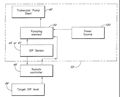
FIG. 8 shows a block diagram illustrating a trabecular pump stent with an IOP
sensor for controlling
the intraocular pressure of an eye. The block elements within the dashed line
55 are to be placed within the
eye in a preferred embodiment. In operation of some embodiments, a target IOP
level 49 is prescribed for a
patient. The information is logged in with a remote controller 46 and
transmitted wirelessly to an implanted
pumping element 54 that is a part of the pump stent system. The target IOP
level is compared to the sensed
IOP data from the IOP sensor 43. The trabecular pump stent will function when
the IOP level is lower than
sensed IOP.
In one preferred embodiment, to achieve the target IOP level, the trabecular
pump 40 with double
check valves starts to pump aqueous out of the anterior chamber 20 toward
Schlemm's canal 22 or
downstream therefrom until the target IOP is reached. The pumping may be
accomplished with a mechanical
pumping element 54 powered by a power source 150, which may comprise a source
of mechanical or
electrical energy.
In a preferred embodiment, the target IOP data is transmitted remotely to the
pumping element 54. In
the meantime, the measured IOP data from the IOP sensor 43 is fed to the
pumping element 54 so as to
activate the pumping operation whenever the measured IOP is higher than a
threshold IOP value.
IOP Sensor and Transmitter
It is one aspect of the present invention to provide a pressure sensor 43 for
transmitting a signal
either continuously or in response to a remote activation signal from a remote
external controller 46. The
sensor may comprise energy means for providing power to the sensor; sensing
means for determining the
pressure and generating a sensing signal indicative thereof and transmitting
means for transmitting the IOP
data to a remote controller 46. In one embodiment, the transmitter is a
radiofrequency transmitter.
An intraocular pressure sensor has been described in, for example, U.S. Patent
No. 6,579,235 to
Abita et al., the entirety of which is hereby incorporated by reference.
In another aspect, a flashing LED (light emitting diode) may be used to
transmit the IOP data to an
external controllerldisplay or to the pumping element for pressure control. In
one embodiment, the flashing
LED is connected to a transducer that converts the IOP data into electrical
signal. The LED technology is well
known to one ordinary skilled in the art.
In another aspect, a pressure sensor is mounted on a trabecular pump stent for
measuring an
intraocular pressure and generating a signal indicative of the measured
pressure. This signal is then
transferred to the pumping element 54.
For continuous monitoring of I~P, a sensor prototype comprises a capacitative-
inductive circuit
formed from a spiral inductor-diaphragm based capacitor. Upon sensing a change
in the IOP level, the
pressure-induced displacement of the diaphragm changes the frequency of the
circuit. The IOP monitoring is
performed telemetrically and does not need to come in contact with the eye. In
some embodiments the sensor
-11-
CA 02488393 2004-12-03
WO 2004/014218 PCT/US2003/024890
relies upon an external pickup coil, which can be placed in an unobtrusive
device such as spectacles. The
prototypes vary from 1.3 mm to 6 mm in diameter, with resolutions of 1.2 to
1.4 mm Hg.
FIG. 9 shows one embodiment of a pressure-pulse driven pump implant with
sensing means for
providing measured IOP data. The trabecular pump stent 40 comprises an inlet
portion 33, an outlet portion
34, and a middle portion 36, bordered by the first check valve 35A and the
second check valve 35B. The
pump stent 40 further comprises an IOP sensor 43, which feeds the data to a
pumping element 54. In one
aspect, the pumping element 54 is intimately adhered to or wrapped around the
wall of the middle portion 36
and has the capability of providing suction enabling the tube wall to expand
and providing pressure enabling
the tube wall to compress. The pumping element 54 can be powered by mechanical
energy or electricity
derived from various energy sources, including the conversion of mechanical to
electrical energy.
Some aspects of the invention provide an intraocular pumping system,
comprising setting a target
IOP level, sensing the real-time IOP and comparing to the target level, and
pumping aqueous out of the
anterior chamber once the sensed IOP is higher than the target IOP.
From the foregoing description, it should be appreciated that a novel approach
for sensing and
controlling the IOP at a target level has been disclosed for regulating
intraocular pressure. While the invention
has been described with reference to specific embodiments, the description is
merely illustrative and is not to
be construed as limiting the invention. Various modifications and applications
may occur to those who are
skilled in the art without departing from the true spirit and scope of the
invention, as described by the
appended claims and their equivalents.
-12-