Note: Descriptions are shown in the official language in which they were submitted.
CA 02489180 2004-12-09
WO 03/104803 PCT/EP03/06110
METHOD FOR MAPPING AND ELIMINATING T-CELL EPITOPES
FIELD OF THE INVENTION
The present invention relates to the field of immunology. The invention
provides
screening methods for.the identification of determinants and epitopes on
protein
molecules able to evoke an immune response. In particular the invention is
concerned
with the identification of epitopes for T-cells in therapeutic proteins.
Finally, the
invention relates to a combined approach of using epitope mapping in concert
with the
identification of MHC class II ligands deriving from said epitope mapping
method and
to design of sequence analogous having a reduced number of such ligands and
epitopes,
respectively.
BACKGROUND OF THE INVENTION
There are many instances whereby the efficacy of a therapeutic protein is
limited by an
unwanted immune reaction to the therapeutic protein. Several mouse monoclonal
antibodies have shown promise as therapies in a number of human disease
settings but in
certain cases have failed due to the induction of significant degrees of a
human anti-
murine .-antibody (HAMA) response [Schroff, R. W. et al (1985) Cancer Res. 45:
879-885;
Shawler, D.L. et al (1985) J. Imrnunol. 135: 1530-1535]. For monoclonal
antibodies, a
2o number of techniques have been developed in attempt to reduce the HAMA
response
[WO 89/09622; EP 0239400; EP 0438310; WO 91106667]. These recombinant DNA
approaches have generally reduced the mouse genetic information in the final
antibody
construct whilst increasing the human genetic information in the final
construct.
Notwithstanding, the resultant "humanized" antibodies have, in several cases,
still elicited
an immune response in patients [Issacs J.D. (1990) Sem. Immunol. 2: 449, 456;
Rebello,
P.R. et al (1999) Transplantation 68: 1417-1420].
Antibodies are not the only class of polypeptide molecule administered as a
therapeutic
agent against which an immune response may be mounted. Even proteins of human
3o origin and with the same amino acid sequences as occur within humans can
still induce an
immune response in humans. Notable examples amongst others include the
therapeutic
use of granulocyte-macrophage colony stimulating factor [Wadhwa, M. et al
(1999) Clin.
Cancer Res. 5: 1353-1361] and interferon alpha 2 [Russo, D. et al (1996) Bri.
J. Haena.
94: 300-305; Stein, R. et al (1988) New E~gl. J. Med. 318: 1409-1413]. In such
situations
where these human proteins are immunogenic, there is a presumed breakage of
CONFIRMATION COPY
CA 02489180 2004-12-09
WO 03/104803 PCT/EP03/06110
- 2 -
immunological tolerance that would otherwise have been operating in these
subjects to
these proteins.
This situation is different where the human protein is being administered as a
replacement
therapy for example in a genetic disease where there is a constitutional lack
of the protein
such as can be the case for diseases such as hemophilia A, Christmas disease,
Gauchers
disease and numerous other examples. In such cases, the therapeutic
replacement protein
may function immunologically as a foreign molecule from the outset, and where
the
individuals are able to mount an immune response to the therapeutic, the
efficacy of the
to therapy is likely to be significantly compromised.
Trrespective of whether the protein therapeutic is seen by the host immune
system as a
foreign molecule, or if an existing tolerance to the molecule is overcome, the
mechanism
of immune reactivity to the protein is the same. Key to the induction of an
immune
response is the presence within the protein of peptides that can stimulate the
activity of T-
cells via presentation on MHC class II molecules, so-called "T-cell.
epitopes". Such T-
cell epitopes are commonly defined as any amino acid residue sequence with the
ability to
bind to MHC Class II molecules. Implicitly, a "T-cell epitope" means an
epitope which
when bound to MHC molecules can be recognized by a T-cell receptor (TCR), and
which
2o can, at least in principle, cause the activation of these T-cells by
engaging a TCR to
promote a T-cell response.
MHC Class II molecules are a group of highly polymorphic proteins that play a
central
role in helper T-cell selection and activation. The human leukocyte antigen
group DR
(HI.A-DR) are the predominant isotype of this group of proteins however,
isotypes HLA-
DQ and HLA-DP perform similar functions. The present invention is applicable
to the
detection of T-cell epitopes presented within the context of DR, DP or DQ MHC
Class II.
In the human population, individuals bear two to four DR alleles, two DQ and
two DP
alleles. The structure of a number of DR molecules has been solved and these
appear as
3o an open-ended peptide binding groove with a number of hydrophobic pockets
which
engage hydrophobic residues (pocket residues) of the peptide [Brown et al
Nature (1993)
364: 33; Stern et al (1994) Nature 368: 215]. Polymorphism identifying the
different
allotypes of class II molecule contributes .to a wide diversity of different
binding surfaces
CA 02489180 2004-12-09
WO 03/104803 PCT/EP03/06110
- 3 -
for peptides within the peptide binding groove and at the population level
ensures
maximal flexibility with regard to the ability to recognize foreign proteins
and mount an
immune response to pathogenic organisms.
An immune response to a therapeutic protein proceeds via the MHC class II
peptide
presentation pathway. Here exogenous proteins are engulfed and processed for
presentation in association with MHC class II molecules of the DR, DQ or DP
type.
MHC Class II molecules are expressed by professional antigen presenting cells
(APCs),
such as macrophages and dendritic cells amongst others. Engagement of a MHC
class II
peptide complex by a cognate T-cell receptor on the surface of the T-cell,
together with
the cross-binding of certain other co-receptors such as the CD4 molecule, can
induce an
activated state within the T-cell. Activation leads to the release of
cytokines further
activating other lymphocytes such as B cells to produce antibodies or
activating T killer
cells as a full cellular immune response.
is
T-cell epitope identification is the first step to epitope elimination,
however there are few
clear cases in the art where epitope identification and epitope removal are
integrated into
a single scheme. Thus W098/52976 and WO00/34317 teach computational threading
approaches to identifying polypeptide sequences with the potential to bind a
sub-set of
human MHC class II DR allotypes. In these teachings, predicted T-cell epitopes
are
removed by the use of judicious amino acid substitution within the protein of
interest.
However with this scheme and other computationally based procedures for
epitope
identification [Godkin, A.J. et al (1998) J. Immureol. 161: 850-858;
Sturniolo, T. et al
(1999) Nat. Biotechnol. 17: 555-561], peptides predicted to be able to bind
MHC class II
molecules may not function as T-cell epitopes in all situations, particularly,
ire vivo due to
the processing pathways or other phenomena. In addition, the computational
approaches
to T-cell epitope prediction have in general not been capable of predicting
epitopes with
DP or DQ restriction.
Equally, ifa vitro methods for measuring the ability of synthetic peptides to
bind MHC
class II molecules, for example using B-cell lines of defined MHC allotype as
a source of
MHC class II binding surface [Marshall K.W. et al. (1994) J. Immunol. 152:4946-
4956;
O'Sullivan et al (1990) J. Immunol. 145: 1799-1808; Robadey C. et al (1997) J.
Immutaol
CA 02489180 2004-12-09
WO 03/104803 PCT/EP03/06110
159: 3238-3246J, may be applied to MHC class II ligand identification.
However, such
techniques are not adapted for the screening multiple potential epitopes to a
wide
diversity of MHC allotypes, nor can they confirm the ability of a binding
peptide to
function as a T-cell epitope.
Recently techniques exploiting soluble complexes of recombinant MHC molecules
in
combination with synthetic peptides have come into use [Kern, F. et al (1998)
Nature
Medicine 4.:975-978; Kwok, W.W. et al (2001) TRENDS ija Immunol. 22:583-588J.
These
reagents and procedures are used to identify the presence of T-cell clones
from peripheral
1o blood samples from human or experimental animal subjects that are able to
bind
particular MHC-peptide complexes and are not adapted for the screening
multiple
potential epitopes to a wide diversity of MHC allotypes.
Biological assays of T-cell activation remain the best practical option to
providing a
15 reading of the ability of a test peptide/protein sequence to evoke an
immune response.
Examples of this kind of approach include the work of Petra et al using T-cell
proliferation assays to the bacterial protein staphylokinase, followed by
epitope mapping
using synthetic peptides to stimulate T-cell lines [Petra, A.M. et al (2002)
J. Immunol.
168: 155-161J. Similarly, T-cell proliferation assays using synthetic peptides
of the
2o tetanus toxin protein have resulted in definition of immunodominant epitope
regions of
the toxin [Reece J.C. et al (1993) J. Immu~iol. 151: 6175-6184J. W099/53038
discloses
an approach whereby T-cell epitopes in a test protein may be determined using
isolated
sub-sets of human immune cells, promoting their differentiation iyi vitro and
culture of the
cells in the presence of synthetic peptides of interest and measurement of any
induced
25 proliferation in the cultured T-cells. The same technique is also described
by Stickler et
al [Stickler, M.M. et al (2000) J. Immufaotherapy 23:654-660J, where in both
instances
the method is applied to the detection of T-cell epitopes within bacterial
subtilisin. 'Such
a technique requires careful application of cell isolation techniques and cell
culture with
multiple cytokine supplements to obtain the desired immune cell sub-sets
(dendritic cells,
3o CD4+ and or CD8+ T-cells) and is not conducive to rapid through-put
screening using
multiple donor samples.
In a variation of these approaches, Hiemstra et al [Hiemstra, H.S. (1997)
Proc. Natl.
Acad. Sci USA 94: I03I3-103I8J have described a procedure for identifying a
peptide
CA 02489180 2004-12-09
WO 03/104803 PCT/EP03/06110
- 5 -
epitope capable of stimulating a known T-cell, such a process is valuable in
the detection
of autoreactive T-cell clones for which the (auto)antigen is unknown.
The above examples and other biological assays involving technical variations
on the
theme of measuring an iya vitro T-cell activation event, usually by the
measurement of an
induced proliferation response, abound. However, none of the procedures
provide a
unified scheme for the detection of biologically relevant epitopes in proteins
of human
origin nor are readily applicable to the detection of epitopes of significance
to a wide
population of MI3C allotypes. The present invention is conceived to provide
such a
to scheme and provides a basis for the identification and removal of T-cell
epitopes from a
given in principal therapeutically valuable but originally immunogenic
peptide,
polypeptide or protein.
Tn summary the invention relates to the following issues:
~ Using a panel of synthetic peptides in a naive T-cell assay to map the
immunogenic
regions) of a protein therapeutic, and in particular a protein therapeutic
whereby the
protein is a human protein;
~ using a panel of synthetic peptides in a recall assay to fine map the
immunogenic
regions) of a protein therapeutic.
~ using a panel of whole protein variants in a naive T-cell assay to select
variants
displaying minimal immunogenicity ire vitro;
~ using a panel of synthetic peptide variants in a naive T-cell assay to
select peptide
sequences displaying minimal immunogenicity in vitro;
~ using biological assays of T-cell stimulation to select a peptide sequence
which
exhibits a stimulation index of less than 2.0 and preferably less than 1.8 in
a naive T-
cell assay;
~ using biological assays of T-cell stimulation to select a protein variant
which exhibits
a stimulation index of less than 2.0 and preferably Iess than 1.8 in a naive T-
cell
assay;
~ a strategy in which T-cell lines are developed from individuals previously
in receipt of
a protein therapeutic and use of those cell lines to map the immunogenic
regions) of
the therapeutic molecule;
CA 02489180 2004-12-09
WO 03/104803 PCT/EP03/06110
- 6 -
~ a strategy according to the above in which in addition B-cell lines are
developed in
parallel to the T-cell lines developed from individuals previously in receipt
of a
protein therapeutic and the combined use of the those lines to map the
immunogenic
regions) of the therapeutic molecule;
~ use of B-cell lines developed from individuals previously in receipt of a
protein
therapeutic and in parallel to the development of T-cell lines from the same
individuals as a source of autologous APC in further rounds of T-cell
stimulation or
optionally as a binding surface for synthetic peptide binding assays;
~ construction of a T-cell epitope map of a subject protein using PBMC
isolated from
to healthy donors and a screening method involving the steps comprising:
i) antigen priming in vztro using synthetic peptide or whole protein
ixnmunogen
for a culture period of up to 7 days;
ii) addition of IL-2 and culture for up to 3 days;
iii) addition of primed T cells to autologous irradiated PBMC and re-challenge
I5 with antigen for a further culture period of 4 days and
iv) measurement of T cell activation e.g. proliferation index by any suitable
method;
~ construction of a T-cell epitope map of a subject protein using PBMC
isolated from
patients in whom there exists an established immune response to the subject
protein or
20 ~ derivatives thereof and application of a screening method involving the
steps
comprising:
i) antigen priming iya vitro using synthetic peptide or whole protein
immunogen
for a culture period of up to 7 days, ii)
ii) addition of IL-2 and culture for up to 3 days, iii)
25 iii) addition of primed T cells to autologous irradiated PBMC and re-
challenge
with antigen for a further culture period of up to 4 days and
iv) measurement of T cell activation e.g. proliferation index by any suitable
method;
~ construction of a T-cell epitope map exploiting polyclonal or monoclonal
cell lines
30 derived from PBMC samples from healthy donors or patients with established
immune responses to a protein of interest. Developing said cell lines into an
immunologically primed state by one or multiple rounds of a priming step
culminating in expansion of cell numbers in the presence of IL-2 +/-
CA 02489180 2004-12-09
WO 03/104803 PCT/EP03/06110
phytohaemagglutinin (PHA) or other mitogenic stimulus. Said primed cells are
contacted with either individual synthetic peptides or peptide pools
comprising
multiple synthetic peptides and lines stimulated into proliferation detected
using any
suitable means. Where stimulation is detected from a pooled peptide immunogen
the
identity of the stimulating peptide is uncovered using further round of
screening with
individual peptides or smaller peptide pools and further primed cells;
~ a concerted method fox mapping the location of T-cell epitopes in protein
sequences
using naive T-cell activation assays and a computational scheme simulating the
binding of the peptide ligand with one or more MHC allotypes;
~ a method for locating T-cell epitopes in protein sequences comprising the
following
steps;
i) use of naive T-cell activation assays and synthetic peptides collectively
encompassing the protein sequence of interest to identify epitope regions
capable of activating T-cells;
ii) use of a computational scheme simulating the binding of the peptide ligand
with one or more MHC'allotypes to analyse the epitope regions identified in
step (i) and thereby identify MHC class II ligands within the epitope region;
iii) use of a computational scheme simulating the binding of the peptide
ligand
with one or more MHC allotypes to identify sequence analogues of the MHC
ligands encompassed within the epitope regions) which no longer bind MHC
class II or bind with lowered affinity to a lesser number of MHC allotypes;
iv) use of naive T-cell activation assays and synthetic peptides encompassing
entirely ox in collection encompassing the epitope regions identified within
the
protein of interest and testing the sequence analogues in naive T-cell
activation
assay in parallel with the wild-type (parental) sequences;
a method accprding to the above scheme wherein steps (ii) and (iii) are
carried out
using a computational approach as taught by WO 02/069232;
a method according to the above scheme where the naive T-cell activation assay
is
conducted using PBMC cells derived from around 20 or more unrelated donors;
~ a method according to the above scheme where the location of a T-cell
epitope is
found when a stimulation index score of around 2.0 is observed in two or more
independent donor samples;
CA 02489180 2004-12-09
WO 03/104803 PCT/EP03/06110
_ g _
~ a method according to the above scheme where the location of a T-cell
epitope is
found when a stimulation index score of around 2.0 is observed in two or more
independent donor.samples and where one or more MHC class II ligands can be
identified within the same sequence locale using a computational system;
~ a method according to the above scheme whereby the computational system is
according to the method as taught by WO 02/069232;
~ identification of protein sequences with reduced ability to promote an
immune
response may be achieved using immunologically primed cells of the
aforementioned
scheme and a screening process whereby multiple variant peptides or whole
protein
antigens are tested in parallel to reference peptide pools or whole protein
antigen
containing only wild-type sequences. Peptides or protein variants with a
lesser
stimulation index to reference pools or wild-type protein are selected for
further
analysis;
identification of protein sequences or protein preparations with increased
ability to
promote an immune response achieved using immunologically primed cells of the
aforementioned scheme and a screening process whereby one or more peptides or
whole protein antigens are tested in parallel to reference peptide pools or
whole
protein antigen giving a known ih vitr~ immune response. Peptides or protein
preparations shown to evoke a different stimulation index profile to the
reference
preparations are selected for further analysis or may be eliminated from the
production process;
~ peptide sequences able to evoke a stimulation index of greater than 1.8 and
preferably
greater than 2.0 in a naive T-cell assay and selected from any therapeutic
protein;
~ peptide sequences selected from any therapeutic protein having a stimulation
index of
greater than 1.8 and preferably greater than 2.0 in a naive T-cell assay
wherein the
peptide is modified to a minimum extent and tested in the naive T-cell assay
and
found to have a stimulation index of less than 2.0;
~ peptide sequences sharing 100% amino acid identity with the wild-type
protein
sequence and able to evoke a stimulation index of 1.8 or greater and
preferably greater
than 2.0 in a T-cell assay;
~ a protein molecule in which the immunogenic regions have been mapped using a
T-
cell assay and then modified such that upon re-testing in a T-cell assay the
modified
CA 02489180 2004-12-09
WO 03/104803 PCT/EP03/06110
- 9 -
protein evokes a stimulation index smaller than the parental (non-modified)
molecule
and most preferably less than 2.0
DETAILED DESCRIPTION OF THE INVENTION
According to the first embodiment of the invention there is provided a method
whereby a
protein antigen may be screened for the presence of determinants within its
sequence
capable of evoking a T-cell driven immune response should that protein be
introduced in
its un-modified state into a human subject. The method thereby provides a
predictive tool
for the identification of T-cell epitopes in proteins with therapeutic
potential in man
l0 where the protein is to be provided for the therapy of an acquired disease
state and where
that protein may be a human protein.
It is particularly desired to provide an epitope map of a protein of interest
where the map
has relevance to a wide spectrum of possible MHC allotypes. It is desired that
the map is
sufficiently representative to allow the design or selection of a modified
protein for which
the ability of the protein to evoke a T-cell driven immune response is
eliminated or at
least ameliorated for the majority of patients to whom the protein is likely
to be
administered. Accordingly in the practice of the invention the screening
process
exploiting PBMC derived T-cells from naive donors is collected from a pool of
donors of
sufficient immunological diversity to provide a sample of at least greater
than 90% of the
MHC class II repertoire (HI,A-DR) extant in the human population and
preferably greater
than 95% of that repertoire. In an ideal situation, equivalence to greater
than 99%
representation is preferred, although it is recognised that there are
practical limitations to
achieving this ideal. Accordingly, where a naive T-cell response is to be
detected to a
given synthetic peptide, the peptide in practice will be contacted with PBMC
preparations
derived from multiple donors in isolation, the numbers of donors or herein
more
preferably described as the "donor pool", is fox practical purposes not likely
to be less
than 20 unrelated individuals (pre-selected according to their MHC class II
haplotypes).
3o The term "naive donor" in the context of the present invention means that
the T-cells
obtained from the individual have not previously been exposed to the protein
or peptide
antigen of interest, and where the protein antigen is a human protein, the
individual has
not been in receipt of any therapeutic or exogenous sources of the protein.
CA 02489180 2004-12-09
WO 03/104803 PCT/EP03/06110
- 10 -
Thus according to the first embodiment of the present, there is provided a
method for T- .
cell epitope mapping exploiting immunologically naive T-cells. The T-cells are
provided
from a peripheral blood sample from a multiplicity of different healthy donors
for whom
the protein of interest may be an endogenous molecule but who have not been in
receipt
of the protein of interest from any exogenous source e.g. administered
therapeutically.
The assay is conducted using PBMC cultured in vitro using procedures common in
the art
and involves contacting the PBMC with synthetic peptide species representative
of the
protein of interest, and following a suitable period of incubation,
measurement of peptide
induced T cell activation such as cellular proliferation. Measurement is by
any suitable
means and may for example be conducted using 3H-thymidine incorporation
whereby the
accumulation of 3H into cellular material is readily measured instrumentally.
The degree
of cellular proliferation for each combination of PBMC sample and synthetic
peptide is
examined relative to that seen in non peptide treated PBMC sample. Reference
may also
be made to the proliferative response seen following treatment with a peptide
or peptides
for which there is an expected proliferative effect. In this regard is
considered
particularly advantageous to use peptide with known broad MHC restriction and
especially peptide epitopes with MHC restriction to the DP or DQ isotypes.
To facilitate assembly of an epitope map for a given protein of interest, a
set of synthetic
peptides representative of the sequence of the protein are produced. A typical
analysis
under the scheme of the present involves the use of peptides containing 15
amino acid
residues although it will be recognised that a peptide containing not less
than 9 amino
acid residues is in principle a suitable peptide. Peptides significantly
exceeding 15 amino
acid residues may also be used but it will equally be recognised that possible
secondary
structural effects or complexities of intracellular processing may obscure the
ability of the
peptide to induce a proliferative response. In order to scan the entire length
of given
protein, a particularly convenient scheme is to produce synthetic peptides
each of 15
amino acid residues in length and each overlapping the next peptide in the
series by 12
amino acid residues, i.e. each successive peptide in the series incrementally
adds a further
3 amino acids to the analysis. Iii this way any given adjacent pair of
peptides will map 18
amino acids of contiguous sequence in the protein of interest. Thus for a
protein of
interest comprising n amino acid residues, the number of 15-mer synthetic
peptides
CA 02489180 2004-12-09
WO 03/104803 PCT/EP03/06110
- 1 ~. -
required for a complete scan of the said protein will be 1+(f2-12)/3. Other
schemes may
be contemplated and be equally efficacious.
Using the scheme outlined above and exemplified in detail within the EXAMPLES
herein, the inventors have discovered regions of protein sequence capable of
evoking a
proliferative response in naive PBMC from different individual healthy donors.
The
protein sequences in question are sequence strings derived from whole human
proteins for
which there could be an expectation of immune tolerance but which none the
less there is
a demonstrable ability to evoke a surrogate immune response zsa vitro. This
ability by
to extension may also apply ira vivo should either of the proteins in question
be administered
for example as therapeutic entities. Specifically these proteins are
interferon a2 and
interferon I3. Both of these proteins are used therapeutically and
significantly for both of
these molecules immunogenic responses to these molecules in patients have been
recorded [Russo, D. et al (1996) ibid; Stein, R. et al (19$$) ibid; Myhr, K.M.
et al (2000)
Neurology 55:1569-1572; Bertolotto, A. et al (2000) Immunopharmacology 48: 95-
I00].
The present invention therefore provides a generalised scheme for the
elucidation of
epitope regions within normal human proteins and demonstrates the ability of
peptides
derived from these proteins to evoke an in vitro proliferative response in
naive PBMC
derived from healthy donors.
A particularly effective method for defining a T-cell map using naive T-cell
assays of the
first embodiment is provided in the EXAMPLES 1 and.2 whereby immunogenic
regions
of the molecules interferon beta (IFNl3) and interferon alpha 2 (IFNa2) are
disclosed. A
particularly preferred method for the identification of T-cell epitopes in
proteins which
are weakly imrnunogenic in vivo is described in EXAMPLE 3.
In a second embodiment where the invention provides for the elucidation of a T-
cell
epitope map, such a map may be used to guide the design of a modified protein
whereby
the epitope regions on the molecule are suitably modified such that they are
no longer
3o able to evoke a proliferative response according to the scheme of the
invention and the
protein of interest is thereby rendered less immunogenic to man.
CA 02489180 2004-12-09
WO 03/104803 PCT/EP03/06110
- 12 -
According to this second embodiment, suitable modifications to the protein may
include
amino acid substitution of particular residues or combinations of residues.
For the
elimination of T-cell epitopes, amino acid substitutions are preferably made
at appropriate
points within the peptide sequence predicted to achieve substantial reduction
or
elimination of the activity of the T-cell epitope. In practice an appropriate
point will
preferably equate to an amino acid residue binding within one of the pockets
provided
within the MHC class II binding groove. It is most preferred to alter binding
within the
first pocket of the cleft at the so-called "Pl" or "PI anchor" position of the
peptide. The
quality of binding interaction between the Pl anchor residue of the peptide
and the first
to pocket of the MHC class 1I binding groove is recognised as being a major
determinant of
overall binding affinity for the whole peptide. An appropriate substitution at
this position
of the peptide will be for a residue less readily accommodated within the
pocket, for
example, substitution to a more hydrophilic residue. Amino acid residues in
the peptide
at positions equating to binding within other pocket regions within the MHC
binding cleft
are also considered and fall under the scope of the present.
It is understood that single amino acid substitutions within a given potential
T-cell epitope
are the most preferred route by which the epitope may be eliminated.
Combinations of
substitution within a single epitope may be contemplated and for example can
be
2o particularly appropriate where individually defined epitopes are in overlap
with each
other. Moreover, amino acid substitutions either singly within a given epitope
or in
combination within a single epitope may be made at positions not equating to
the "pocket
residues" with respect to the MHC class II binding groove, but at any point
within the
peptide sequence. Substitutions may be made with reference to an homologous
structure
or structural method produced using ire silico techniques known in the art and
may be
based on known structural features of the molecule. For example a change may
be
contemplated to restore structure or biological activity of the variant
molecule. Such
compensatory changes and changes may also include deletion or addition of
particular
amino acid residues from the polypeptide.
A particularly effective means of removing epitopes from protein molecules is
the
concerted use of the naive T-cell activation assay scheme as outlined herein
together with
CA 02489180 2004-12-09
WO 03/104803 PCT/EP03/06110
- 13 -
an if2 silico tool developed according to the scheme described in WO 02/069232
which is
incorporated fully herein by reference.
The software simulates the process of antigen presentation at the level of the
peptide
MHC class II binding interaction to provide a binding score for any given
peptide
sequence. Such a score is determined for many of the predominant MHC class II
allotypes extant in the population. As this scheme is able to test any peptide
sequence,
the consequences of amino acid substitutions additions or deletions with
respect to the
ability of a peptide to interact with a MHC class II binding groove can be
predicted.
1o Consequently new sequence compositions can be designed which contain
reduced
numbers of peptides able to interact with the MHC class II and thereby
function as
immmunogenic T-cell epitopes. Where the biological assay using any one given
donor
sample can assess binding to a maximum of 4 DR allotypes, the in silico
process can test
the same peptide sequence using >40 allotypes simultaneously. In practice this
approach
is able to direct the design of new sequence variants which are compromised in
the their
ability to interact with multiple MHC allotypes.
By way of an example of utility of the combined approach to epitiope
identification and
removal, the results of a programme involving the engineering of human
interferon alpha
(IFNoc) are provided herein. The entire human IFNcc sequence was rendered into
a set of
51 15-mer peptides (listed within table 2 of EXAMPLE 2). The T-cell assay was
able to
define three immunogenic regions (termed R1, R2 and R3) within the molecule
and the
software system according to the scheme of WO 021069232 was able to identify
predicted
MHC class II ligands within each of the epitopes Rl R3. Moreover, the system
was
further able to identify amino acid substitutions within the epitopes which
resulted in
significant loss of binding affinity between the peptide sequence and
essentially all of the
MHC class II allotypes represented in the system. A panel of synthetic
peptides were
constucted encompassing the wild-type epitope regions and variant sequences
thereof in
which MHC class II binding was eliminated by amino acid substitution. The
peptides
3o were used in naive T-cell activation assays and the stimulation index
determined for each
peptide and donor PBMC sample combination. In all instances where a donor
sample
was found to be responsive to a wild-type peptide, the variant peptide was
found not to
activate T-cells (FIGURE 3).
CA 02489180 2004-12-09
WO 03/104803 PCT/EP03/06110
- 14 -
A preferred embodiment of the present invention is to use a modified T cell
activation
assay in which measurement of a T Bell response is performed at different
times after
adding a test protein or peptide. This novel format for the assay is
especially useful for
detecting T cell responses in whole proteins or weakly immunogenic
polypeptides. The
assay format counteracts the complexity of components within the T cell assay
mixture
comprising a mixture of leukocytes and different molecules including
cytokines. For any
test protein or peptide, the kinetics of a T~cell response in the assay is
dependant on a
number of factors including the status of T cells within the T cell assay
mixture (for
example, naive versus memory T cells), the concentration of cytokines at
various
1o timepoints, and the rate of generating significant T cell proliferation due
to factors such as
the concentration of specific peptide-MHC class II complexes. For any given
protein or
peptide, the peak of T cell proliferation in the assay system may peak before
or after day
7 after addition of protein or peptide to, the assay mixture such that, by day
7 (the standard
assay timepoint), T cell proliferation is not significant. By testing for T
cell proliferation
over a timecourse, for example on each of days 4, 5, 6, 7, ~ and 9, then T
Bell responses
can be detected which would not necessarily be detected at day 7. An example
of a T cell
assay timecourse is shown in example 3. For whole proteins, the T cell assay
timecourse
provides for a sensitive analysis of T cell immunogenicity and thus provides
for a
sensitive immunogenicity screen for proteins. In addition, as demonstrated in
example 3,
2o this assay may also be used to test for the effects of amino acid
substitutions on
immunogenicity.
The combined approach of using an in silico tool for the identification of MHC
class II
ligands and design of sequence analogues lacking MHC class II ligands, in
concert with
epitope mapping and re-testing using biologically based assays of T-cell
activation is a
particularly effective method and most preferred embodiment of the invention.
The
general method according to this most preferred embodiment comprises the
following
steps:
i) use of naive T-cell activation assays and synthetic peptides collectively
3o encompassing the protein sequence of interest to identify epitope regions
capable
of activating T-cells;
CA 02489180 2004-12-09
WO 03/104803 PCT/EP03/06110
- 15 -
ii) use of a computational scheme simulating the binding of the peptide Iigand
with
one or more MHC allotypes to analyse the epitope regions identified in step
(i)
and thereby identify MHC class II ligands within the epitope region;
iii) use of a computational scheme simulating the binding of the peptide
ligand with
one or more MHC allotypes to identify sequence analogues of the MHC ligands
encompassed within the epitope regions) which no longer bind MHC class II or
bind with lowered affinity to a lesser number of MEiC allotypes;
iv) use of naive T-cell activation assays and synthetic peptides encompassing
entirely
or in collection encompassing the epitope regions identified within the
protein of
to interest and testing the sequence analogues in naive T-cell activation
assay in
parallel with the wild-type (parental) sequences;
It is understood that the software scheme outlined in WO 02/069232 can also be
used to
define with a high degree of certainty the dataset of all peptides comprising
the universe
IS of permissible MHC class ligands for the any human protein such as IFN~.
For reasons
such as the requirement for proteolytic processing and other physiologic steps
leading to
the presentation of immunogenic peptides ifZ vivo, it would be clear that a
relatively minor
sub-set of the entire repertoire of peptides will have ultimate biological
relevance. In
such situations the inventors have established that ex vivo human T-cell
activation assays
2o may be used to identify the biologically relevant peptides. Accordingly,
synthetic
peptides are tested for their ability to evoke a proliferative response in
human T-cell
cultured ifa vitro. Where this type of approach is conducted using nave human
T-cells
taken from healthy donors, the inventors have established that in the
operation of such an
assay, a stimulation index equal to or greater than 2.0 is a useful measure of
induced
25 proliferation. The stimulation index (SI) is conventionally derived by
division of the
proliferation score (e.g. counts per minute of radioactivity if using for
exarnple,3H-
thymidine incorporation) measured to the test peptide .by the score measured
in cells not
contacted with a test peptide. Peptides which evoke no response give SI = 1.0
although in
practice ST values in the range 0.8 -1.2 are unremarkable. A number of
technical
30 proceedures can be inbuilt into the operation of such assays in order to
ensure confidence
in the recorded scores. Typically all determinations are made at least in
triplicate and the
mean score may be computed. Where a computed SI =>2.0 individual scores of the
triplicate can be examined for evidence of outlying data. Similarly the
inclusion of
CA 02489180 2004-12-09
WO 03/104803 PCT/EP03/06110
- 16 -
control peptides for which there is expectation that the majority of PBMC
donor samples
will be responsive may be included in each assay plate. The influenza
haemagglutinin
peptide 307-309, sequence PKYVKQNTLKLA; and the Chlamydia HSP 60 peptide
sequence KWDQIKKISKPVQH are particularly suitable control peptides although
many
other examples may be exploited. Assays should preferably also use a potent
whole
protein antigen such as hemocyanin from Keyhole Limpet to which all PBMC
samples
would be expected to exhibit an SI significantly greater than 2.0
According to the scheme of the present invention there may be a practical need
to test
multiple versions of essentially the same peptide sequence in order to
establish that the
to modification, be it a single amino acid substitution or some other change
or combination
of changes, results in the loss of ability or at least a reduced ability for
the peptides) to
induce a T-cell activation effect. This requirement may be met using a number
of
different practical approaches one of which could involve the screening of
large numbers
of variant peptides from the outset and conducting a selection scheme to
identify those in
which there is a reduced or absent ability to induce proliferation relative to
their parental
(e.g. wild-type) peptide sequence. 'Such an approach could be conducted
entirely using
naive PBMC samples and run concurrently (i.e. in parallel with) the mapping
exercise. It
is understood that this approach need not be limited to the screening of
synthetic peptide
species but may be exploited to the screening of whole protein molecules that
for example
2o may comprise a multiplicity of variants produced as a "library" of variants
from which a
desired member is to be selected. Such a library may be produced for example
by
recombinant means well known in the art or may comprise species produced using
synthetic means for example using the principles of combinatorial chemistry.
In any
event, the desired property to be selected from the library member in this
context would
be the inability to induce a proliferative response in a PBMC preparation.
Alternatively variant peptides may be screened using naive PBMC from entirely
different
donor pool of samples, i.e. epitope mapping is repeated but using modified
peptides
where there is an expectation for little or no proliferative induction.
A further and particularly favoured scheme would involve the testing of
modified
peptides for their ability to induce a proliferative effect in an
immunological recall assay
format. This may be achieved for example using PBMC from a known responding
donor
CA 02489180 2004-12-09
WO 03/104803 PCT/EP03/06110
- 17 -
identified during the initial naive PBMC assay phase and stimulating a sample
of those
cells using a either synthetic peptides (e.g. in a pool) or whole protein
followed by a
suitable period of culture in the presence of cytokines such as IL,-2.
Following this
incubation, the culture may be re-stimulated using the synthetic (modified)
peptide or
modified whole protein of interest and the proliferative effect measured using
any suitable
means. The inventors have classified this assay format as a "recall" assay, so
called as
the T-cell population responsible for the proliferative response is invoked
during a re-
stimulation phase.
to The recall type assay is particularly useful in identifying T cell epitopes
in protein or
peptide antigens that show weak immunogen'icity ifz vivo an can provide
corroborating
evidence for the existence of a T-cell epitope in a given amino acid sequence
where the
epitope was originally identified by other means, fox example by using
computational
techniques or biological assays. In the operation of such a recall assay, PBMC
are
isolated from healthy donors or patients with established immune responses to
a given
therapy. It is necessary to freeze aliquots of autologous PBMC so that they
can be used
as antigen presenting cells (APC) during subsequent procedures. The assay
commences
with an antigen priming step. A typical and preferred protocol requires 2-
4x10s PBMC
are added to each well of 24 well plate. Either whole protein or peptide
antigen or a
2o peptide pool is added to the cells at typical concentrations of 1-10~g/ml
and 1-10~.M,
respectively (total concentration of peptides in peptide pool would be l~,M).
The final
culture volume is 2m1. The cells are incubated for 7 days where on day 7
l0U/ml TL-2 is
added and the cells are incubated for a further 3 days whereupon the cells are
ready for
the antigen re-challenge phase.
The antigen re-challenge requires autologous PBMC as APC. The APC are
incubated
with whole protein or synthetic peptide antigen (for example at a
concentration of 1-
l0ug/ml) for 1 hour at 37°C. The proliferative capability of the APC is
destroyed most
preferably using gamma radiation, for example 4000 rads in a round bottom 96
well plate
(1x10⁵ PBMC/well). 1-10x104 primed T-cells are added to each well containing
the
APC's. It is important to set up untreated control reactions comprising
antigen primed T-
cells cultured with gamma irradiated APC in the absence of re-challenge
antigen. The
cells are incubated for 4 days.before pulsing proliferation assessment for
example 3H-
CA 02489180 2004-12-09
WO 03/104803 PCT/EP03/06110
- 18 -
thymidine incozporation assay. It is understood that such a protocol can
equally be
conducted using enriched or purified populations of cells.
In a third embodiment, there is provided a method whereby a protein antigen
may be
screened for the presence of determinants within its sequence capable of
evoking a T-cell
immune response in individuals for whom the protein of interest is to be
administered for
therapeutic effect against a genetic (constitutional) disease and where, in
effect, the
protein antigen due to the nature of the genetic deficit in the individuals
will constitute a
foreign protein. In this sense, the protein is most likely to represent a
potent antigen in
to vivo and the inventors have established that it is now readily possible to
establish .
polyclonal or mononclonal T-cell lines irc vitro from the PBMC of such
individuals and
these lines may be used as effective reagents in the mapping of T-cell
epitopes within
proteins. This is achieved in essentially the same way as the recall assay of
the foregoing,
with the exception that the T cells are subjected to several rounds of antigen
stimulation
in vitro followed immediately by expansion in the presence of IL-2. For
establishing
polyclonal T cell lines 2-3 rounds of antigen stimulation are generally
sufficient to
generate a Large number of antigen specific cells. These are used to screen
large numbers
of synthetic peptides (for example in the form of peptide pools), and they may
be
cryogenically stored to be used at a later date. After the initial round of
antigen
2o stimulation comprising co-incubation of the antigen and PBMC for 7 days
subsequent re-
challenges with antigen are performed in the presence of most preferably
autologous
irradiated PBMC as antigen presenting cells. These rounds of antigen selection
are
performed for 3-4 days and are interspersed by expansion phases comprising
stimulation
with IL-2 which may be added every 3 days for a total period of around 9 days.
The final
re-challenge is performed using T-cells that have been "rested", that is T
cells which have
not been IL-2 stimulated for around 4 days. These cells are stimulated with
antigen (e.g.
synthetic peptide or whole protein) using most preferably autologous antigen
presenting
cells as previously for around 4 days and the subsequent proliferative
response (if any) is
measured thereafter.
Accordingly the method of the third embodiment comprises the production of T-
cell lines
or oligoclonal cultures derived from PBMC samples taken from an individual
afflicted
with the disease of interest, stimulating in vitro said lines or cultures with
preparations of
CA 02489180 2004-12-09
WO 03/104803 PCT/EP03/06110
- 19 -
synthetic peptides or whole proteins and measuring ih vitro the proliferative
effect if any
of individual synthetic peptides or proteins, producing modified variants of
individual
synthetic peptides or whole proteins and re-testing said modified peptides or
proteins for a
continued ability to promote a significant proliferative response in the T-
cell lines or
cultures.
It is particularly useful to establish T-cell lines of oligoclonal cultures
from individuals
who carry the genetic defect and in whom therapeutic replacement therapy has
been
initiated to and in whom the replacement therapy has resulted in the induction
of an
to immune response to the therapeutic protein. A prominent example of this
kind of subject
is provided by individuals undergoing treatment for hemophillia A but in whom
there is a
significant titre of inhibitory antibodies measurable to the therapeutic
Factor VIII. Under
the scheme of the present invention it would be particularly desired to
exploit PBMC
samples from this class of so called "inhibitor patients" as it could be
expected that the
epitope map of the the Factor VIII protein defined by the T-cell repertoire of
a significant
number of these individuals will be representative of the most prevalent
peptide epitopes
that are capable of presentation in the ih vivo context. In this sense, PBMC
from patients
in whom there is a previously demonstrated immune response constitute the
products of
an i~a vivo priming step and are particularly valuable under the scheme of the
present.
2o EXAMPLE 4 herein provides detailed description of an epitope mapping
programme
conducted on human FVIZI exploiting both naive human T-cells from healthy
donors and
PBMCs derived from haemophilia A patients.
Given that the use of PBMC cell lines from individuals previously in receipt
of the
immunologically foreign protein is in principle a recall assay, it further
provides the
practical benefit of there being the capacity for a much larger magnitude of
proliferative
response to any given stimulating peptide or protein. This reduces the
technical challenge
of conducting a proliferation measurement and in such a situation may give the
opportunity for definition of a possible hierarchy of immunodominant epitopes
where
3o multiple epitopes are uncovered to a target protein. This is certain to be
the case~with
particularly large proteins such as Factor Vfff although as demonstrated
herein, small
human protein molecules (e.g. less than 200 amino acid residues) may be
expected to
harbour multiple or complex (i.e. overlapping) T-cell epitopes.
CA 02489180 2004-12-09
WO 03/104803 PCT/EP03/06110
- 20 -
In a fourth embodiment of the present there is provided a scheme whereby the
assay
format of the foregoing is applied to the screening of production batches' of
therapeutic
biological proteins. The objective of such a screening process is to confirm
the
consistency of the immunogenic profile of the test biologic and for example
may be
particularly valuable in situations where the production process for the
biologic has been
altered by some parameter and although the measured physical properties of the
protein
may be within accepted ranges, there is a consideration that the potential
immunogenic
properties of the protein may have been altered. Thus in order to anticipate
the generation
of an immunogenic response to any new preparation of the molecule of interest
the
to methods set-out herein are particularly effective in providing such a
screening procedure.
Under the fourth embodiment therefore, T-cell lines (oligoclonal or mono-
clonal) derived
as part of the epitope mapping process for the protein of interest or
optionally and in
addition, a panel of naive PBMC preparations for which there has been
established a
population of known responsive preparations, may be used to test the subject
protein for
immunogenicity ih vitro, and the responses scored to the test protein are
compared to a
reference or "gold-standard" preparation of the protein. In this regard where
T-cell lines
are employed, it is particularly preferred to use lines derived from subjects
in whom there
has been a demonstrated previous immune response to the reference protein.
Such lines
2o are expected to provide a high stimulation index score on antigen challenge
in vitro and
are likely to be representative of the most biologically relevant and
immunodominant
epitopes within the protein. These lines under the fourth embodiment provide
indicators
for epitope loss/alteration. By contrast, under the fourth embodiment, panels
of naive
PBMC containing a known set of responding allotypes to the target protein
provide
indication of de novo epitope generation appearing in the test product protein
and are
equally valuable in predicting an unwanted clinical immunogenic response.
The term "T-cell epitope" means according to the understanding of this
invention an
amino acid sequence which is able to bind MHC class II, able to stimulate T-
cells and / or
3o also to bind (without necessarily measurably activating) T-cells in complex
with MHC
class II.
CA 02489180 2004-12-09
WO 03/104803 PCT/EP03/06110
- 21 -
The term "peptide" as used herein and in the appended claims, is a compound
that
includes two or more amino acids. The amino acids are linked together by a
peptide bond
(defined herein below). There are 20 different naturally occurring amino acids
involved
in the biological production of peptides, and any number of them may be linked
in any
order to form a peptide chain or ring. The naturally occurnng amino acids
employed in
the biological production of peptides all have the L-configuration. Synthetic
peptides can
be prepared employing conventional synthetic methods, utilizing L-amino acids,
D-amino
acids, or various combinations of amino acids of the two different
configurations. Some
peptides contain only a few amino acid units. Short peptides, e.g., having
less than ten
amino acid units, are sometimes referred to as "oligopeptides". Other peptides
contain a
large number of amino acid residues, e.g. up to 100 or more, and are referred
to as
"polypeptides". By convention, a "polypeptide" may be considered as, any
peptide chain
containing three or more amino acids, whereas a "oligopeptide" is usually
considered as a
particular type of "short" polypeptide. Thus, as used herein, it is understood
that any
reference to a "polypeptide" also includes an oligopeptide. Further, any
reference to a
"peptide" includes polypeptides, oligopeptides, and proteins. Each different
arrangement
of amino acids forms different polypeptides or proteins. The number of
polypeptides-and
hence the number of different proteins-that can be formed is practically
unlimited.
The invention will now be illustrated by the following examples. The examples
and
foregoing text refer to the following figures:
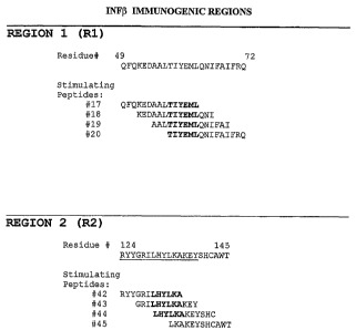
Figure 1 shows the immunogenic regions within IFN13 and details the peptide
sequences
from these regions able to stimulate naive human T-cells.
Figure ~ provides a table indicating the IFN13 peptides capable of promoting
proliferation
of naive human T-cells ifz vitro. For two of the donors, responses are
recorded to multiple
overlapping peptides from either region R1 or R2. Responses to individual
synthetic
peptides mapping to epitope regions Rl. or R2 are scored from six donors.
Figure 3 provides exemplary data from time course T-cell activation assays.
Charts plot
stimulation index (ST) against time (days) for synthetic peptides derived from
the IFNa
CA 02489180 2004-12-09
WO 03/104803 PCT/EP03/06110
- 22 -
Rl, R2 and R3 epitope regions and analogue peptide sequences containing amino
acid
substitutions tested in parallel.
EXAMPLE 1
The interaction between MHC, peptide and T-cell receptor (TCR) provides the
structural
basis for the antigen specificity of T-cell recognition. T-cell proliferation
assays test the
binding of peptides to MHC and the recognition of MHC/peptide complexes by the
TCR.
Ih vitro T-cell proliferation assays of the present example, involve the
stimulation of
peripheral blood mononuclear cells (PBMCs), containing antigen presenting
cells (APCs)
and T-cells. Stimulation is conducted in vitro using synthetic peptide
antigens, and in
some experiments whole protein antigen. Stimulated T-cell proliferation is
measured
using 3H-thymidine (3H-Thy) and the presence of incorporated 3H-Thy assessed
using
scintillation counting of washed fixed cells.
Buffy coats from human blood stored for Iess than 12 hours were obtained from
the
National Blood Service (Addenbrooks Hospital, Cambridge, UI~). Ficoll-paque
was
obtained from Amersham Pharmacia Biotech (Amersham, ITK). .Serum free AIM V
media for the culture of primary human lymphocytes and containing L-glutamine,
50~g/ml streptomycin, 10~g/ml gentomycin and 0.1 % human serum albumin was
from
Gibco-BRL (Paisley, UK). Synthetic peptides were obtained from Pepscan (The
Netherlands) and Babraham Technix (Cambridge, UK).
Erythrocytes and leukocytes were separated from plasma and platelets by gentle
centrifugation of buffy coats. The top phase (containing plasma and platelets)
was
removed and discarded. Erythrocytes and leukocytes were diluted 1:1 in
phosphate
buffered saline (PBS) before layering onto lSml ficoll-paque (Amersham
Pharmacia,
Amersham UK). Centrifugation was done according to the manufacturers
recommended
conditions and PBMCs were harvested from the serum+PBS/ficoll paque interface.
PBMCs were mixed with PBS (1:1) and collected by centrifugation. The
supernatant was
3o removed and discarded and the PBMC pellet resuspended in SOm1 PBS. Cells
were again
pelleted by centrifugation and the PBS supernatant discarded. Cells were
resuspended
using 50m1 AIM V media and at this point counted and viability assessed using
trypan
blue dye exclusion. Cells were again collected by centrifugation and the
supernatant
CA 02489180 2004-12-09
WO 03/104803 PCT/EP03/06110
- 23 -
discarded. Cells were resuspended for cryogenic storage at a density of 3x10
per ml.
The storage medium was 90%(v/v) heat inactivated AB human serum (Sigma, Poole,
UK)
and 10%(v/v) DMSO (Sigma, Poole, UK). Cells were transferred to a regulated
freezing
container (Sigma) and placed at -70°C overnight before transferring to
liquid N2 for long
term storage. When required for use, cells were thawed rapidly in a water bath
at 37°C
before transfernng to lOml pre-warmed AIM V medium.
PBMC were stimulated with protein and peptide antigens in a 96 well flat
bottom plate at
a density of 2x10⁵ PBMC per well. PBMC were incubated for 7 days at
37°C before
1o pulsing with 3H-Thy (Amersham-Phamacia, Amersham, LTK). For the present
study,
synthetic peptides (l5mers) that overlapped by 3aa increments were generated
that
spanned the entire sequence of IFN13. Peptide identification numbers (ID#) and
sequences
are given in Table 1.
Table 1 IFNt3 peptides
Peptide. Peptide
IFN(3-1a; , IFN(3-1a; l5mer
ID l5mer ID
Number N~er sequence
Sequence
1 MSYNLLGFLQRSSNF28 TIVENLLANVYHQIN
2 NLLGFLQRSSNFQCQ29 ENLLANVYHQINHLK
3 GFLQRSSNFQCQKLL30 LANVYHQINHLKTVL
4 QRSSNFQCQKLLWQL31 VYHQINHLKTVLEEK
5 SNFQCQKLLWQLNGR32 QINHLKTVLEEKLEK
6 QCQKLLWQLNGRLEY33 HLKTVLEEKLEKEDF
7 KLLWQLNGRLEYCLK34 TVLEEKLEKEDFTRG
8 WQLNGRI,EYCLKDRM35 EEKLEKEDFTRGKLM
NGRLEYCLKDRMNFD36 LEKEDFTRGKLMSSL
10 LEYCLKDRMNFDIPE37 EDFTRGKLMSSLHLK
11 CLKDRMNFDTPEEIK38 TRGKLMSSLHLKRYY
12 DRMNFDIPEEIKQLQ39 KLMSSLHLKRYYGRI
13 NFDIPEEIKQLQQFQ40 SSLHLKRYYGRILHY
14 IPEEIKQLQQFQKED41 HLKRYYGRILHYLKA
15 EIKQLQQFQKEDAAL42 RYYGRILHYLKAKEY
16 QLQQFQKEDAALTIY43 GRILHYLKAKEYSHC
' 17 QFQKEDAALTIYEML44 LHYLKAKEYSHCAWT
18 KEDAALTIYEMLQNI45 LKAKEYSHCAWTIVR
CA 02489180 2004-12-09
WO 03/104803 PCT/EP03/06110
- 24 -
19 AALTIYEMLQNIFAI46 ICEYSHCAWTIVRVEI
20 TIYEMLQNIFAIFRQ47 SHCAWTIVRVEILRN
21 EMLQNIFAIFRQDSS48 AWTIVRVEILRNFYF
22 ~ QNIFAIFRQDSSSTG49 IVRVEILRNFYFINR
23 FAIFRQDSSSTGWNE50 VEILRNFYFINRLTG
24 FRQDSSSTGWNETIV51 LRNFYFINRLTGYLR
25 DSSSTGWNETIVENL
26 ~ STGWNETIVENLLAN
27 WNETIVENLLANVYH
Each peptide was screened individually against PBMC's isolated from 20 naive
donors.
Two control peptides that have previously been shown to be immunogenic and a
potent
non-recall antigen KLH were used in each donor assay.
The control antigens used in this study were Flu haemagglutinin 307-3I9
(sequence:
PKYVKQNTLKLAT); Chlamydia HSP 60 peptide (sequence: KVVDQIKKISKPVQH) and
Keyhole Limpet herriocyanin.
Peptides were dissolved in DMSO to a final concentration of lOmM, these stock
solutions
were then diluted 1/500 in A1M V media (final concentration 20~M). Peptides
were
added to a flat bottom 96 well plate to give a final concentration of 2 and
20~,M in a
1001. The viability of thawed PBMC's was assessed by trypan blue dye
exclusion, cells
were then resuspended at a density of 2x10⁶ cells/ml, and 100~,I (2x10⁵
PBMC/well) was
transferred to each well containing peptides. Triplicate well cultures were
assayed at each
peptide concentration. Plates were incubated for 7 days in a humidified
atmosphere of
5% C02 at 37°C. Cells were pulsed for 18-21 hours with lp,Ci 3H-
Thy/well before
harvesting onto filter mats. CPM values were determined using a Wallac
microplate beta
top plate counter (Perkin Elmer). Results were expressed as stimulation
indices, where
the stimulation index (SI) is derived by division of the proliferation score
(e.g. counts per
minute of radioactivity) measured to the test peptide by the score measured in
cells not
contacted with a test peptide.
Mapping T cell epitopes in the IFN13 sequence using the T cell proliferation
assay resulted
in the identification of two immunogenic regions Rl and R2 resulting, in each
case, by
responses to four overlapping peptides (Figure 1).
CA 02489180 2004-12-09
WO 03/104803 PCT/EP03/06110
- 25 -
EXAMPLE 2
An epitope map for the human protein interferon a2 (IFNa) was derived using
the method
of EXAMPLE 1. In ail respects the method was as per EXAMPLE 1 except that
synthetic peptides were as given in Table 2 (below) and incubation with the
PBMC
preparations was at a concentration of lOuM
Mapping T cell epitopes in the IFNa sequence resulted in the identification of
three
immunogenic regions R1, R2, R3. This was determined by T cell proliferation to
seven,
four and five overlapping peptides respectively as shown in Figure 2. Region 3
is
considered to contain a potential immunodominant T-cell epitope as
proliferation is
scored in two thirds of donors that responded to IFNa peptides.
Table 2: IFNa peptides
Peptide lFNa2b; l5mer Peptide IFNa2b; l5mer
m se uence m se uence
Number Number
1 CDLPQTHSLGSRRTL 28 DKFYTELYQQLNDLE
2 PQTHSLGSRRTLMLL 29 YTELYQQLNDLEACV
3 HSLGSRRTLMLLAQM 30 LYQQLNDLEACVIQG
4 GSRRTLMLLAQMRRT 31 QLNDLEACVIQGVGV
5 RTLMLLAQMRRISLF 32 DLEACVIQGVGVTET
6 MLLAQMRRISLFSCL 33 ACVIQGVGVTETPLM
7 AQMRRISLFSCLKDR 34 IQGVGVTETPLMKED
8 R.RISLFSCLKDRHDF 35 VGVTETPLMKEDSIL
9 SLFSCLKDRHDFGFP 36 TETPLMKEDSILAVR
SCLKDRHDFGFPQEE 37 PLMKEDSILAVRKYF
11 KDRHDFGFPQEEFGN 38 KEDSTLAVRKYFQRI
12 HDFGFPQEEFGNQFQ 39 SILAVRKYFQRITLY
13 GFPQEEFGNQFQKAE 40 AVRKYFQRITLYLKE
14 QEEFGNQFQKAETIP 41 KYFQRITLYLKEKKY
FGNQFQKAETTPVLH 42 QRITLYLKEKKYSPC
16 QFQKAETIPVLHEMI 43 TLYLKEKKYSPCAWE
17 KAETIPVLHEMIQQI 44 LKEKKYSPCAWEVVR
18 TIPVLHEMIQQIFNL 45 KKYSPCAWEVVRAEI
19 VLHEMIQQIFNLFST 46 SPCAWEVVRAEIMRS
EMIQQIFNLFSTKDS 47 AWEWRAEIMRSFSL
21 QQIFNLFSTKDSSAA 48 VVRAEIMRSFSLSTN
CA 02489180 2004-12-09
WO 03/104803 PCT/EP03/06110
- 26 -
22 FNLFSTKDSSAAWDE 49 AEIMRSFSLSTNLQE
23 FSTKDSSAAWDETLL 50 MRSFSLSTNLQESLR
24 KDSS$AWDETLLDKF 51 FSLSTNLQESLRSKE
25 SAAWDETLLDKFYTE
2 6 V~TDETLLDKFYTELYQ
27 TLLDKFYTELYQQLN
EXAMPLE 3
Method for conducting a time course T cell activation assay
A general protocol for conducting a time course T-cell activation assay
comprises the
following steps:
I. Thaw 1 vial of PBMC per donor
2. Resuspend cells at 2-4x10⁶ cells/ml (in AIM V).
3. Transfer lml to 3 wells of a 24 well plate (giving a final concentration of
2-4x10⁶
PBMC/well), since it is usual to test the antigen at two different
concentrations
to and compare against a non-antigen treated control (e.g. 10-50ug/ml protein
or 1-
5uM peptide).
4. Make stock solutions of antigens typically 100ug/ml for proteins and 2-lOuM
for
peptides. Add Iml of antigen to each well to give a final concentration 10-
50ug/ml protein or 1-5uM peptide.
5. Incubate for 5 days.
6. Gently resuspend the cells in the 2ml cultures by pipetting and from each
condition remove 100u1 cells and place into a well of 96 well plate (round
bottom), repeat this three time of reach culture condition (total of 300u1
removed
from each culture condition per time point).
7. To each well of cells in the 96 well plate add 1 (~Ci/well 3H[Thy] in IOOul
A1M V.
8. Incubate overnight and harvest.
9. Repeat stage 6-8 for days 6, 7, and 8 (day 9 can be included if necessary).
10. Make SI determinations and plot fihe SI versus time for each antigen.
FIGURE 3 shows typical results for the timecourse assay for immunogenicity of
long
peptides.spanning the immunogenic regions of interferon oc2 (cf Example 2).
This novel
timecourse method is especially useful for analysis of whole proteins as a
screen for T
CA 02489180 2004-12-09
WO 03/104803 PCT/EP03/06110
-'27 -
cell immunogenicity (SI's >1.8) and to analyse the effects on imrnunogenicity
of amino
acid modifications within the protein.
EXAMPLE 4
Method for establishment of T cell lines and clones.
. Peripheral blood mononuclear cells (PBMC) were isolated from blood obtained
from
haemophiliac patients, and cryogenically stored under liquid nitrogen.
Blood samples were provided with fully informed consent and working under
local
ethical approval of the Addenbrooke's Health Care Trust.
to
T cell lines were established by stimulating antigen specific T cells in bulk
cultures using
FVIIT followed by several cycles of 1L-2 induced expansion. Initially PBMC
were
incubated (at 37°C in a humidified atmosphere of 5% C02) at 2x10⁶ in
2m1 AIM V media
containing 4ug/ml FVIII (Refacto~) in 24 well plates. After 7 days incubation
100U/ml
IL-2 was added and cultures were incubated for further 3 days. T blasts were
collected
and counted upon completion of the 10 day antigen/11,-2 stimulation. In order
to retain
antigen specificity T blasts wexe subjected to a second round of antigen
stimulation using
'y-irradiated autologous PBMC as antigen presenting cells. This was achieved
by
incubating 1x10⁶ autologous PBMC/well in a 24 well plate with 4p,g/ml FVIII
for 1 hour
2o in 0.75m1 AIM V (containing S% heat inactivated human AB serum) before
being
subjected to 4000 rads y-irradiation. Autologous T blasts were added in 0.25m1
AIM V at
4x10⁵ cells/ml to the y-irradiated antigen presenting cells (pre-loaded with
FVIII) and
incubated for 3 days. T blasts were expanded by stimulating cells with 100U1mI
IL-2 for
3 days; cultures were then supplied with fresh IL-2 (final concentration of
100U/ml) at 3
day intervals for a total of 9 days. To ensure that all expanded T blasts were
antigen
specific a third round of antigen stimulation was performed, where T blasts
were
collected and resuspended at 4x10⁵cells/ml in AIM V media. As described before
antigen
presenting cells were generated by incubating 1x10⁶ Y-irradiated autologous
PBMC in a
24 well plate with,4ug/ml FVIII for 1 hour in 0.75m1 AIM V (containing Solo
heat
3o inactivated human AB serum). Autologous T blasts in 0.25mI AIM V at 4x10⁵
cells/mI
were added to the 'y-irradiated antigen presenting cells and incubated for 3
days. A final
expansion in l0U/ml IL-2 was performed 3 days before T blasts were collected
and used
to screen peptide pools.
CA 02489180 2004-12-09
WO 03/104803 PCT/EP03/06110
_ ~8 _
Cloning from Bulk Cultures
After the third stimulation with FVItI antigen T blasts were collected and
resuspended by
serial dilution to a density of 4x10²-1x10⁴ cells/ml (2 x final culture
density). Autologous
PBMC were thawed and resuspended to 2x10⁶ cells/ml (2 x final culture density)
in a
polyproplene tube. PBMC were then exposed to 4000 rads y-irradiation and were
used as
antigen presenting cells to select antigen reactive T cell clones by limiting
dilution. ~y-
irradiated antigen presenting cells (1x10⁶ final density) were mixed with the
T blasts
(2x10²-5x10³ final density), 1-10~,g1m1 FVIII antigen and 100U/ml IL-2. T cell
clones
were established in Terasaki plates by adding 201 of the APC, T blast, FVIII
and IL-2
to mixture to each well. Limiting dilution cloning was performed using 2-50 T
blasts/well
of a Terasaki plate.
Selection and MairZtenance of T Cell Clohes
T blasts were incubated with FVIII antigen, IL-2 and y-irradiated autologous
antigen
presenting cells for approximately 14 days. After identifying wells that
contained cells
showing unequivocal growth, T blasts were transferred to a single well of a
round bottom
96 well plate containing 1x10⁵ y-irradiated allogenic PBMC, 100U/rnl IL-2 and
l~,g/ml
phytohaemaglutinin (PHA) in a final volume 200~C1 AIM V (with 1 % heat
inactivated
human AB serum). T cell clones were split when cells became confluent, and
ultimately
2o transferred to a single well of 24 well plate containing1x106 y-irradiated
allogenic PBMC
(feeder cells), 100U1m1 IL.-2 and l ~.g/ml phytohaemaglutinin (PHA) in a final
volume of
2m1 AIM V (with 1 % heat inactivated human AB serum). Routine maintenance of T
cell
clones involved stimulation with fresh PHA and allogenic feeder cells every 2-
3 weeks
(depending on cell growth) and twice weekly stimulation with 100U/ml IL-2.
Only T cell
clones that proved to be FVIII specific ware expanded and used to screen FVIIT
peptides.
EBV Transformation of Autologous B Cells.
B cells from PBMC preparations were immortalized to generate B lymphoblastoid
cell
lines (BLCL) by adding 3ml of filtered (0.45,) B95.S supernatant to 4x10⁶ PBMC
and
incubating at 37°C for 1 hour. PBMC were pelleted and resuspended in
2m1 RPMI
containing 5% heat-inactive foetal calf serum (FCS) arid l~g/ml cyclosporin A.
After 7
days incubation lml of culture media was replaced with fresh RPMI containing
5% FCS
CA 02489180 2004-12-09
WO 03/104803 PCT/EP03/06110
- 29 -
and 2~g/ml cyclosporin A (to give a final concentration of l~,g/ml cyclosporin
A). This
feeding regime was repeated on days 14 and 21 after which cells were split
when
necessary using RPMLcontaining 5% FCS and expanded into tissue culture flasks.
Screening FVlll Peptides Using T Cell LiheslClorces
Peptides of 15 residues in length and overlapping with the previous peptide by
increments
of 12 amino acids were synthesized (Pepscan, Netherlands). Peptides were
initially
solubilized at lOmM in 100% dimethylsulphoxide (DMSO) for storage. Peptide
pools
were generated to simultaneously screen a large number of peptides against
FVBI specific
to T cell lines. Pools were organized such that each pool contained
overlapping peptides of
subsequent pools by using this approach T cell epitopes that overlap two
peptides will
result in inducing proliferation two separate pools. Each pool typically
consisted of 8
peptides with each.peptide being tested at either 1 or S~.M.
Autologous PBMC (for T cell Iines) or EBV transformed BLCL (for T cell clones)
were
used as antigen presenting cells by re-suspending 1x10⁵ PBMC or BLCL in 501
AIM V
media which was then added to each well of a round bottom 96 well plate.
Peptide pools
were added in triplicate wells for each pool at both concentrations (1 or
5p,M). Antigen
presenting cells and peptide pools were incubated for 1 hour at 37°C
before exposure to
4000 rads 'y-irradiation. BLCL were pre-treated with l~,g/ml Mitomycin C for 1
hour at
37°C followed by washing 4 times in AIM V when used as antigen
presenting cells
(instead of 'y-irradiated autologous PBMC) for T cell clones . Antigen
specific T cell lines
or T cell clones were then added at Sx104 cells per well and the cultures were
incubated
for 3 days. On the third day each well was pulsed with l~,Ci [3H]-Thymidine
for a
minimum of 8 hours. After harvesting the plates onto filtermats the cpm/well
was
determined using a Wallac Microplate Beta counter.
Na'ave T Cell Epitope Map using PBMC from Healthy Donors
Blood from 40 healthy HLA-DR typed donors was used to isolate PBMC which were
3o used to screen individual FVIII peptides at two concentrations (1 and 5~M).
Since there
were insufficient numbers of PBMC from each donor to screen all FVBI peptides,
donors
were split into two groups where the first 20 donors were used to screen
peptides
spanning the first half of the molecule and the second set of donors used to
screen the
CA 02489180 2004-12-09
WO 03/104803 PCT/EP03/06110
- 30 -
remaining peptides. Donors were selected according to MHC class II allotypes
expressed
in order to cover a large number of allotypes present in the world population.
MHC
allotypes were detected using
The tissue types for all PBMC samples were assayed using a commercially
available
reagent system (Dynal, Wirral, LTK). Assays were conducted in accordance with
the
suppliers recommended protocols and standard ancillary reagents and agarose
electrophoresis systems.
PBMC contain physiological numbers of naive T cells and antigen presenting
cells.
l0 These cells were used at a density of 2x10⁵ cells/well (96 flat bottom
plate) to screen
peptides at 1 and S~M in triplicate 200,1 cultures. Cells were incubated with
peptides at
37°C for 6 days before pulsing each well with l~,Ci [3I~-Thymidine for
a minimum of 8
hours. Cultures were harvested onto filtermats and the cprn/well was
determined using a
Wallac Microplate Beta counter.
TABLE 3 shows an epitope map for human B-domain deleted FVIII generated using
T
cell lines from haemophiliacs and naive T-cell preparations from healthy
individuals.
Where T blasts and nave PBMC derived T-cells were used to identify peptide
pools
containing T cell epitopes, those pools were then decoded to identify the
individual
peptide containing the T cell epitope.
2o TABLE 3
Residue
Peptide Sequence'
196 ILLFAVFDEGKSWSH
406 SYKSQYLNNGPQRIG
415 GPQRIGRKYKKVRFM
511 YKWTVTVRDGPTKSD
610 ASNIMHSINGYVFDS
634 VAYWYILSIGAQTDF
817 MSSSPHVLRNRAQSG
1009 CNIQMEDPTFKENYR
1117 STLFLVYSNKCQTPL
1204 ISQFIIMYSLDGKKW
1251 IARYIRLHPTHYSIRSTLRM
~ Sequence numbering according to B domain deleted sequence
CA 02489180 2004-12-09
WO 03/104803 PCT/EP03/06110
- 31 -
EXAMPLE 5:
Computational scheme
There are a number of factors that play important roles in determining the
total structure
of a protein or polypeptide. First, the peptide bond, i.e., that bond which
joins the amino
acids in the chain together, is a covalent bond. This bond is planar in
structure,
essentially a substituted amide. An "amide" is any of a group of organic
compounds
containing the grouping -CONH-.
The planar peptide bond linking Ca of adjacent amino acids may be represented
as
.______________
/.
' .-C-N
~~ __..____.___Ca
s ~
depicted below:
to Because the O=C and the C-N atoms lie in a relatively rigid plane, free
rotation does not
occur about these axes. Hence, a plane schematically depicted by the
intemzpted line is
sometimes referred to as an "amide" or "peptide plane" plane wherein lie the
oxygen (O),
carbon (C), nitrogen (N), and hydrogen (H) atoms of the peptide backbone. At
opposite
corners of this amide plane are located the Ca atoms. Since there is
substantially no
rotation about the O=C and C-N atoms in the peptide or amide plane, a
polypeptide chain
thus comprises a series of planar peptide linkages joining the Ca atoms.
A second factor that plays an important role in defining the total structure
or
conformation of a polypeptide or protein is the angle of rotation of each
amide plane
about the common Coc linkage. The terms "angle of rotation" and "torsion
angle" are
2o hereinafter regarded as equivalent terms. Assuming that the O, C, N, and H
atoms remain
in the amide plane (which is usually a valid assumption, although there may be
some
slight deviations from planarity of these atoms for some conformations), these
angles of
rotation define the N and R polypeptide's backbone conformation, i.e., the
structure as it
exists between adjacent residues. These two angles are known as ~ and fir. A
set of the
angles ~1, ~1, where the subscript i represents a particular residue of a
polypeptide chain,
thus effectively defines the polypeptide secondary structure. The conventions
used in
defining the ~, t~ angles, i.e., the reference points at which the amide
planes form a zero
degree angle, and the definition of which angle is ~, and which angle is tar,
for a given
CA 02489180 2004-12-09
WO 03/104803 PCT/EP03/06110
- 32 -
polypeptide, are defined in the literature. See, e.g" Ramachandran et al. Adv.
Prot. Che~n.
23:283-437 (1968), at pages 285-94, which pages are incorporated herein by
reference.
The present method can be applied to any protein, and is based in part upon
the discovery
that in humans the primary Pocket 1 anchor position of MHC Class II molecule
binding
grooves has a well designed specificity for particular amino acid side chains.
The
specificity of this pocket is determined by the identity of the amino acid at
position 86 of
the beta chain of the MHC Class II molecule. This site is located at the
bottom of Pocket
1 and determines the size of the side chain that can be accommodated by this
pocket.
Marshall, K.W., J. Irramunol., 152:4946-4956 (1994). If this residue is a
glycine, then all
hydrophobic aliphatic and aromatic amino acids (hydrophobic aliphatics being:
valine,
leucine, isoleucine, methionine and aromatics being: phenylalanine, tyrosine
and
tryptophan) can be accommodated in the pocket, a preference being for the
aromatic side
chains. If this pocket residue is a valine, then the side chain of this amino
acid protrudes
into the pocket and restricts the size of peptide side chains that can be
accommodated
such that only hydrophobic aliphatic side chains can be accommodated.
Therefore, in an
amino acid residue sequence, wherever an amino acid with a hydrophobic
aliphatic or
aromatic side chain is found, there is the potential for a MHC Class II
restricted T-cell
epitope to be present. If the side-chain is hydrophobic aliphatic, however, it
is
approximately twice as likely to be associated with a T-cell epitope than an
aromatic side
chain (assuming an approximately even distribution of Pocket 1 types
throughout the
global population).
A computational method embodying the present invention profiles the likelihood
of
peptide regions to contain T-cell epitopes as follows:
(1) The primary sequence of a peptide segment of predetermined length is
scanned, and
all hydrophobic aliphatic and aromatic side chains present are identified.
(2)The
hydrophobic aliphatic side chains are assigned a value greater than that for
the aromatic
side chains; preferably about twice the value assigned to the aromatic side
chains, e.g., a
value of 2 for a hydrophobic aliphatic side chain and a value of 1 for an
aromatic side
chain. {3) The values determined to be present are summed fox each overlapping
amino
acid residue segment (window) of predetermined uniform length within the
peptide, and
the total value for a particular segment (window) is assigned to a single
amino acid
residue at an intermediate position of the segment (window), preferably to a
residue at
about the midpoint of the sampled segment (window). This procedure is repeated
for
CA 02489180 2004-12-09
WO 03/104803 PCT/EP03/06110
- 33 -
each sampled overlapping amino acid residue segment (window). Thus, each amino
acid
residue of the peptide is assigned a value that relates to the likelihood of a
T-cell epitope
being present in that particular segment (window). (4) The values calculated
and assigned
as described in Step 3, above, can be plotted against the amino acid
coordinates of the
entire amino acid residue sequence being assessed. (5) All portions of the
sequence which
have a score of a predetermined value, e.g., a value of 1, are deemed likely
to contain a T-
cell epitope and can be modified, if desired.
This particular aspect of the present invention provides a general method by
which the
regions of peptides likely to contain T-cell epitopes can be described.
Modifications to the
to peptide in these regions have the potential to modify the MHC Class II
binding
characteristics.
According to another aspect of the present invention, T-cell epitopes can be
predicted
with greater accuracy by the use of a more sophisticated computational method
which
takes into account the interactions of peptides with models of MHC Class II
alleles.
The computational prediction of T-cell epitopes present within a peptide
according to this
particular aspect contemplates the construction of models of at least 42 MHC
Class II
alleles based upon the structures of all known MHC Class II molecules and a
method for
the use of these models in the computational identification of T-cell
epitopes, the
construction of libraries of peptide backbones for each model in order to
allow for the
2o known variability in relative peptide backbone alpha carbon (Coc)
positions, the
construction of libraries of amino-acid side chain conformations fox each
backbone dock
with each model for each of the 20 amino-acid alternatives at positions
critical for the.
interaction between peptide and MHC Class II molecule, and the use of these
libraries of
backbones and side-chain conformations in conjunction with a scoring function
to select
the optimum.backbone and side-chain conformation for a particular peptide
docked with a
particular MHC Class II molecule and the derivation of a binding score from
this
interaction.
Models of MHC Class II molecules can be derived via homology modeling from a
number of similar structures found in the Brookhaven Protein Data Bank
("PDB"). These
may be made by the use of semi-automatic homology modeling software (Modeller,
Sali
A. & Blundell TL., 1993. J. Mol Biol 234:779-~ 15) which incorporates a
simulated
annealing function, in conjunction with the CHARMm force-field for energy
CA 02489180 2004-12-09
WO 03/104803 PCT/EP03/06110
- 34 -
minimisation (available from Molecular Simulations Inc., San Diego, Ca.).
Alternative
modeling methods can be utilized as well.
The present method differs significantly from other computational methods
which use
libraries of experimentally derived binding data of each amino-acid
alternative at each
position in the binding groove for a small set of MHC Class II molecules
(Marshall,
K.W,, et al., Biomed. Pept. Proteif2s Nucleic Acids, 1(3):157-162) (1995) or
yet other
computational methods which use similar experimental binding data in order to
define the
binding characteristics of particular types of binding pockets within the
groove, again
using a relatively small subset of MHC Class II molecules, and then 'mixing
and
to matching' pocket types from this pocket library to artificially create
further 'virtual'
MHC Class II molecules (Sturniolo T., et al., Nat. Biotech, 17(6): 555-561
(1999). Both
prior methods suffer the major disadvantage that, due to the complexity of the
assays and
the need to synthesize large numbers of peptide variants, only a small number
of MHC
Class II molecules can be experimentally scanned. Therefore the first prior
method can
i5 only make predictions for a small number of MHC Class II molecules. The
second prior
method also makes the assumption.~that a pocket lined with similar amino-acids
in one
molecule will have the same binding characteristics when in the context of a
different
Class II allele and suffers further disadvantages in that only those MHC Class
II
molecules can be 'virtually' created which contain pockets contained within
the pocket
2o library. Using the modeling approach described herein, the structure of any
number and
type of MHC Class II molecules can be deduced, therefore alleles can be
specifically
selected to be representative of the global population. In addition, the
number of MHC
Class lI molecules scanned can be increased by making further models further
than
having to generate additional data via complex experimentation.
25 The use of a backbone library allows for variation in the positions of the
Ca atoms of the
various peptides being scanned when docked with particular MHC Class II
molecules.
This is again in contrast to the alternative prior computational methods
described above
which rely on the use of simplified peptide backbones for scanning amino-acid
binding in
particular pockets. These simplified backbones are not likely to be
representative of
30 backbone conformations found in 'real' peptides leading to inaccuracies in
prediction of
peptide binding. The present backbone library is created by superposing the
backbones of
all peptides bound to MHC Class II molecules found within the Protein Data
Bank and
noting the root mean square (RMS) deviation between the Coc atoms of each of
the eleven
CA 02489180 2004-12-09
WO 03/104803 PCT/EP03/06110
- 35 -
amino-acids located within the binding groove. While this library can be
derived from a
small number of suitable available mouse and human structures (currently 13),
in order to
allow for the possibility of even greater variability, the RMS figure for each
C"-a position
is increased by 50%. The average Ca position of each amino-acid is then
determined and
a sphere drawn around this point whose radius equals the RMS deviation at that
position
plus 50%. This sphere represents all allowed Ca positions.
Working from the Ca with the least RMS deviation (that of the amino-acid in
Pocket 1 as
mentioned above, equivalent to Position 2 of the 11 residues in the binding
groove), the
sphere is three-dimensionally gridded, and each vertex within the grid is then
used as a
possible location for a Coc of that amino-acid. The subsequent amide plane,
corresponding
to the peptide bond to the subsequent amino-acid is grafted onto each of these
Ctxs and
the ~ and ~ angles are rotated step-wise at set intervals in order to position
the subsequent
Cet. If the subsequent Cet falls within the 'sphere of allowed positions' for
this Coc than
the orientation of the dipeptide is accepted, whereas if it falls outside the
sphere then the
dipeptide is rejected. This process is then repeated for each of the
subsequent Ca
positions, such that the peptide grows from the Pocket 1 Ca 'seed', until all
nine
subsequent Cots have been positioned from all possible permutations of the
preceding
Cots. The process is then repeated once more for the single Cet preceding
pocket 1 to
create a library of backbone Coc positions located within the binding groove.
The number of backbones generated is dependent upon several factors: The size
of the
'spheres of allowed positions'; the fineness of the gridding of the 'primary
sphere' at the
Pocket 1 position; the fineness of the step-wise rotation of the c~ and 1~
angles used to
position subsequent Cas. Using this process, a large library of backbones can
be created.
The larger the backbone library, the more likely it will be that the optimum
fit will be
found for a particular peptide within the binding groove of an MHC Class II
molecule.
Inasmuch as all backbones will not be suitable for docking with all the models
of MHC
Class II molecules due to clashes with amino-acids of the binding domains, for
each allele
a subset of the library is created comprising backbones which can be
accommodated by
that allele. The use of the backbone library, in conjunction with the models
of MHC Class
II molecules creates an exhaustive database consisting of allowed side chain
conformations for each amino-acid in each position of the binding groove for
each MHC
Class II molecule docked with each allowed backbone. This data set is
generated using a
CA 02489180 2004-12-09
WO 03/104803 PCT/EP03/06110
- 3& -
simple steric overlap function where a MHC Class II molecule is docked with a
backbone
and an amino-acid side chain is grafted onto the backbone at the desired
position. Each of
the rotatable bonds of the side chain is rotated step-wise at set intervals
and the resultant
positions of the atoms dependent upon that bond noted. The interaction of the
atom with
atoms of side-chains of the binding groove is noted and positions are either
accepted or
rejected according to the following criteria: The sum total of the overlap of
all atoms so
far positioned must not exceed a pre-determined value. Thus the stringency of
the
conformational search is a function of the interval used in the step-wise
rotation of the
bond and the pre-determined limit for the total overlap. This latter value can
be small if it
1o is known that a particular pocket is rigid, however the stringency can be
relaxed if the
positions of pocket side-chains are known to be relatively flexible. Thus
allowances can
be made to imitate variations in flexibility within pockets of the binding
groove. This
conformational search is then repeated for every amino-acid at every position
of each
backbone when docked with each of the MHC Class II molecules to create the
exhaustive
database of side-chain conformations.
A suitable mathematical expression is used to estimate the energy of binding
between
models of MHC Class II molecules in conjunction with peptide ligand
conformations
which have to be empirically derived by scanning the large database of
backbone/side-
chain conformations described above. Thus a protein is scanned for potential T-
cell
2o epitopes by subjecting each possible peptide of length varying between 9
and 20 amino-
acids (although the length is kept constant for each scan) to the following
computations:
An MHC Class II molecule is selected together with a peptide backbone allowed
for that
molecule and the side-chains corresponding to the desired peptide sequence are
grafted
on. Atom identity and interatomic distance data relating to a particular side-
chain at a
particular position on the backbone are collected for each allowed
conformation of that
amino-acid (obtained from the database described above). This is repeated for
each side-
chain along the backbone and peptide scores derived using a scoring function.
The best
score for that backbone is retained and the process repeated for each allowed
backbone
for the selected model. The scores from all allowed backbones are compared and
the
3o highest score is deemed to be the peptide score for the desired peptide in
that MHC Class
II model. This process is then repeated for each model with every possible
peptide
derived from the protein being scanned, and the scores for peptides versus
models are
displayed.
CA 02489180 2004-12-09
WO 03/104803 PCT/EP03/06110
- 37 -
In the context of the present invention, each ligand presented for the binding
affinity
calculation is an amino-acid segment selected from a peptide or protein as
discussed
above. Thus, the ligand is a selected stretch of amino acids about 9 to 20
amino acids in
length derived from a peptide, polypeptide or protein of known sequence. The
terms
"amino acids" and "residues" are hereinafter regarded as equivalent terms. The
ligand, in
the form of the consecutive amino acids of the peptide to be examined grafted
onto a
backbone from the backbone library, is positioned in the binding cleft of an
MHC Class II
molecule from the MHC Class II molecule model library via the coordinates of
the C"-
oc atoms of the peptide backbone and an allowed conformation for each side-
chain is
1o selected from the database of allowed conformations. The relevant atom
identities and
interatomic distances are also retrieved from this database and used to
calculate the
peptide binding score. Ligands with a high binding affinity for the MHC Class
II binding
pocket are flagged as candidates for site-directed mutagenesis. Amino-acid
substitutions
are made in the flagged ligand (and hence in the protein of interest) which is
then retested
using the scoring function in order to determine changes which reduce the
binding affinity
below a predetermined threshold value. These changes can then be incorporated
into the
protein of interest to remove T-cell epitopes.
Binding between the peptide ligand and the binding groove of MHC Class II
molecules
involves non-covalent interactions including, but not limited to: hydrogen
bonds,
2o electrostatic interactions, hydrophobic (lipophilic) interactions and Van
der Walls
interactions. These are included in the peptide scoring function as described
in detail
below. It should be understood that a hydrogen bond is a non-covalent bond
which can be
formed between polar or charged groups and consists of a hydrogen atom shared
by two
other atoms. The hydrogen of the hydrogen donor has a positive charge where
the
hydrogen acceptor has a partial negative charge. For the purposes of
peptide/protein
interactions, hydrogen bond donors may be either nitrogens with hydrogen
attached or
hydrogens attached to oxygen or nitrogen. Hydrogen bond acceptor atoms may be
oxygens not attached to hydrogen, nitrogens with no hydrogens attached and one
or two
connections, or sulphurs with only one connection. Certain atoms, such as
oxygens
3o attached to hydrogens or imine nitrogens (e.g. C=NH) may be both hydrogen
acceptors or
donors. Hydrogen bond energies range from 3 to 7 Kcal/mol and are much
stronger than
Van der Waal's bonds, but weaker than covalent bonds. Hydrogen bonds are also
highly
directional and are at their strongest when the donor atom, hydrogen atom and
acceptor
CA 02489180 2004-12-09
WO 03/104803 PCT/EP03/06110
- 38 -
atom are co-linear. Electrostatic bonds are formed between oppositely charged
ion pairs
and the strength of the interaction is inversely proportional to the square of
the distance
between the atoms according to Coulomb's law. The optimal distance between ion
pairs
is about 2.8~. In protein/peptide interactions, electrostatic bonds may be
formed between
arginine, histidine or lysine and aspartate or glutamate. The strength of the
bond will
depend upon the pKa of the ionizing group and the dielectric constant of the
medium
although they are approximately similar in strength to hydrogen bonds.
Lipophilic interactions are favorable hydrophobic-hydrophobic contacts that
occur
between he protein and peptide ligand. Usually, these will occur between
hydrophobic
1o amino acid side chains of the peptide buried within the pockets of the
binding groove
such that they are not exposed to solvent. Exposure of the hydrophobic
residues to solvent
is highly unfavorable since the surrounding solvent molecules are forced to
hydrogen
bond with each other forming cage-like clathrate structures. The resultant
decrease in
entropy is highly unfavorable. Lipophilic atoms may be sulphurs which are
neither polar
nor hydrogen acceptors and carbon atoms which are not polar.
Van der Waal's bonds are non-specific forces found between atoms which are 3-
4A apart.
They are weaker and less specific than hydrogen and electrostatic bonds. The
distribution
of electronic charge around an atom changes with tame and, at any instant, the
charge
distribution is not symmetric. This transient asymmetry in electronic charge
induces a
2o similar asymmetry in neighboring atoms. The resultant attractive forces
between atoms
reaches a maximum at the Van der Waal's contact distance but diminishes very
rapidly at
about 1~ to about 2A. Conversely, as atoms become separated by less than the
contact
distance, increasingly strong repulsive forces become dominant as the outer
electron
clouds of the atoms overlap. Although the attractive forces 'are relatively
weak compared
to electrostatic and hydrogen bonds (about 0.6 Kcal/mol), the repulsive forces
in
particular may be very important in determining whether a peptide ligand may
bind
successfully to a protein.
In one embodiment, the Bohm scoring function (SCOREI approach) is used to
estimate
the binding constant. (Rohm, H.J., J. ComputAided Mol. Des., x(3):243-256
(1994)
3o which is hereby incorporated in its entirety). In another embodiment, the
scoring function
(SCORE2 approach) is used to estimate the binding affinities as an indicator
of a ligand
containing a T-cell epitope (Bohm, H.J., J. ComputAided Mol. Des., 12(4):309-
323
(1998) which is hereby incorporated in its entirety). However, the Bohm
scoring
CA 02489180 2004-12-09
WO 03/104803 PCT/EP03/06110
- 39 -
functions as described in the above references are used to estimate the
binding affinity of
a ligand to a protein where it is already known that the ligand successfully
binds to the
protein and the protein/ligand complex has had its structure solved, the
solved structure
being present in the Protein Data Bank ("PDB"). Therefore, the scoring
function has
been developed with the benefit of known positive binding data. In order to
allow for
discrimination between positive and negative binders, a repulsion term must be
added to
the equation. In addition, a more satisfactory estimate of binding energy is
achieved by
computing the Iipophilic interactions in a pairwise manner rather than using
the area
based energy term of the above Bohm functions. Therefore, in a preferred
embodiment,
to the binding energy is estimated using a modified Bohm scoring function. In
the modified
Bohm scoring function, the binding energy between protein and ligand (OGbind)
is
estimated considering the following parameters: The reduction of binding
energy due to
the overall loss of translational and rotational entropy of the ligand (~Go);
contributions
from ideal hydrogen bonds (OGhb) where at least one partner is neutral;
contributions
from unperturbed ionic interactions (~Gioni~)~ lipophilic interactions between
lipophilic
ligand atoms and Iipophilic acceptor atoms (OGlipo); the loss of binding
energy due to the
freezing of internal degrees of freedom in the ligand, i.e., the freedom of
rotation about
each C-C bond is reduced (dGrot); the energy of the interaction between the
protein and
ligand (Evaw). Consideration of these terms gives equation 1:
(OGbind) - ~ d~'0~ '~' ( OGhb~hb~ '~" ( ~Gionic~ionic~ ~ ~ ~CTlipo~lipo~ +' t
OC'rot'E'Nrot~ + ~~ vdw~
Where N is the number of qualifying interactions for a specific term and, in
one
embodiment, OGo, tlGhb, OGionic, ~G~~po and ~Groc are constants which are
given the
values: 5.4, -4.7, -4.7, -0.17, and I.4, respectively.
The term N~6 is calculated according to eqruation 2:
Nh~, = ~h-bonds ~ ( ~R ~ Da ) x f ( Nneighb ) ~ f pcs .
f(dR, Da) is a penalty function which accounts for large deviations of
hydrogen bonds
from ideality and is calculated according to equation 3:
f (0R, 0-a) - f1 (DR) x f2 (0a)
Where: fl (OR) - 1 if DR <= TOL
or = 1 - ( OR - TOL ) / 0 . 4 i f OR <= 0 . 4 + TOL
Or = 0 if 0R >0 . 4 + TOL
And: f2 (~a) - 1 if ~a <30°
or = 1- ( Da - 30) l50 if Da <=80°
CA 02489180 2004-12-09
WO 03/104803 PCT/EP03/06110
- 40 -
or = 0 if Da >80°
TOL is the tolerated deviation in hydrogen bond length = 0.25
0
DR is the deviation of the H-O/N hydrogen bond length from the ideal value =
1.9A
~a is the deviation of the hydrogen bond angle ~ Nio-H..omr from its idealized
value of
180°
f(Nnei~hb) distinguishes between concave and convex parts of a protein surface
and
therefore assigns greater weight to polar interactions found in pockets rather
than those
found at the protein surface. This function is calculated according to ~uation
4 below:
f (Nneighb) - (Nneighb~Nneighb,0) a where a = 0.5
Noeiohb 1S the number of non-hydrogen protein atoms that are closer than 5~ to
any given
protein atom.
Nnelghb,o is a constant = 25
fps is a function which allows for the polar contact surface area per hydrogen
bond and
therefore distinguishes between strong and weak hydrogen bonds and its value
is
determined according to the following criteria:
fPcs= ~ when Apolar~Nxg < 1
Or fpcs= Z. when Ap°lar~Nxs > 10 AZ
Apol~. is the size of the polar protein-ligand contact surface
NHB is the number of hydrogen bonds
13 is a constant whose value =1.2
For the implementation of the modified Bohm scoring function, the
contributions from
ionic interactions, ~Gion~c~ are computed in a similar fashion to those from
hydrogen
bonds described above since the same geometry dependency is assumed.
The term NliPo is calculated according to equation 5 below:
NliP° - ~m,f (rl~)
f(r1L) is calculated for all lipophilic ligand atoms, l, and all lipophilic
protein atoms, L,
according to the following criteria:
f (r1L) =1 when r1L <= R1f (r1L) _ (riL - R1) / (R2-R1) when R2 <r1L > R1
f (r1L) =0 when r1L >= R2
Where: R2 = rlvaW + r~vaw + 0.5
and R2 = R1 + 3.0
and r1°aW is the Van der Waal's radius of atom 1
and- rL°aW is the Van der Waal's radius of atom L
CA 02489180 2004-12-09
WO 03/104803 PCT/EP03/06110
- 41 -
The term Nrot is the number of rotable bonds of the amino acid side chain and
is taken to
be the number of acyclic spa - sp3 and spa - spa bonds. Rotations of terminal -
CHI or -
NH3 are not taken into account.
The final term, Evdw, is calculated according to equation 6 below:
EVaw = W Ez ( ( r1°aW ~rzvaW ) lz ~ riz - . ( rlVaw +~.z~aw ) s ~ r6 )
~ where:
sl and ~a are constants dependant upon atom identity
rl~aw +r~~aw ~.e the Van der Waal's atomic radii
r is the distance between a pair of atoms.
With regard to Equation 6, in one embodiment, the constants El and ~2 are
given the atom
values: C: 0.245, N: 0.283, O: 0.316, S: 0.316, respectively (i.e. for atoms
of Carbon,
Nitrogen, Oxygen and Sulphur, respectively). With regards to equations 5 and
6, the. Van
0
der Waal's radii are given the atom values C: 1.85, N: 1.75, O: 1.60, S:
2.OOA.
It should be understood that all predetermined values and constants given in
the equations
above are determined within the constraints of current understandings of
protein ligand
interactions with particular regard to the type of computation being
undertaken herein.
Therefore, it is possible that, as this scoring function is refined further,
these values and
constants may change hence any suitable numerical value which gives the
desired results
in terms of estimating the binding energy of a protein to a ligand may be used
and hence
fall within the scope of the present invention. As described above, the
scoring function is
2o applied to data extracted from the database of side-chain conformations,
atom identities,
and interatomic distances. For the purposes of the present description, the
number of
MHC Class II molecules included in this database is 42 models plus four solved
structures. It should be apparent from the above descriptions that the modular
nature of
the construction of the computational method of the present invention means
that new
models can simply be added and scanned with the peptide backbone library and
side-
chain conformational search function to create additional data sets which can
be
processed by the peptide scoring function as described above. This allows for
the
repertoire of scanned MHC Class lI molecules to easily be increased, or
structures and
associated data to be replaced if data are available to create more accurate
models of the
.existing alleles. The present prediction method can be calibrated against a
data set
comprising a large number of peptides whose affinity for various MHC Class II
molecules has previously been experimentally determined. By comparison of
calculated
versus experimental data, a cut of value can be determined above which it is
known that
CA 02489180 2004-12-09
WO 03/104803 PCT/EP03/06110
- 42 -
all experimentally determined T-cell epitopes are correctly predicted. It
should be
understood that, although the above scoring function is relatively simple
compared to
some sophisticated methodologies that are available, the calculations are
performed
extremely rapidly. It should also be understood that the objective is not to
calculate the
true binding energy per se for each peptide docked in the binding groove of a
selected
MHC Class II protein. The underlying objective into obtain comparative binding
energy
data as an aid to predicting the location of T-cell epitopes based on the
primary structure
(i.e. amino acid sequence) of a selected protein. A relatively high binding
energy or a
binding energy above a selected threshold value would suggest the presence of
a T-cell
epitope in the ligand. The ligand may then be subjected to at least one round
of amino
acid substitution and the binding energy recalculated. Due to the rapid nature
of the
calculations, these manipulations of the peptide sequence can be performed
interactively
within the program's user interface on cost-effectively available computer
hardware.
Major investment in computer hardware is thus not required. It would be
apparent to one
is skilled in the art that other available software could be used for the same
purposes. In
particular, more sophisticated software which is capable of docking ligands
into protein
binding-sites may be used in conjunction with energy minimization. Examples of
docking software are: DOCK (Kuntz et al., J. Mol. Biol., 161:269-288 (1982)),
LUDI
(Bohm, H.J., J. Comput Azded Mol. Des., 8:623-632 (1994)) and FLEXX (Rarey M.,
et
al.; ISMB, 3:300-308 (1995)). Examples of molecular modeling and manipulation
software include: AMBER (Tripos) and CHARMm (Molecular Simulations Inc.). The
use of these computational methods would severely limit the throughput of the
method of
this invention due to the lengths of processing time required to make the
necessary
calculations. However, it is feasible that such methods could be used as a
'secondary
screen' to obtain more accurate calculations of binding energy for peptides
which are
found to be 'positive binders' via the method of the present invention. The
limitation of
processing time for sophisticated molecular mechanic or molecular dynamic
calculations
is one which is defined both by the design of the software which mattes these
calculations
and the current technology limitations of computer hardware. It may be
anticipated that,
in the future, with the writing of more efficient code and the continuing
increases in speed
of computer processors, it may become feasible to make such calculations
within a more
manageable time-frame. Further information on energy functions applied to
macromolecules and consideration of the various interactions that take place
within a
CA 02489180 2004-12-09
WO 03/104803 PCT/EP03/06110
- 43 -
folded protein structure can be found in: Brooks, B.R., et al., J. Cornput.
Chem., 4:187-
217 (1983) and further information concerning general protein-ligand
interactions can be
found in: Dauber-Osguthorpe et al., Proteins4(1):31-47(1988), which are
incorporated
herein by reference in their entirety. Useful background information can also
be found,
for example, in Fasman, G.D., ed., Prediction of Protein Stnccture and the
Principles of
Protein Conformation, Plenum Press, New York, ISBN: 0-306 4313-9.