Note: Descriptions are shown in the official language in which they were submitted.
CA 02489495 2004-12-07
WO 2004/002370 PCT/US2003/020642
-1-
THORACIC AORTIC ANEURYSM STENT GRAFT
Description
Technical Field
This invention relates to the field of medical devices and more
particularly to vascular devices.
Background of the Invention
Throughout this specification the term distal with respect to a portion of
the aorta, a deployment device or a prosthesis is intended to refer to the end
of the
aorta, deployment device or prosthesis further away in the direction of blood
flow
away from the heart and the term proximal is intended to refer to the portion
of the
aorta, deployment device or end of the prosthesis nearer to the heart. When
applied to other vessels corresponding terms such as caudal and cranial should
be
understood.
In recent years, endovascular implantable devices have been developed
for treatment of aortic aneurysms, wherein the devices are delivered to the
treatment site through the vascular system of the patient rather than by open
surgery. Such devices generally include a tubular or cylindrical frame work or
scaffolding of one or more stents to which is secured a tubular shape of graft
material such as woven DACRON polyester (trade mark of E I Dupont de Nemours
and Co.), polytetrafluoroethylene (PTFE) and the like. These devices are
initially
reduced to a small diameter and placed into the leading or proximal end of a
catheter delivery system. The delivery system is inserted into the vascular
system
of the patient such as through a femoral incision. The leading end of the
delivery
system is maneuvered to the treatment site over a previously positioned guide
wire. Through manipulation of control systems that extends to the proximal end
of the catheter from the distal end of the system outside the patient, the
device is
then deployed by holding the device at its location and withdrawing the
surrounding sheath, whereafter the stent graft self expands or is expanded
through
use of a balloon therewith that is expanded. The stent graft becomes anchored
in
position to healthy vessel wall tissue in the aorta such as by barbs,
whereafter the
CA 02489495 2004-12-07
WO 2004/002370 PCT/US2003/020642
_2_
delivery system is then removed leaving the device in position traversing the
aneurysm in a manner that channels all blood flow through the stent graft so
that
no blood flow enters the aneurysm thereafter, such that not only does the
aneurysm no longer continue to grow and possibly rupture but the aneurysm
actually beings to shrink and commonly disappears entirely.
For treatment of abdominal aortic aneurysms in particular, bifurcated
stent grafts are known wherein a pair of leg sections extend from the end of
the
stent graft and are disposed in the iliac arteries in the bifurcation of the
aorta and
iliac arteries, while the opposite end of the stent graft is anchored to the
aorta wall
adjacent to the renal arteries, usual ly by means of an attachment stent
having barbs
that penetrate harmlessly into the vessel wall so that blood flow does not
displace
the stent graft from its precise location. One such bifurcation stent graft is
the
ZENITH AAA stent graft sold by William A. Cook Australia Pty Ltd., Brisbane,
Queensland, Australia.
Another example of such a stent graft is disclosed in PCT Publication No.
WO 98/53761 in which the stent graft includes a sleeve or tube of
biocompatible
graft material defining a lumen, and further includes several stents secured
therealong, with the stent graft spanning the aneurysm extending along the
aorta
proximally (ie towards the heart) from the two iliac arteries. The reference
also
?0 discloses the manner of deploying the stent graft in the patient utilizing
an
introducer assembly. The graft material-covered portion of the single-lumen
proximal end of the stent graft bears against the wall of the aorta above the
aneurysm to seal the aneurysm at a location that is spaced distally (ie away
from
the heart) of the entrances to the renal arteries. Thin wire struts of a
proximal stent
?5 extension traverse the renal
arteryentranceswithoutoccludingthem,sincenograft
material is utilized along that portion ofthe proximal stent, while securing
the stent
graft in position within the aorta when the stent graft self-expands. An
extension
is affixed to one of the legs of the stent graft to extend along a respective
iliac
artery and, optionally, extensions may be affixed to both legs.
30 However, for an aneurysm that develops in the thoracic arch ofthe aorta,
stent grafts are needed that are deployable to extend along the substantial
CA 02489495 2004-12-07
WO 2004/002370 PCT/US2003/020642
-3-
curvature ofthe arch without occluding the main branch vessels joined to the
aorta
along the arch's curve, all of which may be involved in and compromised by the
aneurysm.
Summary of the Invention
The foregoing problems are solved and a technical advance is achieved
by a thoracic stent graft having a tubular biocompatible graft material body
with a
lumen therethrough and having a proximal end and a distal end, a sealing stent
at
the proximal end ofthe tubular body and an anchoring device affixed to the
sealing
stent.
Preferably the sealing stent is inside the graft material body and the
anchoring device extends from the sealing stent and through the graft material
body.
Preferably the anchoring device extends towards a distal end of the
tubular body.
The anchoring device can comprise a plurality of barbs extending distally.
There can be further included a distal attachment stent affixed to and
extending from the distal end of the graft material body and the distal
attachment
stent can include at least one anchoring device affixed thereto and extending
proximally. The at least one anchoring device can be a barb.
~0 There can be further included one or more intermediate stents positioned
between the proximal sealing stent and the distal end and at least some of the
one
or more of the intermediate stents can be on the outside surface of the
tubular
body.
Preferably the stents are self-expanding stents such as zigzag self-
?5 expanding Z stents.
Preferably the intermediate stents are spaced apart from five to ten
millimeters to allow for bending of the stent graft.
The graft material can be selected from polyester, expanded
polytetrafluoroethylene (ePTFE) or extra-cellular matrix.
CA 02489495 2004-12-07
WO 2004/002370 PCT/US2003/020642
-4-
The tubular body can have a length of from 75 to 240 mm and a diameter
of from 22 to 42 mm. The tubular body can be substantially cylindrical or have
a
tapered shape with a different diameter at each end.
In one embodiment the tubular graft body comprises a first portion and
a second portion, the first portion including the sealing stent and the second
portion including the distal attachment stent. The first portion can include
at least
one internal sealing stent at its distal end and the second portion can
include at
least two internal sealing stents at its proximal end. Preferably the first
portion and
the second portion have respective lengths to provide at least an overlap of
two
~0 sealing stents. The first portion and the second portion when assembled
together
can have a combined length in use of from 150 to 350 mm and a diameter of from
22 to 42 mm. .
In a further form, the invention is said to reside in a thoracic stent graft
having a tubular biocompatible graft material body with a lumen therethrough
and
having a proximal end and a distal end, a sealing stent at the proximal end of
the
tubular body, a proximal anchoring device affixed to the sealing stent and a
distal
attachment stent affixed to and extending from the distal end of the graft
material
body.
In a still further form, the invention is said to reside in a thoracic stent
?0 graft assembly having a proximal first portion and a distal second portion,
each of
the proximal first portion and distal second portion having a tubular
biocompatible
graft material body with a lumen therethrough and having a proximal end and a
distal end, a sealing stent at the proximal end of the first proximal portion
and a
proximal anchoring device affixed to the sealing stent and a distal attachment
stent
?5 affixed to and extending from the distal end of the distal second portion.
Brief Description of the Drawing
This, then, generally describes the invention, but to assist with
understanding, reference will now be made to the accompanying drawings which
show preferred embodiments of the invention.
30 In the drawings:
Fig. 1 shows a first embodiment of stent graft according to the invention;
CA 02489495 2004-12-07
WO 2004/002370 PCT/US2003/020642
-5-
Fig. 2 shows a cross-sectional view of the stent graft of Fig. 1;
Fig. 3 shows a second embodiment of the stent graft according to the
invention;
Fig. 4 shows a cross-sectional view of the stent graft shown in Fig. 3;
Fig. 5 shows a third embodiment of a stent graft according to this
i nvention;
Fig. 6 shows a cross-sectional view of the stent graft shown in Fig. 5;
Fig. 7 shows the stent graft of one embodiment of the invention flexed
to fit around the thoracic arch;
0 Fig. 8 shows a cross-sectional view of the proximal end of a stent graft
showing a sealing stent; and
Fig. 9 shows further detail of the fastening shown in Fig. 8.
Detailed Description
U.S. Patent No. 5,387,235 entitled "Expandable Transluminal Graft
5 Prosthesis For Repair Of Aneurysm" discloses apparatus and methods of
retaining
grafts onto deployment devices. These features and other features disclosed in
U.S. Patent No. 5,387,235 could be used with the present invention and the
disclosure of U.S. Patent No. 5,387,235 is herewith incorporated in its
entirety into
this specification.
'0 U.S. Patent No. 5,720,776 entitled "Barb And Expandable Transluminal
Graft Prosthesis For Repair of Aneurysm" discloses improved barbs with various
forms of mechanical attachment to a stent. These features and other features
disclosed in U.S. Patent No. 5,720,776 could be used with the present
invention and
the disclosure of U.S. Patent No. 5,720,776 is herewith incorporated in its
entirety
'.5 into this specification.
U.S. Patent No. 6,206,931 entitled "Graft Prosthesis Materials" discloses
graft prosthesis materials and a method for implanting, transplanting
replacing and
repairing a part of a patient and particularly the manufacture and use of a
purified,
collagen based matrix structure removed from a submucosa tissue source. These
s0 features and other features disclosed in U.S. Patent No. 6,206,931 could be
used
CA 02489495 2004-12-07
WO 2004/002370 PCT/US2003/020642
-6-
with the present invention and the disclosure of U.S. Patent No. 6,206,931 is
herewith incorporated in its entirety into this specification.
PCT Patent Publication No. WO 98/53761 entitled "A Prosthesis and a
Method And Means Of Deploying A Prosthesis" discloses an introducer for a
prosthesis which retains the prosthesis so that each end can be moved
independently. These features and other features disclosed in PCT Patent
Publication No. WO 98/53761 could be used with the present invention and the
disclosure of PCT Patent Publication No. WO 98/53761 is herewith incorporated
in
its entirety into this specification.
0 PCT Patent Publication No. WO 99/29262 entitled "Endoluminal Aortic
Stents" discloses a fenestrated prosthesis for placement where there are
intersecting arteries. This feature and other features disclosed in PCT Patent
Publication No. WO 99/29262 could be used with the present invention and the
disclosure of PCT Patent Publication No. WO 99/29262 is herewith incorporated
in
5 its entirety into this specification.
PCT Patent Publication No. WO 03/034948 entitled " Prosthesis for Curved
Lumens" discloses prostheses with arrangements for bending the prosthesis for
placement into curved lumens. This feature and other features disclosed in PCT
Patent Publication No. WO 03/034948 could be used with the present invention
and
!0 the disclosure of PCT Patent Publication No. WO 03/034948 is herewith
incorporated
in its entirety into this specification.
U.S. Provisional Patent Application Serial No. 60/392,682, filed June 28,
2002, entitled "Trigger Wires" discloses release wire systems forthe release
of stent
grafts retained on introducer devices. This feature and other features
disclosed in
'5 U.S. Provisional Patent Application Serial No. 60/392,682 could be used
with the
present invention and the disclosure of U.S. Provisional Patent Application
Serial
No. 60/392,682 is herewith incorporated in its entirety into this
specification.
U.S. Provisional Patent Application Serial No. 60/392,667, filed June 28,
2002, entitled "Thoracic Deployment Device" discloses introducer devices
adapted
30 for deployment of stent grafts particularly in the thoracic arch. This
feature and
otherfeaturesdisclosed in U.S. Provisional Patent Application Serial
No.60/392,667
CA 02489495 2004-12-07
WO 2004/002370 PCT/US2003/020642
_7_
could be used with the present invention and the disclosure of U.S.
Provisional
Patent Application Serial No. 60/392,667 is herewith incorporated in its
entirety into
this specification.
U.S. Provisional Patent Application Serial No. 60/392,599, filed June 28,
2002, entitled "Thoracic AorticAneurysm Stent Graft" discloses stent
graftsthat are
useful in treating aortic aneurysms particularly in the thoracic arch. This
feature
and other features disclosed in U.S. Provisional Patent Application Serial No
60/392,599 could be used with the present invention, and the disclosure is
herewith
incorporated in its entirety into this specification.
0 U.S. Provisional Patent Application Serial No. 60/391,737, filed June 26,
2002, entitled "Stent-Graft Fastening Arrangement" discloses arrangements for
fastening stents onto grafts particularly for exposed stents. This feature and
other
features disclosed in U.S. Provisional Patent Application No. 60/391,737 could
be
used with the present invention and the disclosure of U.S. Provisional Patent
5 Application Serial No. 60/391,737 is herewith incorporated in its entirety
into this
specification.
U.S. Provisional Patent Application Serial No.60/405,367,filedAugust23,
2002, entitled "Asymmetric Stent Graft Attachment" discloses retention
arrangements for retaining onto and releasing prostheses from
introducerdevices.
!0 This feature and other features disclosed in U.S. Provisional Patent
Application
Serial No. 60/405,367 could be used with the present invention and the
disclosure
of U.S. Provisional Patent Application Serial No. 60/405,367 is herewith
incorporated in its entirety into this specification.
U.S. Provisional PatentApplication Serial No. 10/322,862, filed December
!5 18, 2002, entitled "Stent Graft With Improved Adhesion" discloses
arrangements on
stent grafts for enhancing the adhesion of such stent grafts into walls of
vessels in
which they are deployed. This feature and other features disclosed in U.S.
Provisional Patent Application Serial No. 10/322,862 could be used with the
present
invention and the disclosure of U.S. Provisional Patent Application Serial No.
t0 10/322,862 is herewith incorporated in its entirety into this
specification.
CA 02489495 2004-12-07
WO 2004/002370 PCT/US2003/020642
_g_
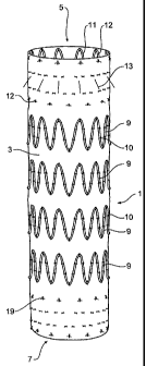
Now looking more closely at the drawings and in particular Figs. 1 and
2 showing external and internal views of a first embodiment of the present
invention, it will be seen that a stent graft 1 includes a tubular body 3
formed from
a biocompatible woven or non-woven fabric or other material. The tubular body
has
a proximal end 5 and a distal end 7. The tubular body may have a diameter in
the
range of 22 to 42 mm and a length of from 100 to 150 mm. The stent graft 1 may
be tapered, outwardly bulging like a balloon or of constant diameter along its
length depending upon the topography of the vasculature.
Along the length of the tubular body, there are a number of self-
0 expanding zigzag stents 9 such as the well-known Gianturco Z or zigzag stent
on the
outside of the body. In this embodiment there are four external stents 9
spaced
apart by a distance of between 5 to 10 mm. The external stents 9 are joined to
the
graft material by means of stitching 10 preferably using a monofilament or
braided
suture material.
5 At the proximal end 5 of the stent graft 1 there is provided an internal
zigzag stent 11 which provides a sealing function for the proximal end of the
stent
graft. The outer surface of the tubular body 3 at the proximal end 5 presents
an
essentially smooth outer surface which with the assistance of the internal
zigzag
stent 11 can engage and seal against the wall of the aorta when it expands and
is
'.0 deployed. The proximal stent 11 is comprised of struts 15 with bends 16 at
each
end of the struts. Affixed to some of the struts 15 are barbs 13 which extend
distally from the struts through the graft material. When the stent graft is
deployed
into a thoracic arch, the barbs 13 engage and/or penetrate into the wall ofthe
aorta
and prevent distal movement of the stent graft caused by pulsating blood flow
!5 through the stent graft.
It will be noted that the stent 11 is joined to the graft material by means
of stitching 12 preferably using a monofilament or braided suture material.
At the distal end 7 of the stent graft 1, there is an internal sealing stent
17 (see Fig. 2) which again is fastened to the graft material body 3 by
stitching 19
30 preferably using a monofilament or braided suture material. The outer
surface of
the tubular body 3 at the distal end 7 presents an essentially smooth outer
surface
CA 02489495 2004-12-07
WO 2004/002370 PCT/US2003/020642
-9-
which with the assistance of the internal zigzag stent 17 can engage and seal
against the wall of the aorta when it is deployed.
The stent graft shown in Figs. 1 and 2 may be used for treatment of
patients with symptomatic acute or chronic dissections and ruptures in the
descending thoracic aorta.
Figs. 3 and 4 show external and internal views of a second embodiment
of the stent graft according to the present invention. In this embodiment, the
stent
graft 20 has a tubular graft material body 22 in the same manner as the
embodiment shown in Fig. 1 with external stents 24 spaced along the body with
a
longitudinal spacing of approximately 5 to 10 mm between the stents. The
length
of the stent graft may be in the range of 75 to 241 mm and a diameter in the
range
of 22 to 42 mm in 2 mm increments.
Also in a similar manner to the embodiment shown in Figs. 1 and 2 at the
proximal end 26 ofthe stent graft there is an internal sealing stent 28 with
barbs 30.
At the distal end 27 of the stent graft 20, there is also an internal distal
sealing stent 32 but in addition, there is a distally extending exposed zigzag
stent
34. This distally extending exposed stent 34 has barbs 36 on some of its
struts and
these barbs 36 are directed proximally. The distally extending exposed zigzag
stent
34 is fastened to the tubular graft material body 22 by stitching 33.
~0 It will be noted that there are provided at the distal end of the stent
graft
radiographic markers 38 to enable correct positioning of the distal end of the
stent
g raft.
Hence, when the stent graft according to this embodiment of the
invention is deployed, the barbs 30 prevent distal migration of the proximal
end of
?5 the stent graft and the barbs 36 on the exposed stent 34 prevent proximal
migration
of the distal end ofthe stent graft 20. This tendency of distal migration of
the distal
end and proximal migration of the distal end may occur ifthe central portion
of the
stent graft is free within an aneurysm and sideways force on a curved stent
graft
caused by pulsating blood flow causes sideways movement of the body ofthe
stent
30 graft with the potential for distal movement of the proximal end and
proximal
movement of the distal end.
CA 02489495 2004-12-07
WO 2004/002370 PCT/US2003/020642
-10-
The stent graft shown in Figs. 3 and 4 may be used for endovascular
repair ofthoracic aortic aneurysms in the descending thoracic aorta and
particularly
for treatment of patients with atherosclerotic aneurysms, symptomatic acute or
chronic dissections, contained ruptures and growing aneurysms, which result in
distal ischaemia.
Figs. 5 and 6 show external and internal views of a third embodiment of
the stent graft according to the present invention. In this embodiment, a
thoracic
stent graft assembly is formed from a first portion 50 and a second portion
52. The
first portion 50 is intended to be deployed proximally ofthe second portion
52. The
0 first portion 50 is substantially identical with the stent graft embodiment
shown in
Figs. 1 and 2. It has proximal and distal internal sealing stents in a tubular
graft
body, barbs extending from the proximal sealing stent and external zigzag
stents
between the proximal and distal sealing stents.
The second portion 52 is substantially the same as the embodiment
5 shown in Figs. 3 and 4 except that there are two internal sealing stents 62,
63 at the
proximal end 60 of the second portion 52. It will be noted that although the
second
portion 52 is substantially similar to the embodiment shown in Figures 3 and 4
it
does not include the distally extending anchoring barbs on the proximal
sealing
stent 62.
!0 The proximal end 60 of the second portion 52 with the internal sealing
stents 62, 63 can be deployed either inside the distal end 58 of the first
portion 50
or outside the distal end 58 of the first portion 50.
Thismeansthatindeployingthestentgraftassemblyofthisembodiment
of the invention either the first or second portions may be deployed first and
the
'.5 other portion subsequently deployed depending upon the requirements in a
particular case.
In either case it is preferable to have at least two stents overlap and it
may be noted that by having this overlap there is at least one stent length of
smooth internal surface ofone of the portions engaging against a smooth
external
30 surface of the other of the portions. By this arrangement, sealing between
the first
and second portions is possible. Also, by having an overlap of at least two
stents,
CA 02489495 2004-12-07
WO 2004/002370 PCT/US2003/020642
-11 -
relative movement between the first and second portions is less likely to
cause
parting of the first and second portions of the thoracic stent graft assembly
when
it is deployed and pulsating blood flow through the stent graft causes
sideways
movement of the centre portion of the stent graft as discussed above.
The stent graft shown in Figs. 5 and 6 may be used for endovascular
repair ofthoracic aortic aneurysms in the descending thoracic aorta and
particularly
for treatment of patients with atherosclerotic aneurysms, symptomatic acute or
chronic dissections, contained ruptures and growing aneurysms. The ability to
adjust the overall length of the device by providing more or less overlap of
the first
0 and second portions (i.e. "tromboning") allows more accurate placement of
the
proximal and distal sealing stents and the anchoring barbs.
Fig. 7 shows a stent graft of the embodiment shown in Fig. 1 to show the
amount of bending which is possible in the stent graft for placement in the
thoracic
arch of a patient. The internal radius of curvature ofthe stent graft
according to this
5 invention may be any radius greater than 35 mm. This can be achieved by
having
the stents longitudinally spaced apart by between five to ten millimetres as
discussed earlier and so far as possible staggering the placement of apices of
adjacent stents. This may not be possible where adjacent stents have different
numbers of struts.
!0 Fig. 8 shows features of a sealing stent that may be present in any of the
above embodiments. A stent graft 70 has a graft material body 72 and an
internal
sealing stent 74 joined to the graft material by stitching 78, also referred
to as
fastenings. Stent 74 is a zigzag stent having struts 75 with bends 76 at each
end of
the struts. Affixed to at least some of the struts 75 are barbs 73 which
extend
!5 distally from the struts through the graft material.
Asdescribed in U.S. Provisional PatentApplication Serial No.60/391,737,
which has been incorporated herein by reference, where the stent graft is
deployed
in a blood vessel, blood flow causes a pull on the graft material tube which
is
resisted by the barbs on the stent. Hence the fastenings of the stent joining
the
30 stent to the graft material take the pull on the prosthesis, and these
fastenings
preferably are sufficiently strong to take that pull. Similarly, the barbs on
the
CA 02489495 2004-12-07
WO 2004/002370 PCT/US2003/020642
-12-
exposed stent used on the distal end ofthe graft material tube resist blood
flow pull
on the graft material tube.
Fig. 9 shows features of a distally extending exposed zigzag stent used
in the embodiments shown in Figs. 3 to 6. As can be seen in the detailed views
in
Fig. 9, the struts 79 and bend 82 of the stent are on the inside of the graft
material
72. At least two fastenings 80 and 81 are used to fasten the stent 83 to the
graft
material 72. As shown here, the first fastening 80 is positioned at the apex
of the
bend 82, and a second fastening 81 is positioned spaced apart adjacent the
transition from the bend 82 to the struts 79 of the stent. The second
fastening 81
0 can be positioned on either strut 79 extending from the bend 82 in a region
extending up to an angle of 50° either side of the first fastening 80
measured
around the radius of the bend from the apex of the bend 82. Generally the
second
fastening 81 is spaced from the first fastening 80 by 0.5 to 2 mm.
The spaced apartfastenings are preferably present at the proximal bends
5 of the distally extending stent 34 of Figs. 3 and 4, and the distally
extending stent
of Figs. 5 and 6.
Throughout this specification various indications have been given as to
the scope of this invention but the invention is not limited to any one of
these but
may reside in two or more of these combined together. The examples are given
!0 for illustration only and not for limitation.
Throughout this specification and the claims that follow unless the
context requires otherwise, the words 'comprise' and 'include' and variations
such
as'comprising' and'including' will be understood to imply the inclusion ofa
stated
integer or group of integers but not the exclusion of any other integer or
group of
!5 integers.