Note: Descriptions are shown in the official language in which they were submitted.
CA 02489584 2004-12-16
WO 03/105659 PCT/IL03/00515
ROBOT FOR USE WITH ORTHOPAEDIC INSERTS
FIELD OF THE INVENTION
The present invention relates to the field of the robotic guidance of
orthopaedic surgical procedures performed on pre-positioned inserts, and
especially for use in the orthopaedic reduction of bone fractures using
intramedullary locking procedures.
BACKGROUND OF THE INVENTION
The need for precise, minimally invasive, surgical interventions has
resulted in the creation of methods of using computers in conjunction with
advanced assistance devices to improve surgical planning and execution. Over
the
past decade, a variety of such Computer Integrated Surgery (CIS) systems have
been developed, with resulting clinical benefits, largely for use in the
fields of
neurosurgery, laparoscopy, maxillofacial surgery and orthopaedics, as for
instance
described in the article by L. Joskowicz et al., entitled "Computers in
imaging and
guided surgery", published in Computers in Science and Engineering, Vol. 3(5),
pp 65-72, 2001.
CIS systems can potentially benefit many orthopaedic surgical procedures,
including total hip and total knee replacement, pedicle screw insertion,
fracture
reduction, and ACL (Anterior Cruciate Ligament) ligament reconstruction. These
procedures are ubiquitous and are performed in high volume in operating rooms
worldwide. They generally involve rigid bone structures that image well,
require
preoperative planning, and employ instruments and tools, such as implants,
screws, drills, and saws that require precise positioning. A number of CIS
systems
for such procedures are currently in use, such as those described in the book
"Computer Assisted Orthopaedic Surgery (CAOS)", edited by L.P. Nolte, and R.
Ganz, published by Hogrefe and Huber (1999).
CA 02489584 2004-12-16
WO 03/105659 PCT/IL03/00515
2
One technique used for fracture reduction is closed intramedullary nailing,
and, according to the article by R.J. Brumback, "Regular and Special Features -
The Rationales of Interlocking Nailing of the Femur, Tibia, and Humerus",
published in Clinical Orthopaedics and Related Research, Vol. 324, pp.586-651,
Lippincott-Raven (1996), it is probably the current routine procedure of
choice for
reducing fractures of the femur and the tibia. This procedure restores the
integrity
of the fractured bone by means of a nail inserted in the medullary canal. In
most
cases, the surgeon inserts lateral proximal and distal interlocking screws, to
prevent fragment rotation and bone shortening. Preparation of the bone for
distal
locking has long been recognized as one of the most challenging steps in the
procedure, at least according to prior art methods of performing the
procedure.
The procedure is performed under X-ray fluoroscopy, which is used to view the
position of the bone fragments, the surgical tools and the implants used, in
order
to determine the position of the holes to be drilled for these screws.
Numerous
X-ray fluoroscopic images are required for this procedure, since the nail
often
deforms by several millimeters to conform to the bone canal shape, and the
exact
position of the distal locking nail holes cannot therefore be determined in
advance.
According to these prior art methods, the surgeon adjusts the entry point and
orientation of the drill, to the best of his judgement and his interpretation
of the
X-ray images, by repeatedly alternating between anterior-posterior and lateral
X-ray fluoroscopic views, such that the drill axis coincides as accurately as
possible with the corresponding nail hole axis. Drilling proceeds
incrementally,
with each advance verified with a new pair of X-ray fluoroscopic images. Once
the pilot hole passing through the distal locking nail hole has been drilled
in the
bone, the locking screws can be inserted and fastened.
Because of the nature of the procedure, depending very largely on the skill
of the surgeon, a number of complications can arise, including inadequate
fixation, malrotation, bone cracking, cortical wall penetration and bone
weakening
due to multiple or enlarged pilot holes. Furthermore, it has been reported in
the
article "Interlocking medullary nails - radiation doses in distal targeting"
by S.
Skejdal and S. Backe, published in Archives of Orthopaedic Trauma Surgery,
Vol.
CA 02489584 2004-12-16
WO 03/105659 PCT/IL03/00515
3
106, pp 179-181, 1987, that the surgeon's direct exposure to radiation per
procedure, using these prior art methods, is 3-30 minutes, depending on the
patient
anatomy and the surgeon's skill. In general, something between about 30 and
50%
of this time is spent on the distal locking process.
Many non-CIS devices have been developed for distal locking, even
recently, such as that described by C. Krettek, et al, in the article "A
mechanical
distal aiming device for distal locking in femoral nails", published in
Clinical
Orthopaedics, Vol. 384, pp. 267-275, 1999. Examples of such devices and
procedures include proximally mounted targeting devices, stereo fluoroscopy,
mechanical guides, and optical and electro-magnetic navigation systems that
help
locate the center of the distal locking nail holes. However, these devices and
techniques generally have some disadvantages, for instance that they are only
selectively applicable, may be cumbersome and difficult to use, may not be
sufficiently accurate, and they thus fail to significantly reduce the
likelihood of
patient complications.
Fluoroscopy-based CIS navigation systems, such as those described by L.
Joskowicz, et al., in the article "FRACAS: A system for computer-aided image-
guided long bone fracture surgery", published in "Journal of Computer-Aided
Surgery", Vol. 3(6), pp. 271-288, 1999, take the guesswork out of targeting.
Such
systems enhance, reduce, or altogether eliminate X-ray fluoroscopic images by
replacing them with a virtual reality view in which the positions of the bone
and
the surgeon's instruments are continuously updated and viewed on-screen as
they
move, using tracking devices and three dimensional registration procedures.
They
can assist the surgeon in aligning the drill axis with the distal locking nail
hole
axis to an accuracy of about lmm and 1 . However, they do not provide any
mechanical guidance for the hand-held drill, which can slip or deviate from
its
planned trajectory as the drilling proceeds. Thus, even using such prior art
CIS
navigation systems, the surgical outcome of the procedure is still dependent
to an
extent on the skill of the surgeon.
Robot-based CIS systems have been developed to assist the surgeon in
implementing the preoperative plan by mechanically positioning and sometimes
CA 02489584 2010-09-22
4
executing the surgical action itself One such system is described by K. Cleary
et
al., in the article "State of the art in surgical robotics: clinical
applications and
technology challenges", published in Journal of Computer-Aided Surgery, Vol.
6(6), pp. 312-328, 2001. The robots are either floor-standing industrial
robots,
adapted for use in the desired surgical application, or table-mounted custom-
designed serial robots. Such robots are generally voluminous and heavy,
despite
the fact that in such surgical applications, they need to operate with
relatively
small workloads and work volumes. In such systems, bone immobilization or
real-time dynamic tracking are important issues, since the relative
configuration of
the bone with respect to the robot must be known precisely at all times. This
may
complicate the registration procedure and may adversely affect the overall
system
accuracy.
There therefore exists a need in the field of orthopaedic surgery, for a
system which overcomes the disadvantages of prior art systems, and enables the
automatic alignment of tools required for the procedure, with the bones or
inserts
involved in the procedure, such that the procedure becomes less dependent on
the
skill of the surgeon, with a concomitant increase in the success rate of the
procedure.
SUMMARY OF THE INVENTION
According to the present invention, there is provided a surgical system
comprising:
a robot connected to a bone, with which is associated an orthopaedic
insert having at least one predrilled hole for attaching said insert to said
bone;
a drill guiding plate carried by said robot, said guiding plate having at
least one hole for guiding a drill into said bone and through said at least
one
predrilled hole; and
CA 02489584 2010-09-22
a computational system utilizing data from at least one x-ray image
including said drill guiding plate and said at least one predrilled hole, to
align said
robot such that the axis of said at least one hole defined by said drill
guiding plate is
aligned essentially colinearly with the axis of said at least one predrilled
hole defined
by said insert.
The present invention seeks to provide a new robotic guidance system, with
the robot mounted directly on the bone on which the operation is being
performed,
or in a configuration which ensures that the robot position has a direct one-
to-one
relationship with the position of the bone, and which assists the orthopaedic
surgeon in performing procedures involving the alignment of surgical tools
with
features of internally located implants utilized in the performance of the
desired
orthopaedic procedure. The robot itself is small, and is thus unobtrusive and
easily
rendered safe for use in the sterile operating room environment. The system
significantly reduces the likelihood of complications arising in the
procedure,
since the surgical tool in use is aligned automatically with its target site,
both
laterally and angularly. Furthermore, the need for numerous, trial and error,
X-ray
fluoroscopic images is reduced, thus increasing radiation safety for all
involved.
The system is particularly suitable for the automatic alignment of a bone
drill with
predrilled holes present in metallic inserts within or in contact with the
patient's
bone. One preferred use of such a system is for the robotically-controlled
guidance
of a target drill to exactly match the position and orientation of the distal
locking
holes in a pre-inserted intramedullary nail, as used in long bone fracture
reduction.
The mounting of the robot directly on the bone, and hence also effectively on
the
nail, which is inserted firmly into the bone, has the advantage that if the
nail
rotates, the robot moves with it, thus preserving the mutual orientation and
position of the guide drill with the distal holes in the nail. If the robot is
alternatively mounted directly on the nail, this advantage applies directly.
CA 02489584 2010-09-22
5a
The system preferably comprises a miniature robot holding a targeting drill
guide. The robot, because of its small size and weight, can be mounted
directly on
the bone on which the desired procedure is being performed. In the case of the
long bone intramedullary distal locking procedure, the robot is, according to
one
preferred embodiment, attached to the proximal nail head. Alternatively and
preferably, it is directly mounted laterally on the patient's bone segment
distal to
the fracture line and proximal to the distal locking nail holes. The targeting
drill
guide preferably has one or more guide holes for the drill, in accordance with
the
number of holes to be drilled, and a targeting fiducial marker pattern for use
in
determining the position and alignment of the targeting drill guide in the X-
ray
fluoroscopic images taken during the procedure. To obtain accurate
registration
results using these X-ray fluoroscopic images, camera distortion correction
and
calibration are performed, preferably using an image calibration ring on the
fluoroscopic X-ray C-arm unit.
Preferably, using only a small number of lateral X-ray fluoroscopic images,
the axis or axes of the targeting drill guide holes are brought into alignment
with the
axis or
CA 02489584 2004-12-16
WO 03/105659 PCT/IL03/00515
6
axes of the distal locking nail holes, by computing the transformation between
them and positioning the robot accordingly. The surgeon can then drill the
holes in
the bone with confidence that they will be accurately in line with the holes
of the
insert. Though the term targeting drill is generally used throughout this
application, in keeping with the common procedure where a smaller target drill
hole is made as a pilot for the correct size drill bore, it is to be
understood that the
invention is not limited to targeting drilling procedures but is equally
applicable to
direct drilling of the final hole, where this procedure is followed.
Mounting the robot directly on the nail or on the patient's bone is
minimally invasive, eliminates the need for limb immobilization or real-time
tracking, and for trial and error positioning, and greatly simplifies
registration.
The system can potentially reduce intra-operative time, reduce complications
associated with malpositioning, and can allow less-experienced surgeons to
perform the locking with high success rates.
There is thus provided in accordance with a preferred embodiment of the
present invention, a surgical system comprising a robot connected to a bone,
with
which is associated an orthopaedic insert having at least one predrilled hole
for
attaching the insert to the bone, a drill guiding plate carried by the robot,
the
guiding plate having at least one hole for guiding a drill into the bone and
through
the at least one predrilled hole, an imaging system generating at least one
image
including the drill guiding plate and the at least one predrilled hole, and a
computational system utilizing data from the at least one image to align the
robot
such that the axis of the at least one hole defined by the drill guiding plate
is
aligned essentially colinearly with the axis of the at least one predrilled
hole
defined by the insert.
In the above described system, the axis of the at least one hole defined by
the drill guiding plate is preferably aligned essentially colinearly with the
axis of
the at least one predrilled hole both laterally and in angular orientation.
The drill
guiding plate preferably comprises a plurality of fiducial markers disposed in
a
predetermined pattern. This plurality of fiducial markers is preferably used
to
align the drill guiding plate by means of the robot such that it images the
drill
CA 02489584 2004-12-16
WO 03/105659 PCT/IL03/00515
7
guiding plate in a fronto-parallel orientation. In any of the above described
embodiments of the system, the imaging system is preferably aligned such that
it
images the at least one predrilled hole in a fronto-parallel orientation. As a
result,
the computational system may align the robot by utilizing data from only one
image of the imaging system.
There is further provided in accordance with yet another preferred
embodiment of the present invention, a surgical system as described above, and
wherein the robot is directly mounted either on the bone or is attached to an
insert
associated with the bone. In any of the above described systems, the robot
maintains its position relative to the bone such that both tracking of the
bone
position and immobilization of the bone are obviated.
In accordance with still more preferred embodiments of the present
invention, in these surgical systems, the bone may be a long bone, and the
orthopaedic insert an intramedullary nail, and the at least one predrilled
hole a
distal locking hole. In such cases, the robot may be attached to the proximal
end of
the intramedullary nail.
Alternatively and preferably, the orthopaedic insert may be an externally
attached connector plate, and the at least one predrilled hole a connecting
hole. In
such cases, the bone may preferably be a femur, and the connector plate a
percutaneous compression plate, and the connecting hole may then be either
such
as to accommodate a screw for connecting the plate to the shaft of the femur,
or
such as to connect a fractured head of the femur to its shaft.
In any of the above embodiments of the surgical system, the imaging
system preferably comprises an image intensifier with a calibration ring
assembly,
adapted to enable at least one of image distortion correction and camera
calibration.
There is further provided in accordance with still another preferred
embodiment of the present invention, a surgical system as described above, and
wherein the robot comprises a miniature parallel robot. Such a robot
preferably
comprises at least three actuators mounted on a base member, the actuators
being
configured for at least one of translational and rotational movement.
CA 02489584 2004-12-16
WO 03/105659 PCT/IL03/00515
8
In accordance with further preferred embodiments of the present invention,
there is also provided an imaging system comprising:
(a) a radiation source for illuminating a target to be imaged, the target
having at least one predefined hole,
(b) a target guide having at least a second predefined hole, whose axis is to
be brought into coincidence with the axis of the at least one predefined hole
of the
target,
(c) a robot on which the target guide is mounted, for bringing the axes of
the at least one target guide hole and the at least one target hole into
coincidence,
(d) an image intensifier generating images of the target and the target
guide, and
(e) a computation system comprising:
(i) a first position localizing module, which computes the position of
the at least one target guide hole from an image thereof,
(ii) a second position localizing module, which computes the
position of the at least one target hole from an image thereof, and
(iii) a registration unit adapted to determine a spatial relationship
between the at least one target guide hole and the at least one target hole.
In the above described imaging system, the image intensifier is preferably
aligned in a fronto-parallel setup, such that the registration unit
determining the
spatial relationship between the at least one target guide hole and the at
least one
target hole, utilizes a two-dimensional image only. In this case, the
alignment is
adjusted by determining when an image of the at least one target hole has a
minimum elliptic shape. The image intensifier preferably incorporates
distortion
correction and camera calibration functions, or alternatively and preferably,
the
system also comprises a separate calibration ring assembly for distortion
correction and camera calibration.
In the above described embodiments of the imaging system, the target
guide preferably comprises a predetermined pattern of fiducial markers, the
images of which are utilized by the first position localizing module in the
computing the position of the at least one target guide hole.
CA 02489584 2004-12-16
WO 03/105659 PCT/IL03/00515
9
In the above described imaging system embodiment, the first position
localizing module, which computes the position of the at least one target
guide
hole, preferably comprises (i) a fiducial marker position locator and position
template determiner, (ii) an image generator producing a new image of the
target
guide, comprising the at least one image of the target guide from which the
positions of the fiducial markers have been morphologically removed, (iii) a
normalized cross correlation calculator for the template at pixel locations
with
negative values, determining the centers of the fiducial markers, (iv) a
fiducial
searcher looking in small areas around local maxima found by the normalized
cross correlator, and (v) a position locator for the targeting guide
localization,
from the locations of the fiducials found by the fiducial searcher, (iv). The
fiducial
marker position locator and position template determiner preferably utilizes a
Hough transform method, and the position locator preferably utilizes a
principal
component analysis procedure.
Furthermore, in the above described imaging system embodiments, the
second position localizing module, which computes the position of the at least
one
target hole from an image thereof, preferably comprises (i) a contour locator
using
an edge detection routine, for determining the longitudinal contours of the
target,
(ii) a hole searcher, determining the position of holes in an area between the
longitudinal contours in an image of the target, using a detector for regions
with
the maximal number of edge elements in windows of dimensions similar to that
of
the hole moved over the contour, and (iii) an ellipse fitter for the edge
elements in
each of the regions detected. The edge detection routine is preferably a Canny
edge detector with sub-pixel edge localization.
Additionally, in the above described imaging system embodiments, the
registration unit adapted to determine a spatial relationship between the at
least
one target guide hole and the at least one target hole, preferably comprises
an
aligner to bring the image intensifier to a fronto-parallel configuration such
that
the at least one image of the target hole has a minimal elliptic shape, a
target guide
aligner routine, such that the at least one image of the target guide hole has
a
CA 02489584 2004-12-16
WO 03/105659 PCT/IL03/00515
minimal elliptic shape, and a target guide lateral translator so that the
positions of
the axes of the target guide hole and the target hole coincide.
Using the imaging system in the above-described various preferred
embodiments, there is further provided a method of bringing the positions of
the
axes of a hole defined by a target guide and a hole defined by the target into
coincidence, comprising the steps of localizing the position of the target
guide
hole from at least one image thereof, localizing the axis of the target hole
from at
least one image thereof, and registering the localized target guide hole with
the
axis of the target hole. An initial step may preferably be performed of image
distortion correction and calibration of the imaging system to obtain the
images of
the target guide hole and the target hole. The individual computing modules of
the
imaging system embodiments described above operate according to more
preferred methods of the present invention, as described in relation to each
functional module, and its parts.
BRIEF DESCRIPTION OF THE DRAWINGS
The present invention will be understood and appreciated more fully from
the following detailed description, taken in conjunction with the drawings in
which:
Figs. 1A and 1B are schematic representations of lateral and frontal views
respectively, such as would be obtained by X-ray fluoroscopic imaging
illustrating
the distal locking stage of the intramedullary nailing procedure;
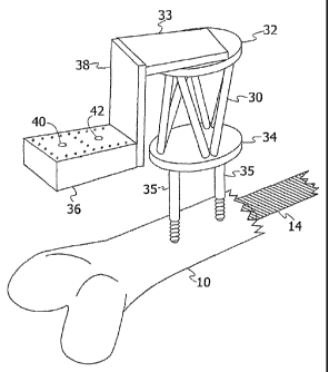
Fig. 2 is a schematic isometric illustration of a robot-based system,
constructed and operative according to a preferred embodiment of the present
invention, for the precise drilling of the pilot holes for locking screw
insertion;
Fig. 3 is a cross-sectional view of the preferred embodiment of Fig. 2,
showing some features which are not visible in the isometric view of Fig. 2;
Fig. 4 is a further cross-sectional view of the robot of Fig. 2, showing an
alternative and preferred method of mounting the robot to the patient's bone;
CA 02489584 2004-12-16
WO 03/105659 PCT/IL03/00515
11
Fig. 5A is a schematic illustration of a complete robot-guided orthopaedic
operating system, according to another preferred embodiment of the present
invention, incorporating the robot assembly shown in Fig. 2;
Fig. 5B is a block diagram of the various computing modules which are
incorporated, according to a further preferred embodiment of the present
invention, into the computing system shown in Fig. 5A;
Fig. 6 is a schematic representation of an X-ray fluoroscopic image,
showing the targeting drill guide and the distal end of the intramedullary
nail with
the two distal locking holes;
Fig. 7 is a schematic representation of another X-ray fluoroscopic image
similar to that of Fig. 6, but showing superimposed fiducial pattern
calculation
lines to illustrate the method by which the localization of the targeting
drill guide
is determined;
Fig. 8 is a schematic representation of another X-ray fluoroscopic image,
similar to that of Fig. 6, but showing superimposed nail longitudinal contour
lines
to illustrate the method by which the localization of the distal locking holes
is
determined; and
Fig. 9 is a schematic cross sectional view of a robot-based system, similar
to that shown in Fig. 2, but being used for the connection of a fractured neck
of
the femur to the shaft by means of robotic-positioned drilling through a
connector
plate.
DETAILED DESCRIPTION OF PREFERRED EMBODIMENTS
Reference is first made to Figs. 1A and 1B, which are schematic
representations of views that would be obtained by X-ray fluoroscopic imaging,
illustrating the distal locking stage of the intramedullary nailing procedure.
Fig.
1A is representative of a lateral image and Fig. 1B a frontal image, of the
distal
part of the femur 10, showing the intramedullary nail 14 with two distal
locking
nail holes 16, 18. The nail 14 is inserted through a minimal opening, usually
in the
proximal part of the bone 10, without the need to surgically expose the
fracture.
CA 02489584 2004-12-16
WO 03/105659 PCT/IL03/00515
12
In performing the procedure, the surgeon first reduces the fracture by
manipulating the proximal and distal bone fragments through the leg 12 until
they
are aligned. The surgeon then inserts a guide wire, reams the canal if
necessary,
and drives in the nail 14. The surgeon then drills the appropriate distal
locking nail
holes in the bone, opposite the pre-prepared holes 16, 18 in the nail, and
inserts
lateral proximal and distal interlocking screws 20, 22, to prevent fragment
rotation
and bone shortening. In Fig. 1B, one locking screw 20 is shown in place, and
the
second one 22, is shown being fastened by means of a screwdriver 24.
Reference is now made to Fig. 2, which is a schematic isometric illustration
of a robot-based system, constructed and operative according to a preferred
embodiment of the present invention, for the precise drilling of the pilot
holes for
locking screw insertion. In the preferred embodiment of Fig. 2, the robot of
the
system is shown being utilized to position the guide drill axis for drilling
through
a patient's femur and into the distal locking holes in a pre-inserted
intramedullary
nail. It is to be understood to those skilled in the art, however, that, with
minor
modifications, if at all necessary, the system can also preferably be used on
any of
the other long bones, the tibia, the humerus, the ulna, the radius and the
fibula, or
for accurately drilling into predisposed orthopaedic inserts other than
intramedullary nails, such as connector plates disposed externally to the
bone.
In Fig. 2, the patient's femur 10 is shown with the intramedullary nail 14
pre-inserted. The robot 30 is preferably a miniature parallel robot having a
base
plate 34 for mounting purposes, and a top plate 32 onto which the operating
load
is attached. The robot shown in the preferred embodiment of Fig. 2 has a
height of
the order of 70 mm and a weight of about 200 grams. The position and
orientation
of its top plate 32 can be adjusted and locked by the robot controller to the
desired
configuration with high accuracy and rigidity. The work volume of the robot is
sufficient for the task at hand, which involves motion of a drilling head from
a
pre-estimated approximate position into the correct position for drilling
through
the distal locking holes in an intramedullary nail.
The robot carries a guiding assembly, which preferably comprises three
components, an adjustable slide head 33, a connecting block 38, and a
targeting
CA 02489584 2010-09-22
13
drill guide 36. The slide head 33 is mounted directly on top of the robot. Its
location along the bone long axis can be manually adjusted over a range of
typically up to 50 mm., to one of several predetermined positions, as defined
by
positioning pins and holes in the members of the slide, depending on the
application envisaged. The use of predetermined positions is necessary in
order to
maintain a known predefined registration between the robot frame of reference
and the position of the drilling guide hole axes. The pin position selected
must be
input to the control system so that the actual registration selected is used
in the
subsequent position calculations. The connecting block 38 and the targeting
drill
guide 36, are made of radiolucent plastic, since they are visible in the X-ray
fluoroscopic images to be taken of the drilling area. The targeting drill
guide 36 is
preferably in the form of a 40 x 55 x 20mm block, and in the preferred example
shown, has two drill guide holes 40, 42, disposed with their axes 30mm apart,
which are predetermined to correspond to the spacing between the centers of
the
distal locking nail holes. The targeting drill guide 36 is parallel to the
robot base
34, and close to the skin of the leg of the patient. The drilling guide has
predetermined patterns of fiducial markers that are used for determining its
spatial
and angular localization in the X-ray fluoroscopic images. In the preferred
embodiment shown in Fig. 2, these fiducials are 2mm. stainless steel spheres
arranged in predetermined patterns resembling the letter "A", in its upper and
lower surfaces.
There are several methods of mounting the robot on the bone. In the
preferred embodiment shown in Fig. 2, the robot base 34 is mounted on two 5mm
diameter by 80mm length, self-tapping screws, mounted 25mm apart, which are
driven into the distal bone cortex.
Reference is now made to Fig. 3, which is a cross-sectional view of the
preferred embodiment of Fig. 2, showing some features which are not visible in
the isometric view of Fig. 2. The set of prepositioning pins and holes 31
between
CA 02489584 2010-09-22
13a
the top plate of the robot 32 and the slide head 33 are shown, though their
number
and positions in Fig. 3 are meant to be purely illustrative and not limiting.
The
holes 16, 18, in the intramedullary nail 14 are visible, as are the two layers
of
CA 02489584 2004-12-16
WO 03/105659 PCT/IL03/00515
14
fiducial marker spheres 60 in the targeting drill guide plate 36. The drill
head 70 is
shown with the guide drill bit 72 accurately positioned by the robot over the
center
of the desired distal locking hole 18 in the nail.
Reference is now made to Fig. 4, which is a further cross-sectional view of
the robot of the present invention, showing an alternative and preferred
method of
mounting the robot to the patient's bone. In this embodiment, the robot is
fitted
with an extended base 39, which is attached by means of a curved support rod
17
to the intramedullary proximal nail head 15. In this embodiment, there is no
need
for the mounting pins 3 5 of the embodiment of Fig. 3. All of the other items
shown in Fig. 3 are numbered like those shown in the embodiment of Fig. 4. It
is
to be understood to those skilled in the art that other preferred mounting
options
besides those shown in Figs. 3 and 4, are equally applicable for use with the
present invention, provided that they provide rigid mounting of the robot
relative
to the bone being operated on.
Reference is now made to Fig. 5A, which is a schematic illustration of a
complete robot-guided orthopaedic operating system, according to another
preferred embodiment of the present invention, incorporating the robot
assembly
shown in Fig. 2. The system preferably includes a stabilized, robotically
controlled, targeting drill guide 36, mounted on a miniature robot 30, which
is
attached to the distal bone fragment 10 of a patient (not shown) lying on a
radiolucent operating table 50. The X-ray source 54 is positioned beneath the
table, and at the top end of the C-arm 52, the fluoroscopic image intensifier
55 is
fitted with an image calibration ring 56, consisting of two parallel plates
with
embedded fiducials. The images from this image intensifier are directed to a
computing system 74 for acquiring fluoroscopic images, either directly in the
case
of an image intensifier with a digital output port, or through a frame grabber
at the
input of the computing system in the case of an image intensifier with an
analog
video output. An image processor analyzes the images obtained, and outputs
them
for displaying on a monitor 76 to the surgeon. A robot controller card 78 is
also
incorporated for providing the correct input signals to direct the robot 30,
according to command signal outputs generated in the computing system 74.
CA 02489584 2004-12-16
WO 03/105659 PCT/IL03/00515
An image calibration ring 56, such as of the type used in calibration and
prior art tracking systems, is preferably attached with clamps to the C-arm
image
intensifier, and according to a preferred embodiment, has two parallel
radiolucent
plate targets, spaced 76 mm apart, with 120 embedded fiducial steel balls of
2mm
and 3mm diameter, arranged in a predetermined asymmetrical pattern. Any
suitable pattern which allows for the performance of accurate calibration and
registration procedures may preferably be used.
Reference is now made to Fig. 5B, which is a block diagram of the various
computing modules which are incorporated, according to a further preferred
embodiment of the present invention, into the computing system 74 shown
schematically in Fig. 5A. The output from the fluoroscopic image intensifier
55 is
directed into an optional fluoroscopic image distortion correction and
calibration
unit 71, for use when such a function is not included with the camera system
itself.
From here, the signal is passed to a target guide localization module 73,
which
analyzes one or more images containing the target guide, in the case of this
preferred embodiment, the drilling guide, such that the target guide
localization is
known, and to the target localization module 75, which analyzes one or more
images containing the target itself, in this embodiment, a distal locking nail
hole,
to determine the localization of the distal locking nail hole. Though these
modules
73, 75, are shown in Fig. 5B operating sequentially, it is to be understood
that they
can process the signal information simultaneously. Finally, the digital
imaging
information is passed to a target guide-target registration module 77, which
in this
preferred embodiment generates a rigid registration between the axes of the
targeting drill guide holes and the distal locking nail holes. Beneath each of
the
three preferred computing modules 73, 75 and 77 of the computing system 74,
there are shown implementation steps or units, according to one preferred
embodiment of the present invention, though it is to be understood that
alternative
individual steps which provide the same end registration result could be
substituted for any of the propose functional units shown. A full description
of the
functional performance of each of these units is given hereinbelow. The
computing system 74 and its separate modules is designed to be robust,
accurate,
CA 02489584 2004-12-16
WO 03/105659 PCT/IL03/00515
16
and to function correctly even with fiducial occlusions in the images, as
described
more fully hereinbelow.
An outline of a preferred surgical protocol, and system operational
procedure to perform it, is as follows. Once the fracture has been reduced and
the
nail 14 has been inserted to its desired position, the image calibration ring
56 is
mounted on the fluoroscopic C-arm image intensifier 55, as shown in Fig. 5A.
Using a distal, lateral fluoroscopic image showing the distal locking nail
holes, the
surgeon determines the location of the self-tapping screws 35 on which the
robot
is to be mounted. Their axes should be roughly parallel to the distal nail
hole axes,
and preferably 40 to 80 mm proximal to them. Two parallel pilot holes,
preferably
approximately 30mm apart and along the axis of the nail, are then drilled by
the
surgeon, preferably with the help of a hand-held jig. The self-tapping screws
are
then fastened, and the robot base mounted on them. The position of the
targeting
drill guide mounted on the robot top, relative to the distal locking nail
holes, is
roughly adjusted between its predefined pin positions so that the drill guide
holes
are approximately above the distal locking nail holes. The orientation of the
C-arm
is then preferably adjusted by the X-ray technician, until it is determined
that the
distal locking nail holes are imaged as optimally as possible as circles,
rather than
ellipses. This indicates that the C-arm imaging axis is parallel with the
distal
locking hole axes, in what is known as the fronto-parallel set up. In order to
increase the precision of the procedure, the determination of the closeness of
the
nail hole images to a circular shape is preferably performed by the system
image
processing software, rather than by means of a visual estimation by the X-ray
technician. The computing system then determines the relative position of the
targeting drill guide with respect to the distal locking nail hole axes, and
computes
the transformation required so that the targeting drill guide hole axes and
the distal
locking nail hole axes coincide.
The controller moves the robot according to this computed transformation,
and locks the robot with the targeting drill guide holes co-linear with the
nail
holes. The surgeon then preferably inserts a K-wire in each drill guide hole,
and
verifies with a new pair of X-ray fluoroscopic images their correct alignment
with
CA 02489584 2004-12-16
WO 03/105659 PCT/IL03/00515
17
respect to the distal locking nail hole centers. The surgeon proceeds to drill
the
screw holes, removes the robot base from its mounting screws, fastens the
lateral
locking screws into the newly drilled holes, such that they pass exactly
through
the distal locking nail holes and firmly lock the intramedullary nail to the
bone,
and then completes the surgery according to the standard protocol.
The preferred procedure described above requires the accurate registration
of the targeting drill guide axes with the distal locking nail hole axes, by
means of
image processing of the X-ray fluoroscopic images obtained, and the use of
registration algorithms to define the mutual spatial relation between the
targeting
drill guide and the locking nail holes. The registration procedure preferably
uses
spherical fiducial markers, as their centers can readily be located accurately
by
well-known image processing techniques. The algorithm preferably provides a
registration error estimate and notifies the surgeon when the registration
cannot be
performed because of poor image quality or because of an excessive number of
fiducial occlusions.
Reference is now made to Fig. 6, which is a schematic representation of a
typical X-ray fluoroscopic image, taken at the initial approximate estimated
location of the targeting drill guide, showing the nail 14, the robot base 34,
and the
targeting drill guide 36. Two sets of fiducials are visible, the set 60 within
the
targeting drill guide, and the set 62 within the image calibration ring. A few
fiducial occlusions, which are due to overlap with other fiducials or other
objects,
generally always occur. The desired accuracy of the lateral alignment of the
drilling guide hole axes 40, 42, is that they should be within 1 mm of the
corresponding nail hole centers 16, 18 in the plane perpendicular to the guide
hole
axes, and the drilling axis angular deviation should preferably be within 0.5
of
the nail hole axes. These tolerances are required to ensure that the locking
screws
can be readily inserted without interference into the distal locking nail
holes.
In order to achieve this accuracy, according to a further preferred
embodiment of the present invention, a model-based method generally consisting
of four main steps is used:
(a) X-ray fluoroscopic image distortion correction and camera calibration;
CA 02489584 2004-12-16
WO 03/105659 PCT/IL03/00515
18
(b) targeting drill guide localization;
(c) distal locking nail hole axes localization; and
(d) registration.
It is to be understood that even though the model-based method is
described below as applied to the orthopaedic system described in the
preferred
embodiments of the present invention, the method is generally applicable to
any
imaging system application, whether medical, industrial or scientific, where a
predefined feature of an image of a target must be made to coincide with a
similar
predefined feature in an image of a target guide. One common application of
such
a system is when the predefined features are holes, and the system is utilized
for
the lining up of a target guide hole in an image, with a hole in an image of
the
intended target itself. A computing system for performing such a lining-up
procedure is described in Fig. 5B hereinabove.
Modern C-arm X-ray fluoroscopic imaging systems are often provided with
a built-in distortion correction and calibration capability, such that step
(a) of this
preferred method may be optionally pre-supplied by such a system.
A brief description of each step follows.
(a) Distortion correction and camera calibration.
A robust automatic C-arm calibration algorithm is provided that includes
fiducial localization, distortion correction and camera calibration. The
algorithm
has been described in the article by H. Livyatan, et al., entitled "Robust
automatic
C-arm calibration for fluoroscopy-based navigation: a practical approach", in
the
Proceedings of the 5th International Conference on Medical Image Computing
and Computer-Aided Intervention, MICCAI 2002, October 2002, Tokyo, Japan,
Elsevier Science Publishers, Amsterdam. This novel algorithm computes the
distortion correction and camera calibration parameters from an X-ray
fluoroscopic image in three steps:
(i) The algorithm first locates the projections of the image calibration ring
fiducials and pairs them with their known spatial location in the pattern;
(ii)
the distortion correction parameters are next computed; and
(iii) finally the calibration parameters themselves are computed.
CA 02489584 2004-12-16
WO 03/105659 PCT/IL03/00515
19
Accurate and robust localization of the fiducials and their pattern is an
important
step, since all other parameters depend on it to provide accuracy. Use of this
algorithm allows the attainment of submillimetric accuracy for the combined
dewarping and camera calibration, even when only some of the fiducials are
detected.
(b) Targeting drill guide localization.
Reference is now made to Fig. 7, which is a schematic representation of
another X-ray fluoroscopic image showing the targeting drill guide, with
superimposed fiducial pattern calculation lines 64, to illustrate the method
by
which the localization of the targeting drill guide is determined.
Targeting drill guide localization is performed by identifying the fiducials
60 and the pattern 64 which they form. In this preferred embodiment of the
present invention, the targeting drill guide contains 28 spherical metal balls
of
2mm diameter, asymmetrically distributed in the form of an "A", disposed on
two
parallel planes 20mm apart, one in the top surface of the targeting drill
guide and
the other in the bottom surface. The targeting drill guide pattern preferably
used
for the registration procedure consists of two orthogonal pairs of parallel
lines 64.
Since the fiducials are spheres, they appear as circles in the fluoroscopic
image.
The white dots inside the spheres show the localization of their centers.
However,
some of the spheres might be occluded, since the dewarping and calibration
fiducials 62 and the nail 14 are also present in the image. Using the
localization
algorithm of the present invention, three fiducials per line are sufficient to
determine the location of the line with an accuracy sufficient to meet the
above-mentioned requirements for the determination of the position of the
drill
guide holes.
The localization algorithm, according to this preferred embodiment of the
present invention uses the following four steps:
(i) Salient circles are detected using the Hough transform, and a circle
template
is inferred from them.
(ii) A new image is generated, comprising the original image from which the
fiducials have been morphologically removed.
CA 02489584 2004-12-16
WO 03/105659 PCT/IL03/00515
(iii) The Normalized Cross Correlation (NCC) value, also known as the Pearson
correlation coefficient, of the circle templates at pixel locations with
negative
values, is computed, to determine the centers of the fiducial spheres. This
procedure is well known, such as is described in Chapter 12 of the book
"Digital
Image Processing", by R.C. Gonzalez and R.E. Woods, Prentice Hall, 2002. Since
the fiducials appear darker than the background, these locations constitute
possible
locations for the fiducials.
(iv) A search for fiducials is conducted in a small area around the local
maxima
of the NCC, using one of the methods known in the art, such as, for example,
the
detection and characterization technique described by H.J. Noordmans H.J. et
al.,
in the article "Detection and characterization of isolated and over-lapping
spots",
published in Computer Vision and Image Understanding, Vol. 70(1), 1998. This
procedure enables detection of most of the fiducials, including those with
partial
occlusions, but not those totally occluded. The major and minor axes of the
targeting drill guide pattern are then determined from the fiducial locations,
preferably using Principal Component Analysis (PCA), or any other suitable
calculation routine.
(c) Distal locking nail holes' axes localization
Reference is now made to Fig. 8, which is a schematic representation of
another X-ray fluoroscopic image, showing the nail with its distal locking
holes
and with superimposed nail longitudinal contour lines 66, to illustrate a
preferred
method by which the localization of the distal locking holes is determined.
The location of the distal locking nail holes in the X-ray fluoroscopic
image is preferably determined by first locating the longitudinal contours 66
of the
nail, and then locating the holes from their expected position with respect to
the
contour. To locate the nail longitudinal contours 66, according to a preferred
embodiment of the present invention, the Canny edge detector with sub-pixel
edge
localization, such as that described by F. Devernay, in "A Non-maxima
suppression method for edge detection with sub-pixel accuracy", INRIA Research
Report No. 2724, Sophia-Antipolis, France, Nov. 1995, is applied to the image.
A
3-D Hough transform is then preferably applied to the image data, whereby the
CA 02489584 2004-12-16
WO 03/105659 PCT/IL03/00515
21
nail is modeled as a band consisting of two parallel lines 66 with a known
distance
between them. The Hough transform voting scheme is constrained so that pixels
which are on parallel lines only cast their vote if the gray level values
between
them are lower than the gray level values outside the band. It is to be
understood
that this scheme is only one possible method for defming the nail contours,
and
that other edge detection algorithms, as known in the art, may equally be
employed for identifying the nail contours.
Having found the nail's longitudinal contours 66, the algorithm now
searches for holes in the area of the image contained between the two lines
representing the contour in a 2-D view. The search is performed by moving a
virtual parallelepiped window, whose lateral dimensions are equal to the nail
width, along the nail's medial axis. The algorithm determines the two
locations 68
containing the maximal number of edge elements, which thus correspond to the
locations of the distal locking nail holes. An ellipse is then fitted to the
edge
elements at these locations, such as by means of the algorithm described in
the
article by R. Halir and J. Flusser, entitled "Numerically stable direct least
squares
fitting of ellipses", published in Proceedings of the 6th International
Conference in
Central Europe on Computer Graphics and Visualization (WSCG), pp. 125-132,
1998.
(d) Registration
The distal locking nail holes are modeled as circles, and the X-ray
fluoroscopic camera as a pinhole camera. According to this model, the circles
in
space are mapped to circles in the image when the camera viewing direction is
perpendicular to the plane of the circle. This requires a fronto-parallel
imaging
setup. The use of the fronto-parallel setup, enables the registration
procedure of
this preferred method of the present invention, to be performed from single
2-dimensional images. To achieve this setup, the X-ray technician images the
nail
in several orientations until the distal locking nail holes appear as close as
possible
to circles. The measure of hole circularity is the aspect ratio of the ellipse
which is
fit to the data points of the edge elements of the holes, as determined in
step (c)
above. Once a ratio close to unity is achieved, the closeness to unity being
decided
CA 02489584 2004-12-16
WO 03/105659 PCT/IL03/00515
22
by a predetermined condition dependent on the accuracy practically required,
the
targeting drill guide is introduced into the imaging field of view, and an
additional
image is acquired. The rigid transformation between the drill guide hole axes
and
the distal locking nail hole axes is computed by the following method. Since
the
targeting drill guide is pre-calibrated, in that the drill guide dimensions
are known,
and the mounting position of the slide head of the drill guide relative to the
robot
top is known, the transformation from the robot coordinate system to the
targeting
drill guide is also known. The transformation between the targeting drill
guide and
the fluoroscopic C-arm camera is determined from the extrinsic camera
parameters and the known geometry of the targeting drill guide. In order to
bring
the drill guide hole axes and the distal locking nail hole axes into
coincidence, the
robot is first orientated so that the drill guide hole axes are aligned with
the
camera axis, as described above, and is then translated laterally according to
the
above-described computation, until the centers of the targeting drill guide
hole
axes and the distal locking nail hole axes coincide. The robot is such as to
provide
sufficient degrees of freedom of movement to allow alignment both in the
lateral
plane, as well as the required angular alignment.
Though the system of the present invention, and the use thereof has been
described hereinabove by means of its preferred application to the drilling of
pilot
holes for distal locking screws in long bone intramedullary nailing surgery,
it is to
be understood that this is only one example of the uses of the system of the
present invention. The robot-guided system can be used to assist orthopaedic
surgeons in performing other orthopaedic surgical procedures involving pre-
positioned bone inserts which have pre-drilled holes for attachment to the
bone
undergoing the procedure, and the holes are invisible to the surgeon's eye.
As an example of another preferred application of the system of the present
invention, reference is now made to Fig. 9, which is a schematic cross
sectional
view of a connector plate 80 as used in the connection of a fractured neck of
the
femur to the bone shaft 88. The connector plate 80 generally has three
predrilled
holes 82 perpendicular to the shaft, for connection of the plate to the bone
shaft
88, and two predrilled holes 84, at an oblique angle, generally of 140 to the
plate,
CA 02489584 2010-09-22
23
for connection of the fractured spherical head of the femur 90. In the prior
art,
such as is described in U.S. Patent 4,465,065 to Y. Gotfried for "Surgical
Device
for Connection of Fractured Bones", such a connector plate 80 is inserted
percutaneously, through a minimal incision, and is slid into place along the
shaft
by means of a two armed handle device attached to the rear end of the
connector
plate. The front end of the connector plate has a chiseled cutting edge to
enable
easy insertion. Guide tubes are attached to holes in one arm of the device and
extending to the predrilled fixing holes in the connector plate. Drilling into
the
bone parts is performed using these tubes as guides. However, this procedure
involves numerous X-ray fluoroscopic images, and insertion of the connector
plate
using the handle device is not always simple to perform.
Using the robot guided system of the present invention, with the robot 30
mounted on the femur by means of its mounting screws 35, it becomes feasible
to
perform the insertion of the connector plate, and to accurately drill the
connecting
screw holes without the use of the prior art handle arm arrangement. A minimal
number of X-ray fluoroscopic images are required, first of all to ensure that
the
plate is inserted with its obliquely aligned holes 84 correctly positioned
opposite
the femur head. The drilling guide 92 is then aligned with its perpendicular
drill
guide holes 94 opposite the corresponding holes 82 in the connector plate 80,
to
the required accuracy, by means of the preferred methods and registration
procedures of the present invention, as described hereinabove. The drilling
guide
plate 92 is, however, different from that used in the intramedullary locking
procedure, in that it preferably has a second angled part connected at an
angle of
140 to the part parallel to the femur shaft, such that each part is parallel
to the
corresponding part of the connecting plate. The adjustable slide head 33 is
moved
such that the angled part of the drilling guide is aligned with its drilling
holes 98
CA 02489584 2010-09-22
23a
approximately opposite the angled holes 84, and the robotic alignment
procedure
is repeated with the C-arm suitably aligned so as to generate the appropriate
fluoroscopic images down the obliquely angled holes, such that they too can be
accurately drilled. Alternatively and preferably, the previously used straight
drilling guide plate can be used, and the robot tilted at the predetermined
angle
CA 02489584 2004-12-16
WO 03/105659 PCT/IL03/00515
24
such that the drilling plate is approximately parallel to the angled part of
the
connector plate.
It is to be understood by one of skill in the art that the robotic system of
the
present invention, and the associated methods of use thereof, are not limited
to the
two preferred applications described hereinabove, but can be used for similar
procedures, where the drilling of holes is required into existing pre-drilled
holes in
orthopaedic inserts inaccessible to the surgeon's eye. Furthermore, the
computing
system and associated algorithms described hereinabove are understood to be
generally applicable to any imaging system application, where a predefined
feature of an image of a targeting guide must be brought to coincide with a
similar
predefined feature in an image of the intended target itself.
It is appreciated by persons skilled in the art that the present invention is
not limited by what has been particularly shown and described hereinabove.
Rather the scope of the present invention includes both combinations and
subcombinations of various features described hereinabove as well as
variations
and modifications thereto which would occur to a person of skill in the art
upon
reading the above description and which are not in the prior art.