Note: Descriptions are shown in the official language in which they were submitted.
CA 02490072 2004-12-17
WO 2004/000126 PCT/US2003/018478
APPARATUS AND METHOD FOR ACCESSING A BODY SITE
FIELD OF THE INVENTION
[0001] The present invention relates generally to the field of biopsy devices
and
the methods of using such devices. More specifically, it relates to a device
and
method for accessing a targeted site of pathologically suspect tissue mass
within a
patient's body, so as to facilitate the taking of a specimen of the tissue
mass.
BACKGROUND OF THE INVENTION
[0002] In diagnosing and treating certain medical conditions, such as
potentially
cancerous tumors, it is usually desirable to perform a biopsy, in which a
specimen of
the suspicious tissue is removed for pathological examination and analysis. In
many
instances, the suspicious tissue is located in a subcutaneous site, such as
inside a
human breast. To minimize surgical intrusion into the patient's body, it is
desirable
to be able to insert a small instrument into the patient's body to access the
targeted
site and then extract the biopsy specimen therefrom.
[0003] After removing the tissue specimens, additional procedures may be
performed at the biopsy site. For example, it may be necessary to cauterize or
otherwise treat the cavity which results from tissue specimen removal to stop
bleeding and reduce the risk of infection or other complications. Also, it may
be
advantageous to mark the site for future surgical procedures should
pathological
tests performed on the biopsy specimen indicate surgical removal or other
treatment
of the suspected tissue mass from which the specimen was removed. Such marking
can be performed, for example, by the apparatus and method disclosed and
claimed
in co-pending U.S. Patent Application Serial No. 09/343,975, filed June 30,
1999,
CA 02490072 2011-12-05
entitled "Biopsy Site Marker and Process and Apparatus for Applying It".
[0004] Electrosurgical techniques have been used in a variety of
circumstances, including certain types of biopsy procedures. In
electrosurgery, high
frequency electrical energy is applied through an active electrode to patient
tissue.
The electrical energy flows through the tissue from the active electrode to a
return
electrode which is in contact with the patient's tissue and which may be on
the
exterior of the patient's body or intracorporeally disposed. Typically, the
return
electrode is attached to the patient at a point remote from where the primary
or
active electrode contacts the tissue. The tissue adjacent the primary
electrode is
ablated, to form an opening in the tissue. An electrosurgical biopsy
instrument is
disclosed and claimed in United States Patent 6,261,241 for "Electrosurgical
Biopsy Device and Method," assigned to the assignee of the subject
application.
SUMMARY OF THE INVENTION
[0005] This invention is directed to a biopsy device that provides ready
access
to a targeted tissue site within a patient's body and provides for the
separation and
capture of a tissue specimen from the target tissue site. The biopsy device of
the
invention generally includes an elongated probe having a proximal end and a
distal
end and an inner lumen extending therein which is configured to be in fluid
communication with a vacuum source. A small-dimensioned distal probe section
is
provided which has transverse dimensions less than adjacent probe portions
distal
to the small-dimensioned section, and which has one and preferably a plurality
of
apertures in a wall thereof in fluid communication with the probe's inner
lumen. A
2
CA 02490072 2011-12-05
circular cutter is slidably disposed about the probe member and configured for
rotation around, and translation along, the probe. Such longitudinal
translation may
be for a partial length, and preferably is for the entire length of the small-
dimensioned distal probe section. The cutting surface of the circular cutter
is
disposed in a plane which is generally transverse and preferably perpendicular
to
the longitudinal axis of the probe.
[0006] The proximal end of the probe is configured to allow the inner lumen
of the probe to be connected to a vacuum source, so that when a vacuum is
applied
to the inner lumen, tissue adjacent to the small-dimensioned distal probe
section is
pulled into contact with the distal probe section and thereby secures the
tissue
specimen to the distal probe section. With the tissue specimen secured to the
distal
probe section, the circular cutter may then be advanced distally, and
preferably also
rotated, to thereby separate the tissue specimen from the surrounding tissue
bed to
which the tissue specimen is secured and supported. The probe and the tissue
specimen secured thereto may then be withdrawn from the patient.
[0007] In a preferred embodiment of the invention, the biopsy device has a
thin, arcuate shaped distal electrode connected to the distal end of the probe
and
spaced distally therefrom as disclosed in U.S. Patent 6,471,700 and U.S.
Patent 6,331,166. The distal arcuate electrode preferably lies in a plane that
is
parallel to and generally passes through a longitudinal axis of the elongated
probe.
The distal arcuate electrode preferably includes two or more electrode
portions
configured to flex or move in radial directions, such as within the plane
parallel to
the longitudinal axis. The maximal chordal dimension of the distal
3
CA 02490072 2004-12-17
WO 2004/000126 PCT/US2003/018478
electrode is typically at least as large as the diameter of the distal end of
the
elongated probe, and is preferably greater than the diameter of the distal end
of the
probe to ensure that an opening made by the electrode is large enough to allow
the
biopsy device to be readily advanced through the tissue to the target site and
through the suspicious tissue that will form at least part of the tissue
specimen.
Moreover, the distal electrode makes a planar cut through the desired specimen
as it
advances through tissue. Thus, when the circular cutter severs a specimen from
supporting tissue as it.advances over the small-dimensioned distal probe
section,
the specimen is typically formed circumferentially around the small-
dimensioned
distal probe section. Where the specimen includes the planar cut made by the
distal
electrode, the specimen may be split into two or more sections.
[0008] In a presently preferred embodiment, the biopsy device is provided with
an
access cannula, within which is disposed a supporting tube that is slidably
disposed
around and along a length of the probe. The supporting tube is disposed so as
to
cover at least part of the small-dimensioned distal probe section during
advancement through tissue. The circular cutter is preferably disposed on the
distal
end of the supporting tube, and is configured to rotate within and to move
longitudinally within the access cannula; the circular cutter is also
configured to
extend beyond the distal end of the access cannula, as it advances distally
around
the small-dimensioned distal probe. The access cannula may retract and advance
as necessary to expose or cover portions of the circular cutter and supporting
tube.
In distal configurations, the access cannula, circular cutter and supporting
tube may
cover at least part of and preferably all of the small-dimensioned probe. When
the
access cannula, circular cutter and supporting tube are disposed in proximal
4
CA 02490072 2004-12-17
WO 2004/000126 PCT/US2003/018478
configurations, at least a portion of the small-dimensioned probe may be
exposed
and configured to allow specimen tissue to be brought into contact with the
small-
dimensioned distal probe section. A vacuum may be applied to the inner lumen
of
the probe effective to pull tissue towards the small-dimensioned probe and to
pull
tissue into contact with the small-dimensioned probe where the specimen is
secured. The circular cutter may be a separate member secured to or formed by
the
distal end of the supporting tube. Longitudinal translation of the circular
cutter and
supporting tube, preferably with rotation, is effective to separate a tissue
specimen,
or specimens, from the adjacent tissue. The supporting tube, with the circular
cutter
attached at its distal end, translates longitudinally at least partially
within the access
cannula, which serves to support and guide the supporting tube and cutter. The
circular cutter and a distal portion of the supporting tube may extend
distally from a
distal end of the access cannula during distal translation and preferably
rotation of
the circular cutter. The access cannula also serves to shield and to protect
body
tissue from contact with a portion of the supporting tube as it translates and
preferably also rotates during cutting operation.
[0009] Distal translation of the supporting tube over the small-dimensioned
distal
probe section effectively encloses and captures the severed tissue specimen(s)
within the interior of the supporting tube.
[0010] After acquisition of a tissue sample, the biopsy device may be
withdrawn
from the patient, and once withdrawn, the specimen or specimen sections may be
removed from the distal probe section for subsequent pathological examination.
Alternatively, the probe, including the small-dimensioned distal probe section
and
the cutter attached to the supporting tube may be withdrawn, and samples
CA 02490072 2012-01-30
recovered, while the access cannula remains in position at least partially
within a
patient's body. The retention of the access cannula in place at least
partially within a
patient's body aids in the recovery of subsequent samples, and aids in the
delivery
of markers, drugs, and the like to the location from which a tissue specimen
was
obtained.
[0011] The distal electrode is connected by means of an electrical conductor
which extends to the proximal extremity of the probe, preferably through the
inner
lumen of the probe to a source of high frequency, e.g. radiofrequency (RF),
electrical power.
[0012] The probe, including the distal radiofrequency cutter, proximal
circular
cutter and the supporting tube, and optionally the access cannula, are
preferably
configured for hand operation, or may be powered by a hand unit connected to a
suitable controller. The probe, or components of the probe, including such
components as the circular cutter and its attached supporting tube, the access
cannula, and other components, are preferably configured to be sterilizable
and to
be disposable.
[0012a] In one aspect, the present invention relates to an elongated device
for
separation of a tissue specimen from a target tissue site, comprising an
elongated
probe which has a proximal end, a distal end, an inner lumen extending within
the
probe and which has a distal extremity with at least one aperture in a wall
thereof
that is in fluid communication with the inner lumen extending within the probe
and
with a transverse dimension less than portions of the probe distal to the
distal
extremity; and a tissue-cutting blade which is at least partially disposed
about the
elongated probe, which lies in a plane transverse to the longitudinal axis of
the
probe, which has an inner dimension greater than the small transverse
dimension of
the distal extremity of the probe, and which is configured for longitudinal
and
rotational movement along a length of the distal extremity of the probe.
6
CA 02490072 2011-12-05
[0012b] In another aspect, the present invention relates to an elongated
tissue
biopsy device, comprising an elongated probe which has a proximal end and a
distal end, an inner lumen extending within a portion of the probe, a distal
extremity
with at least one transverse dimension smaller than an adjacent portion of the
probe distal to the distal extremity, and at least one aperture that is in
fluid
communication with the inner lumen extending within the probe; a proximal
blade
which is at least partially disposed about the elongated probe, which lies in
a plane
that is transverse to the elongated probe and which is configured for
rotational and
longitudinal movement around and along a length of the small-dimensioned
distal
probe section; and an arcuate tissue-cutting electrode which is spaced distal
to the
distal end of the probe, which has a chordal length at least as great as the
largest
transverse dimension of the distal end of the probe and which lies in a plane
parallel to a length of the elongated probe; and an elongated electrical
conductor
having a distal end electrically connected to the arcuate tissue-cutting
electrode and
a proximal end configured for electrical connection to a high frequency
electrical
power source.
[0012c] In another aspect, the present invention relates to an elongated
biopsy
device, comprising an elongated tubular supporting member which has a
longitudinal axis, proximal and distal ends, a first port in the distal end, a
second
port in the proximal end and an inner lumen extending therein and in fluid
communication with the first port in the distal end and the second port in the
proximal end; a circular tissue-cuffing blade secured to or formed from the
distal
end of the elongated tubular supporting member configured to rotate around
said
longitudinal axis and configured to translate along said longitudinal axis; an
elongated probe which is slidably disposed within the inner lumen of the
tubular
supporting member, which has an inner lumen extending therein, which has a
distal
extremity with at least one aperture that is in fluid communication with the
inner
lumen extending within the interior of the probe; an arcuate tissue-cutting
electrode
which is spaced distally of the distal end of the elongated inner probe; and
an
elongated electrical conductor which has a distal end electrically connected
to the
arcuate tissue-cutting electrode and a proximal end configured for electrical
6a
CA 02490072 2012-05-01
connection to a high frequency electrical power source.
[0012d] In another aspect, the present invention relates to the system as
described herein, further comprising means for advancing an elongated probe
member as described herein through the elongated tubular supporting member to
the tissue site and adjust the relative positions of the probe and elongated
tubular
supporting member so as to expose the distal extremity of the probe member.
[0012e] In another aspect, the present invention relates to a system of
obtaining a tissue specimen at a desired site within a patient's body,
comprising the
elongated biopsy device as described herein wherein the accessing cannula is
configured to allow the probe member to be withdrawn therethrough; means for
energizing the tissue-cutting electrode while advancing the elongated biopsy
device
in the patient's body until the distal end of the device has been advanced at
least
partially into tissue at a desired site within the patient's body; means for
partially
withdrawing the elongated tubular supporting member to expose the distal
extremity
of the probe; means for applying a vacuum to the inner lumen of the probe to
secure
tissue to the distal extremity;means for rotating the tissue-cutting blade
while distally
advancing the tissue-cutting blade over the distal extremity of the probe to
separate
a tissue specimen from the tissue site; and means for withdrawing the
elongated
probe member with the tissue specimen attached thereto from the patient
leaving
the accessing cannula in place with the distal end thereof at the tissue site.
[0012f] In another aspect, the present invention relates to a system of
obtaining a tissue specimen at a desired site within a patient's body,
comprising the
elongated biopsy device as described herein wherein the elongated tubular
supporting member is configured to allow the probe member to be withdrawn
therethrough; means for energizing the tissue-cutting electrode while
advancing the
elongated biopsy device in the patient's body until the distal end of the
device has
been advanced at least partially into tissue at a desired site within the
patient's
body; means for partially withdrawing the elongated tubular supporting member
to
expose the distal extremity of the probe; means for applying a vacuum to the
inner
6b
CA 02490072 2011-12-05
lumen of the probe to secure tissue to the distal extremity; means for
rotating the
tissue-cutting blade while distally advancing the tissue-cutting blade over
the distal
extremity of the probe to separate a tissue specimen from the tissue site; and
means for withdrawing the elongated probe member with the tissue specimen
attached thereto from the patient leaving the elongated tubular supporting
member
in place with the distal end thereof at the tissue site.
[0012g] In another aspect, the present invention relates to a biopsy system
for
separation of a tissue specimen from a target tissue site and collection of
the
separated tissue specimen, comprising a handle having a recess in a surface
thereof; and a biopsy device comprising a housing configured to fit within the
recess
provided in the housing; an elongated probe which has a proximal end disposed
within the housing, a distal end, an inner lumen extending within the probe
and
which has a distal extremity with a plurality of apertures in a wall thereof
that is in
fluid communication with the inner lumen extending within the probe and with a
transverse dimension less than portions of the probe distal to the distal
extremity; a
tissue-cutting blade which is at least partially disposed about the elongated
probe,
which lies in a plane traversing the longitudinal axis of the probe, which has
an inner
dimension greater than the small transverse dimension of the distal extremity
of the
probe, and which is secured to a driving member configured for longitudinal
movement and rotational movement along a length of the distal extremity of the
probe;
and a flexible vacuum tube which has an inner lumen, which has a distal end
connected in fluid communication with the inner lumen of the probe member and
which has a proximal end configured to be secured to a vacuum source.
[0012h] In another aspect, the present invention relates to the use of the
elongated biopsy device as defined herein for separation of a specimen of
tissue at
a desired site within a patient's body.
[0012i] In another aspect, the present invention relates to the use of the
elongated biopsy device as defined herein for obtaining a plurality of tissue
specimens at a desired site within a patient's body.
6c
CA 02490072 2012-05-01
[0012j] In another aspect, the present invention relates to a biopsy system
for
separation of a tissue specimen from a target tissue site and collection of
the
separated tissue specimen, comprising a handle having a recess in a surface
thereof; and a biopsy device comprising a housing configured to fit within the
recess
provided in the housing, an elongated probe which has a proximal end disposed
within the housing, a distal end, an inner lumen extending within the probe
and
which has a distal extremity with a plurality of apertures in a wall thereof
that is in
fluid communication with the inner lumen extending within the probe and with a
small
transverse dimension less than portions of the probe distal to the distal
extremity; a
tissue-cutting blade which is at least partially disposed about the elongated
probe,
which lies in a plane traversing the longitudinal axis of the probe, which has
an inner
dimension greater than the small transverse dimension of the distal extremity
of the
probe, and which is secured to a driving member configured for longitudinal
movement along a length of the distal extremity of the probe, and a vacuum
tube
which has an inner lumen, which has a distal end connected in fluid
communication
with the inner lumen of the probe member and which has a proximal end
configured
to be secured to a vacuum source.
[0013] These and other advantages of the invention will become more
apparent from the following detailed description of the invention and the
accompanying exemplary drawings.
BRIEF DESCRIPTION OF THE DRAWINGS
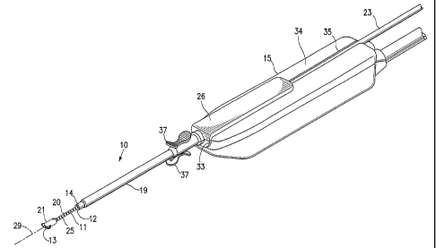
[0014] Figure 1 is a perspective view of a removable biopsy device having
features of the invention seated within a handle with the supporting tube of
the
device in an opened configuration.
6d
CA 02490072 2004-12-17
WO 2004/000126 PCT/US2003/018478
[0015] Figure 2 is a perspective view of the biopsy device shown in Figure 1
removed from the handle.
[0016] Figure 3 is a perspective view of the biopsy device shown in Figure 2
rotated 1801 about its longitudinal axis.
[0017] Figure 4 is an enlarged perspective view of the distal section of the
biopsy
device shown in Figure 2 with the supporting tube in an opened configuration.
[0018] Figure 5 is an enlarged perspective view of the distal section of the
biopsy
device shown in Figure 2 with the supporting tube in a closed configuration.
[0019] Figure 6 is a longitudinal cross-sectional view of the device shown in
Figure 3 taken along the lines 6-6.
[0020] Figure 7 is an enlarged longitudinal cross-sectional view of the distal
section of the device shown in Figure 6.
[0021] Figure 8 is a transverse cross sectional view of the device shown in
Figure
7 taken along the lines 8-8.
[0022] Figure 9 is a transverse cross sectional view of the device shown in
Figure
7 taken along the lines 9-9.
[0023] Figure 10 is a transverse cross sectional view of the device shown in
Figure 7 taken along the lines 10-10.
[0024] Figure 11 is an enlarged longitudinal cross-sectional view of the
distal
section of the device shown in Figure 6 rotated 90 from the view shown in
Figure 7.
[0025] Figure 12 is an enlarged longitudinal cross-sectional view of the
distal
section of the device as shown in Figure 11 with the supporting tube in a
closed
configuration.
7
CA 02490072 2004-12-17
WO 2004/000126 PCT/US2003/018478
[0026] Figure 13 is a transverse cross sectional view of the device shown in
Figure 11 taken along the lines 13-13.
[0027] Figure 14 is a transverse cross sectional view of the device shown in
Figure 12 taken along the lines 14-14.
[0028] Figure 15 schematically illustrates an operative system embodying the
devices of the invention.
[0029] Figure 16 is a transverse cross sectional view of the device shown in
Figure 12 disposed within a tissue site and tissue at the site held against
the surface
of the distal extremity by the action of a vacuum within the inner lumen of
the probe.
[0030] Figure 17 is a transverse cross sectional view of the device shown in
Figure 15 with the supporting tube in a closed configuration with a separated
tissue
specimen within the space between the distal extremity 20 and the interior of
the
supporting tube 14.
[0031] Figure 18 is a perspective view of an alternative probe member for the
biopsy device.
[0032] Figure 19 is a transverse cross-sectional view of the biopsy device
shown
in Figure 18 taken along the lines 19-19.
[0033] Figure 20 is a longitudinal cross-sectional view of a device embodying
features of the invention as shown in Figure 18, with the access cannula and
supporting tube in a closed configuration.
[0034] Figure 21A is a longitudinal cross-sectional view of a device embodying
features of the invention as in Figure 20, shown configured for insertion into
a
patient's body.
8
CA 02490072 2004-12-17
WO 2004/000126 PCT/US2003/018478
[0035] Figure 21B is a longitudinal cross-sectional view of a device embodying
features of the invention as in Figure 21A, shown configured with the access
cannula and supporting tube retracted.
[0036] Figure 21C is a longitudinal cross-sectional view of a device embodying
features of the invention as in Figure 21A, shown after advancement of the
supporting tube and access cannula and cutting of a tissue sample.
[0037] Figure 21 D is a longitudinal cross-sectional view of a device
embodying
features of the invention as in Figure 21A, showing portions of the device
removed
from within the access cannula which remains in place in body tissue.
[0038] Figure 21 E is a longitudinal cross-sectional view of a device
embodying
features of the invention as in Figure 21A, shown configured for removal of a
tissue
sample from the device.
[0039] Figure 21 F is a longitudinal cross-sectional view of a device
embodying
features of the invention as in Figure 21A, shown after re-insertion into a
patient's
body and configured for recovery of another tissue sample.
DETAILED DESCRIPTION OF THE INVENTION
[0040] Reference is made to Figures 1-14 which illustrate a biopsy device 10
embodying features of the invention. The device 10 generally includes an
elongated
probe member 11, a tissue-cutting blade 12, a tissue-cutting electrode 13 and
a
supporting tube 14 carrying tissue-cutting blade 12. The tissue-cutting
electrode 13
preferably includes at least two components, as illustrated in Fig. 1,
although it may
be a single wire electrode, as illustrated in Fig. 2. The supporting tube 14
is slidably
disposed about the probe 11 and is slidably disposed within access cannula 19.
In
one embodiment of the invention shown in Figure 1, the device 10 is a
disposable
9
CA 02490072 2004-12-17
WO 2004/000126 PCT/US2003/018478
device and is configured to be mounted on a handle 15 which is configured to
provide mechanical and electrical power, vacuum, and control to the device.
For
example, a handle 15 may be configured to provide mechanical power effective
to
power the longitudinal translation, rotation, reciprocation, or other movement
of
tissue-cutting blade 12, supporting tube 14, or other movable element of
device 10.
Alternatively, mechanical and/or electrical power may be provided by housing
26, or
by handle 15 and housing 26. As illustrated in the Figures 1, 3, and 6, handle
26
may include finger holders 37 configured to receive a finger or thumb of an
operator.
Finger holders 37 are configured to release housing 26 from handle 15 when
they
are squeezed by an operator.
[0041] The probe member 11 has a proximal section 16 and a distal section 18.
Proximal section 16 is configured for slidable disposition within the inner
lumen 17 of
the supporting tube 14. Proximal section 16 acts to guide supporting tube 14
and to
protect tissue-cutting blade 12 as the supporting tube 14 and cutter 12
translate and
rotate around probe 11 and within access cannula 19. Distal section 18
includes a
distal extremity 20 which is configured to secure tissue from a tissue site
which is to
form the specimen and an enlarged distal end 21 to which the tissue-cutting
electrode 13 is secured. The probe member 11 may be cylindrical, with a
circular
cross-section, or may have a square, rectangular, or other shaped cross-
section.
[0042] As shown in Figures 4 and 5, tissue-cutting blade 12 and supporting
tube
14 are configured to translate longitudinally so as to expose distal extremity
20 when
in a proximal configuration, and to cover distal extremity 20 when in a distal
configuration. Distal extremity 20 may be partially covered when tissue-
cutting blade
12 and supporting tube 14 are in configurations intermediate to those
illustrated in
CA 02490072 2004-12-17
WO 2004/000126 PCT/US2003/018478
Figures 4 and 5, and may be more completely covered or exposed when tissue-
cutting blade 12 and supporting tube 14 are in configurations more distal or
proximal
to those illustrated in Figures 4 and 5. During such longitudinal translation,
tissue-
cutting blade 12 may rotate (in one or more rotational directions) and/or may
reciprocate longitudinally. In preferred embodiments, tissue-cutting blade 12
remains separated by a gap 38 from enlarged distal end 21 of probe 11 at its
most
distal position (e.g., Fig. 20).
[0043] As shown in more detail in Figures 6-10, the probe member 11 is
provided
with an inner lumen 22 which extends from the distal extremity 20 to a
connection
member 23 on the proximal end 24 of the probe member 11 and which is in fluid
communication with the plurality of aspiration ports 25 provided on the distal
extremity 20 of the probe member 11. The proximal end 24 of the probe member
11
and the connection member 23 are secured within the housing 26 as shown in
Figure 6.
[0044] The supporting tube 14 is slidably disposed about the proximal section
of
the probe member 11 and has a proximal end secured to a slidable collar 27
within
the housing 26. The collar 27 is provided with an connector 28 (which may be
an
arm as in Fig. 2 or a gear as in Fig. 3) which is configured to seat within a
receiving
opening on a driver (not shown) provided in the handle 15. The collar 27 is
configured to be slidably disposed within the housing so that the driver on
the handle
can move the arm 28 and as a result translate the outer tubular sheath as
shown by
the arrow 30 in Figure 2 between an opened and closed configuration.
Supporting
tube 14 is also configured to rotate around a longitudinal axis 29 as well as
to
translate longitudinally within access cannula 19. Translation of the
supporting tube
11
CA 02490072 2004-12-17
WO 2004/000126 PCT/US2003/018478
14 and tissue-cutting blade 12 may include reciprocation (i.e., alternated
distal and
proximal translation) as well as rotation, as the supporting tube 14 and
tissue-cutting
blade 12 move generally in a longitudinal direction.
[0045] As illustrated in Fig. 2, a housing 26 may be provided with distal
projection
31 and proximal projection 32 which are designed to tightly seat within
receiving
openings (not shown) provided in the handle 15 to effect a snap fit of the
housing 26
within a recess 33 provided in the upper surface 34 of handle 15 as shown in
Figure
1. A second long recess 35 is provided in the upper surface 34 of handle 15
which
is contiguous with recess 33 and which is configured to receive the connection
member 23 tightly enough to prevent accidental excursions out of the recess.
Connection member 23 has an inner lumen in fluid communication with the inner
lumen 22 of the probe member 11. Distal projection 31 may be connected to
collar
27 attached to supporting tube 14 so that longitudinal translation of proximal
projection 32 towards distal projection 31 causes accessing cannula 19 and
supporting tube 14 to move distally. In preferred embodiments, accessing
cannula
19 and supporting tube 14 move longitudinally in concert, with supporting tube
14
free to rotate within accessing cannula 19.
[0046] The tissue-cutting blade 12, which is circular and disposed about the
probe member 11, has a sharp edge that is preferably beveled to have a sharp
edge
on the outer diameter of the circular blade, although a blade with a leading
edge on
the inner diameter of a tube is also suitable. The tissue-cutting blade 12 is
connected to and supported by the wall of supporting tube 14. This
construction
allows the tissue-cutting blade 12 to travel longitudinally with the
supporting tube 14
within access cannula 19 over the distal extremity 20 of the probe member 11,
and
12
CA 02490072 2004-12-17
WO 2004/000126 PCT/US2003/018478
thus to extend out of access cannula 19. In this configuration, with the
tissue-cutting
blade 12 disposed distally to the end of the access cannula 19, the tissue-
cutting
blade 12 is effective to cut a tissue specimen from tissue held against the
distal
extremity 20 by the action of a vacuum within the inner lumen 22 from the
tissue site,
and at the same time to cover the separated tissue specimen with the
supporting
tube 14. The inner surface of supporting tube 14 may be coated (e.g., with
teflon) to
reduce friction. In preferred embodiments, the inner diameter of the
supporting tube
14 proximal to the tissue cutting blade 12 is greater than the inner diameter
of the
supporting tube 14 at the region of contact between the tissue-cutting blade
12 and
the supporting tube 14, providing greater volume for a tissue sample. Thus,
the
specimen can be removed with device 10 from the patient with the same, or
nearly
the same, movement that severs the specimen from surrounding tissue. As shown
in Figure 3, the collar 27 and the gear 28 are configured to drive and to
translate the
supporting tube 14 both rotationally and longitudinally.
[0047] The tissue-cutting electrode 13 has an arcuate portion which is spaced
distally away from the distal end 21 and has a maximum chord (i.e. distance
between the ends of the arcuate portion) which is preferably larger than the
maximum diameter of the distal end. The maximum width of the tissue-cutting
electrode 13 is preferably about 20 to about 50% greater than the maximum
outside
transverse dimension of the distal end 21 of the probe 11. The tissue-cutting
electrode 13 can be spaced distally from an outer surface of the distal end 21
by a
distance of about 0.01 to about 0.05 inch, preferably about 0.02 to about 0.04
inch.
As shown in Figures 6 and 7, the arcuate tissue-cutting electrode 13 is formed
out of
the distal extremity of electrical conductor 41. The proximal end 42 of the
conductor
13
CA 02490072 2004-12-17
WO 2004/000126 PCT/US2003/018478
41 is electrically connected via a conductor to an electrosurgical generator
which can
supply high frequency electrical power.
[0048] The shaft of the device 10 which extends out from the housing 26 may
have a length of about 3 to about 15 cm, preferably, about 5 to about 13 cm,
and
more specifically, about 8 to about 9 cm for breast biopsy use. To assist in
properly
locating the shaft of device 10 during advancement thereof into a patient's
body, (as
described below), the distal extremity 20 of the probe 11, the access cannula
19,
and the supporting tube 14 may be provided with markers at desirable locations
that
provide enhanced visualization by eye, by ultrasound, by X-ray, or other
imaging or
visualization means. An echogenic polymer coating that increases contrast
resolution in ultrasound imaging devices (such as ECHOCOATTM by STS
Biopolymers, of Henrietta, NY) is suitable for ultrasonic visualization.
Radiopaque
markers may be made with, for example, stainless steel, platinum, gold,
iridium,
tantalum, tungsten, silver, rhodium, nickel, bismuth, other radiopaque metals,
alloys
and oxides of these metals. In addition, the surfaces of the device in contact
with
tissue may be provided with a suitable lubricious coating such as a
hydrophilic
material or a fluoropolymer.
[0049] The proximal portion of the probe 11 generally has an outer dimension
of
about 3 to about 10 mm and a inside dimension of about 2 to about 6 mm and it
may
be desirable in some embodiments to have a close fit between the proximal
section
of the probe 11 and the inner lumen 17 of supporting tube 14 to avoid a gap
therebetween which can catch or snag on adjacent tissue during advancement
through tissue and impede advancement. Similarly, it may be desirable in some
embodiments to have a close fit between the supporting tube 14 and the access
14
CA 02490072 2004-12-17
WO 2004/000126 PCT/US2003/018478
cannula 19, in order to avoid a gap therebetween which can catch or snag on
adjacent tissue during advancement through tissue and impede advancement.
[0050] The tissue-cutting blade 12 is preferably the sharpened edge of a metal
supporting tube 14, or a sharpened metal band ringing the distal end of the
supporting tube 14, although any sharp blade attached to the supporting tube
14 is
suitable. The tissue-cutting blade 12 may be made from any strong, durable
material that can hold a sharp edge, for example, a hard biocompatible metal
such
as stainless steel, titanium, or other metals, alloys, and compounds. A tissue-
cutting
blade may also be made from ceramic, glass, or other material having suitable
strength and ability to maintain a sharp edge.
[0051] The tissue-cutting electrode 13 can be formed with generally conductive
wire formed of metallic materials such as stainless steel, tungsten, titanium,
molybdenum, and other metals and metal alloys, including refractory metals and
alloys containing refractory metals. The shaft components from which the probe
11
and supporting tube 14 are formed may be conventional medical grade polymer
materials such as, for example, polycarbonate and liquid crystal polymer
(LCP),
respectively.
[0052] In preferred embodiments, the supporting tube 14 is stainless steel.
However, metals, ceramics, glasses, and other materials capable of forming a
sharp
edge are also suitable. For example, a supporting tube 14 may be made with an
epoxy-braid material. Although stainless steel and other metals are preferred,
an
advantage of forming a supporting tube 14 from epoxy-braid materials, or from
other
non-conductive materials, is that capacitative coupling with electrical
components
connected to the tissue-cutting electrode 13 is reduced. Where a supporting
tube 14
CA 02490072 2004-12-17
WO 2004/000126 PCT/US2003/018478
is made from a such non-conductive materials, a metal tissue-cutting blade 12
may
be attached to the distal end of the supporting tube 14. Preferably, materials
used in
the construction of a device 10 are sterilizable, and suitable for use in
disposable
medical instruments.
[0053] The biopsy device 10 may be used to obtain a tissue specimen utilizing
the operation system 50 schematically shown in Figure 15. The operating system
50 generally includes a high frequency (e.g. RF) electrical power generator
51,
which is electrically connected to the tissue-cutting electrode 13 on the
biopsy device
through conductors 52 and 53. The power output and the receiving element is
controlled by the controller 54. The RF generator 51 is electrically connected
to the
controller through conductors 55 and 56 and preferably operates at about 300
to
about 1000 KHz, specifically, about 700 to about 900 KHz and has a power
output of
about 50 to about 150 watts, preferably, about 80 to about 100 watts. Vacuum
is
generated by the vacuum pump 57 which is connected in a fluid flow
relationship
with the inner lumen (not shown) provided in conduit 58 which leads to a
vacuum
trap 59. Vacuum is applied to the inner lumen 22 of the probe member 11
through
inner lumen 36 of connection member 23 connected to the vacuum trap. A meter
actuation and control cable 60 is provided to power and control the actuation
elements in handle 15.
[0054] A patient's skin must be breached in order to gain access to a body
site
where a tissue specimen is to be obtained. A scalpel or other surgical
instrument
may be used to make an initial incision in the skin; some physicians may
prefer to
first make an incision with a scalpel through the patient's skin and expose
subcutaneous tissue before passing the device 10 through the tissue.
Alternatively,
16
CA 02490072 2004-12-17
WO 2004/000126 PCT/US2003/018478
access through the skin may be achieved without such an initial incision by
pressing
the energized tissue-cutting electrode 13 of the device 10 against an exterior
site on
the patient's skin proximate to the tissue site where a tissue specimen is to
be
obtained. High frequency electrical power from the generator 51 passes through
the
electrical conductor 41 to energize the tissue-cutting electrode 13.
[0055] Once the skin is breached by any suitable means, the device 10, with
the
tissue-cutting electrode 13 energized is advanced through the tissue until the
distal
end 21 of the device 10 has passed through the tissue which is to form the
specimen. The cutting action of the energized tissue-cutting electrode 13
forms a
planar cut through the desired tissue bed and allows the probe 11 to readily
pass
through the tissue. Very little collateral tissue damage at the margins where
the
tissue cut is made is done by the tissue-cutting electrode 13 as tissue is
accessed.
The device 10 is preferably advanced through the patient's tissue to the
specimen
site with the supporting tube 14 in a closed configuration, the supporting
tube 14
covering distal extremity 20 of probe 11.
[0056] Once the device 10 is in the desired location, the supporting tube 14
can
be withdrawn to an opened configuration to expose the distal extremity 20 of
the
probe 11 by action of the driver (not shown) operatively connected to the arm
28 of
collar 27. With the distal extremity 20 of the probe 11 exposed, a vacuum can
be
generated within the inner lumen 22 of probe 11 by the action of vacuum pump
57.
The vacuum generated in the inner lumen 22, acting through the ports 25 in the
distal extremity 20 draws tissue at the site against the surface of the distal
extremity
20 and holds the tissue against that surface as shown in Figure 16. The tissue-
cutting blade 12 may then be driven distally along with the supporting tube 14
to
17
CA 02490072 2004-12-17
WO 2004/000126 PCT/US2003/018478
which the tissue-cutting blade 12 is secured, effective to sever a generally
cylindrical
shaped tissue specimen 61 from the adjacent tissue site and cover the severed
tissue specimen with the supporting tube 14 as shown in Figure 17.
[0057] In preferred embodiments of methods and devices embodying features of
the invention, tissue-cutting blade 12 rotates, preferably at high speed,
during its
distal translation as it severs tissue from the surrounding tissue bed. Such
rotation
may be in a single rotational direction, or may alternate between clockwise
and
counter-clockwise rotation. Tissue-cutting blade 12 may also reciprocate
longitudinally, with or without rotation, during distal translation as it
severs tissue
from the surrounding tissue bed. Access cannula 19 acts to protect surrounding
tissue from damage during translation, rotation, and/or reciprocation of the
supporting tube 14 and tissue-cutting blade 12.
[0058] The biopsy device may be removed from the patient after a tissue sample
has been collected, and the sample removed for inspection and analysis. The
entire
device 10 may be removed; however, in preferred embodiments, portions of the
device may remain within a patient's body to aid, for example, in the
acquisition of
further tissue specimens and in the placement of markers at the site from
which a
tissue sample was taken. For example, the supporting tube 14 and probe 11 may
be withdrawn together from within access cannula 19, the supporting tube 14
remaining in a closed configuration outside of probe 11 and helping, along
with the
vacuum, to hold the tissue sample. Re-introduction of probe 11 and supporting
tube
14 within access cannula 19 (which remains in place within a patient's body)
allows
further samples to be taken. The access cannula 19 serves as a guide for re-
introduction of the remainder of the device 10 and aids in obtaining
subsequent
18
CA 02490072 2004-12-17
WO 2004/000126 PCT/US2003/018478
tissue samples. Alternatively, the probe 11 may be removed, with a tissue
sample
held by vacuum, from within the supporting tube 14, while supporting tube 14
and
access cannula 19 remain in place within the patient's body. Re-introduction
of
probe 11 within supporting tube 14 allows further samples to be taken.
[0059] Such further samples may be from the same location, or from different
locations. Where subsequent samples are taken from the same location as a
previous sample, so that the tissue-cutting electrode 13 need not be activated
(since
the pathway to the body location has already been formed), further application
of
vacuum draws tissue near to the elongated probe, where the tissue may be
separated from adjacent body tissue by the tissue-cutting blade 12. Due to the
planar cut made by the tissue-cutting electrode 13 through the tissue from
which the
specimen is to be obtained, the initial cylindrical specimen 61 is typically a
split
specimen which greatly aids in its evaluation. Although the initial samples
are
typically split samples, subsequent samples taken from the same location are
typically not split samples.
[0060] Access cannula 19 exterior to the supporting tube 14 can be left in the
patient with its distal end at the site from which the specimen was obtained
in order
to provide access to the site at a later time. Access cannula 19 may thus be
used to
allow a marker or other device to be deposited at the site, or to guide
further
procedures or treatments at the site as necessary or desirable. After the
biopsy
procedure is completed, the incision formed by the initial cut through the
patient's
skin may be appropriately closed.
[0061] An alternative probe member 70 embodying features of the invention is
depicted in Figures 18 and 19. In this alternative the distal extremity 71 of
the probe
19
CA 02490072 2012-01-30
device 70 is of tubular construction as shown. The tissue-cutting electrode 72
on the
enlarged distal end 74 of the distal extremity 71 of the probe member 70 has
an
expandable construction which is disclosed in U.S. Patent 6,471,700, entitled
Apparatus and Method for Accessing A Biopsy Site, by Burbank et al. The
tubular
distal extremity 71 has a plurality of ports 73 which are in fluid
communication with
an inner lumen 75. Tissue-cutting electrode 72 is secured to the enlarged
distal end
74. A proximal enlargement 77 is disposed proximally of the distal extremity
71 on
the probe member 70. An electrical conductor 76 (shown in Figure 19) extends
through inner lumen 75 and is electrically connected to electrode 72. A
supporting
tube 78 carrying a circular cutter 79 extends about the probe member 70 within
access cannula 80. The probe 70 is used with accessing cannula 80, supporting
tube 78 and circular tissue-cutting blade 79 in the same manner as described
above
for the embodiment shown in Figures 1-14. The supporting tube 78 may be
configured to allow the probe 70 to be withdrawn with the specimen for
specimen
removal leaving the distal end of the accessing cannula located at the biopsy
site.
[0062] A cross-sectional view of a device 70 having an expandable tissue-
cutting electrode 72 embodying features of the invention is shown in Figure
20, with
the access cannula 80 shown in a closed configuration. Supporting tube 78
circular
cutter 79 are shown in a distally-disposed, closed configuration (dark lines)
and in a
proximally-disposed, open configuration (dotted lines) within access cannula
80,
which acts as a sheath to enclose the inner elements of the device 70,
particularly
when it is in its distally-disposed, closed configuration.
CA 02490072 2004-12-17
WO 2004/000126 PCT/US2003/018478
[0063] The use of such a device 70 is illustrated in Figures 21A-21 F, which
illustrate a method of using an apparatus for accessing a body site having
features
of the invention. For example, the apparatus may be first inserted into a
patient's
body using radiofrequency energy applied via the tissue-cutting electrode; the
access cannula and tissue-cutting blade may be retracted, followed by
application of
vacuum; the tissue-cutting blade and access cannula may be advanced though
tissue to cut a sample; the sample may then be removed along with the tissue-
cutting blade and tissue-cutting electrode, leaving the access cannula in
place; the
sample may be removed from the apparatus (with the vacuum turned off) by
retracting the supporting tube and tissue-cutting blade; and then the
supporting tube,
tissue-cutting blade, and tissue-cutting electrode may be re-inserted within
the
access cannula for removal of further samples.
[0064] As illustrated in Figures 21A -21F, a first step in obtaining a tissue
sample, or in obtaining several tissue samples, from a location within a
patient's
body, includes inserting a device 70 into a patient's body. A device 70 may be
inserted into a patient's body in a configuration as illustrated in Figure
21A, with
distal cutter 72 activated with RF energy to cut through tissue. Access
cannula 80 is
disposed distally in a closed configuration, with supporting tube 78 and
circular
cutter 79 proximally disposed in an open configuration within the access
cannula 80.
Alternatively, supporting tube 78 and circular cutter 79 may be distally
disposed
within the access cannula 80. If desired, a scalpel or other sharp instrument
may be
used to make an initial incision through a patient's skin 81; however, the
initial
incision and subsequent advancement of the device into a patient's body may be
done solely using a distal cutter 72 under RF power. In preferred embodiments,
the
21
CA 02490072 2004-12-17
WO 2004/000126 PCT/US2003/018478
circular cutter 79 and access cannula 80 move together, remaining in the
configuration shown in Fig. 21 C.
[0065] In a second step, access cannula 80 may be retracted (or probe 71
extended distally into a patient's body tissue) to obtain the configuration
illustrated in
Figure 21B. In this configuration, probe 71 extends distally of access cannula
80,
circular cutter 79 and supporting tube 78, exposing ports 73 to surrounding
tissue.
Vacuum, such as may be supplied by a vacuum system with a vacuum source, may
be applied via ports 73 to urge tissue into contact with the distal extremity
71 of the
probe member 70.
[0066] A further step in a method obtaining a tissue sample, or in obtaining
several tissue samples, from a location within a patient's body is illustrated
in Figure
21 C. Circular cutter 79, followed by access cannula 80, may be advanced into
surrounding tissue by distal movement around distal extremity 71 effective to
sever
tissue from the surrounding tissue bed. This may result in a split tissue
sample (split
due to the action of distal cutter 72 as device 70 is inserted into a desired
location
within a patient's body) disposed within supporting tube 78 and preferably
held
against distal extremity 71 by action of vacuum. Thus, after advancement of
the
supporting tube and access cannula, as shown in Figure 21C, a tissue sample is
held within device 70 for removal from a patient.
[0067] Tissue removal may be performed as illustrated in Figure 21 D. Portions
of
a device 70, including a distal extremity 71, a supporting tube 78, and a
circular
cutter 79, and a tissue sample held within supporting tube 78 and circular
cutter 79,
may be removed proximally by withdrawing them from within an accessing cannula
80, which remains in place at least partially within a patient's body.
22
CA 02490072 2004-12-17
WO 2004/000126 PCT/US2003/018478
[0068] The tissue sample may be removed from the device outside the patient's
body for investigation, analysis and storage as desired. As shown in Figure
21E,
portions of the device 70 may be configured for removal of a tissue sample by
retraction of the supporting tube 78 and circular cutter 79 to expose the
tissue
sample, and by closing the vacuum connection between ports 73 and a vacuum
system with a vacuum source.
[0069] The accessing cannula 80 provides a guide for re-insertion of portions
of
the device 70 that have been removed from the patient, as illustrated in
Figure 21 F.
The device 70 is shown in Figure 21 F after re-insertion into a patient's body
in a
configuration for recovery of another tissue sample. As the configuration in
Figure
21 F is the same as that in Figure 21 A, it will be understood that subsequent
tissue
samples may be acquired by steps described above and as illustrated in Figures
21 B and the following figures. Alternatively, if no further samples are
desired, the
accessing cannula 80 may be removed after the steps illustrated in Figure 21 E
and
standard post-operative care provided to the patient.
[0070] In addition to suction ports 25, the distal extremity 20 (and
optionally the
supporting tube 14) may have features configured to retain a tissue sample.
For
example, a distal extremity 20 may include radial elements configured to
engage
and retain tissue, such as hooks, barbs, hairs, or probes, that may grab
and/or
puncture tissue of an adjacent tissue sample. Such radial elements may be
angled
to be other than perpendicular to a longitudinal axis of probe 11 (e.g.,
angled to point
partially in a distal direction), so that a tissue specimen is retained during
distal
movement of the probe 11.
23
CA 02490072 2011-12-05
[0071] In addition, tissue-cutting electrode may be configured to be able to
retract or otherwise reduce its radial extent before being removed proximally
through
supporting tube 14 during recovery of a tissue specimen. Such retraction is
effective to
reduce the possibility of damage to a tissue-cutting blade 12 as the tissue-
cutting
electrode 13 is withdrawn. Similarly, the possibility of damage to an access
cannula
19 is reduced by retraction of a tissue-cutting electrode 13 before withdrawal
of a
probe 11 through the access cannula 19. The radial extent of a tissue-cutting
electrode 13 may be reduced by, for example, retracting a central supporting
portion of
a tissue-cutting electrode of the type illustrated in Figure 18, or by
retracting a distal
supporting portion of a tissue-cutting electrode of the type illustrated in
Figure 1. Such
retraction may be effected by proximal movement of a connecting element
attached to
such supporting elements. For example, such a connecting element may be, or
may
be connected to, an electrical conductor 41 or 76.
[0072] Those skilled in the art will recognize that various modifications may
be
made to the specific embodiments illustrated above. In addition, it will be
readily
appreciated that other types of instruments may be inserted into the tissue
site
through the supporting tube or a suitable cannula in addition to or in place
of the
instruments described above.
24