Note: Descriptions are shown in the official language in which they were submitted.
CA 02490290 2004-12-21
WO 2004/003195 PCT/US2003/020498
-1-
TRYPTOPHANAS A FUNCTIONAL REPLACEMENT FOR ADRRIBOSE-
ARGININE INRECOMBINANT PROTEINS
PRIORITY CLAIM
This application claims the benefit of U.S. Provisional Application No.
60/393,033, filed June 2~, 2002, which is incorporated herein by reference.
FIELD
The present disclosure relates generally to the modification of proteins to
alter protein activity and stability, specifically, to the substitution of
phenylalanine
or tryptophan for an arginine residue capable of being adenosine-diphosphate
(ADP)-ribosylated in a polypeptide sequence.
BACKGROUND
Mono-ADP-ribosylation of arginine residues in proteins is a reversible
modification that involves the following steps: (i) the transfer of an ADP-
ribose
moiety of nicotinamide adenine dinucleotide (NAD) to an arginine residue of a
target protein, or to a free arginine residue, by an arginine-specific ADP-
ribosyltransferase (ART) and (ii) the cleavage of the bond between ADP-ribose
and
arginine by an ADP-ribosylarginine hydrolase.
A.RTs were first characterized in bacterial toxins, such as cholera toxin,
diphtheria toxin, pertussis toxin, and pseudomonas exotoxin A. ADP-
ribosyltransferase activity has since been identified in eukaryotic cells. The
widespread expression of ARTs in eukaryotes, as well as in prokaryotes,
suggests
that the cycle of ADP-ribosylation/de-ADP-ribosylation of amino acid residues
is
widely involved in regulating protein activity. Moreover, specific ADP-ribose
acceptors, such as arginine, may serve as regulatory switches. For example,
ADP-
ribosylation of a specific axginine residue in the dinitrogenase enzyme of the
nitrogen-fixing bacteria Rlzoc~ospirillium ~ubrum has been shown to regulate
the
activity of this enzyme.
In eukaryotes, ART activity is linked to regulatory signals for critical
cellular
processes such as DNA repair and the maintenance of calcium or phosphorylation
CA 02490290 2004-12-21
WO 2004/003195 PCT/US2003/020498
-2-
levels. In humans, altered cellular ADP-ribosylation levels have been linked
to a
number of diseases including lupus, diabetes and cancer, whereas bacterial
toxins,
such as cholera toxin and diphtheria toxin, catalyze the ADP-ribosylation of
important metabolic or regulatory proteins in their human hosts.
The ability to identify specific amino acids that can be modified in order to
regulate the activity of various proteins is critical in the development of
medical
treatments and therapies. Thus there is a need to identify additional stable
protein
modifications that have an effect on protein activity.
SUMMARY
Methods of producing a protein with an altered activity or stability are
disclosed herein. The method includes replacing an arginine residue capable of
being ADP-ribosylated with either a tryptophan (W) residue or a phenylalanine
(F)
residue, thereby producing a protein with an increased activity or stability.
In one
embodiment, the protein has an antimicrobial activity and the stability or
activity of
the protein is increased. In another embodiment, the protein is a defensin
polypeptide. Substitution of an arginine capable of being ADP-ribosylated with
either a phenylalanine or a tryptophan results in an increased antimicrobial
activity
of the defensin molecule or increased stability of the defensin molecule.
Specific,
non-limiting examples of an antimicrobial activity are T cell chemotaxis,
promotion
of neutrophil recruitment, or cytokine release.
A method is provided for increasing the activity or stability of a defensin
polypeptide comprising an arginine residue capable of being ADP-ribosylated.
The
method includes substituting the arginine residue with a tryptophan or a
phenylalanine, thereby increasing the activity or the stability of the
defensin
polypeptide.
In another embodiment, a method is disclosed for determining if a protein
can be stabilized. The method includes determining if an arginine residue in
the
protein is capable of being ADP-ribosylated. Detection of ADP-ribosylation of
the
arginine residue indicates that the stability of the protein, such as a
protein with
antimicrobial activity, can be increased by substituting the arginine capable
of being
ADP-ribosylated with either a tryptophan or a phenylalanine.
CA 02490290 2004-12-21
WO 2004/003195 PCT/US2003/020498
-3-
A composition is disclosed herein that includes a polypeptide where at least
one arginine residue capable of being ADP-ribosylated is substituted with a
tryptophan or a phenylalanine residue. In one embodiment, the protein has an
antimicrobial activity. In another embodiment, the protein is a defensin
polypeptide.
In yet another embodiment, the amino acid substitution increases the activity
or
stability of the polypeptide.
A pharmaceutical composition is disclosed herein that includes a
therapeutically effective amount of a defensin with at least one arginine
residue
capable of being ribosylated substituted by a tryptophan or a phenylalanine
residue.
In another embodiment, a method is provided for increasing an immune
response in a subject. The method includes administering to the subject a
therapeutically effective amount of a defensin polypeptide comprising an amino
acid
substitution, wherein the amino acid substitution is a replacement of an
arginine
capable of being ADP-ribosylated with a tryptophan or a phenylalanine.
The foregoing and other features and advantages will become more apparent
from the following detailed description of several embodiments, which proceeds
with reference to the accompanying figures.
BRIEF DESCRIPTION OF THE FIGURES
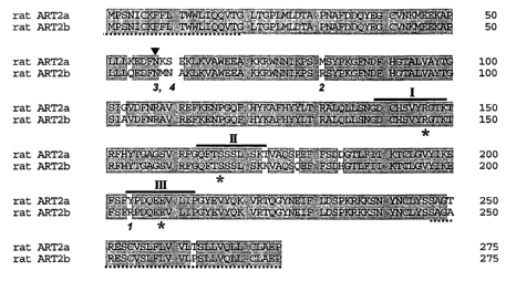
Fig. l is a schematic drawing of the deduced amino acid sequences of rat
ART2a (RT6.1) and rat ART2b (RT6.2). Identical amino acids are shaded.
Arginine 204 and 81 are specific to ART2b. In ART2a, N58 and 58NI~SE61 are in
a putative consensus glycosylation site not present in ART2b. Regions I, II,
and III,
believed to participate in formation of the catalytic site in the bacterial
toxin and
mammalian ADP-ribosyltransferases, are indicated by solid lines and the
putative
catalytic amino acids by an asterisk. Dotted underlines indicate signal
sequences,
which are excised during the export into the endoplasmic reticulum (amino
terminus) and attachment of the glycosylphosphotidylinositol (GPI anchor
(carboxy
terminus).
Fig. 2 is a digital image of a set of blots demonstrating the auto-[32P]ADP-
ribosylation (Fig. 2A) and immunoreactivity (Fig. 2B) of the supernatants from
CA 02490290 2004-12-21
WO 2004/003195 PCT/US2003/020498
-4-
NMU cells transfected with vector alone (lane 1), wild-type ART2b (lane 2),
ART2b(R81K) (lane 3), ART2b(R204K) (lane 4), wild-type ART2a (lane 5),
ART2a(M81R) (lane 6) and ART2a(Y204R) (lane 7). Data shown are
representative of eight independent experiments.
Fig. 3 is a digital image of a set of blots demonstrating the auto-ADP-
ribosyltransferase activity (Fig. 3A) and immunoreactivity (Fig. 3B), as well
as data
demonstrating the NAD glycohydrolase (NADase) activity (nmol per hour) (Fig.
3C) of ART2b, ART2a, and various mutant forms of these proteins. The left
column shows data for the following wild-type and mutant ART2b proteins: ART2b
(lane 1), ART2b(R204K) (lane 2), ART2b(R81K) (lane 3), ART2b(R204Y) (lane 4),
ART2b(R204E) (lane 5), ART2b(R204W) (lane 6), ART2b(R81K,R204K) (lane 7).
The right column shows data for the following wild-type and mutant ART2a
proteins: ART2a (lanel), ART2a(M81R) (lane 2), ART2a(Y204R) (lane 3),
ART2a(M81R,Y204R) (lane 4), ART2a(N58A,Y204R) (lane 5),
ART2a(59NMA61,Y204R) (lane 6). Data shown are representative of two
experiments.
Fig. 4 is a digital image of a set of blots demonstrating the auto-ADP-
ribosylation of ART2b (Fig. 4A), ART2a (Fig. 4B), and their various mutant
forms.
The gels contain samples from cells expressing ART2b wild-type (lane 1),
ART2b(R81K) (lane 2), ART2b(R204W) (lane 3), ART2a(Y204R) (lane 4),
ART2a(Y204R,M81R) (lane 5) and ART2a(59NMA61,Y204R) (lane 6). Data
represent one of two experiments.
Fig. 5 is a digital image of a set of blots demonstrating the SDS-PAGE
separation of auto-ADP-ribosylated ART2b and ART2b(R204K) proteins as
analyzed by a phosphorimager (Fig. SA) and by immunoreactivity (Fig. SB). The
blots contain samples from cells expressing wild type ART2b (lanes 1),
ART2b(R204K) (lanes 2), or a mixtures of samples containing ART2b and
ART2b(R204K) (lanes 3). Samples were incubated with or without 10~M [3zP]NAD
CA 02490290 2004-12-21
WO 2004/003195 PCT/US2003/020498
-5-
followed by either TCA precipitation or further incubation with SmM NAD. The
radiolabeled wild type ART2b and ART2b(R204K) proteins were combined and
then incubated with or without SmM NAD.
Fig. 6 is a digital image of a set of blots demonstrating the SDS-PAGE
separation of proteins tested for their sensitivity to acid, hydroxylamine and
mercuric chloride. Samples from cells expressing wild-type ART2b,
ART2b(R204W), ART2a(M81R,Y204R) and ART2a(N58A,Y204R) were auto-
ADP-ribosylated with 10~,M[32P ]NAD followed by addition of 10% TCA (column
II), or further incubation with SmM NAD at 30°C for lhour before
precipitation
with 10% TCA (column I). Neutralized samples were suspended in O.1M Tris-HCl
pH 7.5 (lane 1), 0.2M HCl (lane 2), lOmM HgCl2 (lane 3), 2M NHzOH (lane 4), or
0.2M NaCI (lane 5) for 2hours at 37° C. The samples were separated by
SDS-
PAGE in 12% gels, transferred to nitrocellulose and analyzed by phosphorlmager
(Fig. 6A) and by immunoblot with antipeptide antibody 1126 (Fig. 6B). Data are
from one experiment, representative of two.
SEQUENCE LISTING
The nucleic and amino acid sequences listed in the accompanying sequence
listing are shown using standard letter abbreviations for nucleotide bases,
and three
letter code for amino acids, as defined in 37 C.F.R. 1.822. Only one strand of
each
nucleic acid sequence is shown, but the complementary strand is understood as
included by any reference to the displayed strand. In the accompanying
sequence
listing:
SEQ ID NO:1 is the amino acid sequence of the human neutrophil peptide
(HNP)-l, HNP-2, HNP-3 prepro-protein.
SEQ ID NO:2 is the amino acid sequence of HNP-1.
SEQ ID N0:3 is the amino acid sequence of HNP-2.
SEQ m N0:4 is the amino acid sequence of HNP-3.
SEQ m NO:S is the amino acid sequence of the HNP-4 prepro-protein.
CA 02490290 2004-12-21
WO 2004/003195 PCT/US2003/020498
-6-
SEQ D7 N0:6 is the amino acid sequence of HNP-4.
SEQ ID N0:7 is the amino acid sequence of the human defensin (HD)-5
prepro-protein.
SEQ ID N0:8 is the amino acid sequence of HD-5.
SEQ ID N0:9 is the amino acid sequence of the HD-6 prepro-protein.
SEQ ID NO:10 is the amino acid sequence of HD-6.
SEQ ID NO:11 is the amino acid sequence of rat ART2a.
SEQ ID NO:12 is the amino acid sequence of rat ART2b.
SEQ ID N0:13 is the primer for introduction of Kozak sequence.
SEQ ID N0:14 is the primer for the ART2a N58A mutation.
SEQ ID NO:15 is the primer for the ART2a K59M, S60N, E61A mutation.
SEQ ID N0:16 is the primer for the ART2a M81R mutation.
SEQ ID N0:17 is the primer for the ART2a Y204R mutation.
SEQ ID N0:18 is the primer for the ART2b R81K mutation.
SEQ ID N0:19 is the primer for the ART2b R204K mutation.
SEQ ID N0:20 is the primer for the ART2b R204E mutation.
SEQ ID N0:21 is the primer for the ART2b R204Y mutation.
SEQ ID N0:22 is the primer for the ART2b R204W mutation.
DETAILED DESCRIPTION
I. Abbreviati~ns
ADP adenosine-diphosphate
ART ADP-ribosyltransferase
ELISA enzyme-linked imrnunosorbent assay
F phenylalanine
GPI glycosylphosphatidylinositol
HD human defensin
HNP human neutrophil peptide
IL interleukin
M81R methionine-to-arginine substitution at position 81
CA 02490290 2004-12-21
WO 2004/003195 PCT/US2003/020498
_7_
MIf macrophage inflammatory protein
N58A asparagine-to-alanine substitution at position 58
NAD nicotinamide adenine dinucleotide
NADase nicotinamide adenine dinucleotide glycohydrolase
PI-PLC phosphatidlyinositol specific phospholipase C
R arginine
R81K arginine-to-lysine substitution at
position 81
R204E arginine-to-glutamic acid substitution
at position 204
R204K arginine-to-lysine substitution at
position 204
R204Y arginine-to-tyrosine substitution
at position 204
R204W arginine-to-tryptophan substitution
at position 204
R:W arginine-to-tryptophan substitution
R:F arginine-to-phenylalanine substitution
W tryptophan
Y204R tyrosine-to-arginine substitution at position 204
59NMA61 asparagine, methionine and alanine at positions 59, 60 and 61,
respectively
Standard one-letter codes for amino acids are utilized herein.
II. Terzzzs
Unless otherwise noted, technical terms are used according to conventional
usage. Definitions of common terms in molecular biology may be found in
Benjamin Lewin, Genes ~, published by Oxford University Press, 1994 (ISBN 0-19-
854287-9); Kendrew et al. (eds.), The Ezzcyclopedia ofMolecular Biology,
published
by Blackwell Science Ltd., 1994 (ISBN 0-632-02182-9); and Robert A. Meyers
(ed.), Molecular Biology and Bioteclafzology: a Comprehensive Desk Referefzce,
published by VCH Publishers, Inc., 1995 (ISBN 1-56081-569-8).
In order to facilitate review of the various embodiments of the disclosure,
the
following explanations of specific terms are provided:
CA 02490290 2004-12-21
WO 2004/003195 PCT/US2003/020498
_g_
Activity: The biological function of a molecule, such as a polypeptide or a
nucleic acid. In one embodiment, an activity is an enzymatic activity. In
another
embodiment, a biological function is an immunologic activity, such as
recruitment
of a cell, or cytokine secretion. An activity can be increased or decreased.
An
increased activity can be, for example, at least about a 20%, about a 50%,
about an
80%, about a 100% or about a 200% increase in activity. An activity can also
be
decreased, such as at least about a 20%, about a 50%, about an 80% or about a
100%
decrease in activity. The activity can be increased or decreased as compared
to a
control, such as the activity of a wildtype protein or a standard value. An
activity
profile is the set of different activities possessed by a molecule, such as an
agent or a
drug. A polypeptide can have a single defined activity, or can have several
defined
activities.
The biological activity of a defensin molecule include modulating T cell
chemotaxis and neutrophil recruitment. In one embodiment, an increased
antimicrobial activity of a defensin molecule includes increased T cell
chemotaxis or
increased neutrophil recruitment, as compared to a control defensin molecule
under
similar conditions.
A specific, non-limiting example of the activity of an ART includes, but is
not limited to, NADase activity. In one embodiment, an increased activity of
an
ART includes increased NADase activity, as compared to a control ART under
similar conditions.
ADP-ribosylation: A reaction in which ADP-ribose is covalently attached
to a compound. Eukaryotic and prokaryotic mono-ARTS catalyze the transfer of
ADP-ribose from nicotinamide adenine dinucleotide (NAD) to an acceptor
nucleophile, such as an amino acid (i.e. the guanidino group of an arginine
residue).
Among the ARTS are bacterial toxins (e.g. cholera toxin, pertussis toxin,
diphtheria
toxin). Periussis toxin and diphtheria toxin use amino acids other than
arginine as
ADP-ribose acceptors.
As disclosed herein, a number of proteins used in host defense are basic and
arginine-rich and thus could serve as acceptors for ADP-ribose. These include,
but
may not be limited to, alpha defensins (HNP-1, HNP-2, HNP-3, HNP-4, HD-5, HD-
6); Beta defensins (hBDl, hBD-2, hBD-3, hBD-4); Major Basic Protein;
Eosinophil
CA 02490290 2004-12-21
WO 2004/003195 PCT/US2003/020498
-9-
Cationic Protein; and Human Cathelicin LL-37 (hCAPlB). In addition, ADP-
ribosyltransferases are capable of auto-ADP-ribosylation. These include, but
may
not be limited to, ART-l, ART2b, ART-3, ART-4, and ART-5.
ADP-ribosyltransferase (ART): An enzyme that catalyzes the transfer of
an ADP-ribose from NAD to an acceptor nucleophile. ARTS can be differentiated
by their corresponding amino acid targets, which include axginine, cysteine,
asparagine, and diphthamide (a post-translationally modified histidine
residue). In
one embodiment, the ART catalyzes the transfer of ADP-ribose to the guanidine
group of an arginine residue on a protein.
Both prokaryotic and eukaryotic ARTs have been identified. Among the
prokaryotic ARTs are bacterial toxins (e.g., cholera toxin, pertussis toxin,
diphtheria
toxin). Five mammalian ARTs (ART-l, ART-2, ART-3, ART-4, ART-5) are
known to exist. Substrates of the five known mammalian ARTS include proteins
that are involved in critical cellular events (e.g., lymphocyte activation and
neutrophil chemotaxis).
A family of mammalian ARTS that are localized on the cell surface through
glycosylphosphatidylinositol (GPI) anchors, are expressed preferentially on
epithelial and inflammatory cells (for example lymphocytes and neutrophils).
ART2a and ART2b are isoenzymes expressed on the surface of mature T cells and
intraepithelial lymphocyte cells of the rat. These proteins express both auto-
ART
and NADase activities, although only ART2b is capable of auto-ADP-ribosylation
at
multiple sites. Of the two proteins, only ARTZa is glycosylated. In addition,
both
are involved in the transmission of transmembrane signals that modulate T cell
activation. Soluble forms of ART have also been identified and circulate in
the
high-density lipoprotein fraction of serum.
Analysis of the crystallographic structure of bacterial toxin ARTS identified
three regions involved in formation of the catalytic site, NAD binding, and
activation of the ribosyl-nicotinamide bond, which is required for ADP-ribose
transfer. These regions appeax to be present in the mammalian transferases as
well.
Region I is defined by an arginine (R) or histidine (H), Region II, by a
sequence rich
in hydrophobic amino acids, or by serine (S) X S, (where X represents
threonine (T),
serine (S) or alanine (A)), and Region III by glutamate (E). (Domenighini et
al.,
CA 02490290 2004-12-21
WO 2004/003195 PCT/US2003/020498
-10-
Mol Microbial 21 (4):667-74, 1996; Bredehorst et al., Adv Exp Med Biol 419:185-
9,
1997; Moss et al., Mol Cell Biochem 193(1-2):109-13, 1999; Takada et al.,
JBiol
Chefn (269(13):9420-3, 1994).
Agent: Any substance, including, but not limited to, a chemical compound,
a drug, a small molecule, a peptide mimetic, a peptide or a protein.
Animal: Living mufti-cellular vertebrate organisms, a category that
includes, for example, mammals and birds. The term mammal includes both human
and non-human mammals. Similarly, the term "subject" includes both human and
veterinary subjects.
Antimicrobial: A compound, such as an agent or a drug, for killing
microorganisms or suppressing their multiplication or growth. An agent has
antimicrobial activity if it results in the death of a microorganism or
suppresses the
growth of a microorganism. In one embodiment, a polypeptide, such as a
defensin
(i.e. an alpha defensin), has antimicrobial activity. An antimicrobial
activity
includes, but may not be limited to, cell lysis (e.g. due to cytotoxicity).
Antimicrobial activity can result from T cell chemotaxis, and neutrophil
recruitment.
In one specific example, an antimicrobial activity is the lysis of a bacterial
cell.
Antimicrobial activity can be modified by the administration of a modified
defensin
polypeptide. In one embodiment, an R:W substituted, R:F substituted HNP-1
polypeptide, or otherwise modified defensin, is administered to a subject.
Arginine (R): An amino acid (C6H14N40a) found in plants and animals that
is essential for the human diet; also produced by the breakdown of proteins.
Also
encompassed are functional analogues of arginine, and structurally modified
arginine molecules (e.g., ADP-ribosylated arginine residues, agmatine) on a
guanidine-containing compound, arginine being one such example. An arginine
residue that is capable of being ADP-ribosylated is an arginine that can be
modified
by the transfer of an ADP-ribose from NAD to the guanidino group of an
arginine.
Asthma: A disorder of the respiratory system characterized by
inflammation, narrowing of the airways and increased reactivity of the airways
to
inhaled agents. Asthma is frequently, although not exclusively, associated
with
atopic or allergic symptoms.
CA 02490290 2004-12-21
WO 2004/003195 PCT/US2003/020498
-11-
B cell or B lymphocyte: One of the two major types of lymphocytes. The
antigen receptor on B lymphocytes, sometimes called the B cell receptor, is a
cell-
surface inununoglobulin. On activation by an antigen, B cells differentiate
into cells
producing antibody molecules of the same antigen-specificity as this receptor.
cDNA (complementary DNA): A piece of DNA lacking internal, non-
coding segments (introns) and regulatory sequences that determine
transcription.
cDNA is synthesized in the laboratory by reverse transcription from messenger
RNA
extracted from cells.
Chronic Bronchitis: An inflammation of the lining of the bronchi. When
the bronchi are inflamed and/or infected, less air is able to flow to and from
the
lungs and a heavy mucus or phlegm is coughed up, resulting in bronchitis. A
brief
attack of acute bronchitis with cough and mucus production can occur with
severe
colds. Chronic bronchitis is defined by the presence of a mucus-producing
cough
most days of the month, three months of a year for two successive years
without
other underlying disease to explain the cough. It may precede or accompany
pulmonary emphysema. Cigarette smoking is the most common cause of chronic
bronchitis. The bronchi of people with chronic bronchitis may also have been
irritated initially by bacterial or viral infections. Air pollution and
industrial dusts
and fumes are also potential etiologic agents. Once the bronchi have been
irritated
over a substantial period of time, excessive mucus is produced constantly, the
lining
of the bronchi becomes thickened, an irritating cough develops, airflow may be
hampered, and the lungs are damaged. The bronchi become susceptible to
infections.
Crohn's Disease: Crohn's disease is an Inflammatory Bowel Disease (the
general name for diseases that cause inflammation in the intestines). Crohn's
Disease causes inflammation in the small intestine. Crohn's Disease usually
occurs
in the lower part of the small intestine (the ileum), but it can affect any
part of the
digestive tract, from the mouth to the anus. The inflammation extends deep
into the
lining of the affected organ, causing pain and diarrhea. Crohn's Disease may
also be
called ileitis or enteritis.
Chronic Obstructive Pulmonary Disease (COPD): Includes emphysema
and chronic bronchitis-diseases that are characterized by obstruction to
airflow.
CA 02490290 2004-12-21
WO 2004/003195 PCT/US2003/020498
-12-
Emphysema and chronic bronchitis frequently coexist. It does not include other
obstructive diseases such as asthma.
Cytokines: Proteins made by cells that affect the behavior of other cells,
such as lymphocytes and neutrophils. In one embodiment, a cytokine is a
chemokine, a molecule that affects cellular trafficking. Cytokines include,
but are
not limited to, MIP-[3, interleukin (IL)-1, IL-8, IL-10, granulocyte-
macrophage
colony stimulating factor (GMCSF), granulocyte colony stimulating factor
(GCSF),
neurokinin, and tumor necrosis factor-alpha (TNF-a).
Defensins: The members of the defensin family are small, cationic peptides
that have six conserved cysteine residues that form three disulfide bonds.
Functional
defensins arise by the sequential post-translational processing of a prepro-
protein of
93-95 amino acids. The members of the defensin family are divided into
different
classes. The alpha-defensins are generally polypeptides containing 29-33
residues.
The beta-defensins are more basic than alpha defensins and are generally
between
34-37 amino acids in length (Raj et al., Biochem J. 347:633-41, 2000). The
recently
identified theta defensins are formed by the head-to-tail linkage of two alpha
defensin-related nonapeptides, generating a circular 18-residue polypeptide
(Tang et
al., Seience 286:498-502, 1999).
Defensins were first identified in neutrophils and have been detected in
human, rabbit, guinea pig, and rat phagocytes. Alpha defensins include, but
are not
limited to, HNP-l, HNP-2, HNP-3, HNP-4, human defensin (HD)-5, and HD-6.
Alpha defensins also include the recently identified HNP-4 homolog, defensin
(Def)-X (see U.S. Patent No. 6, 329, 340 herein incorporated by reference).
HNP-l,
HNP-2, and HNP-3 are products of the same gene (GenBank Accession No. P11479
herein incorporated by reference). HNP-4 is the product of a different gene
(GenBank Accession No. NP_001916 herein incorporated by reference). HD-5
(GenBank Accession No. NP_066290) and HD-6, (GenBank Accession No.
NP_001917 herein incorporated by reference) are two human enteric defensins.
Defensins are antimicrobial peptides that are toxic for a variety of
infectious
agents, such as Gram-negative bacteria, Gram-positive bacteria, fungi, and
certain
enveloped viruses. Defensins act by forming pores in membranes of the
infectious
agent and generating voltage-dependent channels. Antimicrobial activities of
CA 02490290 2004-12-21
WO 2004/003195 PCT/US2003/020498
-13-
defensins include, but are not limited to, lysis of bacteria, fungi, or
viruses; toxicity
for bacteria, fungi or viruses; leukocyte (e.g., T cell) chemotaxis; and
leukocyte
(e.g., neutrophil) recruitment. Without being bound by theory, defensins play
an
important role in the body's natural immunity against infections. Unmodified
defensins are also cytotoxic for several normal and malignant cells.
DNA: Deoxyribonucleic acid. DNA is a long chain polymer which
constitutes the genetic material of most living organisms (some viruses have
genes
composed of ribonucleic acid (RNA)). The repeating units in DNA polymers are
four different nucleotides, each of which contains one of the four bases,
adenine,
guanine, cytosine and thyrnine bound to a deoxyribose sugar to which a
phosphate
group is attached. Triplets of nucleotides (referred to as codons) code for
each
amino acid in a polypeptide. The term codon is also used for the corresponding
(and
complementary) sequence of three nucleotides in the mRNA that is transcribed
from
the DNA.
Electrophoretic mobility: The relative distance that a molecule travels in
the presence of an electric current.
Emphysema: A condition in which there is over-inflation of structures in
the lungs known as alveoli or air sacs. This over-inflation results from a
breakdown
of the walls of the alveoli, which causes a decrease in respiratory function
and often,
shortness of breath.
Encode: A polynucleotide is said to "encode" a~ polypeptide if, in its native
state or when manipulated by methods well knoml to those skilled in the art,
it can
be transcribed and/or translated to produce the mRNA for and/or the
polypeptide or
a fragment thereof. The anti-sense strand is the complement of such a nucleic
acid,
and the encoding sequence can be deduced therefrom.
Functionally Equivalent: Sequence alterations, for example in an ADP-
ribosyltransferase2 (ART2) polypeptide that do not alter a function of the
ART2
polypeptide. In one embodiment, the function is the modulation of T cell
activation.
In another embodiment, the function is to modulate autoimmunity. Such sequence
alterations can include, but are not limited to, substitutions, deletions,
base
modifications, mutations, labeling, and insertions.
CA 02490290 2004-12-21
WO 2004/003195 PCT/US2003/020498
-14-
Immune cell: Any cell involved in a host defense mechanism. These can
include, for example, T cells, B cells, natural killer cells, neutrophils,
mast cells,
macrophages, antigen-presenting cells, basophils, eosinophils, and
neutrophils.
Immune response: A response of a cell of the immune system, such as a
neutrophil, a B cell, or a T cell, to a stimulus. In one embodiment, the
immune
response involves neutrophil recruitment, the phagocytosis of a microbe by the
neutrophil, followed by the release of the contents of the neutrophil's
azurophilic
granules. In another embodiment, the response is specific for a particular
antigen
(an "antigen-specific response"). In yet another embodiment, an immune
response
is an inflammatory response. An immune response can be supplemented by the
administration of a modified defensin polypeptide. In one embodiment, an R: W
substituted, R:F substituted HNP-1 polypeptide, or otherwise modified
defensin, is
administered to a subj ect.
Immune system deficiency: A disease or disorder in which the subj ect's
immune system is not functioning normally, quantitatively or qualitatively, or
in
which it would be useful to boost a subject's immune response. In another non-
limiting example, the subject an immunodeficiency disease resulting from a
human
immunodeficiency virus (HIV) infection.
Infectious~agent: An agent that can infect a subject andlor cause an
infection, including, but not limited to, viruses, bacteria, and fungi.
Examples of infectious virus include: Retroviridae (for example, human
immunodeficiency viruses, such as HIV-1 (also referred to as HTLV-III, LAV or
HTLV-III/LAV, or HIV-III); Paramyxoviridae (for example, parainfluenza
viruses,
mumps virus, measles virus, respiratory syncytial virus); Orthoznyxoviridae
(for
example, influenza viruses); and Herpesviridae (herpes simplex virus (HSV) 1
and
HSV-2, varicella zoster virus, cytomegalovirus (CMV), herpes viruses).
Examples of infectious bacteria include: Helicobacter pyloris, Borelia
buzgdorferi, Legionella pneuznopdzilia, Mycobacteria sps (such as. M.
tuberculosis,
M. avium, M. intracellulare, M. kansaii, M. goz-donae), Staphylococcus aureus,
Neisseria gonorrlzoeae, Neisseria nzeningitidis, Listeria monocytogerzes,
Streptococcus pyogenes (Group A Streptococcus), Streptococcus agalactiae
(Group
B Streptococcus), Streptococcus (viridans group), Streptococcus faecalis,
CA 02490290 2004-12-21
WO 2004/003195 PCT/US2003/020498
-15-
Str eptococcus bo~is, Streptococcus (anaerobic sps.), Streptococcus
pneumorZiae,
pathogenic Campylobacter sp., Enterococcus sp., Haemophilus influerazae,
Bacillus
antracis, Corynebacterium diphtheriae, Corynebacterium sp., Erysipelothrix
rhusiopathiae, Clostridium perfringens, Clostridium tetani, Enterobacter
aerogerzes,
Klebsiella pneumoniae, Pasturella multocida, Bacteroides sp., Fusobacterium
nucleatum, Streptobacillus moniliformis, Treponema palladium, Treponenaa
pertenue, Leptospira, arad ~4ctinomyces israelli.
Examples of infectious fungi include, but are not limited to, Cryptococcus
neoformans, HistoplasnZa capsulatuna, Coccidioides immitis, Blastomyces
dermatitidis, Chlamydia trachomatis, Candida albicans.
Other infectious organisms (such as protists) include: Plasmodium
falciparum and Toxoplasma gondii.
Inflammation: When damage to tissue occurs, the body's response to the
damage is usually inflammation. The damage can be due to trauma, lack of blood
supply, hemorrhage, autoimmunity, transplanted exogenous tissue, or infection.
This generalized response by the body includes the release of many components
of
the immune system (e.g. defensins, IL-1 and tumor necrosis factor), attraction
of
cells (such as neutrophils) to the site of the damage, swelling of tissue due
to the
release of fluid, and other processes.
~0 During the inflammatory processes, a variety of soluble factors are
involved
in leukocyte recruitment through increased expression of cellular adhesion
molecules and chemoattraction. Many of these soluble mediators regulate the
activation of both the resident cells (such as fibroblasts, endothelial cells,
tissue
macrophages, and mast cells) and newly recruited inflammatory cells (such as
monocytes, lymphocytes, neutrophils, and eosinophils). In one embodiment,
activated neutrophils release azurophilic granules that contain defensins.
High
defensin levels can be found in airway secretions of patients with
inflammatory lung
diseases.
Inflammatory Sowel Disease: Two separate diseases (Crolm's Disease and
Ulcerative Colitis) that cause inflammation of the bowel and can cause
arthritis or
inflammation in joints. Crohn's Disease involves inflammation of the colon or
small
intestines. Ulcerative Colitis is characterized by ulcers and inflammation of
the
CA 02490290 2004-12-21
WO 2004/003195 ° PCT/US2003/020498
-16-
lining of the colon. The amount of the bowel disease usually influences the
severity
of arthritis symptoms.
Innate Immunity: Provides the first line of defense against many conunon
microorganisms and is essential for the control of common bacterial
infections.
Includes antimicrobial peptides (e.g., defensins), epithelial barriers,
phagocytic cells
(neutrophils, macrophages), natural killer (NIA) cells, they complement
system, and
cytokines that regulate and coordinate many of the activities of these cells.
Defensin
polypeptides are present at the surface of epithelial cells, such as those
lining the gut
and the lungs, and in microbicidal organelles of the phagocytic cells of the
hematopoietic system (e.g., neutrophils and macrophages) and therefore are an
important component to the innate immune system. Innate immunity can be
supplemented by the administration of a modified defensin polypeptide. Thus,
an
R:W substituted, R:F substituted HNP-1 polypeptide, or otherwise modified
defensin, is administered to a subj ect to increase innate immunity.
Isolated: A biological component (such as a nucleic acid, peptide or
protein) that has been substantially separated, produced apart from, or
purified away
from other biological components in the cell of the organism in which the
component naturally occurs, i.e., other chromosomal and extrachromosomal DNA
and RNA, and proteins. Nucleic acids, peptides and proteins that have been
"isolated" thus include nucleic acids and proteins purified by standard
purification
methods. The term also embraces nucleic acids, peptides and proteins prepared
by
recombinant expression in a host cell as well as chemically synthesized
nucleic
acids.
Leukocyte: Cells in the blood, also termed "white cells," that are involved
in defending the body against infective organisms and foreign substances.
Leukocytes are produced in the bone marrow. There are 5 main types of white
blood cells, subdivided between 2 main groups: polymorphonuclear leukocytes
(neutrophils, eosinophils, basophils) and mononuclear leukocytes (monocytes
and
lymphocytes). When an infection is present, the production of leukocytes
increases
or they may be recruited to the site of infection.
Lymphocytes: A type of white blood cell that is involved in the immune
defenses of the body. There are two main types of lymphocytes: B-cells and T-
cells.
CA 02490290 2004-12-21
WO 2004/003195 PCT/US2003/020498
-17-
Mammal: This term includes both human and non-human mammals.
Similarly, the term "subject" includes both human and veterinary subjects.
Modified Arginine Residue: Any chemical modification of an arginine. In
one embodiment, the modification takes place on the guanidino group of the
arginine residue. Modification of the guanidino group includes, but is not
limited to,
the modification of an arginine residue by ADP-ribosylation, acylation,
alkylation,
or polymer conjugation. An arginine residue that is ADP ribosylated can be
further
modified for example, by the pyrophosphatase/phosphatase cleavage of a
pyrophosphate to yield a ribosyl-arginine residue. In one embodiment, a
decarboxylated arginine residue is a modified arginine residue known as
agmatine
(CSH14N4)~
Neutrophil: Neutrophils are leukocytes of the Polymorphonuclear
Leukocyte subgroup that are also known as granulocytes. Neutrophils contain a
lobed nucleus and abundant cytoplasmic granules that stain with neutral dyes.
Neutrophils form a primary defense against bacterial infection. Like all the
cells of
the immune system, neutrophils are produced in the bone marrow and circulate
in
the bloodstream. However, neutrophils move out of blood vessels into infected
tissue in order to engulf and kill microorganisms (e.g., bacteria, fungus,
virus).
Neutrophils perform their function partially through the phagocytosis of other
cells
and foreign substances. Neutrophils are recruited to a site of infection by
following
a concentration gradient of chemoattractants or cytokines.
Nicotinamide adenine dinucleotide glycohydrolase (NADase): An
enzyme that catalyzes the hydrolysis of NAD+ to nicotinimide and ADP-ribose.
It is
present ubiquitously in organisms from bacteria to, mammals. NADases found in
most eukaryotes are membrane bound and their release by phosphatidyl-inositol-
specific phopholipase C suggests that they are anchored to the membrane via a
GPI
linkage.
Nucleic acid: A deoxyribonucleotide or ribonucleotide polymer in either
single or double stranded form, and unless otherwise limited, encompasses
known
analogues of natural nucleotides that hybridize to nucleic acids in a manner
similar
to naturally occurring nucleotides.
CA 02490290 2004-12-21
WO 2004/003195 PCT/US2003/020498
-18-
Oligonucleotide: A linear polynucleotide sequence of up to about 200
nucleotide bases in length, for example a polynucleotide (such as DNA or RNA)
which is at least 6 nucleotides, for example at least 15, 50, 100 or even 200
nucleotides long.
Operably linked: A first nucleic acid sequence is operably linked with a
second nucleic acid sequence when the first nucleic acid sequence is placed in
a
functional relationship with the second nucleic acid sequence. For instance, a
promoter is operably linked to a coding sequence if the promoter affects the
transcription or expression of the coding sequence. Generally, operably linked
DNA
sequences are contiguous and, where necessary to join two protein coding
regions,
in the same reading frame.
Pharmaceutical agent: A chemical compound or composition capable of
inducing a desired therapeutic or prophylactic effect when properly
administered to
a subject or a cell. "Incubating" includes a sufficient amount of time for a
drug to
interact with a cell. "Contacting" includes incubating a drug in solid or in
liquid
form with a cell.
A "therapeutically effective amount" is a quantity of a specific substance
sufficient to achieve a desired effect in a subject being treated. For
instance, this can
be the amount necessary to alter an immune response and/or to inhibit viral,
fungal,
or bacterial replication or to measurably alter symptoms of the viral, fungal,
or
bacterial infection. When administered to a subject, a dosage will generally
be used
that will achieve taxget tissue concentrations (for example, in lymphocytes)
that has
been shown to achieve a desired in vitro effect.
Pharmaceutically acceptable carriers: The pharmaceutically acceptable
carriers useful in this disclosure are conventional. Remington's
Pharmaceutical
Sciences, by E. W. Martin, Mack Publishing Co., Easton, PA, 15th Edition,
1975,
describes compositions and formulations suitable for pharmaceutical delivery
of
modified alpha defensins.
In general, the nature of the carrier will depend on the particular mode of
administration employed. For instance, parenteral formulations usually
comprise
injectable fluids that include pharmaceutically and physiologically acceptable
fluids
such as Water, physiological saline, balanced salt solutions, aqueous
dextrose,
CA 02490290 2004-12-21
WO 2004/003195 PCT/US2003/020498
-19-
glycerol or the like as a vehicle. For solid compositions (e.g., powder, pill,
tablet, or
capsule forms), conventional non-toxic solid carriers can include, for
example,
pharmaceutical grades of mannitol, lactose, starch, or magnesium stearate. In
addition to biologically-neutral carriers, pharmaceutical compositions to be
administered can contain minor amounts of non-toxic auxiliary substances, such
as
wetting or emulsifying agents, preservatives, and pH buffering agents and the
like,
for example sodium acetate or sorbitan monolaurate.
Phenylalanine (I~: An amino acid (C9H12N02) found in proteins.
Polynucleotide: A linear nucleotide sequence, including sequences of
greater than 100 nucleotide bases in length.
Polypeptide: A polymer in which the monomers are amino acid residues
that are joined together through amide bonds. Either the L-optical isomer or
the D-
optical isomer can be used, the L-isomers being preferred in nature. The term
polypeptide or protein as used herein encompasses any amino acid sequence and
includes, but may not be limited to, modified sequences including, but not
limited
to, substituted polypeptides, ADP-ribosylated polypeptides, ribosyl-
polypeptides,
and glycosylated polypeptides. The term polypeptide is specifically intended
to
cover naturally occur-ing proteins, as well as those that are recombinantly or
synthetically produced.
Substantially purified polypeptide as used herein refers to a polypeptide that
is substantially free of other proteins, lipids, carbohydrates or other
materials with
which it is naturally associated. In one embodiment, the polypeptide is for
example,
at least 80% free of other proteins, lipids, carbohydrates or other materials
with
which it is naturally associated. In another embodiment, the polypeptide is at
least
90% free of other proteins, lipids, carbohydrates or other materials with
which it is
naturally associated. In yet another embodiment, the polypeptide is at least
95% free
of other proteins, lipids, carbohydrates or other materials with which it is
naturally
associated.
Preventing or treating a disease: Preventing a disease refers to inhibiting
completely or in part the development or progression of a disease, for example
in a
person who is known to have a predisposition to a disease. An example of a
person
with a known predisposition is someone with a history of diabetes in the
family, or
CA 02490290 2004-12-21
WO 2004/003195 PCT/US2003/020498
-20-
who has been exposed to factors that predispose the subject to a condition,
such as
lupus or rheumatoid arthritis. Treating a disease refers to a therapeutic
intervention
that ameliorates at least one sign or symptom of a disease or pathological
condition,
or interferes with a pathophysiological process, after the disease or
pathological
condition has begun to develop.
Protein: A biological molecule encoded by a gene and comprised of amino
acids.
Purified: The term purified does not require absolute purity; rather, it is
intended as a relative term. Thus, for example, a purified peptide preparation
is one
in which the peptide or protein is more enriched than the peptide or protein
is in its
natural environment within a cell. Preferably, a preparation is purified such
that the
protein or peptide represents at least 50% of the total peptide or protein
content of
the preparation.
Pyrophosphatase: An enzyme that catalyzes the hydrolysis of
pyrophosphate into two phosphate groups.
Recombinant: A recombinant nucleic acid is one that has a sequence that is
not naturally occurring or was made artificially. Artificial combination is
often
accomplished by chemical synthesis or, more commonly, by the artificial
manipulation of isolated segments of nucleic acids, e.g., by genetic
engineering
techniques. Similarly, a recombinant protein is one encoded by a recombinant
nucleic acid molecule.
Sequence identity: The similarity between two nucleic acid sequences, or
two amino acid sequences, is expressed in terms of the similarity between the
sequences, otherwise referred to as sequence identity. Sequence identity is
frequently measured in terms of percentage identity (or similarity or
homology); the
higher the percentage, the more similar the two sequences are. Homologs or
orthologs of a polypeptide, such as a defensin, and the corresponding cDNA
sequence, will possess a relatively high degree of sequence identity when
aligned
using standard methods. This homology will be more significant when the
orthologous proteins or cDNAs are derived from species that are more closely
related, compared to species more distantly related (e.g., human and marine
sequences).
CA 02490290 2004-12-21
WO 2004/003195 PCT/US2003/020498
-21-
Methods of alignment of sequences for comparison are well known in the art.
Various programs and alignment algorithms are described in Smith and Waterman,
Adv. Appl. Math. 2:482, 1981; Needleman and Wunsch, J. Mol. Biol. 48:443,
1970;
Pearson and Lipman, Proc. Natl. Acad. Sci. U.SA. 85:2444, 1988; Higgins and
Sharp, Gene 73:237-244 9, 1988); Higgins and Sharp, CABIOS 5:151-153, 1989;
Corpet et al., Nuc. Acids Res. 16:10881-90, 1988; Huang et al., Computer
Appls. ih
the Bioscieuces 8:155-65, 1992; and Pearson et al., Meth. Mol. Bio. 24:307-31,
1994. Altschul et al., J. Mol. Biol. 215:403-410, 1990, presents a detailed
consideration of sequence alignment methods and homology calculations.
Stability: The ability of a substance, such as a polypeptide, to maintain its
form, structure or activity. Stability can be increased or decreased. An
increased
stability is an increase in the ability of a substance, such as a polypeptide,
to
maintain its form, structure or activity, as compared to a control substance
under
similar conditions. In one embodiment, the stability of a polypeptide is
increased by
an amino acid substitution, such as an R:F or an R: W substitution, such as at
least
about a 20%, 50%, 80%, 100% or 200% increase, as compared to an unsubstituted
polypeptide or to a wildtype polypeptide. Stability can be measured by any
means
known to one of skill in the part, and includes, but is not limited to,
measurements of
half life.
Subject: Living mufti-cellular vertebrate organisms, a category that includes
both human and non-human mammals.
Substitution; The replacement of one amino acid residue with another
amino acid residue using any technique known to one of ordinary skill in the
art,
including site-directed mutagenesis of nucleic acid sequences encoding the
amino
acid substituted polypeptide or chemical synthesis of the amino acid
substituted
polypeptide.
Conservative amino acid substitution tables providing functionally similar
amino acids are well known to one of ordinary skill in the art. The following
six
groups are non-limiting examples of amino acids that are considered to be
conservative substitutions for one another:
1 ) Alanine (A), Serine (S), Threonine (T);
2) Aspartic acid (D), Glutamic acid (E);
CA 02490290 2004-12-21
WO 2004/003195 PCT/US2003/020498
-22-
3) Asparagine (N), Glutamine (Q);
4) Arginine (R), Lysine (K);
S) Isoleucine (I), Leucine (L), Methionine (M), Valine (V); and
6) Phenylalanine (F), Tyrosine (Y), Tryptophan (V~.
A non-conservative amino acid substitution can result from changes in: (a)
the structure of the amino acid backbone in the area of the substitution; (b)
the
charge or hydrophobicity of the amino acid; or (c) the bulk of an amino acid
side
chain. Substitutions generally expected to produce the greatest changes in
protein
properties are those in which: (a) a hydrophilic residue is substituted for
(or by) a
hydrophobic residue; (b) a proline is substituted for (or by) any other
residue; (c) a
residue having a bulky side chain, e.g., phenylalanine, is substituted for (or
by) one
not having a side chain, e.g., glycine; or (d) a residue having an
electropositive side
chain, e.g., lysyl, arginyl, or histadyl, is substituted for (or by) an
electronegative
residue, e.g., glutamyl or aspartyl.
Any cDNA sequence variant will preferably introduce no more than twenty,
and preferably fewer than ten amino acid substitutions into the encoded
polypeptide.
Variant amino acid sequences may, for example, be 80, 90 or even 95% or 98%
identical to the native amino acid sequence. Programs and algorithms for
determining percentage identity can be found at the NCBI website.
T Cell: A white blood cell critical to the immune response. T cells include,
but are not limited to, CD4+ T cells and CD8+ T cells. A CD4+ T lymphocyte is
an
immune cell that carries a marker on its surface known as "cluster of
differentiation
4" (CD4). These cells, also known as helper T cells, help orchestrate the
immune
response, including antibody responses as well as killer T cell responses.
CD8+ T
cells carry the "cluster of differentiation 8" (CD8) marker. In one
embodiment, a
CD8 T cell is a cytotoxic T lymphocyte. In another embodiment, a CD8 cell is a
suppressor T cell.
T cell chemotaxis: The directed locomotion of a T cell along a
concentration gradient of chemotactically active factors, such as cytokines.
Cells
showing positive chemotaxis move towards areas with higher concentrations of
these agents, those showing negative chemotaxis move away from these areas.
An increase in T cell chemotaxis includes, but may not be limited to, an
increase in the distance or rate of T cell migration, an increase in the
number of T
CA 02490290 2004-12-21
WO 2004/003195 PCT/US2003/020498
-23-
cells migrating, an increase in the types of T cells migrating in a sample in
response
to a chemotactic stimulus, as compared to a control sample which does not
receive
the chemotactic stimulus.
Therapeutically effective dose: A dose sufficient to prevent advancement,
or to cause regression of the disease, or which is capable of relieving
symptoms
caused by the disease, such as pain or swelling.
Treatment: Refers to both prophylactic inhibition of initial infection or
disease, and therapeutic interventions to alter the natural course of an
untreated
infection or disease process, such as a tumor growth or an infection with a
bacteria.
Tryptophan (W): An amino acid (CllHizNzOa) that is essential for growth
and normal metabolism. Tryptophan is a precursor of niacin.
Ulcerative colitis: An Inflammatory Bowel Disease characterized by ulcers
and inflammation of the lining of the colon.
Wildtype: The form of a polypeptide or nucleic acid normally found in
nature. Also referred to as the native form. In one example, a wildtype
polypeptide
is a polypeptide where an arginine residue that is capable of being ADP-
ribosylated
at a position within the amino acid sequence of the polypeptide has not been
substituted.
Unless otherwise explained, all technical and scientific terms used herein
have the same meaning as commonly understood by one of ordinary skill in the
art
to which this disclosure belongs. The singular terms "a," "an," and "the"
include
plural referents unless context clearly indicates otherwise. Similarly, the
word "or"
is intended to include "and" unless the context clearly indicates otherwise.
It is
further to be understood that all base sizes or amino acid sizes, and all
molecular
weight or molecular mass values, given for nucleic acids or polypeptides are
approximate, and are provided for description. Although methods and materials
similar or equivalent to those described herein can be used in the practice or
testing
of the present disclosure, suitable methods and materials are described below.
All
publications, patent applications, patents, and other references mentioned
herein are
incorporated by reference in their entirety. In case of conflict, the present
specification, including explanations of terms, will control. In addition, the
CA 02490290 2004-12-21
WO 2004/003195 PCT/US2003/020498
-24-
materials, methods, and examples are illustrative only and not intended to be
limiting.
Compositions and Administration of Pharmaceutical Compositions
A composition is provided herein that includes a polypeptide with an
arginine-to-tryptophan (R:W) or an arginine-to-phenylalanine (R:F)
substitution at a
position within the amino acid sequence of the polypeptide, wherein the
arginine is
capable of being ADP-ribosylated in the unsubstituted form of the polypeptide.
Substitution of an arginine residue with a tryptophan (W) or a phenylalanine
(F)
residue yields a polypeptide with a modified activity and/or stability.
In one embodiment, a polypeptide includes a substitution of at least one, at
least two, at least three, or at least four arginine residues capable of being
ADP-
ribosylated with a tryptophan or a phenylalanine residue. The arginine residue
that
is capable of being ADP-ribosylated can be substituted with either a
tryptophan or a
phenylalanine at this position. In one specific, non-limiting example one
arginine
capable of being ADP-ribosylated can be substituted with a tryptophan. In
another
specific, non-limiting example, one arginine capable of being ADP-ribosylated
can
be substituted with a phenylalanine. In other specific, non-limiting examples
two
arginines capable of being ADP-ribosylated can be substituted with two
tryptophans
or two phenylalanines, or one tryptophan and one phenylalanine. Specific, non-
limiting examples of a polypeptide with at least one arginine residue capable
of
being ADP-ribosylated include a defensin or an ADP-ribosyltransferase.
Polypeptides with R:W or R:F substitutions disclosed herein include
polypeptides with antimicrobial activity. Specific, non-limiting examples of
antimicrobial activity include the secretion of cytokines, chemotaxis of T
cells, and
neutrophil recruitment. In one embodiment, the polypeptide with antimicrobial
activity is a defensin, such as an alpha defensin.
In one specific embodiment, the alpha defensin is a vertebrate polypeptide,
such as a mammalian polypeptide. In one example, the alpha defensin
polypeptide
is from a human. In other examples, the alpha defensin polypeptide is from a
monkey, a rabbit, a rat, a cat, a dog, a pig, a sheep, or a mouse. The alpha
defensin
CA 02490290 2004-12-21
WO 2004/003195 PCT/US2003/020498
-2S-
can be a human neutrophil peptide (HNP)-1. The alpha defensin polypeptide can
also be HNP-2, HNP-3, HNP-4, HD-S, HD-6, or Def X.
The alpha defensins include HNP-1, HNP-2, HNP-3, and HNP-4. HNP-1,
HNP-2, and HNP-3 are products of the same 94 amino acid prepro-protein. The
S preproprotein has the sequence:
MRTLAILAAILLVALQAQAEPLQARADEVAAAPEQIAADIPEV V V SL
AWDESLAPI~HPGSRKNMDCYCRIPACIAGERRYGTCIYQGRLWAFC
C; (SEQ ID NO:1, see also GenBank Accession No. P 11479, herein
incorporated by reference)
or a conservative variant thereof.
HNP-1 is one member of the family of alpha defensins produced by cleavage
1 S of the preproprotein. In one embodiment, HNP-1 has a sequence as set forth
as:
ACYCRIPACIAGERRYGTCIYQGRLWAFCC; (SEQ ID N0:2)
or a conservative variant thereof.
At least one of the arginine residues at positions S, 14, 1 S, or 24 (counting
from the amino terminal end of the HNP-1 polypeptide sequence) of the HNP-1
polypeptide sequence as set forth as SEQ ID N0:2 is capable of being ADP-
ribosylated. Thus, at least one of the arginine residues at position S, 14, 1
S, or 24
2S can be substituted with a tryptophan or a phenylalanine residue to produce
an
antimicrobial polypeptide with increased stability and/or antimicrobial
activity.
Thus, in one embodiment a tryptophan is included in the HNP-1 polypeptide in
at
least one of positions S, 14, 1 S, or 24. In another embodiment a
phenylalanine is
substituted for an arginine residue in at least one of positions S, 14, 1 S,
or 24. In a
further embodiment the HNP-1 polypeptide includes the substitution of at least
one
tryptophan and at least one phenylalanine with an arginine that is capable of
being
ADP-rybosylated.
CA 02490290 2004-12-21
WO 2004/003195 PCT/US2003/020498
-26-
Specific, non-limiting examples of HNP-1 polypeptides with at least one R:F
or R:W substitution are shown in the table below.
CA 02490290 2004-12-21
WO 2004/003195 PCT/US2003/020498
TABLE 1
SEQ ID NO:
2 Position
No.:
5 14 15
24
Native (SEQ ID R R R R
N0:2)
Substitution W R R R
Substitution R W R R
Substitution R R W R
Substitution R R R W
Substitution F R R R
Substitution R F R R
Substitution R R F R
Substitution R R R F
Substitution W F R R
Substitution F W R R
Substitution R R F W
Substitution R R W F
Substitution R W F R
Substitution R F W R
Substitution F F F F
Substitution W W W W
Substitution W W F F
Substitution F F W W
Substitution F F F W
Substitution F W F F
Substitution F F W F
Substitution W F F F
Substitution W W W F .
Substitution W W F W
Substitution W F W W
Substitution F W W . W
Substitution W R R F
Substitution R W F R
Substitution F R R W
Substitution W R R F
CA 02490290 2004-12-21
WO 2004/003195 PCT/US2003/020498
-28-
HNP-2 is another member of the family of alpha defensins produced by
cleavage of the preproprotein. In one embodiment, HNP-2 has a sequence as set
forth as:
CYCRIPACIAGERRYGTCIYQGRLWAFCC; (SEQ ID N0:3)
or a conservative variant thereof.
At least one of the arginine residues at positions 4, 13, 14, or 23 (counting
from the amino terminal end of the HNP-2 polypeptide sequence) of the HNP-2
polypeptide sequence as in SEQ ID N0:3 is capable of being ADP-ribosylated.
Thus, at least one of the arginine residues at positions 4, 13, 14, or 23
capable of
being ADP-ribosylated can be substituted with either a tryptophan or a
phenylalanine residue to produce an antimicrobial polypeptide with increased
stability and/or antimicrobial activity. One of skill in the art can readily
identify
polypeptides encompassed by the description set forth herein to generate
exemplary
substitutions similar to those shown in Table 2, below.
CA 02490290 2004-12-21
WO 2004/003195 PCT/US2003/020498
-29-
TABLE 2
SEQ ID NO:
3 Position
No.:
4 13 14
23
Native (SEQ ID R ' R R R
N0:3)
Substitution W R R R
Substitution R W R R
Substitution R R W R
Substitution R R R W
Substitution F R R R
Substitution R F R R
Substitution R R F R
Substitution R R R F
Substitution W F R R
Substitution F W R R
Substitution R R F W
Substitution R R W F
Substitution R W F R
Substitution R F W R
Substitution F F F F
Substitution W W W W
Substitution W W F F
Substitution F F W W
Substitution F F F W
Substitution F W F F
Substitution F F W F
Substitution W F F F
Substitution W W W F
Substitution W W F W
Substitution W F W W
Substitution F W W W
Substitution W R R F
Substitution R W F R
Substitution F R R W
Substitution W R R F
CA 02490290 2004-12-21
WO 2004/003195 PCT/US2003/020498
-3 0-
HNP-3 is a third member of the family of alpha defensins produced by
cleavage of the preproprotein. In one embodiment, HNP-3 has a sequence as set
forth as:
DCYCRIPACIAGERRYGTCIYQGRLWAFCC; (SEQ ID N0:4)
or a conservative variant thereof.
At least one of the arginine residues at positions 5, 14, 15, or 24 (counting
from the amino terminal end of the HNP-3 polypeptide sequence ) of the HNP-3
polypeptide sequence as in SEQ ID N0:4 is capable of being ADP-ribosylated.
Thus, at least one of the arginine residues at positions 5, 14, 15, or 24 is
capable of
being ADP-ribosylated and can be substituted with either a tryptophan or a
phenylalanine residue to produce an antimicrobial polypeptide with increased
stability and/or increased antimicrobial activity. One of skill in the art can
readily
identify polypeptides encompassed by the description set forth herein to
generate
exemplary substitutions, such as those shown in Table 3, below.
CA 02490290 2004-12-21
WO 2004/003195 PCT/US2003/020498
-31-
TABLE 3
SEQ ID NO:
4 Position
No.:
5 14 15
24
Native (SEQ ID R R R R
N0:4)
Substitution W R R R
Substitution R W R R
Substitution R R W R
Substitution R R R W
Substitution F R R R
Substitution R F R R
Substitution R R F R
Substitution R R R F
Substitution W F R R
Substitution F W R R
Substitution R R F W
Substitution R R W F
Substitution R W F R
Substitution R F W R
Substitution F F F F
Substitution W W W W
Substitution W W F F '
Substitution F F W W
Substitution F F F W
Substitution F W F F
Substitution F F W F
Substitution W F F F
Substitution W W W F
Substitution W W F W
Substitution W F W W
Substitution F W W W
Substitution W R R F
Substitution R W F R
Substitution F R R W
Substitution W R R F
CA 02490290 2004-12-21
WO 2004/003195 PCT/US2003/020498
-32-
HNP-4 is an alpha defensin that is the product of a prepro-protein having a
sequence as set forth as:
MRIIALLAAILLVALQVRAGPLQARGDEAGQEQRGPEDQDISISFAW
DKSSALQVSGSTRGMVCSCRLVFCRRTELRVGNCLIGGVSFTYCCTR
VD (SEQ ID NO:S, see also GenBank Accession No. NP 001916, herein
incorporated by reference)
or a conservative variant thereof.
In one embodiment, HNP-4 has a sequence as set forth as:
VCSCRLVFCRRTELRVGNCLIGGVSFTYCCTRVD; (SEQ ID N0:6)
or a conservative variant thereof.
At least one of the arginine residues at positions 5, 10, 11, 15, ar 32
(counting from the amino terminal end of the HNP-4 polypeptide sequence) of
the
HNP-4 polypeptide sequence is capable of being ADP-ribosylated. Thus, at least
one of the arginine residues at positions 5, 10, 11, 15, or 32 is capable of
being
ADP-ribosylated and can be substituted with either a tryptophan or a
phenylalanine residue to produce an antimicrobial polypeptide with increased
' stability and/or antimicrobial activity. One of skill in the art can readily
identify
polypeptides encompassed by the description set forth herein to generate
exemplary substitutions similar to those shown in Tables 1, 2, or 3.
HD-5 is produced by cleavage of the following prepro-protein having a
sequence as set forth as:
MRTIAILAAILLVALQAQAESLQERADEATTQKQSGEDNQDLAISFA
GNGLSALRTSGSQARATCYCRTGRCATRESLSGVCEISGRLYRLCCR;
(SEQ ID NO:7, GenBank Accession No. NP 066290, herein incorporated by
reference)
CA 02490290 2004-12-21
WO 2004/003195 PCT/US2003/020498
-33-
or conservative variants thereof.
In one embodiment, HD-5 has a sequence as set forth as:
TCYCRTG RCATRESLSG VCEISGRLYR LCCR; (SEQ ID N0:8)
or conservative variants thereof.
At least one of the arginine residues at positions 5, 8, 12, 24, 27, or 31
(counting from the amino terminal end of the HNP-5 polypeptide sequence) of
the
HNP-5 polypeptide sequence as in SEQ ID N0:8 is capable of being ADP-
ribosylated. Thus, at least one of the arginine residues at positions 5, 8,
12, 24, 27,
or 31 is capable of being ADP-ribosylated and can be substituted with a
tryptophan
or a phenylalanine residue to produce an antimicrobial polypeptide with
increased
stability or antimicrobial activity. One of skill in the art can readily
identify
polypeptides encompassed by the description set forth herein to generate
exemplary
substitutions similar to those shown in Tables l, 2, or 3.
HD-6 is produced by cleavage of the following prepro-protein having a
sequence as set forth as:
MRTLTILTAVLLVALQAKAEPLQAEDDPLQAKAYEADAQEQRGAND
QDFAVSFAEDASSSLRALGSTRAFTCHCRRSCYSTEYSYGTCTVMGI
NHRFCCL; (SEQ ID NO:9, Ge Hank Accession No. NP 001917, herein
incorporated by reference)
In one embodiment, HD-6 has a sequence as set forth as:
TCHCRRSCYS TEYSYGTCTV MGINHRFCCL; (SEQ ID NO:10)
or a conservative variant thereof.
CA 02490290 2004-12-21
WO 2004/003195 PCT/US2003/020498
-34-
At least one of the arginine residues at positions 5, 6, or 26 (counting from
the amino terminal end of the HNP-6 polypeptide sequence) of the HNP-6
polypeptide sequence as in SEQ ID NO:10 is capable of being ADP-ribosylated.
Thus, at least one of the arginine residues at positions 5,, 6, or 26 is
capable of being
ADP-ribosylated and can be substituted with a tryptophan or a phenylalanine
residue
to produce an antimicrobial polypeptide with increased stability and/or
antimicrobial
activity. One of skill in the art can readily identify polypeptides
encompassed by the
description set forth herein to generate exemplary substitutions similar to
those
shown in Tables 1, 2, or 3.
Any ADP-ribose acceptor that contains an arginine residue capable of being
ADP-ribosylated can be substituted with a tryptophan or a phenylalanine
residue to
produce an antimicrobial polypeptide with increased stability and/or
antimicrobial
activity. In one embodiment, a polypeptide with an R:W or an R:F substitution,
where the arginine residue is capable of being ADP-ribosylated, is a
polypeptide
with NADase activity. In another embodiment, a polypeptide with an R:W or an
R:F substitution, where the arginine residue is capable of being ADP-
ribosylated, is
a polypeptide with ART activity, such as an ART. Two specific, non-limiting
examples of a polypeptide with ART activity include ART2a and ART2b. Other
specific, non-limiting examples of polypeptides with ART activity include, but
may
not be limited to, ARTl, ART3, ART4, or ARTS. In one embodiment, ART is a
vertebrate polypeptide. In another embodiment, ART is a mammalian polypeptide.
In yet another embodiment, ART is a rat polypeptide, for example ART2b.
The polypeptides disclosed herein can be produced by any method known to
one of skill in the art. In one embodiment, the polypeptide is synthetic.
Synthetic
polypeptides having fewer than about 100 amino acids, or fewer than about 50
amino acids, and can be generated using known techniques. For example, solid
phase techniques, such as the Merrifield solid-phase synthesis method
(Merrifield, J.
Am. Chem. Soc. 85:2149-2146, 1963), can be used to generate synthetic
polypeptides. Equipment for automated synthesis is commercially available
(e.g.
Perkin Elmer, Applied BioSystems). These automated synthesizers can be used to
produce substitutions (such as an R:W or R:F substitution) of the peptide
sequence
of interest.
CA 02490290 2004-12-21
WO 2004/003195 PCT/US2003/020498
-35-
Polypeptides including an R:W or an R:F substitution of an arginine was
capable of being ADP-ribosylated can be produced recombinantly using a DNA
sequence that encodes the polypeptide, which can be inserted into an
appropriate
expression vector. Methods are known to construct expression vectors encoding
a
polypeptide of interest and appropriate transcriptional and translational
control
elements. In addition, methods are known to one of skill in the art that are
of use to
produce site-directed mutations in a nucleic acid sequence of interest, such
that
translation of the sequence includes R:W or R:F substitution. Materials can
also be
synthesized chemically without requiring recombinant DNA. This is especially
true,
but not limited to, small peptides or other arginine-containing compounds as
noted
above.
Method of Producinb a Polypeptide with Modified Activity and/or Stability
A method is provided herein to produce a polypeptide with modified activity
and/or stability. The method.includes substituting an arginine residue that is
capable
of being ADP-ribosylated, with a tryptophan or a phenylalanine residue in the
amino
acid sequence of the polypeptide. In one embodiment, the polypeptide is an
antimicrobial polypeptide. Specific, non-limiting examples of an antimicrobial
polypeptide are a defensin or an ADP-ribosyltransferase. The method to produce
a
20. polypeptide with modified stability or activity can include substituting
at least one,
at least two, at least three, or at least four arginine residues that are
capable of being
ADP-ribosylated with a tryptophan or a phenylalanine residue within the amino
acid
sequence of a polypeptide. Thus, in one example a polypeptide can be produced
in
which with one arginine capable of being ADP-ribosylated is substituted with a
tryptophan. In another example a polypeptide can be produced in which one
arginine capable of being ADP-ribosylated is substituted with a phenylalanine.
In
other examples two arginines capable of being ADP-ribosylated are substituted
with
two tryptophans or two phenylalanines. In a further example at least one
arginine
capable of being ADP-ribosylated is substituted with a phenylalanine and at
least
one arginine capable of being ADP-ribosylated is substituted with a
tryptophan.
In one embodiment, the method produces a polypeptide with increased
activity, compared to a control polypeptide, by making an R: W substitution or
an
CA 02490290 2004-12-21
WO 2004/003195 PCT/US2003/020498
-3 6-
R:F substitution of an arginine capable of being ADP-ribosylated. In another
embodiment, the method produces a polypeptide with a decreased activity of the
polypeptide, compared to a control polypeptide. In one example, the increased
activity is an enzymatic activity such as, but not limited to, NADase
activity. In
other examples, the increased activity is ART activity, recruitment of an
immune
cell, cytokine secretion, or an antimicrobial or cytotoxic activity: The
activity (for
example the antimicrobial activity or the lysis of a pathogen in response to
administration of the protein) can be increased by at least about 20%, at
least about
S0%, at least about 80%, or at least about 100%. In another embodiment, the
activity can be decreased by at least about 20%, at least about 50%, at least
about
80%, or at least about 100%.
A method is also provided for producing a polypeptide with increased
stability. The method includes producing a polypeptide with an R:W or an R:F
substitution of an axginine capable of being ADP-ribosylated. A specific, non-
limiting example of a control polypeptide is the wildtype polypeptide
including aal
arginine at the position of interest where the arginine is unsubstituted ox
ADP-
ribosylated. Another example of a control is a standard value. In several
embodiments, the increase in stability can be at least about a 20%, at least
about a
50%, at least about an 80%, or at least about a 100% increase in stability.
A substitution of an amino acid residue within a polypeptide, such as the
substitution of an axginine residue capable of being ADP-ribosylated, with a
tryptophan or a phenylalanine residue, can be accomplished by any means known
to
one of skill in the art. As described above, either genetic engineering or
chemical
synthesis techniques can be used. In one specific, non-limiting example,
standard
DNA mutagenesis techniques include oliogonucleotide and PCR-mediated site-
directed mutagenesis. Details of these techniques are provided in Sambrook et
al.
(In Molecular Clohi~g: A Labo~ato~y Manual, CSHL, New York, 2001), Ch. 13. In
addition, as described above, amino acid substitutions can be introduced by
the
chemical synthesis of molecules with the desired amino acids at the specified
residue position.
CA 02490290 2004-12-21
WO 2004/003195 PCT/US2003/020498
-37-
Method of Screening
Disclosed herein are methods fox screening a polypeptide to determine if the
polypeptide can be stabilized or if the activity of the polypeptide can be
altered. In
one embodiment, the ability of an arginine residue to be ADP-ribosylated is an
indication that the polypeptide can be stabilized or that the activity of the
polypeptide can be increased. In one example, the polypeptide can be an
antimicrobial polypeptide such as a defensin. The polypeptide can
alternatively be
an enzyme, such as an ADP-ribosyltransferase.
One skilled in the art can readily determine if an arginine is ADP-
ribosylated. In one specific, non-limiting example, the polypeptide is
incubated with
an ART capable of ADP-ribosylating an arginine residue. The ability of ART to
ADP-ribosylate the polypeptideis then assessed. The polypeptide can have at
least
one, such as at least two, at least three, or at least four, arginine residues
that are
capable of being ADP-ribosylated. Any assay can be used to assess ADP-
ribosylation such as, but not limited to, measurements of the electrophoretic
mobility
of the polypeptide, compared to that of a control polypeptide. A decrease in
the
electrophoretic mobility of the polypeptide, compared to the control
polypeptide, is
an indication that the polypeptide is ADP-ribosylated. A specific, non-
limiting
example of a control polypeptide is a polypeptide known not to be ADP-
ribosylated,
such as a polypeptide that has been incubated with a buffer (in place of the
ART).
Another specific, non-limiting example of a control polypeptide is a
polypeptide
with an R:F and/or an R: W substitution, wherein the arginine is not capable
of being
ADP-ribosylated.
In order to confirm that the polypeptide identified in the screening method
has altered stability or activity, the arginine residue is substituted with a
tryptophan
or a phenylalanine. At least one, at least two, at least three, or at least
four arginine
residues can be substituted with a tryptophan or a phenylalanine residue and
it can
be determined if this polypeptide has a change in stability or activity. In
one
embodiment, the stability of a polypeptide is increased by an amino acid
substitution, such as an R:F or an R:W substitution, such as at least about a
20%,
50%, 80%, 100% or 200% increase, as compared to an unsubstituted polypeptide
or
to a control polypeptide. Stability can be measured by any means known to one
of
CA 02490290 2004-12-21
WO 2004/003195 PCT/US2003/020498
-3 8-
skill in the part, and includes, but is not limited to, measurements of half
life (tl/2) of
the polypeptide. The activity of the polypeptide with the R:W or the R:F
substitution can also be measured and compared to the stability of a control
polypeptide. A specific, non-limiting example of a control is a polypeptide
that has
an arginine at one or more positions of interest, or a standard value.
A polypeptide with at least one, at least two, at least three, or at least
four
R:W or R:F substitutions, can be tested to determine if they have an altered
activity.
In one embodiment, the activity of a polypeptide is increased by an amino acid
substitution, such as an R:F or an R:W substitution, such as at least about a
20%,
50%, 80%, 100% or 200% increase, as compared to ac control polypeptide, such
as
an unsubstituted polypeptide. In another embodiment, the activity of a
polypeptide
is decreased by an amino acid substitution, such as an R:F or an R:W
substitution,
such as at least about a 20%, 50%, 80%, or 100% decrease, as compared to a
control
polypeptide, such as an unsubstituted polypeptide, or a standard value.
Activity can be measured by any means known to one of skill in the ant, and
includes, but is not limited to, an enzymatic activity or an immunologic
activity.
Assays to measure enzymatic activity include kinase assays (such as
serine/threonine
or tyrosine kinase assays), autophosphorylation assays, phosphatase assays,
NADase
assay, ADP-ribosyltransferase assays, phosphodiesterase assays, glutamic acid
decarboxylase assays, oxygenoase assays. Assays to measure immunologic
activity
include chemotaxis assays, cytokine production and secretion assays,
biological
assays for T-cell activity including assays for cytotoxic activity, Thl
activity and
Th2 activity, assays for neutrophil recruitment, and assays to measure B-cell
activation. Other enzymatic and immunologic assays are known to those of skill
in
the art.
Pharmaceutical Compositions and Methods of Using Substituted Polypeptides
As disclosed herein, the amino acid substitution of an arginine residue that
is
capable of being ADP-ribosylated can alter the activity of the polypeptide or
can
alter the stability of the polypeptide. For example, the arginine-to-
tryptophan (R:W)
substitution or an arginine-to-phenylalanine (R:F) substitution in an
antimicrobial
peptide, such as a defensin molecule, can increase the antimicrobial activity
of the
CA 02490290 2004-12-21
WO 2004/003195 PCT/US2003/020498
-39-
peptide and/or modify an immune response in a subject when a therapeutically
effective amount is administered to a subject. Thus, a method is provided
herein for
modulating an immune response against a microbe.
Defensin polypeptides are antimicrobial peptides that are involved in innate
immune defense and are cytotoxic for microbes such as bacteria, fungi, and
certain
types of viruses. In addition, they stimulate IL-8 release from neighboring
cells and
induce an increase in T cell chemotaxis. As disclosed in the methods described
above, the activity of a defensin molecule can also be altered by substituting
an
axginine residue with a tryptophan or a phenylalanine residue, where the
arginine
residue is a residue that is capable of being ADP-ribosylated. Moreover, the
R: W
and/or R:F substituted defensin molecule can have an increased antimicrobial
activity compared to the defensin molecule with an ADP-ribosylated arginine
residue. The activity profile of the defensin molecule can also be altered.
The R:W
and/or R:F substituted defensin molecule can have an increased stability
compared
to a defensin molecule that includes an ADP-ribosylated arginine residue.
In one embodiment, substituting an arginine residue with a tryptophan or a
phenylalanine residue, where the arginine residue is capable of being ADP-
ribosylated, increases the anti-microbial activity of the defensin, compared
to an
unsubstituted defensin molecule when administered to a subject. In several
specific,
non-limiting examples the alpha defensin includes, but is not limited to, HNP-
1,
HNP-2, HNP-3, and HNP-4. The antimicrobial activity can be antibacterial,
antifungal, or antiviral activity. In several specific, non-limiting examples,
the
increase in antimicrobial activity is at least about 20%, at least about 50%,
at least
about 80%, at least about 100%, or at least about 200%. In another embodiment,
the
increased antimicrobial activity is an increase in cytokine production. The
increase
in cytokine expression can be an increase in cytokine secretion, expression,
and/or
release. In one specific, non-limiting example, the cytokine is IL-8. The
antimicrobial activity can be an increase in recruitment of inflammatory
cells, such
as neutrophils. Neutrophil recruitment can be measured by any method known to
one of skill in the art. In one embodiment, promotion of neutrophil
recruitment is
measured by the release of IL-8 from cells. In one specific, non-limiting
example,
IL-8 release is measured by indirect enzyme-linked immunosorbent assay
(ELISA).
CA 02490290 2004-12-21
WO 2004/003195 PCT/US2003/020498
-40-
In yet another embodiment, the altered antimicrobial activity is an increase
in
inflammatory cell chemotaxis. In one specific, non-limiting example the
inflammatory cells are T cells.
Thus, a method of modifying an immune response against a microbe is
provided. The method includes administering a therapeutically effective amount
of
a defensin with an R:W substitution and/or an R:F substitution, where the
arginine
residue is capable of being ADP-ribosylated, to a subject infected with or at
risk of
being infected with the microbe, thereby modulating the immune response
against
the microbe. The immune response in the subject is increased, as compared to a
subject treated with an unsubstituted defensin molecule, or an untreated
subject.
In oneembodiment, modification of an immune response includes increasing
lymphocyte chemotaxis. Thus, the administration of a therapeutically effective
amount of a defensin to a subject, such as an alpha defensin with an R:W
substitution or an R:F substitution where the arginine is capable of being ADP-
ribosylated, modulates T cell chemotaxis in the subject. For example, T-cell
chemotaxis is increased in a subject following substitutions of a modified
alpha
defensin, compared to an unsubstituted alpha defensin molecule. T cell
chemotaxis
can be measured by any means known to one of skill in the art, but is
generally
measured by measuring the length of migration of the T cells, the number of
migrating T cells, or both. In one specific, non-limiting example, T cell
migration is
measured in vitro, such as by measuring T cell migration from one cell culture
chamber to another cell culture chamber through a porous membrane.
In another embodiment, modification of the immune response includes
altering an inflammatory response. Thus, the administration of a
therapeutically
effective amount of a defensin, such as an alpha defensin, results in an
increase in an
inflammatory response. An inflammatory response can be measured by any means
known to one of skill in the art. In one embodiment, an inflammatory response
is '
measured by assessing the number of activated T cells present in the sample.
In
another embodiment, an inflammatory response is measured by a change in
neutrophil recruitment. In yet another embodiment, an inflammatory response is
measured by cytokine production and/or release, such as a change in IL-~
production
and/or release. In several embodiments, increased cytokine production and/or
CA 02490290 2004-12-21
WO 2004/003195 PCT/US2003/020498
-41-
release is a 100%, 200%, or 300% increase in cytokine production and/or
release in
the presence of a defensin with an R:W substitution or an R:F substitution,
where
the arginine residue is capable of being ADP-ribosylated, compared to a
control.
The subject can be any mammal. In one embodiment, the subject is a
human. In other embodiments, the subject may be a monkey, a rabbit, a rat, a
pig, a
sheep, a dog, a cat, or a mouse. In one embodiment, the subject is suffering
from a
disease, such as a pulmonary disease. Specific, non-limiting examples of
pulmonary
diseases are emphysema, adult respiratory distress syndrome, asthma,
bronchopulmonary dysplasia, chronic bronchitis, sarcoidosis, pulmonary
fibrosis, or
cystic fibrosis. In another embodiment, the subject is infected with a
pathogen, such
as a bacteria, fungus, or virus. Specific, non-limiting examples of bacterial
infections affecting the lungs are pneumonia or tuberculosis.
In another embodiment, the subject has a tumor, such as a benign or a
malignant tumor. Specific, non-limiting examples are lung, intestine, colon,
breast,
ovarian, uterine, prostate, testicular, or liver tumors.
In a further embodiment, the subject has an intestinal disease. Specific, non-
limiting examples of intestinal diseases are inflammatory bowel diseases such
as
Crohn's Disease and ulcerative colitis.
In yet another embodiment, the subject is immunodeficient. In one specific,
non-limiting example, the subject is infected with an immunodeficiency virus,
such
as a human immunodeficiency virus (e.g., HIV-1 or HIV-2). In a further
embodiment, the subject has an autoimmune disorder.
Pharmaceutical compositions can include a therapeutically effective amount
of a polypeptide with an R:W or an R:F substitution, where the arginine
residue is
capable of being. ADP-ribosylated, and can be formulated with an appropriate
solid
or liquid carrier, depending upon the particular mode of administration
chosen.
Specific, non-limiting examples of polypeptides with an R:W or an R:F
substitution
that have an altered activity or stability include alpha defensin and ART. The
pharmaceutically acceptable carriers and excipients useful in this disclosure
are
conventional. For instance, parenteral formulations usually comprise
injectabfe
.,
fluids that are pharmaceutically and physiologically acceptable fluid vehicles
such
as water, physiological saline, other balanced salt solutions, aqueous
dextrose,
CA 02490290 2004-12-21
WO 2004/003195 PCT/US2003/020498
-42-
glycerol or the like. Excipients that can be included are, for instance, other
proteins,
such as human serum albumin or plasma preparations. If desired, the
pharmaceutical composition to be administered can also contain minor amounts
of
non-toxic auxiliary substances, such as wetting or emulsifying agents,
preservatives,
and pH buffering agents and the like, for example sodium acetate or sorbitan
monolaurate.
Other medicinal and pharmaceutical agents, for instance other
immunostimulants, also can be included. Immunostimulants include, but are not
limited to, Macrophage Inflammatory Protein (MIP)-(3, IL-1, IL-8, IL-10,
granulocyte-macrophage colony stimulating factor, granulocyte colony
stimulating
factor, neurokinin, and tumor necrosis factor-alpha, for example.
The dosage form of the pharmaceutical composition will be determined by
the chosen mode of administration to the subject. For instance, in addition to
injectable fluids, topical, inhalation, oral and suppository formulations can
be
employed. Topical preparations can include eye drops, ointments, sprays and
the
like. Inhalation preparations can be liquid (e.g., solutions or suspensions)
and
include mists, sprays and the like. Oral formulations can be liquid (e.g.,
syrups,
solutions or suspensions), or solid (e.g., powders, pills, tablets, or
capsules).
Suppository preparations can also be solid, gel, or in a suspension form. For
solid
compositions, conventional non-toxic solid carriers can include pharmaceutical
grades of mannitol, lactose, starch, or magnesium stearate. Actual methods of
preparing such dosage forms are known, or will be apparent, to those skilled
in the
art.
The pharmaceutical compositions that include a therapeutically effective
amount of a polypeptide, such as an alpha defensin, with an R:W or an R:F
substitution, where the arginine residue is capable of being ADP-ribosylated,
can be
formulated in unit dosage form, suitable for individual administration of
precise
dosages. In one specific, non-limiting example, a unit dosage can contain from
about 1 ng to about 1 mg of such a polypeptide. The amount of active
compounds)
administered will be dependent on the subject being treated, the severity of
the
affliction, and the manner of administration, and, for example, can be
determined at
the judgment of the prescribing clinician. Within these bounds, the
formulation to
CA 02490290 2004-12-21
WO 2004/003195 PCT/US2003/020498
-43-
be administered will contain a quantity of the active components) in amounts
effective to achieve the desired effect in the subject being treated.
The compounds of this disclosure can be administered to humans or other
animals an whose tissues they are effective in various manners such as
topically,
orally, intravenously, intramuscularly, intraperitoneally, intranasally,
intradermally,
intrathecally, subcutaneously, via inhalation or via suppository. The
particular mode
of administration and the dosage regimen will be selected by the attending
clinician,
taking into account the particulars of the case (e.g. the subject, the
disease, the
disease state involved, and whether the treatment is prophylactic). Treatment
can
involve daily or mufti-daily doses of compounds) over a period of a few days
to
months, or even years during the course of treatment. However, the effective
amount of the defensin is dependent on the subject being treated, the severity
and
type of the affliction, and the manner of administration of the
therapeutic(s).
A therapeutically effective amount of a polypeptide, such as an alpha
defensin, with an R:W or an R:F substitution, of an arginine residue capable
of being
ADP-ribosylated, can be the amount of a polypeptide necessary to modulate the
immune system of a subj ect. In several examples, a therapeutically effective
amount
is an amount of the substituted defensin sufficient to stimulate antimicrobial
activity,
such as an amount sufficient to stimulate T cell chemotaxis or promote
neutrophil
recruitment.
Site-specific administration of the disclosed compounds can be used, for
instance by applying the amino acid substituted defensin polypeptide (for
example
an R:W or an R:F substituted alpha defensin, of an arginine residue capable of
being
ADP-ribosylated) to a region of inflammation, a region of infection, or a
region
suspected of being prone to inflammation or infection.
The present disclosure also includes combinations of a polypeptide, such as
an alpha defensin, with an R:W or an R:F substitution, of the arginine residue
capable of being ADP-ribosylated, with one or more other agents useful in the
treatment of an immune-related disorder, condition, or disease. For example,
the
compounds of this disclosure can be administered in combination with effective
doses of immunostimulants, anti-cancer agents, anti-inflammatory agents, anti-
infectives, and/or vaccines. The term "administration in combination" or "co-
CA 02490290 2004-12-21
WO 2004/003195 PCT/US2003/020498
-44-
administration" refers to both concurrent and sequential administration of the
active
agents. A subject that is infected with an infectious agent, or displays an
immune
suppression, will be a candidate for treatment using the therapeutic methods
of the
disclosed herein, as described below.
5.
Additional Methods
A method is disclosed herein for inhibiting the cytotoxic activity of a native
alpha defensin polypeptide in a subject. The method includes administering to
a
subject a therapeutically effective amount of a defensin polypeptide with an
arginine-to-tryptophan (R:W) or an arginine-to-phenylalanine (R:F)
substitution,
where the arginine residue is capable of being ADP-ribosylated, to decrease
the
cytotoxic activity of the polypeptide. In one embodiment, the defensin
polypeptide
with an R:W or an R:F substitution, where the arginine residue is capable of
being
ADP-ribosylated, is an alpha defensin polypeptide. For example, if the alpha
defensin polypeptide is HNP-l, the axginine residue at position 14 of the HNP-
1
sequence set forth as SEQ ID NO: l (or a conservative variant thereof) is
capable of
being ADP-ribosylated and can be substituted with a tryptophan residue. In
another
example, an arginine residue at position 14 is substituted with a
phenylalanine
residue.
Cytotoxic activity is measured by the ability of an alpha defensin polypeptide
to lyse a cell. In several embodiments, the lysed cell is a normal cell, a
malignant
cell, or a cell that is resistant to host defense mechanisms. Cell lysis can
be
measured by any means known to detect the number of viable cells remaining in
a
sample, following an incubation period. The number of viable cells in the
sample is
compared to a control sample. For example, the control can be the number of
cells
remaining following incubation with a wildtype alpha defensin.
A method is also provided herein for modulating an activity, such as the
NADase activity or a tranferase activity, of an ART. In one embodiment, an R:
W
substitution andlor an R:F substitution, where the arginine residue is capable
of
being ADP-ribosylated, increases the NADase activity of the ART. In another
embodiment, an R:W substitution and/or an R:F substitution, where the arginine
residue is capable of being ADP-ribosylated, increases the transferase
activity of the
CA 02490290 2004-12-21
WO 2004/003195 PCT/US2003/020498
-4S-
ART. In several specific examples, the ART is ART-1, ART-2a, ART-2b, ART-3,
ART-4, and ART-5.
In several specific, non-limiting examples, the increase in NADase or ART
activity is increased at least about 20%, at least about SO%, at least about
80%, at
least about 100%, or at least about 200%, compared to a control polypeptide.
NADase activity can be measured by any means known to one of skill in the art,
such as measuring an increase in the amount of hydrolyzed NAD+. ART activity
can be measured by any means known to one of skill in the art, including
detecting
an increase in the amount ADP-ribosylated arginine residues.
The disclosure is illustrated by the following non-limiting Examples.
EXAMPLES
Although ADP-ribosylation of specific residues is known to alter the
1S properties of modified proteins, the ADP-ribose bond is readily cleaved by
pyrophosphatases. Thus, there exists a need to identify additional, stable,
protein
modifications that have an effect on protein activity.
Mono-ADP-ribosyltransferases catalyze the transfer of ADP-ribose from
NAD to one of several specific amino acids in an acceptor protein. In place of
an
amino acid, some of these enzymes utilize water as an acceptor, generating ADP-
ribose and nicotinamide from NAD (NADase activity). The properties of these
enzymes have been most studied in the bacterial toxins (e.g., cholera toxin
(CT), an
arginine-specific ADP-ribosyltransferase (ART)) that uses ADP-ribosylation to
modify proteins that alter activity of critical metabolic or regulatory
pathways in
2S mammalian cells (AI~P-ribosylating toxins and G proteins: Insights in
Signal
Transductioy2 (Moss, J., and Vaughan, M., eds.), American Society for
Microbiology, Washington, DC, 1990).
One family of mammalian ARTS have precursor forms with signal sequences
responsible for export into the ER lumen at the amino termini and in some
cases,
signal sequences necessary for addition of a glycosylphosphatidylinositol
(GPI)
anchor, at the carboxy termini (Okazaki et al., JBiol Chem 273(37):23617-20,
1998). Because of their extracellular localization, these ARTs can potentially
CA 02490290 2004-12-21
WO 2004/003195 PCT/US2003/020498
-46-
regulate cell-cell and cell-matrix interactions through modification of ecto-
or
extracellular proteins. For example, ARTl modifies integrin a7 in C2C12 cells
(Zolkiewska et al., JBiol Chem 268(34):25273-6, 1993), and co-receptors of the
TCR (e.g., LFA-1, CDB, CD27, CD45) in mouse T lymphocytes (Nemoto et al., J
Immunol 157(8):3341-9, 1996). These modifications, which have been identified
in
cell culture, require extracellular NAD at millimolar concentrations.
Extracellular
proteins, such as defensins that participate in the innate immune response,
are ADP-
ribosylated by ecto-transferases with resulting alteration in their biological
properties. An ADP-ribosylated HNP-1 was recovered from human bronchoalveolar
lavage fluid consistent with its in vivo modification. An ecto-ART that
catalyzes
this modification has been identified on human airway epithelial cells,
suggesting
that the airway might utilize an ADP-ribosylation pathway to regulate the
immune
response (Balducci et al., Am JRespir Cell Mol Biol 21(3):337-46, 1999).
The amino acid sequences of the ARTS differ significantly from those of the
toxins and each other. ARTl from rabbit skeletal muscle is 30-40% identical in
sequence to rat ART2 NADase (RT6) (Balducci et al., Am JRespir Cell Mol Biol
21(3):337-46, 1999). Analysis of the crystallographic structure of toxin ADP-
ribosyltransferases identified three regions involved in formation of the
catalytic
site, NAD binding, and activation of the ribosyl-nicotinamide bond, which is
required for ADP-ribose transfer (Domenighini et al., Mol Microbiol 21(4):667-
74,
1996 and Bredehorst et al., Adv Exp Med Biol 419:185-9, 1997). These regions
appear to be present in the mammalian transferases as well (Moss et al., Mol
Cell
Biochem 193(1-2):109-13, 1999). Region I is defined by an arginine (R) or
histidine
(H), Region II, by a sequence rich in hydrophobic amino acids, or by serine
(S) X S,
(where X represents threonine (T), serine (S) or alanine (A)), and Region III
by
glutamate (E). In ART1 and the bacterial toxins, site-specific mutagenesis of
Region III verified the importance of Region III glutamate in catalysis and
defined a
role for a second glutamate in Region III (Takada et al., JBiol Clzem
269(13):9420-
3, 1994). Replacement of the second glutamate of human ARTl in the consensus E-
X-E sequence abolished activity. In mouse ART2a (mRt6.1) replacement of the
first
glutamate with glutamine (Q) abolished the arginine-specific transferase
activity
giving rise to an NADase (I~arsten et al., Adv Exp Med Biol 419:175-80, 1997),
CA 02490290 2004-12-21
WO 2004/003195 PCT/US2003/020498
-47-
suggesting that the carboxyl group of the first glutamate was necessary for
proper
positioning of the guanidino group of arginine. The converse was true as well:
the
rat ART2 NADase was converted to an arginine-specific transferase by replacing
the
first glutamine with glutamate (tiara et al., JBiol Cherry 271(47):29552-S,
1996).
Rat ART2b (RT6.2) and ART2a (RT6.1) are encoded by two alleles of a
single copy gene (Thiele et al., Adv Exp Med Biol 419:109-20, 1997); the human
counterpart has stop signals in the coding region and no protein is expressed
(Haag
et al., JMol Biol 243(3):537-46, 1994). To date, only post-thymic peripheral
and
intestinal intraepithelial T lymphocytes are known to express ART2 proteins
(Mojcik et al., Dev Immunol 1(3):191-201, 1991). Both ART2 proteins appear to
be
released from cells in vivo and have been found in soluble form in the high-
density
lipoprotein fraction of serum (Lesma et al., Jlmmuuol 161(3):1212-9, 1998 and
Waite et al., Celllrramunol 152(1):82-95, 1993). Although their biological
functions
are unknown, the absence, depletion or reduction of ART2-expressing T
lymphocytes is associated with autoimmune diabetes (Bortell et al.,
Autoirrznauhity
33(3):199-211, 2001 and Greiner et al., Jlmmunol 136(1):148-51, 1986). Both
proteins are linked to the cell surface by GPI-anchors, and ART2a, but not
ART2b,
is glycosylated (Thiele et al., Immunology 59(2):195-201, 1986 and Koch et
al.,
Immunology 65(2):259-65, 1988). In their mature processed forms, ART2b and
ART2a differ by ten amino acids. Because of the glutamine in Region III (QEE),
ART2 catalyzes the hydrolysis of NAD to ADP-ribose and nicotinamide but, in
contrast to ART1, does not transfer ADP-ribose to arginine or other small
guanidino
compounds. The proteins differed significantly in their abilities to catalyze
auto-
modification, with ART2b, but not ART2a, capable of auto-ADP-ribosylation at
multiple sites. Auto-ADP-ribosylation has been reported to regulate NADase and
transferase activities, most notably those of an erythrocyte NADase, the
activity of
which is decreased by auto-ADP-ribosylation (Yamada et al., Af°eh
Biochem
Biophys 308(1):31-6, 1994; Han et al., Biochem J318(Pt 3):903-8, 1996; and
Weng
et al., JBiol Chem 274(45):31797-803, I999).
As described in the Examples set forth below, to investigate the structural
requirements for auto-ADP-ribosylation and its effects on activity, the amino
acid
sequences of ART2b and ART2a were compared, paying particular attention to the
CA 02490290 2004-12-21
WO 2004/003195 PCT/US2003/020498
-48-
critical catalytic Region III. Replacement of arginine 204 with lysine (R204K)
abolished both the primary and secondary modifications. Replacement with
tryptophan, however, (R204W) resulted in auto-ADP-ribosylation at non-arginine
sites, suggesting that the hydrophobic tryptophan could substitute for an ADP-
ribosyl-arginine, consistent with a regulatory role for amino acid 204 in
modification
of sites elsewhere in the protein.
Example 1
Materials and Methods
Construction of Wild Type ART2a (RT6.1) ahd ART2b (RT6.2) Expression Plasnzids
Rat ART2a open reading frame was amplified by PCR using an ART2a-
pCRILlplasmid as a template and cloned in the pMAMneo mammalian expression
vector (Clontech, Palo Alto, CA) as an NheI l XhoI fragment carrying a Kozak
consensus region (GCCACG) upstream of the first codon. Construction of the rat
ART2b-pMAMneo mammalian expression vector was previously described (Takada
et al., J. Biol. Chem 269:9420-9423, 1994). To improve the level of expression
of
recombinant ART2b in mammalian cells, a Kozak consensus sequence was placed
upstream of the first ATG, using the QuickChange site-directed mutagenesis kit
(Stratagene, La Jolla, CA), according to the manufacturer's instructions with
a pair
of complementary mutant primers corresponding to the following sequence:
CGGACTCACCATAGGGACCAAGCTAGCCGCCATGCC
ATCAAATATTTGCAAGTTCTTCC (SEQ ID N0:13). Plasmid construct
sequences were verified by DNA sequencing of the entire open reading frame.
The amino acid sequences of rat ART2b and ART2a differ considerably
from those of bacterial toxins and other mammalian ADP-ribosyltransferases,
although each has large regions of similarity to rabbit ART l and the
bacterial
toxins, particularly, in three regions believed to be involved in formation of
the
NAD-binding site. The sequences of ART2b and ART2a differ by fourteen amino
acids, ten of which are located N-terminal to the region excised during
addition of
the GPI-anchor (Fig.l). In ART2b, but not other ARTS, an arginine is present
at
position 204 (R204) at the amino end of Region III, which contains the
consensus
glutamate (position 209 in ART2) required for catalytic activity; a putative
CA 02490290 2004-12-21
WO 2004/003195 PCT/US2003/020498
-49-
consensus glycosylation site is present at positions 58-60 in ART2a but not in
ART2b.
Site-directed mutagenesis of wild type RT6.1 and RT6.2
Both ART2b and ART2a have NADase activity, but only ART2b is
significantly auto-ADP-ribosylated. To investigate the structural basis for
these
differences in catalytic function, site-specific mutagenesis was employed with
synthesis of recombinant ART2b and ART2a proteins in rat adenocarcinoma (NMU)
cells under a dexamethasone-sensitive promoter. Point mutations were
introduced in
ART2a and ART2b cDNAs using the QuickChange site-directed mutagenesis kit
(Stratagene, La Jolla, CA) according to the manufacturer's instructions.
Sequences
of sense strands from which pairs of complementary mutation primers were
synthesized to produce the indicated changes in amino acids are indicated in
Tables
4 and 5, below. Altered bases are underlined.
Table 4
Mutation primers for ART2a
Amino Sequence SEQ
acid
substitution ID
NO:
N58A CCCTGCTTTTAAAGGAAGACTTTGCTAAGAGTGAGAAATTAAAAGTTGCG14
K59M,S60N,CCCTGCTTTTAAAGGAAGACTTTAATATGAATGCGAAATTAAAAGTTGCG15
E61 A
M81R CGATGGAACAACATAAAACCTAGTA_GGAGTTATCCCAAAGGTTTCATTGAT16
TTCC
Y204R GGGGGTTTATATCAAAGAATTCTCTTTCCGTCCTGACCAAGAGGAGGT~17
Table 5
Mutation primers for ART2b
Amino Sequence SEQ
acid ID
substitution NO:
R81K CGATGGAACAACATAAAACTAGTAA_GAGTTATCCCAAAGGTTTCAATGAT18
TTC
R204K GGGGGTTTATATCAAAGAATTCTCTTTCAAGCCTGACCAAGAGGAGGTG19
R204E GGGGGTTTATATCAAAGAATTCTCTTTCGAGCCTGACCAAGAGGAGGTG20
R;?04Y GGGGGTTTATATCAAAGAATTCTCTTTCTACCCTGACCAAGAGGAGGTG21
R204W GGGGGTTTATATCAAAGAATTCTCTTTCTGGCCTGACCAAGAGGAGGTG22
CA 02490290 2004-12-21
WO 2004/003195 PCT/US2003/020498
-50-
All clones were screened by restriction digestion and confirmed by DNA
sequencing
(both strands) of the entire open reading frames.
Cell Culture and Protein Expression
Rat mammary adenocarcinoma (NMU) cells were grown in Eagle's minimal
essential medium with 10% fetal calf serum (GIBCO BRL, Carlsbad, CA) at
37°C
in 5% C02. Cells were transfected with the pMAMneo vector (Clontech, Palo
Alto,
CA) containing the ART2a or ART2b construct as indicated using the
Lipofectomine Plus Reagent (Invitrogen, Carlsbad, CA) according to
manufacturer's
instructions. Transfected cells were selected with Geneticin (G-418; Life
Technologies, Inc, Grand Island, NY), O.Smg/ml.
Protein expression was induced with 1 ~,M dexamethasone (Sigma, St.
Louis, MO) for 24 hours. Trypsinized confluent cells were sedimented by
centrifugation (1000 x g), washed with DPBS, and incubated (1 hour,
37°C) with
0.05 units of phosphatidylinositol-specific phospholipase C (PI-PLC) (ICN
Pharmaceuticals, Costa Mesa, CA) in 500 p,l of DPBS to cleave the GPI anchor
and
release the protein from the cell surface. Cells were sedimented by
centrifugation
(1000 x g), and the supernatant containing transferase protein linked to the C-
terminal oligosaccharide was collected.
NAD Glycohyd~olase aid ADP-ribosyltrahsfey~ase Assays
NADase activity was measured in DPBS with 0.1 mM [carbonyl-14C]NAD
(8 x 10⁴ cpm) for 1 hour at 30°C, (total volume =150 p,l). Samples (50
pl) were
applied to AG1-X2 (BIO-RAD, Hercules, CA) columns (0.4 x 4 cm), equilibrated
and eluted with water for liquid scintillation counting as described (Moss et
al., Proc
Natl Acad Sci USA 73(12):4424-7, 1976). Transferase activity was assayed
similarly with or without 20 mM agmatine as ADP-ribose acceptor and with
[adenine-14C]NAD substituted for [carbonyl-14C]NAD.
CA 02490290 2004-12-21
WO 2004/003195 PCT/US2003/020498
-51-
Example 2
Sequence Comparison of ART2b and ART2a
The amino acid sequences of rat ART2b and ART2a differ considerably
from those of bacterial toxins and other marmnalian ADP-ribosyltransferases,
although each has large regions of similarity to rabbit ART l and the
bacterial
toxins, particularly, in three regions believed to be involved in formation of
the
NAD-binding site. The sequences of ART2b and ART2a differ by fourteen amino
acids, ten of which are located N-terminal to the region excised during
addition of
the GPI-anchor (Fig.l). In ART2b but not other ARTs, an arginine is present at
position 204 (R204) at the amino end of Region III, which contains the
consensus
glutamate (position 209 in ART2) required for catalytic activity; a putative
consensus glycosylation site is present at positions 58-60 in ART2a but not in
ART2b.
Example 3
Site Specific Mutagenesis (Amino Acid Substitutions)
Both ART2b and ART2a have NADase activity, but only ART2b is
significantly auto-ADP-ribosylated. To investigate the structural basis for
these
differences in catalytic function, site-specific mutagenesis was employed with
synthesis of recombinant ART2b and ART2a proteins in rat adenocarcinoma cells
(NMU) under a dexamethasone-sensitive promoter. After induction and expression
of the GPI-anchored proteins, cells were incubated with PI-PLC to cleave the
GPI
anchor and then [32P]-NAD was added to the medium to promote auto-ADP-
ribosylation of the released protein (Fig. 2). All recombinant proteins had
NADase
activity and all were reactive with antipeptide antisera NAD2, exhibiting the
expected size of 29 kDa on immunoblots. ART2a, the glycosylated isoform, had
an
additional band at approximately 33 kDa, consistent with its single consensus
sequence for N-glycosylation.
Multiple species of auto-ADP-ribosylated wild-type ART2b were observed
by SDS-PAGE. Substitution of lysine for arginine 81 had no effect on auto-ADP-
ribosylation whereas it was abolished by replacement of arginine-204 by lysine
(R204K) (Fig. 2), consistent with arginine-204 being the primary modification
site.
CA 02490290 2004-12-21
WO 2004/003195 PCT/US2003/020498
-52-
As expected (and shown in Fig. S), the ADP-ribosylarginine bond was sensitive
to
hydroxylamine. Replacement of 8204 with Y, E and K in ART2b(R81K) (Fig. 3)
abolished auto-ADP-ribosylation, although all proteins retained NADase
activity.
Wild-type ART2a with tyrosine in position 204 was not significantly auto-
modified.
However, in ART2a(Y204R) or ART2a(M81R,Y204R), auto-ADP-ribosylation of
both glycosylated 33 kDa and nonglycosylated 29 kDa forms was observed (Fig.
3).
The substitution of arginine at position 81 alone, ART2a(M81R), had no effect.
NADase activities of all ART2a mutants were lower than that of wild-type
ART2a, independent of their ability to be auto-ADP-ribosylated. These results
were
consistent with a role for position 204 in regulating NADase activity. In
ART2a(Y204R), modification of the putative consensus glycosylation site,
replacing
N58 with A, or changing 59KSE61 to 59MNA61, prevented glycosylation, resulting
in a single immunoreactive band after SDS-PAGE. Both non-glycosylated species
were auto-ADP-ribosylated. Thus, a single amino acid, 8204, is responsible for
auto-ADP-ribosylation of ART2b or ART2a(Y204R).
Example 4
Auto-ADP-Ribosylation
To assess the ability of ART2 proteins, ART2a and ART2b, to be auto-ADP-
ribosylated at multiple sites, proteins were incubated first with 10 ~,M [32P]
NAD,
followed by addition of SmM unlabeled NAD and further incubation (Fig. 4).
Wildtype ART2b, ART2b(R81K) and mutants ART2a(Y204R),
ART2a(Y204R,M81R) and ART2a(59MNA61,Y204R), exhibited auto-ADP-
ribosylation following incubation with S mM NAD as evidenced by a decrease in
mobility on SDS-PAGE, consistent with modification of multiple sites. In
contrast,
ART2b(R204K) was not modified by incubation with NAD, suggesting that
primary and secondary auto-ADP-ribosylation sites were lost (Fig.S). It is
unlikely
that auto-modification was due to non-enzymatic addition of [3aP]ADP-ribose
since
reactions were carried out in the presence of 1 mM ADP-ribose, or due to NAD
binding, since the radiolabel was not displaced by incubation with 5 mM NAD.
Thus, arginine at position 204 regulates the auto-ADP-ribosylation of
multiple sites in ART2b and ART2a(Y204R). Since lysine, a conservative
CA 02490290 2004-12-21
WO 2004/003195 PCT/US2003/020498
-53-
substitution, could not replace arginine, ADP-ribosylation of arginine appears
to be
an initiating event required for modifications elsewhere in the protein. When
multiple auto-ADP-ribosylation sites were seen on SDS-PAGE, a decrease in the
rate of NAD hydrolysis by wildtype ART2b was observed. The activity of
wildtype
ART2b decreased during the course of the reaction, as did ART2b(R204W), while
that of ART2b(R204K), which is not auto-ADP-ribosylated, did not change.
These data suggest that the residues which are auto-modified have access to
the catalytic site, and hence, can be modified, can regulate auto-ADP-
ribosylation
activity as in the case of ART2a(Y204R), and can modulate NADase activity.
Without being bound by theory, one possible reason for this in ART2a is
because of
the proximity of the residues to Region III glutamate. All ART2a mutants
capable
of auto-ADP-ribosylation (i. e., arginine at position 204) had reduced NADase
activity compared to wild-type (Fig. 3).
Example 5
Ribosylation of ART2b(R204K) and Characterization of ADP-Ribose Bonds
To investigate whether ART2b(R204K) could be modified, wildtype ART2b
was incubated with its mutant ART2b(R204K) and millimolar NAD (Fig. 5).
Following incubation with NAD with or without ART2b(R204K), ART2b exhibited
decreasing mobility on SDS-PAGE, indicating that it was auto-ADP-ribosylated.
The migration of ART2b(R204K), however, was unchanged, consistent with the
conclusion that ART2b(R204K) is not modified by wildtype ART2b in an
intermolecular reaction.
An ADP-ribose-amino acid linkage can be characterized by its sensitivity to
acid, hydroxylamine, and mercuric chloride (Fig. 6). To characterize the
multiple
ADP-ribose bonds that result from incubation of wildtype ART2b, or
ART2a(M81R,Y204R) with millimolar NAD, their chemical sensitivity was tested.
The PI-PLC released proteins were incubated with 10 ~,M [32P] NAD,
[RT6.1(N58A,Y204R) and RT6.2(R204W)], or also followed by 5 mM NAD. For
auto-ADP-ribosylated ART2b, ART2a(M81R,Y204R) and RT6.1(N58A,Y204R),
hydroxylamine released the [32P]ADP-ribose radiolabel, consistent with the
chemical stability of an ADP-ribose-arginine linkage.
CA 02490290 2004-12-21
WO 2004/003195 PCT/US2003/020498
-54-
Surprisingly, as disclosed herein, a mutant ART2b with tryptophan replacing
arginine-204, ART2b(R204W), had weak auto-ADP-ribosylation activity at
multiple
sites with NADase activity greater than that of wild-type ART2b (Figs. 3,4).
~32P]
ADP-ribose was not released from auto-ADP-ribosylated ART2b(R204W) by
hydroxylamine, mercury chloride or acid suggesting that arginine, cysteine,
and
lysine respectively were not modified by the auto-ADP-ribosylation reaction
(Fig.
6). Thus, the tryptophan mutant is an auto-ADP-ribosyltransferase although it
differs from wild-type ART2b in the ADP-ribose acceptor site(s). Without being
bound by theory, the tryptophan may function in the regulatory role of an ADP-
ribosylated arginine-204. The bulky side chain or hydrophobicity of tryptophan
coupled with its position in Region III in proximity to the catalytic
glutamate (amino
acid 209) could promote auto-ADP-ribosyltransferase, as well as NADase
activity.
The mutant, like the wildtype, however, is unable to transfer ADP-ribose to
agmatine.
The ability of tryptophan to replace some of the function of ADP-
ribosylarginine is likely to be of use in protein design. Although ADP-
ribosylation
has effects on protein function, the modification itself is unstable in
biological
systems. It can be cleaved by pyrophosphatases, with release of AMP, and the
resulting phospho-ribosyl protein, further degraded by phosphatases, yielding
ribosyl-protein. Obviously, synthesis of ADP-ribosylated proteins requires an
additional steps) following production of a recombinant molecule. In contrast,
a
protein containing tryptophan can be produced by standard techniques and
should
have no unusual instability in biological systems. As disclosed herein,
replacement
of an arginine that is capable of being ribosylated with a tryptophan or a
phenylalanine finds use in many systems, including, but not limited to, the
defensins, as described below.
Example 6
Cytotoxicity Assay
The antibacterial activity of different concentrations (16, 32, 64, 12~, 256
nM) of HNP-l, ADP-ribosyl-HNP-l, or HNP-1 with either a R:W substitution or a
R:F substitution, on Escherichia coli ATCC43~27 (American Type Culture
CA 02490290 2004-12-21
WO 2004/003195 PCT/US2003/020498
-55-
Collection, Rockville, Maryland) is evaluated by the radial diffusion assay
(Takemura et al., "Evaluation of susceptibility of gram positive and negative
bacteria to human defensins by using radial diffusion assay,"
Ayztimicr°ob. Agents
Chenzother 40:2280-2284, 1996). The results indicate that HNP-1 with an R:W
substitution or an R:F substitution behaves in a manner similar to ADP-
ribosylated
HNP-1, which is less cytotoxic than unmodified HNP-1 for E. coli ATCC43827 in
the radial diffusion assay (see PCT Application No. PCT/US03/04649, which is
incorporated herein by reference).
EXAMPLE 7
Chromium Release Assay
Chromium-labeled A549 cells (American Type Culture Collection) were
incubated (18 hours, 37°C) in 100 ~,l of serum-free RPMI (Gibco Fluids
Inc.,
Rockville, Maryland) containing HNP-1, ADP-ribosyl-HNP-l, or HNP-1 with either
an R:W substitution or an R:F substitution (1.5 to 24 ~M) to quantify defensin
cytotoxicity. Cytotoxicity is measured as the amount of chromium released from
the
cells (Panyutich et al., "Human neutrophil defensins and serpins form
complexes
and inactivate each other," Am. J. Respir. Cell. Mol. Biol 12:351-357, 1995).
The
results indicate that HNP-1 with either an R:W substitution or an R:F
substitution
and ADP-ribosylated HNP-1 are less cytotoxic than unmodified HNP-1 for A549
cells.
EXAMPLE 8
Radial Diffusion and Chromium Release Assays
HNP-1 (100 nM) is incubated for 1 hour at 37°C with ADP-
ribosylated
HNP-1, or HNP-1 with either an R:W substitution or an R:F substitution (0-800
nM)
before the initiation of the cytotoxicity assay on E. coli.
HNP-1 (12 pM) is incubated with ADP-ribosyl-HNP-1, or HNP-1 with
either an R:W substitution or an R:F substitution (1.5-12 ~.M) for 1 hour at
37°C
before the initiation of the chromium release assay.
CA 02490290 2004-12-21
WO 2004/003195 PCT/US2003/020498
-56-
ADP-ribosylated HNP-1 blocks the cytotoxic activity of HNP-1 in a
concentration-dependent warmer. HNP-1 with either an R:W substitution or an
R:F
substitution has the same effect on HNP-1 as ADP-ribosylated HNP-1.
EXAMPLE 9
IL-8 Production by A549 Cells
A549 cells (3 x 10⁴ cells per well) are incubated in a 96-well plate in 200
~,l
of serum-free RPMI medium (Gibco Fluids Inc) containing ADP-ribosyl-HNP-1,
HNP-1, or HNP-1 with either an R:W substitution or an R:F substitution (0.25,
0.75,
1.5, 3 pM). Culture medium is sampled after 12 or 24 hours of incubation and
IL-8
content is assayed by indirect ELISA according to the manufacturer's
instructions (R
&D System Inc. Minneapolis, Minnesota). At concentrations of 0.75 and 1.5 pM,
IL-8 release is significantly higher with ADP-ribosylated HNP-1, or with HNP-1
with either an R:W substitution or an R:F substitution, than with the
unmodified
peptide.
EXAMPLE 10
Chemotaxis Assay
GD3+ T-cells are isolated from human peripheral blood prepared by
leukapheresis (NIH, Department of Transfusion Medicine, Bethesda, Maryland)
(Chertov et al., J. Biol. Chew: 271:2935-2940, 1996) and suspended in
migration
medium (RPMI 1640, 0.5 % bovine serum albumin, 25 mM HEPES). Inserts coated
with collagen IV (Becton Dickinson Labware, Bedford, Massachusetts) are placed
into 24-well culture plates to form upper and lower chambers in each well.
Upper
chambers are wetted with migration medium, then 500 ~l of migration medium
with
or without ADP-ribosyl-HNP-1, HNP-l, or with HNP-1 with either an R:W
substitution or an R:F substitution (0.025 to 25 nM) are added to the lower
chamber.
Cells are added to the upper chamber and plates are incubated at 37°C
in 5% C02
for 4 hours. Lymphocytes that migrate to the lower chamber are harvested by
centrifugation and counted in a hematocytometer. The results indicate that ADP-
ribosyl HNP-1 and HNP-1 with either an R:W substitution or an R:F substitution
retain their ability to recruit T cells.
CA 02490290 2004-12-21
WO 2004/003195 PCT/US2003/020498
-57-
This disclosure provides methods of producing polypeptides with arginine
residues that are replaced with tryptophan or phenylalanine residues resulting
in
polypeptide with increased activity and/or stability. The disclosure further
provides
methods of screening for polypeptides that can be stabilized, as well as
methods for
modifying the activity of a polypeptide. It will be apparent that the precise
details
of the methods described may be varied or modified without departing from the
spirit of the described invention. We claim all such modifications and
variations
that fall within the scope and spirit of the claims below.
CA 02490290 2004-12-21
WO 2004/003195 PCT/US2003/020498
SEQUENCE LISTING
<110> The Government of the United States of America as represented by the
Secretary of the Department of Health and Human Services
Moss, Joel
Stevens, Linda
Bourgeois, Christelle
Bortell, Rita
<120> TRYPTOPHAN AS A FUNCTIONAL REPLACEMENT FOR ADP-RIBOSE-ARGININE IN
RECOMBINANT PROTEINS
<130> 4239-64830
<150> 60/393,033
<151> 2002-06-28
<160> 22
<170> PatentIn version 3.1
<2l0> 1
<211> 94
<2l2> PRT
<213> Homo Sapiens
<400> 1
Met Arg Thr Leu Ala Ile Leu Ala Ala Ile Leu Leu Val Ala Leu Gln
1 5 10 15
Ala Gln Ala Glu Pro Leu Gln Ala Arg Ala Asp Glu Val Ala Ala Ala
20 25 30
Pro Glu Gln Ile Ala Ala Asp Ile Pro Glu Val Val Val Ser Leu Ala
35 40 45
Trp Asp Glu Ser Leu Ala Pro Lys His Pro Gly Ser Arg Lys Asn Met
50 55 60
Asp Cys Tyr Cys Arg Ile Pro Ala Cys Ile Ala Gly Glu Arg Arg Tyr
65 70 75 80
Gly Thr Cys Ile Tyr Gln Gly Arg Leu Trp Ala Phe Cys Cys
85 90
<210> 2
<211> 30
<212> PRT
<213> Homo Sapiens
<400> 2
Ala Cys Tyr Cys Arg Ile Pro Ala Cys Ile Ala Gly Glu Arg Arg Tyr
1 5 10 15
1
CA 02490290 2004-12-21
WO 2004/003195 PCT/US2003/020498
Gly Thr Cys Ile Tyr Gln Gly Arg Leu Trp Ala Phe Cys Cys
20 25 30
<210> 3
<211> 29
<212> PRT
<213> Homo sapiens
<400> 3
Cys Tyr Cys Arg Ile Pro Ala Cys Ile Ala Gly Glu Arg Arg Tyr Gly
1 5 10 15
Thr Cys Ile Tyr Gln Gly Arg Leu Trp Ala Phe Cys Cys
20 25
<210> 4
<211> 30
<212> PRT
<213> Homo sapiens
<400> 4
Asp Cys Tyr Cys Arg Ile Pro Ala Cys Ile Ala Gly Glu Arg Arg Tyr
1 5 10 15
Gly Thr Cys Ile Tyr Gln Gly Arg Leu Trp Ala Phe Cys Cys
20 25 30
<210> 5
<211> 96
<212> PRT
<213> Homo Sapiens
<400> 5
Met Arg Ile Ile A1a Leu Leu Ala Ala Ile Leu Leu Val Ala Leu Gln
1 5 10 15
Val Arg Ala Gly Pro Leu Gln Ala Arg Gly Asp Glu Ala Gly Gln Glu
20 25 30
Gln Arg Gly Pro Glu Asp Gln Asp Ile Ser Ile Ser Phe Ala Trp Asp
35 40 45
Lys Ser Ser Ala Leu Gln Val Ser Gly Ser Thr Arg Gly Met Val Cys
50 55 60
Ser Cys Arg Leu Val Phe Cys Arg Arg Thr Glu Leu Arg Val Gly Asn
65 70 75 80
2
CA 02490290 2004-12-21
WO 2004/003195 PCT/US2003/020498
Cys Leu Ile Gly Gly Val Ser Phe Thr Tyr Cys Cys Thr Arg Val Asp
85 90 95
<210> 6
<211> 34
<212> PRT
<213> Homo Sapiens
<400> 6
Val Cys Ser Cys Arg Leu Val Phe Cys Arg Arg Thr Glu Leu Arg Val
1 5 10 15
Gly Asn Cys Leu Ile Gly Gly Val.Ser Phe Thr Tyr Cys Cys Thr Arg
20 25 30
Val Asp
<210> 7
<211> 94
<212> PRT
<213> Homo Sapiens
<400> 7
Met Arg Thr Ile Ala Ile Leu Ala Ala Ile Leu Leu Val Ala Leu Gln
1 5 10 15
Ala Gln Ala Glu Ser Leu Gln Glu Arg Ala Asp Glu Ala Thr Thr Gln
20 25 30
Lys Gln Ser Gly Glu Asp Asn Gln Asp Leu Ala Ile Ser Phe Ala Gly
35 40 45
Asn Gly Leu Ser Ala Leu Arg Thr Ser Gly Ser Gln Ala Arg Ala Thr
50 55 60
Cys Tyr Cys Arg Thr Gly Arg Cys Ala Thr Arg Glu Ser Leu Ser Gly
65 70 75 80
Val Cys Glu Ile Ser Gly Arg Leu Tyr Arg Leu Cys Cys Arg
85 90
<210> 8
<211> 31
<212> PRT
<213> Homo Sapiens
3
CA 02490290 2004-12-21
WO 2004/003195 PCT/US2003/020498
<400> 8
Thr Cys Tyr Cys Arg Thr Gly Arg Cys Ala Thr Arg Glu Ser Leu Ser
1 5 10 15
Gly Val Cys Glu Ile Ser Gly Arg Leu Tyr Arg Leu Cys Cys Arg
20 25 30
<210> 9
<211> 100
<212> PRT
<213> Homo Sapiens
<400> 9
Met Arg Thr Leu Thr Ile Leu Thr Ala Val Leu Leu Val Ala Leu Gln
1 5 10 15
Ala Lys Ala Glu Pro Leu Gln Ala Glu Asp Asp Pro Leu Gln Ala Lys
20 25 30
Ala Tyr Glu Ala Asp Ala Gln Glu Gln Arg Gly Ala Asn Asp Gln Asp
35 40 45
Phe Ala Val Ser Phe Ala Glu Asp Ala Ser Ser Ser Leu Arg Ala Leu
50 55 60
Gly Ser Thr Arg Ala Phe Thr Cys His Cys Arg Arg Ser Cys Tyr Ser
65 70 75 80
Thr Glu Tyr Ser Tyr Gly Thr Cys Thr Val Met Gly Ile Asn His Arg
85 90 95
Phe Cys Cys Leu
100
<210> 10
<211> 30
<212> PRT
<213> Homo Sapiens
<400> 10
Thr Cys His Cys Arg Arg Ser Cys Tyr Ser Thr Glu Tyr Ser Tyr Gly
1 5 10 15
Thr Cys Thr Val Met Gly Ile Asn His Arg Phe Cys Cys Leu
20 25 30
<210> 11
4
CA 02490290 2004-12-21
WO 2004/003195 - - PCT/US2003/020498
<211> 275
<212> PRT
<213> Rattus norvegicus
<400> 1l
Met Pro Ser Asn Ile Cys Lys Phe Phe Leu Thr Trp Trp Leu Ile Gln
1 5 10 15
Gln Val Thr Gly Leu Thr Gly Pro Leu Met Leu Asp Thr Ala Pro Asn
20 25 30
Ala Phe Asp Asp Gln Tyr Glu Gly Cys Val Asn Lys Met Glu Glu Lys
35 40 45
Ala Pro Leu Leu Leu Lys Glu Asp Phe Asn Lys Ser Glu Lys Leu Lys
50 55 60
Val Ala Trp Glu Glu Ala Lys Lys Arg Trp Asn Asn Ile Lys Pro Ser
65 70 75 80
Met Ser Tyr Pro Lys Gly Phe Asn Asp Phe His Gly Thr Ala Leu Val
85 90 95
Ala Tyr Thr Gly Ser Ile Gly Val Asp Phe Asn Arg Ala Val Arg Glu
100 105 110
Phe Lys Glu Asn Pro Gly Gln Phe His Tyr Lys Ala Phe His Tyr Tyr
115 120 125
Leu Thr Arg Ala Leu Gln Leu Leu Ser Asn Gly Asp Cys His Ser Val
130 135 140
Tyr Arg Gly Thr Lys Thr Arg Phe His Tyr Thr Gly Ala Gly Ser Val
145 150 155 160
Arg Phe Gly Gln Phe Thr Ser Ser Ser Leu Ser Lys Thr Val Ala Gln
165 170 175
Ser Pro Glu Phe Phe Ser Asp Asp Gly Thr Leu Phe Ile Ile Lys Thr
180 185 190
Cys Leu Gly Val Tyr Ile Lys Glu Phe Ser Phe Tyr Pro Asp Gln Glu
195 200 205
Glu Val Leu Ile Pro Gly Tyr Glu Val Tyr Gln Lys Val Arg Thr Gln
210 215 220
CA 02490290 2004-12-21
WO 2004/003195 PCT/US2003/020498
Gly Tyr Asn Glu Ile Phe Leu Asp Ser Pro Lys Arg Lys Lys 5er Asn
225 230 235 240
Tyr Asn Cys Leu Tyr Ser Ser Ala Gly Thr Arg Glu Ser Cys Val Ser
245 250 255
Leu Phe Leu Val Val Leu Thr Ser Leu Leu Val Gln Leu Leu Cys Leu
260 265 270
Ala Glu Pro
275
<210> 12
<211> 275
<212> PRT
<213> Rattus norvegicus
<400> 12
Met Pro Ser Asn Ile Cys Lys Phe Phe Leu Thr Trp Trp Leu Ile Gln
1 5 10 15
Gln Val Thr Gly Leu Thr Gly Pro Leu Met Leu Asp Thr Ala Pro Asn
20 25 30
Ala Phe Asp Asp Gln Tyr Glu Gly Cys Val Asn Lys Met Glu Glu Lys
35 40 45
Ala Pro Leu Leu Leu Gln Glu Asp Phe Asn Met Asn Ala Lys Leu Lys
50 55 60
Val Ala Trp Glu Glu Ala Lys Lys Arg Trp Asn Asn Ile Lys Pro Ser
65 70 75 80
Arg Ser Tyr Pro Lys Gly Phe Asn Asp Phe His Gly Thr Ala Leu Val
85 90 95
Ala Tyr Thr Gly Ser Ile Ala Val Asp Phe Asn Arg Ala Val Arg Glu
100 105 110
Phe Lys Glu Asn Pro Gly Gln Phe His Tyr Lys Ala Phe His Tyr Tyr
115 120 125
Leu Thr Arg Ala Leu Gln Leu Leu Ser Asn Gly Asp Cys His Ser Val
130 135 140
Tyr Arg Gly Thr Lys Thr Arg Phe His Tyr Thr Gly Ala Gly Ser Va1
6
CA 02490290 2004-12-21
WO 2004/003195 PCT/US2003/020498
145 150 155 160
Arg Phe Gly Gln Phe Thr Ser Ser Ser Leu Ser Lys Lys Val Ala Gln
165 170 175
Ser Gln Glu Phe Phe Ser Asp His Gly Thr Leu Phe Ile Ile Lys Thr
180 185 190
Cys Leu Gly Val Tyr Ile Lys Glu Phe Ser Phe Arg Pro Asp Gln Glu
195 200 205
Glu Val Leu Ile Pro Gly Tyr Glu Val Tyr Gln Lys Val Arg Thr Gln
210 215 220
Gly Tyr Asn Glu Ile Phe Leu Asp Ser Pro Lys Arg Lys Lys Ser Asn
225 230 235 240
Tyr Asn Cys Leu Tyr Ser Ser Ala Gly Ala Arg Glu Ser Cys Val Ser
245 250 255
Leu Phe Leu Val Val Leu Pro Ser Leu Leu Val Gln Leu Leu Cys Leu
260 265 270
Ala Glu Pro
275
<210> 13
<211> 59
<212> DNA
<213> Artificial sequence
<220>
<223> Primer
<400> 13
cggactcacc atagggacca agctagccgc catgccatca aatatttgca agttcttcc 59
<210> 14
<211> 50
<212> DNA
<213> Artificial sequence
<220>
<223> Primer
<400> 14
ccctgctttt aaaggaagac tttgctaaga gtgagaaatt aaaagttgcg 50
<210> 15
<211> 50
7
CA 02490290 2004-12-21
WO 2004/003195 PCT/US2003/020498
<212> DNA
<213> Artificial sequence
<220>
<223> Primer
<400> 15
ccctgctttt aaaggaagac tttaatatga atgcgaaatt aaaagttgcg 50
<210> 16
<211> 55
<212> DNA
<213> Artificial sequence
<220>
<223> Primer
<400> 16
cgatggaaca acataaaacc tagtaggagt tatcccaaag gtttcattga tttcc 55
<210> 17
<211> 49
<212> DNA
<213> Artificial sequence
<220>
<223> Primer
<400> 17 '
gggggtttat atcaaagaat tctctttccg tcctgaccaa gaggaggtg 49
<210> 18
<211> 53
<212> DNA
<213> Artificial sequence
<220>
<223> Primer
<400> 18
cgatggaaca acataaaact agtaagagtt atcccaaagg tttcaatgat ttc 53
<210> 19
<211> 49
<212> DNA
<2l3> Artificial sequence
<220>
<223> Primer
<400> 19
gggggtttat atcaaagaat tctctttcaa gcctgaccaa gaggaggtg 49
<210> 20
<211> 49
<212> DNA
g
CA 02490290 2004-12-21
WO 2004/003195 PCT/US2003/020498
<213> Artificial sequence
<220>
<223> Primer
<400> 20
gggggtttat atcaaagaat tctctttcga gcctgaccaa gaggaggtg 49
<2l0> 21
<211> 49
<212> DNA
<213> Artificial sequence
<220>
<223> Primer
<400> 21
gggggtttat atcaaagaat tctctttcta ccctgaccaa gaggaggtg 49
<210> 22
<211> 49
<212> DNA
<213> Artificial sequence
<220>
<223> Primer
<400> 22
gggggtttat atcaaagaat tctctttctg gcctgaccaa gaggaggtg 49
9