Note: Descriptions are shown in the official language in which they were submitted.
CA 02491367 2004-12-30
WO 2004/004539 PCT/IL2003/000556
APPARATUS FOR MONITORING CHF PATIENTS USING BIO-IMPEDANCE
TECHNIQUE
FIELD OF THE INVENTION
The present invention relates to the field of instrumentation for monitoring
and
evaluating patients with heart disease, particularly congestive heart failure.
BACKGROUND OF THE INVENTION
Congestive heart failure (CHF) is a condition in which the heart does not
adequately maintain circulation of blood. It is characterized by an increase
in retained
body water, especially extracellular water, often in the lungs (pulinonary
edema). A
decrease in extracellular fluid in CHF patients typically indicates an
improvement in heart
performance. Conventional methods of monitoring CHF patients either require
expensive
equipment and trained personnel (e.g. measuring pulmonary artery and central
venous
pressure with catheters, measuring blood flow through the mural annulus and
pulmonary
veins with doppler echocardiography) or are not very accurate (e.g. monitoring
changes in
body weight, observing neck vein distension, measuring ankle dimensions).
Impedance
measurements of the chest, both resistive and reactive (capacitive) impedance,
have been
shown to correlate with total body water, extracellular body water, and the
ratios of these
quantities to fat free mass (U.S. Patent 5,788,643). Monitoring trends in
these quantities in
congestive heart failure patients is a particularly useful way to determine
whether
medication doses need to be increased or decreased, As stated in U.S, Patent
5,788,643:
"Subramanyan, et al. and others have shown that both the resistive and
reactive
components of the body's impedance to the flow of relatively high frequency
(50 kliz)
electrical current is sensitive to the amount of fluid retained by a patient
with CHF. As the
CHF resolves, resistance and reactance both increase as does the [ratio of
reactance to
resistance]. See Subramanyan, et al., "Total Body Water in Congestive Heart
Failure,"
Jour. Asso. Phys. Ind., Vol. 28, September, 1980, pages 257-262...It would be
most
desirable to provide a simple way of detecting increases in body water of
patients with
CHF before hospitalization is necessary and permitting adjustments in
medication and/or
diet in time to prevent an episode of acute heart failure." The patent
describes a figure of
merit, calculated from impedance measurements, for deciding when medical
intervention
may be needed for a CHF patient.
There are several parameters that affect the impedance of the thorax. The
impedance of the chest cavity is small compared to changes in the impedance of
the skin,
CA 02491367 2004-12-30
WO 2004/004539 PCT/IL2003/000556
and chest cavity impedance changes substantially during the respiratory
cardiac cycle, due
to the changing volume of air in. the lungs, and during the cardiac cycle due
to the
changing blood perfusion of the lungs. Various techniques are used to separate
out the part
of the impedance due to excess body water, and to meaningfully compare such
impedance
measurements taken in the same patient on different days. For example, U.S.
Patent
5,749,369, and Charach, G. et al., "Transthoracic Monitoring of the Impedance
of the
Right Lung in Patients with Cardiogenic Pulmonary Edema," Crit. Care Med.
2001, Vol.
29, No. 6, pages 1137-1144 discuss ways to compensate for drifting skin
impedance.
In addition to the techniques used in bulls measurements of impedance,
impedance
imaging is also useful for separating out the different contributions to the
impedance. In
impedance unaging, a set of many electrodes (usually 16 or 32) is placed on
the body, for
example encircling the chest, and the voltage is measured' at each electrode,
while a known
current is applied between different pairs of the electrodes. The resulting
data is used to
produce a map of the internal impedance of the body, using various
mathematical
techniques, some of them similar to those used in x-ray tomography. Some image
reconstruction techniques are described in a review paper by D. C. Barber,
Med. Phys.,
(1989), Vol. 16, pages 162-169.
The finite element method, finite difference method, and boundary element
method are different techniques used to solve differential equations
numerically. Solving
Poisson's equation to find the potential distribution in the body due to known
current
sources and impedance distribution, together with boundary conditions, is
lrnown as the
forward problem. These numerical methods are used in the field of bio-
impedance to solve
the forward problem. Rosenfeld, M. et al., "Numerical Solution of the
Potential Due to
Dipole Sources in Volume Conductors With Arbitrary Geometry and Conductivity,"
IEEE
Transactions on Biomedical Engineering, July 1996, Vol. 43, No. 7, pages 679-
689 use a
different technique, the finite volume method, to solve the forward problem.
Finding the
impedance distribution with known potential distribution at the surface
(measured with
surface electrodes, for example), and known current sources (flowing from one
surface
electrode to another), is called the inverse problem. Some of the inverse
problem solvers
use the forward problem solver as a step in an iterative solution.
An early paper on impedance imaging by Eyuboglu, B. M. et al., "In Vivo
Imaging
of Cardiac Related Impedance Changes," March 1989, IEEE Engineering in
Medicine and
Biology Magazine, Vol. 8, pages 39-45 discusses the use of gating and time-
averaging to
2
CA 02491367 2004-12-30
WO 2004/004539 PCT/IL2003/000556
separate out the contributions of the respiratory and cardiac cycles to the
chest impedance
and impedance images, including impedance images of pulmonary embolisms. The
authors state, "[T]he resistivity of most tissue changes significantly with
blood perfusion
into the tissue. . . [I]t has been shown that the thoracic resistivity changes
during the cardiac
cycle can be imaged by ECG-gated EIT [electrical impedance tomography]...The
average
resistivity of lung tissue increases with the amount of air inspired.. . [by]
approximately
300 percent...from maximal expiration to maximal inspiration...The resistivity
of lung
tissue also changes with the perfusion of blood following ventricular
systole...This change
has been calculated as 3 percent. . . [which] may be as small as the noise
level. . . Therefore,
to piclc up the cardiac-related resistivity variations within the thorax
during normal
breathing, the respiratory component and the noise must be eliminated....The
respiratory
component may be rejected by temporal averaging...Experience has shown that
averaging
over at least 100 cardiac cycles is needed during shallow breathing to
attenuate the
respiratory component and to improve S/N ratio. Cardiac gating is required..."
Brown and
Barber develop numerical methods to reduce noise in U.S. Patent 5,311,878, and
they use
differences in impedance at different electrical frequencies between 10 kHz
and 600 kHz
to distinguish between cardiac and respiratory effects in U.S. Patent
5,746,214. Newell, T.
C. et aL, "Assessment of Acute Pulmonary Edema in Dogs by Eletrical Impedance
Imaging," February 1996, IEEE Transactions on Biomedical Engineering, Vol. 43,
No. 2,
pages 133-138 demonstrate the use of impedance imaging to detect pulmonary
edemas in
dogs, and discuss the variability in impedance over time and from day to day,
which
makes it difficult to measure long-term changes.
SUMMARY OF THE INVENTION
An aspect of some embodiments of the invention concerns the use of an
electrocardiograph (ECG) to measure the depth, frequency, and/or timing of the
breathing
cycle, in order to be able to correct for the effect of breathing on the chest
impedance,
which would otherwise mask the effects of pulmonary edema and other symptoms
of
congestive heart failure on the chest impedance. The breathing cycle is
correlated with the
RR Intervals extracted from ECG data, because breathing modulates the heart's
pacemaker located at the sinuatrial node. Breathing depth also affects the
amplitude of the
raw ECG data, since the higher impedance of the chest when the lungs are
expanded
reduces the voltage at the ECG electrodes. By tracking changes in the ECG data
at a given
point in the cardiac cycle, for example the minirnurn voltage or the-maximum
voltage
3
CA 02491367 2004-12-30
WO 2004/004539 PCT/IL2003/000556
between electrodes during each cardiac cycle, the breathing cycle can be
monitored.
Although the breathing cycle can also be monitored directly, by measuring air
flow into
and out of the lungs, this requires more patient cooperation than taking ECG
data does,
and requires extra equipment, so it is easier to monitor breathing by using
ECG data. ECG
data is usually obtained anyway in impedance imaging, in order to monitor the
cardiac
cycle, and no extra equipment is needed if the ECG data is used to monitor the
breathing
cycle at the same time. Optionally, the system is adapted to be used as home
monitoring
system, with the information transferred to a remote location where a
physician views and
diagnoses the condition of a patient. The data can be transferred, for
example, by a modem
over telephone lines, through secure broadband Internet lines, or by another
means of
communication.
An aspect of some embodiments of the invention concerns solving the inverse
problem, i.e, calculating an impedance image of the chest from measured
voltages
between different pairs from a set of electrodes on the surface of the body,
using the finite
volume method. The finite volume method offers several advantages over the
finite
element method and boundary element method for solving the inverse problem,
but it has
not previously been used for solving the inverse problem in. impedance
imaging.
An aspect of some embodiments of the invention concerns using ECG data,
together with impedance imaging, to evaluate the condition of a congestive
heart failure
patient, for example in order to determine whether to increase or decrease
doses of
medication. Diuretics, for example, which are prescribed to reduce pulmonary
edema and
other symptoms of congestive heart failure, may induce cardiac arrhythmia if
taken in too
high a dose. In determining the optimal dose, patient outcome is likely to be
better if
treatment is determined by looking at the overall picture, including symptoms
of
congestive heart failure and symptoms that may indicate incipient arrhythmia,
as well as
other symptoms that may be seen in ECG data, rather than simply starting or
stopping
medication based on isolated symptoms. U.S. Patent 5,788,643 describes a
figure of merit
fox deciding when medical intervention is called for in a CHF patient, but
this figure of
merit is based only on impedance measurements, not on ECG data.
Optionally, the ECG data is also used to measure the breathing cycle to
correct the
impedance imaging, as described above. Optionally, the electrodes used for the
ECG are
also used for the impedance imaging.
4
CA 02491367 2004-12-30
WO 2004/004539 PCT/IL2003/000556
There is thus provided, in accordance with an embodiment of the invention, a
method for generating impedance images of the chest, comprising:
acquiring electrical data of the chest;
obtaining electrocardiograph data of a patient;
analyzing the electrocardiograph data to obtain information about breathing
parameters at the time the electrical data was acquired; and
reconstructing at least one impedance image of the chest from the electrical
data
and the information about breathing parameters;
wherein the information about breathing parameters reduces the sensitivity of
the
at least one impedance image to breathing parameters.
Optionally, reconstructing at least one impedance image comprises:
reconstructing at least one preliminary impedance image of the chest from the
electrical data; and
correcting the at least one preliminary impedance images to form the at least
one
impedance image, taking into account the breathing parameters.
Optionally, analyzing the electrocardiograph data comprises analyzing changes
in
RR intervals.
Alternatively or additionally, analyzing the electrocardiograph data comprises
analyzing changes in a voltage measured at a same phase in each cardiac cycle.
Alternatively or additionally, analyzing the electrocardiograph data comprises
analyzing the average over one or more cardiac cycles of a voltage measured by
the
electrocardiograph.
In an embodiment of the invention, reconstructing at least one preliminary
image
comprises reconstructing a plurality of preliminary images, and correcting the
at least one
impedance images comprises sorting the preliminary images into a plurality of
bins
according to the breathing parameters.
Optionally, sorting the preliminary images into bins comprises sorting
according to
the state. of expansion of the lungs.
Alternatively or additionally, sorting the preliminary images into bins
comprises
sorting according to the elapsed time since the last maximum expansion of the
lungs.
Alternatively or additionally, sorting the preliminary images into bins
comprises
sorting according to the elapsed time since the last minimum expansion of the
lungs.
5
CA 02491367 2004-12-30
WO 2004/004539 PCT/IL2003/000556
Optionally, sorting the preliminary images into bins comprises sorting
according to
a cardiac volume.
Alternatively or additionally, sorting the preliminary images into bins
comprises
sorting according to a heart rate.
Alternatively or additionally, sorting the preliminary images into bins
comprises
sorting according to a phase of the cardiac cycle.
In an embodiment of the invention, acquiring the electrical data comprises
gating
by the cardiac cycle.
Optionally, correcting the at least one preliminary impedance images comprises
averaging the impedance data acquired over one or more breathing cycles.
Alternatively or additionally, reconstructing at least one preliminary image
comprises reconstructing a plurality of preliminary images for which the
impedance data
was acquired at a plurality of phases in the breathing cycle, and correcting
the at least one
preliminary impedance images comprises averaging the preliminary impedance
images.
Optionally, the method includes measuring the air flow into the lungs, and
calibrating the information about breathing parameters obtained from the
electrocardiograph using said measured air flow.
Alternatively or additionally, the method includes measuring the air flow out
of the
lungs, and calibrating the information about breathing parameters obtained
from the
electrocardiograph using said measured air flow.
Optionally, reconstructing at least one preliminary impedance image of the
chest
comprises using a finite volume method.
. There is further provided, according to an embodiment of the invention, a
method
for generating an impedance image of the chest, comprising:
acquiring electrical data of the chest; and
using a finite volume method to calculate an impedance image from the
electrical
data.
Optionally, the method includes:
formulating an initial impedance image;
using a finite volume method to calculate an expected set of electrical data
if the
impedance distribution of the chest matched the initial impedance image;
determining a difference between the acquired electrical data and the expected
electrical data; and
6
CA 02491367 2004-12-30
WO 2004/004539 PCT/IL2003/000556
calculating a new impedance image based on said difference.
Optionally, calculating an expected set of electrical data and calculating a
new
impedance image are iterated at least one time, using the new impedance image
calculated
in at Ieast one previous iteration to calculate the expected set of electrical
data in each
iteration except the first iteration.
Optionally, calculating an expected set of electrical data and calculating a
new
impedance image are iterated until the difference between the acquired
electrical data and
the expected set of electrical data is small enough to satisfy a stopping
condition.
Optionally, calculating the new impedance image comprises calculating with a
Newton-Raphson method.
Alternatively or additionally, calculating the new impedance image comprises
calculating with a modified Newton-Raphson method.
In an embodiment of the invention, formulating the initial impedance image
comprises ascribing typical impedances to different parts of the chest
according to at least
one image of the chest.
Optionally, ascribing impedances according to at least one image of the chest
comprises ascribing impedances according to at least one x-ray image.
Optionally, ascribing impedances according to at Ieast one x-ray image
comprises
ascribing impedances according to at least one x-ray computed tomography
image.
Alternatively or additionally, ascribing impedance according to at least one
image
of the chest comprises ascribing impedances according to at least one magnetic
resonance
image.
Alternatively or additionally, ascribing impedances according to at least one
image
of the chest comprises ascribing impedances according to at least one
ultrasound image.
In an embodiment of the invention, using the finite volume method comprises
inverting a_matrix with a technique that is adapted for inverting sparse
matrixes.
Optionally, inverting a matrix comprises inverting a matrix with the
successive
over relaxation method.
Optionally, acquiring electrical data of the chest comprises measuring
potentials at
a plurality of locations on the body, while known currents are applied at a
plurality of
locations on the body.
There is further provided, in accordance with an embodiment of the invention,
a
method for monitoring a congestive heart-failure patient, comprising:
7
CA 02491367 2004-12-30
WO 2004/004539 PCT/IL2003/000556
generating at least one impedance image of the patient's chest;
acquiring electrocardiograph data of the patient; and
calculating a parameter characterizing medical treatment of the patient, from
electrocardiograph data and at least one impedance image of the chest.
Optionally, calculating at Ieast one parameter comprises calculating a
recommended dose of a medication.
Optionally, calculating a recommended dose of medication comprises calculating
a
recommended dose of a diuretic.
Optionally, using the electrocardiograph data comprises using the QT interval.
Optionally, using the QT interval comprises using the QT interval to detect
hypokalemia.
Alternatively or additionally, using the electrocardiograph data comprises
using the
U wave amplitude.
Optionally, using the U wave amplitude comprises using the U wave amplitude to
detect hypokalemia.
There is further provided, in accordance with an embodiment of the invention,
an
apparatus for making corrected impedance images of the chest, comprising:
an impedance imaging data acquisition system which acquires impedance imaging
data of the chest;
an electrocardiograph which obtains electrocardiograph data of a patient; and
a data analyzer which analyzes the electrocardiograph data to obtain
information
about breathing parameters at the time the impedance imaging data was
acquired, and
reconstructs, from the impedance imaging data and the information abut
breathing
parameters, at least one impedance image of the chest with reduced sensitivity
to breathing
parameters.
There is further provided, in accordance with an embodiment of the invention,
an
apparatus for malting impedance images of the chest, comprising:
an impedance imaging data acquisition system which acquires impedance imaging
data of the chest;
a data analyzer which reconstructs an impedance image of the chest from said
impedance imaging data, using a finite volume method.
8
CA 02491367 2004-12-30
WO 2004/004539 PCT/IL2003/000556
BRIEF DESCRIPTION OF THE DRAWINGS
Exemplary erribodiments of the invention are described in the following
sections
with respect to the drawings. The drawings are generally not to scale.
Features found in
one embodiment can also be used in other embodiments, even though not all
features are
shown in all drawings.
Fig. 1 is schematic view of a cross-section of the chest, showing the
placement of
electrodes for impedance imaging, according to prior art;
Fig. 2 is a flowchart showing how ECG data is used to distinguish the effect
of
breathing from the effect of the cardiac cycle on an impedance image of the
chest,
according to an exemplary embodiment of the invention;
Figs. 3A, 3B and 3C show breathing data and ECG data, illustrating how the ECG
data is affected by breathing;
Fig. 4 is a schematic drawing of a hardware configuration for impedance
imaghig,
according to an exemplary embodiment of the invention;
Fig. 5 is a flowchart showing how the finite volume method is used to
calculate an
impedance image, according to an exemplary embodiment of the invention; and
Fig. 6 is a flowchart showing how ECG data and impedance images are used to
assess the condition of a congestive heart failure patient, according to an
exemplary
embodiment of the invention.
DETAILED DESCRIPTION OF EXEMPLARY EMBODIMENTS
Aspects of some embodiments of the invention concern improved systems fox
making impedance images of the chest, and for using these images to monitor
congestive
heart failure patients. In order to describe the embodiments of the invention
shown in Figs.
2-5, it will be convenient to first describe some prior art shown in Fig. 1.
The various
options described for Fig. 1 are also options for the embodiments of the
invention shown
in Figs. 2-5.
Fig. 1 shows a cross-section of a chest 100, including lungs 102 and a heart
104.
Sixteen electrodes 106 are shown placed on the skin all around the chest. The
number of
electrodes used is optionally great enough to obtain a desired resolution in
the impedance
image, but not so great that the measurements and data analysis take too long.
Sixteen and
thirty-two are numbers that are commonly used, but other numbers of electrodes
may be
used. To take a set of data for an impedance image, current is first passed
through two of
the electrodes, and the voltage is measured at all of the electrodes. Then
another pair of
9
CA 02491367 2004-12-30
WO 2004/004539 PCT/IL2003/000556
electrodes is chosen for passing current through, and the process is repeated
for many
different pairs of electrodes. Optionally, the voltage is not measured on the
electrodes with
current passing through them, since for those electrodes the voltage tends to
be dominated
by the voltage drop between the electrode and the skin, so it is difficult to
obtain accurate
potential measurements on those electrodes. Optionally, more than one pair of
electrodes
has current passing through it, for one or more of the measurements. In this
case, different
electrodes optionally have different currents flowing through them. Although
this may
make the data analysis simpler, it has the disadvantage that there are more
electrodes for
which it is difficult to get good potential measurements. Optionally, one or
more of the
electrodes are also used to obtain ECG data.
In Fig. 1, the electrodes are arranged in a single circle around the body,
similar to .
the arrangement used by Eyuboglu, Brown and Barber (loc, cit.). This
arrangement may
not provide any information about the axial distribution of impedance inside
the body, but
provides a two-dimensional cross-sectional map of impedance, a weighted
average over
the axial direction of the three-dimensional impedance distribution.
Optionally, the
electrodes are arranged not in a single circle, but in two or more circles at
different axial
positions. Such a two-dimensional grid of electrodes provides data for
constnzcting a
three-dimensional map of impedance. More than one circle of electrodes is
optionally used
for other reasons as well. For example, optionally the positive electrode
supplying current
is always located in one circle, and the negative electrode with current is
always in the
other circle. This arrangement provides more independent measurements than if
the
positive and negative electrodes were chosen from the same circle of
electrodes, since in
that case switching the two electrodes would not provide any new information.
Having
one circle of electrodes for potential measurements, and one or two separate
circles of
electrodes for supplying current, also avoids the problem of measuring
potential on an
electrode that is supplying current.
Typical currents used for impedance imaging are 1 to 5 milliamps. A current of
this magnitude is not dangerous, but is high enough to provide a reasonable
signal to noise
ratio when measuring the voltage, In order to obtain reactive (capacitive)
impedance data
as well as resistance data, the currents optionally are AC, typically at
frequencies between
10 lcHz and several hundred kHz. However, lower frequencies may also be used.
For
safety reasons, DC current is typically not used in medical procedures, even
if reactive
impedance data is not needed. Reactive impedance is related to the capacitance
of cell
CA 02491367 2004-12-30
WO 2004/004539 PCT/IL2003/000556
membranes, and resistive impedance is related to the volume of water_ Because
low
frequency currents cannot penetrate the cell membranes, low frequency
resistive
impedance tends to measure only the volume of exfxacellular water, while high
frequency
resistive impedance measures the volume of water within cells as well.
S Fig. 2 is a flowchart describing a procedure for using ECG data to monitor
the
state of expansion of the lungs, and to calibrate impedance images of the
chest according
to the state of expansion of the lungs. Using this procedure, it may be
possible to detect
the relatively small changes in impedance associated with changes in thoracic
fluid
volume, in spite of the Iarger changes in impedance associated with breathing.
At 202, a pair of electrodes is chosen to apply current. At 204, the voltage
is
measured and recorded on each electrode, while current is flowing through the
chosen
electrodes. Optionally, as discussed above, the voltage is not measured on the
electrodes
carrying current, or certain electrodes are dedicated to carrying current and
other
electrodes are dedicated to measuring the potential. At 206, the flow goes
back to 202 and
another pair of electrodes is chosen to carry current, until data has been
taken with every
possible pair of electrodes, or until it is decided, based on some criterion,
that a sufficient
set of data has been taken. The potential data is then stored, at 208,
together with ECG
data talcen at the same time. At 210, the procedure goes to 212, and a new set
of potential
measurements is initiated, until it is decided that a sufficient number of
data sets have
been taken. Optionally, data sets are taleen at intervals short compared to
the cardiac cycle
time, and data is taken over a period corresponding to several breathing
cycles, at least.
This allows the impedance images to be correlated with the cardiac and
breathing cycles.
At 214, after all the data has been taken, an impedance image is computed for
each data
set, and associated with the ECG data taken at the same time. Optionally, the
image is
computed using the finite volume method, according to the procedure detailed
below in
the description of Fig. 4.
At 216, the impedance images are sorted by the phase of the cardiac cycle, and
by
the state of expansion of the lungs, as indicated by the ECG data taken at the
same time
the impedance data was measured for that image. The state of expansion of the
lungs is
optionally inferred from one or both of tvvo different features of the ECG
data. When the
lungs are in a more expanded state, the R.R interval increases, since the
expansion of the
lungs affects the heart's pacemaker located at the sinuatrial node.
Optionally, in using the
RR interval to infer the state of expansion of the lungs, variations in the -
RR interval at
11
CA 02491367 2004-12-30
WO 2004/004539 PCT/IL2003/000556
frequencies much lower than the breathing frequency are filtered out, since
these could be
due to other factors which affect the Rlt interval, for example stress. In,
addition, the
expansion of the lungs increases the resistive impedance of the chest, and
this reduces the
voltage measured by the ECG electrodes. Normally, in ECG systems, the raw
voltage
signals are adjusted by pre-amps, which compensate for the slow changes in
voltage
associated with the breathing cycle, which are not usually of interest. In
order to use this
aspect of the ECG data to monitor breathing, the pre-amps may be bypassed.
Optionally, the state of expansion of the lungs as inferred from ECG data is
calibrated by direct measurements of lung expansion, for example by measuring
the air
flow into andlor out of the lungs. Optionally, the impedance images are also
sorted into
bins by the rate of expansion or contraction of the lungs, or other
characteristics of the
breathing that may affect the impedance image, especially the appearance of
pulmonary
edemas in the impedance image. If the heartbeat is irregular in strength or
timing, then the
images are also optionally sorted by systolic volume, interval of ventricular
contraction,
and other characteristics of the heartbeat that may affect the impedance
image.
At 21 ~, the sorted impedance images are converted to a canonical impedance
image in which the appearance of pulmonary edema, or the measured thoracic
fluid
volume, is independent of the cardiac and breathing cycles. At 220, the
canonical image is
stored. Such a canonical image may be used to meaningfully compare thoracic
fluid
volume, or other characteristics of a pulmonary edema, at different times,
hours or days or
weeks apart, and to detect trends which may indicate the need to increase or
decrease
doses of medication, or to stop or start a given medication, ox to intervene
medically in
other ways.
Optionally, instead of computing preliminary impedance images at 214 and then
sorting them at 216, the data sets are sorted at 216, with or without some
preliminary
processing, and the sorted data sets are used to produce a canonical impedance
image at
218. Since the data sets contain the information used to produce the
preliminary images, it
should be understood that any manipulations performed on the preliminary
images to
produce a corrected image might instead be performed directly on the data sets
without
first producing preliminary images.
Several different concepts may optionally be used, singly or in any
combination, in
processing the images to produce a canonical image:
12
CA 02491367 2004-12-30
WO 2004/004539 PCT/IL2003/000556
1. Averaging the images in a given.bin (for example, the images taken at a
given
state of expansion of the lungs, and a given phase of the cardiac cycle), and
then taking a linear combination of images in different bins.
2. The coefficients of this linear combination may be negative. For example,
if
' the change in impedance of the lungs associated with a pulmonary edema is
correlated with the cardiac cycle, then images taken at one phase in the
cardiac
cycle may be subtracted from images taken 180 degrees apart in the cardiac
cycle. Such a procedure may emphasize pulmonary edemas in the resulting
canonical image, and de-emphasize other features of chest impedance that are
not of interest.
3. Changes in chest impedance at the breathing frequency, which are likely not
to
be of interest, are eliminated or reduced by averaging aver bins that
represent
different phases in the breathing cycle, at the same phase in the cardiac
cycle.
4. Converting an image taken at any state of lung expansion to an equivalent
image at a canonical state of lung expansion, for example with the lungs fully
expanded, or the lungs emptied, or half way in between. An algorithm which
does this could make use of a series of impedance images taken at different
states of expansion of the lungs.
Optionally, the algorithm for producing a canonical impedance image is
adjusted
for the particular patient, based on previous data taken fox that patient.
Additionally or
alternatively, the algorithm is based on previous data taken from one or more
other
patients, possibly from a large number of other patients.
Fig. 3A shows lung volume as a function of time for six breathing cycles, Fig
3B
shows the raw ECG data, and Fig. 3C shows RR interval derived from the ECG
data,
plotted for the same time period. When the lungs are more expanded, the chest
impedance
is greater, and the voltage at the ECG electrodes is lower. Hence there is a
negative
correlation between ECG voltage and lung volume. The RR interval is also
correlated
negatively with lung volume, because respiration affects the pacemaker of the
heart in the
sinuatrial node. The correlations between lung volume, raw ECG voltage, and RR
interval
axe strong enough so that ECG voltage and RR interval may be usefully used to
monitor
the state of expansion of the lungs during breathing.
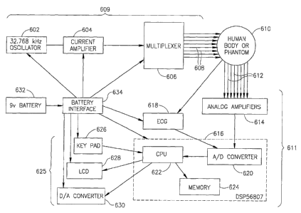
Fig. 4 schematically shows a hardware configuration for an impedance imaging
system which uses ECG data to determine breathing parameters, in accordance
with an
13
CA 02491367 2004-12-30
WO 2004/004539 PCT/IL2003/000556
embodiment of the invention. The hardware comprises a current injection module
609, a
potential measuring and processing module 611, and a user intexface-module
625. Tn the
current injection module, a 32.768 kHz oscillator 602 generates a stable
sinusoidal current
of a few micro-amperes, which is amplified to the desired current, 1 to S
milliamperes, by
current amplifier 604. A dual 1-to-4 multiplexes 606 is used to inject the
cuzrent through
any desired pair chosen from 8 electrodes 608, which are placed around the
thorax of a
human body 610, or around a phantom. Potential measuring and processing module
611
includes eight electrodes 612, which are applied to the thorax and sense
voltage, analog
amplifiers 614, and a Motorola DSPS6807 chip 616. An electrocardiogram 618
also feeds
voltage measurements into chip 616. Chip 616 includes an analog to digital
convertor 620
which converts the analog voltage data to digital form, a central processing
unit 622, and a
memory 624. The digital data is stored in the memory, for each pair of
electrodes used to
inject current, and is then used by the CPU to reconstruct an impedance image.
The CPU
also uses the data from the ECG to calculate parameters such as RR and QT
intervals,
which are used to infer breathing parameters. User interface module 625
includes a keypad
626 used to enter data or feedback from the user into the CPU, a liquid
crystal display 628
for presenting the results or for giving instructions to the patient during
the measurement
process, and a digital to analog convertor 630 for plotting data during
development of the
system. A 9 volt battery 632 provides power for all three modules, via a
battery interface
634, which provides positive and negative voltage and a ground.
Optionally, user ,interface module 62S is located remotely, with the data
transmitted (for example, over phone lines with a modem, or over a secure
broadband
Internet connection), or user interface module 625 includes hardware for
transmitting the
impedance imaging data from memory 624 to a remote location. Optionally,
current
amplifier 604 and multiplexes 606 axe also controlled remotely, or they are
controlled by a
computer, optionally chip 616, which is programmed to inject a given sequence
of
currents through the different electrodes. These options may be useful, for
example, for
monitoring the condition of a patient who is at home, without the need for
hiln to come
into a hospital every time.
Fig. 5 is a flowchart outlining how the finite volume method is used to
calculate an
impedance image from the potential data taken with different pairs of
electrodes carrying
current. Initially, in 402, an image is made of the chest of the patient,
using, for example,
magnetic resonance imaging, computerized x-ray tomography, or ultrasound.
14
CA 02491367 2004-12-30
WO 2004/004539 PCT/IL2003/000556
Alternatively, with some loss of accuracy, the patient's chest is modeled by
some standard
body model, perhaps parameterized by characteristics such as weight, height,
gender, and
body type. Optionally, the model or image includes the whole body, or more of
the body,
rather than just the chest, which makes it possible to more accurately account
for current
paths that are not confined to the chest.
At 404, the chest or body model is used to create a three-dimensional grid.
Optionally, the grid conforms to the surface of the body. Optionally, the grid
conforms to
the surfaces of the lungs and/or the heart, which generally have substantially
different
impedance from other parts of the chest, and from each other, Optionally, the
grid changes
during the breathing cycle and heart beat, so that it can continue to conform
to the surfaces
of the lungs and heart. Alternatively, the grid conforms only to some
approximate average
surfaces of the lungs and heart, or does not conform to the surfaces of the
Lungs and heart
at all. The grid coordinates of the various electrodes (including their
orientations and
outlines, as well as their positions) are determined and stored.
In 406, potential data is read at each electrode, for each pair of current-
carrying
electrodes, as described above in the description of Fig. l and Fig. 2. In
408, an initial
guess is made of the impedance distribution of the chest, for example, using
information
about the location of the Lungs and heart obtained from the image made in 402,
and/or
from a chest model used in 402. Optionally, the initial' guess for the
impedance
distribution simply assigns typical values of impedance for lung tissue,
cardiac tissue, and
the rest of the chest cavity.
In 410, the finite volume method is used to solve the forward problem,
calculating
the expected surface potential at each electrode where voltage is measured,
for each choice
of current carrying electrodes, using the initial guess for impedance
distribution as a
starting point. The finite volume method uses the integral form of Poisson's
equation,
which becomes a set of simultaneous linear equations when Poisson's equation
is
discretized and the integral is xeplaced by a sum. The boundary conditions for
Poisson's
equation are Neumann-type conditions, stating the current flux normal to the
boundary.
The finite volume method is more accurate than the (mite element method, the
most
commonly used method in the field of bio-impedance, at solving Poisson's
equation with
Neumann boundary conditions, because it can treat discontinuous impedance
distributions
and discontinuous current sources (B. Lucquin and O. Pironneau, Intt~oduction
to
Scientific Computing, John Wiley & Sons, 1998, pp. 300-304). The finite volume
method
CA 02491367 2004-12-30
WO 2004/004539 PCT/IL2003/000556
also makes more efficient use of computational resources and CPU time than the
finite
element method Abboud, S. et al, Comput. Biomed. Res., (1994), Vol. 27, pages
441-455.
The set of linear equations can be represented in sparse matrix form, and
relaxation
methods can be used that are very fast and efficient for sparse matrixes, for
example the
successive over relaxation (SOR) method.
In 412, the surface potential calculated at each electrode in 410, for each
chosen
pair of current-carrying electrodes, is compared to the voltages measured at
each electrode
in 406. If difference between the measured and calculated potentials is small
enough, then
the initial guess made in 40$ for the impedance disixibution is a good match
to the actual
impedance distribution. Otherwise, the Newton-Raphson method or a similar
method may
be used in 414 to make an improved guess for the impedance distribution, and
step 410
(solving the forward problem) is repeated, using the new guess. The Newton-
Raphson
method involves differentiating (finding the Jacobian of) the matrix
associated with the set
of linear equations in 4I0, with respect to changes in the impedance
distribution. Here the
finite volume method offers another advantage over the finite element method,
since the
finite volume method allows the matrix elements to be expressed symbolically
in terms of
the impedance distribution, and the expressions can be mathematically
manipulated to find
their derivatives, and hence the Jacobian. With the finite element method, on
the other
hand, the matrix is found only in numerical form, and finding the Jacobian is
then much
more time consuming, for a large matrix.
The Newton-Raphson method involves inverting a matrix, called the Hessian
matrix, which depends on the Jacobian and on the difference between the
measured and
calculated potentials. Because the Hessian matrix is often ill-conditioned,
the Newton-
Raphson method may be unstable. Optionally, the stability of the convergence
is improved
by using a modified Newton-Raphson method, for example the Marquardt method.
These
methods involve addilg to the Hessian matrix a regularization matrix, which
makes it
better conditioned.
At each iteration of the loop shown in Fig. 4, the calculated potential is
compared
to the measured voltages on the electrodes. When the difference between them
is small
enough, the latest guess for the impedance distribution is accepted as a good
approximation to the actual impedance distribution. In 416, this impedance
distribution is
stored, and optionally displayed on a monitor or printed.
16
CA 02491367 2004-12-30
WO 2004/004539 PCT/IL2003/000556
Fig. 6 is a flowchart showing how impedance imaging is combined with ECG data
to produce an overall evaluation of a patient suffering from congestive heart
failure, and to
decide on appropriate treatment. ECG data is recorded in 502. This data is
used both for
determining breathing parameters in 504, as described above in Fig. 2, and for
detecting
problems with heart function, for example arrhythmia or incipient arrhythmia,
in 506. At
the same time, in 508, impedance imaging is used to estimate the thoracic
fluid volume in
510, and this estimate is adjusted by taking into account the breathing
parameters
determined in 504. This leads in 512 to a canonical impedance image, as
discussed above
in Fig. 2, which characterizes the thoracic fluid volume, and the presence of
pulmonary
edema, independently of the state of expansion of the lungs and the phase of
the cardiac
cycle at the time the image was made.
In 514, the canonical impedance image in 512 is used, together with the
information on cardiac performance in 506, as input to an algorithm which
generates an
evaluation of the patient's overall condition, with a view toward determining
the optimal
treatment in 516. For example, an abnormally high thoracic fluid volume by
itself might
indicate the need for the patient to take an increased dose of diuretic
medication. But some
diuretics, such as. thiazide, furosemide, and ethacrynic acid, can cause or
enhance
hypokalemia, which if not treated can lead to arrhythmia. If the ECG data in
506 shows
abnormally long QT intervals, especially with prominent U waves, then this by
itself
might indicate hypokalemia and the need to decrease the dose of diuretics.
Only by
lookiilg at both ECG data in 506 and impedance imaging in 512, is it possible
to
determine the optimum dose of medication. An algorithm which uses both ECG
data and
impedance imaging, and fads the optimum treatment, is optionally based, for
example, on
experience with the outcomes of other patients with similar combinations of
symptoms.
The word "data analyzer" as used herein means any equipment used to analyze
data, even if it is not a single unit. For example, when a data analyzer is
described as
analyzing electrocardiograph data and reconstructing an impedance image, this
does not
necessarily mean that a single piece of equipment does both the analyzing and
the
reconstructing. The word "data analyzer" can include one or more ordinary
computers
running software, one or more pieces of specially designed hardware, or both.
The words
"comprise", "include" and their conjugates as used herein mean "include but
are not
necessarily limited to". While the invention has been described with reference
to certain
exemplary embodiments, various modifications will be readily apparent to and
may be
17
CA 02491367 2004-12-30
WO 2004/004539 PCT/IL2003/000556
readily accomplished by persons skilled in the art without departing from the
spirit and
scope of the above teachings.
1g