Note: Descriptions are shown in the official language in which they were submitted.
CA 02491493 2005-O1-04
WO 2004/004584 PCT/US2003/020550
EXPANDABLE PERCUTANEOUS SHEATH
Back~,round of the Invention
Field of the Invention
S The present invention relates to medical devices and, more particularly, to
methods
and devices for forming a percutaneous channel. In one application, the
present invention
relates to a minimally invasive procedure to insert an orthopedic fixation or
stabilization
implant into the body, such as a formed in situ spinal stabilization rod.
Descrption of the Related Art
The vertebrae and associated connective elements are subject to a variety of
diseases
and conditions which cause pain and disability. Among these diseases and
conditions are
spondylosis, spondylolisthesis, vertebral instability, spinal stenosis and
degenerated,
herniated, or degenerated and herniated intervertebral discs. Additionally,
the vertebrae and
associated connective elements are subject to injuries, including fractures
and torn
ligaments and surgical manipulations, including laminectomies.
The pain and disability related to these diseases, conditions, injuries and
manipulations often result from the displacement of all or part of a vertebra
from the
remainder of the vertebral column. A variety of methods have been developed to
restore
the displaced vertebrae or portions of displaced vertebrae to their normal
position and to fix
them within the vertebral column. For example, open reduction with screw
fixation is one
currently used method. The surgical procedure of attaching two or more parts
of a bone
with pins, screws, rods and plates requires an incision into the tissue
surrounding the bone
and the drilling of one or more holes through the bone parts to be joined. Due
to the
significant variation in bone size, configuration, and load requirements, a
wide variety of
bone fixation devices have been developed in the prior art. In general, the
current standard
of care relies upon a variety of metal wires, screws, rods, plates and clamps
to stabilize the
bone fragments during the healing or fusing process. These methods, however,
are
associated with a variety of disadvantages, such as morbidity, high costs,
lengthy in-patient
hospital stays and the pain associated with open procedures.
Therefore, devices and methods are needed for repositioning and fixing
displaced
vertebrae or portions of displaced vertebrae which cause less pain and
potential
complications. Preferably, the devices are implantable through a minimally
invasive
CA 02491493 2005-O1-04
WO 2004/004584 PCT/US2003/020550
procedure.
In addition, a wide variety of diagnostic or therapeutic procedures involve
the
introduction of a device through a natural or artificially created access
pathway. A general
objective of access systems which have been developed for this purpose, is to
minimize the
cross-sectional area of the puncture, while maximizing the available space for
the
diagnostic or therapeutic instrument. These procedures include, among others,
a wide
variety of laproscopic diagnostic and therapeutic interventional procedures.
Accordingly, a
need remains for access technology which allows a device to be percutaneously
passed
through a small diameter tissue tract, while accommodating the introduction of
relatively
large diameter instruments.
Summary of the Invention
A percutaneous access sheath is provided according to an aspect of the present
invention. In one application, the percutaneous access sheath is used to
facilitate the
insertion of an orthopedic fixation or stabilization implant that is formed in
situ, such as a
1 S spinal stabilization rod.
The percutaneous access sheath may be used in conjunction with a deployment
catheter, which is provided with a balloon at its distal end. The percutaneous
access sheath
has a proximal section and a variable diameter distal section. The deployment
catheter may
be disposed within the percutaneous access sheath such that the balloon is
positioned within
the distal section of the percutaneous access sheath.
The distal section of the percutaneous access sheath is restrained in a first,
small
diameter by a releasable restraint such as a perforated insertion sheath. The
distal section of
the percutaneous access sheath is creased, folded inwards and inserted into a
distal section
of the insertion sheath. This gives the percutaneous access sheath a smaller
cross-sectional
profile, facilitating its insertion.
The percutaneous access sheath is inserted as packaged above. Following
insertion,
the insertion sheath may be torn away along its perforations. To facilitate
this the balloon
may be partially inflated, expanding the distal section of the percutaneous
access sheath
sufficiently to tear the insertion sheath along its perforations. After the
insertion sheath is
removed, the balloon may be fully inflated to distend the distal section of
the percutaneous
access sheath to its full cross-sectional profile. Afterwards, the balloon may
be deflated to
allow the removal of the deployment catheter, leaving the percutaneous access
sheath in
-2-
CA 02491493 2005-O1-04
WO 2004/004584 PCT/US2003/020550
place.
In one embodiment where the percutaneous access sheath is used to facilitate
the
insertion of an orthopedic spinal stabilization implant that is formed in
situ, a percutaneous
access sheath may advantageously be first inserted through the portals of
adjacent bone
anchors, by the method described above. This provides a smooth channel to
facilitate the
passage of another deployment catheter carrying an inflatable orthopedic
fixation device at
its distal end.
Other applications of the percutaneous access sheath include a variety of
diagnostic
or therapeutic clinical situations which require access to the inside of the
body, through
either an artificially created or natural body lumen.
Brief Description of the Drawings
Figure 1 is a side elevational view of a percutaneous access sheath.
Figure 2 is a side elevational view of a insertion sheath.
Figure 3 illustrates the percutaneous access sheath in a reduced cross-
sectional
configuration and inserted into the insertion sheath.
Figure 4 is a side elevational view of an access sheath expansion catheter.
Figure 5 is an enlarged view of the distal end of the expansion catheter.
Figure 6 is an enlarged view of the proximal end of the expansion catheter.
Figure 7 illustrates the percutaneous access sheath assembly, with the
expansion
catheter inserted into the structure illustrated in Figure 3.
Figure 8 is a side elevational view of a bone anchor.
Figure 9 is a side elevational view of the bone anchor of Figure 8, rotated
90° about
its longitudinal axis.
Figure 10 is a longitudinal cross-sectional view of the bone anchor of Figure
9.
Figure 11 is a side elevational view of an alternative embodiment of a bone
anchor.
Figure 12-15 illustrate one embodiment of a method of threading a guide wire
through the portals of bone anchors that have been implanted into adjacent
vertebrae in a
vertebral column.
Detailed Description of the Preferred Embodiment
Figure 1 is an overview of the percutaneous access sheath 100. It generally
comprises an elongate tubular body with an axial lumen, and is designed to
provide
percutaneous access to a diagnostic or treatment site in the body. The
elongate tubular
-3-
CA 02491493 2005-O1-04
WO 2004/004584 PCT/US2003/020550
body has a proximal section and a distal section 110. The length of these two
sections can
be varied according to clinical need, as will be understood by those skilled
in the art with
reference to this disclosure. The distal section 110 is expandable from a
first, smaller
cross-sectional profile to a second, larger cross-sectional profile. The
first, smaller cross-
sectional profile of the distal section 110 eases its insertion into the
percutaneous treatment
site. After insertion, the distal section 110 is expanded to a second, larger
cross-sectional
profile to provide a larger passageway for surgical instruments to reach the
percutaneous
treatment site.
In the illustrated embodiment, the percutaneous access sheath 100 is made of a
double-layered co-extruded tubing 102, with an inner layer 104 and an outer
layer 106. The
inner layer 104 defines a lumen 108. The inner layer 104 extends further
distally than the
outer layer 106, such that the distal section 110 of the tubing 102 is of a
single layer, the
inner layer 104. The inner layer 104 may be made of PTFE and the outer layer
106 may be
made of HDPE. Other suitable materials, such as nylon, PEBAX or PEEK, may be
used for
either layer.
In this embodiment, the distal section 110 is creased, folded inwards, and
collapsed
from a larger to a smaller cross-sectional profile to ease its insertion. As
discussed below,
in one application of the invention, the distal section 110 is inserted
through adjacent bone
screws or anchors. Its length is thus determined by the distance between such
adjacent
bone screws, and is generally in the range of 4-l2cm. The proximal end 112 of
the tubing
102 is flared and fitted onto a handle 114. A distal cap 116 may be threaded
onto the
handle 114 to secure the proximal end 112 of the tubing 102. Additionally a
proximal cap
118 may be threaded onto the handle 114. The overall length of the tubing 102
depends on
the distance between the insertion and treatment locations, and is generally
in the range of
15-60cm for orthopedic fixation surgery of the vertebrae. In the illustrated
embodiment the
length of the tubing is approximately 20cm, with the distal section 110
accounting for
approximately half of that length.
Figure 2 is an overview of the insertion sheath 200. It is preferably made of
a thin,
smooth and flexible material. The insertion sheath 200 has a proximal section
and a distal,
restraint section 210. The restraint section 210 has a smaller cross-sectional
profile than the
proximal section of the insertion sheath 200. The restraint section 210 is
adapted to restrain
the distal section 110 of the percutaneous access sheath 100 in its smaller
cross-sectional
-4-
CA 02491493 2005-O1-04
WO 2004/004584 PCT/US2003/020550
profile. This is achieved by inserting the percutaneous access sheath 100 into
the insertion
sheath 200 such that the distal section 110 of the percutaneous access sheath
100 lies within
the restraint section 210 of the insertion sheath 200.
In the illustrated embodiment, the insertion sheath 200 may be made of PTFE.
The
proximal end 202 of the insertion sheath 200 terminates at a pull tab 204,
which may be
formed by a threaded luer lock. The insertion sheath 200 is provided with a
slit 206 near its
proximal end 202. The insertion sheath 200 tapers at a first tapering point
208 into a
restraint section 210, which tapers again into the distal tip 212. As
discussed above, the
restraint section 210 restrains the distal section 110 of the percutaneous
access sheath 100
in its smaller cross-sectional profile. Thus the length of the restraint
section 210 is
approximately the same as or slightly longer than the distal section 110, and
generally falls
in the range of 4-l3cm.
The diameter of the restraint section 210 is preferably smaller than the
diameter of
the eye of the bone screw used, as discussed below. The insertion sheath 200
may be
1 S perforated or otherwise provided with a tear line distally from the first
tapering point 208 to
its distal tip 212. The distance between the slit 206 and the distal tip 212
is generally
approximately equal to or slightly shorter than the length of the tubing 102,
and thus is
generally in the range of 12-57cm. In the illustrated embodiment this distance
is
approximately l5cm, and the overall length of the insertion sheath 200 is
approximately
24cm.
Figure 3 illustrates the percutaneous access sheath 100 inserted into the
insertion
sheath 200 via the slit 206 provided near its proximal end 202. The diameter
of the
restraint section 210 of the insertion sheath 200 is smaller than the diameter
of the distal
section 110 of the tubing 102. The distal section 110 is creased and folded
inwards to
decrease its effective diameter, and inserted into the restraint section 210.
As discussed
above, the restraint section 210 restrains the distal section 110 of the
percutaneous access
sheath 100 in its smaller cross-sectional profile. The restraint section 210
is approximately
the same length as or just longer than the distal section 110. Thus inserted,
the distal
section 110 extends to a point just proximal of the distal tip 212 of the
insertion sheath 200.
In certain embodiments an insertion sheath 200 may not be necessary if the
distal
section 110 of the percutaneous access sheath 100 is made of a stretchable
material that
may be stretched from a first, smaller cross-sectional profile to a second,
larger cross-
-5-
CA 02491493 2005-O1-04
WO 2004/004584 PCT/US2003/020550
sectional profile. In these embodiments the outer surface of the distal
section 110 is
preferably made of a smooth material to facilitate the insertion of the
percutaneous access
sheath 100 into a treatment site.
Figure 4 is an overview of the deployment catheter 300. It is provided with an
expansion element such as balloon 310 at its distal end. The deployment
catheter 300 is
inserted into the lumen 108 of the percutaneous access sheath 100 such that
the balloon 310
is arranged within the distal section 110. The balloon 310 may be inflated to
expand the
distal section 110 from its first, smaller cross-sectional profile to its
second, larger cross
sectional profile following the insertion of the percutaneous access sheath
100 into a
treatment site.
An inner tube 302 extends the entire length of the deployment catheter 300. A
guide wire lumen 304 is defined by the interior of the inner tube 302. The
deployment
catheter 300 can travel along a guide wire extending through the guide wire
lumen 304.
The inner tube 302 carries coaxially on its exterior an outer tube 306. The
outer tube 306
terminates proximally into the distal end of a handle 308, and distally into
the proximal end
of a balloon 310. The balloon 310 may be made of PET. The handle 308 may be
provided
with an optional support tube 312 extending from its distal end and over a
proximal section
of the outer tube 306, to increase the rigidity of the deployment catheter 300
during
insertion. This support tube 312 may be made of aluminum.
Figure 5 is an enlarged view of the distal end of the deployment catheter 300.
Both
the inner tube 302 and the guide wire lumen 304 extend through the distal end
314 of the
balloon 310. The inner tube 302 carries coaxially on its exterior a marker
ring 316 near the
distal end 314 of the balloon 310. Alternatively the marker ring 316 may be
carned by the
distal end 314 of the balloon 310. The marker ring 316 is preferably made of
gold,
tantalum, or another radio-opaque material. Additional marker rings may be
provided in
the balloon 310 to aid in visualizing its location. A balloon inflation lumen
318, defined in
the space between the inner tube 302 and the outer tube 306, communicates with
the
interior of the balloon 310. As discussed above, the balloon 310 may be
inflated to expand
the distal section 110 of the percutaneous access sheath 100 from its first,
smaller cross-
sectional profile to its second, larger cross-sectional profile. Thus the
length of the balloon
310 is approximately equal to or slightly longer than the length of the distal
section 110. In
the illustrated embodiment the length of the balloon 310 is approximately
lOcm.
-6-
CA 02491493 2005-O1-04
WO 2004/004584 PCT/US2003/020550
Figure 6 is an enlarged view of the proximal end of the deployment catheter
300.
Both the inner tube 302 and the guide wire lumen 304 extend through the
proximal end of
the handle 308. The balloon inflation lumen 318, defined in the space between
the inner
tube 302 and the outer tube 306, opens into a port 320 in the handle 308. A
stopper 322
supports the inner tube 302 within the handle 308 and prevents the balloon
inflation lumen
318 from communicating with the space 324 in the main branch of the handle
308. Thus
only the port 320 communicates via the balloon inflation lumen 318 with the
interior of the
balloon. A pump may be connected to the port 320 to inflate or deflate the
balloon. To
enable visualization of the state of the balloon, it may be inflated with
contrast media.
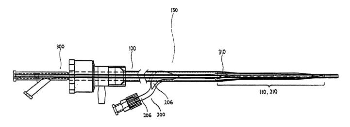
Figure 7 illustrates the percutaneous access sheath assembly 150. The
percutaneous
access sheath assembly 150 comprises the percutaneous access sheath 100, the
insertion
sheath 200 and the deployment catheter 300. It is assembled by inserting the
deployment
catheter 300 into the percutaneous access sheath 100 and inserting the
percutaneous access
sheath 100 into the insertion sheath 200 such as via the slit 206 or other
proximal opening
provided near its proximal end 202. The balloon 310 of the deployment catheter
300 is
deflated, folded and inserted into the distal section 110 of the access sheath
100. The distal
section 110, as discussed above, is creased and folded inwards to decrease its
effective
diameter, and inserted into the restraint section 210 of the insertion sheath
200. As
discussed, the balloon 310 is approximately the same length as or just longer
than the distal
section 110 and the restraint section 210.
Figures 8-11 illustrate one embodiment of a bone anchor 410 as mentioned
above.
It is provided with at least one connector 422 at or near its proximal end (or
top end, as
illustrated). This connector 422 is used to engage an orthopedic spinal
stabilization implant
that is formed in situ, as discussed below. The connector 422 is preferably an
aperture 422,
to achieve a more secure engagement. In one embodiment the percutaneous access
sheath
100 extends through the apertures 422 of two or more bone anchors 410 to
establish a
passageway to facilitate the insertion of a formed in situ orthopedic spinal
stabilization
implant.
An embodiment with two bone anchors is now described. The percutaneous access
sheath 100 is extended through the aperture 422 of a first bone anchor 410,
then through the
aperture 422 of a second bone anchor 410. The first bone anchor 410 is
preferably
implanted within a first bone. The second bone anchor 410 may be implanted
within the
CA 02491493 2005-O1-04
WO 2004/004584 PCT/US2003/020550
second bone. The bones may be adjacent vertebral bodies or vertebrae, or first
and second
vertebrae spaced apart by one or more intermediate vertebrae. The clinical
procedure is
described in further detail below.
The bone anchors 410 of Figures 8-11 are made of a biocompatible material such
as
titanium or stainless steel. Alternatively, the bone anchors 410 may be made
of a
composite material. The bone anchors 410 may also be made of a suitable
medical grade
polymer. In one embodiment, the bone anchors 410 have a length between about
40 mm
and 60 mm, preferably about 50 mm. However, the actual length is dependent on
the
location of the fracture, size of patient, etc.
The bone anchor 410 comprises a proximal portion 412 having a proximal end 414
and a distal portion 416 having a distal end 418. The proximal portion 412
typically
comprises a head 420 and a portal 422. In a preferred embodiment, the head 420
comprises
a proximal portion 424 configured to mate with the tip of a screwdriver. The
head 420 may
comprise a standard or Phillips slot for mating with the screwdriver. A
variety of slot
configurations are also suitable, such as hexagonal, Torx, rectangular,
triangular, curved, or
any other suitable shape. The bone anchor of Figure 11 has a raised platform
434 having a
plurality of substantially flat sides, such as a hexagonal platform,
configured to mate with a
corresponding depression in the distal end of a screwdriver. Platform 434 may
come in a
variety of shapes suitable mating with a screwdriver.
The portal 422 of the bone anchor 410 may extend through the head 420 and is
generally between about 4 mm and 8 mm in diameter, preferably about 6 mm to
about
8 mm in diameter. The portal 422 may be of any suitable shape; however, the
portal 422 is
preferably round to facilitate the insertion of the percutaneous tube 100 as
well as the in situ
forming orthopedic spinal stabilization implant.
The distal portion 416 of the bone anchor 410 typically comprises threads 426
and a
sharp tip 428. The bone anchor 410 also preferably comprises a central lumen
430
extending coaxially through the length of the bone anchor 410 from its
proximal end 414 to
its distal end 418 and configured to receive a guidewire. The bone anchor 410
may also
include one or more perforations 432, as shown in Figure 11. These
perforations 432 are in
communication with the central lumen 430 of the bone anchor 410. The
perforations 432
may be aligned axially, as illustrated, or may be staggered axially. The
perforations 432
permit bone to grow into bone anchor 410, stabilizing bone anchor 410 within
the bone.
_g_
CA 02491493 2005-O1-04
WO 2004/004584 PCT/US2003/020550
Additionally, bone matrix material such as a hydroxyapatite preparation can be
injected into
the central lumen 430 and through the perforations 432 to promote bone in-
growth.
The method of using the percutaneous access sheath 100 to facilitate the
insertion of
an orthopedic spinal stabilization implant formed in situ according to one
aspect of the
present invention is described in the following figures. In this embodiment a
smooth
channel is first established between two or more adjacent bone anchors to
facilitate the
passage of another deployment catheter carrying an inflatable orthopedic
fixation device at
its distal end. While the method is disclosed and depicted with reference to
only two
vertebrae, one of which is either unstable, separated or displaced and the
other of which is
neither unstable, separated or displaced, the method can also be applied to
three or more
vertebrae simultaneously. Further, the method can be used to stabilize the LS
vertebrae,
using the cranial-ward portion of the sacrum as the "vertebrae" with which LS
is anchored.
Although the method is disclosed and depicted as applied on the left side of
the vertebral
column, the method can also be applied on the right side of the vertebral
column or,
preferably, on both sides of the vertebral column, as will be understood by
those skilled in
the art with reference to this disclosure. Other applications include the
stabilization of
other bones and skeletal elements of the body.
Figure 12 illustrates bone anchors 410 that have been inserted through the
periosteal
surface and into the anterior vertebral body or another suitable portion of
the vertebrae 500
and 502. As discussed above, bone matrix material such as a hydroxyapatite
preparation
can be injected into the central lumen 430 of a bone anchors 410 and through
its
perforations (not visible in this figure) to promote bone in-growth. The bone
anchors 410
are arranged such that their portals 422 are substantially coaxial in relation
to each other.
A hollow needle 436 is inserted percutaneously and advanced into the portal
422 of
one of the bone anchors 410, with the aid of fluoroscopy. The hollow needle
436 may be
16 or 18 gauge. While the hollow needle 436 is shown engaging the bone screw
410 in the
cranial-ward vertebrae 502, it can alternatively first engage the bone screw
410 in the
caudal-ward vertebrae 500, as will be understood by those skilled in the art
with reference
to the disclosure. Figure 13 is an enlarged view of the distal end of the
hollow needle 436.
A semi-rigid guide wire 438 is introduced through the lumen of the hollow need
436 and
the portal 422 of the bone anchor 410 in the cranial-ward vertebrae 502. The
hollow needle
436 preferably has a Tuohy needle tip which causes the guide wire 438 to exit
the hollow
-9-
CA 02491493 2005-O1-04
WO 2004/004584 PCT/US2003/020550
needle 436 perpendicularly to the central lumen 430 of the bone anchor 410, or
coaxially
with the axis of the portal 422 of the bone anchor 410. Alternatively, the
bending of the
guide wire 438 through the portal 422 of the bone anchor 410 may be
accomplished by an
angled-tip modified Ross needle or another suitable structure as will be
understood by those
skilled in the art with reference to the disclosure.
Figure 14 illustrates an optional guide wire directing device 440, according
to one
aspect of the present invention, inserted percutaneously between the bone
anchors 410. The
guide wire directing device 440 may have a forked end used to direct the guide
wire 438
through the portal 422 of the bone anchor 410 in the caudal-ward vertebrae
500. In another
embodiment a guide wire capture device 442, such as a snare or forceps, may be
inserted
percutaneously caudal to the portal 422 of the bone anchor 410 in the caudal-
ward vertebrae
500. The guide wire capture device 442 engages the distal end of the guide
wire 438 after
the guide wire 438 has passed through portal 422 of the bone anchor 410 in the
caudal-ward
vertebrae 500, and pulls it through the skin dorsally, so that both ends of
the guide wire 438
are secured.
Figure 15 illustrates the guide wire 438 in place after the procedure
described above
in Figures 12-14.
The guide wire 438 may be inserted into the guide wire lumen 304 of the
deployment catheter 300 of the percutaneous access sheath assembly 150. The
entire
assembly 150 may travel over the guide wire 438 until its distal tapered
portion is inserted
through the portals 422 of the bone anchors 410. The insertion sheath 200,
which is on the
exterior of the percutaneous access sheath assembly 150, facilitates the
insertion because of
its smooth, low profile exterior. As discussed above, it may be made of PTFE.
Following the insertion of the percutaneous access sheath assembly 150, the'
insertion sheath 200 is removed. This may be accomplished by pulling on the
pull tab 204
and tearing the insertion sheath 200 along the perforations, crease line, or
other structure for
facilitating tearing provided along its restraint section 210. This may be
facilitated by first
partially inflating the balloon 310 of the deployment catheter 300. As
discussed above, the
balloon 310 is arranged within the distal section 110 of the percutaneous
access sheath 100,
which is itself arranged within the restraint section 210 of the insertion
sheath 200. Thus,
inflating the balloon 310 causes the distal section 110 of the percutaneous
access sheath
100 to expand, tearing the restraint section 210 of the insertion sheath 200
along its
-10-
CA 02491493 2005-O1-04
WO 2004/004584 PCT/US2003/020550
perforations.
After the removal of the insertion sheath 200, the balloon 310 may be fully
inflated
to expand the distal section 110 of the percutaneous access sheath to its full
cross-sectional
profile. Afterwards the balloon 310 may be deflated to ease the removal of the
deployment
catheter 300. As discussed above, the inflation and deflation of the balloon
310 may be
done via a pump connected to the port 320 of the deployment catheter 300, and
preferably
with contrast media being pumped, to better convey the state of the balloon.
Thus the percutaneous access sheath 100 is inserted through the portals 422 of
the
bone anchors 410. The establishment of this smooth channel through the portals
422 of the
bone anchors 410 facilitates the passage of another deployment catheter
carrying an
inflatable orthopedic fixation device at its distal end. An example of such a
deployment
catheter with an inflatable orthopedic fixation device at its distal end as
well as the
associated anchors and methods are disclosed in United States Patent
Application Serial
No. 10/161,554 filed on May 31, 2002, the disclosure of which is hereby
incorporated by
1 S reference in its entirety.
Although the present invention has been described in terms of certain
preferred
embodiments, other embodiments of the invention including variations in
dimensions,
configuration and materials will be apparent to those of skill in the art in
view of the
disclosure herein. In addition, all features discussed in connection with any
one
embodiment herein can be readily adapted for use in other embodiments herein.
The use of
different terms or reference numerals for similar features in different
embodiments does not
imply differences other than those which may be expressly set forth.
Accordingly, the
present invention is intended to be described solely by reference to the
appended claims,
and not limited to the preferred embodiments disclosed herein.
-11-