Note: Descriptions are shown in the official language in which they were submitted.
CA 02491690 2015-05-07
SOMATOGENIC PLASTID TRANSFORMATION
STATEMENT REGARDING FEDERALLY SPONSORED RESEARCH
Investigations reported in this application were supported in part by funding
from NIH R 01 GM63879.
FTFLD OF THE INVENTION
The field of this invention relates to genetically engineering a plant
plastid.
More specifically, this invention relates throught the transformation of non-
green plant
cells through plastid transformation, and the subsequent regeneration the non-
green
plant cells through somatic embryogenesis.
BACKGROUND
Plastids are ideal for genetic engineering because it offers a number of
attractive advantages, including high-level transgene expression (Daniell et
al., 2002),
multi-gene engineering in a single transformation event (DeCosa et al., 2001;
Ruiz at
al., 2003; Daniell & Dhingra, 2002), transgene containment via maternal
inheritance
(Daniell 2002), lack of gene silencing (Lee et al., 2003; DeCosa et al.,
2001), position
effect due to site specific transgene integration (Daniell et al., 2002) and
pleiotropic
effects (Daniell at al., 2001; Lee et al., 2003). Chloroplast genetic
engineering is most
suitable for hyper-expression of vaccine antigens and production of valuable
therapeutic proteins. Ever since we demonstrated expression of human-elastin
derived
polymers for various biomedical applications (Guda et al., 2000), we have
extended
this approach to express vaccines antigens for Cholera, Anthrax (Daniell et
al., 2001,
Daniell 2003), monoclonal antibody (Daniell et al., 2001) and human
therapeutic
proteins, including Human Serum Albumin, (Fernandez et al., 2003), Magainin
(DeGray at al., 2001), Interferon (Daniell 2003) and Insulin like Growth
Factor
(Daniell, 2003). Several other laboratories have expressed Human Somatotropin
Interferon-GUS fusion proteins to improve stability (Reddy et al., 2003) and
tetanus
vaccine antigens (Tregoning et al., 2003) in transgenic chloroplasts. Without
any
exception, all of these therapeutic proteins have been expressed in transgenic
tobacco
CA 02491690 2005-01-04
WO 2004/005480
PCT/US2003/021157
chloroplasts, meeting the zero-tolerance of food crops for plant-derived
pharmaceuticals advocated by various environmental groups.
However, there is an urgent need for oral delivery of therapeutic proteins and
vaccine antigens to dramatically reduce their production, purification,
storage and
transportation costs and minimize complications associated with intravenous
delivery.
Carrot (Daucus carota L.) is one of the most important vegetables used
worldwide for
human and animal consumption, due to its excellent source of sugars, vitamins
A, C
and fiber in the diet. The carrot plant is biennial, completing its life cycle
in two years.
In the first year the plant produces the fleshy taproot, which is edible. If
left in the
ground, plants flower in the second year after passing through a cold season
(Yan, W.
& Hunt, L.A Reanalysis of Vernalization Data of Wheat and Carrot, Annals of
Botany
84, 615-619 (1999). In addition, chloroplast genomes in the cultivated carrot
crop are
transmitted strictly through maternal inheritance (Vivek et al 1999). Thus,
carrot is
environmentally safe and is doubly protected against transgene flow via pollen
and
seeds to achieve zero-tolerance on transgene flow advocated for food crops.
Carrot
somatic embryos are single cell derived and multiply through recurrent
embryogenesis;
this provides uniform source of cell culture, which is one of the essential
requirements
for producing therapeutic proteins (homogeneous single source of origin).
Carrot cells
divide rapidly and large biomass is produced using bioreactors. Cultured
carrot cells
are edible and could be used directly to deliver precise dose of vaccine
antigens or
biopharmaceuticals. When delivered via edible carrots, there is no need to
cook and
this would preserve the structural integrity of therapeutic proteins during
consumption.
Viable for long duration on culture medium, encapsulated embryos are used as
synthetic seeds for cryopreservation and controlled germination (Tessereau,
H., B.
Florin, M. C. Meschine, C. Thierry and V. Petiard, 1994). Thus, transgenic
carrot with
enhanced medicinal or nutritional value can play a vital role in improving
human or
animal health
However, in order to engineer the carrot chloroplast genome, one has to
overcome several major hurdles. So far, the chloroplast genome has been
transformed
only by using green leaves as explants, which contain large chloroplasts. The
first
challenge is to introduce foreign DNA into small proplastids and identify
appropriate
regulatory sequences and selectable markers that function in non-green
plastids. The
2
CA 02491690 2005-01-04
WO 2004/005480
PCT/US2003/021157
second challenge is to regenerate chloroplast transgenic plants via somatic ,
embryogeneis and achieve homoplasmy, which lacks the benefit of subsequent
rounds
of selection offered by organogenesis, while using leaves as explants. Because
of these
reasons, chloroplast genetic engineering has been = achieved only in a few
solanaceous
crops other than tobacco, such as tomato (Ruf et al., 2001) and potato
(Sidorov et al.,
1999), although the later crop remained sterile. Non-solanaceous crops
continue to be
a challenge to transform, even though regeneration was possible from green
leaves via
organogenesis. For example, Arabidopsis transgenic plants were sterile (Sidkar
et al.,
1998) and even stable integration or homoplasmy could not be achieved in
oilseed rape
(Bing Kai Hou et al., 2003).
Table 1 shows an exemplary list of the development of transgene expression in
chloroplasts.
3
CA 02491690 2005-01-04
WO 2004/005480 PCT/US2003/021157
TABLE 1
Transgene Expression in chloroplasts
Agronomic traits Gene Promoter 5'/3' Regulatory elements Reference
Insect resistance Cry1A(c) Prrn rbc1_, I Trps16
Mc Bride et al
1995
Herbicide CP4 (petunia) Prrn ggagg / TpsbA Daniell et al
resistance 1998
Insect resistance Cry2Aa2 Prrn ggagg (native) /
TpsbA Kota et al 1999
Herbicide CP4 (bacterial or Prrn rbeL or T7 gene 10 / Tips16 Ye at al
2001
resistance synthetic)
Insect resistance Cry2Aa2 operon Puri Native 5'UTRs /
TpsbA DeCosa et al
2001
Disease resistance MSI-99 Prrn ggagg / TpsbA
DeGray et al
2001
Salt and drought tps Prrn ggagg TpsbA Lee et al 2003
tolerance
Phytoremediation merAalmerBb Prrn ggagga b TpsbA
Ruiz et al 2003
Biopharmaceutical Gene Promoter 5'/ 3' % tsp Reference
proteins regulatory expression
elements
Protein based polymer EG121 Prrn T7genel0 / Not tested
Guda et al
TpsbA 2000
Human somatotropin hST Prma, T7genelOa or 7.0 % a and Staub et
al
PpsbAb psbAb I Trps16 1 %b 2000
Cholera toxin caB Prrn ggagg TpsbA 4% Daniell et al
2002
Tetanus toxin TetC Prrn T7 gene 10a, 25% a, 10%b Tregoning
et al
(bacterial atpBb I Trbc1_, 2003
and
synthetic)
Human Serum Albumin hsa Puna, ggagga, psbAb I 0.02% a, Fernandez-
San
PpsbAb TpsbA 11.1% b Milan et al
2003
Interferon alpha ,5 INF 05 Prrn PpsbAlTpsbA ND Torres
Interferon alpha 2B INF 02B Prrn PpsbAlTpsbA 19%
Falconer
4
CA 02491690 2005-01-04
WO 2004/005480 PCT/US2003/021157
Table 1(continued)
Interferon gamma ifn-g PpsbA PpsbAlTpsbA 6% Leelavathi and
Reddy, 2003
Monoclonal antibodies Prrn ggagg / TpsbA ND Daniell et al
(photosynthesi
s)
Insulin like growth Igf-1 Prrn PpsbAlTpsbA 33% Ruiz
G
factor
Anthrax protective Fag Prrn PpsbAlTpsbA 4-5% Watson
antigen
Plague vaccine CaF1.--Ecr Prrn PpsbAlTpsbA 4.6 % Singleton
V
CA 02491690 2005-01-04
WO 2004/005480
PCT/US2003/021157
SUMMARY OF THE INVENTION
One aspect of this invention describes methods for transforming plastids using
a highly efficient process for carrot plastid transformation through somatic
embryogenesis. Still other aspects of this invention provide for vectors which
are
capable plastid transformation through somatic embryogenesis. Still another
aspect
provides for transformed plastids, plants, and plant parts, which have been
transformed
through somatic embryogenesis through the methods and vectors described
herein.
This application, along with the knowledge of the art, provides the necessary
guidance
and instructions to engineer the plastid genome of several major crops in
which
regeneration is mediated through somatic embryogenesis. Cereal crops (wheat,
rice,
corn, sugarcane), legumes (soybean, alfalfa), oil crops (sunflower, olive),
cash crops
(cotton, coffee, tea, rubber, flex, cork oak, pines), vegetables (eggplant,
cucumber,
cassava, chili pepper, asparagus etc.), fruits (apple, cherry, banana,
plantain, melons,
grape, guava), nuts (cashew, walnuts, peanuts), and trees (date palm etc.) are
regenerated through somatic embryogenesis. Another aspect of this invention
shows
the constructs of chloroplast vectors for a variety of different species using
the same
primers( universal plastid primers).
BRIEF DESCRIPTION OF THE FIGURES
Fig. 1 (A-B). shows the physical map of the carrot chloroplast transformation
vectors.
Fig 1(A) shows carrot chloroplast transformation vector pDD-Dc-gfpIBADH
carries the gfp and BADH genes expressed under the regulation of T7 gene 10 5'
untranslated region (LTTR)/ rps16 3'UTR and PpsbA 5' and 3' UTR respectively.
The
Prrn promoter of 16S r-RNA gene, having both PEP and NEP recognition sites,
drives
expression of the cassette.
Fig 1(B) shows the carrot chloroplast transformation vector pDD-Dc-
aadAIBADH carries the aadA and BADH gene. Expression of aadA gene is under the
regulation of Shine-Dalgarno sequence and psbA 3'UTR while that of BADH is
' regulated by genel0 5' and rps16 3'UTR. AflIII/Pvull digested ¨ 4.9 kb DNA
fragment used as a probe for Southern analysis of the transgenic plants and
landing
6
CA 02491690 2005-01-04
WO 2004/005480
PCT/US2003/021157
sites for primers 3P/3M and 16SF/1M used to confirm the presence of the
transgene
integration into carrot plastids are shown.
Fig. 2(A-C) shows the expression of GFP in carrot cultures transformed with
chloroplast vector pDD-Dc-gfp/BADH; visible under confocal fluorescent
microscope
at fluorescence emission in green at 488nm blue Argon (laser).
Fig. 2(A) shows the untransformed control carrot culture,
Fig. 2(B) shows the transformed embryogenic calli,
Fig.2(C) shows the transformed embryogenic carrot calli differentiated into
globular somatic embryos and
Fig. 2(D) shows a somatic embryo differentiated into cotyledonary stage.
Fig. 3 (A-D) shows the visual selection of green transgenic cells versus
yellow
non-transgenic carrot cells culture.
Fig. 3(A-B) shows the transgenic carrot cell culture turned green due to the
expression BADH (Plate A) while wild type culture remained yellow (Plate B).
Transgenic carrot cell culture can be distinguished as green-transgenic cell
culture vs
yellow non-transgenic carrot cell culture, when heteroplasmic transgenic cell
line was
placed on medium without any selection agent (Plate C and D).
Fig. 4 (A-C) shows the transgene (aadA and badh) integration into the carrot
plastid genome was confirmed by PCR and Southern blot analysis.
Fig 4(A) shows the use of internal primers 3P (land on flanking sequence) and
3M (land on aadA gene) ¨1.65 kb size PCR product was amplified at 64 C
annealing
temperature, confirmed transgene integration into plant cell lines.
Fig. 4(B) shows the use of a set of primer 16SF (landing on the native
chloroplast genome) and 1M (landing on the aadA gene) yield ¨ 2.5 kb size PCR
product at 64 C annealing temperature, confirmed plastid specific integration
of the
transgenes. Lane 2 stand for DNA from non-transgenic carrot cells and lanes 2-
9
represents DNA from seven transgenic carrot cell lines. Primers landing sites
for
primer pairs (3P/3M and 165F/1M) is shown in Fig. 1B.
Fig 4(C) shows the southern blot analysis of plastid genome of untransformed
and transformed carrot with vector pDD-Dc-aadA/BADH. Carrot genomic DNA (5 g
per lane) digested with AflIII and PvuTI and transferred to nitrocellulose
membrane,
was hybridized with the 4.9 kb radioactive labeled P32 DNA probe (containing
2.4 kb
7
CA 02491690 2005-01-04
WO 2004/005480
PCT/US2003/021157
flanking sequence and 2.5 kb transgene sequence, see Fig. 1B). Lane 1, control
DNA
from untransformed transgenic plant showed 2.4 kb size band while
heteroplasmic
transgenic plant from cell line one, lane 2 showed both bands. Homoplasmy in
transgenics plants from different cell lines (lanes 3-8) was achieved by
repetitive
subcultures of transgenic cells in liquid medium.
Fig. 5 (A-B) shows BADH enzyme activity (nmol/min/mg/protein) and BADH
expression was analyzed in protein extracts from untransformed and transformed
carrot
with plastid vector pDD-Dc-aadA/BADH.
Fig 5(A) shows the reduction of NAD+ dependent BADH enzyme was analyzed
for the formation of NADH at 340 nm in presence of betaine aldehyde. Very low
BADH activity was detected in untransformed cells suspension (U), carrot root
(U) and
leaf (U). On the other hand, high BADH activity was recorded about 54 % in
transformed carrot cells suspension (T), about 72% in root (T) in comparison
to leaf
(T) tissues.
Fig. 5(B) shows that the BADH expression was analyzed by western blot.
Whole cell extracts from transformed and untransformed carrot cell culture,
root and
leaf tissues were prepared and 50 fig total soluble protein from each sample
was run on
10% SDS-PAGE and protein transferred to hmnunoblotTM PVDF membrane and
hybridized with polyclonal anti-BADH serum, raised in rabbits against native
BADH.
Antigenic peptides were detected using horseradish peroxidase-linked secondary
antibody. Lane 1, 2, 3 contain whole carrot extract from untransformed cell
culture,
root and leaf and lane 4, 5, 6 contain whole carrot extract from transformed
cell
culture, root and leaf. Transgenic cells expresses about 50 % less (lane 4),
root about
% less (lane 5) BADH protein than leaf (lane 6).
25 Fig. 6
(A-C) shows the effect of different salt concentrations on growth of
untransformed and transformed carrot cell suspension cultures with chloroplast
vector
pDD-Dc-aadA/BADH.
Fig. 6(A) shows the dry mass in untransformed carrot cell culture.
Fig. 6(B) shows transformed cell cultures produced in liquid medium
containing 100 mM NaCl.
Fig. 6(C) shows the stimulation of BADH activity in presence of salt.
Untransformed and transformed carrot cells in suspension cultures were placed
on
8
CA 02491690 2005-01-04
WO 2004/005480
PCT/US2003/021157
shaker at 130 rpm speed for two weeks in liquid medium containing 0, 100, 200
and
300 mM NaCl. Elevated level of BADH activity in transgenic cell cultures was
noticed
when liquid growth medium containing 100 and 200 mM NaCl.
Fig. 7 Effect of salt (100-500 mM NaCl) on untransformed (U) and
transformed (T) carrot plants. Transgenic plants were tested for one month on
different concentration of NaCl. Plants were irrigated with water containing
different
concentrations NaC1 at alternative days up to four weeks.
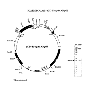
Fig. 8 shows the plasmid pDD-Ta-aphA-6/nptII. More particularly the plasmid
illustrates pDA-76 (aphA-6/nptII expression cassette), having a backbone
vector
pBluescript II KS, a selectable marker/host cell Ampicillin/Kan/ XL-1 Blue
MRF' Tc,
and a flanking region from Triticum aestivum (Ta).
Fig 9 shows the plasmid pDD-So-aphA-6/nptII. More particularly the plasmid
illustrates pDA,76 (aphA-6/nptII expression cassette), having a backbone
vector
pBluescript II KS, a selectable marker/host cell Ampicillin/Kan/ XL-1 Blue
MRF' Tc,
and a flanking region from Saccharum officinarum (So).
Fig 10 shows the plasmid pDD-Dc-aphA-6/nptII. More particularly the
plasmid illustrates pDA-76 (aphA-6/nptII expression cassette), having a
backbone
vector pBluescript II KS, a selectable marker/host cell Ampicillin/Kan/ XL-1
Blue
MRF' Tc, and a flanking region from Daucus carota (Dc).
Fig 11 shows the plasmid pDD-Dc-aadA/BADH. More particularly the
plasmid illustrates pDA-29 (aadA/BADH expression cassette) having a backbone
vector pBluescript II KS, a selectable marker/host cell
Ampicillin/spectinomycinXL-1
Blue MRF' Tc, and a flanking region from Daucus carota (Dc).
Fig. 12 shows the plasmid pDD-Dc-gfp/BADH. More particularly the plasmid
pDA-30 (gfp/BADH expression cassette), having a backbone vector pBluescript II
KS,
a selectable marker/host cell Ampicillin/Kan/ XL-1 Blue MRF' Tc, and a
flanking
region from Daucus carota (Dc).
Fig. 13 shows the plasmid pDD-Gh-aphA-6/nptII. More particularly the
plasmid illustrates pDA-76 (aphA-6/nptII expression cassette), having a
backbone
vector pBluescript II KS, a selectable marker/host cell Ampicillin/Kan/ XL-1
Blue
MRF' Tc, and a flanking region from Gossypium hirsutum (Gh).
9
CA 02491690 2005-01-04
WO 2004/005480
PCT/US2003/021157
Fig. 14 shows the plasmid pDD-Gh-aadA/BADH. More particularly the
plasmid illustrates pDA-29 (aadA/BADH expression cassette), having a backbone
vector pBluescript II KS, a selectable marker/host cell
Ampicillin/spectinomycinXL-1
Blue MRF' Tc and a flanking region from Gossypium hirsutum (Gh).
Fig. 15 shows the plasmid pDD-Gh-gfp/BADH. More particularly the plasmid
illustrates pDA-30 (gfp/BADH expression cassette), having a backbone vector
pBluescript II KS, a selectable marker/host cell AmpicillinJXL-1 Blue MRF' Tc,
and a
flanking region from Gossypium hirsutum (Gh).
Fig. 16 shows the plasmid pDD-Zm-aadA/BADH. More particularly the
plasmid illustrates pDA-29 (aadA/BADH expression cassette), having a backbone
vector pBluescript II KS, a selectable marker/host cell
Ampicillin/spectinomycinXL-1
Blue MRF' Tc, and a flanking region from Zea mays (Zm).
Fig. 17 shows the plasmid pDD-Zm-gfp/BADH. More particularly the plasmid
illustrates pDA-30 (gfp/BADH expression cassette), having a backbone vector
pBluescript II KS, a selectable marker/host cell Ampicillin/XL-1 Blue MRF' Tc,
and a
flanking region from Zea mays (Zm).
Fig. 18 shows the plasmid pDD-Zm-aphA-6/nptII. More particularly the
plasmid illustrates pDA-76 (aphA-6/nptII expression cassette), having a
backbone
vector pBluescript II KS, a selectable marker/host cell Ampicillin/Kan/ XL-1
Blue
MRF' Tc, and a flanking region from Zea mays (Zm).
Fig. 19 shows the plasmid pDD-Pv-aphA-6/nptII (switchgrass). More
particularly the plasmid illustrates pDA-76 (aphA-6/nptII expression
cassette), having
a backbone vector pBluescript II KS, a selectable marker/host cell
Ampicillin/Kan/
XL-1 Blue MRF' Tc, and a flanking region from Panicum virgatum (Pv).
Fig. 20 shows the plasmid pDD-Pv-aadA/BADH (switchgrass). More
particularly the plasmid illustrates pDA-29 (aadA/BADH expression cassette),
having
a backbone vector pBluescript II KS, a selectable marker/host cell :
Ampicillin/spectinomycinXL-1 Blue MRF' Tc, and a flanking region from Panicum
virgatum (Pv).
Fig.21 shows the plasmid pDD-Cd-aphA-6/nptII (bermudagrass). More
particularly the plasmid illustrates pDA-76 (aphA-6/nptII expression
cassette), having
CA 02491690 2005-01-04
WO 2004/005480
PCT/US2003/021157
a backbone vector pBluescript II KS, a selectable marker/host cell:
Ampicillin/Kan/
XL-1 Blue MRF' Tc, and a flanking region from Cynodon dactylon (Cd).
Fig. 22 shows the plasmid pDD-Nt-aphA-6/nptII. More particularly the
plasmid illustrates pDA-76 (aphA-6/nptll expression cassette), having a
backbone
vector pBluescript II KS, a selectable marker/host cell Ampicillin/Kan/ XL-1
Blue
MRF' Tc, and a flanking region from Nicotiana tabacum (Nt).
Fig. 23 shows the plasmid pDD-Os-aphA-6/nptII. More particularly the
plasmid illustrates pDA-76 (aphA-6/nptII expression cassette), having a
backbone
vector pBluescript II KS, a selectable marker/host cell Ampicillin/Kan/ XL-1
Blue
MRF' Tc, and a flanking region from Oryza sativa (Os).
Fig. 24 shows the plasmid pDA-66. More particularly the plasmid illustrates
psbA 5'UTR BACKBONE VECTOR pUC 19, which is a Derivative of pLD-CtV
basic vector (modified MCS) having a selectable marker/host cell
Ampicillin/Kan/ XL-
1 Blue MRF' Tc, and a flanking region from Tobacco.
Fig. 25 shows the plasmid pDD-Ta-aadA/BADH. More particularly the
plasmid illustrates pDA-29 (aadA/BADH expression cassette), having a backbone
vector pBluescript II KS, a selectable marker/host cell
Ampicillin/spectinomycinXL-1
Blue MRF' Tc, and a flanking region from Triticum aestivum (Ta).
Fig. 26 shows the plasmid pDD-Ta-gfp/BADH. More particularly the plasmid
illustrates pDA-30 (gfp/BADH expression cassette), having a backbone vector
pBluescript II KS, a selectable marker/host cell Ampicillin/XL-1 Blue MRF' Tc,
and a
flanking region from Triticum aestivum (Ta).
Fig. 27 shows the plasmid pDD-Hv-aphA-6/nptII. More particularly the
plasmid illustrates pDA-76 (aphA-6/nptII expression cassette), having a
backbone
vector pBluescript II KS, a selectable marker/host cell Ampicillin/Kan/ XL-1
Blue
MRF' Tc, and a flanking region from Hordeunt vulgare (Hv).
Fig. 28 is a schematic view of a Double Barreled Plastid Vector harboring
aphA-6 and aphA-2 genes conferring resistance to aminoglycosides according to
the
=
description contained herein.
Fig. 29A and 30A illustrate the construction of maize chloroplast
transformation vector, where flanking regions were amplified using PCR. The
PCR
products were cloned and the expression cassette was inserted in the
transcriptionally
11
CA 02491690 2005-01-04
WO 2004/005480
PCT/US2003/021157
active spacer region between tml/trnA genes. The expression cassette of Fig.
30A has
the Prrn promoter driving the expression of GFP and BADH, which are regulated
by
(5') gene10/rps16 3' and psbA 5'/3' UTRs respectively. The expression cassette
of
31A has the Prrn promoter driving the expression of aadA and BADH. The latter
gene
is regulated by (5') gene10/rps16 3' UTRs.
Figs. 29B and 30B shows the functions of the genes in the maize chloroplast
transformation vectors which were tested in E. coli. For observing GFP
expression,
cells were plated on LB agar (Amp) plates and incubated at 37 C overnight.
Cells
harboring pDD34-ZM-GFP-BADH were seen to fluoresce when exposed to UV light,
as is seen in Fig 30B. To test the aadA gene expression, cells harboring pDD33-
ZM-
aadA-BADH plasmid were plated on LB agar plates containing spectinomycin
(100mg/m1) and incubated at 37 C overnight.
Transformed cells grow on
spectinomycin, as can be seen in Fig. 31B.
Fig. 31 shows GFP expression in embryogenic maize cultures studied under the
confocal microscope. Fig. 32A is a non-transgenic control, while Figs. 32B-C
are
transfamied maize embryogenic calli. The selection in Figs. 30-31 was
initiated two
days after bombardment by transferring the bombarded calli to callus induction
medium containing BA or streptomycin. After eight weeks, a number of the
healthy
growing calli from different bombardment experiments were examined for GFP
expression under the fluorescent stereomicroscopeand the confocal microscope.
Somatic embryos were regenerated on maize regeneration medium containing BA or
streptomycin.
Figs. 32(A-B) shows maize plants on regeneration medium containing
streptomycin or betaine aldehyde. Fig. 32A illustrates maize chloroplast
transgenic
plants which were capable of growth on the selection agent indicating that
construction
of transgenic maize, while untransfomed maize plants did not grow on the
selection
medium.
Fig 32B shows PCR confirmation of chloroplast transgenic plants using
appropriate primers. Lanes 1-3, plants transformed with pDD34-ZM-gfp-BADH and
Lanes 4-5, plants transformed with pDD33-ZM-aadA-BADH. Lanes ¨ and + represent
the negative and positive controls respectively. Genomic DNA was isolated from
the
12
CA 02491690 2005-01-04
WO 2004/005480
PCT/US2003/021157
leaf tissues and PCR was performed on transformed and non-transformed tissues
using
appropriate primers.
Fig. 33(A-C) shows the Transformed cotton cultures (Gossypium hirsutum cv.
Coker310FR) with chloroplast vector pDD-C-aphA6/aphA2; selected on medium
MST1 (0.1 mg/1 2,4-D and 0.5 mg/1 kinetin) supplemented with 50 mg/1
kanamycin.
Fig. 33(A) shows the untransformed control cotton calli.
Fig. 33(B) shows the transformed primary cotton calli,
Fig. 33(C) shows the transformed cotton calli subcultured from transgenic
primary cotton calli (Plate B).
Fig 34 (A-B) show the transgene (aphA6 and aphA2) integration into the cotton
plastid genome was confirmed by PCR.
Fig. 34(A) shows the use of internal primers 3P (land on flanking sequence)
and aphA6-rev (land on aphA6 gene) ¨1.7 kb size PCR product was amplified at
64 C
annealing temperature, confirmed transgene integration into cotton calli.
Fig 34(B) shows the use of a set of primer 16SF (landing on the native
chloroplast genome) and aphA6-rev (landing on the aphA6 gene) yield ¨ 2.5 kb
size
PCR product at 64 C annealing temperature, confirmed plastid specific
integration of
the transgenes. Lane 1 represents the lkb Plus molecular marker (ladder). Lane
2
stand for DNA from non-transgenic cotton calli and lane 3 represents DNA from
transgenic cotton calli selected on 50 mg/1 kanamycin.
Fig. 35 shows the sequence of the aadA/BADH expression cassette (SEQ ID
No. 1).
Fig. 36 shows the sequence of the gfp/BADH expression cassette (SEQ ID No.
2).
Fig. 37 shows the sequence of the aphA-6/nptII expression cassette (SEQ ID
No. 3).
Fig. 38 shows a schematic view of a general plastid transformation vector.
DETAILED DESCRIPTION OF THE INVENTION
In one aspect of the invention homoplasmic plants regenerated from the plant
cell cell culture via somatic embryogenesis is provided. Another aspect of
this
invention is plastid transformation vectors capable for use in non-green
explants which
13
CA 02491690 2005-01-04
WO 2004/005480
PCT/US2003/021157
can lead to somatic embryogenesis of the plant cells. Yet another aspect of
this
invention provides for transgene expression in non-green edible plant parts.
Aspects of
this invention further describe transformation of monocots, legumes,
vegtables, fruit
crops, and transgene expression within the non-green plant parts of these
plants.
Another aspect of this invention provides for the expression of heterologous
proteins
using a plastid transofimation vector suitable for transforming the non-green
plant
parts. In other aspects methods of transforming plastid genomes to express,
via
somatic embryogenesis, heterologous proteins is provided transformed plants
and
progeny thereof, which express the protein of interest. Yet another aspect of
this
invention is the introdcution of foreign DNA into the small proplastids of
plants, and
the identification of selectable markers which function in non-green plastids.
Still
other aspects of this invention provide for regenerated chloroplast transgenic
plants via
somatic embryogenesis to achieve homoplasmy.
The preferred aspects of this application are applicable to all plastids of
higher
plants. These plastids include the chromoplasts, which are present in the
fruits,
vegetables, and flowers; amyloplasts which are present in tubers such as
potato;
proplastids in the roots of higher plants; leucoplasts and etioplasts (which
express in
the dark), both of which are present in the non-green parts of plants. The
aspects of
this application are also applicable to various developemental stages of
chloroplast,
wherein the chloroplast are not fully green.
Definitions
To better understand the current disclosure, the following definitions, which
shall hold their meaning throughout this application unless otherwise noted,
are
provided to put the application in proper context.
Heterologous generally means derived from a separate genetic source. Of
course this invention contemplates the use of heterologous and homologous DNA,
as
well as operons suitable for expression in plant plastid.
An expression cassette, is generally understood in the art as a cloning vector
that contains the necessary regulatory sequences to allow transcription and
translation
of a cloned gene or genes.
14
CA 02491690 2005-01-04
WO 2004/005480
PCT/US2003/021157
Properly folded should be understood to mean a protein that is folded into its
normal conformational configuration, which is consistent with how the protein
folds as
a naturally occurring protein expressed in its native host cell.
When refering to plants throughout the application it should be understood
that
the current aspects disclosed herein contemplate the transformation of the
plastids of
all organisms and plants which contain plastids. For purposes of clarity the
phrase
"higher plants" generally includes solanaceous and non-solanaceous plants, and
the
exemplary list of crops, fruits, flowers, vegtables, beans, medicinal plants,
and all other
plants which one skilled in the art would recognize as being a higher plant.
Substantially homologous as used throughout the ensuing specification and
claims, is meant a degree of homology to the native Human Serum Albumin
sequence
in excess of 50%, most preferably in excess of 80%, and even more preferably
in
excess of 90%, 95% or 99%. Substantial sequence identity or substantial
homology as
used herein, is used to indicate that a nucleotide sequence or an amino acid
sequence
exhibits substantial structural or functional equivalence with another
nucleotide or
amino acid sequence. Any structural or functional differences between
sequences
having substantial sequence identity or substantial homology will be de
minimis; that
is, they will not affect the ability of the sequence to function as indicated
in the desired
application. Differences may be due to inherent variations in codon usage
among
different species, for example. Structural differences are considered de
minimis if
there is a significant amount of sequence overlap or similarity between two or
more
different sequences or if the different sequences exhibit similar physical
characteristics
even if the sequences differ in length or structure. Such characteristics
include, for
example, ability to maintain expression and properly fold into the proteins
conformational native state, hybridize under defined conditions, or
demonstrate a well
defined immunological cross-reactivity, similar biopharmaceutical activity,
etc. Each
of these characteristics can readily be determined by the skilled practitioner
in the art
=
using known methods.
Non-green plastids generally refers to any plastid that is not green. Examples
of such plastids include, the chromoplasts, which are present in the fruits,
vegetables,
and flowers; amyloplasts which are present in tubers such as potato;
proplastids in the
roots of higher plants; leucoplasts and etioplasts (which express in the dark)
and
CA 02491690 2011-01-20
different develop stages of chloroplast, wherein the chloroplast is not green.
Further
the non-green part of plants and plant cells is well characterized and
understood in the
art.
Spacer region is understood in the art to be the region between two genes. The
chloroplast genome of plants contains spacer regions which highly conserved
nuclear
tide sequences. The highly conserved nature of the nuclear tide sequences of
these
spacer regions chloroplast genome makes the spacer region ideal for
construction of
vectors to transform chloroplast of a wide variety of plant species, without
the
necessity of constructing individual vectors for different plants or
individual crop
species. It is well understood in the art that the sequences flanking
functional genes
are well-known to be called "spacer regions".
It was well-known that there are at least sixty transcriptionally-active
spacer regions within the higher plant chloroplast genomes (Sugita, M.,
Sugiura. M.,
Regulation of Gene Expression in Chloroplasts of Higher Plants, Plant Mol.
Biol., 32:
315-326, 1996). Specifically, Sugita et al. reported sixty transcriptionally-
active
spacer regions referred to as transcription units, as can be seen in Table 11
of the article.
Because the transcriptionally active spacer regions are known, a universal
vector
can be used in
the identified spacer regions contained within a variety of the higher plant
chloroplast
genomes. By utilizing the teachings in Sugita et al., intergenic spacer
regions are
easily located in the plastid genome.
Selectable marker provides a means of selecting the desired plant cells,
vectors
for plastid transformation typically contain a construct which provides for
expression
16
CA 02491690 2011-01-20
of a selectable marker gene. Marker gene S are plant-expressible DNA sequences
which
express a polypeptide which resists a natural inhibition by, attenuates, or
inactivates a
selective substance, i.e., antibiotic, herbicide, or an aldehyde dehydrogenase
such as
Betaine aldehyde dehydrogenase (described in. the Applicant's Application No.
U.S. 2002-0137214 published on September 26, 2001).
Alternatively, a selectable marker gene may provide some other visibly
reactive
response, i.e., may cause a distinctive appearance or growth pattern relative
to plants or
plant cells not expressing the selectable marker gene in the presence of some
substance, either as applied directly to the plant or plant cells or as
present in the plant
or plant cell growth media.
In either case, the plants or plant cells containing such selectable marker
genes
will have a distinctive phenotype for purposes of identification, i.e., they
will be
distinguishable from non-transformed cells. The characteristic phenotype
allows the
identification of cells, cell groups, tissues, organs, plant parts or whole
plants
containing the construct. Detection of the marker phenotype makes possible the
selection of cells having a second gene to which the marker gene has been
linked.
The use of such a marker for identification of plant cells containing a
plastid
construct has been described in the literature. In the examples provided
below, a
bacterial aadA gene is expressed as the marker. Expression of the aadA gene
confers
resistance to spectinomycin and streptomycin, and thus allows for the
identification of
plant cells expressing this marker. The aadA gene product allows for continued
growth and greening of cells whose chloroplasts comprise the selectable marker
gene
product. Numerous additional promoter regions may also be used to drive
expression
of the selectable marker gene, including various plastid promoters and
bacterial
promoters which have been shown to function in plant plastids.
Inverted Repeat Regions are regions of homology, which are present in the
inverted repeat regions of the plastid genome (known as IRA and 1RB), two
copies of
the transgene are expected per transformed plastid. Where the regions of
homology are
present outside the inverted repeat regions of the plastid genome, one copy of
the
transgene is expected per transformed plastid.
17
CA 02491690 2005-01-04
WO 2004/005480
PCT/US2003/021157
Structural equivalent should generally be understood meaning a protein
maintaining the conformational structure as the native protein expressed in
its natural
cell.
18
CA 02491690 2011-01-20
Vectors
The current application contemplates the use .of vectors capable of plastid
transformation, particularly for plastid transformation. Such vectors include
plastid
expression vectors such as pUC, pBR322, pBLUESCRIPT, pGEM, and all others
identified by Daniel in U.S. Patent No. 5,693,507 and U.S. Patent No.
5,932,479.
Included are also vectors whose flanking sequences are located outside of the
embroidered repeat of the chloroplast genome.
The universal vector is described in WO 99/10513 which was published. on
March 4, 1999,
Basic pLD vector, developed in this laboratory for chloroplast transformation,
was used (Daniell et al., 1998; Daniell et al., 2001b; De Cosa et al., 2001;
Guda et al.,
2000; Kota et al., 1999). High levels of foreign protein expression in
chloroplasts (3-
21% of tsp) have been shown for different proteins using the SD 5' sequence
(Daniell
et al., 2001b; DeGray et al., 2001; Kota et al., 1999).
It should be noted that the vectors described herein are illustrative examples
and vectors can be constructed with different promoters,
different selectable markers such as those
described in U.S. Publication No. 2002-0137214,
and different flanking sequences
suitable for integration into a variety of plant plastid genomes.
GENERAL METHODOLGY FOR TRANSFORMING THE PLASTID GENOME
This illustrative example shows generally all of the necessary steps to
practice
the Applicants invention. Of course other suitable methods, which are known in
the art
may be substituted or used to supplement the example methodology described
herein.
Isolation of genomic DNA from plants.
Mortar and pestle, liquid nitrogen, fresh dark green leaves. DNeasy Plant Mini
Kit (QIAGEN Inc.)
19
CA 02491690 2011-01-20
PCR amplification of chloroplast flanking sequence.
Materials for pa reaction: Genomic DNA (50-100ng/g1), dNTPs, 10x pfu
buffer, Forward primer, Reverse primer, autoclaved distilled H20 and Turbo pfu
DNA
Polymerase.
Vector construction.
1. Plasmid pUC19 or pBlueScript SK (+4
2. Species specific PCR amplified chloroplast DNA flanking sequences.
3. A promoter functional in plastids, 5'UTR of chloroplast gene, selectable
marker gene, gene of interest and chloroplast 3'UTR.
4. Restriction enzymes and buffers.
5. T4 DNA polymerase to remove 3' overhangs to form blunt ends
and
fill-in of 5' overhangs to form blunt ends or Klenow large fragment (fill-in
of 5'
overhangs to form blunt ends), alkaline phoshatase for dephoshorylation of
cohesive
ends, DNA ligase to form phosphodiester bonds and appropriate buffers.
6. Water baths or incubators set at different temperatures.
Preparation for biolistics.
TM
1. Autoclaved Whatman filter paper #1(55 mm in diameter) dried in oven.
2. 100% ethanol.
3. Autoclaved tips in box, autoclaved kimwipes tissues wrapped in
aluminum foil.
4. Sterile gold particles stored at ¨20 C in 50% glycerol (see Notes 1 and
2).
5. Sterile rupture discs (1100 psi) and macrocarriers sterilized by dipping
in 100% ethanol.
6. Autoclaved steel macrocarrier holders and stopping screens.
7. Freshly prepared 2.5 mM CaC12: weigh 1.84 g and dissolve in 5 mL
1120 and filter sterilized with 0.2 gm filter.
8. 0.1 M spermidine (highly hygroscopic): dilute 1M spennidine stock
tolOx and aliquot 100 L in 1.5 mL Eppendrop tubes to store at ¨20 C. Discard
each
tube after single use.
Medium preparation for plant tissue culture.
2.5.1. Tobacco.
CA 02491690 2005-01-04
WO 2004/005480
PCT/US2003/021157
Medium for 1000 mL: 4.3 g MS salts (INVITROGEN Inc.), 1120 (molecular
biology grade), 100 mg/L myo-inositol, 1 mg/L thiamine-HC1, 3% sucrose for
shoot
induction and 2% sucrose for root induction, lmg/L 6-benzyl aminopurine (BAP;
use 1
mL from lmg/mL stock), 0.1 mg/L indole-3- acetic acid (use 0.1 mL from 1 mg/mL
IAA stock), 1 mg/L indole-3-butyric acid for root induction (use 1 mL from
lmg/mL
IBA stock). Add 500 mg/L spectinomycin in autoclaved medium when it cools to
45 C
- 50 C (use 5 mL filter sterilized spectinomycin from 100 mg/mL stock).
Edible crops.
Potato.
Medium for 1000 mL: 4.3 g MS salts, B5 vitamins (make 100x solution in 100
mL 1120 by dissolving: 1 g myo-inositol, 10 mg nictonic acid, 10 mg pyridoxine-
HC1,
100 mg thiamine-HC1; use 10 mL, store remaining solution at 4PC), 5 mg/1
zeatin
riboside (use 0.5 mL from 1 mg/mL ZR stock), 0.1 mg/1 a-napthaleneacetic acid
(use
0.1 mL from 1 mg/mL NAA stock), 40 to 500 mg/L spectinomycin.
Tomato
Medium for 1000 mL: 4.3 g MS salts, B5 vitamins (10 mL from 10x stock), 0.2
mg/1 indole-3-acetic acid (use 0.2 mL from 1 mg/mL IAA stock), 3 mg/1 of 6-
benzylaminopurine (use 3 mL from 1 mg/mL BAP stock). 300 or 500 mg/L
spectinomycin.
For all plant growth media adjust to pH 5.8 with 1N KOH or 1N NaOH and add
6g/L phytagel (Sigma) before autoclaving at 121 C for 20 min. For preparation
of
lmg/mL stock of BAP, IAA, IBA, NAA, ZR respectively: weigh 10 mg powder and
dissolve first in 1 or 2 drops of 1N NaOH and make up the final volume to 10
mL;
store all plant growth regulators at 4 C for 1-2 months).
Molecular analysis of transgenic plants.
PCR analysis for gene integration into tobacco chloroplasts
PCR reaction for 50 L: 1.0 1 genomic DNA (50-100 ng/ 1), 1.5 jtl dNTPs
(stockl 0 mM), 5.0 1.1.1 (10x PCR buffer), 1.5 1 Forward primer (to land on
the native
chloroplast genome; stock 10 OW), 1.5 1 Reverse primer (to land on the
transgene;
stock 10 M), 39.0 j.tl autoclaved distilled 1120 and 0.5 ill Taq DNA
polymerase.
21
CA 02491690 2005-01-04
WO 2004/005480
PCT/US2003/021157
Analysis of homoplasmy by Southern blots.
1. Depurination solution: 0.25 N HC1 (use 0.4 mL HC1 from 12.1 N HC1;
Fisher Scientific USA, to make up final volume 500 mL with distilled 1120).
2. Transfer buffer: 0.4 N NaOH, 1 M NaCl (weigh 16 g NaOH and 58.4 g
NaC1 and dissolve in distilled 1120 to make up the final volume to 1000 mL).
3. 20X SSC: 3M NaC1, 0.3 M sodium citrate trisodium salt (weigh 175.3 g
NaCl, 88.2 g Na3C6H507.2H20 900 mL H20 and adjust pH 7.0 using 1 N HC1 and
make up the final volume to 1000 mL with distilled 1120 and autoclave).
4. 2X SSC: Add 20 mL of 20X SSC in 180 mL of distilled H20.
Protein analysis by Western blots.
1. Acrylamide/Bis: ready made from Fischer (USA), stored at 4 C.
2. 10% SDS: dissolve 10 g SDS in 90 mL deionized water, make up the
volume to 100 mL, store at room temperature.
3. Resolving gel buffer: 1.5 M Tris-HC1 (add 27.23 g Tris base in 80 mL
water, adjust to pH 8.8 with 6 N HC1 and make up the final volume to 150 mL.
Store at
4 C after autoclaving).
4. Stacking gel buffer: 0.5 M Tris-HC1 (add 6.0 g Tris base in 60 mL
water. Adjust to pH 6.8 with 6 N HC1. Make up the volume to 100 mL. Store at 4
C
after autoclaving).
5. Sample Buffer
(SDS Reducing Buffer): In 3.55 mL water add 1.25 mL
0.5 M Tris-HC1 (pH 6.8), 2.5 mL glycerol, 2.0 mL (10% SDS), 0.2 mL (0.5%
Bromophenol blue). Store at room temperature. Add 50 AL f3-Mercaptoethanol
(I3ME)
to 950 AL sample buffer prior to its use.
6. 10X running buffer (pH 8.3): Dissolve 30.3 g Tris Base, 144.0 g
Glycine and 10.0 g SDS in - 700 mL water (add more water if not dissolving).
Bring
up the volume to 1 L and store at 4 C.
7. 10x PBS: Weigh 80 g NaC1, 2 g I(C1, 26.8 g Na2-11'047 H20 (or 14.4 g
Na2HPO4), 2.4 g KH2PO4 in 800 mL water. Adjust pH to 7.4 with HC1 and make up
the volume to 1 L. Store at room temperature after autoclaving.
8. 20% APS:
Dissolve 200 mg ammonium persulfate in 1 mL water (make
fresh every two weeks).
22
CA 02491690 2011-01-20
9. Transfer buffer for 1500 mL: Add 300 mL 10x running buffer,
300 mL
methanol, 0.15 g SDS in 900 rnL water and make volume to 1 L.
Plant Extraction Buffer:
Used Concentration Final Concentration
60 I 5M NaC1 100 mM
60p1 0.5 M EDTA 10 mM
600p1 1 M Tris-HC1 200 mM
Tm
2 pi Tween-20 .05%
30 p.L 10% SDS 0.1%
3 L BME 14 mM
1.2 mL 1 M Sucrose 400 inM
1 mL Water
60 pi 100 mM PMSF 2mM
Add PMSF just before use (vortex to dissolve PMSF crystals).
PMSF (Phenylmethyl sulfonyl fluoride): Dissolve 17.4 mg of powdered PMSF
in 1 mL of methanol by vortexing and store at -20 C for up to a month.
Methods
Isolation of genomic DNA from plants.
Extract the genomic DNA from fresh green leaves using DNeasy Plant kit
(QIAGEN Inc.) following vender's instructions.
Amplification of chloroplast flanking sequence.
Species-specific flanking sequences from the chloroplast DNA or genomic
DNA of a particular plant species is amplified with the help of PCR using a
set of
primers that are designed using known and highly conserved sequence of the
tobacco
chloroplast genome.
Conditions for running PCR reaction: There are three major steps in a PCR,
which are repeated for 30 to 40 cycles. (1) Denaturation at 94 C: to separate
double
stranded chloroplast DNA. (2) Annealing at 54 to 64 C: primers bind to single
stranded DNA with formation of hydrogen bonds and the DNA polymerase starts
copying the template. (3) Extension at 72 C: DNA Polymerase at 72 C extends to
the
template that strongly forms hydrogen bond with primers. Mismatched primers
will not
form strong hydrogen bonds and therefore, all these temperatures may vary
based on
23
CA 02491690 2005-01-04
WO 2004/005480
PCT/US2003/021157
DNA sequence homology. The bases complementary to the template are coupled to
the
primer on the 3' side. The polymerase adds dNTPs from 5' to 3', reading the
template in
3' to 5' direction and bases are added complementary to the template.
Chloroplast transformation vector.
The left and right flanks are the regions in the chloroplast genome that serve
as
homologous recombination sites for stable integration of transgenes. A strong
promoter
and the 5' UTR and 3' UTR are necessary for efficient transcription and
translation of
the transgenes within chloroplasts. For multiple gene expression, a single
promoter
may regulate the transcription of the operon, and individual ribosome binding
sites
must be engineered upstream of each coding sequence (2) (Fig. 10). The
following
steps are used in vector construction:
1. Amplification
of flanking sequences of plastid with primers that are
designed on the basis of known sequence of the tobacco chloroplast genome
(between
16S-23S region of chloroplast).
2. Insert the PCR
product containing the flanking sequence of the
chloroplast genome into pUC19 plasmid digested with Pvull restriction enzyme
(to
eliminate the multiple cloning site), dephoshorylated with the help of
alkaline
phoshatase (CIP) for 5 min at 50 C (to prevent recircularization of cloning
vector).
Inactivate CIP enzyme at 68 C for 10 min.
Clone chloroplast transformation cassette (which is made blunt with the help
of
T4 DNA polymerase or Klenow filling) into a cloning vector digested at the
unique
PvuII site in the spacer region, which is conserved in all higher plants
examined so far.
Delivery offoreign genes into chloroplasts via particle gun.
This is most successful and a simple technique to deliver transgenes into
plastids and is referred as Biolistic PDS-1000/ He Particle Delivery System
(18,19).
This technique has proven to be successful for delivery of foreign DNA to
target
tissues in a wide variety of plant species and integration of transgenes has
been
achieved in chloroplast genomes of tobacco (2), Arabidopsis (20), potato (21),
tomato
(25) and transient expression in wheat (22), carrot, marigold and red pepper
(23) (see
Note 5).
24
CA 02491690 2005-01-04
WO 2004/005480
PCT/US2003/021157
Preparation of gold particle suspension.
1. Suspend 50-60 mg gold particles in 1 mL 100% ethanol and vortex for 2
mm.
2. Spin at maximum speed ¨10, 000 x g (using tabletop microcentrifuge)
for 3 min.
3. Discard the supernatant.
4. Add lml fresh 70% ethanol and vortex for 1 mm.
5. Incubate at room temperature for 15 min and shake intermittently.
6. Spin at 10, 000 x g for 2 mm.
7. Discard supernatant, add lml sterile distilled 1120, vortex for lmin,
leave at room temperature for lmin, and spin at 10, 000 x g for 2 mm.
8. Repeat above washing process three times with 1120 (step 7).
9. Resuspend the gold-pellet in 1 mL 50% glycerol, store stock in ¨20 C
freezer.
Precipitation of the chloroplast vector on gold particles for five samples.
1. Take 50 1 the gold particles in 1.5 mL tube after vortexing for a 1
min.
2. Add 10 1.1.1 DNA (about 1 g/u1 plasmid DNA), and vortex the mixture
for 30 sec.
3. Add 50 111 of 2.5 M CaC12 and vortex the mixture for 30 sec.
4. Add 20 'al of 0.1 M speunidine and vortex the mixture for 20 min at
4 C.
Washing of chloroplast vector coated on gold particles.
1. Add 200 1100% ethanol and vortex for 30 sec.
2. Spin at 3000 x g for 40 sec.
3. Pour off ethanol supernatant.
4. Repeat ethanol washings five times.
5. In the last step, pour off ethanol carefully and add 35-40 1 ethanol
(100%).
CA 02491690 2005-01-04
WO 2004/005480
PCT/US2003/021157
Preparation of macrocarriers.
1. Sterilize
macrocarriers by dipping in 100% ethanol for 15 min and
insert them into sterile steel ring holder with the help of a plastic cap when
air-
dried.
2. Vortex the gold-
plasmid DNA suspension and pipet 8-10 ill in the
center of macrocarrier and let it air dry.
Gene gun setup for bombardment of samples.
1. Wipe the gun
chamber and holders with 100% ethanol using fine tissue
paper (do not wipe the door with alcohol).
2. Turn on the vacuum pump.
3. Turn on the valve (Helium pressure regulator) of Helium gas tank (anti-
clockwise).
4. Adjust the gauge valve (adjustable valve) ¨200 to 250 psi above the
desired rupture disk pressure (clockwise) using adjustment handle.
5. Turn on the gene gun.
6. Place the rupture disc (sterilized by dipping in 100% ethanol for 5 min)
in the rupture disc-retaining cap and tightly screw to the gas acceleration
tube.
7. Place a stopping screen in the macrocarrier launch assembly and above
that place macrocarrier with gold particles with chloroplast vector facing
down towards
screen. Screw assembly with a macrocarrier coverlid and insert in the gun
chamber.
8. Place an intact leaf or explants to be bombarded on a filter paper
(Whatman No. 1) soaked in medium containing no antibiotics. Place sample plate
over
target plate shelf, insert in the gun chamber and close the bombardment
chamber door.
9. Press Vac switch to build pressure (up to 28 inches of Hg) in the
vacuum gauge display. Turn same switch down at hold point and press Fire
switch
until you hear a burst sound of the ruptured disc.
10. Press Vent switch to release the vacuum and open the chamber to
remove sample.
11. Shut down the system by closing the main valve (Helium pressure
regulator) on the Helium gas cylinder. Create some vacuum in the gene gun
chamber
and keep using fire switch on and off until both pressure gauges' show zero
reading.
Release the vacuum pressure and turn off the gene gun and vacuum pump.
26
CA 02491690 2005-01-04
WO 2004/005480
PCT/US2003/021157
12.
Incubate bombarded sample plates in the culture room for two days in
the dark (i.e. covered with aluminum foil) and on the third day cut explants
in
appropriate pieces and place on the selection medium.
Plant tissue culture and chloroplast transformation.
Tobacco chloroplast transformation.
A highly efficient and reproducible protocol has been established for
Nicotiana
tabacum cv. Petit Havana (Daniell, H. (1997) Methods in Mol. Biol. Recombinant
gene
expression protocols. 62,463-489.
1. Bombard 4 weeks old dark green tobacco leaves on the abaxial (bottom
side) side with the chloroplast vector and incubate leaves in the dark for 2
days on
selection free medium.
2. On the third day cut bombarded leaf explants into small square pieces (5
mm) and place explants facing abaxial surface towards selection medium
containing
MS salts, 1mg/1 thiamine HC1, 100mg/1 myo-inositol, 3% sucrose, lmg/1 BAP and
0.1
mg/1 IAA along with 500 mg/1 spectinomycin as a selective agent.
3. Transgenic shoots should appear after three to five weeks of
transformation. Cut the shoot leaves again into small square explants (2 mm)
and
subject to a second round of selection for achieving homoplasmy on fresh
medium.
4. Regenerate transgenic shoots (confirmed by PCR for transgene
integration) on rooting medium containing MS salts, 1mg/1 thiamine HC1,
100mg/1
myo-inositol, 2% sucrose and 1mg/1 IBA with 500mg/1 spectinomycin.
5. Transfer transgenic plants into pots under high humidity and move them
to green house or growth chamber for farther growth and characterization.
Plastid transformation of edible crops.
The concept of universal vector for using the chloroplast DNA from one plant
species to transform another species (of unknown sequence) was developed by
the
Daniell group (8). Using this concept both tomato and potato chloroplast
genomes
were transformed as described below.
Potato chloroplast transformation.
Using the tobacco chloroplast vector, leaf tissues of potato cultivar FL1607
was
transformed via biolistics, and stable transgenic plants were recovered using
the
27
CA 02491690 2005-01-04
WO 2004/005480
PCT/US2003/021157
selective aadA gene marker and the visual green fluorescent protein (GFP)
reporter
gene (21).
1.
Bombard potato leaves (3-4 week old) and incubate in the dark for 2
days on selection free medium.
2. Third day
excise leaves into small square pieces (5 mm) and place on
MS medium containing B5 vitamins, 5 mg/1 ZR, 0.1 NAA, and 3% sucrose.
Gradually
increase spectinomycin selection pressure (40 to 400 mg/1) after every two
weeks
subculture under diffuse light.
3. Regenerate shoots from transgenic potato calli on MS medium
containing B5 vitamins, 0.01mg/L NAA, 0.1mg/L GA3, 2% sucrose and 40-400 mg/1
spectinomycin.
4. Transfer transgenic shoots on basal MS medium containing B5
vitamins, 2% sucrose and 40-400 mg/1 spectinomycin for root induction.
Transfer
transgenic plantlets to growth chamber.
Tomato chloroplast transformation.
Using the tobacco chloroplast vector, tomato (Lycopersicon esculentum cv.
IAC Santa Clara) plants with transgenic plastids were generated using very low
intensity of light (25).
1. Bombard four-week-old tomato leaves and incubate in the dark for 2
days on selection free medium.
2. Excise bombarded leaves into small pieces and place on shoot induction
medium containing 0.2 mg/L IAA, 3 mg/L BAP, 3% sucrose and 300 mg/L
spectinomycin.
3. Select spectinomycin-resistant primary calli after a three to four month
duration without any shoot induction.
4. Regenerate shoots in about four weeks after transfer of transgenic calli
to shoot induction medium containing 0.2 mg/L IAA, 2 mg/L ZR, 2% sucrose and
300
mg/L spectinomycin then root on hormone-free medium. Transfer regenerated
transgenic plants into the greenhouse.
Molecular analysis of transgenic plants.
PCR screening of transgenic shoots.
28
CA 02491690 2005-01-04
WO 2004/005480
PCT/US2003/021157
This method has been used to distinguish between mutants, nuclear and
chloroplast transgenic plants. By landing one primer on the native chloroplast
genome
adjacent to the point of integration and a second primer on the aadA gene (26.
PCR
product of an appropriate size should be generated in chloroplast
transformants. Since
this PCR product cannot be obtained in nuclear transgenic plants or mutants,
the
possibility of nuclear integration or mutants should be eliminated.
1)
Extract the genomic DNA from transgenic leaf tissue using DNeasy
Plant kit (QIAGEN Inc.) by following vender's instructions. For lower amount
of
transgenic tissues, volume of buffers may be reduced appropriately.
2) Run PCR
reaction with Taq DNA Polymerase (QIAGEN Inc.) using
appropriate primers following the same conditions as described above for
amplification
of flanking sequences.
Analysis of homoplasmy by Southern blot.
In Southern blot analysis, tobacco plastid genome digested with suitable
restriction enzymes should produce a smaller fragment (flanking region only)
in wild
type plants compared to transgenic chloroplast that include transgene cassette
as well
as the flanking region. In addition, homoplasmy in transgenic plants is
achieved when
only the transgenic fragment is observed.
Transfer of DNA to membrane.
1. Digest the
genomic DNA (-2 to 10 pig) with suitable restriction
enzymes from transgenic samples (including wild type as a control) and run
digested
DNA on 0.8% agarose gel containing 5 EtBr (from 10 mg/mL stock) in 100 mL for
four hours at 40V.
2. Soak gel in 0.25 N HC1 (depurination solution) for 15 minutes and rinse
gel twice in distilled 1120 for 5 minutes.
3. Soak gel for 20 minutes in transfer buffer to denature DNA.
4. Transfer overnight DNA from gel to nylon membrane (pre-soak first in
water, then in transfer buffer for 5 minutes) using the transfer buffer.
5. Next day, rinse membrane twice with 2x SSC buffer for 5 minutes each
and air-dry for 5 minutes on filter papers. Cross-link transferred DNA to
membrane
using GS GeneLinker UV Chamber (Bio-Rad) at appropriate (C3) setting.
29
CA 02491690 2005-01-04
WO 2004/005480
PCT/US2003/021157
Preparation of probe.
1. Digest any plasmid (containing flanking sequences of the chloroplast
genome) with appropriate restriction enzymes.
2. Denature 45 AL flanking DNA fragment (50-250 ng) at 95 C for 5
minutes, then place on ice for 2-3 minutes.
3. Add denatured probe to Ready-To-Go DNA Labeling Beads (-dCTP)
tube (Amersham Biosciences, USA) and gently mix by flicking the tube.
4. Add 5 AL radioactive c22P (dCTP; Amersham Biosciences, USA) to
probe mixture and incubate at 37 C for lhour and filter the probe using
ProbeQuant G-
50 Micro Columns (Amersham Pharmacia Biotech Inc. USA).
Prehybridization and hybridization.
Place the blot (DNA transfer side facing towards the solution) in a
hybridization bottle and add 10 mL Quik-Hyb (Stratagene, USA).
Incubate for 1 hour at 68 C. Add 100 ILL sonicated salmon sperm (10 mg/mL
stock; Stratagene, USA) to the labeled probe and heat at 94 C for 5 minutes
and add to
bottle containing membrane and Quik-Hyb solution. Incubate for 1 hour at 68 C.
Washing and autoradiography.
1.
Discard Quik-Hyb solution with probe and wash membrane twice in 50
mL (2x SSC buffer and 0.1% SDS) for 15 minutes at room temperature.
2. Wash membrane
twice in 50 mL (0.1x SSC buffer and 0.1% SDS) for
15 minutes at 60 C.
3.
Wrap the wash membrane in saran wrap and expose blot to x-ray film in
the dark and leave at -70 C until ready for development.
Determination of transgene expression by Western blot.
Extraction of plant protein.
1. Grind 100 mg of leaf in liquid nitrogen and add 200 1.1", of extraction
buffer to samples on ice.
2. Add appropriate volume of freshly prepared 2x Sample loading buffer
to an aliquot plant extract (from a stock containing 50 L (3-mercaptoethanol
and 950
,L sample loading buffer).
3. Boil samples for 4 minutes with loading dye and centrifuge for 2
minutes at 10, 000 x g, then immediately load 20 pt samples into gel.
CA 02491690 2005-01-04
WO 2004/005480
PCT/US2003/021157
Running gel.
Load samples on gel and run for half hour at 100 V, then 1 hour at 150 V until
the marker bands corresponding to your protein are in middle.
Transfer of protein to membrane.
Transfer protein from gel to membrane using Mini Transfer Blot Module at 30
V overnight or 65 V for 2 hours or 100 V for 1 hour. Membrane wrapped in saran
wrap
can be stored at -20 C for a few days if necessary.
Membrane blocking
1. After transfer, rinse membrane with water and incubate membrane in
PTM (100 mL lx PBS, 50 [iL 0.05% Tween 20, and 3 g dry milk (3%) for 1 hour at
room temperature.
2. Add primary antibody in suitable dilution for 15 mL and incubate for 2
hours at room temperature. Wash membrane twice with lx PBS for 5 minutes each.
3. Add secondary antibody in proper dilution for 20 mL. Incubate for 1.5
hours at room temperature on a shaker.
4. Wash twice with PT (100 ml lx PBS + 50 viL Tween 20) for 15 minutes
and finally with lx PBS for 10 minutes.
Exposure of the blot to X-ray film.
1. Mix 750 mt of
each chemiluminescent solution (Luminol Enhancer and
Stable Peroxide) in 1.5 mL tube and add to membrane, cover thoroughly.
2. Wipe out extra
solution and expose blot to x-ray film for appropriate
duration and develop film.
Seed sterilization.
1. Vortex small amount of seeds into microcentrifuge tube with 1 mL 70%
ethanol for 1 minute. Discard ethanol after brief spin.
2. Add 1 mL disinfecting solution (1.5% Bleach and 0.1% Tween 20) in tube
and vortex intermittently for 15 min. Discard solution after brief spin.
3. Wash the seed thrice with sterile distilled water.
4. Spray seeds with sterile water on plate containing RMOP basal medium
supplemented with 500 p,g/mL spectinomycin to determine maternal inheritance
in
transgenic chloroplast plants.
31
CA 02491690 2005-01-04
WO 2004/005480
PCT/US2003/021157
Evaluation of results.
Maternal inheritance in chloroplast transgenic plants.
Transgenes integrated into chloroplast genomes are inherited maternally. This
is evident when transgenic seed of tobacco are germinated on RMOP basal medium
containing 500 ,g/mL spectinomycin. There should be no detrimental effect of
the
selection agent in transgenic seedlings whereas untransformed seedlings will
be
affected.
CTB-GM1-gangliosides binding ELISA assay.
1. Coat microtiter plate (96 well ELISA plate) with monosialoganglioside-
GM1 {3.0 ttg/mL in bicarbonate buffer (15 mM Na2CO3, 35 mM NaHCO3, pH 9.6)}
and as a control, coat BSA (3.0 ug/mL in bicarbonate buffer) in few wells.
2. Incubate plate overnight at 4 C.
3. Block wells with 1% (w/v) bovine serum albumin (BSA) in 0.01 M
phosphate-buffered saline (PBS) for two hours at 37 C.
4. Wash wells thrice with PBST buffer (PBS containing 0.05% Tween 20).
5. Incubate plate by adding soluble protein from transformed and
untransformed plants and bacterial CTB in PBS.
6. Add primary antibodies (rabbit anti cholera serum diluted 1:8000 in
0.01 M PBST containing 0.5% BSA) and incubate plate for 2 hours at 37 C.
7. Wash well thrice with PBST buffer.
8. Add secondary antibodies diluted 1:50,000 (mouse anti rabbit IgG-
alkaline phosphatase conjugate in 0.01 M PBST containing 0.5% BSA) and
incubate
plate for 2 hours at 37 C.
9. Develop plate with Sigma Fast pNPP substrate. Stop reaction by adding
3 M NaOH and read plate absorbance at 405 nm.
The macrophage lysis assay is as follows:
1. Isolate crude extract protein from 100 mg transgenic leaf using 200 ,L
of extraction buffer containing CHAPS detergent (4% CHAPS, 10 mM EDTA, 100
mM NaCl, 200 mM Tris-HCI, pH 8.0, 400 mM sucrose, 14 mMO-mercaptoethanol, 2
mM PMSF) and one without CHAPS detergent.
2. Spin samples for five minutes at 10, 000 x g and use both supernatant
and homogenate for assay
32
CA 02491690 2005-01-04
WO 2004/005480
PCT/US2003/021157
3. Plate macrophage cells RAW 264.7 (grown to 50% confluence) into 96-
wells plate, incubated in 120 jut Dulbecco's Modified Eagle's Medium (DMEM;
from
Invitrogen life technologies).
4. Aspirate medium from wells and add 100 ILL medium containing 250
ng/mL proteins in crude leaf extract.
5. In control plate, add only DMEM with no leaf fraction to test toxicity
of
plant material and buffers.
6. In another plate, add 40 AL dilutions onto RAW 264.7 cells from plant
samples, which serially diluted 2 fold, so that the top row had plant extract
at 1:14
dilution.
7. Add 20 DL of
MTT 3- [4,5-dimethylthiazol-2-yl] -2,5-
diphenyltetrazolium bromide; Sigma) to each well containing cells (from a
stock
5mg/m1 MTT dissolved in 1xPBS and filter sterilize) after 5 hours to assess
the cell
death.
8. Incubate the
plate at 37 C for 5 hours. Remove media with needle and
syringe. Add 200 In of DMSO to each well and pipette up and down to dissolve
crystals. Transfer to plate reader and measure absorbance at 550nm.
Active PA was found in both the supernatant and homogenate fractions.
However, maximum macrophage lysis activity was noticed in supernatant when
extraction buffer was used with CHAPS detergent.
Cholera toxin (CTB) antigen as an edible vaccine.
Chloroplast transgenic plants are ideal for production of vaccines. The
heatlabile toxin B subunits of E. coli enterotoxin (LTB), or cholera toxin of
Vibrio
cholerae (CTB) have been considered as potential candidates for vaccine
antigens.
Integration of the unmodified native CTB gene into the chloroplast genome has
demonstrated high levels of CTB accumulation in transgenic chloroplasts
(Daniell, H.,
et al. (2001). J. MoL Biol. 311,1001-1009.). This new approach not only
allowed the
high level expression of native CTB gene but also enabled the multimeric
proteins to
be assembled properly in the chloroplast, which is essential because of the
critical role
of quaternary structure for the function of many vaccine antigens. The
expression level
of CTB in transgenic plants was between 3.5% and 4.1% tsp and the
functionality of
the protein was demonstrated by binding aggregates of assembled pentamers in
plant
33
CA 02491690 2011-01-20
extracts similar to purified bacterial antigen, and binding assays confirmed
that both
chloroplast-synthesized and bacterial CTB bind to the intestinal membrane GM1-
ganglioside receptor, confirming correct folding and disulfide bond formation
of CTB
pentamers within transgenic chloroplasts (Fig. 11).
Further, this invention contemplates the examples of edible vaccines expressed
via the
plastid, such as CTB( as described above), Anthrax, Plague, and all other
vaccines
known and desribed in the art including those described in PCT/US02/41503,
filed on
12/26/02. The
aspects of this invention further contemplate the expression of other
therapeutic
proteins such as interferon, IGF-1, insulin, which will be expressed in non-
green plant
cells for oral delivery.
Oral delivery of vaccines and selection of transgenic plants without the use
of
antibiotic selectable markers.
Betaine aldehyde dehydrogenase (BADH) gene from spinach has been used as a
selectable marker to transform the chloroplast genome of tobacco (Daniell, H.
et al.,
(2001) Curr. Genet. 39,109-116). Transgenic plants were selected on media
containing betaine aldehyde (BA). Transgenic chloroplasts carrying BADH
activity
convert toxic BA to the beneficial glycine betaine (GB). Tobacco leaves
bombarded
with a construct containing both aadA and BADH genes showed very dramatic
differences in the efficiency of shoot regeneration. Transformation and
regeneration
was 25% more efficient with BA selection, and plant propagation was more rapid
on
BA in comparison to spectinomycin. Chloroplast transgenic plants showed 15 to
18
fold higher BADH activity at different developmental stages than untransformed
controls. Expression of high BADH level and resultant accumulation of glycine
betaine
did not result in any pleiotropic effects and transgenic plants were
morphologically
normal and set seeds as untransformed control plants.
34
CA 02491690 2011-01-20
Production of human therapeutic proteins in transgenic chloroplasts
Human serum albumin (HSA) protein.
Human Serum Albumin (HSA) accounts for 60% of the total protein in blood
and widely used in a number of human therapies. Chloroplast transgenic plants
were
generated expressing HSA (Fernandez-San Milian et al., (2003) Plant Bitechnol.
J.
1,71-79). Levels of HSA expression in chloroplast transgenic plants was
achieved up
to 11.1% tsp. Formation of HSA inclusion bodies within transgenic chloroplasts
was
advantageous for purification of protein. Inclusion bodies were precipitated
by
centrifugation and separated easily from the majority of cellular proteins
present in the
soluble fraction with a single centrifugation step. Purification of inclusion
bodies by
centrifugation may eliminate the need for expensive affinity columns or
chromatographic techniques.
Purification of HSA.
1. Solubilize the HSA inclusion bodies from transformed tissues using
extraction buffer containing 0.2M NaCI, 25 mM Tris-HC1 (pH 7.4), 2mM PMSF and
0.1% Tritoiri X-100.
2. Spin at 10, 000 x g. Suspend the pellet in buffer containing 6M Gu-HC1, .
0.1M (3ME and 0.25 mM Tris-HC1 (pH 7.4).
3. Dilute plant extract 100-fold in buffer containing 100 mM NaC1, 50 mM
Tris-HC1 (pH 8.5) and 1 mM EDTA.
4. Concentrate HSA protein by precipitation using a polyethylenglycol
treatment at 37%.
5. Separate protein fractions by running a SDS-PAGE gel and stain gel with
silver regent, following vender's instruction (Bio-Rad, USA).
Electron microscopy and immuno gold labeling.
1. Cut the transformed and untransforrned leaf in 1-3 mm squares.
2. Fix them in 0.1 M cacodylate buffer pH 7.4 (2.5% glutaraldehyde, 2%
paraformaldehyde and 5 mM CaCl2) for 15 minutes under vacuum and 12 hours at
4 C.
3. Rinse samples twice in 0.1M cacodylate buffer (pH 7.4) after fixation.
4. Dehydrate fixed samples through a graded ethanol series to 95%, then
implant in LRW resin at 60 C for 24 hours.
CA 02491690 2005-01-04
WO 2004/005480
PCT/US2003/021157
5. Cut ultra-thin sections using a Leica Ultracut T ultramicrotome and collect
sections onto nickel grids.
6. Incubate sections in 0.05M glycine prepared in PBS buffer for 15 minutes
to inactivate residual aldehyde groups.
7. Place grids onto drops of blocking solution (PBS containing 2% non-fat dry
milk) and incubate for 30 minutes
8. Incubate sections for 1 hour in a goat anti-human albumin polyclonal
antibody (dilution range from 1:1000 to 1:10,000 in blocking solution).
9. Wash sections with blocking solution 6 X 5 minutes each.
10. Incubate sections for 2 hours with a rabbit anti-goat IgG secondary
antibody
conjugate to 10 nm gold diluted 1:40 in blocking solution.
11. Wash sections 6 X 5 minutes in blocking solution and 3 X 5 minutes with
PBS, and fixed sections in 2% glutaraldehyde diluted in PBS for 5 minutes.
12. Wash fixed sections in PBS 3 X 5 minutes, then in distilled water 5 X 2
min
each.
13. Stain sections using uranyl acetate and lead citrate and examine samples
under transmission electron microscope at 60kv.
Notes
1. Gold particles suspended in 50% glycerol may be stored for several
months at -20 C. Avoid refreezing and thawing spermidine stock; use once after
thawing and discard the remaining solution. Use freshly prepared CaCl2
solution after
filter sterilization. Do not autoclave.
2. Precipitation efficiency of DNA on gold and spreading of DNA-gold
particles mixture on macrocarriers is very important. For high transformation
efficiency via biolistics, a thick film of gold particles should appear on
macrocarrier
disks after alcohol evaporation. Scattered or poor gold precipitation reduces
the
transformation efficiency.
3. Generally, a 1000 bp flanking sequence region on each side of the
expression cassette is adequate to facilitate stable integration of
transgenes.
4. Use of the 5'
untranslated region (5' UTR) and the 3' untranslated region
(3' UTR) regulatory signals are necessary for higher levels of transgene
expression in
plastids (13). The expression of transgene in the plant chloroplast depends on
a
36
CA 02491690 2005-01-04
WO 2004/005480
PCT/US2003/021157
functional promoter, stable mRNA, efficient ribosomal binding sites; efficient
translation is determined by the 5' and 3' untranslated regions (UTR).
Chloroplast
transformation elements Prrn, psbAS 'UTR, 3 'UTR can be amplified from tobacco
chloroplast genome.
5. Bombarded
leaves after two-days dark incubation should be excised in
small square pieces (5-7 mm) for first round of selection and regenerated
transgenic
shoots should be excised into small square pieces (2-4 mm) for a second round
of
selection.
6. Temperature for plant growth chamber should be around 26-28 C for
appropriate growth of tobacco, potato and tomato tissue culture. Initial
transgenic
shoot induction in potato and tomato require diffuse light. However, higher
intensity is
not harmful for tobacco.
7. Transformation efficiency is very poor for both potato and tomato
cultivars compared to tobacco.
8. Tobacco
chloroplast vector gives low frequency of transformation if
used for other plant species. For example, when petunia chloroplast flanking
sequences were used to transform the tobacco chloroplast genome (DeGray, G. et
al.,
(2001), Plant Physiol. 127,852-862.), it resulted in very low transformation
efficiency.
Under diffuse light conditions, highly regenerating tomato cultivar (Microtom)
shoots produce premature flowering that inhibit further growth of transgenic
plants.
Therefore, after the first shoot induction phase, shoots should be moved to
normal light
conditions.
ILLUSTRATIVE EXAMPLE 1: SOMATIC EMBRYOGENESIS VIA
CARROT TRANSFORMATION
Homoplasmic transgenic carrot plants exhibiting high levels of salt tolerance
(up to 500 mM NaC1) were rapidly regenerated from carrot cell cultures, via
somatic
embryogenesis. Carrot chloroplast genome is strictly maternally inherited and
plants do
not produce seeds in the first year, offering complete containment of
transgene flow.
Carrot cells multiply rapidly and large biomass is produced using bioreactors;
somatic
embryos are derived from single cells; viable for long duration on culture
medium,
encapsulated embryos are used as synthetic seeds for cryopreservation and
controlled
37
CA 02491690 2005-01-04
WO 2004/005480
PCT/US2003/021157
germination; these features provide an ideal production system for plant made
pharmaceutical proteins and their oral delivery. BADH expressing cells offer a
visual
selection by their green color, distinguishing them from untransformed yellow
cells. A
useful trait has been engineered via the chloroplast genome for the first time
in a non-
tobacco crop. This is the first plastid transformation achieved using non-
green
explants and somatic embryogenesis, opening the door to transform monocots,
legumes, vegetables, fruit crops and transgene expression in non-green edible
parts.
This Example presents the first report of stable and highly efficient plastid
transformation of carrot using non-green tissues as explants, regenerated via
somatic
embryogenesis. An useful plant trait (salt tolerance) has been expressed for
the first
time in a non-solanaceous crop via the chloroplast genome. So far, only the
tobacco
chloroplast genome has been engineered to confer herbicide resistance (Daniell
et al.,
1998), insect resistance (McBride et al., 1995; Kota et al., 1999; DeCosa et
al., 2001),
disease resistance (DeGray et al., 2001), drought tolerance (Lee et al., 2003)
and
phytoremediation of toxic metals (Ruiz et al., 2003). In addition, expression
of the
BADH enzyme facilitated the visual selection of transgenic green cells from
non-
transgenic yellow cells. As a major crop consumed world-wide, this report
should
serve as a model for genetic engineering of plastid genomes in higher plant
species that
require the use of non-green tissues as explants and somatic embryogenesis for
regeneration.
Results and Discussion
Construction of carrot plastid transformation vectors.
Carrot chloroplast vectors target the expression cassette to the 16SItrnI ¨
trn,4123S region of the chloroplast genome for integration via homologous
recombination. The site of integration is similar to the universal chloroplast
transformation vector (pLD) reported earlier from our laboratory (Daniell et
al., 1998;
Guda et al., 2000) but the size of the flanking sequence on either side of the
expression
cassette was doubled to enhance efficiency of homologous recombination. As a
result
the flanking sequence were increased to approximately 4kb. The chloroplast
transformation vector pDD-Dc-gfp/BADH (Fig. 1A) is a carrot specific vector
that
harbors the gfp gene regulated by the gene 10 5'UTR / rps16 3'UTR (to
facilitate
expression in green as well as non-green tissues and screen transformants by
GFP
38
CA 02491690 2011-01-20
fluorescence) and the badh gene expressed under the psbA 5' and 3' UTRs (to
facilitate
expression in green tissues, regulated by light). All the 5' and 3' regulatory
elements
were PCR amplified from tobacco genomic DNA except for the gene 10 5'UTR which
was derived from phage T7 gene 10. Transcription of both transgenes in this
cassette is
driven by the full length Prrn 16S rRNA promoter which contains regulatory
elements
for both the nuclear encoded and plastid encoded RNA polymerases, thereby
facilitating transcription in green as well as non-green tissues. The
chloroplast
transformation vector pDD-Dc-aadAJBADH (Fig. 1B) harbors the aadA and badh
genes whose expression is driven by the full length 16S rRNA promoter under
the
regulation of RBS (Ribosome Binding Site) / 3'psbA UTR and the gene 10 5'UTR /
rps16 3'UTR regulatory elements, respectively.
Transformation of carrot plastids and plant regeneration.
Yellow fine cell suspension culture induced from stem segments of carrot
(Daucus carota L. cv. Half long) was bombarded with carrot chloroplast vectors
pDD-
Dc-gfi9/BADH and pDD-Dc-aadA/BADH as described (Daniell, 1997).
Carrot cultures transformed with pDD-Dc-,gffi/BADH vector, produced
GFP expressing calli and somatic embryos that were observed under confocal
microscope (Fig. 2 A-C) when selected on 20 mM betaine aldehyde (BA). Even
though
it was quite evident that somatic embryos expressing GFP could be regenerated
rapidly
on BA selection, there were two major limitations for using this vector. BA is
quite
expensive ($2000-$3000/ gm) and this limited the number of experiments that
could be
performed and the badh gene was regulated by the psbA promoter and UTR
elements,
that was under developmental and light regulation. Unfortunately, cultured
carrot cells
were non-green and in early stages of development, thus minimizing badh
expression.
Therefore, more efforts were made to transform carrot cells using another
chloroplast
vector. Using the chloroplast vector pDD-Dc-aadA/BADH, seven independent
transgenic cell lines were recovered within 2-3 months from five bombardments
on
solid medium (MSB+ 3 mg/L 2,4-D, 1 mg/L kinetin) containing different
concentrations of spectinomycin (150-450 mg/L). Further, transgenic calli were
39
CA 02491690 2005-01-04
WO 2004/005480
PCT/US2003/021157
transferred to 350-mg/L spectinomycin for a month and multiplied using 500-
mg/L
spectinomycin. All Transgenic cell cultures were incubated under 100 lx light
intensity
at 26 2 C temperature. The MSB medium supplemented with 3-mg/L 2,4-D and 1
mg/L kinetin along with the selection agent was used as the growth medium to
induce
and multiply the transgenic cell cultures. In order to multiply transgenic
cultures, they
were subcultured on solid medium every 2-3 week and were grown in liquid
medium
(MSB+0.1 mg/L 2,4-D) at 130 rpm under diffuse light (50 lx). The medium
without
2,4-D (MSB+0.2 mg/L kinetin) was used as the plant-regeneration-medium to
convert
embryogenic calli into plantlets. Transgenic plants maintained on basal MSB
medium
(containing 500 mg/L spectinomycin) were transferred to soil in pots to induce
the
mature taproot system and used for further characterization.
Visible selection of transgenic carrot cells. During in vitro cell culture
studies
of transgenic and non-transgenic carrot, it was interesting to note that
carrot cells could
be distinguished on the basis of color. Transgenic carrot cells, which carry
the badh
transgene, were always green in color whereas non-transgenic cells were yellow
in
color (see Fig. 3A-B). To test that transgenic bright green cells were truly
transgenic,
hetroplasmic (partially transformed plastids) carrot cell cultures were placed
on a
growth medium without selection and were allowed to segregate; green and
yellow
cells visually segregated within 3-4 weeks (Fig. 3C-D). Transgenic-green cells
were
confirmed positive for transgene integration while yellow cells were found to
be
untransformed. Further, suppression of untransformed cell division was
possible, when
cells were exposed to different concentrations of NaCl. This visible selection
system
would be vital to distinguish transformed from untransformed cells while
eliminating
the selectable antibiotic marker genes using direct repeats (Iamtham and Day,
2002)
from transformed chloroplast genomes. It will be interesting to investigate
the role of
BADH enzyme in enhancing chlorophyll biosynthesis or stability in transgenic
chloroplasts.
Confirmation of transgenes integration into carrot plastids.
The carrot chloroplast vector pDD-Dc-aadA/BADH integrates the aadA and
badh genes into the 16S-23S-spacer region of the plastid genome by homologous
recombination. Transgene integration into carrot cell lines was confirmed by
PCR (Fig.
4A) using internal primer set 3P (that lands on trill region of plastids) and
3M (that
CA 02491690 2005-01-04
WO 2004/005480
PCT/US2003/021157
lands on the aadA gene) producing 1.6 kb PCR product. This eliminates mutants
that
may be obtained on spectinomycin selection, caused by mutation of the
chloroplast
16S rRNA gene. In order to distinguish between nuclear and chloroplast
transgenic
cell lines (Fig. 4B), 16S-F primer was landed on the native chloroplast
genome, 200 bp
upstream of integration site and 1M primer was landed on the aadA gene; this
generated 2.5 kb size PCR product. Since this PCR product cannot be obtained
in
nuclear transgenic plants, the possibility of nuclear integration was
eliminated. Thus,
PCR analyses allow rapid screening of large number of putative transgenic
lines and
eliminate mutants or nuclear transgenic lines.
Determination of homoplasmy in transgenic carrot plastids.
PCR confirmed transgenic carrot cell lines were repeatedly subcultured each
week in liquid medium for several rounds of selection in the presence of
selection
agent. Southern blot analysis was performed using total genomic DNA isolated
from
untransformed and transformed carrot plants, developed from different
transgenic cell
lines and was digested with AfiIII and PvuII (Fig. 4C). Presence of AfIIII
restriction
site in the 16S-rRNA region (left flank) in both transfon-ned and
untransformed carrot
plastids and a unique PvuII site in between trnI and trnA flanking regions of
untransformed plastids as well as in the mid region of badh transgene, allowed
excision of predicted size fragments in both the untransformed and transgenic
lines. In
order to confirm heteroplasmy or homoplasmy, genomic DNA of carrot plants
digested
with AflIII and PvuII was hybridized with the 4.9 kb radioactive DNA probe.
This
probe fragment was isolated from the chloroplast vector pDD-Dc-aadA/BADH, by
digesting with AflIII and PvuII; this fragment includes the 2.4 kb trnI
flanking
sequence and 2.5 kb transgene sequences of the chloroplast vector (Fig. 1B).
Transgenic plants developed after two subcultures in liquid medium on the
selection
agent (350 mg/L spectinomycin) showed heteroplasmy (Fig. 4C, lane 2), whereas
plants that were developed from cell lines after 8-10 subcultures in liquid
medium
supplemented with high concentration of selecting agent (500 mg/L
spectinomycin)
showed homoplasmy (Fig. 4C, lanes 3-8).
BADH enzyme activity in carrot cells, root and shoot.
BADH enzyme activity was assayed in crude extracts from untransformed and
transformed carrot cell cultures, tap roots (carrot) and leaves as described
(Daniell et al
41
CA 02491690 2005-01-04
WO 2004/005480
PCT/US2003/021157
2001). By assessing BADH enzyme activity, the expression of badh transgene was
observed in cells and different parts of carrot plants, using 50 jig crude
extract of
protein from each sample, desalted using G-25 column. BADH enzyme in the
presence
of betaine aldehyde converts NAD+ to NADH and the reaction rate was measured
by
an increase in absorbance at 340 nm due to the reduction of NAD+. Crude
extracts
from transgenic plastids (cells, tap roots and leaves) demonstrate elevated
levels of
BADH activity compared to untransformed tissues of carrot (Fig. 5A). Higher
BADH
activity was observed in leaves, tap roots of carrot plant and transgenic
cells in
suspension culture, confirming that full length 16S promoter Prrn and gene/0
UTR are
highly suitable for expressing transgenes in various tissues. Because these
regulatory
elements are anticipated to function uniformly in all tissues, we presume that
the
difference in BADH activity observed might be due to variation in the plastid
genome
copy numbers. It is known that plastid genome copy numbers vary significantly
in
different tissues, with only 5% observed in roots compared to leaves (Sasaki
et al
1990). However, the high BADH enzyme activity observed in carrot tap root (75%
of
leaves) may be due to the large number of chromoplasts present; this was
evident by
their orange or purple color, instead of colorless roots normally observed in
plants that
contain only 5% of plastid copies.
BADH protein expression in carrot cells, root and shoot.
To further confirm the results of BADH activity in cells, tap roots and
leaves,
western blot analysis was done using crude extracts prepared from transformed
and
untransformed carrot tissues and a fraction from each sample (50 jig protein)
was
subjected to 10% SDS-PAGE. Protein transferred to nitrocellulose membrane was
hybridized with polyclonal anti-BADH serum, raised in rabbits against native
BADH
(kindly provided by Dr. Elisa Soto; Figueroa-Soto et al 1999) and antigenic
peptides
were detected using horseradish peroxidase-linked secondary antibody. No badh
expression was detected in untransformed carrot tissues (cells, tap roots and
leaves;
lanes 1-3). However, in transgenic samples (Fig. 5B), higher expression was
observed
in leaves (lane 6) and tap roots (lane 7) compared to carrot cell suspension
cultures
(lane 4). BADH protein accumulation in carrot root and leaf tissues were
parallel to the
results obtained with BADH enzyme activity in transgenic roots and shoots.
42
CA 02491690 2005-01-04
WO 2004/005480
PCT/US2003/021157
Salt tolerance and BADH activity in cell suspension cultures of carrot.
In order to test whether salt stress affects the BADH activity, experiments
were
performed under different salt concentrations (0-500 mM NaC1) on carrot cell
suspension cultures. Study performed for two weeks on carrot cells showed that
transformed cells were able to sustain and proliferate in higher
concentrations of NaC1
in the liquid medium than untransformed cells (Fig. 6A-B). Using 500 mL
conical
flask containing 100 ml medium, carrot cultures produced about 11. 82 g cells
in both
transformed and untransformed (1475%) while, in presence of 100 mM NaC1, 8.75
g
transgenic cells (1096%) and 1.29 g untransformed cells (161%) were obtained
after
two weeks (when initial culture used contained 0.8 g per flask, see Fig. 6A-
B).
Furthermore, BADH enzyme activity was stimulated in transgenic carrot cell
cultures
when they were exposed to 100 to 300 mM NaCl. Maximum noticeable BADH
activity was seen in 100 and 200 mM NaC1 (Fig. 6C) and such increase was
insignificant in untransformed cells. This shows that full length Prrn
promoter and
gene /0 5' UTR facilitate efficient transcription and translation in all
tissues,
irrespective of the developmental stage, despite low copy number of plastid
genomes
in non-green cells or roots.
Effect of salt on carrot plants.
Different salt concentrations (100-500 mM NaC1) were tested on transformed
and untransformed carrot plants transferred to soil in pots. Transgenic plants
carrying
the badh transgene thrive well up to 400 mM NaC1 (Fig. 7) whereas,
untransformed
plants could not survive in pots after two weeks over 200 mM NaCl.
One aspect of this invention describes methods for transforming plastids using
a highly efficient process for carrot plastid transformation through somatic
embryogenesis. Still other aspects of this invention provide for vectors which
are
capable plastid transformation through somatic embryogenesis. Still another
aspect
provides for transformed plastids, plants, and plant parts, which have been
transformed
through somatic embryogenesis through the methods and vectors described
herein.
This application, along with the knowledge of the .art, provides the necessary
guidance
and instructions to engineer the plastid genome of several major crops in
which
regeneration is mediated through somatic embryogenesis. Cereal crops (wheat,
rice,
corn, sugarcane), legumes (soybean, alfalfa), oil crops (sunflower, olive),
cash crops
43
CA 02491690 2005-01-04
WO 2004/005480
PCT/US2003/021157
(cotton, coffee, tea, rubber, flex, cork oak, pines), vegetables (eggplant,
cucumber,
cassava, chili pepper, asparagus etc.), fruits (apple, cherry, banana,
plantain, melons,
grape, guava), nuts (cashew, walnuts, peanuts), and trees (date palm etc.) are
regenerated through somatic embryogenesis.
Previously, chloroplast transformation has been reported through bombardment
of large number of leaf explants to obtain few transgenics. For example, in
potato out
of 200 bombardments only 2 transgenic shoots were recovered (Sidorov, 1999)
after
prolonged tissue culture. Authors reported that transformation of tomato
plastids took
almost two years (Ruf etal 2001). In contrast, stable transgene integration
was
confirmed in seven transgenic cell lines of carrot within 8-10 weeks from five
bombardments and homolplasmy was achieved in cell cultures by repetitive 8-10
weekly subcultures in liquid medium. Carrot transgenic plants (ready to go in
pots)
were generated within a month from homoplasmic cell lines. Such high frequency
of
transformation obtained in carrot for transgene integration into plastid
genomes might
be due to several reasons. Long flanking sequences for homologous
recombination
were used from the same species i.e. native chloroplast genome of carrot
(Daucus
carota L. cv. Half long). In contrast, chloroplast vectors used for plastid
transformation
in both tomato and potato contained flanking sequences from the tobacco
chloroplast
genome. Similarly, when petunia chloroplast flanking sequences were used to
transform the tobacco chloroplast genome (DeGray etal 2001), it resulted in
very low
transformation efficiency.
Therefore, in order to advance this field of research,
chloroplast genomes of useful crops should be sequenced. It is quite
challenging to
obtain flanking sequences for homologous recombination when no chloroplast DNA
sequence is available in the Genbank (as it was the case for carrot).
Another significant observation in this study is high levels of transgene
expression in proplastids of cultured carrot cells. Sidorov et al reported 50-
fold less
GFP accumulation in amyloplasts of potato tubers compared to leaves. In sharp
contrast we report 53% BADH activity observe in cells comparison to leaves.
This is
the first report of transgene expression in proplastids even though
chromoplasts have
been reported to accumulate reasonable amounts of foreign gene products (Ruf
et al
2001). The heterologous gene 10 5' UTR that regulates translation of badh gene
was
indeed promoterless. Chlorop last psbA 3' UTR is known to stabilize
transcripts and is
44
CA 02491690 2005-01-04
WO 2004/005480
PCT/US2003/021157
poor in transcription termination, with 30% termination efficiency. However,
if higher
levels of expression are desired in proplastids (such as corn seeds or in
roots for
nematode resistance), it may be desirable to insert a promoter upstream of
gene 10
5'UTR to enhance transcription.
In order to achieve homoplasmy in carrot system, several rounds of selection
(8-10) were required along with high selection pressure. In liquid culture,
all surfaces
of carrot cells are directly exposed to the growth medium. This implies a high
selection
pressure, which might possibly block the growth of untransformed plastids.
Carrot
cells in liquid medium multiply fast compared to solid medium containing 2,4-
D. Once
2,4-D is removed from the carrot cells, they rapidly convert into mature
somatic
embryos and profusely give rise to plantlets. Yet another requirement to
achieve
homoplasmy is the availability of templates for integration into thousands of
plastid
genomes per cell. We have previously shown that the use of chloroplast
flanking
sequence that contain the complete chloroplast origin of replication offers
large
number of templates within chloroplasts (Daniell et al., 1990) and helps to
achieve
homoplasmy even in the first round of selection (Guda et al., 2000). Inclusion
of the
chloroplast origin of replication within the carrot chloroplast vectors
(assuming that the
on is present in the same location as in other plant species) should have
helped achieve
homoplasmy within a few rounds of cell division.
Carrot cells have a great potential for rapid proliferation through somatic
embryos, which produce secondary somatic embryos (recurrent embryogenesis)
until
exposed to maturation medium for conversion into plants and this culture can
be
maintained for several year in vitro culture. In Daucus carota L. cv. US-
Harumakigosun, embryogenic carrot calli maintained on suitable growth medium
for
five year produced a large number of plants (4x10⁷ plantlets/L-medium/day)
continuously for 245 days without any decrease in the productivity (Nagamori
etal
1999) through liquid cell culture using a rotating bioreactor. Another
advantage with
somatic embryos is that they can be used as synthetic seeds. Synthetic or
artificial
seeds have been defined as somatic embryos encapsulated in sodium aliginate
beads or
matrix for use in the commercial propagation of plants. For example, carrot
somatic
embryos encapsulated in alginate-gellan gum (dehydrated to 15% RH and
rehydrated
in moistured air to 90% RH) germinated up to 73% in soil (Timbert etal 1996)
and the
CA 02491690 2005-01-04
WO 2004/005480
PCT/US2003/021157
frequency of immobilized cell regeneration was improved about 1.5 times higher
than
that obtained through non-immobilized cells (Nagamori etal 1999). With
synthetic seed
technology, desirable elite genotypes in carrot and other plant species can be
cryopreserved for long time (Tessereau etal 1994) and on demand these
propagules can
be used for synchronized planting of plants in the greenhouse or field. A
single source
for production in highly desired to minimize heterogeneity of therapeutic
proteins.
Glycine betaine (GB) is a commonly occurring compound in all-living
organisms and studies suggests that many higher plants, bacteria, marine and
mammalian species accumulate GB under water deficiency or salt stress. In
plants,
bacteria and in mammalian kidney tissues, GB acts as an osmoprotectant. In
porcine
kidney BADH is localized in cortex and in the inner medulla, which is
specifically
localized on cells surrounding the tubules that are exposed to urea and high
salt
compounds (Figueroa-Soto etal 1999). Glycine betaine is one of the major
compatible
organic osmolytes identified in renal inner medulla of rats, which help renal
cells to
survive and function normally, despite being exposed to hypertonicity and
lethal levels
of urea.
These facts suggest that the presence of the BADH enzyme or accumulation of
GB would be beneficial in foods consumed by humans or animals. In plants,
salinity
stress is a continuing and increasingly deleterious to the growth and yield of
crop
plants, owing to irrigation practices and increasing demands on fresh water
supply;
therefore, engineering of salt-tolerant crop plants has been a long-held and
intensively
sought objective (Apse MP and Blumwald E, 2002). One effective mechanism to
reduce damage from these stresses is the accumulation of high intracellular
levels of
osmoprotectant compounds like glycine betaine in plants through genetic
engineering
(Rontein etal 2002).
Experimental protocol.
Construction of plastid transformation vectors.
DNA fragment representing carrot flanking sequence was amplified from carrot
genomic DNA that was isolated from the leaves using DNeasy Plant mini kit
(Qiagen
Inc.) following manufacturer's protocol. The flanking sequence fragment was
amplified with the primers generated from the tobacco chloroplast genome
sequence,
using Platinum Pfx DNA polymerase (Invitrogen Inc.). The amplified fragment
46
CA 02491690 2011-01-20
represents the 16S/tnzI-trnA/23S region of the chloroplast genome and is
approximately 4.2 kb in size. The PCR amplified DNA fragment was treated with
T4
polynucleotide kinase (Promega) and cloned into PvulI digested pBluescript II
KS,
dephosphorylated with Shrimp Alakaline phosophatase (Promega). The kinase and
dephosphorylation reactions were performed as per the manufacturer's
instructions.
The chloroplast promoters and regulatory sequences were amplified using PCR
based
on the information available for the tobacco chloroplast genome (Accession
number -
ONC_ 0 1879). The carrot specific chloroplast transformation vector pDD-Dc-
gNBADH (Fig. 1A) was constructed by inserting a blunt ended ClaI-SacI fragment
representing the gfiilBADH expression cassette into PvuII site of carrot
chloroplast
DNA flanking sequences. The sm-gfp gene was obtained from TAIR. Similarly, the
carrot chloroplast transformation vector pDD-Dc-aadAIBADH (Fig. 1B) was
constructed by inserting blunt ended ApaI fragment representing the aadA/BADH
expression cassette into carrot chloroplast DNA flanking sequences at the
Pvul1 site,
after dephoshorylation. Bacterial and DNA manipulations were performed as per
standard molecular biology protocols.
Of note, a potential vector for use is the double barrel plastid vector as
described in U.S. Patent Application No. PCT/us02/41503, filed December 26,
2002,
for purposes of
simplicity we have included particular passages which describe the chloroplast
double
vector. The description provide below will help in the understanding of the
plasmids
shown in Fig 9-28 of this Application.
Chloroplast transformation has been accomplished only in a few Solanaceous
crops so far. There are several challenges in extending this technology to
other crops.
So far, only green chloroplasts have been transformed in which the leaf has
been used
as the explant. However, for many crops, including monocots, cultured non-
green
cells or other non-green plant parts are used as explants. These non-green
tissues
contain proplastids instead of chloroplasts, in which gene expression and gene
regulation systems are quite different. During transformation, transformed
proplastids
should develop into mature chloroplasts and transformed cells should survive
the
selection process during all stages of development. Therefore, the major
challenge is
to provide chloroplasts the ability to survive selection in the light and the
dark, at
47
CA 02491690 2005-01-04
WO 2004/005480
PCT/US2003/021157
different developmental stages. This is absolutely critical because only one
or two
chloroplasts are transformed in a plant cell and these plastids should have
the ability to
survive the selection pressure, multiply and establish themselves while all
other
untransformed plastids are eliminated in the selection process. The Double
Barrel
Plastid Vectors accomplish this by using genes coding for two different
enzymes
capable of detoxifying the same selectable marker (or spectrum of selectable
markers),
driven by regulatory signals that are functional in proplastids as well as in
mature
chloroplasts.
The plastid vector described herein is one of several such non-limiting
examples. The chloroplast flanking sequence contains appropriate coding
sequences
and a spacer region into which the transgene cassette is inserted. Any spacer
sequence
within the plastid genome could be targeted for transgene integration,
including
transcribed and transcriptionally silent spacer regions. Both aphA-6 and aphA-
2
(nptII) genes code for enzymes that belong to the aminoglycoside
phosphotransferase
family but they originate from different prokaryotic organisms. Because of
prokaryotic nature of the chloroplast genome, these genes are ideal for use in
transgenic chloroplasts without any codon optimization. Genes of prokaryotic
origin
have been expressed at very high levels in transgenic chloroplasts (up to 47%
of total
soluble protein, DeCosa et al., 2001). Both enzymes have similar catalytic
activity but
the aphA-6 gene product has an extended ability to detoxify kanamycin and
provides a
wider spectrum of aminoglycoside detoxification, including amikacin. The
advantage
of choosing kanamycin as a selectable marker is that it has no natural
resistance, unlike
spectinomycin resistance observed in most monocots or spontaneous point
mutation of
the 16 S rRNA gene observed during the selection process. In addition,
kanamycin is
not in human clinical use as an antibiotic and several crops containing
kanamycin
resistant nuclear transgenes have been already approved by FDA for human
consumption (e.g. flavor savor tomatoes) and currently in the market place.
As shown in Fig 36, in this non-limiting example, all transgenes are regulated
by the plastid Prrn promoter; this 16S rRNA promoter drives the entire rRNA
operon
in the native chloroplast genome and contains binding sites for both the
nuclear
encoded ,and plastid encoded RNA polymerases. Therefore, this promoter is
capable of
functioning in both proplastids and chloroplasts (green and non-green, in the
light and
48
CA 02491690 2005-01-04
WO 2004/005480
PCT/US2003/021157
dark). The aphA-6 gene is further regulated by the gene 10 5' UTR capable of
efficient translation in the dark, in proplastids present in non-green tissues
(see GFP
expression in proplastids of non-green cells of corn and carrot in Figs 32 and
2
regulated by the 16S rRNA promoter and gene 10 UTR). The rps16 3' UTR has been
used to stabilize aphA-6 gene transcripts. The aphA-2 (nptII) gene, on the
other hand
is regulated by the psbA promoter, 5' and 3' UTRs, which are light regulated
and
highly efficient in the light, in chloroplasts (see A. Fernandez-San Millan,
A. Mingeo-
Castel, M. Miller and H. Daniell, 2003, A chloroplast transgenic approach to
hyper-
express and purify Human Serum Albumin, a protein highly susceptible to
proteolytic
degradation. Plant Biotechnology Journal, in press; also see WO 01/72959).
Therefore, a combination of both aphA-6 and aphA-2 genes, driven by regulatory
signals in the light and in the dark in both proplastids and chloroplasts,
provides
continuous protection for transformed plastids/chloroplasts around the clock
from the
selectable agent. The gene(s) of interest with appropriate regulatory signals
(gene X)
are inserted downstream or upstream of the double barrel selectable system.
Because
multiple genes are inserted within spacer regions (DeCosa et al 2001, Daniell
&
Dhingra, 2002), the number of transgenes inserted does not pose problems in
transcription, transcript processing or translation of operons (WO 01/64024).
In a
variation of this example, aphA-6 and aphA-2 genes, coupled with different
trans genes
are inserted at different spacer regions within the same chloroplast genome
using
appropriate flanking sequences and introduced via co-transformation of both
vectors
Plant material transformation and regeneration of transgenic plants.
Sterile carrot plants (Daucus carota L. cv. Half long) were raised in plant
tissue
culture tubes containing MS salts (Murashige and Skoog, 1962), B5 vitamins
(Gamborg et al 1968), 2% sucrose and 0.8% agar in the medium. The hypocotyls
were
cut into 0.5 mm segments and placed in 50 ml MSB liquid medium containing 3%
sucrose, 0.1 mg/1 2,4-dichlorophenoxyacetic acid (2,4-D) and pH 5.7. After 3
weeks of
continuous shaking at 2612 C and 120 rpm, liberated cells were collected on a
100 [NI
mesh, centrifuged (150 x g for 10 min) and resuspended in fresh medium.
Rapidly
growing homogenous yellow cells were subcultured weekly. Fine cell suspension
culture of carrot, filtered through a 100 uM mesh, were evenly spread on MSB
solid
medium supplemented with 3 mg/L 2,4-D and 1 mg/L kinetin and bombarded with
49
CA 02491690 2011-01-20
carrot specific plastid vectors (Fig. 1A-B). Bombarded calli incubated for 2
days in the
dark were selected on MSB medium containing 3 mg/L 2,4-D, 1 mg/L kinetin and
different concentrations of betaine aldehyde (10, 15, 20 and 25 mM BA) and
spectinomycin (150, 250, 350 and 450 mg/L). Cultures were incubated in 16/8 h
day/night cycle at 50-100 lx light intensity and 2612 C temperature.
Transgenic
cultures were multiplied using both solid and liquid medium supplemented with
selection agent. Transgenic plants produced through somatic embryogenesis (on
MSB
medium containing 0.2 mg/L) were transferred to soil in pots. The pots were
covered
with plastic bags to maintain high humidity for the first week and irrigated
with
progressively reduced concentrations of MS salts for the first week, followed
by tap
water in the second week.
DNA extraction, PCR and Southern blot analysis. Total genomic DNA was
isolated using a Plant DNeasy kit (Quiagen Inc. USA) for PCR and Southern blot
analysis of transgenic carrot. PCR reactions were performed by denaturing 50
ng DNA
template at 94 C for 5 min, running 25 PCR cycles (lmin at 94 C, lmin at 64 C,
3min
at 72 C), final extension at 72 C for 10 min, using Taq DNA polymerase with
10x
PCR buffer and primers pairs 3P/3M (land on flanking sequence/land on aadA
gene)
and 16SF/1M (landing on the native chloroplast genome/ the aadA gene).
For Southern blot analysis, plant DNA was digested with Pvull and ARM and
transferred to nylon membranes for hybridizing with a 4.9 kb probe, generated
by
digesting pDD-Dc-aadA/badh vector DNA with PvulI and Afill1 and labeled with
32P
using the ProbeQuant G-50 Columns (Amersharn, USA). Blot was hybridized with
probe using the Stratagene Quick-HYB hybridization solution and vender's
instructions (Stratagene, USA).
BADH enzyme activity and immunoblot analysis. Extraction and assay for
BADH (Betaine aldehyde dehydrogenase) activity was done as described (Daniell
etal
2001). 1 g carrot tissues were grounded in 2 mL buffer containing 50 mM Hepes-
KOH
(pH 8.0), 1 mM EDTA, 20 mM Sodium metabisulfite, 10 mM Sodium borate, 5 mM
ascorbic acid and 5 mM DTT. Crude extract was centrifuged at 10, 000 x g at 4
C for
10 min and the supernatant was desalted using Sephade;cmG-25 Columns (Amersham
Pharmacia biotech, USA). NAD+ reduction by BADH was measured
spectrophotometrically at 340 nm after 1 min and 10 mM in 1 mL assay buffer
(50 mM
CA 02491690 2005-01-04
WO 2004/005480
PCT/US2003/021157
Hepes-KOH, pH 8.0; '1 mM EDTA, 5 mM DTT, 1 mM NAD+) added with 1 mM BA
at 25 C to start reaction.
For immunoblot analysis total soluble protein was isolated using 2x Laernmli
buffer from 100 mg carrot tissues. The mixture was boiled for 5 min and
centrifuged
for 5 min at 10,000 x g. Supernatant containing 50 jig total soluble protein
(quantified
with Bradford assay) was loaded on a 10% SDS-PAGE gel and transferred on
blotting
membrane. Membrane was hybridized with polyclonal anti-BADH serum, raised in
rabbits against BADH (provided by Dr. Elisa Soto). Hybridized peptides were
detected
using horseradish peroxidase-linked secondary antibody, using LumiPhosTM WB
chemiluminescent reagent (Pierce, USA).
Analysis of transgenic plants for salt tolerance. Transgenic and non-
transgenic carrot plants transferred to soil in pots of same molphogenic
growth phase
and height were analyzed for salt tolerance containing 0, 100, 200, 300, 400
and 500
mM NaC1, respectively. Plants were maintained in growth chamber and irrigated
daily
with saline water containing different levels of salt for one month.
ILLUSTRATIVE EXAMPLE 2 COTTON TRANSFORMATION
Material and Methods
Plant material and transformation: Delinted cotton (Gossypium hirsutum L. cv.
Coker310FR) seeds were sterilized by dipping in 70% ethanol for 2 minutes
followed
an 8 minutes treatment with sodium hypochlorite solution containing
approximately
4% available chlorine and then by treatment with 0.1% mercuric chloride
solution
(w/v) for 5 minutes. After surface sterilization and four to five washes with
sterile
water, seeds were kept in sterile water for 4-5 hours for softening the seed
coat which
was completely removed before the seeds were placed on 1/2 MSB medium
containing
half strength MS salts (Murashige and Skoog, 1962) and B5 vitamins (Gamborg
etal
1968) with 1.5% sucrose. Hypocotyl explants (4-6 mm long) of 5 day old
seedlings
were placed vertically on MST1 medium (containing MS salts, B5 vitamins, 0.1
mg/1
2,4-D, 0.5 mg/1 kinetin and 3% glucose) for the induction of callus. Uniformly
distributed pro-embryognic callus were bombarded with gold particle coated
with
chloroplast vector using the Helium-based biolistics particle gun (Bio-Rad).
The
transformation of cotton callus was optimized using the parameters: 0.61am
gold
51
CA 02491690 2005-01-04
WO 2004/005480
PCT/US2003/021157
particles macro-carrier; 27 in. Hg chamber vacuum; 1550 psi Helium pressure; 6
mm
rupture disc macro-carrier gap; 6 mm macro-carrier flying distance, 6 cm
target
distance and 10 1 chloroplast vector coated on gold particles. Cultures after
bombardment were incubated in dark for 24 h and thereafter transferred to
selection
medium MST1 supplemented with 50 mg/1 kanamycin and incubated in 16/8 h
day/night cycle at 750 lux light intensity and 28 2 C temperature. Transgenic
embryogenic cultures were multiplied on MST1 medium supplemented with 50 mg/1
kanamycin (Fig 34) and transgene aphA-6 gene integration in cultures was
confirmed
by PCR (Fig 35).
ILLUSTRATIVE EXAMPLE 3 EXPRESSION CASSETTE
CONSTRUCTION
Materials and Methods:
Amplification and cloning of flanking sequences: DNA fragment
representing flanking sequences were amplified from plant genomic DNA that was
isolated from the leaves using Qiagen plant extraction kit following
manufacturer's
protocol. The flanking sequence fragment was amplified with the primers, ADLF
¨ 5'
gtgtcagtgtcggcccagcagag 3' and ADLR ¨ 5' aacaggggtcaaggtcggccag 3' using
Platinum pa DNA polymerase (Invitrogen Inc.). The amplified fragment
represents
the 16S/trnI-trnA/23S region of the chloroplast genome and is approximately
4.2 kb in
size. The PCR amplified DNA fragment was treated with T4 polynucleotide kinase
(Promega) and cloned into Pvull digested pBluescript II KS dephosphorylated
with
Shrimp Alakaline phosophatase (Promega). The kinase and dephosphorylation
reactions were performed as per the manufacturer's instructions. The clone
harboring
carrot specific flanking region was designated as pDA-35.
Construction of the expression cassettes: pDA-29 is a chloroplast specific
expression cassette cloned in pBluescript II KS that carries the aadA gene
conferring
resistance to spectinomycin and streptomycin and badh gene that metabolizes
the
breakdown of toxic betaine aldehyde to glycinebetaine. Expression of the first
gene is
driven by the 16S Prrn promoter, under the regulation of a Shine-Dalgarno
sequence at
the 5' end and psbA 3' UTR at the 3' end. Expression of badh gene is regulated
by
heterologous T7 gene 10 5'UTR and rps16 3'UTR. The aadA gene was derived from
52
CA 02491690 2011-01-20
pLD-CtV (Daniell et al. 1998) and the badh gene was derived from pLD-BADH
(Daniell et al. 2001). Second chloroplast expression cassette pDA-30 carries
the sm-gfp
gene encoding for soluble modified green fluorescent protein (obtained from
TAR)
and the badh gene. Expression of sm-gfp is driven by the 16S Prrn promoter and
is
regulated by heterologous T7 gene 10 5'UTR and rps16 3'UTR. The expression of
badh gene is regulated by psbA promoter/5' UTR and 3' UTR. The promoters and
regulatory sequences were amplified using PCR based on the information
available for
tobacco chloroplast genome.
Construction of chloroplast transformation vectors: Chloroplast
transformation vector pDD-XX-aadAlbadh is a derivative of pDA vectors that
harbor
the flanking sequences with the pDA-29 expression cassette. The expression
cassette
from pDA-29 was obtained as an Apal fragment, blunt-ended using klenow DNA
polymerase (NEB) as per manufacturer's instructions and cloned into Pvull
digested
and dephosphorylated pDA-35. The other species specific chloroplast
transformation
vector pDD-XX-gffilbadh harbors the carrot flanking sequence and the pDA-30
expression cassette. The expression cassette from pDA-30 was derived as a
Claidad
fragment, blunt-ended and cloned into Pvull digested and dephosphorylated pDA-
35.
Bacterial and DNA manipulations were performed as per standard molecular
biology
protocols.
ILLUSTRATIVE EXAMPLE 4: CORN TRANSFORMATION
For genetic engineering of the corn chloroplast genome, corn specific
sequences, flanking the targeted integration site in the corn chloroplast
genome (trnl
and trnA) were amplified with specific PCR primers and subcloned to flank the
betaine
aldehyde dehydrogenase (BADH) selectable marker, and green fluorescent protein
(GFP) reporter gene expression cassette.
Callus cultures were initiated from aseptically excised immature zygotic
embryos (1-2 mm in length), produced on self-pollinated ears of Hill (F1)
maize
plants. Ears were surface sterilized in a solution containing 2.6% Sodium
hypochlorite
(prepared with commercial bleach) containing .1% Tween 20 (polyoxyethylene
sorbitan monolaurate) for 20 miniutes under continuous shaking, then rinsed 4
timesin
sterile distilled water. The Embryos were then placed on the callus induction
medium
53
CA 02491690 2005-01-04
WO 2004/005480
PCT/US2003/021157
CI-1, which contained N6 salts and vitamins (463.0mg/1 (NH4)2SO4,
2830.0mg/1KNO3, 400mg/1 KH2PO4, 166.0 mg/1 CaC12, 185 mg/1 MgSO4. 7H20,
37.3 mg/1 Na2-EDTA, 27.85mg/1 FeSO4.7H20, 1.6 mg/1 H3B03, 4.4 mg/1
MnSO4.H20, .8, KI, 1.5mg/1 ZnSO4. 7H20), 2% sucrose and 1.0 mg/1 2,4-D (2,4
dichloro-phenoxy acetic acid), with the rounded scutellar side exposed and the
flat
plumule-radicle axis side in contact with the medium. Callus cultures were
maintained
in darkness at 25-28 C and subcultured every two weeks.
Particle bombardment of embryogenic calli
Micro projectiles were coated with DNA (pDA34-ZM-gfp-BADH and pDA33-
ZM-aadA-BADH) and bombardment was carried out with the biolistic device
PDS1000/He (Bio-Rad).
Prior to bombardment, embryogenic calli were selected, transferred over
sterile
filter paper (Watman No.1), and placed on the surface of a fresh medium in
standard
Petrti plates (100x 15mm). Gold particles (0.6p,m) were then coated with
plasmid
DNA as follows: 50n1 of washed gold particles were mixed with 10 1 DNA (1 g/
1),
50 1 of 2.5M CaC12, 20 1 of 0.1M spermidine and vortexed. Particles were
cneterfuged for a few seconds at 3000rpm and then the ethanol was poured off.
Ethanol washing was repeated five times, then the pellet was resuspended in 30
1 of
100% ethanol and placed on ice until it was used for bombardment (the coated
particles were used within 2hours). Bombardment was carried out with the
biolistic
device PDS1000/He (Bio Rad) by loading the target sample at level 2 in the
sample
chamber under a partial vacuum (28 inches Hg).
The callus cultures were bombarded with the maize chloroplast transformation
vectors using 1100 psi rupture discs. Following bombardment, theexplants were
transferred to a fresh medium; plates Were sealed with micropore tape and
incubated in
darkness at 25-28 C.
Selection
Selection was intiated two days after bombardment. The bombarded calli were
transferred to callus induction medium containing 5-20mM BA (betaine aldehyde)
or
25-100mg/1 streptomycin. Selection was also carried out using 50-150 mM NaC1
in
combination with the BA to maintain osmostic pressure.
Regeneration
54
CA 02491690 2005-01-04
WO 2004/005480
PCT/US2003/021157
Regeneration was initiated 6 to 8 weeks after bombardment by transferring the
calli to a medium R1 containing Ms salts and vitamins supplemented with 1.0
mg/1
NAA (a-naphthalene acetic acid),2% sucrose, 2g/1 myoinositol and .3% phytagel
at pH
5.8. Regenerated plants were transferred to R2 containing 1/2 MS salts and
vitamins,
3% sucrose and .3% phytagel at pH 5.8. Regenerated plants were maintained in
light
(16 /8 hr photoperiod).
Shoot multiplication
The surface steriliztion and germination of corn seeds
Corn seeds were surface sterilized in a solution containg 2.6% Sodium
hypochlorite (prepared from commercial bleach) containing .1% Tween 20 for 20
minutes under continuos shaking, then rinsed four times in sterile distilled
water.
Seeds were grown on MS medium at pH 5.8 in darkness. Nodal sections were
excised
aseptically from three day old seedlings. The nodal sections appear as clear
demarcations on the germinated seedlings and represent the seventh node. When
excised, the nodal cross sections are 1.3 to 1.5 mm in length.
Particle bombardment of nodal sections
Prior to bombardment, 20-30 nodal sections were placed in the center of each
petri plate with acropitila end up. Bombardment was carried out with the maize
chloroplast vectors, using 1100, 1300 and 1550 psi rupture discs.
Multiple shoot induction and selection
Nodal section explants are placed acropital end up on shoot multiplication
medium SM1 composed of Ms salts and vitamins, 1.0 mg/1 6BA ( 6-Benzyl amino
purine), 3% sucrose and 5g/1 phytagel at pH 5.8 under continuous light at 25
C.
Initiation of the shoot-tip clumps from the original shoot tips occurred 2 to
4 weeks
after culture. Two days after bombardment, transformed nodal sections were
transferred to shoot multiplicaton medium containing 5-20mM BA or 50-100 mg/1
streptomycin selective agents. Subsequent subcultures at two week intervals
were
carried out by selecting, dividing and subculturing green clumps on selective
shoot
multiplication medium containing 5-20mM BA or 25-100 mg/1 streptomycin.
Regeneration
The Multiple shoot clumps were regenerated by transferring them to
regeneration medium M1 containing MS salts and Vitamins, 5 mg/1 IBA and 3%
CA 02491690 2005-01-04
WO 2004/005480
PCT/US2003/021157
sucrose at pH 5.8. The developed shoots were regenerated by transferring the
shoot tip
clumps to M2 medium containing 1/2 MS salts and vitamins, 3 % sucrose and 3g/1
phytagel at pH 5.8. It should be further rnoted that all the regeneration
media are
supplemented with 5-20mM BA or 25-100mg/1 streptomycin as the selective
agents.
To engineer the corn chloroplast genome free of antibiotic resistance genes,
maize calli were bombarded with a chloroplast expression vector containing the
green
fluorescent protein (GFP) and the betaine adehyde dehydrogenase (BADH) genes
as
selectable or screenable markers. To compare the betaine aldehyde (BA)
selection
with streptomycin, another chloroplast expression vector was constructed
containing
the aadA and the BADH genes. The number of putative transgenic events was
higher
on BA selection than on streptomycin. Transgenic corn tissues screened on BA
were
examined using a laser-scanning confocal microscope. The GFP fluorescence was
observed throughout the somatic embryos of corn. Chloroplast transformation of
corn
provides a suitable avenue for the production of edible vaccines and oral
delivery of
biopharmaceuticals.
Corn chloroplast transformation vector
Corn chloroplast transformation vector facilitates the integration of
transgene
into the inverted repeat (IR) region of the corn chloroplast genome. The
vector pLD-
Corn-BADH contains the chimeric aadA gene and the BADH gene driven by the
constitutive 16 S rRNA promoter and regulated by the 3' UTR region of psbA
gene
from petunia plastid genome. In this construct both, aadA and BADH possess the
chloroplast preferred ribosomal binding site, GGAGG. Another vector used for
corn
chloroplast transfonnation pLD-corn-UTR-BADH has the constitutive 16 S rRNA
promoter driving the expression of the dicistron, but BADH is under the
regulation of
the promoter and the 5' UTR of the psbA gene and the 3' UTR of psbA gene, for
enhanced expression. Since the expression of the foreign protein is desired in
chromoplasts of corn seeds, the gene of interest needs to be under the control
of a
regulatory sequence that is free from cellular control. In this context,
examples of
sUitable candidate regulatory sequences are the T7 gene 10-leader sequence and
cry2Aa2 UTR. The T7 gene 10-leader sequence is used to express foreign
proteins in
transgenic chromoplasts. The cry2Aa2 UTR has been shown by the inventor to
accumulate as *much foreign protein in chromoplasts as efficient as the psbA
UTR in
56
CA 02491690 2011-01-20
green tissues. Therefore the selectable marker for additional vectors use the
BADH
gene under the regulation of psbA promoter and 5'UTR, as psbA is one of the
most
efficiently translated chloroplast genes in green tissues. When green tissue
or non-
green embryogenic calli are used for introducing the transgene into the corn
chloroplast genome, it is preferred to use the light regulated psbA promoter/
UTR or 16
S rRNA promoter/gene 10 UTR, respectively.
The following list of examples wherein the Applicant has demonstrated somatic
embryogenesis in corn, cotton, and carrot clearly .demonstrate that the
transformation
of any of a number of plants is suitable owing to the Applicants desription
and
examples contained herein. The ability to transform any of a number of plants
so that
these plants perform embryogenesis is further bolstered by the number of
exemplary
references listed below which illustrate a methodology of plant transformation
through
somatic embryogenesis in a wide variety of plants. The following references
are
examples of regeneration of crops via embryogenesis and/or a crop/plant
transformation via the nuclear genome. As a result, one skilled in the art can
use the
the protocols for somatic embryogenesis described in the exemplary references
below,
in concert with the chloroplast vectors described in the specification, to
achieve plastid
transformation through non-green plant cells. In other word, one skilled the
art will
recognize that the protocols below are applicable to the description in the
content of
the specification. This following list of references
describe plant transformation through somatic embryogenesis:
Crops:
Maize:
J. F. Petolino, N. L. Hopkins, B. D. Kosegi, M. Skokut genetic transformation
and hybridization: Whisker-mediated transformation of embryogenic callus of
maize.
19: 781-786 (2000).
Soybean:
H. N. Trick , J. J. Finer. Sonication-assisted Agrobacterium-mediated
transformation of soybean [Glycine max (L.) Merrill] embryogenic suspension
culture
tissue. Plant Cell Reports 17: 482-488 (1998).
Wheat:
57
CA 02491690 2011-01-20
A. Pellegrineschi , R. M. Brito , L. Velazquez , L. M. Noguera , W. Pfeiffer,
S.
McLean , D. Hoisington. The effect of pretreatment with mild heat and drought
stresses on the explant and biolistic transformation frequency of three durum
wheat
cultivars. Plant Cell Rep 20: 955-960 (2002).
Barley:
M. Manoharan , L. S. Da.hleen. Genetic transformation of the commercial
barley (Horde= vulgare L.) cultivar Conlon by particle bombardment of callus
Plant Cell Rep 21: 76-80 (2002).
Meyeong-Je C., Wen J. and Lemaux P.G. Transformation of recalcitrant barley
cultivars through improvement of regenerability and decreased albinism. Plant
Science
138: 229-244.
Rice:
Sung-Ho Lee, Nigel W. Blackhall, J. Brian Power, Edward C. Cocking, David
Tepfer and Michael R. Davey. Genetic and morphological transformation of rice
with
the rolA gene from the Ri TL-DNA of Agrobacterium rhizogenes, Plant Science,
161:
917-925 (2001).
Sunflower:
S. Weber, W. Friedt , N. Landes , J. Molinier, , C. Himber , P. Rousselin , G.
Hahne , R. Horn. Improved Agobacterium -mediated transformation of sunflower (
Helianthus annuus L.): assessment of macerating enzymes and sonication. Plant
Cell
Rep 21: 475-482 (2003).
Cotton:
B. Chaudhary, K.
V. S. K. Prasad , G. S. Oinam , P. K. Burma, D.
Pental. Slow desiccation leads to high-frequency shoot recovery from
transformed
somatic embryos of cotton (Gossypium hirsutum L. cv. Coker 310 FR). Plant Cell
Rep.
21: 955-960 (2003).
Sugarcane:
Gil A. Enriquez-Obregon , Roberto I. Vazquez-Padron , Dmitri L. Prieto-
Samsonov, , Gustavo A. De la Riva , Guillermo Selman-Housein. Herbicide-
resistant
sugarcane (Saccharum officinarum L.) plants by Agrobacteriurn-mediated
transformation Planta 206: 20-27 (1998).
58
CA 02491690 2005-01-04
WO 2004/005480
PCT/US2003/021157
Sugar beet:
S. Kifle , M. Shao , C. Jung , D. Cai. An improved transformation protocol for
studying gene expression in hairy roots of sugar beet (Beta vulgaris L.) Plant
Cell
Reports 18: 514-519 (1999).
Banana:
T. R. Ganapathi , N. S. Higgs , P. J. Balint-Kurti , C. J. Arntzen , G. D.
May, J.
M. Van Eck. Agrobacterium -mediated transformation of embryogenic cell
suspensions of the banana culfivar Rasthali (AAB) Plant Cell Reports 20: 157-
162
(2001).
*Cassava:
P. Zhang , G. Legris , P. Coulin , J. Puonti-Kaerlas. Production of stably
transformed cassava plants via particle bombardment. Plant Cell Reports 19:
939-
945(2000).
Oat:
H. F. Kaeppler, , G. K. Menon , R. W. Skadsen , A. M. Nuutila , A. R. Carlson.
Transgenic oat plants via visual selection of cells expressing green
fluorescent protein.
Plant Cell Reports 19: 661-666 (2000).
Meyeong-Je C., Wen J. and Lemaux P.G. High-frequency transformation of oat
via microprojectile bombardment of seed-derived highly regenerative cultures.
Plant
Science 148: 9-17 (1999).
Medicinal plants:
Selection of interspecific somatic hybrids of Medicago by using agrobacterium-
transformed tissues, Plant Science, Volume 69, Issue 2, 1990, Pages 189-198
M. R. Thomas, L. B. Johnson and F. F. White
Genetic transformation of mature Taxus: an approach to genetically control the
in vitro production of the anticancer drug, taxol, Plant Science, Volume 95,
Issue 2,
1994, Pages 187-196. Kyung-Hwan Han, Paul Fleming, Kevin Walker, Matthew
Loper, W. Scott Chilton, Ursula Mocek, Milton P. Gordon and Heinz G. Floss
Gin, A., Banerjee, S., Ahuj a, P.S. and Gin, C.C., 1997. Production of hairy
roots in Aconitum heterophyllum Wall. using Agrobacterium rhizogenes. In Vitro
Cellular and Developmental Biology Plant 33 4, pp. 293-296.
59
CA 02491690 2005-01-04
WO 2004/005480
PCT/US2003/021157
Pradel, H., Dumkelehmann, U., Diettrich, B. and Luckner, M., 1997. Hairy root
cultures of Digitalis lanata. Secondary metabolism and plant regeneration. J
Plant
Physiol 151, pp. 209-215.
Beans, vegetable and fruits:
Regeneration of Fertile Plants from Callus in Phaseolus polyanthus Greenman
(Year Bean), Annals of Botany, Volume 88, Issue, 3, September 2001, Pages 371-
377.
Mukund Zambre, Pascal Geerts, Alain Maquet, Marc Van Montagu, Willy Dillen and
Geert Angenon
Expression of a bifunctional green fluorescent protein (GFP) fusion marker
under the control of three constitutive promoters and enhanced derivatives in
transgenic grape (Vitis vinifera), Plant Science, Volume 160, Issue 5, April
2001,
Pages 877-887. Zhijian Li, Subramanian Jayasankar and D. J. Gray
Agrobacterium-mediated genetic transformation of grapevine somatic embryos
and regeneration of transgenic plants expressing the coat protein of grapevine
chrome
mosaic nepovirus (GCMV), Plant Science, Volume 102, Issue 2, 1994, Pages. 161-
170. 0. Le Gall, L. Torregrosa, Y. Danglot, T. Candresse and A. Bouquet
A reliable system for the transformation of cantaloupe charentais melon
(Cucumis melo L. var. cantalupensis) leading to a majority of diploid
regenerants,
Scientia Horticulturae, Volume 84, Issues 1-2, 28 April 2000, Pages 91-99
Monique Guis, Mohamed Ben Amor, Alain Latche, Jean-Claude Pech and
Jean-Paul Roustan
M.A. McLean and P.J. Charest , The regulation of transgenic trees in North
America. Silvae Genet. 49 (2000), pp. 233-239. (For Papaya)
Regeneration of transgenic peanut plants from stably transformed embryogenic
callus, Plant Science, Volume 93, Issues 1-2, 1993, Pages 185-194
Peggy Ozias-Akins, Jennifer A. Schnall, William F. Anderson, Chong Singsit,
Thomas E. Clemente, Michael J. Adang and Arthur K. Weissinger
Transformation of cucumber tissues by microprojectile bombardment:
identification of plants containing functional and non-functional transferred
genes,
Gene, Volume 118, Issue 2, 10 September 1992, Pages 255-260. Paula P. Chee and
Jerry L. Slightom
CA 02491690 2005-01-04
WO 2004/005480
PCT/US2003/021157
H. Tsukazaki , Y. Kuginuki , R. Aida, T. Suzuki. Agrobacterium-mediated
transformation of a doubled haploid line of cabbage. Plant Cell Rep (2002) 21:
257-
262
Flowers:
Biolistic Transformation of Rose (Rosa hybridaL.), Annals of Botany, Volume
81, Issue 1, January 1998, Pages 109-114. R. MARCHANT, J. B. POWER, J. A.
LUCAS and M. R. DAVEY
Production of transgenic plants of the Liliaceous ornamental plant Agapanthus
praecox ssp. orientalis (Leighton) Leighton via Agrobacterium-mediated
transformation of embryogenic calli, Plant Science, Volume 161, Issue 1, June
2001,
Pages 89-97. Sakae Suzuki, Kanyaratt Supaibulwatana, Masahiro Mii and Masaru
Nakano
Somatic embryogenesis and Agrobacterium-mediated transformation of
chrysanthemum, Plant Science, Volume 97, Issue 1, 1994, Pages 95-101. Daniela
Pavingerova, Josef Dostal, Rena Biskova and Vojtch Benetka
Trees:
High levels of expression of full-length crylA(c) gene from Bacillus
thuringiensis in transgenic somatic walnut embryos, Plant Science, Volume 131,
Issue
2, 2 February 1998, Pages 181-193. Abhaya M. Dandekar, Gale H. McGranahan,
Patrick V. Vail, Sandra L. Uratsu, Charles A. Leslie and J. Steven Tebbets
M. Delledonne et al., Transformation of white poplar (Populus alba L.) with a
novel Arabidopsis thaliana cysteine proteinase inhibitor and analysis of
insect pest
resistance. Mol. Breed. 7 (2001), pp. 35-42.
L.H. Zhu et al., Transformation of the apple rootstock M.9/29 with the rolB
gene and its influence on rooting and growth. Plant Sci. 160 (2001), pp. 433-
439.
R.L. Bell et al., Transformation of 'Beurre Bose' pear with the roIC gene. J.
Am. Soc. Hort. Sci. 124 (1999), pp. 570-574.
C. El Euch et al., Expression of antisense chalcone synthase RNA in transgenic
hybrid walnut microcuttings. Effect on flavonoid content and rooting ability.
Plant
Mol. Biol. 38 (1998), pp. 467-479.
61
CA 02491690 2005-01-04
WO 2004/005480
PCT/US2003/021157
R. Scorza et al., Post-transcriptional gene silencing in plum pox virus
resistant
transgenic European plum containing the plum pox potyvirus coat protein gene.
Transgenic Res. 10 (2001), pp. 201-209.
J.L. Norelli et al., Transgenic 'Mailing 26' apple expressing the attacin E
gene
has increased resistance to Erwinia amylovora. Euphytica 77 (1994), pp. 123-
128.
J.P. Reynoird et al., First evidence for improved resistance to fire blight in
transgenic pear expressing the attacin E gene from Hyalophora cecropia. Plant
Sci. 149
(1999), pp. 23-31.
V. Levee et al., Stable genetic transformation of white pine (Pinus strobus
L.)
after cocultivation of embryogenic tissues with Agrobacterium tumefaciens.
Mol.
Breed. 5 (1999), pp. 429-440.
A.R. Wenck et al., High-efficiency Agrobacterium-mediated transformation of
Norway spruce (Picea abies) and loblolly pine (Pinus taeda). Plant Mol. Biol.
39
(1999), pp. 407-416.
D.D. Ellis et al., Stable transformation of Picea glauca (white spruce) by
particle acceleration. Bio/Technology 11 (1993), pp. 84-89.
M. Fladung , Gene stability in transgenic aspen (Populus). I. Flanking DNA
sequences and T-DNA structure. Mol. Gen. Genet. 260 (1999), pp. 574-581.
Introduction and expression of marker genes in sandalwood (Santalum album
L.) following Agrobacterium-mediated transformation, Plant Science, Volume
131,
Issue 1,15 January 1998, Pages 53-63.Veena Shin i and K. Sankara Rao
Regeneration of a transgenic woody legume (Robinia pseudoacacia L., black
locust) and morphological alterations induced by Agrobacterium rhizogenes-
mediated
transformation, Plant Science, Volume 88, Issue 2,1993, Pages 149-157. Kyung-
Hwan
Han, Daniel E. Keathley, John M. Davis and Milton P. Gordon
Conifer species:
K. Klimaszewska , D. Lachance , M. Bernier-Cardou , R. G. Rutledge.
Transgene integration patterns and expression levels in transgenic tissue
lines of Picea
mariana, P. glauca and P. abies. Plant Cell Reports published online first
DOI10.1007/s00299-003-0626-5 (2003).
62
CA 02491690 2005-01-04
WO 2004/005480
PCT/US2003/021157
V. Levee, M.-A. Lelu , L. Jouanin , D. Cornu , G. Pilate. Agrobacterium
tumefaciens-mediated transformation of hybrid larch (Larix kaempferi T L.
decidua)
and transgenic plant regeneration. Plant Cell Reports 16: 680-685 (1997).
S.L. Bishop-Hurley, R.I. Zabldewicz , L. Grace , R.C. Gardner, A. Wagner,
C. Walter. Conifer genetic engineering: transgenic Pinus radiata (D. Don) and
Picea
abies (Karst) plants are resistant to the herbicide Buster. Plant Cell Reports
20: 235-
243 (2001)
L. -N. Tian, P. J. Charest , A. Seguin , R. G. Rutledge. Hygromycin resistance
is an effective selectable marker for biolistic transformation of black spruce
(Picea
mariana) 19: 358-362 (2000).
Sweet potato:
Transformation of sweet potato (Ipomoea batatas (L.) Lam.) with
Agrobacterium tumefaciens and regeneration of plants expressing cowpea trypsin
inhibitor and snowdrop lectin, Plant Science, Volume 107, Issue 2, 1 June
1995, Pages
215-227. C. A. Newell, J. M. Lowe, A. Merryweather, L. M. Rooke and W. D. 0.
Hamilton
Flax:
Transgenic flax plants from Agrobacterium mediated transformation: incidence
of chimeric regenerants and inheritance of transgenic plants, Plant Science,
Volume
91, Issue 2, 1993, Pages 139-148. Jin-Zhuo Dong and Alan McHughen
Coffee:
T. Hatanaka , Y. E. Choi , T. Kusano , H. Sano. Transgenic plants of coffee
Coffea canephora from embryogenic callus via Agrobacterium tumefaciens-
mediated
transformation. 19: 106-110 (1999).
Rubber:
P Montoro, N Teinseree, W Rattana, P Kongsawadworakul and N Michaux-
Ferriere (2000) Effect of exogenous calcium on Agrobacterium tumefaciens-
mediated
gene transfer in Hevea brasiliensis (rubber tree) friable calli. Plant Cell
reports. 19:
851-855 (2000).
Tea:
63
CA 02491690 2011-01-20
Mondal , Bhattacharya , Ahuja , Chand. Transgenic tea [Camellia sinensis (L.)
0. Kuntze cv. Kangra Jat] plants obtained by Agrobacterium-mediated
transformation
of somatic embryos. Plant Cell Reports 20: 712-720 (2001)
Cocoa:
Moxalactam as a counter-selection antibiotic for Agrobacterium-mediated
transformation and its positive effects on Theobtoma cacao somatic
embryogenesis,
Plant Science, Volume 164, Issue 4, April 2003, Pages 607-615. Gabriela
Anninez de
Mayolo, Siela N. Maximova, Sharon Pishak and Mark J. Guiltinan.
64
CA 02491690 2005-01-04
WO 2004/005480
PCT/US2003/021157
REFERENCES
1. Apse MP and Blumwald E (2002) Engineering salt tolerance in plants.
Current
opinion in Biotechnol. 13: 146-150.
2. Arakawa K., Mizuno K., Kishitani, S. & Takabe T. Immunological studies
of
betaine aldehyde dehydrogenase in barley. Plant Cell Physiol. 33, 833-840
(1992
3. Bing-Kai Hou, Yi-Hua Zhou, Li-Hong Wan, Zhong-Lin Zhang, Gui-Fang
Shen, Zheng-Hua Chen, Zan-Min Hu. Chloroplast Transformation in Oilseed
Rape. Transgenic Res. 12, 111-114 (2003).
4. Corneille S, Lutz K, Svab Z and Maliga P (2001) Efficient elimination of
selectable marker genes from the plastid genome by the CRE-lox site-specific
recombination system. Plant J. 27: 171-178.
5. Daniell, H. & Dhingra, A. Multigene engineering: Dawn of an exciting new
era
in biotechnology. Curr. Opin.BiotechnoL 13,136-141(2002).
6. Daniell, H. et al. Transient foreign gene-expression in chloroplasts of
cultured
tobacco cells after biolistic delivery of chloroplast vectors. Proc. Natl.
Acad.
Sci. U. S. A. 87, 88-92 (1990).
7. Daniell, H. Medical molecular farming: expression of antibodies,
biopharmaceuticals and edible vaccines via chloroplast genome (Vasil, I.K.
ed.), Kluwer Academic Publishers, Netherlands, pp 371-376 (2003).
8. Daniell, H. Molecular strategies for gene containment in transgenic
crops.
Nature Biotechnol. 20, 581-586 (2002).
9. Daniell, H. Transformation and foreign gene expression in plants
mediated by
micoprojectile bombardment. Methods MoL Biol. 62, 463-489 91997).
10. Daniell, H., Datta, R., Varma, S., Gray, S. and Lee, S.B. Containment
of
herbicide resistance through genetic engineering of the chloroplast genome.
Nat. BiotechnoL 16, 345-348 (1998).
11. Daniell, H., Dhingra, A. & San-Milan, A.F. in 12th International
Congress on
Photosynthesis, Vol. S40-04 1-6 (CSIRO Publishing, Brisbane, Australia;
2001).
CA 02491690 2005-01-04
WO 2004/005480
PCT/US2003/021157
12. Daniell, H., Khan, M.S. and Alison L. Milestones in chloroplast genetic
engineering: an environmentally friendly era in biotechnology. Trends Plant
Sci. 7, 84-91 (2002).
13. Daniell, H., Lee, S.B., Panchal, T. & Wiebe, P.O. Expression of cholera
toxin B
subunit gene and assembly as functional oligomers in transgenic tobacco
chloroplasts. J Mol. Biol. 311,1001-1009 (2001).
14. Daniell, H., Muthukumar, B. and Lee, S.B. Marker free transgenic
plants:
engineering the chloroplast genome without the use of antibiotic selection.
Curr. Genet. 39, 109-116 (2001).
15. DeCosa B., Moar W., Lee S.B., Miller M. and Daniell H. Overexpression
of the
Bt Cry2Aa2 operon in chloroplasts leads to formation of insecticidal crystals.
Nat. Biotechnol. 19, 71-74 (2001).
16. DeGray, G., Kanniah, R., Franzine, S., John, S. and Daniell, H. (2001)
Expression of an antimicrobial peptide via the chloroplast genome to control
phytopathogenic bacteria and fungi. Plant Physiol. 127: 852-862.
17. E. Harlow and D. Lane. Antibodies: A Laboratory Manual, Cold Spring
Harbor
Laboratory, Cold Spring Harbor, NY (1988).
18. Eiji Nagamori, Hiroyuki Honda and Takeshi Kobayashi. Release of
Embryogenic Carrot Cells with High Regeneration Potency from Immobilized
Alginate Beads, Journal of Bioscience and Bioengineering, Volume 88, Issue 2,
1999, Pages 226-228
19. Esteban C. Serra and Nestor Carrillo. DNA polymerase activity of tomato
fruit
chromoplasts. FEBS Letters. 275, 102-106 (1990)
20. Fernandez-San Millan, A., Mingo-Castel, A. & Daniell, H. A chloroplast
transgenic approach to hyper-express and purify human serum albumin, a
protein highly susceptible to proteolytic degradation. Plant Biotechnol. J.
1,71-
79 (2003).
21. Figueroa-Soto CG, Lopez-CervantesG and Valenzuela-Soto EM (1999)
Immunolocalization of Betaine Aldehyde Dehydrogenase in Porcine Kidney
Biochemical and Biophysical Research Communications. 258: 732-736.
22. Gamborg, 0.L., Miller, R.A. & Ojima, K. Nutrient requirements of
suspension
cultures of soybean root cells. Exp. Cell Res. 50, 151-158 (1968).
66
CA 02491690 2011-01-20
23. Guda, C., Lee, S.B. & Daniell, H. Stable expression of biodegradable
protein
based polymer in tobacco chloroplasts. Plant Cell Rep. 19, 257-262 (2000).
24. H. Tessereau, B. Florin, M. C. Meschine, C. Thierry and V. Petiard.
Cryopreservation of Somatic Embryos: A Tool for Germplasm Storage and
Commercial Delivery of Selected Plants. Ann. of Botany. 74, 547-555 ( 1994)
25. Hajdukiewicz PTJ, Gilbertson L and Staub JM (2001) Multiple
pathways for Cre/lox-mediated recombination in plastids. Plant J. 27: 161-170.
26. Huang FC, Klaus SMJ, Herz S, Zou Z, Koop HU, Golds TJ (2002) Efficient
plastid transformation in tobacco using the aphA-6 gene and kanamycin
selection. Mol. Gen. Genomics 268: 19-27.
27. Iamtham S and Day A (2000): Removal of antibiotic resistance genes from
transgenic tobacco plastids. Nat. BiotechnoL 18: 1172-1176.
28. Kota, M. et al. Overexpression of the Bacillus thuringiensis (Bt)
Cry2Aa2
protein in chloroplasts confers resistance to plants against susceptible and
Bt-
resistant insects. Proc. Natl. Acad. Sci. USA 96, 1840-1845 (1999).
29.
30. Larkin, P.J. and Scocrowt, W.R.; 1981; Somaclonal variation - a novel
source
of variability from cell cultures for plant improvement. Theor. app!. Genet.
60;
p. 197-214.
31. Lee, S.B. et al. Accumulation of trehalose within transgenic
chloroplasts
confers drought tolerance. MoL Breeding 11, 1-13 (2003).
32. Leelavathi, S. & Reddy, V.S. Chloroplast expression of His-tagged GUS-
fusion: a general strategy to overproduce and purify foreign proteins using
transplastomic plants as bioreactors. MoL Breeding 11, 49-58 (2003).
33. M.M. Bradford, A rapid and sensitive method for the quantification of
microgram quantities of protein utilising the principle of protein¨dye-
binding.
Analytical Biochemistry 72, 248-254 (1976)
34. Mans P. Apse and Eduardo Blumwald. Engineering salt tolerance in
plants.
Curr. Openion in Bitechnol. 13, 146-150 (2002)
67
CA 02491690 2005-01-04
WO 2004/005480
PCT/US2003/021157
35. McBride, K.E. et al. Amplification of a chimeric Bacillus gene in
chloroplasts
leads to an extraordinary level of an insecticidal protein in tobacco.
Bio/Technology 13, 362-365 (1995).
36. Moghaieb, REA., Tanaka, N., Saneoka, H., Hussein, HA., Yousef, SS.,
Ewada,
MAF., Aly, M and Fujita, K. Expression of betaine aldehyde dehydrogenase
gene in transgenic tomato hairy roots leads to the accumulation of glycine
betaine and contributes to the maintenance of the osmotic potential under salt
stress. Soil Science and Plant Nutrition, 46, 873-883 (2000).
37. Murashige T and Skoog F (1962) A revised medium for rapid growth and
bioassays with tobacco tissue cultures. PhysioL Plant. 15: 473-497.
38. Nagamori, E., Honda, H. & Kobayashi, T. Release of Embryogenic Carrot
Cells with High Regeneration Potency from Immobilized Alginate Beads.
Journal of Bioscience and Bioengineering 88, 226-228 (1999).
39. O.L. Gamborg, R.A. Miller and K. Ojima, Nutrient requirements of
suspension
cultures of soybean root cells. Exp. Cell Res. 50 (1968), pp. 151-158.
40. Predieri S.; 2001; Mutation induction and tissue culture in improving
fruits;
Plant cell, tissue and organ culture 64; p. 185-210
41. R. Timbert, J. N. Barbotin and D. Thomas. Enhancing carrot somatic
embryos
survival during slow dehydration, by encapsulation and control of dehydration,
Plant Sci. 120,215-222 (1996)
42. Rontein D, Basset G, Denis and Hanson AD (2002) Metabolic Engineering
of
Osmoprotectant Accumulation in Plants. Metabolic Engineering 4: 49-56.
43. Ruf S, Hermann M, Berger I, Caner H, and bock R (2001) Stable genetic
transformation of tomato plastids and expression of a foreign protein in
fruit.
Nat. BiotechnoL 19: 870-875.
44. Ruiz, 0.N., Hussein, H., Terry, N. & Daniell, H. Phytoremediation of
organomercurial compounds via chloroplast genetic engineering. Plant Physiol.
132, 1-9 (2003).
45. Salt stress enhances choline uptake in the halotolerant cyanobacterium
Aphanothece halophytica, Biochimica et Bioplzysica Acta (BBA) 1621, 102-109
(2003)
68
CA 02491690 2011-01-20
46. Sasaki, Y. Morioka, S. & Matsuno, R. Correlation of plastid DNA copy
number
with plastid gene-expression in various organs in mature pea-plants (Pisum
sativum L). Plant Cell Physiol. 31, 925 ¨ 931 (1990).
47.
48. Sidorov VA, Kasten D, Pang SZ, Hajduldewicz PT, Staub JM and Nehra NS
(1999) Technical advance: stable chloroplast transformation in potato: use of
green fluorescent protein as a plastid marker. Plant J. 19: 209-216.
49. Sikadar SR, Serino G, Chaudhuri S and Maliga P (1998) Plastid
transformation
in Arabidopsis thaliana. Plant Cell. Rep. 18: 20-24.
50. Staub, J.M. et al. High-yield production of a human therapeutic protein
in
tobacco chloroplasts. Nat. Biotechnol. 18, 333-338 (2000).
51. Tessereau, H., Florin, B., Meschine, M.C., Thierry, C. & Petiard, V.
Cryopreservation of somatic embryos: A tool for germplasm storage and
commercial delivery of selected plants. Annals of Botany 74, 547-555 (1994).
52. Timbert, R., Barbotin, J.N. & Thomas, D. Enhancing carrot somatic
embryos
survival during slow dehydration, by encapsulation and control of dehydration.
Plant Sci. 120, 215-222 (1996).
53.
54. Tregoning, J.S. et al. Expression of tetanus toxin Fragment C in
tobacco
chloroplasts = Nucleic Acids Res. 31, 1174-1179 (2003).
55. U.K. Laemmli, Cleavage of structural proteins during the assembly of
the head
of bacteriophage T4. Nature 259 (1970), pp. 680-685.
56. Valenzuela-Soto, E.M. and Mufioz-Clares, R.A., 1994. Purification and
properties of betaine aldehyde dehydrogenase extracted from detached leaves
of Amaranthus hypochondriacus L. subjected to water deficit. J. Plant Physiol.
143, pp. 145-152
57. Velasco-Garcia R, Gonzalez-Segura L and Munoz-Clares R A, (2000) Steady-
state kinetic mechanism of the NADP+ -and NAD+ -dependent reaction
catalysed by betaine aldehyde dehydrogenase from Pseudonzonas aeruginosa.
Biochem. J. 352: 675-683.
69
CA 02491690 2005-01-04
WO 2004/005480
PCT/US2003/021157
58. Vivek B.S., Ngo, Q.A. & Simon PW. Evidence for maternal inheritance of
the
chloroplast genome in cultivated carrot (Daucus carota L. ssp. Sativus) Theor
Appl Genet. 98, 669-672 (1999).
59. Yan, W. & Hunt, L.A. Reanalysis of vernalization data of wheat and
carrot.
Annals of Botany 84, 615-619 (1999).