Note: Descriptions are shown in the official language in which they were submitted.
CA 02492475 2005-O1-13
WO 2004/006860 PCT/US2003/022264
COMPOSITIONS AND USES THEREOF FOR IDENTIFYING AND
TARGETING PROVASOPRESSIN-EXPRESSING CANCER CELLS
Related Applications
The present application claims the benefit of priority to U.S. provisional
application 60/396,121, filed July 16, 2002, which is hereby incorporated by
reference in its entirety.
Field Of The Invention
The present invention relates to novel compositions and methods for
identifying and targeting cancer cells which express vasopressin gene-related
surface
antigens, and optionally an angiotensin II type-2 receptor. More particularly,
the
present invention relates to novel monoclonal antibodies and protein fragments
which are highly selective for cancer cells which present the proteins
structurally
related to provasopressin precursor protein as a cell-surface antigen. More
particularly, the present invention relates to methods of screening, methods
of
phenotyping, methods of treatment, and kits.
Federal Funding
Some of the funding for the research leading to the development of the
inventions may have been provided under the following Federal research grants:
Department of Defense Contract No. DAM D17-94-J-4288; National Cancer
Institute Contract Nos. CA 19613 and DID 07508; and Department of Defense
Breast
Cancer Research Program Fellowship BC011026.
Baclc~round Of The Invention
Lung cancer is the leading cause of cancer-related deaths worldwide, and
SCLC comprises about 16% of all lung cancer cases in the United States (Travis
et
-1-
CA 02492475 2005-O1-13
WO 2004/006860 PCT/US2003/022264
al. (1995) Ca~cei~ 75: 191-202). CmTently, SCLC is diagnosed on the basis of
gross
morphological and histological data, and is too often identified after the
disease has
reached its advanced stages (Junket et al. (2000) J. Cancer Res. Clip. Oszcol.
126:
361-368). Although there is a high response rate to present treatments
consisting of
high-dose chemotherapy with or without radiotherapy, disease recurrence is
frequent, and tumors become resistant to these approaches, resulting in 2-year
survival rates of only 6-12% (Johnson et al. (1998) J. Natl. Cancer Inst.
(Bethesda)
90: 1335-1345). Considerable toxicity is also associated with these therapies.
The expression of the vasopressin gene is largely restricted to hypothalamic
neurons, and it encodes for a protein product of ~17 kDa, to which an N-
glycosidic
side-chain of ~4 kDa is added, resulting in the ~20 kDa provasopressin (pro-
VP)
precursor. This protein is normally packaged into secretory vesicles where it
undergoes enzymatic cleavage to generate vasopressin (VP), VP-NP, and VAG
(North, W.G. Ih: D. Gash and G. Boer (eds.), Vasopressin: Principles and Pro
erties,
pp. 175-209. New York: Plenum Press, 1987). These components are then secreted
into the circulation. SCLC tumors and cultured cells also express the VP gene,
however intact provasopressin protein can become localized to the cell surface
plasma membrane (Friedmann et al. (1994) B. J. Cancer 69: 260-263; North et
al.
(1993) Ann. NYAcad. Sci. 689: 107-121). Polyclonal antibodies raised against
VP-
NP bind specifically to the surface of cultured SCLC cells, as determined by
immunofluorescent analysis (Friedmann et al. (1995) Nez~ropeptides 28: 183-
189;
North et al. (1983) Prog. Brain Res. 60: 217-225; North and Yu (1993)
Regulatory
Peptides 45: 209-216). Thus, the target of these antibodies has been termed
neurophysin-related cell surface antigen (NRSA) (North et al. (1993) Peptides
14:
303-307). Polyclonal anti-VP-NP antibodies recognize proteins of ~20 lcDa and
~40
kDa in total protein extracts from SCLC cultured cells by Western analysis
(North et
al. (1993) Peptides 14: 303-307). The ~20 kDa protein corresponds in size to
the
provasopressin protein, and the ~40 leDa protein is believed to be a related
form
(Gamier et al. (1979) FEBS Lett., 108: 369-373; Lauber et al. (1979) FEBS
Lett., 97:
343-347; Lauber et al. (1981) Proc. Natl. Acad. Sci. USA, 78: 6086-6090; Moore
and Rosenior. (1983) Prog. Brain Res., 60: 253-256; Nicolas et al. (1980)
Ps°oc.
Natl. Acad. Sci. USA, 77: 2587-2591; Rosenior et al. (1981) E~docri~rology,
109:
-2-
CA 02492475 2005-O1-13
WO 2004/006860 PCT/US2003/022264
1067-1072). Polyclonal antibodies that have been raised against the
vasopressin,
VP-NP, or VAG regions of the pro-VP protein display specific staining of SCLC
tumor sections, whereas they exhibit a very low incidence of immunoreactivity
with
the non-neuroendocrine tumors examined (Friedmann et al. (1994) B. J. Cancer
69:
260-263; Friedmann et al. (1993) Cancer Letters 75: 79-85).
Breast cancer is a leading cause of death among women throughout the
world, and accounts for the death of approximately 50,000 women in the United
States each year (American Cancer Society. Cancer Facts and Figures, Atlanta,
GA:
American Cancer Society, 1993). Although there have been many recent advances
for effectively treating this disease (Silverstein, M.J. et al., The Breast
Jom°vcal
(2002) 8:70-76), successful intervention still heavily relies on early
detection
through mammography and surgical removal of cancerous tissue. As for small
cell
lung cancer (SCLC), products of the vasopressin (VP) gene appear to present a
universal tumor marker system for breast cancer/ductal carcinoma iu situ
(DCIS)
that could provide advanced warnings of early post-oncogenic tissue changes,
precise methods for identifying and evaluating changes in tumor burden, and
new
non-surgical methods of treatment that are effective in providing long-term
survival
for patients (North et al. Br. Cah. Res. Ty°eat. (1995) 34: 229-235;
and North Exper.
Physiol. (2000) 855: 27-40). Alternatively, no evidence has been found for
expression by normal breast tissues or by various fibrocystic conditions,
including
atypical hyperdisplasia (North et al., Ehdocrin. Pathology, In Press, June,
2003).
Expression of the VP gene in breast cancer gives rise to surface markers named
GRSA (North Expe~: Physiol. (2000) 855: 27-40). These marlcers interact with
polyclonal antibodies recognizing provasopressin and seem to have molecular
weights of 40 and 20 lcilodaltons. Since the antibodies were first found to
interact
with glycopeptide moiety of provasopressin, the antigen has been called GRSA
(i.e.,
Glycopeptide-Related cell Surface Antigen).
SUMMARY OF THE INVENTION
-3-
CA 02492475 2005-O1-13
WO 2004/006860 PCT/US2003/022264
The present invention provides effective therapeutic methods, compositions,
diagnostic methods, kits, and pharmaceutical packages for diseases associated
with
tumor cells.
The compositions according to the invention comprise peptides, antibodies,
antigen binding fragments, and peptidomimetics that are immunoreactive with
different regions or provasopressin (e.g., vasopressin (VP), neurophysin (NP)
and
vasopressin-associated glycopeptide (VAG)), in the cell or as it presents
itself as
Neurophysin-Related/Glycopeptide-Related Surface Antigen (NRSA/GRSA).
Antibodies of the present invention can be monoclonal antibodies which are
immunoreactive with a C-terminal epitope of the VAG domain of provasopressin.
One embodiment of the present invention is the monoclonal antibody MAG-1.
Also encompassed by the present invention are antibodies including an antigen
binding site comprising a heavy chain variable region sequence represented in
SEQ
ID NO: 26 andlor a light chain variable region sequence represented in SEQ ID
NO:
27, wherein the antibody is immunoreactive with a C-terminal epitope of the
VAG
domain of provasopressin. Single chain variable fragment (scFv), a Fab
fragment, a
F(ab')2 fragment, a heavy chain, and a light chain of the antibodies are
encompassed
by the present invention, as are humanized antibodies. Antibodies, antigen
binding
fragments, and peptides of the present invention are immunoreactive with the C-
terminal epitope of the VAG domain of provasopressin expressed in invasive
breast
cancer, ductal carcinoma irr situ, and small cell lung cancer.
One embodiment of the present invention includes a method of identifying a
patient susceptible to breast cancer or ductal carcinoma i~ situ comprising
obtaining
a test sample from a patient, rendering the test sample amenable to
immunoassay,
contacting the rendered sample with a peptide or an antibody, or an antigen
binding
fragment under conditions that allow for binding to provasopressin; and
determining
if the cells of the rendered sample overexpress provasopressin compared to a
control
tissue, wherein if the test sample overexpresses provasopressin, a patient
susceptible
to breast cancer or ductal carcinoma in situ has been identified. The test
sample can
be further reacted with an antibody immunoreactive with the angiotensin II
type-2
receptor. If the test sample is positive for staining for provasopressin and
negative
for staining the angiotensin II type-2 receptor, the test sample is diagnosed
as
-4-
CA 02492475 2005-O1-13
WO 2004/006860 PCT/US2003/022264
invasive breast cancer. If the test sample is positive for staining both
provasopressin
and the angiotensin II type-2 receptor, the test sample is diagnosed as ductal
carcinoma in situ. If the test sample is negative for staining for
provasopressin and
positive for staining the angiotensin II type-2 receptor, the test sample is
diagnosed
as atypical ductal hyperplasia.
One embodiment of the present invention is a kit useful for phenotyping a
biopsy tissue sample for breast cancer, ductal carcinoma ih situ, and atypical
ductal
hyperplasia comprising a preparation of an antibody or peptide immunoreactive
with
provasopressin and a preparation of an antibody immunoreactive with an
angiotensin II type-2 receptor. The antibody/peptide preparations can be used
in any
immunoassay.
If the test biopsy sample is positive for staining for provasopressin and
negative for staining the angiotensin II type-2 receptor, the test biopsy
sample is
phenotyped as an invasive form of breast cancer.
If the test biopsy sample is positive for staining both provasopressin and the
angiotensin II type-2 receptor, the test biopsy sample is phenotyped as ductal
carcinoma in situ.
If the test biopsy sample is negative for staining for provasopressin and is
positive for staining the angiotensin II type-2 receptor, the test biopsy
sample is
phenotyped as atypical ductal hyperplasia.
The antibody preparation immunoreactive with provasopressin can be
polyclonal antibodies, monoclonal antibodies, humanized antibodies, chimeric
antibodies, recombinant antibodies, or antigen binding fragments thereof
Antibody
preparations of the lcit can be monoclonal antibodies, such as a mAb which
binds to
the C-terminal VAG domain of provasopressin produced by the hybridoma having
ATCC No. . More specifically, the monoclonal antibody can be MAG-1.
The antibody preparation immunoreactive with the angiotensin II type-2
receptor can be polyclonal antibodies, monoclonal antibodies, humanized
antibodies,
chimeric antibodies, recombinant antibodies, or antigen binding fragments
thereof.
More specifically, the antibody preparation immunoreactive with the
angiotensin IT
type-2 receptor is a polyclonal ATZ antibody (Santa Cruz Biotechnology, Santa
Cruz, CA).
-5-
CA 02492475 2005-O1-13
WO 2004/006860 PCT/US2003/022264
Peptides of the kits of the present invention comprise a portion of
provasopressin and are immunoreactive with
provasopressin. Preferably, the peptide
of the present invention is any one of: TSLSMQYGPLDS(SEQ ID 4);
NO:
FPFPVRPSPLAM (SEQ ID NO: 5); ILPNTRPSNYLM (SEQ ID 6);
NO:
HHI~RPTPLLQVT (SEQ ID NO: 7); KLI~I,HDGTPYNL(SEQ ID 8);
NO:
WQQI~GHTPTPMP (SEQ ID NO: 9); QGWPQSSKLGLT (SEQ ID 10);
NO:
NNQSPHLRPTGS (SEQ ID NO: 11); TITDMSPHWGLR (SEQ ID 12);
NO:
TYQSNLGLSSPR (SEQ ID NO: 13); YPYWSNAMSMAS (SEQ ID 14);
NO:
FPNHALSKRWGI (SEQ ID NO: 15); HQNHLHVPVSWS (SEQ ID 16);
NO:
TMDPFRSVWPRL (SEQ ID NO: 17); MNYTSTPGPRSW(SEQ ID 18);
NO:
LLDPYHPRKLSR (SEQ ID NO: 19); IIRGAQVDHSTW and
(SEQ ID NO: 20);
LWAHSYNFRLLS (SEQ ID NO: 21).
One embodiment of the present invention includes a method of phenotyping
breast tissue samples from patients to distinguish fibrocystic and cancerous
lesions
comprising obtaining a test biopsy sample from a patient, rendering the test
biopsy
sample amenable to immunoassay, contacting the rendered sample with an
antibody
or peptide irnmunoreactive with provasopressin under conditions that allow for
binding to provasopressin, contacting the rendered sample with an antibody
immunoreactive with an angiotensin II type-2 receptor, and determining if the
cells
express one of both of provasopressin and angiotensin II type-2 receptor.
If the test biopsy sample is positive for staining for provasopressin and
negative for staining the angiotensin II type-2 receptor, the test biopsy
sample is
phenotyped as an invasive form of breast cancer.
If the test biopsy sample is positive for staining both provasopressin and the
angiotensin II type-2 receptor, the test biopsy sample is phenotyped as ductal
carcinoma irc situ.
If the test biopsy sample does not stain for provasopressin or the angiotensin
II type-2 receptor, the test biopsy sample is phenotyped as fibrocystic (e.g.,
ADH).
Antibody preparations of the present invention can be polyclonal antibodies,
monoclonal antibodies, humanized antibodies, chimeric antibodies, recombinant
antibodies, or antigen binding fragments thereof. Preferably, antibodies of
the
methods can be monoclonal antibodies, such as one which binds to the C-
terminal
-6-
CA 02492475 2005-O1-13
WO 2004/006860 PCT/US2003/022264
VAG domain of provasopressin is produced by the hybridoma having ATCC No.
More preferably, the monoclonal antibody can be MAG-1. Preferably, the
antibody immunoreactive with the angiotensin II type-2 receptor is a
polyclonal AT2
antibody (Santa Cruz Biotechnology).
Peptides of the present invention comprise a portion of provasopressin and
are immunoreactive with provasopression. Preferably, the peptide of the
present
invention is any one of: TSLSMQYGPLDS (SEQ ID NO: 4); FPFPVRPSPLAM
(SEQ ID NO: 5); ILPNTRPSNYLM (SEQ ID NO: 6); HHl=IRPTPLLQVT (SEQ ID
NO: 7); KLKLHDGTPYNL (SEQ ID NO: 8); WQQKGHTPTPMP (SEQ ID NO:
9); QGWPQSSKLGLT (SEQ ID NO: 10); NNQSPHLRPTGS (SEQ ID NO: 11);
TITDMSPHWGLR (SEQ ID NO: 12); TYQSNLGLSSPR (SEQ ID NO: 13);
YPYWSNAMSMAS (SEQ ID NO: 14); FPNHALSKRWGI (SEQ ID NO: 15);
HQNHLHVPVSWS (SEQ ID NO: 16); TMDPFRSVWPRL (SEQ ID NO: 17);
MNYTSTPGPRSW (SEQ ID NO: 18); LLDPYHPRKLSR (SEQ ID NO: 19);
IIRGAQVDHSTW (SEQ ID NO: 20); and LWAHSYNFRLLS (SEQ ID NO: 21).
BRIEF DESCRIPTION OF THE DRAWINGS
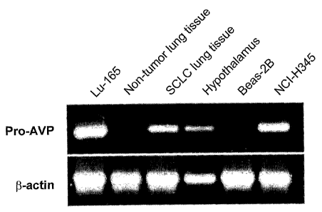
Figure 1 illustrates detection of NRSA in cultured SCLC cells and human SCLC
tissue by RT-PCR analysis. RT-PCR was performed on total RNA extracts from the
indicated cell lines or human tissue. Products were separated on a 1.5%
agarose gel
and visualized with ethidium bromide. The PCR primers were designed to amplify
the entire coding region for the pro-VP protein, which spans 2 introns. The
predicted size for the amplification is 570 bp. Lu-165 and NCI-H345; SCLC cell
lines, Beas-2B; transformed normal human epithelial cell line, lung and
hypothalamus; human tissue extracts were tested.
Figure 2 shows detection of NRSA in cultured SCLC cells and human SCLC tissue
by Western analysis. Figure 2A illustrates staining with MAG-1 mAb or MAG-I
Fab. Figure 2B illustrates staining with MAG-1 mAb or a rabbit polyclonal
antibody against VP-NP. Approximate molecular mass is indicated on the left of
each figure. Total cellular or tissue protein extracts (40 p,g) were separated
by SDS-
_7_
CA 02492475 2005-O1-13
WO 2004/006860 PCT/US2003/022264
PAGE, blotted onto polyvinylidene difluoride (PDVF) membrane, and reacted with
(A) MAG-1 mAb or MA.G-1 Fab, and with (B) MAG-1 mAb or a rabbit polyclonal
antibody against VP-NP.
S Figure 3 shows flow cytometry analysis of MAG-I binding to the surface of
cultured SCLC cells. Figure 3A illustrates MA.G-1 Fab staining of NCI-H82
cells at
two different concentrations compared to a control IgGl antibody. Figure 3B
illustrates MAG-1 mAb staining of NCI-H345 cells compared to a control IgGl
antibody. Figure 3C illustrates MAG-1 mAb staining of NCI-H82 cells at two
different concentrations compared to a control IgGl antibody. Figure 3D
illustrates
MAG-1 mAb staining of Lul6S cells compared to a control IgGI antibody.
Antibody staining procedures prior to flow cytometty were performed using
conditions that minimize plasma membrane internalization. MAG-1 mAb and
MAG-1 Fab were employed at a concentration of 100 ~,ghnl except where noted.
1S
Figure 4 shows confocal analysis of 1VIAG-I binding to the surface of cultured
SCLC cells. SCLC cells were reacted with MAG-1 or an isotype control mAb,
followed by FITC-labeled secondary antibodies. Figure 4A illustrates combined
differential interference contrast (DIC) transmitted, red fluorescent, and
green
fluorescent light channel images of NCI-H82 cells viewed with a 40x objective
(NA
1.4) and a S.8x magnification zoom setting. The cells were incubated with
propidium iodide to stain the nuclei (gray) for contrast. Figure 4B NCI-H34S
illustrates imaged by confocal microscopy and the top panels display the
control
IgGl observed with a 20x objective (O.S NA) and the lower panels display the
2S MAG-1 stained cells observed with a 40x objective (1.3 NA). Figure 4C
illustrates
Lu-I65 cells imaged by confocal microscopy with a 40x objective. For Figures
4B
and 4C, the left panels depict the transmitted light channel images, the
middle panes
depict the green fluorescent light channel images, and the right panels depict
the two
combined images.
Figure S illustrates immunohistochemical analysis of human tissue sections
using
MAG-1 mAb. Figure SA depicts MAG-1 immunoreactivity with human SCLC
_g_
CA 02492475 2005-O1-13
WO 2004/006860 PCT/US2003/022264
tumor cells (brown staining). Figure SB depicts control normal epithelial
cells of the
alveoli of the lung. Figure SC depicts control normal epithelial cells of the
bronchioles of the lung.
Figure 6 illustrates the amino acid sequence and nucleic acid sequence of a
single
chain variable fragment immunoreactive with the C-terminal 18-amino acid
residues
of VAG. Figure 6A depicts the amino acid sequence (SEQ ID NO: 2). Figure 6B
depicts the nucleic acid sequence (SEQ ID NO: 3).
Figure 7 illustrates staining of human ductal carcinoma in situ (DCIS) tumor
tissue
sections examined by immunohistochemical analysis using MAG-1.
Figure 8 illustrates lack of staining of human aplastic ductal hyperplasia
tumor tissue
sections examined by immunohistochemical analysis using MAG-1.
DETAILED DISCLOSURE OF THE INVENTION
I. Overview
The pressing need for effective screening and non-toxic treatment methods
has spawned a search for new approaches to combat breast cancer and small cell
lung cancer (SCLC), taking advantage of the numerous molecular and genetic
abnormalities that have been described for breast cancer and SCLC (Junlcer et
al.
(2000) J: Cazzcez~ Res. Clizz. O>zcol. 126: 361-368; Wistuba et al. (2001)
Seyzziu.
O>zcol. 28: 3-13 2001; Zangemeister-Wittlce and Stahel (1999) Cell. Mol. Life
Sci.
55: 1585-1598; and Popescu NC and Zimonjic DB (Oct. 30, 2002) Am. J. Med.
Genet. 115(3): I42-I49). The prospect of antibodies directed against cell-
surface
tumor-specific antigens is attractive not only for use in the differential
diagnosis of
SCLC, but also for use in localizing and eradicating tumors since they have
the
potential for eliciting minimal side effects (Weiner, L.M. (1999) Semin.
Ozzcol. 26:
41-50). This strategy is most effective when directed against tumor-specific
antigens that are not lost or modulated, and products of the vasopressin gene
may
provide for such an antigen.
-9-
CA 02492475 2005-O1-13
WO 2004/006860 PCT/US2003/022264
The present invention discloses the detection of NRSA in cultured SCLC
cells and human SCLC tumor tissue using a mAb designated MAG-1, which was
generated using a synthetic peptide representing the C-terminal portion of the
VAG
region of the pro-VP protein. MAG-1 recognizes the ~20 lcDa and ~40 lcDa NRSA
proteins in cultured SCLC cell lysate by Western analysis, while
immunofluorescent
cytometl~ic and microscopic analyses indicates that it binds to the surface of
these
cells. More importantly, the ~20 kDa and ~40 kDa NRSA proteins were detected
in
the lysate of human SCLC tumor biopsy samples by Western analysis using MAG-
1, but they were riot detected in the lysate of non-tumor human lung tissue.
Immunohistochemical analysis revealed that MAG-I reacts with human SCLG
tumor, but not with normal lung tissue. Since NRSA is not typically found on
the
surface of normal cells, it is anticipated that it can serve as an excellent
target in a
MAG-I-based approach for tumor localization in the diagnosis and therapy of
SCLC.
1 S Currently, screening for breast cancer requires direct
immunohistochemistry
and a battery of antibodies directed against several tumor markers which
individually occur in less than 50% incidence in these tumors.
It is currently very difficult for pathologists to distinguish invasive breast
cancer and ductal carcinoma irc situ from atypical ductal hyperplasia, and a
method
for achieving this is urgently needed because it can save women undergoing
unnecessary surgery. A diagnosis of the former will generally result in
surgery, a
diagnosis of the latter, in no intervention.
One embodiment of the present invention encompasses a monoclonal
antibody (MAG-1) directed to the C-terminal region of the glycopeptide
component
of the provasopressin protein (VAG), and that the two major immunoreactive
forms
of NRSA are detected using MAG-I in protein extracts from cultured SCLC cells
as
well as in protein extract from human SCLC tumor. However, these proteins were
not detected in protein extract from non-tumor human lung tissue by Western
analysis using MAG-1. Some cancers, including SCLC, are lenown to express the
vasopressin gene, although it appears that not all of the precursor protein is
enzymatically processed and secreted into the circulation as in the
hypothalamus.
Polyclonal anti-VP-NP antibodies have been shown to recognize proteins ~20
lcDa
-10-
CA 02492475 2005-O1-13
WO 2004/006860 PCT/US2003/022264
and ~40 kDa in SCLC cell extracts (North et al., (1993) Peptides 14: 303-307).
The
predicted size of the glycosylated pro-VP protein product of the normal VP
message
is ~20 lcDa, and this has been demonstrated using a cell-free translation
assay
(Giudice et al. (1979) J. Biol. Chem. 254: 11767-11770; Lin et al. (1979)
Biocheyra.
Biophys. Res. Commute. 89: 943-950; Sclnnale et al. (1979) FEBS Lett. 108: 311-
316). Previous studies identified an extended VP message which was though
might
account for the ~40 lcDa protein (North et al., (1993) Peptides 14: 303-307;
Rosenbaum et al. (1990) PNAS 87: 9928-9932). However, only one form of the
predicted size for the normal VP message was detected by RT-PCR in the SCLC
cell
lines and tumor tissue used in this study, and this corresponded in size to
that
detected in human hypothalamus.
Polyclonal antibody preparations were shown to specifically stain human
SCLC and hypothalamus tissue sections, as well as bind to the plasma membrane
of
cultured SCLC cells in a similar manner to what was observed using MAG-1
(Friedmann et al. (1994) Br. J. CasZCer 69:260-263; Friedmann et al. (1995)
Neuropeptides 28: 183-189; North and Yu (I993) Regulatory Peptides 45: 209-
216;
Friedmann et al. (1993) Cancer Lett. 75: 79-85; North et al. (1983) Irc: F.
Greco
(ed.), Biology and Mana~yement of Lung Cancer, pp. 143-169. Boston: Martinus
Nijhoff). We have determined that the ~20 l~Da and ~40 lcDa NRSA proteins are
VP gene related products can serve as tumor-specific antigens and can be
targeted
by antibodies (North and Yu (1993) Regulatory Peptides 45: 209-216; North et
al.
(1993) Peptides 14: 303-307; North, W.G., et al. (1995) Breast cancer Research
aad
Treatment 34:229-235; Friedmann et al. (I994) Br. J. Cancer 69:260-263;
Friedmann et al. (1993) Ca~rcef~ Lett. 75: 79-85; North et al. (1989) Nuc.
Med.
Coynmutz.10:643-652).
Although the level of NRSA expression by the different SCLC cell types has
not been measured, we demonstrated that MAG-I can recognize NRSA on the
surface of cultured cell lines derived from SCLC tumors of both the classical
and
variant sub-types. Fluorescent microscopy revealed that MAG-1 staining was
observed with each SCLC cell type examined with differing levels of
immunoreactivity.
-11-
CA 02492475 2005-O1-13
WO 2004/006860 PCT/US2003/022264
Additionally, we demonstrated the specificity of 1VIAG-I for SCLC tumor by
immunohistochemical analysis. MAG-1 reacted with human SCLC tumor, but not
with normal lung tissue. The staining appeared to be localized to the surface,
as well
as the cytoplasm on the SCLC cells in the section of tumor. MAG-1 also reacted
with human hypothalamus tissue, the staining appeared to be localized to the
cytoplasm. These results indicate that MAG-1 can be used to effectively target
NRSA on SCLC tumors. MAG-1 did not react with the normal lung epithelial cells
in the tissue used in the immunohistochemical screening, however staining was
observed in pulmonary neuroepithelial bodies. Although not obvious in the
figure,
VP message was detected in the non-tumor lung sample used in the RT-PCR
reaction (Figure 1).
The expression of the VP message could represent the presence of
pulmonary neuroendocrine cells in the lung tissue sample used (Reynolds et al.
(2000) Am. J. Physiol. Lung Cell. Mol. Physiol. 278: L1256-L1263), but the
I S prohormone may be enzymatically processed and the products secreted as
occurs in
the hypothalamus. This theory is supported by the immunohistochemical findings
depicted in Figure 5.
The potential uses for the MAG-1 mAb are significant, not only as a tumor
targeting agent for the localization and treatment of SCLC, but also fox
distinguishing SCLC from other forms of lung cancer, and aiding its early
diagnosis
(Friedmann et al. (1994) B~. J. Cancer 69:260-263; Friedmann et al. (1993)
Cancer
Lett. 75: 79-85; North et al. (1989) Nuc. Med. Commun. 10: 643-652). The
ability
of the MAG-1 Fab fragment to recognize synthetic antigen, as well as NRSA in
protein extracts from SCLC tumor and cultured cells was also evaluated since
antibody fragments may be better suited for some in vivo applications. While
the
Fab was able to recognize NRSA by Western analysis, not surprisingly it
displayed a
lower binding affinity for synthetic antigen. However, the localization of
antibody
molecules to tumor tissue, and their ability to penetrate solid tumor depends
on a
number of factors including size, affinity, rate of clearance, and antigen
density
(Adams et al. (2001) Cancer Res. 61: 4750-4755; Todorovska et al. (2001) J.
Imrraunol. Methods 248: 47-66).
-12-
CA 02492475 2005-O1-13
WO 2004/006860 PCT/US2003/022264
Variable region fi~agments (Fv) of this antibody have been produced to assess
their potential for ira vivo tumor targeting because they may provide
additional
benefits for use in unaging and therapy (Kortt et al. (2001) Bio~zol. Ehg. 18:
95-
201).
NRSA is not typically found on the surface of normal cells, it is not
modulated between classical and variant SCLC, and there is a low incident of
its
expression by non-neuroendocrine lung carcinomas (Friedmann et al. (1993)
Cahce~~
Letters 75: 79-~5). Therefore, NRSA should serve as an excellent target for
the
localization of SCLC tumors in diagnosis and therapy, employing MAG-1 mAb and
its fragments.
This invention pertains to the discovery of the provasopressin precursor
protein as a novel tumor specifc marker that can be targeted with antibodies,
antibody fragments, their derivatives (such as monospecific or bispecific scFv
fragments or Fd fragments), or binding peptides. To date we have screened 70
small
cell lung cancer (SCLC), 62 breast cancer, and 55 ductal carcinoma in situ
(DCIS)
human tissue sections with various antibodies to provasopressin, and they all
have
displayed positive staining. Normal lung and breast tissue sections, including
ADH
sections, do not display staining with these antibodies. The discovery that
the
provasopressin precursor protein can be used as a specific tumor antigen
constitutes
a novel finding. The concept to develop of antibodies, antibody fragments,
their
derivatives, or binding peptides for use in targeting cancers that express the
provasopressin precursor protein, for the purpose of research, early
detection,
diagnosis, therapy, and prevention represent direct applications of that
finding.
Under normal physiological conditions, the vasopressin gene is expressed for
the most part by hypothalamic neurons, where the resultant provasopressin
precursor
protein is enzymatically cleaved into its peptide hormone components, which
include the neuropeptide vasopressin (VP), vasopressin-associated neurophysin
(VP-
NP), and vasopressin-associated glycopeptide (VAG). Certain cancers, including
small-cell lung cancer (SCLC), and breast cancer, also express the vasopressin
gene,
and vasopressin production by tumors has been described in the literature
beginning
around the early 1970s, where elevated levels of vasopressin were detected in
the
blood of patients with SCLC. However, North et al. first described the
possibility
-13-
CA 02492475 2005-O1-13
WO 2004/006860 PCT/US2003/022264
that the provasopressin precursor protein could localize to the surface of
cancer
cells, and serve as a tumor antigen, in 1983. The terms Neurophysin- and
Glycopeptide-Related Cell-Surface antigen (NRSA and GRSA) were applied to the
provasopressin precursor protein as it presents itself as a useful antigen for
tumor
S targeting. The difference in the terminology stemmed from the use of
antibodies
directed against two different regions of the provasopressin protein.
Antibodies can be used for targeting provasopressin (NRSA/GRSA) on
tumors. Previous work indicates that SCLC tumors can be localized and imaged
in
humans using radiolabeled antibody directed against the neurophysin portion of
provasopressin. Subsequent studies show that polyclonal antibodies, monoclonal
antibodies, and antibody Fab fragments directed against different regions of
the
provasopressin protein bind specifically to cultured SCLC and breast cancer
cells, as
well as to human tumor sections, but not to tissue that is devoid of tumor. We
have
developed polyclonal and monoclonal antibodies, and their Fab fragment
derivatives, to NRSA/GRSA and have demonstrated that they can bind to cultured
human cancer cells and human cancer tissue. Since the NRSA/GRSA is not
typically found in normal cells, it is anticipated that it can serve as an
excellent
target for tumor localization in the early detection, diagnosis, and treatment
of
cancers that express the vasopressin gene. NRSA/GRSA also provides for a
attractive candidate for use in vaccine development strategies for the
prevention of
those cancers that express the vasopressin gene.
Single-chain antibodies fragments and small binding peptides can be used for
targeting provasopressin (NRSA/GRSA) on tumors. We have produced single-chain
variable region fragments (scFv) of an antibody, as well as peptides, that
bind to
NRSAIGRSA. The use of such smaller molecules will provide added benefits
(tumor penetration, ease of manufacturing) for ih vivo tumor targeting.
Although the
expression of vasopressin by various tumoxs has been lmown for some time,
targeting the precursor protein with antibodies and antibody fragments is a
novel
concept.
Mechanisms of vasopressin gene expression can be targeted for tumor
therapy. Additionally, vasopressin is involved in autocrine regulation of
tumor
-14-
CA 02492475 2005-O1-13
WO 2004/006860 PCT/US2003/022264
survival, and preliminary results demonstrate that the vasopressin mRNA
message is
a viable target for antisense-based methods for the inhibition of tumor
growth.
The term "Immuno-based" refers to the use of antibodies, antibody
fragments, their derivatives, or binding peptides.
S 1.) Early Detection
a.) Measurement blood levels of provasopressin components for indication of
certain tumors. Antibodies directed against various portions of the
provasopressin
precursor protein would be useful in the clinical screening assay to measure
their
levels in the blood of patients suspected of having certain tumors, or who
have had
those tumors in the past. This would be a useful, non-invasive or less
invasive test
to possibly justify further, more invasive tests/biopsies, and aid in
monitoring
recurrence of disease.
b.) Immuno-based imaging. With the use of antibodies directed against
various portions NRSA/GRSA, current imaging techniques, such as mammography,
could be greatly enhanced, and new imaging protocols for diseases such as
SCLC/breast cancer could be developed and effectively implemented for clinical
use. These types of techniques would be especially useful for the detection of
metastatic disease.
2.) Diagnosis
a) hnmuno-based pathological screening of biopsies. Currently, SCLC is
diagnosed on the basis of gross morphological and histological data obtained
from
biopsied tissue, and is far too often identified after the disease has reached
its
advanced stages. Additionally, DCIS is often difficult to discern from
atypical
ductal hyperplasia (ADH), generally considered to be a benign affliction, on
biopsied tissue sections. These biopsied tissue samples can be stained using
antibodies directed against various portions NRSA/GRSA, allowing for critical
differential diagnoses to be made, which can then effect subsequent treatment
procedures and outcomes.
b.) In situ immuno-based imaging. Similar to that outlined in l.a. above, the
use of antibodies directed against various portions NRSA/GRSA could be used in
combination with mammographic imaging techniques to allow for non-invasive or
less invasive diagnoses of breast disease versus hyperplastic conditions.
-15-
CA 02492475 2005-O1-13
WO 2004/006860 PCT/US2003/022264
Compositions of the present invention can be used for immuno-based
targeting of tumors and delivery of chemotoxic/radiologic agents. As mentioned
above, SCLC tumors can be localized and imaged using an antibody to the
neurophysin region of the provasopressin protein. Thus, antibodies, antibody
fragments, their derivatives, or binding peptides could be radiolabeled,
conjugated to
or used in conjunction with chemotoxic agents, or serve as an attractor for
endogenous immune system cells to kill NRSA/GRSA-expressing tumors. Since all
SCLC, breast cancer, and DCIS cells appear to express NRSA/GRSA, treatments
that target this antigen would provide for significantly more potent therapy
than
currently available strategies for these diseases.
Targeting the inhibition of vasopressin gene transcription and/or vasopressin
mRNA message translation to prevent tumor growth. Since vasopressin provides
for
autocrine growth stimulation in cancer cells that express the vasopressin
gene,
inhibition of it production would inhibit tumor survival. By using antisense
molecules to block gene transcription translation, a powerful, non-invasive
tool for
therapy could be developed.
Cancer vaccines are based on tumor antigens, such as NRSA and GRSA.
Because of its unique expression in certain cancers, vaccine strategies based
on
NRSAIGRSA, such as anti antibodies or utilizing antigenic motifs on the
NRSA/GRSA structure, could be developed that would enable the initial
prevention
and/or recurrence of these diseases.
Use of monoclonal antibodies against the region in provasopressin bridging
vasopressin and neurophysin moieties (referred to here as "MAP"s), and
modified
forms of these MAPS, or monoclonal antibodies against vasopressin (referred to
here as "MAV"s) and modified forms of these MAVs, or monoclonal antibodies
against vasopressin-associated glycopeptide (referred to here as "MAG"s) and
modified forms of MAGs, or monoclonal antibodies against tumor-specific
regions
of GRSA proteins (referred to here as "MAT"s) and modified forms of these
MATS,
for: a) screening fresh and fixed biopsied material for the presence of breast
cancer;
b) non-invasive diagnostic imaging of breast cancer in patients and; c)
targeting
therapy of breast cancer in patients.
- 16-
CA 02492475 2005-O1-13
WO 2004/006860 PCT/US2003/022264
MAPS are generated against an undecapeptide, PRGGKRAMSDL (SEQ ID
NO: 1) antigen but primarily recognize the tripeptide bridge structure GKR,
and the
amino acid residues that skirt this structure. MAGs are generated against an
18-
residue polypeptide representing the C-terminal half of the vasopressin-
associated
glycopeptide (hereinafter "VAG"). MAVs, MAPS, MAGs, and MATS, are
designed to recognize what we propose to be the universal provasopressin-
related
cell-surface antigens) on breast cancer called GRSA.
In accordance with the present invention, monoclonal antibodies against the
xegion of provasopressin bridging vasopressin and neurophysin moieties (MAPS),
and modified forms of these MAPS, or monoclonal antibodies against vasopressin
(MAVs), and modified forms of these MAVs, or monoclonal antibodies against
vasopressin-associated glycopeptide (MAGs), and modified forms of these MAGs,
or monoclonal antibodies against tumor-specific regions of GRSA proteins
(MATS),
and modified forms of these MATS, are employed for the screening, ih vivo
diagnosis, and treatment of breast cancer. From immunohistochemical studies
with
polyclonal antibodies against vasopressin and polyclonal antibodies against
human
vasopressin-associated glycopeptide (also called human copeptin), the inventor
and,
colleagues determined there was a possibility that all breast cancers are
reactive
with these polyclonal antibodies.
Monoclonal antibodies have been developed not only against vasopressin
(MAVs), but also against human VAG, and the bridging structure of human
provasopressin. MAGs and MAVs provide positive immunostaining for all breast
cancers making them ideal in a single-regimen screening test for breast cancer
in
fresh and fixed biopsied tissues. Modeling studies show that fox GRSA proteins
in
the plasma membrane of breast cancer cells, not only can the vasopressin
moiety be
exposed to the outside of the cell but also the glycopeptide region against
which
MAGs are generated and the provasopressin bridging structure against which
MAPS
are generated can also be exposed. Thus MAGs, MAPS, MAVs, and MATS also can
identify and bind to GRSA proteins on viable cells in culture. They can
therefore
bind to viable tumor cells in patients, and be used to locate tumors
(particularly of
the metastatic and/or recurrent disease) in patients and be effectively
adapted for
targeted immunotherapy. Modification of MAGs, or MAPS or MAVs or MATs,
-17-
CA 02492475 2005-O1-13
WO 2004/006860 PCT/US2003/022264
required to make them effective in vivo tools include conversion to Fab and
F(ab')2
forms. Diagnostic localization of tumors in patients include (though not
exclusively) use of 99Technetium-, 131lodine-, 111lndium-, and/or ferric-
containing-
labeled forms of MAGs, MAPS, MAVs, or MATS.
The invention provides a much needed rapid, inexpensive, sensitive, and
specific method for: 1) early detection of breast cancer; and 2) identifying
and
localizing breast cancer, particularly metastatic and/or recurrent disease, in
patients.
It also provides valuable tools for developing new immuno-targeted treatments,
applicable to all patients with breast cancer, that are effective with both
primary
disease, and with recurrent drug-resistant disease. In this respect it should
be useful
to all hospitals and physicians examining and treating patients with breast
cancer.
Detection kits are simple enough to be set up in any local hospital
laboratory, and
MAGs, MAVs, MAPs and/or MATs and their modified forms can readily be made
available to all hospitals treating patients with breast cancer.
Antibodies against an abnormal form of the vasopressin V2 receptor may be
used for (a) distinguishing DCIS from ADH, and for; (b) detection and
targeting of
breast cancer.
We have discovered that breast cancer cells make a tumor-specific abnormal
form of vasopressin V2 receptor and predict antibodies directed against this
abnormal protein can be used in the early immunohistochemical detection, not
only
of breast cancer, but also of DCIS. They are also predicted to be valuable in
the
diagnosis of breast cancer in patients, particularly for detecting and
localizing
metastatic disease, and in the targeted treatment of breast cancer.
Two forms of vasopressin V2 receptor mRNAs were found by the inventors
in breast cancer cells. One of these has a sequence identical to the receptor
of
normal human tissues; the other is an enlarged form and found to contain the
entire
106 bases of intron 2 in addition to the sequence for V2 receptor mRNA. Breast
cancer cells produce both these forms of vasopressin V2 receptors. Inclusion
of
intron 2 introduces a stop colon into the reading frame and therefore the
abnormal
vasopressin V2 receptor mRNA gives rise to a C-terminally truncated protein
lacking the seventh transmembrane segment and carboxyl tail of the normal
receptor. We have demonstrated this, and the other vasopressin receptor mRNAs
-18-
CA 02492475 2005-O1-13
WO 2004/006860 PCT/US2003/022264
are translated into proteins by the cancer cells through Western analysis
using
specific antibodies. The antibodies to the abnormal protein are directed
against its
unique carboxyl terminal region.
The same abnormal receptor was found in SCLC using cell lines NCI-H82,
NCI-H146, NCI-H345, and DMS-57 (North et al. (1998) Cancer Research 58:
1866-1871 and North et al. Vasopressin and Oxytocin. (eds) Zingg et al. Plenum
Press, pp. 335-338, 1998). Structures were determined by RT-PCR, cloning, and
sequencing. Protein products were verified by Western analysis with specific
antibodies.
Breast cancer expresses the vasopressin gene and suspected vasopressin
receptor expression always accompanies the vasopressin gene cancer cells. The
vasopressin gene is expressed by DCIS but not by various fibrocystic breast
conditions, including ADH. Since we expect the abnormal vasopressin V2
receptor
will, like breast cancer, be expressed by DCIS, we predict antibodies against
this
abnormal receptor can be used in methods such as immunohistochemistry to
successfully evaluate biopsies and distinguish DCIS and breast cancer from
ADH.
Since metastatic as well as localized breast cancer seems to express the
heretofore unknown abnormal vasopressin V2 receptor, suitably labeled or
modified
forms of monoclonal or polyclonal antibodies against this receptor, through
injection
and different means of detection, are useful in providing a needed very
sensitive and
specific means of detecting and localizing metastatic and/or early recurrent
disease
in patients. These antibodies, and their modified form, it is here claimed,
are
important new tools for effectively targeting different treatments to tumors
in
patients. This is potentially especially valuable in treating recurrent and
generally
estrogen-resistant forms of breast cancer.
The disclosed invention can be used for:
1) providing a much needed rapid, inexpensive, sensitive, and specific
method for distinguishing DCIS and breast cancer from hyperplastic conditions
such
as ADH. In this respect it should be useful to all hospitals and physicians
examining
patients for breast cancer, and of benefit to all women undergoing such
diagnosis;
2) providing a much needed sensitive and specific method for non-invasively
detecting and localizing recurrent disease and metastatic disease in all
breast cancer
-19-
CA 02492475 2005-O1-13
WO 2004/006860 PCT/US2003/022264
patients. With refinements, this method will be particularly useful in
performing
regular screening of all patients who have recovered from breast cancer to
detect for
any return of the cancer; and
3) providing an effective means for targeting new and effective treatments
for all breast cancers including those that are estrogen-sensitive. Modified
antibodies can be used either as an additive to treatments with anti-estrogens
and
chemotherapeutics, or as an effective alternative. Modified forms of the
antibodies
such as forms carrying a destructive radiochemical or chemical poison, or
complexing this immune cells, should be potentially effective in treating alI
tumors.
Also encompassed by the present invention are methods for distinguishing
DCIS from ADH using antibodies against VP or against VP-associated
glycopeptide
(VAG) or Copeptin; and by performing RT-PCR for VP mRNA on RNA from
biopsied material and fixed material from stored tissue bloclcs.
It is highly likely that all breast cancers express the VP gene. Even though
the vasopressin gene is expressed by all examined DCIS, it is generally
regarded to
be a pre-cursor of invasive breast cancer. The gene is not expressed by
various
fibrocystic breast conditions, including ADH. The expression of the VP gene is
believed by the inventor to be part of the process of oncogenic transformation
of
cells in the breasts. Vasopressin gene expression produces vasopressin and
vasopressin gene-related proteins of approximately 20,000 and 40,000 daltons.
Antibodies against VP and VP -associated glycopeptide react with these
vasopressin
gene products in methods such as immunohistochemistry, and this positive
reaction
(staining) can be used in a new method to successfully and automatically
evaluate
biopsies and distinguish DCIS and breast cancer from the benign ADH.
Monoclonal
antibodies to vasopressin (MAVs) and to vasopressin-related glycopeptide
(MAGs)
have been developed for this purpose. Alternatively, a method of RT-PCR for VP
mRNA, representing the expressed gene, can be utilized, with both fresh and
fixed
tissue, to identify DCIS.
The vasopressin gene is largely expressed in hypothalamic neurons, where
the resultant provasopressin protein is enzymatically cleaved into its peptide
hormone components, which include the neuropeptide vasopressin (VP),
vasopressin-associated neurophysin (VP-NP), and vasopressin-associated
- 20 -
CA 02492475 2005-O1-13
WO 2004/006860 PCT/US2003/022264
glycopeptide (VAG). Small cell lung cancer (SCLC) tumors also express the
vasopressin gene, but the tumor provasopressin protein can remain intact and
localize to the cell surface membrane.
The present invention discloses a monoclonal antibody (mAb), designated
MAG-l, was raised using a synthetic peptide representing the C-terminal
sequence
of the VAG domain of human provasopressin. The MAG-1 mAb recognizes NRSA
in SCLC cell and tissue lysates by Western analysis, while immunofluorescent
cytometric and microscopic analyses indicate that MA.G-1 reacts specifically
with
NRSA on the surface of viable SCLC cells of both the classical and the variant
subtype. hnmunohistochemical analysis was performed to demonstrate that MAG-1
reacts with human SCLC tumor, but not with normal lung tissue. Additionally, a
MAG-I Fab fragment was generated which was also able to recognize NRSA. This
is the disclosure demonstrating that a monoclonal antibody directed to the VAG
region of the provasopressin protein has the potential fox development into an
ih vivo
diagnostic and therapeutic tool that targets plasma membrane-incorporated
NRSA.
A further discovery of the present invention is the ability to distinguish
between fibrocystic lesions and cancerous lesions. We have found that if a
test
samples is positive for provasopressin and negative for the angiotensin II
type-2
receptor, a patient is susceptible to invasive breast cancer. If the test
sample is
positive for both provasopressin and the angiotensin II type-2 receptor, a
patient is
susceptible to ductal carcinoma in. situ. If a test sample is negative for
provasopressin and the angiotensin II type-2 receptor, the patient Iilcely has
a
f brocystic lesion, such as hyperplastic tissue. Thus, we have discovered a
powerful
tool whereby health care providers can conclusively distinguish non-invasive
fibrocystic tissue from cancerous lesions in test samples from patients
suspected of
having cancer.
An additional discover of the present invention is that cocl~tails of
chemotherapeutic agents can be administered to a patient in need thereof in
combination therapy with pharmaceutical compositions of an antibody, antigen
binding fragment, or peptide immunoreactive with provasopressin, whereby the
combination therapy is effective at inhibiting proliferation of tumor cells.
-21 -
CA 02492475 2005-O1-13
WO 2004/006860 PCT/US2003/022264
II. Definitions
As used herein the term "species" or "animal" refers to mammals, preferably
mammals such as humans. Likewise, a "patient" or "subject" to be treated by
the
method of the invention can mean either a human or non-human animal.
As used herein, "immunoreactive" refers to binding agents, antibodies or
fragments thereof that are specific to a tumor target cell antigen, yet if are
cross-
reactive to other proteins, are not toxic at the levels at which they are
formulated for
administration to human use. "Specifically binds" means that the binding agent
binds to the antigen on the target cell with greater affinity than it binds
unrelated
antigens. Preferably such affinity is at least 10-fold greater, more
preferably at least
100-fold greater, and most preferably at least 1000-fold greater than the
affinity of
the binding agent for unrelated antigens. The terms "immunoreactive" and
"specifically binds" axe used interchangeably herein.
The terms "protein", "polypeptide" and "peptide" are used interchangeably
herein when referring to a gene product, e.g., as may be encoded by a coding
sequence. Peptides of the present invention broadly to refer to portions of
the
provasopressin amino acid sequence. Preferably, the peptides are portions of
the
VAG domain of provasopressin. More preferably, the peptides are between 5-50
amino acid residues in length, 5-30 amino acid residues in length, 5-20 amino
acid
residues in length" or 10-15 amino acid residues in length. The peptides of
the
present invention are immunoreactive with provasopressin, or portions thereof.
The term "antibody" as used herein, unless indicated otherwise, is used
broadly to refer to both antibody molecules and a variety of antibody-derived
molecules. Such antibody derived molecules comprise at least one variable
region
(either a heavy chain of or a light chain variable region), as well as
individual
antibody light chains, individual antibody heavy chains, chimeric fusions
between
antibody chains and other molecules, and the like. Functional immunoglobulin
fragments according to the present invention may be Fv, scFv, disulfide-linked
Fv,
Fab, and F(ab')2. Antibodies, or fragments thereof, of the present invention,
can be
used to image target cells when labeled with a detectable label.
Also encompassed by the term "antibody" are polyclonal antibodies ("pAb"),
monoclonal antibodies ("MAb" or "mAb"), preferably IgGI antibodies; chimeric
-22-
CA 02492475 2005-O1-13
WO 2004/006860 PCT/US2003/022264
monoclonal antibodies ("C-MAb"); humanized antibodies; genetically engineered
monoclonal antibodies ("G-MAb").
Preferably, the antibody is the monoclonal antibody, MAG-1, which
recognizes the 18 C-terminal amino acid residues of the VAG domain of
S provasopressin. Humanizing antibodies is a technique well-known in the art
wherein portions of the framework regions are modified such that they do not
cause
adverse reactions when administered to a human patient. One of ordinary skill
in the
art would readily recognize portions of MAG-1 to be mutated such that the
antibody
was humanized.
The antibodies and peptides of the present invention may be labeled. As
used herein, "label" is used to mean a detectable label which is used to
visualize the
binding of an antibody to its target protein or receptor. Alternatively,
antibodies and
peptides of the present invention may be labeled with a radiolabel, an iron-
related
compound, or a toxin which would kill the cell to which it binds. Radiolabels
and
1S toxins are well known in the al-t and include, for example, 32P, 33P~ 43R~
a7Sca s2Fe,
57CC' 64Cu' 67Ga' 67Cu' 68Ga' 7lGe' 75Br' 76Br' 77Bt,' 77As' 77Br'
8lRb/8lIvl~r' s7Msr~
90Y' 97Ru' 99TC' IOOPd' lol~' 103Pb' lose 109Pd' 111Ag' IIlIn' 113In' 119Sb
121Sn' 123h
125f 1275' 128Ba' 129CS~ 131f 131Cs' 143Pr' 153sm' 161Tb, 166H~' 169Eu' 177Lu'
186Re,
188Re' ls9Re~ 191 OS' 193Pt~ 194Ir~ 197Hg~ 199Au' 203Pb? 211At' 212Pb' 212Bi
and 213B1, rICIn,
ricin A chain (ricin toxin), Pseudomonas exotoxin (PE), diphtheria toxin (DT),
Clostridium perfi~ingehs phospholipase C (PLC), bovine pancreatic ribonuclease
(BPR), pokeweed antiviral protein (PAP), abrin, abrin A chain (abrin toxin),
cobra
venom factor (CVF), gelonin (GEL), saporin (SAP), modeccin, viscumin and
vollcensin. Iron-related compounds include, for example, Fe203 and Fe304.
"Tumor cell specific antibody" and "tumor cell specific peptide" are defined
herein as the ability of an antibody or peptide to specifically bind to the
target cell
antigen. As used herein, the specificity of the antibody or peptide for a
tumor cell
antigen can be measured wherein the affinity of the antibody/peptide to the
tumor
cell antigen is greater then to other cells not associated with the tumor.
Antigen
binding fragments and peptidomimetics having the same function of specifically
binding to a target cell antigen are also contemplated by the present
invention.
-23-
CA 02492475 2005-O1-13
WO 2004/006860 PCT/US2003/022264
"Immunoassay" of the present invention include any assay that can be used
to determine the presence of a target cell antigen. Non-limiting examples of
immunoassays known to one of ordinary skill in the art include
immunohistochemistry, ELISAs, MRI, and Western Blots.
"Test samples" of the present invention can obtained from patients in an
invasive or non-invasive way. Non-invasive test samples include, for example,
urine. Invasive test samples include, for example, blood or blood products,
lymph
tissue or fluid, breast fluid or tissue. Wherein one term is used in the
present
invention, the other terms are meant to be interchangeable.
"Administering" is defined herein as a means providing the composition to
the patient in a manner that results in the composition being inside the
patient's
body. Such an administration can be by any route including, without
limitation,
subcutaneous, intradermal, intravenous, intra-arterial, intraperitoneal, and
intr amuscular.
A physician or veterinarian having ordinary skill in the art can readily
determine and prescribe the "effective amount" (EDSO) of the pharmaceutical
composition required. For example, the physician or veterinarian could start
doses of
the compounds of the invention employed in the pharmaceutical composition at
levels lower than that required in order to achieve the desired therapeutic
effect and
gradually increase the dosage until the desired effect is achieved.
Each of the embodiments of the present invention can be used as a
composition when combined with a pharmaceutically acceptable carrier or
excipient.
"Carrier" and "excipient" are used interchangeably herein.
The phrase "pharmaceutically acceptable" is employed herein to refer to
those compounds, materials, compositions, and/or dosage forms which are,
within
the scope of sound medical judgment, suitable for use in contact with the
tissues of
human beings and animals without excessive toxicity, irritation, allergic
response, or
other problem or complication, commensurate with a reasonable benefit/rislc
ratio.
"Pharmaceutically acceptable carrier" is defined herein as a carrier that is
physiologically acceptable to the administered patient and that retains the
therapeutic properties of the antibodies. Pharmaceutically-acceptable carriers
and
their formulations are well-lmown and generally described in, for example,
-24-
CA 02492475 2005-O1-13
WO 2004/006860 PCT/US2003/022264
Reminuton's pharmaceutical Sciences (18th Edition, ed. A. Gennaro, Maclc
Publishing Co., Easton, PA, 1990). On exemplary pharmaceutically acceptable
carrier is physiological saline. The phrase "pharmaceutically acceptable
carrier" as
used herein means a pharmaceutically acceptable material, composition or
vehicle,
such as a liquid or solid filler, diluent, excipient, solvent or encapsulating
material,
involved in carrying or transporting the subject antibodies from the
administration
site of one organ, or portion of the body, to another organ, or portion of the
body.
Each carrier must be "acceptable" in the sense of being compatible with the
other
ingredients of the formulation and not injurious to the patient. Nor should a
pharmaceutically acceptable carrier alter the specific activity of the
antibodies.
Some examples of materials which can serve as pharmaceutically acceptable
carriers
include: (1) sugars, such as lactose, glucose and sucrose; (2) starches, such
as corn
starch and potato starch; (3) cellulose, and its derivatives, such as sodium
carboxymethyl cellulose, ethyl cellulose and cellulose acetate; (4) powdered
tragacanth; (5) malt; (6) gelatin; (7) talc; (8) excipients, such as cocoa
butter and
suppository Waxes; (9) oils, such as peanut oil, cottonseed oil, safflower
oil, sesame
oil, olive oil, corn oil and soybean oil; (10) glycols, such as propylene
glycol; (11)
polyols, such as glycerin, sorbitol, mannitol and polyethylene glycol; (12)
esters,
such as ethyl oleate and ethyl Iaurate; (I3) agar; (14) buffering agents, such
as
magnesium hydroxide and aluminum hydroxide; (15) alginic acid; (16) pyrogen-
free
water; (17) isotonic saline; (18) Ringer's solution; (19) ethyl alcohol; (20)
phosphate
buffer solutions; and (21) other non-toxic compatible substances employed in
pharmaceutical formulations.
As used herein, the term "cancer" is used to mean a condition in which a cell
in a patient's body undergoes abnormal, uncontrolled proliferation. Non-
limiting
examples of cancers include breast cancer, small cell lung cancer, and ductal
carcinoma ijz situ.
As used herein, the term "fibrocystic" is used to mean tissue that is non-
cancerous, such as atypical ductal hyperplasia.
As used herein, "transformed cells" refers to cells that have spontaneously
converted to a state of unrestrained growth, i.e., they have acquired the
ability to
grow through an indefinite number of divisions in culture. Transformed cells
may be
- 25 -
CA 02492475 2005-O1-13
WO 2004/006860 PCT/US2003/022264
characterized by such terms as neoplastic, anaplastic and/or hyperplastic,
with
respect to their loss of growth control. For purposes of this invention, the
terms
"transformed phenotype of malignant mammalian cells" and "transformed
phenotype " are intended to encompass, but not be limited to, any of the
following
phenotypic traits associated with cellular transformation of mammalian cells:
immortalization, morphological or growth transformation, and tumorigenicity,
as
detected by prolonged growth in cell culture, growth in semi-solid media, or
tumorigenic growth in immuno-incompetent or syngeneic animals.
By "treating" a patient suffering from cancer it is meant that the patient's
symptoms are partially or totally alleviated, or remain static following
treatment
according to the invention. A patient that has been treated can exhibit a
partial or
total alleviation of symptoms and/or tumor load. The term "treatment" is
intended to
encompass prophylaxis, therapy and cure.
A "therapeutically effective amount" is defined herein an effective amount
of composition for producing some desired therapeutic effect by inducing tumor-
specific killing of tumor cells in a patient and thereby blocking the
biological
consequences of that pathway in the treated cells eliminating the tumor cell
or
preventing it from proliferating, at a reasonable benefit/rislc ratio
applicable to any
medical treatment.
The term "sample" is defined herein as blood, blood product, biopsy tissue,
serum, and any other type of fluid or tissue that can be extracted fiom a
patient.
The terms "apoptosis" or "programmed cell death," refers to the
physiological process by which unwanted or useless cells are eliminated during
development and other normal biological processes. Apoptosis, is a mode of
cell
death that occurs under normal physiological conditions and the cell is an
active
participant in its own demise ("cellular suicide"). It is most often found
during
normal cell turnover and tissue homeostasis, embryogenesis, induction and
maintenance of immune tolerance, development of the nervous system and
endocrine-dependent tissue atrophy. Cells undergoing apoptosis show
characteristic
morphological and biochemical features. These features include chromatin
aggregation, nuclear and cytoplasmic condensation, partition of cytoplasm and
nucleus into membrane bound vesicles (apoptotic bodies), which contain
ribosomes,
-26-
CA 02492475 2005-O1-13
WO 2004/006860 PCT/US2003/022264
morphologically intact mitochondria and nuclear material. Ih vivo, these.
apoptotic
bodies are rapidly recognized and phagocytized by either macrophages,
dendritic
cells or adjacent epithelial cells. Due to this efficient mechanism for the
removal of
apoptotic cells in vivo no inflammatory response is elicited. Ifz vit~~o, the
apoptotic
bodies as well as the remaining cell fragments ultimately swell and finally
lyse. This
terminal phase of i~a vitro cell death has been termed "secondary necrosis."
Apoptosis can be measured by methods known to those skilled in the art lilce
DNA
fragmentation, exposure of Annexin V, activation of caspases, release of
cytochrome
c, etc. A tumor cell that has been induced to die is termed herein as an
"apoptotic
tumor cell".
As used herein, the term "nucleic acid" refers to polynucleotides such as
deoxyribonucleic acid (DNA), and, where appropriate, ribonucleic acid (RNA).
The
term should also be understood to include, as equivalents, derivatives,
variants and
analogs of either RNA or DNA made from nucleotide analogs, and, as applicable
to
the embodiment being described, single (sense or antisense) and double-
stranded
polynucleotides.
Operably linked is intended to mean that the nucleotide sequence is linked to
a regulatory sequence in a manner which allows expression of the nucleotide
sequence. Regulatory sequences are art-recognized and are selected to direct
expression of the subject peptide. Accordingly, the term transcriptional
regulatory
sequence includes promoters, enhancers and other expression control elements.
Such regulatory sequences are described in Goeddel; Gene Exp~essioh
Technology:
Metlaods i~ Ehzyynology 185, Academic Press, San Diego, CA (1990).
The term "gene construct" refers to a vector, plasmid, viral genome or the
like which includes a coding sequence, can transfect cells, preferably
mammalian
cells, and can cause expression of the antibody, antigen binding fragment,
peptide or
peptidomimetic of the cells transfected with the construct.
The "growth state" of a cell refers to the rate of proliferation of the cell
and/or the state of differentiation of the cell. An "altered growth state" is
a growth
state characterized by an abnormal rate of proliferation, e.g., a cell
exhibiting an
increased or decreased rate of proliferation relative to a normal cell.
_27_
CA 02492475 2005-O1-13
WO 2004/006860 PCT/US2003/022264
As used herein, "proliferating" and "proliferation" refer to cells undergoing
mitosis.
The term "amino acid residue" is known in the art. In general the
abbreviations used herein for designating the amino acids and the protective
groups
are based on recommendations of the IUPAC-IUB Commission on Biochemical
Nomenclature (see Biochemistry (1972) 11:1726-1732). In certain embodiments,
the amino acids used in the application of this invention are those naturally
occurring amino acids found in proteins, or the naturally occurring anabolic
or
catabolic products of such amino acids which contain amino and carboxyl
groups.
Particularly suitable amino acid side chains include side chains selected from
those
of the following amino acids: glycine, alanine, valine, cysteine, leucine,
isoleucine,
serine, threonine, methionine, glutamic acid, aspantic acid, glutamine,
asparagine,
lysine, arginine, proline, histidine, phenylalanine, tyrosine, and tryptophan.
The term "amino acid residue" further includes analogs, derivatives and
congeners of any specific amino acid referred to herein, as well as C-terminal
or N-
terminal protected amino acid derivatives (e.g. modified with an N-termi?~al
or C-
terminal protecting group). For example, the present invention contemplates
the use
of amino acid analogs wherein a side chain is lengthened or shortened while
still
providing a carboxyl, amino or other reactive precursor functional group for
cyclization, as well as amino acid analogs having variant side chains with
appropriate functional groups). For instance, the subject compound can include
an
amino acid analog such as, for example, cyanoalanine, canavanine, djenlcolic
acid,
norleucine, 3-phosphoserine, homoserine, dihydroxy-phenylalanine, 5-
hydroxytryptophan, 1-methylhistidine, 3-methylhistidine, diaminopimelic acid,
ornithine, or diaminobutyric acid. Other naturally occurring amino acid
metabolites
or precursors having side chains which are suitable herein will be recognized
by
those skilled in the art and are included in the scope of the present
invention.
Also included are the (D) and (z) stereoisomers of such amino acids when the
structure of the amino acid admits of stereoisomeric forms. The configuration
of the
amino acids and amino acid residues herein are designated by the appropriate
symbols (D), (L) or (b1.), furthermore when the configuration is not
designated the
amino acid or residue can have the configuration (D), (z) or (~L). It will be
noted
- 28 -
CA 02492475 2005-O1-13
WO 2004/006860 PCT/US2003/022264
that the structure of some of the compounds of this invention includes
asymmetric
carbon atoms. It is to be understood accordingly that the isomers arising from
such
asymmetry are included within the scope of this invention. Such isomers can be
obtained in substantially pure form by classical separation techniques and by
S sterically controlled synthesis. For the purposes of this application,
unless expressly
noted to the contrary, a named amino acid shall be construed to include both
the (D)
or (L) stereoisomers. D- and L-a-Amino acids are represented by the following
Fischer projections and wedge-and-dash drawings. In the majority of cases, D-
and
L-amino acids have R- and S absolute configurations, respectively.
Peptidomimetics are compounds based on, or derived from, peptides and
proteins. The peptidomimetics of the present invention typically can be
obtained by
structural modification of a lcnown peptide sequence using unnatural amino
acids,
conformational restraints, isosteric replacement, and the like. The subject
peptidomimetics constitute the continuum of structural space between peptides
and
1S non-peptide synthetic structures; peptidomimetics may be useful, therefore,
in
delineating pharmacophores and in helping to translate peptides into non-
peptide
compounds with the activity of the parent peptides.
Moreover, as is apparent from the present disclosure, mimetopes of the
subject antibodies, antigen binding fragments, peptides, and peptidomimetics
can be
provided. Such peptidomimetics can have such attributes as being non-
hydrolyzable
(e.g., increased stability against proteases or other physiological conditions
which
degrade the corresponding peptide), increased specificity and/or potency, and
increased cell permeability for intracellular localization of the
peptidomimetic. For
illustrative purposes, peptide analogs of the present invention can be
generated
2S using, for example, benzodiazepines (e.g., see Freidinger et al. in
Peptides:
Chemistry and Biology, G.R. Marshall ed., ESCOM Publisher: Leiden,
Nethexlands,
1988), substituted gama lactam rings (Garvey et al. in Peptides: Che~iist~y
acrd
Biology, G.R. Marshall ed., ESCOM Publisher: Leiden, Netherlands, 1988, p123),
C-7 mimics (Huffman et al. in Peptides: Chefyaistry and Biology~r, G.R.
Marshall ed.,
ESCOM Publisher: Leiden, Netherlands, 1988, p. 10S), lceto-methylene
pseudopeptides (Ewenson et al. (1986) JMed Chena 29:295; and Ewenson et al. in
Peptides: Structure and FurrctiorZ (Proceedings of the 9th American Peptide
- 29 _
CA 02492475 2005-O1-13
WO 2004/006860 PCT/US2003/022264
Symposium) Pierce Chemical Co. Roclcland, IL, 1985), [3-turn dipeptide cores
(Nagai et al. (1985) Tetrahedron. Lett 26:647; and Sato et al. (1986) J Chem
Soc
Perkin Trans 1:1231), (3-aminoalcohols (Gordon et al. (1985) Biochem Biophys
Res
Commun126:419; and Dann et al. (1986) BiochenZ Biophys Res Commun 134:71),
diaminoketones (Natarajan et al. (1984) Biochem Bioplays Res Gomrnun 124:141),
and methyleneamino-modifed (Roark et al. in Peptides: Chernistfy and Biology,
G.R. Marshall ed., ESCOM Publisher: Leiden, Netherlands, 1988, p134). Also,
see
generally, Session III: Analytic and synthetic methods, in Peptides: Chemistry
and
Biology, G.R. Marshall ed., ESCOM Publisher: Leiden, Netherlands, 1988)
III. Exemplary Embodiments
A. Compounds and Compositions
1. Antibodies and antigen binding fragments inununoreactive with
provasopressin
Antibodies are immunoreactive with different regions of human
*provasopressin (e.g., vasopressin (VP), neurophysin (NP) and vasopressin-
associated glycopeptide (VAG)), in the cell or as it presents itself as
Neurophysin-
Related/Glycopeptide-Related Surface Antigen (NRSA/GRSA).
Antibodies of the present invention are immunoreactive with provasopressin.
In a preferred embodiment, the antibodies are irnmunoreactive with the C-
terminal
portion of VAG domain of human provasopressin. In a preferred embodiment, the
antibodies are immunoreactive with the C-terminal 18 amino acid portion of the
VAG domain of human provasopressin, characterized by SEQ ID NO: 44. In a
preferred embodiment, the antibody is a monoclonal antibody. In a more
preferred
embodiment, the antibody MAG-1 is produced by a hybridoma having ATCC No.
Monoclonal antibodies of the present invention include, for example, those
immunoreactive with the region of human provasopressin bridging vasopressin
and
neurophysin moieties (referred to here as "MAP"s), and modified forms of these
-30-
CA 02492475 2005-O1-13
WO 2004/006860 PCT/US2003/022264
MAPS, or monoclonal antibodies immunoreactive with vasopressin (referred to
here
as "MAV"s) and modified forms of these MAVs, or monoclonal antibodies
immunoreactive with vasopressin-associated glycopeptide (referred to here as
"MAG"s) and modified forms of MAGs, or monoclonal antibodies immunoreactive
with tumor-specific regions of GRSA proteins (referred to here as "MAT"s) and
modified forms of these MATS.
MAPS are generated against an undecapeptide, PRGGKRAMSDL (SEQ ID
NO: 1) antigen but primarily recognize the tripeptide bridge structure GI~R,
and the
surrounding amino acid residues. MAGs are generated against an 18-residue
polypeptide representing the C-terminal half of the human vasopressin-
associated
glycopeptide (hereinafter "VAG"). MAVs, MAPS, MAGs, and MATS, are
designed to recognize the universal provasopressin-related cell-surface
antigens) on
breast cancer, i.e., GRSA, and SCLC, i.e., NRSA.
The present invention discloses a monoclonal antibody (mAb), designated
MAG-1, was raised using a synthetic peptide representing the C-terminal
sequence
of the VAG domain of human provasopressin. The MAG-I mAb recognizes NRSA
in SCLC cell and tissue lysates by Western analysis, while immunofluorescent
cytometric and microscopic analyses indicate that MAG-1 reacts specifically
with
NRSA on the surface of viable SCLC cells of both the classical and the variant
subtype. Immunohistochemical analysis was performed to demonstrate that MAG-1
reacts with human SCLC tumor, but not with normal lung tissue. Additionally, a
MAG-1 Fab fragment was generated which was also able to recognize NRSA. This
is the disclosure demonstrating that a monoclonal antibody directed to the VAG
region of the provasopressin protein has the potential for development into an
i~z vivo
diagnostic and therapeutic tool that targets plasma membrane-incorporated
NRSA.
Antibodies of the present invention also encompass single-chain variable
region fragment (scFv), Fab fragments, and F(ab')2 fragments. More preferably,
a
single-chain variable region fragment (scFv) comprises the amino acid sequence
of
SEQ ID NO: 2 which is encoded by the nucleic acid sequence of SEQ ID NO: 3.
In a preferred embodiment, the antibody fragment has a variable heavy chain
amino acid sequence of SEQ ID NO: 26. In a preferred embodiment, the antibody
fragment has a variable light chain amino acid sequence of SEQ ID NO: 27.
-31-
CA 02492475 2005-O1-13
WO 2004/006860 PCT/US2003/022264
A hybridoma cell line which produces the monoclonal antibody MAG-1 was
deposited on July 1S, 2003, at the American Tissue Culture Collection, 10801
University Blvd., Manassas, VA 20110-2209, and was given the ATCC accession
number MAG-1 will be maintained under the conditions of the Budapest
Treaty. All restrictions upon public access to the deposited material will be
irrevocably removed upon the grant of a patent of this application.
Antibodies of the present invention can be made recombinantly. Linkers
may be added to the nucleic acid sequences of the heavy and light chains to
increase
flexibility of the antibody. In the case of a scFv, the linkers are added to
connect the
Vh and Vl chains and the varying composition can effect solubility,
proteolytic
stability, flexibility, and folding. In a preferred embodiment, a linker of
the present
invention has the amino sequence GSTSG (SEQ ID NO: 22). In a preferred
embodiment, a linker of the present invention has the amino sequence GGSSRSS
(SEQ ID NO: 28). Linkers are well-known in the art and can comprise varied
amino
acid residues depending on the flexibility needed in the resulting recombinant
protein to allow for biological activity.
2. Antibodies immunoreactive with ahgioterZSih II type-2 receptof~
Antibodies of the present invention are immunoreactive with the angiotensin
II type-2 receptor. Polyclonal antibodies against the angiotensin II type-2
receptors
are commercially available from Santa Cruz Biotechnology (Polyclonal AT2,
Santa
Cruz, CA). Monoclonal antibodies, humanized antibodies, and antigen binding
fragments immunoreactive with the angiotensin II type,2 receptor are also
2S contemplated by the present invention.
3. Peptides ahd peptidomir~aetics
One embodiment of the present inventions are peptides, and compositions
thereof, which may be used in a screening assay to identify tumor cells
expressing
neurophysin, VP, pro-VP, or VAG. Peptides of the present invention can
comprise
S-SO amino acid residues. More preferably, peptides of the present invention
comprise S-30 amino acid residues. More preferably, peptides of the present
-32-
CA 02492475 2005-O1-13
WO 2004/006860 PCT/US2003/022264
invention comprise 5-20 amino acid residues. More preferably, peptides of the
present invention comprise 10-15 amino acid residues.
Preferably, the peptide has the sequence TSLSMQYGPLDS (SEQ ID NO:
4); FPFPVRPSPLAM (SEQ ID NO: 5); ILPNTRPSNYLM (SEQ ID NO: 6);
I~PTPLLQVT (SEQ ID NO: 7); KLKLHDGTPYNL (SEQ ID NO: 8);
WQQKGHTPTPMP (SEQ ID NO: 9); QGWPQSSKLGLT (SEQ ID NO: 10);
NNQSPHLRPTGS (SEQ ID NO: 11); TITDMSPHWGLR (SEQ ID NO: 12);
TYQSNLGLSSPR (SEQ ID NO: 13); YPYWSNAMSMAS (SEQ ID NO: 14);
FPNHALSKRWGI (SEQ ID NO: 15); HQNHLHVPVSWS (SEQ ID NO: 16);
TMDPFRSVWPRL (SEQ ID NO: 17); MNYTSTPGPRSW (SEQ ID NO: 18);
LLDPYHPRKLSR (SEQ ID NO: 19); IIRGAQVDHSTW (SEQ ID NO: 20); and
LWAHSYNFRLLS (SEQ ID NO: 21).
Another aspect of the invention provides a peptide or peptidomimetic, e.g.,
wherein one or more backbone bonds is replaced or one or more side chains of a
naturally occurring amino acid are replaced with sterically and/or
electronically
similar functional groups.
In certain embodiments, the peptide or peptidomimetic is formulated in a
pharmaceutically acceptable excipient.
4. Compositions
Each of the embodiments of the present invention can be used as a
composition when combined with a pharmaceutically acceptable carrier or
excipient.
Pharmaceutically acceptable carrier are physiologically acceptable to the
administered patient and that retains the therapeutic properties of the
antibodies or
peptides with which it is administered. Pharmaceutically-acceptable carriers
and
their formulations are well-known and generally described in, for example,
Remin~ton's pharmaceutical Sciences (18th Edition, ed. A. Gennaro, Maclc
Publishing Co., Easton, PA, 1990). On exemplary pharmaceutically acceptable
carrier is physiological saline. The phrase "pharmaceutically acceptable
carrier" as
used herein means a pharmaceutically acceptable material, composition or
vehicle,
such as a liquid or solid filler, diluent, excipient, solvent or encapsulating
material,
involved in carrying or transporting the subject antibodies or peptides from
the
-33-
CA 02492475 2005-O1-13
WO 2004/006860 PCT/US2003/022264
administration site of one organ, or portion of the body, to another organ, or
portion
of the body. Each carrier must be "acceptable" in the sense of being
compatible with
the other ingredients of the formulation and not injurious to the patient. Nor
should
a pharmaceutically acceptable carrier alter the specific activity of the
antibodies,
antigen binding fragments, or peptides.
B. Labels
The antibodies, antigen binding fragments, and peptides of the present
invention may be associated with a toxin, a radionuclide, an iron-related
compound,
or a chemotherapeutic agent which would be toxic when delivered to a cancer
cell.
The antibodies, antigen binding fragments, and peptides of the present
invention may be associated with detectable Iabel, such as a radionuclide,
iron-
related compound, or a fluorescent agent for immunodetection of target
antigens.
The antibodies and peptides of the present invention which are
immunoreactive with the VAG domain of provasopressin can be labeled with a
detectable label, such as a radiolabel, a toxin, or fluorescent label
Non-limiting examples of radiolabels include, for example, 32P, 33P~ 43K~
47SC 52Fe 57C~ 64Cu~ 67Ga' 67Cu' 68Ga' 7lGe' 7sBr~ 768r' 778r' 77AS~ 77Br~
8lRb/81M~
> > > >
87Msr' 90Y' 97Ru~ 99Tc, 100Pd' loll' 103Pb' los~l' 109Pd' 111Ag~ lllln' 1131n'
119sb
121sn~ 123f 1251' 127Cs' 128Ba' 129CS? 131f 131Cs' 143Pr' 153Sm' 161Tb'
1661I0' 169Eu' 177Lu,
186Re' IsaRe~ 1891~e~ 1910$' 193Pt' 1941r' 1971Ig' 199Au' 203Pb' 211At~ zl2Pb~
21281 and
21381.
Non-limiting examples of toxins include, for example, ricin A chain (ricin
toxin), Pseudomonas exotoxin (PE), diphtheria toxin '(DT), Clostridium
perfringens
phospholipase C (PLC), bovine pancreatic ribonuclease (BPR), pokeweed
antiviral
protein (PAP), abrin, abrin A chain (abrin toxin), cobra venom factor (CVF),
gelonin
(GEL), saporin (SAP), modeccin, viscumin and vollcensin.
Non-limiting examples of fluorescent labels include, for example, FITC,
Texas Red, phycoerythrin (PE), and cytochrome c.
Non-limiting examples of iron-related compounds include, for example,
magnetic iron-oxide particles, ferric or ferrous particles, Fe203, and Fe304.
lron-
related compounds and methods of labeling antibodies and polypeptides can be
-34-
CA 02492475 2005-O1-13
WO 2004/006860 PCT/US2003/022264
found, for example, in U.S. Patents 4,101,435 and 4,452,773, and U.S.
published
applications 20020064502 and 20020136693, all of which are hereby incorporated
by reference in their entirety.
Additionally, other labels, such as biotin followed by streptavidin-alkaline
phosphatase (AP), horseradish peroxidase (HItP) are contemplated by the
present
invention.
Methodology for labeling proteins, such as antibodies, antigen binding
fragments, and peptides are well lcnown in the art. When the antibodies,
antigen
binding fragments, and peptides of the present invention are labeled with a
radiolabel or toxin, the antibodies, antigen binding fragments, and peptides
can be
prepared as pharmaceutical compositions which are useful for therapeutic
treatment
of patients exhibiting increased levels of provasopressin wherein the
pharmaceutical
compositions are administered to the patient in an effective amount.
C. Chemotherapeutic agents
Chemotherapeutic agents contemplated by the present invention include
chemotherapeutic drugs that are commercially available.
Merely to illustrate, the chemotherapeutic can be an inhibitor of chromatin
function, a topoisomerase inhibitor, a rnicrotubule inhibiting drug, a DNA
damaging
agent, an antimetabolite (such as folate antagonists, pyrimidine analogs,
purine
analogs, and sugar-modified analogs), a DNA synthesis inhibitor, a DNA
interactive
agent (such as an intercalating agent), andlor a DNA repair inhibitor.
Chemotherapeutic agents may be categorized by their mechanism of action
into, for example, the following groups: anti-metabolites/anti-cancer agents,
such as
pyrimidine analogs (5-fluorouracil, floxuridine, capecitabine, gemcitabine and
cytarabine) and purine analogs, folate antagonists and related inhibitors
(mercaptopurine, thioguanine, pentostatin and 2-chlorodeoxyadenosine
(cladribine));
antiproliferative/antimitotic agents including natural products such as vinca
alkaloids (vinblastine, vincristine, and vinorelbine), microtubule disruptors
such as
taxane (paclitaxel, docetaxel), vincristin, vinblastin, nocodazole,
epothilones and
navelbine, epidipodophyllotoxins (etoposide, teniposide), DNA damaging agents
- 35 -
CA 02492475 2005-O1-13
WO 2004/006860 PCT/US2003/022264
(actinomycin, amsacrine, anthracyclines, bleomycin, busulfan, camptothecin,
carboplatin, chlorambucil, cisplatin, cyclophosphamide, cytoxan, dactinomycin,
daunorubicin, doxorubicin, epirubicin, hexamethyhnelamineoxaliplatin,
iphosphamide, melphalan, merchlorehtamine, mitomycin, mitoxantrone,
nitrosourea,
plicamycin, procarbazine, taxol, taxotere, teniposide,
triethylenethiophosphoramide
and etoposide (VP16)); antibiotics such as dactinomycin (actinomycin D),
daunorubicin, doxorubicin (adriamycin), idarubicin, anthracyclines,
mitoxantrone,
bleomycins, plicamycin (mithramycin) and mitomycin; enzymes (L-asparaginase
which systemically metabolizes L-asparagine and deprives cells which do not
have
the capacity to synthesize their own asparagine); antiplatelet agents;
antiproliferative/antimitotic alkylating agents such as nitrogen mustards
(mechlorethamine, cyclophosphamide and analogs, melphalan, chlorambucil),
ethylenimines and methylmelamines (hexamethylmelamine and thiotepa), alkyl
sulfonates-busulfan, nitrosoureas (carmustine (BCNU) and analogs,
streptozocin),
trazenes - dacarbazinine (DTIC); antiproliferative/antimitotic antimetabolites
such as
folic acid analogs (methotrexate); platinum coordination complexes (cisplatin;
carboplatin), procarbazine, hydroxyurea, mitotane, aminoglutethimide;
hormones,
hormone analogs (estrogen, tamoxifen, goserelin, bicalutamide, nilutamide) and
aromatase inhibitors (letrozole, anastrozole); anticoagulants (heparin,
synthetic
heparin salts and other inhibitors of thrombin); fibrinolytic agents (such as
tissue
plasminogen activator, streptokinase and urolcinase), aspirin, dipyridamole,
ticlopidine, clopidogrel, abciximab; antimigratoiy agents; antisecretory
agents
(breveldin); immunosuppressives (cyclosporine, tacrolimus (FTC-506), sirolimus
(rapamycin), azathioprine, mycophenolate mo~etil); anti-angiogenic compounds
(TNP-470, genistein) and growth factor inhibitors (vascular endothelial growth
factor (VEGF) inhibitors, fibroblast growth factor (FGF) inhibitors);
angiotensin
receptor bloclcer; nitric oxide donors; anti-sense oligonucleotides;
antibodies
(trastuzumab, rituximab); cell cycle inhibitors and differentiation inducers
(tretinoin); mTOR inhibitors, topoisomerase inhibitors (doxorubicin
(adriamycin),
amsacrine, camptothecin, daunorubicin, dactinomycin, eniposide, epirubicin,
etoposide, idarubicin, irinotecan (CPT-11) and mitoxantrone, topotecan,
irinotecan),
corticosteroids (cortisone, dexamethasone, hydrocortisone, methylpednisolone,
-36-
CA 02492475 2005-O1-13
WO 2004/006860 PCT/US2003/022264
prednisone, and prenisolone); growth factor signal transduction lcinase
inhibitors;
mitochondria) dysfunction inducers, toxins such as Cholera toxin, ricin,
Pseudomonas exotoxin, Bordetella pertussis adenylate cyclase toxin, or
diphtheria
toxin, and caspase activators; and chromatin disruptors. Preferred dosages of
the
chemotherapeutic agents are consistent with currently prescribed dosages.
D. Variants/Mutants
One embodiment of the invention describes an isolated polypeptide
consisting of MAG-1, or an antigen binding fragment thereof, which functions
as the
binding site when folded in the proper 3-D orientation. One eznbodiznent is an
isolated polypeptide consisting of SEQ ID NOS: 2, 26, or 27, or a sequence
that is at
least 70%, 75%, 80%, 85%, 90%, 95%, 98% or 99% homologous to the amino acid
sequence represented by SEQ ID NOS: 2, 26, or 27.
One embodiment of the invention comprises variants of the amino acid
sequence of MAG-1. Variants of the present invention may have an amino acid
sequence that is different by one or more amino acid substitutions to the
amino acid
sequence disclosed in SEQ ID NOS: 2, 26, or 27. Embodiments which comprise
amino acid deletions and/or additions are also contemplated. The variant may
have
conservative changes (amino acid similarity), wherein a substituted amino acid
has
similar structural or chemical properties, for example, the replacement of
leucine
with isoleucine. Guidance in determining which and how many amino acid
residues
may be substituted, inserted, or deleted without abolishing biological or
proposed
pharmacological activity may be reasonably inferred in view of this disclosure
and
may further be found using computer programs well known in the art, for
example,
DNAStar~ software.
Amino acid substitutions may be made, for instance, on the basis of
similarity in polarity, charge, solubility, hydrophobicity, hydrophilicity,
and/or the
amphipathic nature of the residues as long as a biological and/or
pharmacological
activity of the native molecule is retained.
Negatively charged amino acids include aspartic acid and glutamic acid;
positively charged amino acids include lysine and arginine; amino acids with
-37-
CA 02492475 2005-O1-13
WO 2004/006860 PCT/US2003/022264
uncharged polar head groups having similar hydrophilicity values include
leucine,
isoleucine, and valine; amino acids with aliphatic head groups include
glycine,
alanine; asparagine, glutamine, serine; and amino acids with aromatic side
chains
include tryptophan, phenylalanine, and tyrosine.
Example substitutions are set forth in Table 1 as follows:
Table 1:
Original Residue Example conservative substitutions
Ala (A) Gly; Ser; Val; Leu; Ile;
Pro
Arg (R) Lys; His; Gln; Asn
Asn (I~ GIn; His; Lys; Arg
Asp (D) Glu
Cys (C) Ser
Gln (Q) Asn
Glu (E) Asp
Gly (G) Ala; Pro
His (H) Asn; Gln; Arg; Lys
Ile (I) Leu; Val; Met; Ala; Phe
Leu (L) Ile; Val; Met; Ala; Phe
Lys (K) Arg; Gln; His; Asn
Met (M) Leu; Tyr; IIe; Phe
Phe (F) Met; Leu; Tyr; Val; Ile;
Ala
Pro (P) Ala; Gly
Ser (S) Thr
Thr (T) Ser
Trp (W) Tyr; Phe
Tyr (Y) Trp; Phe; Thr; Ser
Val (V) Ile; Leu; Met; Phe; Ala
"Homology" is a measure of the identity of nucleotide sequences or amino
acid sequences. In order to characterize the homology, subject sequences are
aligned so that the highest percentage homology (match) is obtained, after
introducing gaps, if necessary, to achieve maximum percent homology. N- or C-
-38-
CA 02492475 2005-O1-13
WO 2004/006860 PCT/US2003/022264
terminal extensions shall not be construed as affecting homology. "Identity"
peg se
has an art-recognized meaning and can be calculated using published
techniques.
Computer program methods to determine identity between two sequences, for
example, include DNAStar~ software (DNAStar Inc. Madison, WI); the GCG~
program package (Devereux, J., et al. Nucleic Acids Re,rearch (1984) 12(1):
387);
BLASTP, BLASTN, FASTA (Atschul, S.F. et al., J. Molec Biol (1990) 215: 403).
Homology (identity) as defined herein is determined conventionally using the
well-
known computer program, BESTFIT~ (Wisconsin Sequence Analysis Package,
Version 8 for Unix, Genetics Computer Group, University Research Parlc, S7S
Science Drive, Madison, WI, 53711). When using BESTFIT~ or any other
sequence alignment program (such as the Clustal algorithm fiom MegAlign
software
(DNAStar~) to determine whether a particular sequence is, for example, about
90%
homologous to a reference sequence, according to the present invention, the
parameters are set such that the percentage of identity is calculated over the
full
1 S length of the reference nucleotide sequence or amino acid sequence and
that gaps in
homology of up to about 90% of the total number of nucleotides in the
reference
sequence are allowed.
Ninety percent of homology is therefore determined, for example, using the
BESTFIT~ program with parameters set such that the percentage of identity is
calculated over the full length of the reference sequence, e.g., SEQ ID NOS:
2, 26,
or 27, and wherein up to 10% of the amino acids in the reference sequence may
be
substituted with another amino acid. Percent homologies are likewise
determined,
for example, to identify preferred species, within the scope of the claims
appended
hereto, which reside within the range of about 70% to 100% homology to SEQ ID
2S NOS: 2, 26, or 27, as well as the binding site thereof. As noted above, N-
or C-
terminal extensions shall not be construed as affecting homology. Thus, when
comparing two sequences, the reference sequence is generally the shorter of
the two
sequences. This means that, for example, if a sequence of SO nucleotides in
length
with precise identity to a 50 nucleotide region within a 100 nucleotide
polynucleotide is compared, there is 100% homology as opposed to only SO%
homology.
-39-
CA 02492475 2005-O1-13
WO 2004/006860 PCT/US2003/022264
Although the naturally polypeptide of SEQ ID NOS: 2, 26, or 27, and a
variant polypeptide may only possess 90% identity, they are actually likely to
possess a higher degree of similarity, depending on the number of dissimilar
codons
that are conservative changes. Conservative ammo acid substitutions can
frequently
be made in a protein without altering either the conformation or function of
the
protein. Similarity between two sequences includes direct matches as well a
conserved amino acid substitutes which possess similar structural or chemical
properties, e.g., similar charge as described in Table 1.
Percentage similarity (conservative substitutions) between two polypeptides
may also be scored by comparing the amino acid sequences of the two
polypeptides
by using programs well known in the art, including the BESTFIT program, by
employing default settings for determining similarity.
A further embodiment of the invention is a heavy or light chain of MAG-1,
wherein the amino acid sequence is represented by SEQ ID NOS: 2, 26, or 27. A
further embodiment of the invention is a heavy or light chain of MAG-1, or
fragment thereof, wherein the amino acid sequence is at least 75%, 80%, 85%,
90%,
95%, 98% or 99% homologous to the amino acid sequence represented by SEQ ID
NOS: 2, 26, or 27.
E. Linkers
It may be necessary in some instances to introduce an unstructured
polypeptide linker region between a label of the present invention and
portions of
the antibodies, antigen binding fragments, peptides, or peptidomimetics. The
linlcer
can facilitate enhanced flexibility, and/or reduce steric hindrance between
any two
fragments. The linker can also facilitate the appropriate folding of each
fragment to
occur. The linker can be of natural origin, such as a sequence determined to
exist in
random coil between two domains of a protein. An exemplary linker sequence is
the
linker found between the C-terminal and N-terminal domains of the RNA
polymerase a subunit. Other examples of naturally occurring linkers include
linkers
found in the 1cI and LexA proteins.
Within the linker, the amino acid sequence may be varied based on the
preferred characteristics of the linker as determined empirically or as
revealed by
-40-
CA 02492475 2005-O1-13
WO 2004/006860 PCT/US2003/022264
modeling. For instance, in addition to a desired length, modeling studies may
show
that side groups of certain amino acids may interfere with the biological
activity, e.g.
DNA binding or transcriptional activation, of the protein. Considerations in
choosing a linker include flexibility of the linleer, charge of the linker,
and presence
S of some amino acids of the linker in the naturally-occurring subunits. The
linker can
also be designed such that residues in the linker contact DNA, thereby
influencing
binding affinity or specificity, or to interact with other proteins. For
example, a
linker may contain an amino acid sequence which can be recognized by a
protease
so that the activity of the chimeric protein could be regulated by cleavage.
In some
cases, particularly when it is necessary to span a longer distance between
subunits or
when the domains must be held in a particular configuration, the liillcer may
optionally contain an additional folded domain.
In some embodiments it is preferable that the design of a linker involve an
arrangement of domains which requires the linker to span a relatively short
distance,
preferably less than about 10 Angstroms (A). However, in certain embodiments,
depending, e.g., upon the selected domains and the configuration, the linker
may
span a distance of up to about SO Angstroms.
F. Toxins and Imaging Reagents
In certain embodiments, the subject antibodies, antigen binding fragments,
peptides and peptidomimetics can be covalently or non-covalently coupled to a
cytotoxin or other cell proliferation inhibiting compound, in order to
localize
delivery of that agent to a tumor cell. For instance, the agent can be
selected from
the group consisting of allcylating agents, enzyme inhibitors, proliferation
inhibitors,
lytic agents, DNA or RNA synthesis inhibitors, membrane permeability
modifiers,
DNA intercalators, metabolites, dichloroethylsulfide derivatives, protein
production
inhibitors, ribosome inhibitors, inducers of apoptosis, and neurotoxins.
Chemotherapeutics useful as active moieties which when conjugated to
antibodies, antigen binding fragments, peptides and peptidomimetics of the
present
invention are specifically delivered to tumorigenic cells are typically, small
chemical
entities produced by chemical synthesis. Chemotherapeutics include cytotoxic
and
cytostatic drugs. Chemotherapeutics may include those which have other effects
on
-41-
CA 02492475 2005-O1-13
WO 2004/006860 PCT/US2003/022264
cells such as reversal of the transformed state to a differentiated state or
those which
inhibit cell replication. Examples of known cytotoxic agents useful in the
present
invention are listed, for example, in Goodman et al., "The Pharmacological
Basis of
Therapeutics," Sixth Edition, A. G. Gihnan et al, eds./Macmillan Publishing
Co.
New York, 1980. These include taxanes, such as paclitaxel (Taxol") and
docetaxel
(Taxotere°°); nitrogen mustards, such as mechlorethamine,
cyclophosphamide,
melphalan, uracil mustard and chlorambucil; ethylenimine derivatives, such as
thiotepa; alkyl sulfonates, such as busulfan; nitrosoureas, such as
carmustine,
lomustine, semustine and streptozocin; triazenes, such as dacarbazine; folic
acid
analogs, such as methotrexate; pyrimidine analogs, such as fluorouracil,
cytarabine
and azaribine; purine analogs, such as mercaptopurine and thioguanine; vinca
alkaloids, such as vinblastine and vincristine; antibiotics, such as
dactinomycin,
daunorubicin, doxorubicin, bleomycin, mithramycin and mitomycin; enzymes, such
as L-asparaginase; platinum coordination complexes, such as cisplatin;
substituted
urea, such as hydroxyurea; methyl hydrazine derivatives, such as procarbazine;
adrenocortical suppressants, such as mitotane; hormones and antagonists, such
as
adrenocortisteroids (prednisone), progestins (hydroxyprogesterone caproate,
medroprogesterone acetate and megestrol acetate), estrogens
(diethylstilbestrol and
ethinyl estradiol), antiestrogens (tamoxifen), and androgens (testosterone
propionate
and fluoxymesterone).
Drugs that interfere with intracellular protein synthesis can also be used;
such drugs are known to those skilled in the art and include puromycin,
cycloheximide, and ribonuclease.
Most of the chemotherapeutic agents currently in use in treating cancer
possess functional groups that are amenable to chemical cross-linking directly
with
an amine or carboxyl group of an agent of the present invention. For example,
free
amino groups are available on methotrexate, doxorubicin, daunorubicin,
cytosinarabinoside, bleomycin, gemcitabine, fludarabine, and cladribine while
free
carboxylic acid groups are available on methotrexate, melphalan, and
chlorambucil.
These functional groups, that is free amino and carboxylic acids, are targets
for a
variety of homobifunctional and heterobifunctional chemical cross-linking
agents
-42-
CA 02492475 2005-O1-13
WO 2004/006860 PCT/US2003/022264
which can crosslinlc these drugs directly to a free amino group of an
antibody,
antigen binding fragment, peptide or peptidomimetics.
Peptide and polypeptide toxins are also useful as active moieties, and the
present invention specifically contemplates embodiments wherein the
antibodies,
antigen binding fragments, peptides and peptidomimetics of the present
invention
are coupled to a toxin. In certain preferred embodiments, the antibodies,
antigen
binding fragments, peptides and peptidomimetics and toxin are both
polypeptides
and are provided in the form of a fusion protein. Toxins are generally complex
toxic
products of various organisms including bacteria, plants, etc. Examples of
toxins
include but are not limited to: ricin, ricin A chain (ricin toxin),
Pseudomonas
exotoxin (PE), diphtheria toxin (DT), Clostridium perfringens phospholipase C
(PLC), bovine pancreatic ribonuclease (BPR), pokeweed antiviraI protein (PAP),
abrin, abrin A chain (abrin toxin), cobra venom factor (CVF), gelonin (GEL),
saporin (SAP), modeccin, viscumin and volkensin.
1 S The invention fuuher contemplates embodiments in which the antibodies,
antigen binding fragments, peptides and peptidomimetics are coupled to a
polymer
or a functionalized polymer (e.g., a polymer conjugated to another molecule).
Preferred examples include water soluble polymers, such as, polyglutamic acid
or
polyaspartic acid, conjugated to a drug such as a chemotherapeutic or
antiangiogenic
agent, including, for example, paclitaxel or docetaxel.
In certain preferred embodiments, particularly where the cytotoxic moiety is
chemically cross-linked to the antibody, antigen binding fragment, peptide and
peptidomimetic moieties, the linkage is hydrolyzable, e.g., such as may be
provided
by use of an amide or ester group in the linking moiety.
2S In certain embodiments, the subject antibodies, antigen binding fragments,
peptides and peptidomimetics can be coupled with an agent useful in imaging
tumors. Such agents include: metals; metal chelators; lanthanides; lanthanide
chelators; radiometals; radiometal chelators; positron-emitting nuclei;
microbubbles
(for ultrasound); liposomes; molecules microencapsulated in liposomes or
nanosphere; monocrystalline iron oxide nanocompounds; magnetic resonance
imaging contrast agents; light absorbing, reflecting and/or scattering agents;
colloidal particles; fluorophores, such as near-infi~ared fluorophores. In
many
- 43 -
CA 02492475 2005-O1-13
WO 2004/006860 PCT/US2003/022264
embodiments, such secondary functionality will be relatively large, e.g., at
Least 25
amu in size, and in many instances can be at least 50, 100 or 250 amu in size.
In certain preferred embodiments, the secondary functionality is a chelate
moiety for chelating a metal, e.g., a chelator for a radiometal or
paramagnetic ion.
In preferred embodiments, it is a chelator for a radionuclide useful for
radiotherapy
or imaging procedures.
Radionuclides useful within the present invention include gamma-emitters,
positron-emitters, Auger electron-emitters, X-ray emitters and fluorescence-
emitters,
with beta- or alpha-emitters preferred for therapeutic use. Examples of
radionuclides useful as toxins in radiation therapy include: 3zP~ 33P~ 43K~
a7Sc~ s2Pe,
s7C~' 64Cu' 67Ga' 67~u' 68Ga' 7lGe' 7sBr' 76Br' 77Br' 77AS~ 77Br' slRb/8lMI~r,
87MSr~
90Y' 97Ru' 99TC' 100Pd? 101' 103Pb' IOS~1' 109Pd' 111Ag' Illln~ 113In~ 119sb
121sn~ 123f
125I? 127~S' 128Ba' 129~5~ 131h 131CS' 143Pr~ Is3sln, 161Tb, 166H~' 169Eu'
177Lu' 186Re,
188Re' 189Re~ 191OS~ 193Pt~ 194Ir' 197Hg' 199Au' 203Pb' 211At~ 2I2Pb~ 212Bi
and 213Bi.
Preferred therapeutic radionuclides include 188Re, 186Re, 2°3Pb, 212Pb,
212Bi~ lo9Pd,
64Cu~ 67Cu' 90,Y' 125f 131h 77Br~ 211At~ 97Ru, 105~~ 198Au and 199Ag~ 166H0 or
177Lu.
Conditions under which a chelator will coordinate a metal are described, for
example, by Gansow et al., U.S. Pat. Nos. 4,831,175, 4,454,106 and 4,472,509.
Within the present invention, "radionuclide" and "radiolabel" are
interchangeable.
99mTC 1S a particularly attractive radioisotope for diagnostic applications,
as it
is readily available to all nuclear medicine departments, is inexpensive,
gives
minimal patient radiation doses, and has ideal nuclear imaging properties. It
has a
half Life of six hours which means that rapid targeting of a technetium-
labeled
antibody is desirable. Accordingly, in certain preferred embodiments, the
modified
antibodies, antigen binding fragments, peptides and peptidomimetics include a
chelating agent for technium.
In still other embodiments, the secondary functionality can be a
radiosensitizing agent, e.g., a moiety that increases the sensitivity of cells
to
radiation. Examples of radiosensitizing agents include nitroimidazoles,
metronidazole and misonidazole (see: DeVita, V. T. Jr, in Harrison's
Principles of
Internal Medicine, p.68, McGraw-Hill Boolc Co., N.Y. 1983, which is
incorporated
herein by reference). The modified antibodies, antigen binding fragments,
peptides
-44-
CA 02492475 2005-O1-13
WO 2004/006860 PCT/US2003/022264
and peptidomimetics that comprise a radiosensitizing agent as the active
moiety are
administered and localize at the target cell. Upon exposure of the individual
to
radiation, the radiosensitizing agent is "excited" and causes the death of the
cell.
There are a wide range of moieties which can serve as chelators and which
can be derivatized to the antibodies, antigen binding fragments, peptides and
peptidomimetics of the present invention. For instance, the chelator can be a
derivative of 1,4,7,10-tetraazaeyelododecanetetraacetic acid (DOTA),
ethylenediaminetetraacetic acid (EDTA), diethylenetriaminepentaacetic acid
(DTPA) and 1-p-Isothiocyanato-benzyl-methyl-diethylenetriaminepentaacetic acid
(ITC-MX). These chelators typically have groups on the side chain by which the
chelator can be used for attachment to subject antibodies, antigen binding
fragments,
peptides and peptidomimetics. Such groups include, e.g., benzylisothiocyanate,
by
which the DOTA, DTPA or EDTA can be coupled to, e.g., an amine group.
In one embodiment, the chelate moiety is an "NXSy" chelate moiety. As
1 S defined herein, the term "NXSy chelates" includes bifunctional chelators
that are
capable of coordinately binding a metal or radiometal and, preferably, have
N2S2 or
N3S cores. Exemplary NXSy chelates are described, e.g., in Fritzberg et al.
(1988)
PNAS 85:4024-29; and Weber et al. (1990) Bioconju~ate Chem. 1:431-37; and in
the references cited therein.
The Jacobsen et al. PCT application WO 98/12156 provides methods and
compositions, i.e. synthetic libraries of binding moieties, for identifying
compounds
which bind to a metal atom. The approach described in that publication can be
used
to identify binding moieties which can subsequently be added to antibodies,
antigen
binding fragments, peptides and peptidomimetics to derive the modified
antibodies,
antigen binding fragments, peptides and peptidomimetics of the present
invention.
A problem fiequently encountered with the use of conjugated proteins in
radiotherapeutic and radiodiagnostic applications is a potentially dangerous
accumulation of the radiolabeled moiety fragments in the kidney. When the
conjugate is formed using a acid-or base-labile linker, cleavage of the
radioactive
chelate from the protein can advantageously occur. If the chelate is of
relatively low
molecular weight, as most of the subject modified antibodies, antigen binding
fragments, peptides and peptidomimetics are expected to be, it is not retained
in the
- 45 -
CA 02492475 2005-O1-13
WO 2004/006860 PCT/US2003/022264
ludney and is excreted in the urine, thereby reducing the exposure of the
lcidney to
radioactivity. However, in certain instances, it may be advantageous to
utilize acid-
or base-labile linkers in the subject ligands for the same reasons they have
been used
in labeled proteins.
Accordingly, certain of the subject labeled/modified antibodies, antigen
binding fragments, peptides and peptidomimetics can be synthesized, by
standard
methods lalown in the art, to provide reactive functional groups which can
form
acid-labile linkages with, e.g., a carbonyl group of the ligand. Examples of
suitable
acid-labile linkages include hydrazone and thiosemicarbazone functions. These
are
formed by reacting the oxidized carbohydrate with chelates bearing hydrazide,
thiosemicarbazide, and thiocarbazide functions, respectively.
Alternatively, base-cleavable linkers, which have been used for the enhanced
clearance of the radiolabel from the kidneys, can be used. See, for example,
Weber
et al. 1990 Bioconju~. Chem. 1:431. The coupling of a bifunctional chelate to
antibodies, antigen binding fragments, peptides and peptidomimetics via a
hydrazide
linkage can incorporate base-sensitive ester moieties in a linker spacer arm.
Such an
ester-containing linker unit is exemplified by ethylene glycolbis(succinimidyl
succinate), (EGS, available from Pierce Chemical Co., Roclcford, Ill.), which
has
two terminal N-hydroxysuccinimide (NHS) ester derivatives of two 1,4-dibutyric
acid units, each of which are linked to a single ethylene glycol moiety by two
alkyl
esters. One NHS ester may be replaced with a suitable amine-containing BFC
(for
example 2-aminobenzyl DTPA), while the other NHS ester is reacted with a
limiting
amount of hydrazine. The resulting hyrazide is used for coupling to the
antibodies,
antigen binding fragments, peptides and peptidomimetics, forming an ligand-BFC
linkage containing two alkyl ester functions. Such a conjugate is stable at
physiological pH, but readily cleaved at basic pH.
Antibodies, antigen binding fragments, peptides and peptidomimetics labeled
by chelation are subject to radiation-induced scission of the chelator and to
loss of
radioisotope by dissociation of the coordination complex. In some instances,
metal
dissociated from the complex can be re-complexed, providing more rapid
clearance
of non-specifically localized isotope and therefore less toxicity to non-
target tissues.
For example, chelator compounds such as EDTA or DTPA can be infused into
-46-
CA 02492475 2005-O1-13
WO 2004/006860 PCT/US2003/022264
patients to provide a pool of chelator to bind released radiometal and
facilitate
excretion of free radioisotope in the urine.
In still other embodiments, the antibodies, antigen binding fragments,
peptides and peptidomimetics are coupled to a Boron addend, such as a
carborane.
For example, carboranes can be prepared with carboxyl functions on pendant
side
chains, as is well known in the art. Attachment of such carboranes to an amine
functionality, e.g., as may be provided on the antibodies, antigen binding
fragments,
peptides and peptidomimetics, can be achieved by activation of the carboxyl
groups
of the carboranes and condensation with the amine group to produce the
conjugate.
I0 Such modified antibodies, antigen binding fragments, peptides and
peptidomimetics
can be used for neutron capture therapy.
The present invention also contemplates the modification of the subject
peptides with dyes, for example, useful in photodynamic therapy, and used in
conjunction with appropriate non-ionizing radiation. The use of light and
porphyrins
in methods of the present invention is also contemplated and their use in
cancer
therapy has been reviewed by van den Bergh, Chemistry in Britain, 22: 430-437
(1986), which is incorporated by reference herein in its entirety.
One embodiment of the present invention includes antibodies, antigen
binding fragments thereof, peptides, and peptidomimetics labeled with a
fluorescent
label. Common fluorescent labels include, for example, FITC, PE, Texas Red,
cytochrome c, etc. Techniques for labeling polypeptides and proteins are well-
known in the art.
One embodiment of the present invention includes antibodies, antigen
binding fragments thereof, peptides, and peptidomimetics labeled with a metal
compound, such as iron which can be used in MRI imaging and/or for treatment.
Iron-containing compounds include both ferrous and ferric-containing
compounds,
such as ferric-oxides. Specific examples include Fe203 and Fe304. Iron-
containing
compounds and methods of malting iron-coupled antibodies and fragments thereof
are described in U.S. Patents 4,101,435 and 4,452,773 and published U.S.
patent
applications 20020064502 and 20020136693, all of which axe hereby incorporated
by reference in their entireties.
-47-
CA 02492475 2005-O1-13
WO 2004/006860 PCT/US2003/022264
G. Diagnostic Assays
1. Methods of phenotyping breast and lung samples.
One embodiment of the present invention comprises a method of
phenotyping breast tissue samples from patients suspected of having breast
cancer,
DCIS, or SCLC comprising the steps of (i) obtaining a test biopsy sample from
a
patient, (ii) rendering the test biopsy sample amenable to immunoassay, (iii)
contacting the rendered sample with an antibody or a peptide that is
immunoreactive
with provasopressin under conditions that allow for binding to provasopressin,
and
(iv) determining if the cells of the rendered sample overexpress
provasopressin
compared to a control tissue.
In one embodiment of the present invention, if provasopressin is over
expressed in the biopsy tissue sample, the patient is lilcely to have invasive
breast
cancer, DCIS, or small cell lung cancer.
In a further embodiment, the method can additionally comprise contacting
the rendered sample with an antibody immunoreactive with the angiotensin II
type-2
receptor.
In the present invention, if the tissue biopsy sample is positive for
provasopressin and negative for the angiotensin II type-2 receptor, a patient
susceptible to invasive breast cancer has been identified.
In the present invention, if the tissue biopsy sample is positive for
provasopressin and the angiotensin II type-2 receptor, a patient susceptible
to ductal
carcinoma in situ has been identified.
In the present invention, if the tissue biopsy sample is negative
provasopressin and positive for angiotensin II type-2 receptor, a patient
susceptible
to atypical ductal hyperplasia has been identified.
2. Methods of phenotyping breast tissue samples from patief2ts to distinguish
fibrocystic and cancerous lesions.
- 48 -
CA 02492475 2005-O1-13
WO 2004/006860 PCT/US2003/022264
One embodiment of the present invention comprises a diagnostic assay
wherein fibrocystic tissue can be distinguished from cancerous lesions in
breast
biopsy samples. The method comprises the steps of obtaining one or more test
biopsy samples) fiom a patient, rendering the test biopsy sample amenable to
immunoassay, contacting a rendered sample with an antibody or peptide
immunoreactive with provasopressin under conditions that allow for binding to
provasopressin, contacting a rendered sample with an antibody immunoreactive
with
an angiotensin II type-2 receptor, and determining if the cells of the
rendered
samples express one or both of provasopressin and angiotensin II type-2
receptor.
In one embodiment of the present invention, determining if the cells of the
rendered samples express one or both of provasopressin and angiotensin II type-
2
receptor is accomplished wherein the antibodies and/or peptide are labeled
with a
detectable label. If the antibodies of the present invention are unlabeled, a
secondary antibody can be added to the rendered samples wherein the secondary
I5 antibody is labeled with a detectable label. Visualization of the
detectable labels can
be accomplished using immunohistochemistry methodology as described in the
Examples of the instant specification.
In the present invention, if the tissue biopsy sample is positive for
provasopressin and negative for the angiotensin II type-2 receptor, a patient
susceptible to cancerous lesions, such as invasive breast cancer or SCLC, has
been
identified.
In the present invention, if the tissue biopsy sample is positive for
provasopressin and the angiotensin II type-2 receptor, a patient susceptible
to
cancerous lesions, such as ductal carcinoma in situ, has been identified.
In the present invention, if the tissue biopsy sample is negative
provasopressin and positive fox angiotensin II type-2 receptor, a patient
susceptible
to fibrocystic, non-invasive atypical ductal hyperplasia has been identified.
Antibodies ofthe screerzirrg assays
One embodiment of the methods comprises antibodies or antigen binding
fragments immunoreactive with human provasopressin. In a preferred embodiment,
-49-
CA 02492475 2005-O1-13
WO 2004/006860 PCT/US2003/022264
the antibody immunoreactive with human provasopressin is a polyclonal
antibody, a
monoclonal antibody, a humanized antibody, a chimeric antibody, a recombinant
antibody, or a fragment thereof. In a more preferred embodiment, the antibody
is a
monoclonal antibody immunoreactive with the C-terminal 18 amino acid residues
of
the VAG domain of human provasopressin. In a more preferred embodiment, the
monoclonal antibody is MAG-1.
In a preferred embodiment, the antigen binding fragment is a single chain
variable fragment (scFv), a Fab fragment, a F(ab')2 fragment, a heavy chain ,
or a
light chain immunoreactive with provasopressin. In a more preferred
embodiment,
the amino acid sequence of the single chain variable fragment (scFv) is
characterized by SEQ ID NO: 2, wherein the fragment is encoded by the nucleic
acid sequence of SEQ ID NO: 3. In one embodiment, the antigen binding fragment
is a heavy chain having the variable heavy chain amino acid sequence of SEQ ID
NO: 26. In a preferred embodiment, the antigen binding fragment is light chain
having the variable light chain amino acid sequence of SEQ ID NO: 27.
In one embodiment, the antibody immunoreactive with angiotensin II type-2
receptor is a polyclonal antibody, a monoclonal antibody, a humanized
antibody, a
chimeric antibody, a recombinant antibody, or a fragment thereof. More
preferably,
the antibody immunoreactive with angiotensin II type-2 receptor is a
polyclonal AT2
antibody (Santa Cruz Biochemicals).
Antibodies of the present invention can be unlabeled or labeled with a
detectable label as described above. If the antibody of the present invention
is
unlabeled, a secondary antibody specific for the antibody can be added to the
method, wherein the secondary antibody is labeled with a detectable label.
Peptides of the phenotypifzg assays
One embodiment of the present invention are peptides immunoreactive with
human provasopressin. In a preferred embodiment, the peptides immunoreactive
with the C-terminal VAG domain of human provasopressin. In a more preferred
embodiment, the peptide has one of the following amino acid sequences:
TSLSMQYGPLDS (SEQ ID NO: 4); FPFPVRPSPLAM (SEQ ID NO: 5);
ILPNTRPSNYLM (SEQ ID NO: 6); HHHRPTPLLQVT (SEQ ID NO: 7);
-50-
CA 02492475 2005-O1-13
WO 2004/006860 PCT/US2003/022264
KLKLHDGTPYNL (SEQ ID NO: 8); WQQKGHTPTPMP (SEQ ID NO:
9);
QGWPQSSKLGLT (SEQ ID NO: 10); NNQSPHLRPTGS (SEQ ID NO:
11);
TITDMSPHWGLR (SEQ ID NO: 12); TYQSNLGLSSPR (SEQ ID NO:
13);
YPYWSNAMSMAS (SEQ ID NO: 14); FPNHALSKRWGI (SEQ ID NO:
15);
HQNHLHVPVSWS (SEQ ID NO: TMDPFRSVWPRL (SEQ ID NO:
16); 17);
MNYTSTPGPRSW (SEQ ID NO: 18); LLDPYHPRKLSR (SEQ ID NO:
19);
IIRGAQVDHSTW (SEQ ID NO: 20); LWAHSYNFRLLS (SEQ TD NO:
and 21).
Binding of the peptide to provasopressin
can be imaged wherein the
peptide
has been labeled with a detectableAcceptable labels have
label. been previously
described and are well-known in the art.
H. Kits for phenotyping biopsy tissue samples for breast cancer, ductal
carcinoma i~ situ, or atypical ductal hyperplasia.
1. Breast Cancer/Small Cell Lung Cancer
One embodiment of the present invention includes for a kit useful for
screening a biopsy tissue sample for invasive breast cancer or small cell lung
cancer
comprising a preparation of an antibody, antigen binding fragment, peptide, or
peptidomimetic immunoreactive with provasopressin, wherein the antibody
immunoreactive with provasopressin indicates the presence of carcinogenic,
invasive breast cancer or small cell lung cancer tissue. If a the biopsy
tissue sample
is positive for provasopressin, an invasive form of cancer, such as breast
cancer or
small cell lung cancer, as been identified
The kit can further comprise a preparation of an antibody, or an antigen
binding fragment thereof, immunoreactive with an angiotensin II type-2
receptor. If
the biopsy tissue sample is negative for the angiotensin II type-2 receptor, a
sample
has been confirmed as invasive breast cancer.
2. Ductal Carcinoma ih situ (DCIS).
One embodiment of the present invention includes for a lcit useful for
screening a biopsy tissue sample for breast ductal carcinoma in situ
comprising a
-51-
CA 02492475 2005-O1-13
WO 2004/006860 PCT/US2003/022264
preparation of an antibody, antigen binding fragment, peptide, or
peptidomimetic
immunoreactive with provasopressin, and a preparation of an antibody
immunoreactive with an angiotensin II type-2 receptor.
In the present invention, if the biopsy tissue sample is positive for
provasopressin and the angiotensin II type-2 receptor, the biopsy tissue
sample
contains carcinogenic breast ductal carcinoma ih situ cells.
3. Atypical Ductal Hyperplasia (ADH).
One embodiment of the present invention includes for a kit useful for
screening a biopsy tissue sample for atypical ductal hyperplasia comprising a
preparation of an antibody, antigen binding fragment, peptide, or
peptidomimetic
immunoreactive with provasopressin, and a preparation of an antibody
immunoreactive with an angiotensin II type-2 receptor.
In the present invention, if the biopsy tissue sample is negative for
provasopressin and positive for the angiotensin Ix type-2 receptor, the biopsy
tissue
sample contains hyperplastic cells.
A~tihody p~epa~atiohs of the kits
One embodiment of the kits axe preparations of antibodies or antigen binding
fragments immunoreactive with provasopressin. Antibodies and antigen binding
fragments can be lyophilized or in solution. Additionally, the preparations
can
contain stabilizers to increase the shelf life of the kits, e.g., bovine serum
albumin
(BSA). Wherein the antibodies and antigen binding fragments are lyophilized,
the
lcit can contain further preparations of solutions to reconstitute the
preparations.
Acceptable solutions are well known in the art, e.g., PB S.
In one embodiment, the antibody is a polyclonal antibody, a monoclonal
antibody, a humanized antibody, a chimeric antibody, a recombinant antibody,
or
fragment thereof. In a preferred embodiment, the antibody, or fragment thereof
is
immunoreactive with the C-terminal 18 amino acid residues of the VAG domain of
provasopressin. In a preferred embodiment, the antibody, or fragment thereof
is
immunoreactive with the angiotensin II type-2 receptor.
-52-
CA 02492475 2005-O1-13
WO 2004/006860 PCT/US2003/022264
In a more preferred embodiment, the antibody is a monoclonal antibody
immunoreactive with the C-terminal 18 amino acid residues of the VAG domain of
provasopressin. In a more preferred embodiment, the monoclonal antibody is MAG-
1. In a preferred embodiment, the antigen binding fragment is a single chain
variable fragment (scFv), a Fab fragment, a F(ab')2 fragment, a heavy chain,
or a
light chain immunoreactive with provasopressin. In a more preferred
embodiment,
the amino acid sequence of the single chain variable fragment (scFv) is
characterized by SEQ ID NO: 2, wherein the fragment is encoded by the nucleic
acid sequence of SEQ ID NO: 3. In one embodiment, the antigen binding fragment
IO is a heavy chain having the vaxiable heavy chain amino acid sequence of SEQ
ID
NO: 26. In a preferred embodiment, the antigen binding fragment is light chain
having the variable light chain amino acid sequence of SEQ ID NO: 27.
Peptide p~~epa~~atiohs of tl~e kits
One embodiment of the present invention are peptides immunoreactive with
the C-terminal region of the VAG region of provasopressin. In a preferred
embodiment, the peptide has one of the following amino acid sequences:
TSLSMQYGPLDS (SEQ ID NO: 4); FPFPVRPSPLAM (SEQ ID NO: 5);
ILPNTRPSNYLM (SEQ ID NO: 6); HHHRPTPLLQVT (SEQ ID NO: 7);
KLKLHDGTPYNL (SEQ ID NO: 8); WQQKGHTPTPMP (SEQ ID NO: 9);
QGWPQSSKLGLT (SEQ ID NO: 10); NNQSPHLRPTGS (SEQ ID NO: 11);
TITDMSPHWGLR (SEQ ID NO: 12); TYQSNLGLSSPR (SEQ ID NO: 13);
YPYWSNAMSMAS (SEQ ID NO: 14); FPNHALSKRWGI (SEQ ID NO: 15);
HQNHLHVPVSWS (SEQ ID NO: 16); TMDPFRSVWPRL (SEQ ID NO: 17);
MNYTSTPGPRSW (SEQ ID NO: 18); LLDPYHPRKLSR (SEQ ID NO: 19);
IIRGAQVDHSTW (SEQ ID NO: 20); and LWAHSYNFRLLS (SEQ ID NO: 21).
Binding of the peptide to provasopressin can be imaged wherein the peptide
has been labeled with a detectable label. Acceptable labels have been
previously
described and are well-known in the art.
Peptide preparations can be lyophilized or in solution. Additionally, the
preparations can contain stabilizers to increase the shelf life of the kits,
e.g., bovine
serum albumin (BSA). Wherein the peptides are lyophilized, the kit can contain
-53-
CA 02492475 2005-O1-13
WO 2004/006860 PCT/US2003/022264
further preparations of solutions to reconstitute the preparations. Acceptable
solutions are well known in the art, e.g., PBS.
Packaging
S Kits of the present invention can further include the components for an
ELISA assay for measuring provasopressin and fragments thereof as tumor
markers
in body fluids. Samples to be tested in this application include, for example,
plasma,
urine, lymph, breast ductal secretions and products thereof.
Alternatively, preparations of the kits are used in immunoassays, such as
immunohistochemistry to test patient tissue biopsy sections.
Units
The compositions of the lcit of the present invention can be formulated in
single or multiple units for either a single test or multiple tests.
1 S In preferred embodiments, the preparations of the kit are free of
pyrogens.
Instructions
Kits of the present invention can include instructions for the use of the
compositions in an immunoassay.
I. Methods of treatment
In one preferred embodiment, pharmaceutical compositions of the present
invention can be administered to a patient by any convenient route, including,
for
example, subcutaneous, intradermal, intravenous, intra-arterial,
intraperitoneal, or
2S intramuscular injection.
A physician or veterinarian having ordinary skill in the art can readily
determine and prescribe the effective amount (EDSO) of the pharmaceutical
composition required. For example, the physician or veterinarian could start
doses
of the compounds of the invention employed in the pharmaceutical composition
at
levels lower than that required in order to achieve the desired therapeutic
effect and
gradually increase the dosage until the desired effect is achieved.
-54-
CA 02492475 2005-O1-13
WO 2004/006860 PCT/US2003/022264
Pharmaceutical compositions of ,the present invention are administered in a
therapeutically effective amount which are effective for producing some
desired
therapeutic effect by inducing tumor-specific killing of tumor cells in a
patient and
thereby blocking the biological consequences of that pathway in the treated
cells
eliminating the tumor cell or preventing it from proliferating, at a
reasonable
benefit/risk ratio applicable to any medical treatment. For the administration
of the
present pharmaceutical compositions to human patients, the pharmaceutical
compositions of the present invention can be formulated by methodology Ienown
by
one of ordinary skill in the art to be free of pyrogens.
An effective immune response of the present invention is achieved when the
patient experiences partial or total alleviation or reduction of signs or
symptoms of
illness, and specifically includes, without limitation, prolongation of
survival. The
patient's symptoms remain static, and the tumor burden does not increase.
1. Antibodies, antigen binding fragments, and peptides immunoreactive with
provasopressin.
One embodiment of the present invention are methods of treating patient
susceptible to breast cancer and DCIS with pharmaceutical compositions of
antibodies, antigen binding fragments, and peptides as described above. In a
preferred embodiment, the patient receiving treatment is a human patient.
Pharmaceutical compositions of the antibodies, antigen binding fragments, and
peptides can be administered to a patient in need there of by injection. In a
preferred
embodiment, the antibodies, antigen binding fragments, or peptides are labeled
with
a radiolabel or a toxin that kills the target cell upon binding of the
antibodies,
antigen binding fragments, or peptides to provasopressin.
In one embodiment of the present methods, the toxin is any one of ricin, ricin
A chain (ricin toxin), Pseudomonas exotoxin (PE), diphtheria toxin (DT),
Clost~~idiurn peffringens phospholipase C (PLC), bovine pancreatic
ribonuclease
(PBR), pokeweed antiviral protein (PAP), abrin, abrin A chain (abrin toxin),
cobra
venum factor (CVF), gelonin (GEL), saporin (SAP) modeccin, viscumin or
vollcensin.
- 55 -
CA 02492475 2005-O1-13
WO 2004/006860 PCT/US2003/022264
In one embodiment of the present methods, the radiolabel is any one of the
following radionuclides: 32p, 33p~ 43~' 47SC' szFe~ s7C0, 64Cu, 67Ga, 67Cu,
6sGa, 7lGe,
75Br' 76Br' 77Br' 77A5' 77Br' RIRb/8lIvl~l,' 87Msr' 90Y' 97811, 99TC' 100pd'
1011' 103pb'
1051' 109pd~ IIlAg' lIlIll, llsln~ 119sb 121611, 123h 125I' 127Cs~ 128Ba'
129CS' 131I' 1315'
143pr~ lS3Sm, 161Tb' 166H~~ 169E11, 177LL1, 186Re' ls8Re' 189Re~ 191OS' 193pt'
194I1,' 197Hg'
199Au~ 203pb~ 211At~ 212pb~ 212Bi and 213B1. preferred therapeutic
xadionuclides
include 188Re' 186Re' 203pb' 212pb' 212Bi' 109pd' 64011' 67Cu, 90y' 125I'
131I' 77Br' 211At,
9~Ru~ los~l~ l9sAu and 199Ag~ 166Ho or 177Lu.
I O 2. Combination therapy
Subject antibodies, antigen binding fragments, peptides and peptidomimetics
of the present invention can be used in combination therapy with
chemotherapeutic
agents. A novel aspect of the present invention is that the MA.G-1 antibody in
combination with a cocktail of chemothexapeutic agents is effective at
inhibiting
15 proliferation of cancerous cells when administered in an effective amount.
Accordingly, one method of treating a cancer of the subject invention
involves administering to a subject in need thereof a pharmaceutical
composition
comprising an antibody immunoreactive with provasopressin; and a
pharmaceutical
composition comprising a chemotherapeutic agent and epinephrine.
Alternatively,
20 the subject in need thereof could be administered a pharmaceutical
composition
comprising a peptide immunoreactive with provasopressin; and a pharmaceutical
composition comprising a chemotherapeutic agent and epinephrine. The
pharmaceutical compositions can be administered separately or concomitantly.
In
one aspect of the present invention, the pharmaceutical compositions are
25 administered in a single formulation. In one aspect of the present
invention, the
pharmaceutical compositions are administered as separate formulations.
One embodiment of the present invention includes a method of treating a
cancer of the present invention comprising administering to a subject in need
thereof
a pharmaceutical composition comprising antibody immunoreactive with
30 provasopressin; and a pharmaceutical composition comprising a cocktail of
dexamethasone, IBMX, and 8-bromoadenosine 3',5'-cyclic monophosphate (8br-
cAMP). Alternatively, the method comprises administering a pharmaceutical
-56-
CA 02492475 2005-O1-13
WO 2004/006860 PCT/US2003/022264
composition comprising a peptide immunoreactive with provasopressin; and a
pharmaceutical composition comprising a cocktail of dexamethasone, IBMX, and 8-
bromoadenosine 3',5'-cyclic monophosphate (8br-cAMP).
One embodiment of the present invention includes a method of treating a
cancer of the present invention comprising administering to a subject in need
thereof
a pharmaceutical composition comprising antibody immunoreactive with
provasopressin; and a pharmaceutical composition comprising a cocktail of IBMX
and forskolin. Alternatively, the method comprises administering a
pharmaceutical
composition comprising a peptide immunoreactive with provasopressin; and a
pharmaceutical composition comprising a cocktail of IBMX and forskolin.
One of ordinary skill in the art could prepare a formulation of any of the
chemotherapeutic agents as described above to be administered with a
preparation
MAG-1 to treat a cancer of the present invention.
Antibodies, antigen binding fragments, and peptides immunoxeactive with
provasopressin have been described above.
J. Medicaments
Pharmaceutical compositions contemplated by the pxesent invention have
been described above. In one embodiment of the present invention, the
pharmaceutical compositions are formulated to be free of pyrogens such that
they
are acceptable for administration to human patients. Testing pharmaceutical
compositions for pyrogens and preparing pharmaceutical compositions free of
pyrogens are well understood to one of ordinary skill in the art.
One embodiment of the present invention contemplates the use of any of the
pharmaceutical compositions of the present invention to make a medicament for
treating a cancer of the present invention. Medicaments can be formulated
based on
the physical characteristics of the patient/subject needing treatment, and can
be
formulated in single or multiple formulations based on the stage of the
cancerous
tissue. Medicaments of the present invention can be packaged in a suitable
pharmaceutical paclcage with appropriate labels for the distribution to
hospitals and
clinics wherein the label is for the indication of treating breast cancer,
ductal
-57-
CA 02492475 2005-O1-13
WO 2004/006860 PCT/US2003/022264
carcinoma i~ situ, or small cell lung cancer in a subject. Medicaments can be
packaged as a single or multiple units. Instructions for the dosage and
administration of the pharmaceutical compositions of the present invention can
be
included with the pharmaceutical packages.
IV. Equivalents
Those skilled in the art will recognize, or be able to ascertain using no more
than xoutine experimentation, many equivalents to the specific embodiments of
the
invention described herein. Such equivalents are intended to be encompassed by
the
following claims.
All of the above-cited references and publications axe hereby incorporated by
reference in their entireties.
V. Examples
The present invention is illustrated by the following examples which should
not be construed as limiting in any way. The contents of all cited references
(including literature references, issued patents, published patent
applications as cited
throughout this application) are hereby expressly incorporated by reference.
The practice of the present invention will employ, unless otherwise
indicated, conventional techniques of cell biology, cell culture, molecular
biology,
microbiology, and recombinant DNA, which are within the skill of the art. Such
techniques are explained fully in the literature. See, for example, Molecular
Cloning: A Laboratory Manual, 2nd Ed., ed. by Sambrook, Fritsch and Maniatis
(Cold Spring Harbor Laboratory Press: 1989); DNA Cloning, Volumes I and IT (D.
N. Glover ed., 1985); Oligonucleotide Synthesis (M. J. Gait ed., 1984); Mullis
et al.
U.S. Patent No: 4,683,195; Nucleic Acid Hybridization (B. D. Hames & S. J.
Higgins eds. 1984); Transcription And Translation (B. D. Hames & S. J. Higgins
eds. 1984); and Current Protocols in Immunology, Molecular Biology, Cell
Biology,
Human Genetics, Protein Science, and Nucleic Acid Chemistry (John Wiley &
Sons,
Inc., Edison, NJ).
-58-
CA 02492475 2005-O1-13
WO 2004/006860 PCT/US2003/022264
Materials afad Methods
Cultured Cell Lines and Hurnan Tissues
Cultured cell lines were maintained at 37°C and 5% COZ. The NCI-
H82
S variant-type SCLC cell line was obtained from the American Type Culture
Collection (ATCC) (Manassas, VA) and cultured in RPMI 1640 (Mediatech,
Herndon, PA) with 10% FBS (Hyclone, Logan, UT). The NCI-H345 classical-type
SCLC cell line, a gift from Dr. J-J. Legros (Liege, Belgium), was maintained
in
RPMI 1640 supplemented with 10% FBS, 10-8 M 13-estradiol, 10-8 M
hydrocortisone, 5 pg/ml insulin, 5 p.g/ml transferrin, and 5 ng/ml sodium
selenite
(ITS, Sigma, St. Louis, MO), and 0.01 M HEPES. The Lu-165 classical-type
SCLC cell line (Terasaki et al. (1994) Jph. J. Cafzcey° Res. 85: 718-
722), a gift from
Dr. J. Coulson (Liverpool, UK), was maintained in RPMI 1640 with 10% FBS.
The Beas-2B transformed human bronchial epithelial cell line, a gift from I.
Pitha-
Rowe in the laboratory of Dr. E. Dmitrovsky (Dartmouth Medical School), was
maintained in LHC-8 media with epinephrine. The mouse myelomal spleen hybrid
cell line Sp2/0-Agl4 was obtained from the ATCC and vvas maintained in DMEM
(Mediatech) with 10% FBS. Human SCLC tumor and non-tumor lung tissue
samples were obtained either through the Cooperative Human Tissue Network
(University of Alabama, Birmingham), or from the Pathology Department of
Dartmouth Medical School. Both the non-tumor and SCLC tumor samples were
taken from lung tissue that was removed by lobectomy from patients with
emphysema. Human hypothalamus tissue was obtained at autopsy with the
assistance of Dr. C. Harker Rhodes (Dartmouth Medical School).
Monoclonal Arr.tibodies ahd Fab Fs~agynents
All procedures involving animals were conducted with the approval of the
American Association for the Accreditation of Laboratory Animal Care (AAALAC)
certified Dartmouth College and Dartmouth Hitchcoclc Medical Center
Institutional
Animal Care and Use Committee (IACUC). MAG-1 mAb was generated against a
synthetic 18-amino acid peptide representing the COOH-terminal VAG region of
the
pro-VP protein (VAGcl8: VQLAGAPEPFEPAQPDAY; SEQ ID NO: 23) coupled
-59-
CA 02492475 2005-O1-13
WO 2004/006860 PCT/US2003/022264
to bovine thyroglobulin using glutaraldehyde. This complex was used as a 1
mg/ml
solution (peptide equivalent concentration) in 0.05 M sodium phosphate (pH
7.0)
that had been sonicated with an equal volume of complete Fruend's adjuvant
(CFA)
to immunize BALB/c mice. A follow-up immunization was performed 21 days later
using an mixture of antigen with incomplete Freund's adjuvant (IFA), and
spleen
cells were harvested after an additional five days. The spleen cells were
hybridized
with Sp2/0-Agl4 cells, and viable hybridomas were selected using DMEM
containing 10% FBS and supplemented with hypoxanthine-aminopterin-thymidine
(HAT) (Sigma). Clones were screened for the production of antibodies using
lasl-
VAGclB peptide by displacement RIA (North et al. (1978) E~doc~i~rology, 103:
1976-1984). The MAG-1-producing clone was isolated, used to generate ascites
fluid in BALBIc mice, and the mAb was purified by immunoaffinity
chromatography using a column comprised of VAGcI8 conjugated to cyanogen
bromide-activated Sepharose 4B (Sigma). MAG-1 was determined to be of isotype
IgGI using a Clonotyping System lcit (Southern Biotechnology Associates,
Birmingham, AL). Fab fragments of MAG-1 were generated using an ImmunoPure
IgGI Fab and F(ab)2 Preparation Kit (Pierce, Rockford, IL) following the
manufacturer's instructions, and complete ficin digestion of the IgG molecule
was
confirmed by Western analysis.
Reverse-Trahscr iptase Polyrnerase Chain Reactiofz (RT PCR) of the pro-IrP
protein
Total RNA was isolated from cultured cells or human tissues using Trizol
(Life Technologies, Inc., Rockville, MD), and 1 p,g was used together with
oligo(dT)
primers and RNase H- reverse transcriptase (Life Technologies, Inc.) in a
standard
reverse transcription reaction following the manufacture's instructions.
Polymerase
chain reaction (PCR) was performed using primers AVPfwd (5'-
aggatgcctgacaccatgctg-3'; SEQ ID NO: 24) and AVPrev (5'-attggcggaggtttattgtc-3
;
SEQ ID NO: 25) in a reaction employing MasterTaq enzyme, TagMaster PCR
Enhancer buffer, and a Mastercycler Gradient thermocycler (Eppendorf,
Westbury,
NY). These primers were designed to span the 2 introns of the VP gene and
amplify
the entire coding sequence of the pro-VP protein. Cycle conditions were as
follows:
1 X (95°C for 5 min), 4 X (95°C for 1 min, 62°C for 1 min
minus 1°C per cycle,
-60-
CA 02492475 2005-O1-13
WO 2004/006860 PCT/US2003/022264
72°C for 1 min), 30 X (9S°C for 1 min, S7°C for 1 min,
72°C for 1 min) followed by
a final extension at 72°C for 10 min.
Reverse-Ti~ansc~~iptase Polymerase Chain Reaction (RT PCR) of antibody
fi~agmeszts
Total RNA was isolated from cultured cells or human tissues using Trizol
(Life Technologies, Inc., Roclcville, MD), and I p.g was used together with
oligo(dT)
primers and RNase H- reverse transcriptase (Life Technologies, Inc.) in a
standard
reverse transcription reaction following the manufacture's instructions.
Polymerase chain reaction (PCR) was performed using primers Vh fwd: (5'-
AGGTSMARCTGCAGSAGTCWGG-3'; SEQ ID NO: 37); Vh rev: (S'-
CCAGGGGCCAGTGGATAGACAAGCTTGGGTGTCGTTTT-3'; SEQ ID NO:
3~); Vl fwd: (S'-GGTGATATCWTGMTGACCCAAWCTCCACTCTC-3'; SEQ DJ
NO: 39); and Vl rev: (5'-GGGAAGATGGATCCAGTTGGTGCAGCATCAGC-
3'; SEQ ID NO: 40); in a reaction employing MasterTaq enzyme, TagMaster PCR
Enhancer buffer, and a Mastercycler Gradient thermocycler (Eppendorf,
Westbury,
NY). These primers were designed to span the IgG antibody Fv-DNA and amplify
the entire coding sequence of the IgG antibody Fv-DNA. Cycle conditions were
as
follows: Initial denaturation of 94°C for 5 minutes; 35 cycles (1 min
at 94°C, 2 min
at 52°C, 2 min at 72°C), followed by a final extension at
72°C for 10 minutes.
Primers were developed based on the MAG-1 sequence with a Sfil
restriction site for insertion into the pComb3X vector (The Scripps Research .
Institute, La Jolla, CA). Primers used in the PCR reaction for the Vh-Vl
orientation
are as follows: Vh-fwd: (S'-
GGGCCCAGGCGGCCGAGGTCAAGCTGCAGGAGTCA-3'; SEQ ID NO: 29);
Vl-rev: (5'-CCTGGCCGGCCTGGCCTTTI~ATTTCCAGYTTGGTCCC-3'; SEQ
ID NO: 30); Vh rev: (5'
ACCGGAAGTAGAGCCGGAGACTGTGAGAGTGGAGCC-3'; SEQ ID NO: 31);
and Vl fwd: (5'-GGCTCTACTTCCGGTGATATCGTTATGACCCCAACT-3'; ,
SEQ ID NO: 32). PCR conditions were as follows: initial denaturation of
94°C for 5
minutes; 35 cycles (30 sec at 90°C, 30 sec at 52°C, 90 sec at
72°C); followed by a
final extension at 72°G for 10 minutes.
-61-
CA 02492475 2005-O1-13
WO 2004/006860 PCT/US2003/022264
Primers used in the PCR reaction for the Vl-Vh orientation are as follows:
Vl-fwd: (5'-GGGCCCAGGCGGCCGAGCTCGAYATCCAGCTGACTCAGCC-
3'; SEQ ID NO: 33); Vh-rev: (5'-
CCTGGCCGGCCTGGCCACTAGTGACAGATGGGGSTGTYGTTTTGG-3';
SEQ ID NO: 34); Vl-rev: (5'-
GGAAGATCTAGAGGAACCACCTTTKATTTCCAGYTTGGTCCC-3'; SEQ ID
NO: 35); and Vh-fwd: (5'-
GGTGGTTCCTCTAGATCTTCCCTCGAGGTRMAGCTTCAGGAGTC-3'; SEQ
ID NO: 36). PCR conditions were as follows: initial denaturation of
94°C for 5
minutes; 35 cycles (30 sec at 90°C, 30 sec at 56°C, 90 sec at
72°C); followed by a
' final extension at 72°C for 10 minutes.
For the above primers, the degenerate primers axe as follows: K = G or T; R
=Aorta;Y=CorT;S=GorC;M=AorC;andW=AorT.
Weste~~» A»alysis
Total protein lysates were prepared from cell or lung tissue samples by
extraction in 0.1 M HCl containing 0.1% Tween 20, and protein concentrations
were
determined using differential absorbance measurements taken at 215 nm and 225
nm
(Waddell, W. J. (1956) J. Lab. Cli». Med. 48: 31I-314). Lysates (40 p,g) were
separated on 14% gels by SDS-PAGE using in Tris/glycine/SDS buffer (25 mM
Tris, 192 mM Glycine, 0.1% SDS, pH 8.3), and the proteins were transferred
onto
Immobilon-P polyvinylidene difluoride (PVDF) membrane (Millipore, Bedford,
MA) in Tris/glycine/SDS buffer with 20% methanol added, using the MiniProtean
3
system (BioRad, Hercules, CA). To block the membranes, they were dried using a
Model 583 Gel Dryer (Life Technologies, Inc.), and NRSA/GRSA was detected by
sequential incubation with MAG-1 and horseradish peroxidase (HRP)-conjugated
protein L (Pierce). For detection of VP-NP, a rabbit polyclonal antibody
produced
in this laboratory was utilized (North et al. (1993) Peptides 14: 303-307).
For
detection of VP, VAG, or proVP, both monoclonal and polyclonal antibodies were
used. This was followed by an HRP-labeled goat anti-rabbit antibody (Gibco
BRL).
Signal was generated using Lumi-Light Western blotting substrate (Roche,
Indianapolis, IL), and the membranes were exposed to autoradiography film.
-62-
CA 02492475 2005-O1-13
WO 2004/006860 PCT/US2003/022264
ImnZUnofluorescent Cytometric and Microscopic Analyses
Approximately 10° cultured SCLC cells were incubated with varying
dilutions of MAG-1 mAb or Fab in PBS with 0.1% BSA and 0.01% sodium azide,
followed by fluorescent isothiocyanate (FITC)-conjugated Fab-specific goat
anti-
mouse antibody (Sigma). Each step was performed at 4°C, and the cells
were
washed in the interim. The cells were then fixed in 1 % paraformaldehyde at
4°C,
washed, and the fluorescence was measured on a FACStar flow cytometer (Becton
Dickinson, Mountain View, CA). An aliquot of the cells was removed and
resuspended in SlowFade Light (Molecular Probes, Eugene, OR) and mounted for
visualization using an Axioslcop microscope (Zeiss, Thornwood, NY) with Plan
NeoFluar optics connected to a BioRad MRC 1024. An IgGI isotype control
(Hybridoma Library, Dartmouth Medical School) was used to assess non-specific
binding.
Immunohistoclaemistry
Sections of 4-6 pm from each formalin-fixed, or acetone-fixed, paraffin-
embedded specimens of human SCLC, normal lung, or hypothalamus tissue were
stained for NRSA with MAG-1 mAb. Fixed preparations of breast cancer, DCIS,
fibrocystic disease, or normal breast tissue were stained for GRSA with MAG-1.
All steps were performed at ambient temperature unless otherwise stated. The
sections were de-paraffinized by heat exposure (60°C for 2 h) followed
by xylene
washes (2 x IO min), and tissues were re-hydrated by washes (10 min) in
descending
concentrations of ethanol (100%-70%). Endogenous peroxide activity was blocked
by incubation in 0.6% hydrogen peroxidase in methanol for 10 min. After
washing
with PBS (2 X 3 min), the tissues were subjected to antigen retrieval by
proteolytic
digestion with trypsin solution (BioGenex, San Ramon, CA) for 10 min at
37°C,
washed in 95% ethanol for one minute, and then in PBS for 10 min. Slides were
blocleed with Power Bloclc Universal Blocking Reagent (BioGenex) for 20 min
and
incubated with MAG-1 mAb (0.25 ~g/ml) in PBS with 0.1% BSA overnight at
4°C.
Following washes with PBS (2 X 3 min), the slides were incubated with
MultiLink
biotinylated goat anti-immunoglobulins solution (BioGenex) for 20 minutes,
washed
- 63 -
CA 02492475 2005-O1-13
WO 2004/006860 PCT/US2003/022264
with PSS (2 X 3 min), and incubated with Label peroxidase-conjugated
streptavidin
solution (BioGenex) for 20 min. After washing, staining was achieved using
3,3'-
diaminobenzidine (DAB) substrate solution (BioGenex) for 2-5 minutes. Tissues
were then counterstained with hematoxylin, dehydrated in ascending
concentrations
of ethanol, washed in xylene, and cover-slipped using SuperMount mounting
medium (BioGenex).
Alternatively, sections of 4-6 pm from each formalin-fixed paraffin-
embedded specimens of human SCLC, normal lung, or hypothalamus tissue were
stained for NRSA with MAG-1 mAb. All steps were performed at ambient
temperature unless otherwise stated. The sections were deparaffinized by heat
exposure to (60°C for 10 min) followed by xylene washes (2 x 5 min),
and tissues
were re-hydrated by washes (2 X 5 min) in descending concentrations of ethanol
(100%, 95%, and 70%). After washing with PBS (2 X 5 min), the tissues were
subjected to antigen retrieval by 0.01 M sodium citrate (pH 8.5) fox 30 min at
80°C.
Slides were washed in PBS (2 X 5 min) and then incubated in Power Bloclc
Universal Blocking Reagent (BioGenex) for 5 min and incubated with MAG-1 mAb
(1 p,g/ml) in PBS with 0.1% BSA and 0.02% Tween 20 and reacted with the tissue
sections for 1 h. After washes with PBS (3 X 5 min), the slides were incubated
with
MultiLink biotinylated goat anti-immunoglobulins solution (BioGenex) for 30
minutes, washed with PBS (3 X 5 min), and incubated with Label peroxidase-
conjugated sh~eptavidin solution (BioGenex) for 30 min. After washing in PBS
(3 X
5 min), staining was achieved using 3,3'-diaminobenzidine (DAB) substrate
solution
(BioGenex) for 3 minutes. Tissues were then counterstained with hematoxylin,
dehydrated in ascending concentrations of ethanol, washed in xylene, and cover-
slipped using SuperMount mounting medium (BioGenex).
EXAMPLE I
Detection ofNRSA in SCLC Turnor Tissue and Cultured Cells.
Total RNA was extracted from human lung SCLC tumor and non-tumor
tissue samples and analyzed for the presence of the vasopressin message by RT-
PCR. The PCR reaction was carried out with sequence-specific primers that
spanned the two introns of the vasopressin gene.
-64-
CA 02492475 2005-O1-13
WO 2004/006860 PCT/US2003/022264
Only one product was detected in the reactions using RNA extracted from
the SCLC tumor, SCLC cultured cells, and human hypothalamus tissue (Figure 1).
This band corresponds in size to that predicted (570 bp) for the VP message
employing these particular amplification primers. There was no VP message
detected in total RNA extract from the Beas-2B cells, while there was a faint
band at
570 by detected in the non-tumor lung tissue extract. It is possible that this
represents VP expression by small, undetected SCLC tumor cells embedded in the
lung tissue sample, or expression by pulmonary neuroendacrine cells (Reynolds
(2000) Ay~a. .I. Physiol. Lung Cell. Mol. Physiol. 278: L1256-L1263). However,
when SDS-PAGE and Western analysis was performed using the MAG-1 mAb and
Fab fragment, NRSA was detected in protein extracts from the cultured SCLC
cells
and tumor tissue, but not in protein extract from the non-tumor lung tissue
(Figure
2A). When cultured SCLC cell protein extract was examined using a polyclonal
antibody raised against VP-NP (North, (1993) Peptides 14: 303-307), a banding
pattern identical to that produced using the MAG-1 mAb was observed (Figure
2B).
The MAG-1 mAb and Fab, as well as the polyclonal anti-VP-NP antibody recognize
proteins with molecular masses of ~20 and ~40 lcDa, along with what appear to
be
degradation products and/or deglycosylated forms of the pro-VP protein. The
~20
lcDa protein corresponds to the expected size for the pro-VP protein.
EXAMPLE II
Detection of NRSA at the Surface Of Cultured SCLC Cells.
NCI-H82 cells were reacted with MAG-I mAb or Fab, followed by FITC
labeled goat anti-mouse Fab-specific antibody, and fluorescence was measured
on a
FACStar apparatus (Figure 3). A similar level of staining was observed using a
1
p.g/ml concentration of MAG-1 mAb or Fab, however the mean fluorescence
measured was increased only ~2-fold when the concentration of Fab was used at
100
,ug/ml, whereas it increased ~IO-fold when using the mAb at that
concentration.
Since reactions were performed at 4°C in the presence of sodium azide
to inhibit
internalization of proteins from the plasma membrane, these results indicate
that the
MAG-1 mAb has a higher binding capacity than MAG-1 Fab for NRSA on the
surface of cultured SCLC cells. The mAb was also used to detect NRSA on the
-65-
CA 02492475 2005-O1-13
WO 2004/006860 PCT/US2003/022264
surface of Lu-165 and NCI-H345 cultured SCLC cells. The intensity of the
fluorescence measured after staining with the isotype control mouse mAb was
equivalent to that measured in unstained cells. The binding of the MAG-1 mAb
to
NRSA on the surface of cultured SCLC cells was also assessed by fluorescent
microscopy. In all cases, a non-uniform pattern of staining of the cell
surface was
observed on SCLC cells while almost no staining was present when the isotype
control antibody was used (Figure 4). Propidium iodide was used to stain the
nuclei
of the NCI-H82 cells for contrast, after the cells had been incubated with MAG-
I,
FITC-conjugated anti-mouse antibody, and fixed paraformaldehyde (Figure 4A).
When the NCI-H345 and Lu-165 cells were viewed by confocal microscopy,
punctuate plasma membrane staining was observed (Figures 4B and 4C). While a
quantitative assessment concerning the percentage of cells that were found to
be
immunoreactive with MAG-1 was not made, it is clear that there was a varied
level
of labeling of individual cells within the population of each cell type.
EXAMPLE III
Detection of NRSA on Hun2are SCLC Tissue Sections
Human tissue sections were examined by immunohistochemical analysis
using MAG-I, and staining was observed with small cell lung cancer (SCLC)
tumor
tissue (Figure 5A), but not with normal lung tissue alveoli (Figure 5B) or
bronchioles (Figure 5C). Both surface and intracellular staining are evident
in the
SCLC tumor section. Human hypothalamus was used as a positive control,
however, only intracellular staining was observed on the hypothalamus tissue
section (data not shown).
EXAMPLE IV
Ir~amufzohistochenaistry of Ductal Ca~ci~oma In Situ and Aplastic Ductal
Hypefplasia Tissue Sections
Human tissue sections were examined by immunohistochemical analysis
using MAG-l, and staining was observed with ductal carcinoma in situ (DCIS)
tumor tissue (Figure 7), but not with aplastic ductal hyperplasia (Figure 8).
-66-
CA 02492475 2005-O1-13
WO 2004/006860 PCT/US2003/022264
It will be apparent to those skilled in the art that various modifications and
variations can be made in the present invention without departing from the
scope or
spirit of the invention. Other embodiments of the invention will be apparent
to those
skilled in the art form consideration of the specification and practice of the
invention
disclosed herein. It is intended that the specification and examples be
considered as
exemplary only, with the true scope and spirit of the invention being
indicated by the
following claims.
-67-
CA 02492475 2005-O1-13
WO 2004/006860 PCT/US2003/022264
References
1. Adams et al. (2001) Cancer Res. 61: 4750-4755.
2. Birnbaumer, M., et al. (1993) In: P. Gross, D. Richter, G.I. Robertson
(eds)
Vasopressin. John Libbey Eurotext, Paxis, pp. 19-31.
3. Carrier et al. (1979) FEBS Lett., 108: 369-373.
4. Du et al. (2001) Caf2cer Letters 165: 211-218.
5. Drobnik et al. (2000) J. Physiol. Pharmacol. 51(3): 521-533.
6. Fay et a1. (1994) Car~cerLetters 82: 167-174.
7. Fay, M.J., et al. (1996) Peptides 17:477-481.
8. Friedmann et aI. (1993) Cancer Letters 75: 79-85.
9. Friedmann et al. (1994) B. J. Ca~rcer 69: 260-263.
10. Friedmann et al. (1995) Neuropeptides 28: 183-189.
11. Giudice et al. (1979) J. Biol. Chem. 254: 11767-11770.
12. Johnson et al. (1998) J. Natl. Cancer Inst. (Bethesda) 90: 1335-1345.
13. Junker et al. (2000) J. Ca~.cer Res. Cli~r. Orzcol. 126: 361-368.
14. Keegan et al. (Nov. 2002) Molecular Cancer Therapeutics 1: 1153-1159.
15. Kortt et al. (2001) Biomol. Eng. 18: 95-201.
16. Lauber et al. (1979) FEBSLett., 97: 343-347.
17. Lauber et al. (1981) Proc. Natl. Acad. Sci. USA, 78: 6086-6090.
18. Lin et al. (1979) Biochem. Biophys. Res. Comm.un. 89: 943-950.
19. North et al. (1983) Ih: F. Greco (ed.), Biol~o y and Management of Lung
Cancer, pp. 143-169. Boston: Martinus Nijhoff.
20. Moore and Rosenior. (1983) Prog. Biair~ Res., 60: 253-256.
21. Nicolas et al. (1980) Proc. Natl. Acad. Sci. USA, 77: 2587-2591.
22. North et aI. (I978) Endocrinology, 103: 1976-1984.
23. North et al. (1983) Prog. Brain Res. 60: 217-225.
24. North, W.G. In: D. Gash and G. Boer (eds.), Vasopressin: Principles and
Properties, pp. 175-209. New Yorlc: Plenum Press, 1987.
25. North et al. (1989) Nucl. Med. Commu~r.. 10: 643-652.
26. William G. North (199I) J. Clin. E~.do. Metab. 73(6): 1316-1320.
27. North et al. (1993) Ann. NYAcad. Sci. 689: 107-121.
28. North et al. (1993) Peptides 14: 303-307.
-68-
CA 02492475 2005-O1-13
WO 2004/006860 PCT/US2003/022264
29. North and Yu (1993) Regulatory Peptides 45: 209-216.
30. North, W.G., et al. (1995) Breast cancer Research and Treatment 34:229-
235.
31. North et al. (1997) Peptides 18(7): 985-993.
32. North, W.G., et al. (1998) Carrcei°Reseay~ch 58: 1866-1871.
33. North et al. (1998) Ado. Exp. Med. Biol. 449: 335-338.
34. North et al. (1998) Peptides 19(10): 1743-1747.
35. North, W.G., et al. (1999) Peptides 20:837-842.
36. No~~th, W.G. (2000) Exp. Physiol. 855: 27S-405.
37. Reynolds et al. (2000) Am. J. Physiol. Lung Cell. Mol. Physiol. 278: L1256-
L1263.
38. Rosenior et al. (1981) Endocrinology, 109: 1067-1072.
39. Schmale et al. (1979) FEBS Lett. 108: 311-316.
40. Terasaki et al. (1994) Jpn. J. Cancer Res. 85: 718-722.
41. Travis et al. (1995) Cancer 75: 191-202.
42. Waddell, W. J. (1956) J. Lab. Clin. Med. 48: 311-314.
43. Weiner, L.M. (1999) Semin. Oncol. 26: 41-50
44. Wistuba et al. (2001) Sernan. Oncol. 28: 3-13 2001.
45. Todorovska et al. (2001) J. InZmunol. Methods 248: 47-66.
46. Zangemeister-Witthe and Stahel (1999) Cell. Mol. Life S'ci. 55: 1585-1598.
-69-
CA 02492475 2005-O1-13
WO 2004/006860 PCT/US2003/022264
SEQUENCE LISTING
<110> Woomera Therapeutics, Tnc. et al.
<120> Compositions and Uses Thereof fox Identifying and Targeting
Provasopressin-Expressing Cancer Cells
<130> WOO-PWO-001
<160> 40
<170> Patentln version 3.2
<210> 1
<211> 11
<212> PRT
<213> Artificial Sequence
<220>
<223> peptide used to inject mice to make antibodies
<400> 1
Pro Arg Gly Gly Irys Arg Ala Met Ser Asp Zeu
1 5 l0
<210> 2
<211> 253
<212> PRT
<213> Artificial Sequence
<220>
<223> sequence of antibody fragment
<220>
<221> misc_feature
<222> (26) . . (26)
<223> Xaa can be any naturally occurring amino acid
<220>
<221> misc_feature
<222> (221)..(221)
<223> Xaa can be any naturally occurring amino acid
<220>
<221> misc_feature
<222> (225)..(225)
<223> Xaa can be any naturally occurring amino acid
<220>
<221> misc_feature
<222> (231)..(231)
<223> Xaa can be any naturally occurring amino acid
<220>
<221> misc_feature
<222> (247)..(247)
1
CA 02492475 2005-O1-13
WO 2004/006860 PCT/US2003/022264
<223> Xaa can be any naturally occurring amino acid
<220>
<221> misc_feature
<222> (253)..(253)
<223> Xaa can be any naturally occurring amino acid
<400> 2
Met Lys Lys Thr Ala Ile Ala Ile Ala Val Ala Leu Ala Gly Phe Ala
1 5 10 Z5
Thr Val Ala Gln Ala Glu Val Lys Leu Xaa Glu Ser Gly Gly Gly Leu
20 25 30
Val His Pro Gly Gly Ser Met Lys Leu Ser Cys Val Ala Ser Gly Phe
35 40 45
Thr Phe Ser Asn Tyr Trp Met Asn Trp Val Arg Gln Ser Pro Glu Lys
50 55 60
Gly Leu Glu Trp Val Ala Glu Ile Arg Leu Lys Ser Asn Asn Tyr Ala
65 70 75 80
Thr His Tyr Ala Glu Ser Val Lys Ala Arg Phe Thr Ile Ser Arg Asp
85 90 95
Asp Ser Lys Ser Thr Val Tyr Leu Gln Met Asn Asn Leu Arg Gly Glu
100 105 110
Asp Thr Gly Ile Tyr Tyr Cys Thr Arg Asp Val G1y Arg Asp Tyr Trp
115 120 125
Gly His Gly Ser Thr Leu Thr Va1 Ser Gly Ser Thr Ser Gly Asp Ile
130 135 140
Val Met Thr Pro Thr Pro Leu Ser Leu Ser Val Thr Ile Gly Gln Pro
145 150 155 160
Ala Ser Tle Ser Cys Lys Ser Ser Gln Ser Leu Leu Tyr Ser Asn Gly
165 170 175
Lys Thr Tyr Leu Asn Trp Leu Gln Gln Arg Pro Gly Gln Ala Pro Lys
180 285 190
His Leu Met Tyr Gln Val Ser Lys Leu Asp Pro Gly Ile Pro Asp Arg
195 200 205
2
CA 02492475 2005-O1-13
WO 2004/006860 PCT/US2003/022264
Phe Sex Gly Ser Gly Ser Zys Thr Asp Phe Thr T~eu Xaa Ile Ser Arg
210 215 220
Xaa Glu Ala Glu Asp Trp Xaa Val Tyr Tyr Cys Phe Gln Gly His Ile
225 230 235 240
Ile Arg Thr Arg Thr Gly Xaa Pro Ala Gly Arg Ala Xaa
245 250
<210> 3
<211> 759
<212> DNA
<213> Artificial Sequence
<220>
<223> sequence of antibody fragment
<220>
<221> misc_feature
<222> (77) .(77)
<223> n is a, c, g, or t
<220>
<221> misc_feature
<222> (661)..(661)
<223> n is a, c, g, or t
<220>
<221> misc_feature
<222> (674)..(674)
<223> n is a, c, g, or t
<220>
<221> misc_feature
<222> (692)..(692)
<223> n is a, c, g, or t
<220>
<221> misc_feature
<222> (741)..(741)
<223> n is a, c, g, or t
<220>
<221> misc_feature
<222> (757)..(758)
<223> n is a, c, g, or t
<400> 3
atgaaaaaga cagctatcgc gattgcagtg gcactggctg gtttcgctac cgtagcgcag
gccgaggtca agctgcntga gtcaggagga ggcttggtgc atcctggagg atccatgaaa
120
3
CA 02492475 2005-O1-13
WO 2004/006860 PCT/US2003/022264
ctctcctgtg ttgcctctgg attcactttc agtaactact ggatgaactg ggtccgccag
180
tctccagaga aggggcttga gtgggttgct gaaattagat tgaaatctaa taattatgca
240
acacattatg cggagtctgt gaaagcgagg ttcaccatct caagagatga ttccaaaagt
300
actgtctacc tgcaaatgaa caacttaaga ggtgaagaca ctggcattta ttactgtacc
360
agggacgtgg gacgtgacta ctggggccat ggctccactc tcacagtctc cggctctact
420
tccggtgata tcgttatgac cccaactcca ctctctttgt cggttaccat tggacaacca
480
gcctctatct cttgcaagtc aagtcagagc ctcttatata gtaatggaaa gacatatttg
540
aattggttac aacagaggcc tggccaggct ccaaagcacc taatgtatca ggtgtccaaa
600
ctggaccctg gcatccctga caggttcagt ggcagtggat caaaaacaga ttttacacct
660
naaatcagca gagnggaggc tgaagattgg gnagtttatt actgcttcca gggacatata
720
atccgtactc gtacgggccc nccagctgga agggcannc
759
<210> 4
<211> 12
<212> PRT
<213> Artificial Sequence
<220>
<223> primer
<400> 4
Thr Ser Zeu Ser I~et Gln Tyr Gly Pro Zeu Asp Ser
1 5 10
<210> 5
<211> 12
<212> PRT
<213> Artificial Sequence
<220>
<223> primer
<400> 5
4
CA 02492475 2005-O1-13
WO 2004/006860 PCT/US2003/022264
Phe Pro Phe Pro Val Arg Pro Ser Pro T~eu Ala Met
1 5 10
<210> 6
<211> 12
<212> PRT
<213> Artificial Sequence
<220>
<223> primer
<400> 6
Ile Leu Pro Asn Thr Arg Pro Ser Asn Tyr Zeu Met
1 5 l0
<210> 7
<211> 12
<212> PRT
<213> Artificial Sequence
<220>
<223> primer
<400> 7
His His His Arg Pro Thr Pro Zeu Zeu Gln Val Thr
Z 5 10
<210> 8
<211> 12
<212> PRT
<213> Artificial Sequence
<220>
<223> primer
<400> 8
Zys Zeu Zys Zeu His Asp Gly Thr Pro Tyr Asn T~eu
1 ~ 5 10
<210> 9
<211> 12
<212> PRT
<213> Artificial Sequence
<220>
<223> primer
<400> 9
Trp Gln Gln T~ys Gly His Thr Pro Thr Pro Met Pro
1 5 10
CA 02492475 2005-O1-13
WO 2004/006860 PCT/US2003/022264
<210> 10
<211> 12
<212> PRT
<213> Artificial Sequence
<220>
<223> primer
<400> 10
Gln Gly Trp Pro Gln Ser Ser Lys Leu Gly Leu Thr
1 5 10
<210> 11
<211> 12
<212> PRT
<213> Artificial Sequence
<220>
<223> primer
<400> 11
Asn Asn Gln Sex Pro His Leu Arg Pro Thr G1y Ser
1 5 10
<210> 12
<211> 12
<212> PRT
<213> Artificial Sequence
<220>
<223> primer
<400> 12
Thr I1e Thr Asp Met Ser Pro His Trp Gly Leu Arg
1 5 10
<210> 13
<211> 12
<212> PRT
<213> Artificial Sequence
<220>
<223> primer
<400> 13
Thr Tyr Gln Ser Asn Leu Gly Leu Ser Ser Pro Arg
1 5 10
<210> 14
<211> 12
6
CA 02492475 2005-O1-13
WO 2004/006860 PCT/US2003/022264
<212> PRT
<213> Artificial Sequence
<220>
<223> primer
<400> 14
Tyr Pro Tyr Trp Ser Asn Ala Met Ser Met Ala Ser
1 5 10
<210> 15
<211> 12
<212> PRT
<213> Artificial Sequence
<220>
<223> priemr
<400> 15
Phe Pro Asn His Ala Zeu Ser Zys Arg Trp Gly Tle
1 5 10
<210> 16
<211> 12
<212> PRT
<213> Artificial Sequence
<220>
<223> primer
<400> 16
His Gln Asn His Zeu His Val Pro Va1 Ser Trp Ser
1 5 10
<210> 17
<211> 12
<212> PRT
<213> Artificial Sequence
<220>
<223> priemr
<400> 17
Thr Met Asp Pro Phe Arg Ser Val Trp Pro Arg Leu
1 5 10
<210> 18
<211> 12
<212> PRT
<213> Artificial Sequence
CA 02492475 2005-O1-13
WO 2004/006860 PCT/US2003/022264
<220>
<223> primer
<400> 18
Met Asn Tyr Thr Ser Thr Pro Gly Pro Arg Ser Trp
1 5 10
<210> 19
<211> 12
<212> PRT
<213> Artificial Sequence
<220>
<223> primer
<400> 19
Leu Leu Asp Pro Tyr His Pro Arg Lys Leu Ser Arg
1 5 10
<210> 20
<211> 12
<212> PRT
<213> Artificial Sequence
<220>
<223> primer
<400> 20
Ile T1e Arg Gly Ala Gln Va1 Asp His Ser Thr Trp
1 5 10
<210> 21
<211> 12
<212> PRT
<213> Artificial Sequence
<220>
<223> primer
<400> 21
Leu Trp Ala His Ser Tyr Asn Phe Arg Leu Leu Ser
1 5 10
<210> 22
<211> 5
<212> PRT
<213> Artificial Sequence
<220>
<223> primer
8
CA 02492475 2005-O1-13
WO 2004/006860 PCT/US2003/022264
<400> 22
Gly Ser Thr Ser Gly
1 5
<210> 23
<211> 18
<212> PRT
<213> Artificial Sequence
<220>
<223> primer
<400> 23
Val Gln Leu A1a Gly Ala Pro G1u Pro Phe Glu Pro Ala Gln Pro Asp
1 5 10 15
Ala Tyr
<210> 24
<211> 21
<212> DNA
<213> Artificial Sequence
<220>
<223> primer
<400> 24
aggatgcctg acaccatgct g
21
<210> 25
<211> 20
<212> DNA
<213> Artificial Sequence
<220>
<223> primer
<400> 25
attggcggag gtttattgtc
<210> 26
<211> 137
<212> PRT
<213> Artificial Sequence
<220>
<223> Variable heavy chain of scFv1
9
CA 02492475 2005-O1-13
WO 2004/006860 PCT/US2003/022264
<220>
<221> misc_feature
<222> (26) . (26)
<223> Xaa can be any naturally occurring amino acid
<400> 26
Met Lys Lys Thr Ala Ile Ala Ile Ala Val Ala Leu Ala Gly Phe Ala
1 5 10 15
Thr Val Ala Gln Ala Glu Val Lys Leu Xaa Glu Ser Gly Gly Gly Leu
20 25 30
Val His Pro Gly Gly Ser Met Lys Leu Ser Cys Val Ala Ser Gly Phe
35 40 45
Thr Phe Ser Asn Tyr Trp Met Asn Trp Val Arg Gln 5er Pro Glu Lys
50 55 60
Gly Leu Glu Trp Val Ala Glu Ile Arg Leu Lys Ser Asn Asn Tyr Ala
65 70 75 80
Thr His Tyr Ala Glu Ser Val Lys Ala Arg Phe Thr Tle Ser Arg Asp
85 90 95
Asp Ser Lys Ser Thr Val Tyr Leu Gln Met Asn Asn Leu Arg Gly Glu
100 105 110
Asp Thr Gly Ile Tyr Tyr Cys Thr Arg Asp Val Gly Arg Asp Tyr Trp
115 120 125
Gly His Gly Ser Thr Leu Thr Val Ser
130 135
<210> 27
<211> 111
<212> PRT
<213> Artificial Sequence
<220>
<223> Variable light chain of scFv1
<220>
<221> misc_feature
<222> (79) . . (79)
<223> Xaa can be any naturally occurring amino acid
<220>
<221> misc feature
L~
4
CA 02492475 2005-O1-13
WO 2004/006860 PCT/US2003/022264
<222> (83)..(83)
<223> Xaa can be any naturally occurring amino acid
<220>
<221> misc_feature
<222> (89) . (89)
<223> Xaa can be any naturally occurring amino acid
<220>
<221> misc_feature
<222> (105)..(105)
<223> Xaa can be any naturally occurring amino acid
<220>
<221> misc_feature
<222> (111)..(111)
<223> Xaa can be any naturally occurring amino acid
<400> 27
Asp Ile Va1 Met Thr Pro Thr Pro Zeu Ser Leu Ser Val Thr Ile Gly
1 5 10 15
Gln Pro Ala Ser I1e Ser Cys Lys Ser Ser Gln Ser heu T~eu Tyr Ser
20 25 30
Asn Gly Lys Thr Tyr Zeu Asn Trp Zeu Gln Gln Arg Pro Gly Gln Ala
35 40 45
Pro Zys His heu Met Tyr Gln Val Ser Lys Leu Asp Pro G1y Tle Pro
50 55 60
Asp Arg Phe Ser G1y Ser Gly Ser Zys Thr Asp Phe Thr Leu Xaa Tle
65 70 75 80
Ser Arg Xaa Glu Ala Glu Asp Trp Xaa Val Tyr Tyr Cys Phe Gln Gly
85 90 95
His Ile Ile Arg Thr Arg Thr Gly Xaa Pro Ala Gly Arg Ala Xaa
100 105 110
<210> 28
<211> 7
<212> PRT
<213> Artificial Sequence
<220>
<223> used in recombinant techniques to encode a linker between
antibody heavy and light chains
<400> 28
11
CA 02492475 2005-O1-13
WO 2004/006860 PCT/US2003/022264
Gly Gly Ser Ser Arg Ser Ser
1 5
<210> 29
<211> 35
<212> DNA
<213> Artificial Sequence
<220>
<223> primer
<400> 29
gggcccaggc ggccgaggtc aagctgcagg agtca
<210> 30
<211> 37
<212> DNA
<213> Artificial Sequence
<220>
<223> primer
<400> 30
cctggccggc ctggcctttk atttccagyt tggtccc
37
<210> 31
<211> 36
<212> DNA
<213> Artificial Sequence
<220>
<223> primer
<400> 32
accggaagta gagccggaga ctgtgagagt ggagcc
36
<210> 32
<211> 36
<212> DNA
<213> Artificial Sequence
<220>
<223> primer
<400> 32
ggctctactt ccggtgatat cgttatgacc ccaact
36
<210> 33
<211> 40
<212> DNA
12
CA 02492475 2005-O1-13
WO 2004/006860 PCT/US2003/022264
<213> Artificial Sequence
<220>
<223> primer
<400> 33
gggcccaggc ggccgagctc gayatccagc tgactcagcc
<210> 34
<211> 45
<212> DNA
<213> Artificial Sequence
<220>
<223> primer
<400> 34
cctggccggc ctggccacta gtgacagatg gggstgtygt tttgg
<210> 35
<211> 42
<212> DNA
<213> Artificial Sequence
<220>
<223> primer
<400> 35
ggaagatcta gaggaaccac ctttkatttc cagyttggtc cc
42
<210> 36
<211> 44
<212> DNA
<213> Artificial Sequence
<220>
<223> primer
<400> 36
ggtggttcct ctagatcttc cctcgaggtr magcttcagg agtc
44
<210> 37
<211> 22
<212> DNA
<213> Artificial Sequence
<220>
<223> primer
<400> 37
13
CA 02492475 2005-O1-13
WO 2004/006860 PCT/US2003/022264
aggtsmarct gcagsagtcw gg
22
<210> 38
<211> 38
<212> DNA
<213> Artificial Sequence
<220>
<223> primer
<400> 38
ccaggggcca gtggatagac aagcttgggt gtcgtttt
38
<210> 39
<211> 32
<212> DNA
<213> Artificial Sequence
<220>
<223> primer
<400> 39
ggtgatatcw tgmtgaccca awctccactc tc
32
<210> 40
<211> 32
<212> DNA
<213> Artificial Sequence
<220>
<223> primer
<400> 40
gggaagatgg atccagttgg tgcagcatca gc
32
14