Note: Descriptions are shown in the official language in which they were submitted.
CA 02492871 2005-O1-18
WO 2004/008937 PCT/IL2003/000597
IMPROVED INTUSSUSCEPTION AND ANASTOMOSIS APPARATUS
FIELD OF THE INVENTION
The present invention relates, generally to intussusception and anastomosis
and, more specifically, to intussusception and anastomosis apparatus.
GLOSSARY
Intussusception: a drawing in of something from without, especially, the
slipping of a length of intestine into an adjacent portion.
Anastomosis: the union of parts or branches (as of streams, blood vessels, or
leaf veins) so as to intercommunicate therebetween.
Proximal: situated close to .the user.
Distal: situated away or distant from the user (relative to Proximal)
BACKGROUND OF THE INVENTION
It is known in the art to provide an excision of a segment of diseased colon
or intestine as a result, for example, of a perforation, bleeding,
inflammation, or
tumor and to provide an anastomosis of the cut end portions. This can be
conducted
by opening the peritoneal cavity or laparoscopically. However, there are two
significant problems associated with these procedures.
The integrity of the anastomosis must be sound so that there is no risk of the
anastomosis rupturing or leaking into the peritoneal cavity, thllS causing
contamination of the clean interior of the peritoneal cavity. Further, opening
the
bowel and exposing the clean peritoneal cavity to contamination increases the
risk
of postoperative complications. There have been a number of improvements in
the
anastomosis procedure over the past decade.
SUBSTITUTE SHEET (RULE 26)
CA 02492871 2005-O1-18
WO 2004/008937 PCT/IL2003/000597
Reference is made to United States Patent No. 5,197,648 to Gingold on
March 30, 1993 entitled "Surgical stapling apparatus." There is disclosed an
improved circular anastomosis surgical stapling instrument for joining hollow
tubular organs. The instrument includes a staple-carrying assembly at its
distal end,
a centered longitudinally extensible and retractable main shaft centered in
the body,
and an anvil opposed to the staple-carrying assembly. In a preferred form, the
end
of the main shaft is provided with a plurality of radially extendable arms
positioned
to overlie the main shaft having spring hinges biasing them radially outwardly
away from the main shaft. The instrument also has a second shaft segment in
the
hollow of the main shaft, which has a conical pointed unit at its distal end.
Reference is also made to United States Patent No. 5,312,024 to Grant, et al.
on May 17, 1994 entitled "Surgical anastomosis stapling instrument with
flexible
support shaft and anvil adjusting mechanism." There is disclosed a stapling
instrument for circular anastomosis stapling. The instrument includes a
stapling .
head flexibly mounted by a support shaft to an actuator handle. The support
shaft is
radially flexible and suitable for insertion into a patient. The flexible
support shaft
includes a dual coil structure, to be self supporting in any curved
configuration and
to resist deflection upon insertion into the patient during actuation of the
stapler.
The stapling head includes a driver assembly, which is operable to separate
staple
forming and tissue cutting actions. The actuator handle includes a staple
actuator
and a cam follower assembly, to facilitate the operation of the instrument by
a
surgeon. The actuator handle includes a thumb wheel for opening and closing
the
anvil and an adjusting knob for adjusting the anvil gap. There is also
provided a
control lever for pivoting the stapling head relative to the flexible support
shaft.
Reference is further made to United States Patent No. 5,344,059 to Green, et
al. on September 6, 1994 entitled "Surgical apparatus and anvil delivery
system
therefor." There is disclosed a detachable anvil assembly for use with a
circular
anastomosis apparatus for tubular organs. This includes an anvil rod with an
anvil
head mounted on the distal end thereof. The distal end portion is adapted to
pivot
by about ninety degrees relative to the axis of the rod. A delivery member
facilitates delivery of the anvil assembly to the operative site. The pivoting
feature
2
CA 02492871 2005-O1-18
WO 2004/008937 PCT/IL2003/000597
of the distal end reduces the transverse profile of the assembly, consequently
facilitating introduction and advancement of the anvil assembly into the
organ.
Referring, additionally, to United States Patent No. 5,411,508 to Bessler, et
al. on May 2, 1995 entitled "Gastrointestinal approximating and tissue
attaching
device," there is disclosed a steerable intestinal endoscopic stapler. The
stapler
comprises a circular anvil with a circular stapling anvil surface and a
cutting block
surface, disposed radially inwardly of the stapling surface. A circular
stapler drives
staples in an array corresponding to the anvil surface and a circular cutting
blade
operates corresponding to the cutting block. A scope in the hand piece,
optically
connected to a lens in the head assembly, is provided for viewing beyond the
head
assembly. A steering arrangement is provided for steering the head assembly.
An
activator at the hand piece is for driving staples toward the anvil and for
driving the
cutting blade toward the cutting block. Tubular tissue ends are joined by
staples
and excess tissue is trimmed off with the blade.
In addition, reference is made to United States Patent No. 5,639,008 to
Gallagher, et al. on June 17, 1997 entitled "Anvil for circular stapler."
There is
disclosed an anvil for a fastening instrument. The anvil pivots relative to a
shaft to
facilitate movement of the anvil and instrument. The anvil also has an
improved
surface for severing tissue and a sloped surface for guiding a knife during
use.
In order to avoid opening the bowel and exposing the clean peritoneal cavity,
intussusception of the colon or intestine enables the excision to be conducted
extra
corporeally, that is, outside the body cavity; preventing contamination of the
body
cavity. There has been a development recently whereby the intussusception,
anastomosis and resection of the intussuscepted segment is facilitated.
Reference is made to United States Patent No. 6,117,148 to Ravo, et al. on
September 12, 2000 entitled "Intraluminal anastomotic device." There is
disclosed
a bowel intussusception, anastomosis and severing mechanism for the resected
bowel. The device enables these procedures, without exposing the contaminated
intraluminal content to the clean abdominal or thoracic cavities. By tying the
bowel
to a post, which is withdrawn, intussusception is accomplished. Thereafter,
anastomosis by stapling and finally intraluminal resection is carried out.
3
CA 02492871 2005-O1-18
WO 2004/008937 PCT/IL2003/000597
Each of the foregoing inventions utilizes stapling for causing anastomosis of
the portions of bowel or intestine to be joined. It would be advantageous to
utilize a
procedure and apparatus that did not rely on applying a plurality of staples
or other
connecting devices, which, of necessity, remain in the bowel and which,
despite the
utmost care by the surgeon, may leak or rupture. There are also advantages to
further facilitating and improving the intussusception procedure.
SUMMARY OF THE INVENTION
The present invention aims to provide an apparatus for intratubular removal
of a diseased portion of a hollow organ, which has, for example, a tumor,
inflammation, ulcer or other trauma, without exposing the peritoneal cavity to
contaminants generally present within such hollow organs. The apparatus
includes
elements for intussusception, anastomosis and excision. The diseased organ
portion
is removed, and the severed ends crimped together using an anastomosis ring
formed of a shape memory alloy and a crimping support element, without
exposure
of the peritoneal cavity to, for example, bowel contaminants. Initial patency
of the
gastrointestinal tract is immediately created. The anastomosis ring and
crimping
support element subsequently become detached from the organ when anastomosis
is
complete and are passed through the bowel.
According to a preferred embodiment of the present invention, there is
provided an intratubular anastomosis apparatus for joining organ wall portions
of a
hollow organ after_ intussusception. The apparatus includes an anastomosis
ring,
including a length of a wire formed of a shape memory alloy defining a closed
generally circular shape, having a central opening, and having overlapping end
portions, the anastomosis ring provided for crimping adjacent intussuscepted
organ
wall portions against a crimping support element so as to cause anastomosis
therebetween. The anastomosis ring and the shape memory alloy assume a plastic
or
malleable state, when at a first, lower temperature and an elastic state, when
reaching at least a second, higher temperature, thereby enabling the
anastomosis
ring to retain a preselected configuration at the first, lower temperature,
and an
elastic crimping configuration upon reverting to the second, higher
temperature.
4
CA 02492871 2005-O1-18
WO 2004/008937 PCT/IL2003/000597
The apparatus also includes the crimping support element for intratubular
insertion
so as to provide a support for crimping the organ wall portions against the
support
element. The crimping support element has a generally cylindrical side-wall;
proximal and distal end walls formed generally transversely to the side-wall,
thereby to define therewith the crimping support element; a generally axial
aperture
for providing flow communication therethrough; and attachment means for
operationally engaging the crimping support element to a crimping applicator
member so as to position the crimping support element adjacent to the
anastomosis
ring for facilitating crimping of preselected wall portions of a hollow organ
therebetween.
Also in accordance with a preferred embodiment of the present invention
there is provided apparatus for intratubular intussusception and anastomosis
of a
preselected wall portion of a hollow organ. The apparatus includes a generally
cylindrical enclosure member having a proximal and a distal end; and an
intratubular intussusception device, generally coaxially disposed within the
enclosure member, for intussusception of a preselected hollow organ portion to
be
excised from the hollow organ. The intussusception device includes clamping
means disposed at the distal end, and activating means, operationally
connected to
the clamping means, disposed at the proximal end. The apparatus further
includes
an intratubular anastomosis apparatus disposed within the enclosure member for
joining the wall portions of the hollow organ after intussusception. The
anastomosis
apparatus includes an anastomosis ring, including a length of a wire formed of
a
shape. memory alloy defining a closed generally circular shape, having a
central
opening, and having overlapping end portions, the anastomosis ring for
crimping
adjacent intussuscepted organ wall portions against a crimping support element
so
as to cause anastomosis therebetween. The anastomosis ring and the shape
memory
alloy assumes a plastic state, when at a first, lower temperature and an
elastic state,
when reaching at least a second, higher temperature. This enables the
anastomosis
ring to retain a preselected configuration at the first, lower temperature,
and an
elastic crimping configuration upon reverting to the second, higher
temperature.
The anastomosis apparatus additionally includes the crimping support element
for
intratubular insertion so as to provide a support for crimping the organ wall
5
CA 02492871 2005-O1-18
WO 2004/008937 PCT/IL2003/000597
portions against the support element. The crimping support element has a
generally
cylindrical side-wall; proximal and distal end walls arranged generally
transversely
to the side-wall, thereby to define therewith the crimping support element; a
generally axial aperture for providing flow communication therethrough; and
attachment means for operationally engaging the crimping support element to a
crimping support element applicator member so as to position the crimping
support
element adjacent to the anastomosis ring. The apparatus further includes a
surgical
excising means, for excising preselected intussuscepted hollow organ portion,
the
excising means operatively associated with the intratubular anastomosis
apparatus,
selectably operable, after crimping adjacent intussuscepted organ walls
against the
crimping support element with the anastomosis ring.
According to a first embodiment of the present invention, the intratubular
anastomosis apparatus, includes the length of wire which is formed having a
cross-
sectional shape substantially circular and elliptical, thereby to control
pressure
applied to tissue compressed between the anastomosis ring and the crimping
support element.
According to a second embodiment of the present invention, the anastomosis
ring is a contracting or an expanding anastomosis ring at the second higher
temperature.
According to a third embodiment of the present invention, the crimping
support member has a circumferential recess formed in an outer or an inner
surface
thereof for facilitating retaining the contracting anastomosis ring in a
predetermined position therein.
According to a fourth embodiment of the present invention, the intratubular
anastomosis apparatus in which the crimping support member is configured as a
crimping support helix includes one or more coils formed of shape memory alloy
such that the crimping support helix is an expanding support helix at the
second
higher temperature.
According to a fifth embodiment of the present invention, the apparatus for
intratubular intussusception and anastomosis has a clamping means which
includes
6
CA 02492871 2005-O1-18
WO 2004/008937 PCT/IL2003/000597
a coaxial pair of jaw elements having a generally disc-like configuration
operatively disposed to move relative to each other and to the apparatus.
According to a sixth embodiment of the present invention, the apparatus for
intratubular intussusception and anastomosis has activating means,
operationally
connected to the clamping means, which is remotely disposed therefrom.
According to a seventh embodiment of the present invention, the apparatus
for intratubular intussusception and anastomosis has surgical excising means
which
includes a generally cylindrical cutting blade member operative axially.
According to an eighth embodiment of the present invention, the apparatus
for intratubular intussusception and anastomosis has surgical excising means
operatively associated with an excising controller remotely disposed
therefrom.
According to a ninth embodiment of the present invention, the apparatus for
intratubular intussusception and anastomosis includes an optical device, the
optical
device affixed to the apparatus, for permitting viewing of the organ being
intussuscepted and anastomosed.
Furthermore, there is provided a method for intratubular intussusception and
anastomosis of a hollow organ wall portion. The method includes steps of
inserting
a distal end portion of an apparatus for intratubular intussusception and
anastomosis a preselected distance into an hollow organ; clamping a
preselected
portion of the hollow organ utilizing a clamping means of the intussusception
and
anastomosis apparatus; intussuscepting the preselected organ portion by
withdrawing the clamping means a preselected distance into an enclosure
member;
disengaging an anastomosis ring from an anastomosis ring applicator member so
as
to crimp the intussuscepted organ portion against a crimping support element;
and
excising the intussuscepted organ portion.
According to a tenth embodiment of the present invention, the method step
of inserting includes a step of demountably engaging the anastomosis ring
formed
of a shape memory alloy to the anastomosis ring applicator member. According
to a
variation of the tenth embodiment of the present invention the method step of
7
CA 02492871 2005-O1-18
WO 2004/008937 PCT/IL2003/000597
demountably engaging includes a step of cooling the anastomosis ring below a
first
transition temperature so as to assume a plastic state thereof.
According to an eleventh embodiment of the present invention, the method
step of clamping a preselected portion of the hollow organ includes a step of
drawing a substantially middle portion of the preselected organ portion within
the
clamping means.
According to a twelfth embodiment of the present invention, the method, in
which the preselected wall portion of a hollow organ has been subject to
prolapse,
includes the method steps of inserting a distal end portion of an apparatus
for
intratubular intussusception and anastomosis a preselected distance into an
hollow
organ so as to juxtapose a crimping support element and an anastomosis ring to
the
prolapsed organ portion; disengaging the anastomosis ring from an anastomosis
ring applicator member so as to crimp the intussuscepted organ portion against
the
crimping support element; and excising the intussuscepted organ portion.
According to a thirteenth embodiment of the present invention, the method,
in which the preselected wall portion of a hollow organ has been subject to
surgical
excision, includes the method steps of inserting a distal end portion of an
apparatus
for intratubular intussusception and anastomosis into a hollow organ to the
surgically excised wall portion of a hollow organ; clamping of surgically
excised
bowel portions prior to anastomosis; intussuscepting the preselected organ
portions
by withdrawing the clamping means a preselected distance into an enclosure
member; disengaging an anastomosis ring-- from- an anastomosis ring applicator
member so as to crimp the intussuscepted organ portion against a crimping
support
element; and excising the intussuscepted organ portion.
BRIEF DESCRIPTION OF THE DRAWINGS
The present invention will be more fully understood and its features and
advantages will become apparent to those skilled in the art by reference to
the
ensuing description, taken in conjunction with the accompanying drawings, in
which:
8
CA 02492871 2005-O1-18
WO 2004/008937 PCT/IL2003/000597
Figure lA illustrates a perspective view of an anastomosis ring;
Figure 1B illustrates a cross-sectional view of overlapping ends of an
anastomosis ring as illustrated in Figure lA;
Figures 1C and 1D illustrate cross-sectional views of an anastomosis ring as
illustrated in Figure lA, in accordance with alternative embodiments of the
present
invention;
Figure 2A illustrates a perspective view of an anastomosis ring having a
constant circular cross-sectional area;
Figure 2B illustrates a cross-sectional view of the anastomosis ring shown in
Figure 2A;
Figure 3A illustrates a perspective view of an anastomosis ring having a
constant elliptical cross-sectional area;
Figure 3B illustrates a cross-sectional view of the anastomosis ring shown in
Figure 3A;
Figure 4A illustrates a perspective view of an anastomosis ring in crimping
engagement with a crimping support element in accordance with a preferred
embodiment of the present invention;
Figure 4B illustrates a cross-sectional view of the anastomosis ring and
crimping support element shown- in Fig-ure 4A; -
Figure 5 illustrates a cross-sectional view of the anastomosis ring in
crimping engagement with an intussuscepted organ portion and a crimping
support
element as shown in Figures 4A and 4B;
Figure 6 illustrates a cutaway view of an anastomosis ring in crimping
engagement with a crimping support element in accordance with an alternative
embodiment of the present invention;
9
CA 02492871 2005-O1-18
WO 2004/008937 PCT/IL2003/000597
Figure 7 illustrates a cross-sectional view of a crimping support element
release mechanism in accordance with an alternative embodiment of the present
invention;
Figure 8A illustrates a perspective view of an alternative anastomosis ring;
Figure 8B illustrates the anastomosis ring as shown in Figure 8A in crimping
engagement with a cylindrical crimping support element;
Figure 9 illustrates an apparatus for intussusception and anastomosis in
accordance with an embodiment of the present invention;
Figure 10 illustrates a schematic view of an apparatus for intussusception
and anastomosis flexibly connected to a remote operating mechanism;
Figure 11 illustrates a partial view of an apparatus prior to intussusception
and anastomosis;
Figure 12 illustrates a cut away view of an apparatus for intussusception and
anastomosis;
Figures 13-14 illustrate clamping mechanisms in accordance with alternative
embodiments of the present invention;
Figure 15 illustrates a rotating cutting blade according to an alternative
embodiment of the present invention;
Figure 16 illustrates a partial cross-sectional view of bowel drawn into the
clamping means of the present invention;
Figure 17 illustrates a cross-sectional view of an intussuscepted bowel;
Figure 18 illustrates a cross-sectional view of an intussuscepted bowel with a
crimping support element positioned for crimping;
Figure 19 illustrates a cross-sectional view of an intussuscepted bowel
crimped between an anastomosis ring and a crimping support element;
CA 02492871 2005-O1-18
WO 2004/008937 PCT/IL2003/000597
Figure 20 illustrates a cross-sectional view of a cylindrical cutting blade in
cutting engagement with an intussuscepted bowel in accordance with a preferred
embodiment of the present invention;
Figure 21 illustrates a cross-sectional view of the disengaged anastomosis
ring and crimping support element joining organ walls;
Figure 22 illustrates a cross-sectional view of an anastomosed bowel with
the anastomosis ring and crimping support element providing patency to the
bowel;
Figure 23 illustrates a cross-sectional view of an anastomosed bowel after
the anastomosis ring and crimping support element become detached therefrom;
Figure 24 illustrates a partial cross-sectional view of an expandable
anastomosis ring in crimping engagement with an external crimping support
element;
Figure 25 illustrates an anastomosis release mechanism for applying the
expandable anastomosis ring and the external crimping support element as shown
in
Figure 24;
Figure 26 illustrates a perspective view of an expandable anastomosis ring in
crimping engagement with an expandable helix crimping support element in
accordance with an alternative embodiment of the present invention;
Figure 27 illustrates a cross-sectional view of the expandable anastomosis
ring and expandable helix crimping support element as shown in Figure 26;
Figure 28 illustrates a cross-sectional view of the expandable anastomosis
ring and an expandable helix crimping support element formed from square
section
wire generally as shown in Figure 26;
Figure 29 illustrates a perspective view of an expandable anastomosis ring in
crimping engagement with an expandable coiled flat section crimping support
element in accordance with another embodiment of the present invention;
11
CA 02492871 2005-O1-18
WO 2004/008937 PCT/IL2003/000597
Figure 30 illustrates a perspective cross-sectional view of the expandable
anastomosis ring in crimping engagement with the coiled flat section crimping
support element as shown in Figure 29;
Figure 31 illustrates an anastomosis mechanism for applying the expandable
anastomosis ring and the expandable helix crimping support element as shown in
Figures 26 to 30;
Figure 32 illustrates surgically excised bowel portions clamped prior to
anastomosis in accordance with an alternative embodiment of the present
invention;
Figure 33 illustrates crimped surgically excised bowel portions with a
cylindrical cutting blade positioned prior to cutting engagement therewith;
Figure 34 illustrates a partial cross-sectional view of apparatus inserted
into
a prolapsed bowel for anastomosis;
Figure 35 illustrates a cross-sectional view of a crimping support element
positioned prior to crimping of a prolapsed bowel;
Figure 36 illustrates anastomosis of a prolapsed bowel with a cylindrical
cutting blade in cutting engagement therewith;
Figure 37 illustrates a schematic view of an apparatus to provide controlled
retraction of intussusception and anastomosis apparatus;
Figure- 3 ~ illustrates a schematic representation of the method - steps in
accordance with a preferred embodiment of the present invention; and
Figure 39 illustrates a schematic representation of the method steps relating
to a prolapsed bowel in accordance with an alternative embodiment of the
present
invention.
12
CA 02492871 2005-O1-18
WO 2004/008937 PCT/IL2003/000597
DETAILED DESCRIPTION OF THE INVENTION
The incidence of tumors, ulcers, inflammation and other traumas in the lower
large intestine and in other sections of the intestinal tract is significantly
high. To
excise a diseased section of bowel represents a risk of causing contamination
to the
peritoneal cavity by the discharge from the exposed bowel interior. Also,
joining
the bowel portions after excising a section of bowel results in the risk of
leakage or
rupture of the join between the bowel portions.
The present invention seeks to provide a solution to both problems by
providing apparatus for an improved excision procedure and an improved joining
technique. The removal of a troublesome portion of bowel is carried out by
intratubular intussusception of that portion. Joining or anastomosis is then
accomplished using an intratubular anastomosis apparatus concurrently with the
intussusception of the bowel. The preferred fastening apparatus includes an
anastomosis ring formed from a shape memory alloy in conjunction with a
crimping
support element, which become detached from the site when anastomosis is
complete. In addition, the preferred fastening apparatus may also be used to
achieve
anastomosis following conventional or laparoscopic excision of a diseased
intestinal portion.
With reference to Figures lA-1D, there is seen, in accordance with a
preferred embodiment of the present invention, in Figure lA an anastomosis
ring
generally referenced 10, which is configured from a length of shape mefilory
alloy
wire 12 as a closed generally circular shaped ring, having a central opening
referenced 14, a predetermined wire thickness and overlapping end portions
referenced 16 and 18.
In Figure 1B there is seen a cross-sectional view of overlapping end portions
16 and 18 of anastomosis ring 10 as taken along line 1-1 (Figure 1A). Each of
end
portions 16 and 18 has a flat contact surface referenced 20 formed thereon so
as to
provide a similar cross-sectional profile at overlapping portions 16 and 18 as
wire
12.
13
CA 02492871 2005-O1-18
WO 2004/008937 PCT/IL2003/000597
In order to control the pressure on the tissue walls at the point of contact
with anastomosis ring 10, the cross-section of the wire forming ring 10 may be
varied, in accordance with alternative embodiments of the present invention.
In
Figures 1C and 1D there are seen cross-sectional views, which are non-limiting
examples only, of alternative profiles taken along line 2-2 of surgical clip
10
(Figure lA). In Figure 1C there is seen a generally circular cross-sectional
profile
referenced 22. According to an alternative embodiment of the present
invention,
there is seen in Figure 1D an elliptical profile referenced 24.
The shape memory alloy wire 12 forming anastomosis ring 10 assumes a
plastic or malleable state, when cooled to or below a first, lower temperature
and an
elastic state, when reaching and exceeding a second, higher temperature. This
cooling enables anastomosis ring 10 to retain a malleable configuration at the
first,
lower temperature. Once the temperature of ring 10 has risen above the
transition
temperature, ring 10 returns fully to an elastic phase, and, as seen in Figure
18 as
referred to hereinbelow, ring 10 contracts and presses against and remains
positioned on anastomosis ring applicator 420 in recess 424.
Referring now to Figure 2A, there is seen a perspective view of an
anastomosis ring generally referenced 30 formed such that the circular cross-
sectional area remains constant about the periphery of the ring. A cross-
sectional
view is seen in Figure 2B as taken along line 3-3 in Figure 2A. This constant
cross-
section ensures that a uniform radial force is exerted as ring 30 contracts or
expands.
Referring now to Figures 3A and 3B, there is seen, respectively, a
perspective and a cross-sectional view taken along line 4-4 of an anastomosis
ring
generally referenced 40, formed such that the elliptical cross-sectional area
remains
constant about the periphery of the ring, in accordance with an alternative
embodiment of the present invention. This ensures that a uniform radial force
is
exerted as ring 40 contracts or expands. Furthermore, using, for example,
various
elliptical cross-sections provides a means for controlling the radial pressure
exerted
by ring 40.
14
CA 02492871 2005-O1-18
WO 2004/008937 PCT/IL2003/000597
Referring now to Figures 4A and 4B, there is seen, respectively, a
perspective and a cross-sectional view of a contractible anastomosis ring
referenced
50 in crimping engagement with a crimping support element referenced generally
52, in accordance with a preferred embodiment of the present invention. The
cross-
sectional view seen in Figure 4B is taken along line 5-5 in Figure 4A.
Crimping
support element 52 includes a short cylindrical section referenced 54,
proximal lugs
referenced 56 and distal lugs referenced 58 (as disclosed in relation to
Figure 9
hereinbelow). Anastomosis ring 50 is caused to contract in position in
crimping
engagement with organ portions (not shown) against crimping support element
52,
as indicated, such that proximal and distal lugs 56 and 58, respectively,
ensure that
ring 50 remains in position over cylindrical section 54. Crimping support
element
52 has an opening referenced 60 to permit passage therethrough.
Referring now to Figure 5, there is seen a cross-sectional view of
anastomosis ring 50 in crimping engagement with organ wall portions referenced
62
and 64 and crimping support element 52 so as to cause anastomosis between the
adjacent wall portions 62 and 64, in accordance with a preferred embodiment of
the
present invention. Crimping of organ portion 62 to portion 64 (as related
hereinbelow with reference to Figures 16-23) results in anastomosis thereof.
Opening 60 (as seen in Figure 4A) in crimping support element 52 provides
immediate patency to anastomosed organ portions 62 and 64.
Referring also to Figure 6, in accordance with an alternative embodiment of
the present invention, there is seen a cutaway view of contractible
anastomosis ring
50 (as disclosed hereinabove in relation to Figures lA-3B) in crimping
engagement
with organ portions (not shown) against a crimping support element generally
referenced 68. Anastomosis ring 50 is employed to crimp adjacent
intussuscepted
organ wall portions (not shown, and as related hereinbelow with reference to
Figures 16-23) against crimping support element 68 to cause anastomosis of the
organ portions. Referring further to Figure 6, crimping support element 68,
has a
side-wall referenced 70 defining a generally cylindrical, outward facing
surface
referenced 72. Crimping support element 68, further has a proximal and a
distal end
wall referenced 74 and 76 respectively, (as disclosed in relation to figures
11-12
hereinbelow) arranged generally transversely to side-wall 70. A generally
axial
CA 02492871 2005-O1-18
WO 2004/008937 PCT/IL2003/000597
aperture referenced 78 is formed through crimping support element 68 for
providing flow communication therethrough after anastomosis is accomplished by
crimping of adjacent organ walls with anastomosis ring 50 thereto (as
disclosed
hereinbelow in relation to Figures 16-23). Axial aperture 78 also defines an
inner
wall surface referenced 80 of crimping support element 68. Formed in inner
wall
surface 80 are bayonet engagement recesses referenced 82 and bayonet locking
recesses 84 (referred to hereinbelow in relation to Figures 11-12). Further,
retaining recesses referenced 86 are formed in proximal end wall 74. Formed in
outward facing surface 72 is a circumferential recess referenced 88 to ensure
precise positioning and retention of contractible anastomosis ring 50 therein.
Referring now to Figure 7, in accordance with an alternative embodiment of
the present invention, there is seen an external view of an apparatus
generally
referenced 100 for intussusception and anastamosis in accordance with an
embodiment of the present invention, having a release mechanism generally
referenced 102 formed to retain crimping support element referenced 104 to
applicator member generally referenced 106 (referred to hereinbelow in
relation to
Figures 11-12). Crimping support element 104 is retained in position by
retention
pins referenced 112, which are kept in a retention mode by springs referenced
110.
Further, to release crimping support element 104 from applicator member 106,
anastomosis ring 50 is brought into crimping engagement with organ portions 62
and 64 and crimping support element 104. Thereupon, anastomosis ring 50
depresses release pins referenced 108, which, in turn, depress retention pins
112,
and thereby release crimping support element 104 from applicator member 106.
Referring now to Figures 8A and 8B, there is seen a perspective view of an
alternative anastomosis ring generally referenced 120 having a three-
dimensional
closed waveform. In Figure 8B, anastomosis ring 120 is seen in crimping
engagement with organ portions (not shown) against a generally cylindrical
crimping support element referenced 122. Utilizing an anastomosis ring having,
for
example, a three-dimensional closed waveform, provides a means for controlling
and specifically spreading the pressure applied to anastomosed organ portions
between the ring and crimping support element and for providing a crimping
force
16
CA 02492871 2005-O1-18
WO 2004/008937 PCT/IL2003/000597
over a larger surface area, especially when a single coil ring could cause
damage to
organ portion walls by applying excessive pressure thereto.
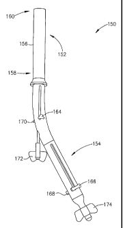
Referring now to Figure 9, there is seen an external view of an apparatus
generally referenced 150 for intussusception and anastomosis in accordance
with an
embodiment of the present invention. Apparatus 150 is comprised of an
operative
portion referenced generally 152 and a control or activating portion
referenced
generally 154. Operative portion 152 has an outer cylindrical enclosure
referenced
156 having a proximal end generally referenced 158 and a distal end generally
referenced 160. Disposed within outer cylindrical member 156 are
intussusception
and anastomosis apparatus (as referred to hereinbelow in relation to Figures
11-15).
Control or activating portion 154 includes various control elements to
facilitate
operation of the intussusception and anastomosis apparatus. The control
elements
include an anastomosis ring applicator lever referenced 164, proximal and
distal
clamp jaw levers 166 and 168, respectively, an excising applicator lever
referenced
170, crimping support applicator control referenced 172 and intussusception
and
anastomosis apparatus positioning controller 174. In order to carry out
intussusception and anastomosis, a user (not shown) grasps control portion 154
of
apparatus 150 and inserts distal end 160 of operative portion 152 a
preselected
distance into a hollow organ such as the bowel via the anus. It is sometimes
necessary to provide controlled positioning and retraction of intussusception
and
anastomosis apparatus 150, (as disclosed hereinbelow in relation to Figure
37).
Included with apparatus 150, according to an alternative embodiment of the
present
invention, -is an optical device (not shown) fo-r providing a view o-f the
interior of
the organ being intussuscepted and anastomosed.
Referring now to Figure 10, there is seen, in accordance with an embodiment
of the present invention, an intratubular intussusception and anastomosis
apparatus
generally referenced 180, including operative portion referenced 182 and
operationally connected thereto operating mechanism referenced 184. A flexible
control cable referenced 186 connects operating mechanism 184 to a remote
activator apparatus referenced 188. Included in control cable 186, according
to an
alternative embodiment of the present invention, is an optical device (not
shown)
17
CA 02492871 2005-O1-18
WO 2004/008937 PCT/IL2003/000597
for providing a view of the interior of the organ being intussuscepted and
anastomosed.
With reference to Figures 11 and 12, in accordance with a preferred
embodiment of the present invention, there is seen apparatus generally
referenced
200 (generally as disclosed hereinabove as operative portion 152 in Figure 9
and as
operative portion 182 in Figure 10) for intussusception and anastomosis of a
hollow
organ (not shown) from which a portion is to be excised. Figure 12 is a cut-
away
view of apparatus 200 as seen in Figure 11. Apparatus 200 has an outer,
cylindrical
enclosure referenced 202 having a retaining lip referenced 204 (Figure 12)
formed
at the distal end generally referenced 206 thereof.
Slidably disposed within and coaxial with enclosure 202 is a generally
tubular anastomosis ring applicator referenced 208 (Figure 12), having a
recessed
portion referenced 210 at the distal extremity thereof, thereby to demountably
engage an anastomosis ring referenced 212 thereto (as referred to hereinabove
in
relation to Figures lA-3B and 8A). Ring applicator 208 is either rigidly or
flexibly
operatively connected to a control device (not shown, as disclosed hereinabove
with reference to Figures 9-10) to cause an advancing and retracting movement
of
ring applicator 208. Ring applicator 208 is advanced to facilitate demountably
engaging expanded anastomosis ring 212 thereto while precooled below the
transition temperature and in a malleable or plastic state (as disclosed
hereinabove
in relation to Figures lA-1D). Ring 212 is permitted or caused to warm so as
to
reach and exceed the transition temperature, thereby reverting to a
contractible
elastic state. Thereafter, by retracting ring applicator 208, anastomosis ring
212 is
disengaged from ring applicator recess 210 thereby crimping adjacent organ
walls
to crimping support element referenced 214 which has been pre-aligned with lip
204 (as disclosed hereinbelow in relation to Figures 18-19).
Further, there is seen an intratubular intussusception device, generally
referenced 216 disposed coaxially within enclosure 202, for intussusception of
a
preselected hollow organ portion to be excised from the hollow organ (as
disclosed
hereinbelow with reference to Figures 16-17). Intussusception device 216
includes
proximal and distal clamping jaws referenced 218 and 220, respectively.
18
CA 02492871 2005-O1-18
WO 2004/008937 PCT/IL2003/000597
In accordance with one embodiment of the present invention, circular
clamping jaws 218 and 220 are slidingly operable in accordance with movements
of
coaxial operating supports referenced 222 and 224, respectively, beyond and
retractable within enclosure 202 and ring applicator 208. Jaws 218 and 220 are
caused to move axially with respect to enclosure 202 to be disposed at a
preselected
mid-position relative to a diseased organ portion (as referred to hereinbelow
with
reference to Figure 16). After drawing substantially the mid-portion of the
diseased
organ portion to within jaws 218 and 220, distal jaw 220 is retractable
relative to
proximal jaw 218 and, similarly, proximal jaw 218 is advancible relative to
distal
jaw 220, so as to clamp a preselected organ portion (as disclosed hereinbelow
with
reference to Figure 16). Thereafter, intussusception of the preselected organ
portion
occurs by simultaneously retracting jaws 218 and 220 to within enclosure 208
(as
disclosed hereinbelow with reference to Figure 17).
Activating or control means (not shown; as disclosed hereinabove with
reference to Figures 9-10) for intussusception is operationally connected to
coaxial
clamping jaws 218 and 220 which are operative independently or in clamping
engagement by operating supports 222 and 224 thereby to advance or retract
clamping jaws 218 and 220. This activating means is disposed at a proximate
end of
apparatus 200 or remotely therefrom (not shown; as disclosed hereinabove with
reference to Figures 9-10), in accordance with embodiments of the present
invention. According to alternative embodiments of the present invention, (as
disclosed hereinbelow in relation to Figures 13-14) there are alternative
intussusception clamping devices, wherein clamping movement of proximal and
distal jaws 218 and 220 is caused without necessitating independent movement
of
supports 222 and 224, respectively.
In accordance with a preferred embodiment of the present invention, there is
also seen in Figures 11-12 a crimping support applicator member generally
referenced 226, which is slidingly disposed coaxially within coaxial clamp
supports
222 and 224 and operating supports (not shown) therewithin. Crimping support
applicator member 226 is configured as a tubular support shaft referenced 228
having a transverse holder referenced 230 formed at the distal end thereof.
Crimping support element 214 (as disclosed hereinabove with reference to
19
CA 02492871 2005-O1-18
WO 2004/008937 PCT/IL2003/000597
Figures 4A-8B) is demountably fastened to transverse holder 230. Crimping
support
applicator member 226 has activating means (not shown; as disclosed
hereinabove
with reference to Figures 9-10) according to embodiments of the present
invention,
which are operationally connected directly or remotely to crimping support
applicator member 226 to facilitate advancement or retraction thereof.
According to an alternative embodiment of the present invention, crimping
support element 214 (as disclosed hereinabove in relation to Figure 6), is
demountably fastened to an alternatively configured crimping support
applicator
member, by means of a bayonet fastening mechanism (not shown) formed at a
distal
end thereof. Bayonet fastening mechanism of crimping support applicator member
226 is engaged into bayonet engagement recesses 82 and locked into bayonet
locking recesses 84 of crimping support element 68 (seen in Figure 6), by
rotating
an inner coaxial shaft (not shown) within tubular support shaft 228.
Activating
means (not shown, as disclosed hereinabove with reference to Figures 9-10) for
operating crimping support applicator member 226, according to embodiments of
the present invention, are operationally connected directly or remotely to
crimping
support applicator member 226.
In accordance with a preferred embodiment of the present invention, an
intratubular anastomosis crimping support element 214 is aligned with lip 204.
Thereafter, anastomosis ring 212 (as referred to hereinabove in relation to
Figures
lA-3B) disengages from anastomosis ring applicator recess 210, and thereby
crimps
adjacent intussuscepted wall portions of a hollow organ (not shown) against
crimping support element 214 (as disclosed hereinbelow with reference to
Figures
18-19).
In Figures 11-12, in accordance with a preferred embodiment of the present
invention, there is further seen a surgical excising means generally
referenced 232
(Figure 12), including an excise applicator referenced 234. Distally attached
thereto
is a cylindrical cutting blade referenced 236, operatively associated with
transverse
holder 230 of crimping support applicator member 226. Following intratubular
intussusception and crimping of adjacent organ wall portions with anastomosis
ring
212 against crimping support element 214, cylindrical cutting blade 236 is
CA 02492871 2005-O1-18
WO 2004/008937 PCT/IL2003/000597
selectably operable to excise the diseased organ portion (as disclosed
hereinbelow
in relation to Figure 20). Excise applicator 234 is distally advanced towards
transverse holder 230 until reaching operative engagement therewith.
Activating
means (not shown; as disclosed hereinabove in relation to Figures 9-10) for
surgical
excising means 232 is operationally connected thereto, either directly or
remotely.
In accordance with an alternative embodiment of the present invention, another
example of a surgical excising means is disclosed hereinbelow in relation to
Figure
15.
Referring now to Figures 13-14 there are seen additional configurations of
clamping jaw mechanisms in accordance with alternative embodiments of the
present invention. The clamping between proximal and distal jaws 218 and 220,
respectively, is caused without necessitating independent movement of supports
222 and 224 respectively (as disclosed hereinabove in relation to Figures 11-
12).
There is seen in Figure 13 a toggle clamping apparatus generally referenced
250,
including circular proximal and distal jaws 252 and 254 respectively, fixably
connected to a distal end generally referenced 256 of a common clamping
support
shaft referenced 258. Proximal jaw 252 includes, disposed in surface
referenced
259, a preselected number of toggle clamps referenced 260, each rotationally
mounted on a pivot pin referenced 262. Toggle clamp 260 rotates to position
260-1
as a result of a force applied in the direction of arrow 264 to arm 266, which
thereupon rotates to position 266-1. In sympathy therewith, arm referenced 268
rotates to position referenced 268-1 and is held in this position by a spring-
operated
ratchet pin referenced 270- which -operatively engages ratchet teeth
referenced 272.
Distal jaw 254 includes concentric rings referenced 274 formed facing towards
proximal j aw 252. When an organ portion (not shown) to be subj ected to
intussusception and anastomosis is drawn between jaws 252 and 254 in the
direction of arrow 264, consequent to the resulting force exerted on arm 266,
toggle
clamp 260 rotates to position 260-1 such that arm 268 moves to position 268-1.
The
drawn-in organ portion is thereby clamped against distal jaw 254 and held in
position by rings 274 in preparation for intussusception of the organ portion.
In accordance with another embodiment of the present invention, there is
seen, in Figure 14, a spring-loaded clamping apparatus generally referenced
280. A
21
CA 02492871 2005-O1-18
WO 2004/008937 PCT/IL2003/000597
proximal and a distal circular jaw referenced 282 and 284, respectively, are
disposed at a distal end referenced generally 286 of a common clamping support
shaft referenced 288, distal jaw 284 being fixably attached thereto. Proximal
jaw
282 is slidingly and elastically mounted on clamping support shaft 288,
supported
by springs referenced 290, thereby causing proximal jaw 282 to elastically
engage
distal jaw 284 in a clamping configuration. To maintain a predetermined
separation
between jaws 282 and 284, there are disposed therebetween two or more stay
members referenced 292, rotationally attached to distal jaw 284 and engaging
proximal jaw 282. The clamping of organ portion referenced 294 is effected by
suture referenced 296 drawing organ portion 294 inward between jaws 282 and
284.
Stay members 292 are radially depressed in an axial direction, such that they
pass
through apertures referenced 298, permitting proximal jaw 282 to move
elastically
toward distal jaw 284 and to hold organ portion 294 in a clamping engagement
therebetween. There are also formed, in jaws 282 and 284, a series of
concentric
rings referenced 297, disposed therebetween to retain a grip on organ portion
294
prior to intussusception thereof.
In accordance with an alternative embodiment to the present invention,
referring now to Figure 15, there is seen an alternative surgical excising
means
referenced generally 300. This includes an axially rotary excise applicator
referenced 302 coaxially disposed within outer cylindrical enclosure 202 (as
disclosed hereinabove in relation to Figures 11-12), having distally attached
thereto
a cutting blade referenced 304, pivotally attached to pivot pin referenced
306.
Cutting blade 304 is operatively associated with a cutting anvil element
referenced
308 axially slidably disposed within anastomosis ring applicator 208 (as
disclosed
hereinabove in relation to Figures 11-12). Following intratubular
intussusception
and crimping of the adjacent organ wall portions with an anastomosis ring
against a
crimping support element (not shown; as disclosed hereinbelow in relation to
Figures 16-19), cutting blade 304 is selectably operable to excise the
diseased
organ portion (as disclosed hereinbelow in relation to Figure 20). By rotation
of
rotary excise applicator 302, blade 304 pivots as shown, from a non-operative
position referenced 304-1 and engages intussuscepted organ portions (not
shown) in
a cutting engagement against anvil 308. Activating means (not shown) for
surgical
22
CA 02492871 2005-O1-18
WO 2004/008937 PCT/IL2003/000597
excising means 300 is operationally connected thereto, either directly or
remotely
(as disclosed hereinabove with reference to Figures 9-10).
Referring now to Figures 16-23, in accordance with embodiments of the
present invention, the method for performing an intussusception and
anastomosis
procedure to excise a diseased portion of a hollow organ follows hereinbelow.
In
Figure 16 there is seen a cross-sectional view of apparatus generally
referenced 400
for intussusception and anastomosis (generally as disclosed hereinabove in
relation
to Figures 11-12). Apparatus 400 is disposed within a hollow organ generally
referenced 402, such that proximal clamp j aw 404 and distal clamp j aw 406
are
aligned with substantially the middle of an organ portion referenced generally
408,
of hollow organ portion 402, to be excised. Utilizing either Laparoscopic or
open
surgery, substantially the middle of organ portion 408 to be excised is drawn
within
clamping jaws 404 and 406 by means of an external tie referenced 410. Jaws 404
and 406 are brought into clamping engagement with the drawn in organ portion
408.
Distal jaw 406 is retracted or proximal jaw 404 is advanced to cause the
middle of organ portion 408 to be clamped between jaws 404 and 406 as shown.
Thereupon, j aws 404 and 406 are simultaneously retracted while remaining in a
clamping configuration to within enclosure 412 as seen in Figure 17, causing
intussusception of organ portion 408 of organ 402. Additionally, there is
seen, in
Figure 18, crimping support element 414 retracted so as to align
circumferential
recess 416 therein with the distal lip 418 of enclosure 412.
As seen in Figure 19, anastomosis ring applicator 420 is slidingly retracted.
Thereupon, anastomosis ring 422 is disengaged from ring applicator recess 424
and
crimps adj acent organ wall portions 408 against crimping support element 414
thereby to effect anastomosis of adjacent organ wall portions 408. As seen in
Figure
20, by advancing cutting blade 426 along the axis of apparatus 400,
cylindrical
cutting blade 426 is brought into cutting engagement with intussuscepted organ
wall portion 408, in accordance with a preferred embodiment of the present
invention. Further, as seen in Figure 21, apparatus 400 is withdrawn from
organ
23
CA 02492871 2005-O1-18
WO 2004/008937 PCT/IL2003/000597
402, causing anastomosis ring 422 and crimping support element 414 to
disengage
from crimping support applicator member 425.
According to an alternative embodiment of the present invention, tubular
support shaft 428 (Figure 21) is rotated thereby to disengage the bayonet
fastening
mechanism (not shown) formed at a distal end of the alternatively configured
crimping support applicator member 425, from bayonet locking recesses 84 and
engagement recesses 82 of crimp support element 68 (as disclosed hereinabove
in
relation to Figure 6). Retracting apparatus 400 causes disengagement of crimp
support element 414 from crimping support applicator member 425.
As seen in Figure 22, anastomosis ring 422, by crimping adjacent organ
portions against crimping support element 414, provides immediate patency to
organ 402, bringing portions referenced 430 and 432 into flow communication
through axial aperture referenced 434 of crimping support element 414. Organ
402
remains sealed to flow or leakage into the surrounding peritoneal cavity (not
shown).
As a result of the pressure exerted by anastomosis ring 422 on wall portions
430 and 432 of organ 402, respective wall areas referenced 436 and 438 are
pressed
tightly against each other. Blood supply to end wall portions referenced 440
and to
areas 436 and 438 ceases, resulting in eventual necrosis of wall areas 436,
438 and
440. While these begin to die-off, wall tissue portions referenced 442,
immediately
externally adjacent thereto, begin anastomosis such that portions 442 of wall
portions 430 and 432 of organ 402 become joined, and function as one
continuous-
organ.
Referring now to Figure 23, once wall areas 436, 438 and 440 become fully
necrotic, these areas together with anastomosis ring 422 and crimping support
element 414 become separated from wall portions 430 and 432. This results in
an
aperture referenced 444 in organ 402 substantially similar to the original
opening in
organ 402, providing little or no restriction to normal organ flow. Necrotic
wall
areas 436, 438 and 440 together with anastomosis ring 422 and crimping support
element 414 are passed out of organ 402, by normal organ activity.
24
CA 02492871 2005-O1-18
WO 2004/008937 PCT/IL2003/000597
With regard to embodiments of the present invention disclosed hereinabove,
the relationship between the anastomosis ring and crimping support element
relates
to having a crimping support element within the anastomosed organ walls and a
contractible anastomosis ring external to the organ walls. The anastomosis
ring is
brought into contracting crimping engagement with the organ walls against the
crimping support element. In accordance with additional embodiments of the
present invention, an expandable anastomosis ring is disposed within an organ
to be
anastomosed and brought into crimping engagement with an external crimping
support element.
Referring now to Figure 24, there is seen an alternative configuration of
anastomosis ring and crimp support element, generally referenced 450 including
an
anastomosis ring referenced 452 in crimping engagement with an organ (not
shown)
against a generally cylindrical external crimping support element referenced
454.
External crimping support element 454 has a retaining recess referenced 456,
formed in an interior surface referenced 458 to ensure that anastomosis ring
452
remains engaged therein, and, also, a mounting recess referenced 462 (as
disclosed
further herein below in relation to Figure 25).
Referring now to Figure 25, there is seen an intussusception and anastomosis
apparatus, generally referenced 460, including anastomosis apparatus generally
referenced 470 for causing anastomosis of portions of a hollow organ (not
shown)
by bringing an intratubular expandable anastomosis ring 452 into a crimping
engagement with organ wall portions (not shown) against external crimping
support
element 454 (as disclosed hereinabove in relation to Figure 24). Crimping
support
element 454 is demountably attached to cylindrical enclosure referenced 472 by
engaging mounting projections referenced 474 thereof into mounting recess 462
of
crimping support element 454. Coaxially disposed within cylindrical enclosure
472
there is an intussusception apparatus generally referenced 476 which includes
proximal and distal clamping jaws respectively referenced 478 and 480
operatively
attached to coaxial tubular clamp operating members respectively referenced
482
and 484 to bring jaws 478 and 480 into clamping engagement with an organ
portion
(not shown). Further, coaxially disposed within enclosure 472 there is
anastomosis
apparatus 470 including an anastomosis ring mounting member referenced
CA 02492871 2005-O1-18
WO 2004/008937 PCT/IL2003/000597
generally 486, which includes proximal and distal anastomosis ring holders
respectively referenced 488 and 490, axially operable by a coaxial slidingly
operable tubular mounting shaft referenced 492.
In order to position expandable anastomosis ring 452 between holders 488
and 490 as indicated, anastomosis ring 452 is cooled to or below the
transition
temperature so as to become expandably .malleable. To prevent expandable
anastomosis ring 452 from expanding away from mounting member 486, there is a
coaxial ring applicator member generally referenced 496 having a retaining
member
operating shaft referenced 498 coaxially slidingly disposed within tubular
mounting
shaft 492 and a generally cylindrical ring retaining wall referenced 497. As
anastomosis ring 452 warms above the transition temperature, the memory alloy
thereof enters the elastic state and expands into engagement with cylindrical
retaining wall 497. Apparatus 470 is now ready for use.
After inserting apparatus 460 into an organ portion (not shown) requiring
excision of a diseased portion, intussusception apparatus 476 clamps a
substantially
mid-portion thereof (generally as disclosed hereinabove in relation to Figures
16-
18) causing intussusception thereof. Following intussusception, mounting
member
486 and applicator member 496 are aligned with recess 456 of crimping support
element 454. With mounting member 486 fixed in this position, applicator
member
496 is distally advanced, thereby releasing anastomosis ring 452 therefrom, to
expand so as to bring organ walls (not shown) into crimping engagement against
crimping support element 454. Excision of the intussuscepted organ portion is
then
carried out (generally as disclosed hereinabove in relation to Figures 18-20).
Thereafter, withdrawing apparatus 460 from the anastomosed hollow organ causes
crimping support element 454 together with anastomosis ring 452 to become
detached from mounting projections 474 of apparatus 460.
The consequence of utilizing apparatus 460 together with intratubular
expandable anastomosis ring 452 and external crimping support element 454 is
the
provision of a generally larger aperture formed within the organ at the site
of
anastomosis, compared with that formed when using an internal crimping support
member. Nevertheless, the aperture is limited by the wall thickness and
external
26
CA 02492871 2005-O1-18
WO 2004/008937 PCT/IL2003/000597
diameter of external crimping support element 454. External crimping support
element 454 is selected in accordance with the internal diameter of the organ
to be
treated. Inevitably, an aperture formed at the site of the anastomosis is
smaller than
the original organ diameter. In order to further increase the anastomosed
aperture,
in accordance with further embodiments of the present invention, an expandable
crimping support element and apparatus for utilizing this expandable crimping
support element is disclosed hereinbelow in relation to Figures 26-31.
Following
crimping the organ walls using an expandable anastomosis ring against an
expandable crimping support element, the aperture formed at the site of
anastomosis will be in accordance with the expanded size of the expandable
crimping support element.
Referring now to Figures 26-28, there is seen an expandable anastomosis
ring referenced 500 in crimping engagement with an organ portion (not shown)
against an inner face referenced 502 of an expandable helical crimping support
element referenced 504 configured from a length of substantially circular
cross-
section memory alloy wire, in accordance with an alternative embodiment of the
present invention. In Figure 27 there is seen a cross-section of expandable
anastomosis ring 500 and expandable helical crimping support element 504 taken
along line 6-6 in Figure 26. In Figure 28 there is seen a cross-section
generally as
taken along line 6-6 in Figure 26 of expandable anastomosis ring 500 and an
expandable helical crimping support element referenced 506, formed from a
generally square section memory alloy wire, thereby forming a generally
flatter
inner face referenced 508.
Referring now to Figures 29 and 30, in accordance with a variation of an
embodiment of the present invention, there is seen expandable anastomosis ring
500 in crimping engagement with organ portions (not shown) against a
substantially
single coil expandable crimping support element referenced 510. Figure 30 is a
cross-section taken along line 7-7 of Figure 29. Expandable crimping support
element 510 is formed from a substantially flat section strip of memory alloy,
having a generally cylindrical configuration and having a generally smooth
internal
surface referenced 512.
27
CA 02492871 2005-O1-18
WO 2004/008937 PCT/IL2003/000597
Referring now to Figure 31, there is seen an intussusception and anastomosis
apparatus, generally referenced 520 including anastomosis apparatus generally
referenced 530 for causing anastomosis of portions of a hollow organ (not
shown)
by bringing an intratubular expandable anastomosis ring 500 into crimping
engagement with intussuscepted organ wall portions (not shown) against
external
expandable crimping support element 504 (as disclosed hereinabove in relation
to
Figures 26-30).
Crimping support element 504 is cooled to below the transition temperature
so that the memory alloy thereof becomes malleable thereby allowing crimping
support element 504 to become compressible for insertion within retaining
collets
referenced 534 formed at a distal end 536 of enclosure referenced 532. Collets
534
are rendered outwardly flexible as a result of recesses referenced 538 formed
in an
outer face thereof.
Coaxially disposed within cylindrical enclosure 532 is an intussusception
apparatus (not shown; generally as disclosed in relation to Figure 25).
Further,
coaxially disposed within enclosure 532 there is anastomosis apparatus 530
including an anastomosis ring mounting member referenced generally 542, which
includes proximal and distal anastomosis ring holders respectively referenced
544
and 546, axially operable by a coaxial slidingly operable tubular mounting
shaft
referenced 548.
In order to position expandable anastomosis ring 500 between holders 544
and 5-46 as indicated, anastomosis ring 500 is cooled to or below the
transition
temperature so as to become expandably malleable. To prevent expandable
anastomosis ring 500 from expanding away from mounting member 542, there is a
coaxial anastomosis ring applicator member generally referenced 550 having an
applicator operating shaft referenced 552 coaxially slidingly disposed within
tubular mounting shaft 548. Applicator member 550 further has a generally
cylindrical anastomosis ring retaining wall referenced 554. As anastomosis
ring
500, positioned in mounting member 542, warms above the transition
temperature,
the memory alloy thereof enters the elastic state and expands into engagement
with
cylindrical retaining wall 554. Apparatus 520 is now ready for use.
28
CA 02492871 2005-O1-18
WO 2004/008937 PCT/IL2003/000597
After inserting apparatus 520 into an organ (not shown) requiring excision of
a diseased portion, intussusception apparatus (not shown) causes
intussusception of
the diseased organ portion (generally as disclosed hereinabove in relation to
Figures 16-18). Following intussusception, mounting member 542 and applicator
member 550 are generally centrally aligned with crimping support element 504.
With mounting member 542 fixed in this position, applicator member 550 is
distally advanced, thereby releasing anastomosis ring 500 therefrom, to expand
so
as to bring organ walls (not shown) into crimping engagement against crimping
support element 504. Excision of the intussuscepted organ portion is carried
out
(generally as disclosed hereinabove in relation to Figures 18-20). Thereafter,
disengaging member referenced 556 is distally advanced causing crimping
support
element 504 together with anastomosis ring 500 to push against and thereby to
force collets 534 to flex outwards at recesses 538. Crimping support element
504
together with anastomosis ring 500 is thereby detached from apparatus 520 and
both crimping support element 504 and anastomosis ring 500 expand further to a
preselected size.
Utilizing apparatus 520 together with intratubular expandable anastomosis
ring 500 and one of external expandable crimping support elements 504, 506 or
510
(Figures 26-30), a generally larger aperture is formed within the organ at the
site of
anastomosis, which is not limited by the wire thickness and diameter of
external
crimping support elements 504, 506 or 510. Rather, in accordance with further
embodiments of the present invention, the anastomosed aperture is formed in
accordance with the expanded diameters of anastomosis ring 500 and of
expandable
crimping support elements 504, 506 or 510.
Under certain circumstances, the surgeon may decide to carry out a
conventional, open surgery excising procedure in order to excise a portion of
diseased or problematic bowel. The conventional method of joining the bowel
portions is utilizing staples or sutures. However, according to an alternative
embodiment of the present invention, using an anastomosis ring and a crimping
support element, anastomosis is achieved whereby the risk of leakage is
substantially reduced and no staples or sutures remain in the anastomosed
bowel.
Referring now to Figure 32, there is seen a modified intussusception and
29
CA 02492871 2005-O1-18
WO 2004/008937 PCT/IL2003/000597
anastomosis apparatus generally referenced 600, inserted into organ portion
referenced 604, clamping surgically excised bowel portions referenced 602 and
604. The intussusception and anastomosis apparatus (as disclosed hereinabove
in
relation to Figures 11-12), is modified, insofar as an additional clamping jaw
referenced 606 is disposed immediately proximate to transverse crimping
support
applicator referenced 608 to facilitate clamping surgically excised organ
portion
602 therebetween. Organ portion 604 is clamped between jaws referenced 610 and
612.
Referring now to Figure 33, clamped organ portions 602 and 604 are
partially intussuscepted by simultaneously retracting clamping jaws 610 and
612 in
clamping engagement with organ portion 604 into enclosure referenced 614 and
thereafter simultaneously retracting transverse crimping support applicator
608 and
clamping jaw 606 in clamping engagement with organ portion 602. Both pairs of
clamps (the underside surface of crimping support applicator 608 providing a
clamping surface in clamping engagement with clamp 606), 606 and 608 and 610
and 612 are further retracted to cause crimping support element referenced 616
to
be aligned with lip referenced 618 of enclosure 614. Anastomosis ring
applicator
referenced 620 is then retracted so as to release anastomosis ring referenced
622
from recess referenced 624 thereby to crimp organ portions 602 and 604 against
crimping support element 616. Cylindrical cutting blade referenced 626 is
distally
advanced to provide cutting engagement with crimped organ portions 602 and 604
and to excise clamped portions referenced 628 and 630 therefrom. Thereafter,
crimping support element 616--and anastomosis ring- 622 are disengaged from
crimping support applicator referenced 632 (as disclosed hereinabove in
relation to
Figures 21-23) to provide patency to anastomosed organ portions 602 and 604.
Referring now to Figures 34, 35 and 36, in accordance with a further
embodiment of the present invention, there is seen an intratubular anastomosis
apparatus generally referenced 650 inserted into a prolapsed bowel generally
referenced 652 for bringing about anastomosis of organ 652 followed by
excising
of prolapsed portion referenced 654. In Figure 35 there is seen crimping
support
element referenced 656, supported on crimping support applicator referenced
670,
proximally retracted within enclosure referenced 660 to be brought into
alignment
CA 02492871 2005-O1-18
WO 2004/008937 PCT/IL2003/000597
with recess referenced 666 of crimping support element 656 in preparation for
crimping prolapsed bowel portion 654 against crimping support element 656.
Retraction of anastomosis ring applicator referenced 662, causes anastomosis
ring
referenced 664 to disengage from recess 666 and to crimp prolapsed organ
portion
654 as seen in Figure 36. Thereafter, cylindrical cutting blade referenced 668
is
brought into cutting engagement with crimping support applicator referenced
672
thereby to excise prolapsed organ portion 654. Crimping support element 656
and
anastomosis ring 664 are disengaged from crimping support applicator 672 (as
disclosed hereinabove in relation to Figures 21-23) to provide patency to
anastomosed organ 652.
Referring now to Figure 37, there is seen an apparatus generally referenced
680 to provide controlled positioning and retraction of intussusception and
anastomosis apparatus referenced generally 682 including crimping support
element
656 in relation to the buttocks wall referenced 684 of a patient. A curved
positioning plate referenced 686 having a generally central opening referenced
688
is placed against the buttocks of the patient. Clamp referenced 690 is fixably
attached to apparatus 682 at a preselected position. Apparatus 680 is inserted
through opening 688 into the anus and into the patient's rectum, where
intussusception and anastomosis is to be carried out. Adjusting screw
referenced
692 provides means for adjusting the position of apparatus 682 within the
patient.
Also, retraction of apparatus 682 is carried out by means of screw 692.
Referring now to Figure 38, there is seen, in accordance with an embodiment
of the present invention, a schematic representation of method steps generally
referenced 700 relating to the present invention, namely
Step 701 of inserting a distal end portion of an apparatus for intratubular
intussusception and anastomosis a preselected distance into a hollow
organ;
Step 702 of clamping a preselected portion of the hollow organ utilizing a
clamping means of the intussusception and anastomosis apparatus;
Step 703 of intussuscepting the preselected organ portion by withdrawing the
clamping means a preselected distance into an enclosure member;
31
CA 02492871 2005-O1-18
WO 2004/008937 PCT/IL2003/000597
Step 704 of disengaging an anastomosis ring from an anastomosis ring
applicator member so as to crimp the intussuscepted organ portion to a
crimping support element; and
Step 705 of excising the intussuscepted organ portion.
In accordance with other embodiments of the present invention, step 701 of
inserting includes a step of demountably engaging the anastomosis ring formed
of a
shape memory alloy to the anastomosis ring applicator member. Also, the step
of
demountably engaging includes a step of cooling the anastomosis ring to or
below a
transition temperature so as to assume a plastic state. Further, step 702 of
clamping
a preselected portion of the hollow organ includes a step of drawing a
substantially
middle portion of the preselected organ portion within the clamping means.
Referring now to Figure 39, there is seen a schematic representation of
method steps referenced generally 800 relating to a prolapsed bowel in
accordance
with an alternative embodiment of the present invention, namely
Step 801 of inserting a distal end portion of an apparatus for intratubular
intussusception and anastomosis a preselected distance into a hollow
organ so as to juxtapose a crimping support element and an anastomosis
ring to the prolapsed organ portion;
Step 802 of disengaging the anastomosis ring from an anastomosis ring
applicator member so as to crimp the prolapsed organ portion against the
crimping support element; and
Step 803 of excising the prolapsed organ portion.
It will, further, be appreciated by persons skilled in the art that there is a
direct relationship between the size and thickness of the anastomosis ring and
crimping support element used in the surgical procedure disclosed above and
the
size and shape of the organ to be treated. An anastomosis ring and crimping
support
element of a particular size is selected so as to achieve an aperture of a
requisite
size as appropriate to the situation and the hollow organ to be subjected to
32
CA 02492871 2005-O1-18
WO 2004/008937 PCT/IL2003/000597
intussusception and anastomosis. Clearly, a smaller size is appropriate for
use in
the upper bowel and a larger size in the lower bowel.
Additionally, it will be appreciated by persons skilled in the art, that an
apparatus employing a shape memory alloy, such as an anastomosis ring,
referred
to hereinabove according to embodiments of the present invention, may be
described as being of one of two different types. A first type of apparatus
employs
a shape memory alloy, which is in an easily deformable, martensitic state when
it is
cooled to below room temperature, called a "Cold" type. This first apparatus
achieves a fully or partially austenitic state at room temperature, and a
completely
austenitic state when heated to at least its upper phase transition
temperature,
between room and body temperature. In a second type of apparatus, the shape
memory alloy is in an. easily deformable, martensitic state at room
temperature,
called a "Hot" type, whereat the apparatus is deformed and applied, and the
shape
memory alloy achieves a completely austenitic state when heated to above room
temperature. The temperature range over which the shape memory alloy is easily
deformable defines the difference between the two types of apparatus. Thus,
utilizing an apparatus including a shape memory alloy of the second Hot type
allows more freedom in application without necessitating cooling below room
temperature. The present invention disclosed hereinabove relates to an
apparatus of
the first Cold type, necessitating cooling below room temperature.
Considering the "Hot" type, in which the transformation temperature is
higher, the clip is martensitic at room temperature and heated to about 42-
45°C to
assume an austenitic state. When the temperature drops to 37°C, that
is, body
temperature, the martensitic transformation is not complete, leaving the clip
in a
transition state, with inferior mechanical characteristics.
It should be understand that the so-called transformation temperature of the
alloy, in fact, is a process of transformation. Transition from a martensitic
to an
austenitic state starts at a temperature AS and ends with a temperature Af at
which
the state becomes fully austenitic. When transforming from austenitic to
martensitic
state, by dropping the temperature, the alloy starts to become martensitic at
temperature MS, and reaches a full martensitic state at temperature Mf.
33
CA 02492871 2005-O1-18
WO 2004/008937 PCT/IL2003/000597
In the Cold type, generally preferred in accordance with embodiments of the
present invention, Af is lower than body temperature, generally about
25°C. In the
Hot type, Mf is below body temperature, so that the alloy does not become
fully
martensitic at body temperature.
It will be appreciated by persons skilled in the art that the present
invention
is not limited by the drawings and description hereinabove presented. Rather,
the
invention is defined solely by the claims that follow.
34