Note: Descriptions are shown in the official language in which they were submitted.
CA 02493122 2011-08-04
WO 2004/010959 PCT/US2003/024839
-1-
HE' MATOPOIETIC STEM CELLS AND METHODS OF
TREATMENT OF NEOVASCULAR EYE DISEASES THEREWITH
Statement of Government Interest
A portion of the work described herein was supported by grant number
CA92577 from the National Cancer Institute and by grants number EY1 1254,
EY12598
and EY125998 from the National Institutes of Health. The United States
Government
has certain rights in this invention.
Field of the Invention
This invention relates to isolated, mammalian, lineage negative
hematopoietic stem cells (Lin HSC) derived from bone marrow. The invention
also
relates to treatment of vascular diseases of the eye by administering
Lin HSC and transfected Lin HSC to the retina.
Background of the Invention
Age Related Macular Degeneration (ARMD) and Diabetic Retinopathy
(DR) are the leading causes of visual loss in industrialized nations and do so
as a result of
abnormal retinal neovascularization. Since the retina consists of well-defined
layers of
neuronal, glial, and vascular elements, relatively small disturbances such as
those seen in
vascular proliferation or edema can lead to significant loss of visual
function. Inherited
retinal degenerations, such as Retinitis Pigmentosa (RP), are also associated
with
vascular abnormalities, such as arteriolar narrowing and vascular atrophy.
While
significant progress has been made in identifying factors that promote and
inhibit
CA 02493122 2005-01-20
WO 2004/010959 PCT/US2003/024839
-2-
angiogenesis, no treatment is currently available to specifically treat ocular
vascular
disease.
For many years it has been known that a population of stem cells exists in
the normal adult circulation and bone marrow. Different sub-populations of
these cells
can differentiate along hematopoietic lineage positive (Lin*) or non-
hematopoietic, lineage
negative (Lin) lineages. Furthermore, the lineage negative hematopoietic stem
cell
(HSC) population has recently been shown to contain endothelial progenitor
cells (EPC)
capable of forming blood vessels in vitro and in vivo. Asahara et al. Science
275,
964-7 (1997). These cells can participate in normal and pathological postnatal
angiogenesis (See Lyden et al. Nat. Med. 7, 1194-201 (2001); Kalka et al.
Proc. Natl.
Acad. Sci. U. S. A. 97, 3422-7 (2000); and Kocher et al Nat. Med. 7, 430-6
(2001))
as well as differentiate into a variety of non-endothelial cell types
including hepatocytes
(See Lagasse et al. Nat. Med. 6, 1229-34 (2000)), microglia (See Priller et
al. Nat.
Med. 7, 1356-61 (2002)), cardiomyocytes (See Orlic et al. Proc. Natl. Acad.
Sci. U.
S. A. 98, 10344-9 (2001)) and epithelium (See Lyden et al. Nat. Med. 7, 1194-
201
(2001)). Although these cells have been used in several experimental models of
angiogenesis, the mechanism of EPC targeting to neovasculature is not known
and no
strategy has been identified that will effectively increase the number of
cells that
contribute to a particular vasculature.
- -Summary of the-Invention _ . _,.. .. _ _
The present invention provides isolated, mammalian, lineage negative
hematopoietic stem cell populations (Lin' HSC) derived from bone marrow, which
contain endothelial progenitor cells (EPC; also known as endothelial precursor
cells) that
selectively target activated retinal astrocytes. At least about 50% of the
cells of the
isolated Lin HSC populations of the present invention have cell markers for
CD31 and
c-kit.
The EPC's in the lineage negative HSC populations of the present
CA 02493122 2005-01-20
WO 2004/010959 PCT/US2003/024839
-3-
invention extensively incorporate into developing retinal vessels and remain
stably
incorporated into neovasculature of the eye. The isolated, lineage negative
HSC
populations of the present invention can be used to rescue and stabilize
degenerating
retinal vasculature in mammals. In one embodiment of the isolated Lin HSC
populations
of the present invention, the cells are transfected with a therapeutically
useful gene. The
transfected cells can selectively target neovasculature and inhibit new vessel
formation
without affecting already established vessels through a form of cell-based
gene therapy.
Cells from isolated, lineage negative HSC population of the present invention
that have
been transfected with a gene encoding angiogenesis inhibiting peptides are
useful for
modulating abnormal blood vessel growth in diseases such as ARMD, DR and
certain
retinal degenerations associated with abnormal vasculature.
A particular advantage of ocular treatments with the isolated
Lin- HSC population of the present invention is a vasculotrophic and
neurotrophic rescue
effect observed in eyes intravitreally treated with the
Lin HSC. Retinal neurons and photoreceptors are preserved and visual function
is
maintained in eyes treated with the isolated Liri HSC of the invention.
The present invention also provides a method of isolating lineage negative
hematopoietic stem cell populations containing endothelial progenitor cells
from bone
marrow, preferably adult bone marrow.
-20-- Brief-Description-,of the Drawings.
Figure 1 (a and b) depicts schematic diagrams of developing mouse
retina. (a) Development of primary plexus. (b) The second phase of retinal
vessel
formation. GCL, ganglion cell layer; IPL, inner plexus layer; INL, inner
nuclear layer;
OPL, outer plexus layer; ONL, outer nuclear layer; RPE, retinal pigment
epithelium; ON,
optic nerve; P, periphery.
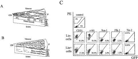
Figure 1 c depicts flow cytometric characterization of bone
marrow-derived Lin+ HSC and Liri HSC separated cells. Top row: Dot plot
distribution
CA 02493122 2005-01-20
WO 2004/010959 PCT/US2003/024839
-4-
of non-antibody labeled cells, in which R1 defines the quantifiable-gated area
of positive
PE-staining; R2 indicates GFP-positive; Middle row: Liri HSC (C57B/6) and
Bottom
row: Link HSC (C57B/6) cells, each cell line labeled with the PE-conjugated
antibodies
for Sca-1, c-kit, Flk-1/KDR, CD31. Tie-2 data was obtained from Tie-2-GFP
mice.
Percentages indicate percent of positive-labeled cells out of total Lin HSC or
Link HSC
population.
Figure 2 depicts engraftment of Liri HSC cells into developing mouse
retina. (a) At four days post-injection (P6) intravitreally injected eGFP'
Liri HSC cells
attach and differentiate on the retina (b) Lin HSC (B6.I29S7-Gtrosa26 mice,
stained
with (3-gal antibody) establish themselves ahead of the vasculature stained
with collagen
IV antibody (asterisk indicates tip of vasculature). (c) Most of Link HSC
cells (eGFP') at
four days post-injection (P6) were unable to differentiate. (d) Mesenteric
eGFP' murine
EC four days post-injection (P6). (e) Lin HSCs (eGFP) injected into adult
mouse
eyes. (f) Low magnification of eGFP' Lin- HSCs (arrows) homing to and
differentiating
along the pre-existing astrocytic template in the GFAP-GFP transgenic mouse.
(g)
Higher magnification of association between Lin cells (eGFP) and underlying
astrocyte
(arrows). (h) Non-injected GFAP-GFP transgenic control. (i) Four days post-
injection
(P6), eGFP' Lin HSC cells migrate to and undergo differentiation in the area
of the
future deep plexus. Left figure captures
- Lin- HSC cells activity in a whole mounted retina; right-figure
indicates.lacation -of the
Lin cells (arrows) in the retina (top is vitreal side, bottom is scleral
side). (j) Double
labeling with a-CD31-PE and (X-GFP-alexa 488 antibodies. Seven days after
injection,
the injected Lin- HS Cs (eGFP), red) were incorporated into the vasculature
(CD3 1).
Arrowheads indicate the incorporated areas. (k) eGFP' Lin HSC cells form
vessels
fourteen days post-injection (P17). (I and m) Intra-cardiac injection of
rhodamine-dextran indicates that the vessels are intact and functional in both
the primary
(1) and deep plexus (m).
CA 02493122 2005-01-20
WO 2004/010959 PCT/US2003/024839
-5-
Figure 3 (a and b) shows that eGFP-" Lin HSC cells home to the gliosis
(indicated by GFAP expressing-astrocytes, far left image) induced by both
laser (a) and
mechanical (b) induced injury in the adult retina (asterisk indicates injured
site). Far right
images are a higher magnification, demonstrating the close association of the
Lin-HSCs
and astrocytes. Calibration bar= 20 M.
Figure 4 shows that Liri HSC cells rescue the vasculature of the retinal
degeneration mouse. (a-d) Retinas at 27 days post-injection (P33) with
collagen IV
staining; (a) and (b), retinas injected with Lin+ HSC cells (Balb/c) showed no
difference
in vasculature from normal FVB mice; (c) and (d) retinas injected with Lin
HSCs
(Balb/c) exhibited a rich vascular network analogous to a wild-type mouse; (a)
and (c),
frozen sections of whole retina (top is vitreal side, bottom is scleral side)
with DAPI
staining; (b) and (d), deep plexus of retinal whole amount; (e) bar graph
illustrating the
increase in vascularity of the deep vascular plexus formed in the Lin HSC cell-
injected
retinas (n=6). The extent of deep retinal vascularization was quantified by
calculating the
total length of vessels within each image. Average total length of
vessels/high power field
(in microns) for Liri HSC, Lin+ HSC or control retinas were compared. (f)
Comparison
of the length of deep vascular plexus after injection with Lin HSC (R, right
eye) or
Lin+ HSC (L, left eye) cells from rd/rd mouse. The results of six independent
mice are
shown (each color represents each mouse). (g) and (h) Liri HSC cells also
(Balb/c)
- rescued the rd/rd-vasculature when injected into P 15 eyes.. The
intermediate and deep -
vascular plexus of Liri HSC (G) or Lin+ HSC (H) cell injected retinas (one
month after
injection) are shown.
Figure 5 depicts photomicrographs of mouse retinal tissue: (a) deep layer
of retinal whole mount (rdlyd mouse), five days post-injection (P11) with
eGFP+ Liri HSCs (green). (b) and (c) P60 retinal vasculature of Tie-2-GFP
(rdlyd)
mice that received Balb/c Lin cells (A) or Lin} HSC cell (B) injection at P6.
The
vasculature was stained with CD31 antibody (red) and only endogenous
endothelial cells
CA 02493122 2005-01-20
WO 2004/010959 PCT/US2003/024839
-6-
present green color. Arrows indicate the vessels stained with CD31 but not
with GFP.
(d) x-SMA staining of Lin HSC injected and control retina.
Figure 6 shows that T2-TrpRS-transfected Lift- HSCs inhibit the
development of mouse retinal vasculature. (a) Schematic representation of
human
TrpRS, T2-TrpRS and T2-TrpRS with an Igk signal sequence at the amino
terminus.
(b) T2-TrpRS transfected Lin cells injected retinas express T2-TrpRS protein
in vivo.
1, Recombinant T2-TrpRS produced in E.coli; 2, Recombinant T2-TrpRS produced
in
E.coli; 3, Recombinant T2-TrpRS produced in E.coli; 4, control retina; 5, Lift-
HSC +
pSecTag2A (vector only) injected retina;
6, Liri HSC + pKLel35 (Igk-T2-TrpRS in pSecTag) injected retina. (a);
endogenous
TrpRS b; recombinant T2-TrpRS c; T2-TrpRS of Liri HSC injected retina). (c-f)
Representative primary (superficial) and secondary (deep) plexuses of injected
retinas,
seven days post-injection; (c) and (d), Eyes injected with empty plasmid-
transfected
Liri HSC developed normally; (e) and (f), the majority of T2-TrpRS-transfected
Lin HSC injected eyes exhibited inhibition of deep plexus; (c) and (e),
primary
(superficial) plexus; (d) and (f), secondary (deep) plexus). Faint outline of
vessels
observed in F are "bleed-through" images of primary network vessels shown in
(e).
Figure 7 shows the DNA sequence encoding His6-tagged
T2-TrpRS, SEQ ID NO: 1.
- - -- - - -Figure -8 shows-the amino acid-sequence of
His6-tagged T2-TrpRS, SEQ ID NO: 2.
Figure 9 illustrates photomicrographs and electroretinograms (ERG) of
retinas from mice whose eyes were injected with the Lin HSC of the present
invention
and with Lin+HSC (controls).
Figure 10 depicts statistical plots showing a correlation between neuronal
rescue (y-axis) and vascular rescue (x-axis) for both the intermediate (Int.)
and deep
vascular layers of rdlyd mouse eyes treated with Liri HS C.
CA 02493122 2005-01-20
WO 2004/010959 PCT/US2003/024839
-7-
Figure 11 depicts statistical plots showing no correlation between
neuronal rescue (y-axis) and vascular rescue (x-axis) for rd/rd mouse eyes
that were
treated with Lin+ HSC.
Figure 12 is a bar graph of vascular length (y-axis) in arbitrary relative
units for rd/rd mouse eyes treated with the Lin- HSC (dark bars) and untreated
(light
bars) rd/rd mouse eyes at time points of 1 month (IM), 2 months (2M), and 6
months
(6M) post-injection.
Figure 13 includes three bar graphs of the number of nuclei in the outer
neural layer (ONR) of rd/rd mice at 1 month (1M), 2 months (2M) and 6 months
(6M),
post-injection, and demonstrates a significant increase in the number of
nuclei for eyes
treated with Lin- HSC (dark bars) relative to control eyes treated with Lin+
HSC (light
bars).
Figure 14 depicts plots of the number of nuclei in the outer neural layer
for individual rd/rd mice, comparing the right eye (R, treated with
Lin HSC) relative to the left eye (L, control eye treated with Lin} HSC) at
time points
(post injection) of 1 month (1M), 2 months (2M), and 6 months (6M); each line
in a
given plot compares the eyes of an individual mouse.
Detailed Description of Preferred Embodiments
The present invention provides an isolated, mammalian, bone marrow-
-20 - - derived -lineage negative hematopoietic stem cell population
containing endothelial _ .
progenitor cells. The isolated Lin HSC populations of the present invention
preferably
comprise HSC in which at least about 50% of the cells
contain CD31 and c-kit cell marker antigens. In a preferred embodiment, at
least about
75% of the HSC cells include the CD31 marker, more preferably about 81% of the
cells. In another preferred embodiment, at least about 65% of the cells
include the c-kit
cells marker, more preferably about 70% of the cells.
In a particularly preferred embodiment of the isolated Lin- HSC
CA 02493122 2005-01-20
WO 2004/010959 PCT/US2003/024839
-8-
populations of the present invention, about 50% to about 85% of the cells
include the
CD31 marker, about 70% to about 75% of the cells include the c-kit marker,
about 4%
to about 8% of the cells include the Sca-1 marker, and about 2% to about 4% of
the
cells include the Flk-1/KDR marker.
The isolated Lin HSC populations of the present invention can also
comprise up to about 1% of cells having the Tie-2 antigen marker.
In preferred embodiments, the isolated Liri HSC populations of the
present invention are derived from mouse or human bone marrow, preferably from
human bone marrow.
The isolated Liri HSC populations of the present invention selectively
target and incorporate into the retinal neovasculature when intravitreally
injected into the
eye of the mammalian species from which the cells were isolated.
The isolated Lin HSC populations of the present invention contain EPC
cells that differentiate to endothelial cells and generate vascular structures
within the
retina. In particular, the Lin HSC compositions of the present invention are
useful for
the treatment of retinal neovascular and retinal vascular degenerative
diseases, and for
repair of retinal vascular injury.
The present invention also provides a method of treating ocular diseases
in a patient comprising isolating from the bone marrow of the patient a
lineage negative
--20 hematopoietic stem cell population that. includes endothelial progenitor
cells, and
intravitreally injecting the isolated stem cells into an eye of the patient in
a number
sufficient to arrest the disease. The present method can be utilized to treat
ocular
diseases such as retinal degenerative diseases, retinal vascular degenerative
diseases,
ischemic retinopathies, vascular hemorrhages, vascular leakage, and
choroidopathies.
Examples of such diseases include age related macular degeneration (ARMD),
diabetic
retinopathy (DR), presumed ocular histoplasmosis (POHS), retinopathy of
prematurity
(ROP), sickle cell anemia, and retinitis pigmentosa, as well as retinal
injuries.
CA 02493122 2011-08-04
WO 2004/010959 PCT/US2003/024839
-9-
The number of stem cells injected into the eye is sufficient for arresting
the disease state of the patient's eye. For example, the number of cells can
be effective
for repairing retinal damage of the patient's eye, stabilizing retinal
neovasculature,
maturing retinal neovasculature, and preventing or repairing vascular leakage
and vascular
hemorrhage.
Cells present in the isolated Lin HSC populations of the present
invention can be transfected with therapeutically useful genes, such as genes
encoding
antiangiogenic proteins for use in ocular, cell-based gene therapy.
The transfected cells can include any gene which is therapeutically useful
for treatment of retinal disorders. Preferably, the transfected cells in the
Lin HSC
populations of the present invention include a gene encoding an antiangiogenic
peptide,
protein, or protein fragment such as TrpRS or antiangiogenic fragments
thereof, such as
the Ti and T2 fragments thereof, which are described in detail in co-owned, co-
pending
U.S. patent application Serial No. 10/080,839.
The present invention also provides a method of isolating a lineage
negative hematopoietic stem cell population containing endothelial progenitor
cells from
bone marrow. The method entails the steps of (a) extracting bone marrow from a
mammal; (b) separating a plurality of monocytes from the bone marrow; (c)
labeling the
- monocytes-with biotin.conjugated lineage panel antibodies to CD45, CD3,_Ly-
6G,
CDI 1 and TER-119; and (d) removal of monocytes that are positive for CD45,
CD3,
Ly-6G, CD 11 and TER-119 from the plurality of monocytes to provide a
population of
lineage negative hematopoietic stem cells containing endothelial progenitor
cells.
The present invention also provides methods for treating ocular
angiogenic diseases by administering transfected Lin- HSC compositions of the
present
invention by intravitreal injection of the cells into the eye. Such
transfected Linz HSC
CA 02493122 2005-01-20
WO 2004/010959 PCT/US2003/024839
-10-
compositions comprise Lin HSC transfected with a therapeutically useful gene,
such as a
gene encoding anti-angiogram gene product.
Preferably, at least about 1 x 10⁵ Lin- HSC cells or transfected
Lin HSC cells are administered by intravitreal injection to an eye suffering
from a retinal
degenerative disease. The number of cells to be injected may depend upon the
severity
of the retinal degeneration, the age of the patient and other factors that
will be readily
apparent to one of ordinary skill in the art of treating retinal diseases. The
Lin- HSC may
be administered in a single dose or by multiple dose administration over a
period of time,
as determined by the physician in charge of the treatment.
The Lin HSC populations of the present invention are useful for the
treatment of retinal injuries and retinal defects involving an interruption in
or degradation
of the retinal vasculature.
The transfected Liri HSC populations of the present invention are useful
for delivery of therapeutic genes to the retina, particularly to the retinal
vasculature.
In a preferred embodiment of the gene delivery method of the present
invention, cells in the Lin HSC populations of the present invention are
transfected with
a gene encoding an antiangiogenic peptide such as antiangiogenic fragment of
tryptophan
RNA synthetase (TrpRS). Particularly preferred fragments of TrpRS include the
Ti and
T2 fragments of TrpRS. The transfected cells in the Lin HSC compositions
encoding an
20- antiangiogenic peptide-of the present invention are useful for treatment-
of retinal disease
involving abnormal vascular development, such as Diabetic Retinopathy and like
diseases.
Methods
Example 1. Cell Isolation and Enrichment; Preparation of a Lin HSC
Populations A and B.
General Procedure. All in vivo evaluations were performed in
accordance with the NIH Guide for the Care and Use of Laboratory Animals, and
all
CA 02493122 2005-01-20
WO 2004/010959 PCT/US2003/024839
-11-
evaluation procedures were approved by The Scripps Research Institute (TSRI,
La
Jolla, CA) Animal Care and Use Committee. Bone marrow cells were extracted
from
B6.129S7-Gtrosa26, Tie-2GFP, ACTbEGFP, FVB/NJ (rdlyd mice) or Balb/cBYJ
adult mice (The Jackson Laboratory, ME).
Monocytes were then separated by density gradient separation using
HISTOPAQUE polysucrose gradient (Sigma, St. Louis, MO) and labeled with
biotin
conjugated lineage panel antibodies (CD45, CD3, Ly-6G, CD 11, TER-119,
Pharmingen, San Diego, CA) for Lin- selection. Lineage positive (Lin+) cells
were
separated and removed from Lin HSC using a magnetic separation device
(AUTOMACSTM sorter, Miltenyi Biotech, Auburn, CA). The resulting Lin- HSC
population, containing endothelial progenitor cells was further characterized
using a
FACSTM Calibur flow cytometer (Becton Dickinson, Franklin Lakes, NJ) using
following
antibodies: PE-conjugated-Sca-1, c-kit, KDR, and CD31 (Pharmingen, San Diego,
CA).
Tie-2-GFP bone marrow cells were used for characterization of Tie-2.
To harvest adult mouse endothelial cells, mesenteric tissue was surgically
removed from ACTbEGFP mouse and placed in collagenase (Worthington, Lakewood,
NJ) to digest the tissue, followed by filtration using a 45 m filter. Flow-
through was
collected and incubated with Endothelial Growth Media (Clonetics, San Diego,
CA).
Endothelial characteristics were confirmed by observing morphological
cobblestone
- appearance, staining with CD31 mAb (Pharmingen) and examining cultures for
the
formation of tube-like structures in MATRIGELTM matrix (Beckton Dickinson,
Franklin
Lakes, NJ).
Lin- HSC Population A. Bone marrow cells were extracted from
ACTbEGFP mice by the General Procedure described above. The Lin HSC cells were
characterized by FACS flow cytometry for CD3 1, c-kit, Sca-1, Flk-1, and Tie-2
cell
surface antigen markers. The results are shown in FIG. 1 c. About S 1 % of the
Lin- HSC
exhibited the CD31 marker, about 70.5% of the Lin HSC exhibited the c-kit
marker,
CA 02493122 2005-01-20
WO 2004/010959 PCT/US2003/024839
-12-
about 4% of the Lin HSC exhibited the Sca-1 marker, about 2.2% of the Lin- HSC
exhibited the Flk-1 marker and about 0.91% of the Lin HSC cell exhibited the
Tie-2
marker. In contrast, the Lin+ HSC that were isolated from these bone marrow
cells had
a significantly different cell marker profile (i.e., CD31: 37.4%; c-kit: 20%;
Sca-1: 2.8%;
Flk-:0.05%).
Lin- HSC Population B. Bone marrow cells were extracted from
BalbC, ACTbEGFP, and C3H mice by the General Procedure described above. The
Lin- HSC cells were analyzed for the presence of cell surface markers (Seal,
KDR,
cKit, CD34, CD31 and various integrins: al, a2, (0, a4, a5, a6, aM, a\,, ax,
am,,,
(31, P4, 33, (34, (35 and (37). The results are shown in Table 1.
CA 02493122 2005-01-20
WO 2004/010959 PCT/US2003/024839
-13-
Table 1. Characterization of Lin HSC Population B.
Cell Marker Lin HSC
a1 0.10
a2 17.57
a3 0.22
a4 89.39
U5 82.47
a6 77.70
aL 62.69
am 35.84
ax 3.98
ON 33.64
aiIb 0.25
f31 86.26
02 49.07
03 45.70
(34 0.68
05 9.44
07 11.25
CD31 51.76
CD34 55.83
Flk-1/KDR 2.95
c-kit 74.42
Sca-1 7.54
CA 02493122 2005-01-20
WO 2004/010959 PCT/US2003/024839
-14-
Example 2. Intravitreal Administration of Cells.
An eyelid fissure was created with a fine blade to expose the P2 to P6
eyeball. Lineage negative HSC Population A of the present invention
(approximately 105
cells in about 0.5 l to about 1 l of cell culture medium) was then injected
intravitreally
using a 33-gauge (Hamilton, Reno, NV) needled-syringe.
Example 3. EPC Transfection.
Liri HSC (Population A) were transfected with DNA encoding the T2
fragment of TrpRS also enclosing a His6 tag (SEQ ID NO: 1, FIG. 7) using
FuGENE'6 Transfection Reagent (Roche, Indianapolis, IN) according to
manufacturer's protocol. Cells from a Lin HSC composition (about 106 cell per
ml)
were suspended in opti-MEM medium (Invitrogen, Carlsbad, CA) containing stem
cell
factor (PeproTech, Rocky Hill, NJ). DNA (about 1 g) and FuGENE reagent (about
3
l) mixture was then added, and the mixtures were incubated at about 37 C for
about
18 hours. After incubation, cells were washed and collected. The transfection
rate of
this system was approximately 17% that was confirmed by FACS analysis. T2
production was confirmed by western blotting. The amino acid sequence of His6-
tagged
T2-TrpRS is shown as SEQ ID NO: 2, FIG. 8.
Example 4. Immunohistochemistry and Confocal Analysis.
Retinas were harvested at various time points and were prepared for
either whole mounting or frozen sectioning. For whole mounts,, retinas were
fixed with
4% paraformaldehyde, and blocked in 50% fetal bovine serum (FBS) and 20%
normal
goat serum for one hour at ambient room temperature. Retinas were processed
for
primary antibodies and detected with secondary antibodies. The primaries used
were:
anti-Collagen IV (Chemicon, Temecula, CA, anti-p-gal (Promega, Madison, WI),
anti-GFAP (Dako Cytomation, Carpenteria, CA), anti-a-smooth muscle actin ((X-
SMA,
Dako Cytomation). Secondary antibodies used were conjugated either to Alexa
488 or
594 fluorescent markers (Molecular
CA 02493122 2005-01-20
WO 2004/010959 PCT/US2003/024839
- 15-
Probes, Eugene, OR). Images were taken using an MRC 1024 Confocal microscope
(Bio-Rad, Hercules, CA). Three-dimensional images were created using
LASERSHARP software (Bio-Rad) to examine the three different layers of
vascular
development in the whole mount retina. The difference in GFP pixel intensity
between
enhanced GFP (eGFP) mice and GFAP/wtGFP mice, distinguished by confocal
microscopy was utilized to create the 3D images.
Example 5. In vivo Retinal Angiogenesis Quantification Assay.
For T2-TrpRS analysis, the primary and deep plexus were reconstructed
from the three dimensional images. Primary plexus was divided into two
categories:
normal development, or halted vascular progression. The categories of
inhibition of deep
vascular development were construed based upon the percentage of vascular
inhibition
including the following criteria: complete inhibition of deep plexus formation
was labeled
"Complete", normal vascular development (including less than 25% inhibition)
was
labeled "Normal" and the remainder labeled "Partial. " For the rd/rd mouse
rescue data,
four separate areas of the deeper plexus in each whole mounted retina were
captured
using a 1 Ox lens. The total length of vasculature was calculated for each
image,
summarized and compared between the groups. To acquire accurate information,
Lin HSC were injected into one eye and Lint HSC into another eye of the same
mouse.
Non-injected control retinas were taken from the same litter.
Example 6. Adult Retinal Injury Models.
Laser and scar models were created using either a diode laser (150 mW,
1 second, 50 mm) or mechanically by puncturing the retina with a 27 gauge
needle. Five
days after injury, cells were injected using the intravitreal method. Eyes
were harvested
five days later.
Example 7. Neurotrophic Rescue of Retinal Regeneration.
Adult bone marrow derived lineage hematopoietic stem cells (Lin- HSC)
have a vasculotrophic and neurotrophic rescue effect in a mouse model of
retinal
CA 02493122 2005-01-20
WO 2004/010959 PCT/US2003/024839
-16-
degeneration. Right eyes of 10-day old mice were injected intravitreally with
about 0.5
microliters containing about 105 Lin HSC of the present invention and
evaluated 2
months later for the presence of retinal vasculature and neuronal layer
nuclear count. The
left eyes of the same mice were injected with about the same number of Link
HSC as a
control, and were similarly evaluated. As shown in FIG. 9, in the Lin HSC
treated eyes,
the retinal vasculature appeared nearly normal, the inner nuclear layer was
nearly normal
and the outer nuclear layer (ONL) had about 3 to about 4 layers of nuclei. In
contrast,
the contralateral Lin' HSC treated eye had a markedly atrophic middle retinal
vascular
layer, a completely atrophic outer retinal vascular layer; the inner nuclear
layer was
markedly atrophic and the outer nuclear layer was completely gone. This was
dramatically illustrated in Mouse 3 and Mouse 5. In Mouse 1, there was no
rescue
effect and this was true for approximately 15% of the injected mice.
When visual function was assessed with electroretinograms (ERG), the
restoration of a positive ERG was observed when both the vascular and neuronal
rescue
was observed (Mice 3 and 5). Positive ERG was not observed when there was no
vascular or neuronal rescue (Mouse 1). This correlation between vascular and
neurotrophic rescue of the rd/rd mouse eyes by the
Liri HSC of the present invention is illustrated by a regression analysis plot
shown in
FIG. 10. A correlation between neuronal (y-axis) and vascular (x-axis)
recovery was
observed for the intermediate vasculature type- (r=0.45) and for the deep
vasculature
(r=0.67).
FIG. 11 shows the absence of any statistically significant correlation
between vascular and neuronal rescue by Lin+ HSC. The vascular rescue was
quantified
and the data are presented in Figure 12. Data for mice at 1 month (1M), 2
months
(2M), and 6 months (6M), post-injection shown in FIG. 12, demonstrate that
vascular
length was significantly increased in eyes treated with the LinHSC of the
present
invention (dark bars) relative to the vascular length in untreated eyes from
the same
CA 02493122 2005-01-20
WO 2004/010959 PCT/US2003/024839
-17-
mouse (light bars), particularly at 1 month and 2 months, post-injection. The
neurotrophic rescue effect was quantified by counting nuclei in the inner and
outer nuclear
layers about two months after injection of Lin- HSC or Lin+ HSC. The results
are
presented in Figures 13 and 14.
Results.
Murine Retinal Vascular Development; A Model for Ocular Angiogenesis
The mouse eye provides a recognized model for the study of mammalian
retinal vascular development, such as human retinal vascular development.
During
development of the murine retinal vasculature, ischemia-driven retinal blood
vessels
develop in close association with astrocytes. These glial elements migrate
onto the third
trimester human fetus, or the neonatal rodent, retina from the optic disc
along the ganglion
cell layer and spread radially. As the murine retinal vasculature develops,
endothelial
cells utilize this
already established astrocytic template to determine the retinal vascular
pattern (See FIG.
1 a and b). FIG. 1 (a and b) depicts schematic diagrams of developing mouse
retina.
FIG. 1 a depicts development of the primary plexus (dark lines at upper left
of the
diagram) superimposed over the astrocyte template (light lines) whereas, FIG.
lb depicts
the second phase of retinal vessel formation. In the Figures, GCL stands for
ganglion cell
layer; IPL stands for inner plexus layer; INL stands for inner nuclear layer;
OPL stands
for outer plexus layer; ONL stands for outer nuclear layer; RPE stands for
retinal
pigment epithelium; ON stands for optic nerve; and P stands for periphery.
At birth, retinal vasculature is virtually absent. By postnatal day 14 (P14)
the retina has developed complex primary (superficial) and secondary (deep)
layers of
retinal vessels coincident with the onset of vision. Initially, spoke-like
peripapillary
vessels grow radially over the pre-existing astrocytic network towards the
periphery,
becoming progressively interconnected by capillary plexus formation. These
vessels
grow as a monolayer within the nerve fiber through P10 (FIG. la). Between P7-
P8
CA 02493122 2005-01-20
WO 2004/010959 PCT/US2003/024839
- 18-
collateral branches begin to sprout from this primary plexus and penetrate
into the retina
to the outer plexiform layer where they form the secondary, or deep, retinal
plexus. By
P21, the entire network undergoes extensive remodeling and a tertiary, or
intermediate,
plexus forms at the inner surface of inner nuclear layer (FIG. 1b).
The neonatal mouse retinal angiogenesis model is useful for studying the
role of HSC during ocular angiogenesis for several reasons. In this
physiologically
relevant model, a large astrocytic template exists prior to the appearance of
endogenous
blood vessels, permitting an evaluation of the role for cell-cell targeting
during a
neovascular process. In addition, this consistent and reproducible neonatal
retinal
vascular process is known to be hypoxia-driven, in this respect having
similarities to
many retinal diseases in which ischemia is known to play a role.
Enrichment of Endothelial Progenitor Cells (EPC) From Bone Marrow
Although cell surface marker expression has been extensively evaluated
on the EPC population found in preparations of HSC, markers that uniquely
identify EPC
are still poorly defined. To enrich for EPC, hematopoietic lineage marker
positive cells
(Lin+), i.e., B lymphocytes (CD45), T lymphocytes (CD3), granulocytes (Ly-6G),
monocytes (CD 11), and erythrocytes (TER-119), were depleted from bone marrow
mononuclear cells. Sca-1 antigen was used to further enrich for EPC. A
comparison of
results obtained after intravitreal injection of identical numbers of either
Lin Sca-1' cells
- 20 or Lin cells, no difference was detected between-the two groups. In fact,
when only
Lin Sea-l- cells were injected, far greater incorporation into developing
blood vessels
was observed.
The Liri HSC of the present invention are enriched for EPC based on
functional assays. Furthermore, Lin+ HSC populations functionally behave quite
differently from the Lin HSC populations. Epitopes commonly used to identify
EPC for
each fraction (based on previously reported in vitro characterization studies)
were also
evaluated. While none of these markers were exclusively associated with the
Lin-
CA 02493122 2005-01-20
WO 2004/010959 PCT/US2003/024839
-19-
fraction, all were increased about 70 to about 1800% in the Lin HSC, compared
to the
Lin' HSC fraction (FIG. 1 c). FIG. 1 c illustrates flow cytometric
characterization of bone
marrow-derived Lin+ HSC and Liri HSC separated cells. The top row of FIG. 1 c
shows a hematopoietic stem cell dot plot distribution of non-antibody labeled
cells. Ri
defines the quantifiable-gated area of positive PE-staining; R2 indicates GFP-
positive.
Dot plots of Lin HSC are shown in the middle row and dot plots of Lint HSC are
shown in the bottom row. The C57B/6 cells were labeled with the PE-conjugated
antibodies for Sca-l, c-kit, Flk-1/KDR, CD31. Tie-2 data was obtained from
Tie-2-GFP mice. The percentages in the corners of the dot plots indicate the
percent of
positive-labeled cells out of total Lin or Lin+ HSC population. Interestingly,
accepted
EPC markers like Flk-1/K.DR, Tie-2, and Sca-1 were poorly expressed and, thus,
not
used for further fractionation.
Intravitreally Injected HSC Lin- Cells Contain EPC That Target Astrocytes and
Incorporate into Developing Retinal Vasculature
To determine whether intravitreally injected Lin HSC can target specific
cell types of the retina, utilize the astrocytic template and participate in
retinal
angiogenesis, approximately 105 cells from a Lin HSC composition of the
present
invention or Lin' HSC cells (control, about 105 cells) isolated from the bone
marrow of
adult (GFP or LacZ transgenic) mice were injected into postnatal day 2 (P2)
mouse
eyes. Four days after injection (P6), many cells from the
Lin HSC composition of the present invention, derived from GFP or LacZ
transgenic
mice were adherent to the retina and had the characteristic elongated
appearance of
endothelial cells (FIG. 2a). FIG. 2 illustrates engraftment of Lin cells into
developing
mouse retina. As shown in FIG. 2a, the four days post-injection (P6)
intravitreally
injected eGFP+ Lin HSC attach and differentiate on the retina.
In many areas of the retinas, the GFP-expressing cells were arranged in a
pattern conforming to underlying astrocytes and resembled blood vessels. These
CA 02493122 2005-01-20
WO 2004/010959 PCT/US2003/024839
-20-
fluorescent cells were observed ahead of the endogenous, developing vascular
network
(FIG. 2b). Conversely, only a small number of Lin+ HSC (FIG. 2c), or adult
mouse
mesenteric endothelial cells (FIG. 2d) attached to the retinal surface. In
order to
determine whether cells from an injected Lin HSC composition could also attach
to
retinas with already established vessels, we injected a Lin- HSC composition
into adult
eyes. Interestingly, no cells were observed to attach to the retina or
incorporate into
established, normal retinal blood vessels (FIG. 2e). This indicates that the
Lin` HSC
compositions of the present invention do not disrupt a normally developed
vasculature
and will not initiate abnormal vascularization in normally developed retinas.
In order to determine the relationship between an injected Lin- HSC
compositions of the present invention and retinal astrocytes, a transgenic
mouse was used,
which expressed glial fibrillary acidic protein (GFAP, a marker of astrocytes)
and
promoter-driven green fluorescent protein (GFP). Examination of retinas of
these
GFAP-GFP transgenic mice injected with Lin- HSC from eGFP transgenic mice
demonstrated co-localization of the injected eGFP EPC and existing astrocytes
(FIG. 2f-h,
arrows). Processes of eGFP+Liri HSC were observed to conform to the underlying
astrocytic network (arrows, FIG. 2g). Examination of these eyes demonstrated
that the
injected, labeled cells only attached to astrocytes; in P6 mouse retinas,
where the retinal
periphery does not yet have endogenous vessels, injected cells were observed
adherent to
astrocytes in these-not yet t-vascularized areas. Surprisingly, injected,
labeled cells were
observed in the deeper layers of the retina at the precise location where
normal retinal
vessels will subsequently develop (FIG. 2i, arrows).
To determine whether injected Lin HSC of the present invention are stably
incorporated into the developing retinal vasculature, retinal vessels at
several later time
points were examined. As early as P9 (seven days after injection),
Lin HSC incorporated into CD31'structures (FIG. 2j). By P16 (14 days after
injection),
the cells were already extensively incorporated into retinal vascular-like
structures (FIG.
CA 02493122 2005-01-20
WO 2004/010959 PCT/US2003/024839
-21-
2k). When rhodamine-dextran was injected intravascularly (to identify
functional retinal
blood vessels) prior to sacrificing the animals, the majority of Lin HSC were
aligned with
patent vessels (FIG. 21). Two patterns of labeled cell distribution were
observed: (1) in
one pattern, cells were interspersed along vessels in between unlabeled
endothelial cells;
and (2) the other pattern showed that vessels were composed entirely of
labeled cells.
Injected cells were also incorporated into vessels of the deep vascular plexus
(FIG. 2m).
While sporadic incorporation of Lin HSC-derived EPC into neovasculature has
been
previously reported, this is the first report of vascular networks being
entirely composed of
these cells. This demonstrates that cells from a population of bone marrow-
derived
Lin HSC of the present invention injected intravitreally can efficiently
incorporate into any
layer of the forming retinal vascular plexus.
Histological examination of non-retinal tissues (e.g., brain, liver, heart,
lung,
bone marrow) did not demonstrate the presence of any GFP positive cells when
examined
up to 5 or 10 days after intravitreal injection. This indicates that a sub-
population of cells
within the Lin HSC fraction selectively target to retinal astrocytes and
stably incorporate
into developing retinal vasculature. Since these cells have many
characteristics of
endothelial cells (association with retinal astrocytes, elongate morphology,
stable
incorporation into patent vessels and not present in extravascular locations),
these cells
represent EPC present in the
Lin HSC population. The targeted astrocytes are of the same type observed in
many of
the hypoxic retinopathies; it is well known that glial cells are a prominent
component of
neovascular fronds observed in DR and other forms of retinal injury. Under
conditions of
reactive gliosis and ischemia-induced neovascularization, activated astrocytes
proliferate,
produce cytokines, and up-regulate GFAP, similar to that observed during
neonatal retinal
vascular template formation in many mammalian species including humans.
To test whether Lin- HSC compositions of the present invention will target
activated astrocytes in adult mouse eyes as they do in neonatal eyes,
CA 02493122 2005-01-20
WO 2004/010959 PCT/US2003/024839
-22-
Lin HSC cells were injected into adult eyes with retinas injured by photo-
coagulation (FIG. 3a) or needle tip (FIG. 3b). In both models, a population of
cells with
prominent GFAP staining was observed only around the injury site (FIG. 3a and
b). Cells
from injected Lin HSC compositions localized to the injury site and remained
specifically
associated with GFAP-positive astrocytes (FIG. 3a and b). At these sites, Lin`
HSC cells
were also observed to migrate into the deeper layer of retina at a level
similar to that
observed during neonatal formation of the deep retinal vasculature (data not
shown).
Uninjured portions of retina contained no Lin- HSC cells, identical to that
observed when
Liri HSC were injected into normal, uninjured adult retinas (FIG. 2e). These
data indicate
that Lin` HSC compositions can selectively target activated glial cells in
injured adult retinas
with gliosis as well as neonatal retinas undergoing vascularization.
Intravitreally Injected Lin` HSC Can Rescue and Stabilize Degenerating
Vasculature
Since intravitreally injected Liri HSC compositions target astrocytes and
incorporate into the normal retinal vasculature, these cells also stabilize
degenerating
vasculature in ischemic or degenerative retinal diseases associated with
gliosis and vascular
degeneration. The rd/rd mouse is a model for retinal degeneration that
exhibits profound
degeneration of photoreceptor and retinal
vascular layers by one month after birth. The retinal vasculature in these
mice develops
normally until P 16 at which time the deeper vascular plexus regresses; in
most mice the
deep and intermediate plexuses have nearly completely degenerated by P30.
To determine whether HSC can rescue the regressing vessels, Lin' or
Lin HSC (from Balb/c mice) were injected into rd/rd mice intravitreally at P6.
By P33,
after injection with Lin' cells, vessels of the deepest retinal layer were
nearly completely
absent (FIG. 4a and b). In contrast, most Lin- HSC-injected retinas by P33 had
a nearly
normal retinal vasculature with three parallel, well-formed vascular layers
(FIG. 4a and 4d).
Quantification of this effect demonstrated that the average length of vessels
in the deep
CA 02493122 2005-01-20
WO 2004/010959 PCT/US2003/024839
- 23 -
vascular plexus of Lin injected rd/rd eyes was nearly three times greater than
untreated or
Lin+ cell-treated eyes (FIG. 4e). Surprisingly, injection of a Lin HSC
composition derived
from rd/rd adult mouse (FVB/N) bone marrow also rescued degenerating rd/rd
neonatal
mouse retinal vasculature (FIG. 4f). Degeneration of the vasculature in rd/rd
mouse eyes in
observed as early as 2-3 weeks post-natally. Injection of Lin- HSC as late as
P15 also
resulted in partial stabilization of the degenerating vasculature in the rd/rd
mice for at least
one month (FIG. 4g and 4h).
A Lin HSC composition injected into younger (e.g., P2) rd/rd mice also
incorporated into the developing superficial vasculature. By P11, these cells
were
observed to migrate to the level of the deep vascular plexus and form a
pattern identical to
that observed in the wild type outer retinal vascular layer (FIG. 5a). In
order to more
clearly describe the manner in which cells from injected
Lin HSC compositions incorporate into, and stabilize, degenerating retinal
vasculature in
the rd/rd mice, a Lin HSC composition derived from Balb/c mice was injected
into
Tie-2-GFP FVB mouse eyes. The FVB mice have the rd/rd genotype and because
they
express the fusion protein Tie-2-GFP, all endogenous blood vessels are
fluorescent.
When non-labeled cells from a Lin` HSC composition are injected into
neonatal Tie-2-GFP FVB eyes and are subsequently incorporated into the
developing
vasculature, there should be non-labeled gaps in the endogenous, Tie-2-GFP
labeled
vessels that correspond to.the incorporated, non-labeled Lin HSC that were
injected.
Subsequent staining with another vascular marker (e.g., CD-3 1) then
delineates the entire
vessel, permitting determination as to whether non-endogenous endothelial
cells are part of
the vasculature. Two months after injection, CD31-positive, Tie-2-GFP
negative, vessels
were observed in the retinas of eyes injected with the Lin HSC composition
(FIG. 5b).
Interestingly, the majority of rescued vessels contained Tie-2-GFP positive
cells (FIG. 5c).
The distribution of pericytes, as determined by staining for smooth muscle
actin, was not
changed by Liri HSC injection, regardless of whether there was vascular rescue
(FIG. 5d).
CA 02493122 2005-01-20
WO 2004/010959 PCT/US2003/024839
-24-
These data clearly demonstrate that intravitreally injected Liri HSC
compositions of the
present invention migrate into the retina, participate in the formation of
normal retinal blood
vessels, and stabilize endogenous degenerating vasculature in a genetically
defective mouse.
Inhibition of Retinal Angiogenesis by Transfected Cells from Liri Hsc
The majority of retinal vascular diseases involve abnormal vascular
proliferation rather than degeneration. Transgenic cells targeted to
astrocytes can be used
to deliver an anti-angiogenic protein and inhibit angiogenesis. Cells from Lin
HSC
compositions were transfected with T2-tryptophanyl-tRNA synthetase (T2-TrpRS).
T2-TrpRS is a 43 kD fragment of TrpRS that potently inhibits retinal
angiogenesis (FIG.
6a). On P12, retinas of eyes injected with a control plasmid-transfected Lin
HSC
composition (no T2-TrpRS gene) on P2 had normal primary (FIG. 6c) and
secondary
(FIG. 6d) retinal vascular plexuses. When the T2-TrpRS transfected Liri HSC
composition of the present invention was injected into P2 eyes and evaluated
10 days later,
the primary network had significant abnormalities (FIG. 6e) and formation of
the deep
retinal vasculature was nearly
completely inhibited (FIG. 6f). The few vessels observed in these eyes were
markedly
attenuated with large gaps between vessels. The extent of inhibition by
T2-TrpRS-secreting Lin- HSC cells is detailed in Table 2.
T2-TrpRS is produced and secreted by cells in the Lin- HSC composition
in vitro and after injection of these transfected cells into the vitreous, a
30 kD fragment of
T2-TrpRS in the retina (FIG. 6b) was observed. This 30 kD fragment was
specifically
observed only in retinas injected with transfected Liri HSC of the present
invention and this
decrease in apparent molecular weight compared to the recombinant or in
vitro-synthesized protein may be due to processing or degradation of the T2-
TrpRS in
vivo. These data indicate that Lin- HSC compositions can be used to deliver
functionally
active genes, such as genes expressing angiostatic molecules, to the retinal
vasculature by
targeting to activated astrocytes. While it is possible that the observed
angiostatic effect is
CA 02493122 2005-01-20
WO 2004/010959 PCT/US2003/024839
-25-
due to cell-mediated activity this is very unlikely since eyes treated with
identical, but non-
T2-transfected Lin HSC compositions had normal retinal vasculature.
Table 2. Vascular Inhibition by T2-TrpRS-secreting Lin- HSC Cells
Primary Plexus Deep Plexus
Inhibited Normal Complete Partial Normal
TsTrpRs 60% 40% 33.3% 60% 6.7%
(15 eyes) (9 eyes) (6 eyes) (5 eyes) (9 eyes) (1 eye)
Control 0% 100% 0% 38.5% 61.5%
(13 eyes) (0 eyes) (13 eyes) (0 eyes) (5 eyes) (8 eyes)
Intravitreally injected Liri HSC compositions localize to retinal astrocytes,
incorporate into vessels, and can be useful in treating many retinal diseases.
While most
cells from injected HSC compositions adhere to the astrocytic template, small
numbers
migrate deep into the retina, homing to regions
where the deep vascular network will subsequently develop. Even though no
GFAP-positive astrocytes were observed in this area prior to 42 days
postnatally, this does
not rule out the possibility that GFAP-negative glial cells are already
present to provide a
signal for Lin- HSC localization. Previous studies have shown that many
diseases are
associated with reactive gliosis. In DR, in particular, glial cells and their
extracellular matrix
are associated with pathological angiogenesis.
Since cells from injected Lin HSC compositions specifically attached to
GFAP-expressing glial cells, regardless of the type of injury, Lin HSC
compositions of the
present invention can be used to target pre-angiogenic lesions in the retina.
For example, in
the ischemic retinopathies such as diabetes, neovascularization is a response
to hypoxia.
By targeting Lin HSC compositions to sites of pathological neovascularization,
developing
neovasculature can be stabilized preventing abnormalities of neovasculature
such as
CA 02493122 2005-01-20
WO 2004/010959 PCT/US2003/024839
-26-
hemorrhage or edema (the causes of vision loss associated with DR) and can
potentially
alleviate the hypoxia that originally stimulated the neovascularization.
Abnormal blood
vessels can be restored to normal condition. Furthermore, angiostatic
proteins, such as
T2-TrpRS can be delivered to sites of pathological angiogenesis by using
transfected
Liri HSC compositions and laser-induced activation of astrocytes. Since laser
photocoagulation is a commonly used in clinical ophthalmology, this approach
has
application for many retinal diseases. While such cell-based approaches have
been
explored in cancer therapy, their use for eye diseases is more advantageous
since
intraocular injection makes it possible to deliver large numbers of cells
directly to the site of
disease.
Neurotrophic and Vasculotrophic Rescue by Liri HSC
MACS was used to separate Lin- HSC from bone marrow of enhanced
green fluorescent protein (eGFP), C3H (rd/rd), FVB (rd/rd) mice as described
above.
Lin- HSC containing EPC from these mice were injected intravitreally into P6
OH or FVB
mouse eyes. The retinas were collected at
various time points (1 month, 2 months, and 6 months) after injection. The
vasculature was
analyzed by scanning laser confocal microscope after staining with antibodies
to CD31 and
retinal histology after nuclear staining with DAPI. Microarray gene expression
analysis of
mRNA from retinas at varying time points was also used to identify genes
potentially
involved in the effect.
Eyes of rd/rd mice had profound degeneration of both neurosensory retina
and retinal vasculature by P21. Eyes of rd/rd mice treated with Lin HSC
on P6 maintained a normal retinal vasculature for as long as 6 months; both
deep and
intermediate layers were significantly improved when compared to the controls
at all
timepoints (1M, 2M, and 6M) (see FIG. 12). In addition, we observed that
retinas treated
with Liri HSC were also thicker (1M; 1.2-fold, 2M; 1.3-fold, 6M; 1.4-fold) and
had
greater numbers of cells in the outer nuclear layer (lM; 2.2-fold, 2M; 3.7-
fold, 6M; 5.7-
CA 02493122 2005-01-20
WO 2004/010959 PCT/US2003/024839
-27-
fold) relative to eyes treated with Lin+ HSC as a control. Large scale genomic
analysis of
"rescued" (e.g., Lin HSC) compared to control (untreated or non-Lin- treated)
rd/rd
retinas demonstrated a significant up-regulation of genes encoding sHSPs
(small heat shock
proteins) and specific growth factors that correlated with vascular and neural
rescue,
including factors shown in Table 3.
The bone marrow derived Lin- HSC of the present invention significantly
and reproducibly induce maintenance of a normal vasculature and dramatically
increase
photoreceptor and other neuronal cell layers in the rd/rd mouse. This
neurotrophic rescue
effect is correlated with significant
up-regulation of small heat shock proteins and growth factors and, thus,
provides insights
into therapeutic approaches to currently untreatable retinal degenerative
disorders.
CA 02493122 2005-01-20
WO 2004/010959 PCT/US2003/024839
-28-
Table 3. Genes Upregulated in Lin` HSC Injected Mouse Retinas
Common Control
Name Lin - CD31 (-) rd mice Genbank # Comments
Tgtp 11.855 0.526 0.664 L38444 T-cell-specific protein
H-2D4(q) 7.091 0.916 0.694 X52914 transplantation antigen
H2-K2; H-2K2 4.507 0.705 0.547 M27134 cell surface glycoprotein
Lzp-s 6.514 0.648 0.987 X51547 lysozyme; lysozyme P
Kcnj5 4.501 0.855 0.722 U33631 G-protein gated K+ channel
EST 2.905 1.000 0.750 AA087373 EST
Scya8 5.186 0.470 0.996 AB023418 MCP-2 precursor
Ly6a 4.020 0.962 0.792 X04653 Ly-6 alloantigen
Anxal 2.490 0.599 0.510 AV003419 EST
Pip5k1 c 3.405 0.944 0.782 AB006916 phosphatidylinositolkinase
EST 3.999 0.502 0.975 AU042276 EST
MAD 3.763 0.560 0.892 X83106 MAX dimerization protein
Cxadr 3.977 0.814 1.000 U90715 CAR
lsg15 2.218 0.642 0.449 X56602 interferon inducible protein
EST 3.512 0.901 0,978 AA790936 EST
Tm4sfl 3.022 0.493 0.697 AV087000 EST
IgG VH-II 2.644 0.948 0.909 X02463 Ig heavy chain; variable region
Yyl 2.967 0.854 0.874 M74590 delta-transcription factor
EST 2.952 0.869 0.822 AA739246 EST
EST 2.575 0.486 0.650 AW046243 EST
Psmb9 3.288 0.492 0.975 D44456 polypeptide complex subunit 2
EST 2.195 0.873 0.904 AV172782 EST
H2-Aa 2.627 0.878 0.940 X52643 I-E alpha NON, MHC
EST 2.697 0.791 0.869 AV076889 EST
Crystallin
genes
Crybb2 8.726 0.552 0.831 M60559 beta-B2-crystallin
Cryaa 3.995 0.567 1.000 J00376 alpha-A-crystallin
CrygD 2.090 0.740 0.972 AJ224342 gamma-D-crystallin
Crybal 6.520 0.930 0.603 AJ239052 beta-A3/A1-crystallin
Crygs 2.892 0.971 0.854 AF032995 gamma-S-crystallin
CrygC 5.067 1.000 0.826 Z22574 gamma-C-crystallin
CrygF 1.942 0.999 0.688 AJ224343 gamma-F-crystallin
CA 02493122 2005-01-20
WO 2004/010959 PCT/US2003/024839
-29-
Discussion.
Markers for lineage-committed hematopoietic cells were used to negatively
select a population of bone marrow-derived Lin- HSC containing EPC. While the
sub-population of bone marrow-derived Lin HSC that can serve as EPC is not
characterized by commonly used cell surface markers, the behavior of these
cells in
developing or injured retinal vasculature is entirely different than that
observed for Lin' or
adult endothelial cell populations. Further subfractionation of HSC using
markers such as
Sca-1, indicated that Liri Scal'cells did not show any substantial difference
from the use of
Lin HSC cells alone. These cells selectively target to sites of retinal
angiogenesis and
participate in the formation of patent blood vessels.
Inherited retinal degenerative diseases are often accompanied by loss of
retinal vasculature. Effective treatment of such diseases requires restoration
of function as
well as maintenance of complex tissue architecture. While several recent
studies have
explored the use of cell-based delivery of trophic factors or stem cells
themselves, some
combination of both may be necessary. For example, use of growth factor
therapy to treat
retinal degenerative disease resulted in unregulated overgrowth of blood
vessels resulting in
severe disruption of the normal retinal tissue architecture. The use of neural
or retinal stem
cells to treat retinal degenerative disease may reconstitute neuronal
function, but a functional
vasculature will also be necessary to maintain retinal functional integrity.
Incorporation of
-cells from a Lin- HSC composition of the present invention into the retinal
vessels of rd/rd
mice stabilized the degenerative vasculature without disrupting retinal
structure. This rescue
effect was also observed when the cells were injected into PIS rd/rd mice.
Since vascular
degeneration begins on P16 in rd/rd mice, this observation expands the
therapeutic
window for effective Lin HSC treatment. Retinal neurons and photoreceptors are
preserved and visual function is maintained in eyes injected with the Lin HSC
of the
present invention.
CA 02493122 2005-01-20
WO 2004/010959 PCT/US2003/024839
-30-
Liri HSC compositions of the present invention contain a population of
EPC that can promote angiogenesis by targeting reactive astrocytes and
incorporate into an
established template without disrupting retinal structure. The Liri HSC of the
present
invention also provide a surprising long-term neurotrophic rescue effect in
eyes suffering
from retinal degeneration. In addition, genetically modified, autologous Lin
HSC
compositions containing EPC can be transplanted into ischemic or abnormally
vascularized
eyes and can stably incorporate into new vessels and continuously deliver
therapeutic
molecules locally for prolonged periods of time. Such local delivery of genes
that express
pharmacological agents in physiologically meaningful doses represents a new
paradigm for
treating currently untreatable ocular diseases.
CA 02493122 2005-01-20
WO 2004/010959 PCT/US2003/024839
1/4
SEQUENCE LISTING
<110>The Scripps Research Institute
Friedlander, Martin
Otani, Atsushi
DaSilva, Karen
<120> Hematopoietic Stem Cells and Methods of
Treatment of Neovascular Eye Diseases Therewith
<130> TSRI 900.1PC
<150> 60/398,522
<151> 2002-07-25
<150> 60/467,051
<151> 2003-05-02
<160> 2
<170> FastSEQ for Windows Version 4.0
<210> 1
<211> 4742
<212> DNA
<213> Artificial Sequence
<220>
<223> DNA encoding His-tagged human T2-TrpRS
<400> 1
tggcgaatgg gacgcgccct gtagcggcgc attaagcgcg gcgggtgtgg'tggttacgcg 60
cagcgtgacc gctacacttg ccagcgccct agcgcccgct cctttcgctt tcttcccttc 120
ctttctcgcc acgttcgccg gctttccccg tcaagctcta aatcgggggc tccctttagg 180
gttccgattt agtgctttac ggcacctcga ccccaaaaaa cttgattagg gtgatggttc 240
acgtagtggg ccatcgccct gatagacggt ttttcgccct ttgacgttgg agtccacgtt 300
ctttaatagt ggactcttgt tccaaactgg aacaacactc aaccctatct cggtctattc 360
ttttgattta taagggattt tgccgatttc ggcctattgg ttaaaaaatg agctgattta 420
acaaaaattt aacgcgaatt ttaacaaaat attaacgttt acaatttcag gtggcacttt 480
tcggggaaat gtgcgcggaa cccctatttg tttatttttc taaatacatt caaatatgta 540
tccgctcatg agacaataac cctgataaat gcttcaataa tattgaaaaa ggaagagtat 600
gagtattcaa catttccgtg tcgcccttat tccctttttt gcggcatttt gccttcctgt 660
ttttgctcac ccagaaacgc tggtgaaagt aaaagatgct gaagatcagt tgggtgcacg 720
igtgggttac atcgaactgg atctcaacag cggtaagatc cttgagagtt ttcgccccga 780
agaacgtttt ccaatgatga gcacttttaa agttctgcta tgtggcgcgg tattatcccg 840
tattgtcgcc gggcaagagc aactcggtcg ccgcatacac tattctcaga atgacttggt 900
tgagtactca ccagtcacag aaaagcatct tacggatggc atgacagtaa gagaattatg 960
cagtgctgcc ataaccatga gtgataacac tgcggccaac ttacttctga caacgatcgg 1020
aggaccgaag gagctaaccg cttttttgca caacatgggg gatcatgtaa ctcgccttga 1080
tcgttgggaa ccggagctga atgaagccat accaaacgac gagcgtgaca ccacgatgcc 1140
tgcagcaatg gcaacaacgt tgcgcaaact attaactggc gaactactta ctctagcttc 1200
ccggcaacaa ttaatagact ggatggaggc ggataaagtt gcaggaccac ttctgcgctc 1260
ggcccttccg gctggctggt ttattgctga taaatctgga gccggtgagc gtgggtctcg 1320
cggtatcatt gcagcactgg ggccagatgg taagccctcc cgtatcgtag ttatctacac 1380
gacggggagt caggcaacta tggatgaacg aaatagacag atcgctgaga taggtgcctc 1440
CA 02493122 2005-01-20
WO 2004/010959 PCT/US2003/024839
2/4
actgattaag cattggtaac tgtcagacca agtttactca tatatacttt agattgattt 1500
aaaacttcat ttttaattta aaaggatcta ggtgaagatc ctttttgata atctcatgac 1560
caaaatccct taacgtgagt tttcgttcca ctgagcgtca gaccccgtag aaaagatcaa 1620
aggatcttct tgagatcctt tttttctgcg cgtaatctgc tgcttgcaaa caaaaaaacc 1680
accgctacca gcggtggttt gtttgccgga tcaagagcta ccaactcttt ttccgaaggt 1740
aactggcttc agcagagcgc agataccaaa tactgtcctt ctagtgtagc cgtagttagg 1800
ccactcattc aagaactctg tagcaccgcc tacatacctc gctctgctaa tcctgttacc 1860
agtggctgct gccagtggcg ataagtcgtg tcttaccggg ttggactcaa gacgatagtt 1920
actggataag gcgcagcggt cgggctgaac ggggggttcg tgcacacagc ccagcttgga 1980
gcgaacgacc tacaccgaac tgagatacct acagcgtgag ctatgagaaa gcgccacgct 2040
tcccgaaggg agaaaggcgg acaggtatcc ggtaagcggc agggtcggaa caggagagcg 2100
cacgagggag cttccagggg gaaacgcctg gtatctttat agtcctgtcg ggtttcgcca 2160
cctttgactt gagcgtcgat ttttgtgatg ctcgtcaggg,gggcggagcc tatggaaaaa 2220
cgccagcaac gcggcctttt tacggttcct ggccttttgc tggccttttg ctcacatgtt 2280
ctttcctgcg ttatcccctg attctgtgga taaccttatt accgcctttg agtgtgctga 2340
taccgctcgc cgcagccgaa cgaccgagcg cagcgagtca gtgagcgagg aagcggaaga 2400
gcgcctgatg cggtattttc tccttacgca tctgtgcggt atttcacacc gcatatatgg 2460
tgcactctca gtacaatctg ctctgatgcc gcatagttaa gccagtatac actccgctat 2520
cgctacgtga ctgggtcatg gctgcgcccc gacacccgcc aacacccgct gacgcgccct 2580
gacgggcttg tctgctcccg gcatccgctt acagacaagc tgtgaccgtc tccgggagct 2640
gcatgtgtca gaggttttca ccgtcatcac cgaaacgcgc gaggcagctg=cggtaaagct 2700
catcagcgtg gtcgtgaagc gattcacaga tgtctgcctg ttcatccgcg tccagctcgt 2760
tgagtttctc cagaagcgtt aatgtctggc ttctgataaa gcgggccatg ttaagggcgg 2820
ttttttcctg tttggtcact gatgcctccg tgtaaggggg atttctgttc atgggggtaa 2880
tgataccgat gaaacgagag aggatgctca cgatacgggt tactgatgat gaacatgccc 2940
ggttactgga acgttgtgag ggtaaacaac tggcggtatg gatgcggcgg gaccagagaa 3000
aaatcactca gggtcaatgc cagcgcttcg ttaatacaga tgtaggtgtt ccacagggta 3060
gccagcagca tcctgcgatg cagatccgga acataatggt gcagggcgct gacttccgcg 3120
tttccagact ttacgaaaca cggaaaccga agaccattca tgttgttgct caggtcgcag 3180
acgttttgca gcagcagtcg cttcacgttc gctcgcgtat cggtgattca ttctgctaac 3240
cagtaaggca accccgccag cctagccggg tcctcaacga caggagcacg atcatgcgca 3300
cccgtggcca ggacccaacg ctgcccgaga tctcgatccc gcgaaattaa tacgactcac 3360
tatagggaga ccacaacggt ttccctctag aaataatttt gtttaacttt aagaaggaga 3420
tatacatatg agtgcaaaag gcatagacta cgataagctc attgttcggt ttggaagtag 3480
taaaattgac aaagagctaa taaaccgaat agagagagcc accggccaaa gaccacacca 3540
cttcctgcgc agaggcatct tcttctcaca cagagatatg aatcaggttc ttgatgccta 3600
tgaaaataag aagccatttt atctgtacac gggccggggc ccctcttctg aagcaatgca 3660
tgtaggtcac ctcattccat ttattttcac aaagtggctc caggatgtat ttaacgtgcc 3720
cttggtcatc cagatgacgg atgacgagaa gtatctgtgg aaggacctga ccctggacca 3780
ggcctatggc gatgctgttg agaatgccaa ggacatcatc gcctgtggct ttgacatcaa 3840
caagactttc atattctctg acctggacta catggggatg agctcaggtt tctacaaaaa 3900
tgtggtgaag attcaaaagc atgttacctt caaccaagtg aaaggcattt tcggcttcac 3960
tgacagcgac tgcattggga agatcagttt tcctgccatc caggctgctc cctccttcag 4020
caactcattc ccacagatct tccgagacag gacggatatc cagtgcctta tcccatgtgc 4080
cattgaccag gatccttact ttagaatgac aagggacgtc gcccccagga tcggctatcc 4140
taaaccagcc ctgttgcact ccaccttctt cccagccctg cagggcgccc agaccaaaat 4200
gagtgccagc gacccaaact cctccatctt cctcaccgac acggccaagc agatcaaaac 4260
caaggtcaat aagcatgcgt tttctggagg gagagacacc atcgaggagc acaggcagtt 4320
tgggggcaac tgtgatgtgg acgtgtcttt catttacctg accttcttcc tcgaggacga 4380
cgacaagctc gagcagatca ggaaggatta caccagcgga gccatgctca ccggtgagct 4440
caagaaggca ctcatagagg ttctgcagcc cttgatcgca gagcaccagg cccggcgcaa 4500
ggaggtcacg gatgagatag tgaaagagtt catgactccc cggaagctgt ccttcgactt 4560
tcagaagctt gcggccgcac tcgagcacca ccaccaccac cactgagatc cggctgctaa 4620
caaagcccga aaggaagctg agttggctgc tgccaccgct gagcaataac tagcataacc 4680
ccttggggcc tctaaacggg tcttgagggg ttttttgctg aaaggaggaa ctatatccgg 4740
at 4742
CA 02493122 2005-01-20
WO 2004/010959 PCT/US2003/024839
3/4
<210> 2
<211> 392
<212> PRT
<213> Artificial Sequence
<220>
<223> His-tagged human T2-TrpRS
<400> 2
Met Ser Ala Lys Gly Ile Asp Tyr Asp Lys Leu Ile Val Arg Phe Gly
1 5 10 15
Ser Ser Lys Ile Asp Lys Glu Leu Ile Asn Arg Ile Glu Arg Ala Thr
20 25 30
Gly Gln Arg Pro His His Phe Leu Arg Arg Gly Ile Phe Phe Ser His
35 40 45
Arg Asp Met Asn Gln Val Leu Asp Ala Tyr Glu Asn Lys Lys Pro Phe
50 55 60
Tyr Leu Tyr Thr Gly Arg Gly Pro Ser Ser Glu Ala Met His Val Gly
65 70 75 80
His Leu Ile Pro Phe Ile Phe Thr Lys Trp Leu Gln Asp Val Phe Asn
85 90 95
Val Pro Leu Val Ile Gln Met Thr Asp Asp Glu Lys Tyr Leu Trp Lys
100 105 110
Asp Leu Thr Leu Asp Gln Ala Tyr Gly Asp Ala Val Glu Asn Ala Lys
115 120 125
Asp Ile Ile Ala Cys Gly Phe Asp Ile Asn Lys Thr Phe Ile Phe Ser
130 135 140
Asp Leu Asp Tyr Met Gly Met Ser Ser Gly Phe Tyr Lys Asn Val Val
145 150 155 160
Lys Ile Gln Lys His Val Thr Phe Asn Gln Val Lys Gly Ile Phe Gly
165 170 175
Phe Thr Asp Ser Asp Cys Ile G1y Lys Ile Ser Phe Pro Ala Ile Gln
180 185 190
Ala Ala Pro Ser Phe Ser Asn Ser Phe Pro Gln Ile Phe Arg Asp Arg
195 200 205
Thr Asp Ile Gln Cys Leu Ile Pro Cys Ala Ile Asp Gln Asp Pro Tyr
210 215 220
Phe Arg Met Thr Arg Asp Val Ala Pro Arg Ile Gly Tyr Pro Lys Pro
225 230 235 240
Ala Leu Leu His Ser Thr Phe Phe Pro Ala Leu Gln Gly Ala Gln Thr
245 250 255
Lys Met Ser Ala Ser Asp Pro Asn Ser Ser Ile Phe Leu Thr Asp Thr
260 265 270
Ala Lys Gln Ile Lys Thr Lys Val Asn Lys His Ala Phe Ser Gly Gly
275 280 285
Arg Asp Thr Ile Glu Glu His Arg Gln Phe Gly Gly Asn Cys Asp Val
290 295 300
Asp Val Ser Phe Met Tyr Leu Thr Phe Phe Leu Glu Asp Asp Asp Lys
305 310 315 320
Leu Glu Gln Ile Arg Lys Asp Tyr Thr Ser Gly Ala Met Leu Thr Gly
325 330 335
Glu Leu Lys Lys Ala Leu Ile Glu Val Leu Gln Pro Leu Ile Ala Glu
340 345 350
His Gln Ala Arg Arg Lys Glu Val Thr Asp Glu Ile Val Lys Glu Phe
355 360 365
Met Thr Pro Arg Lys Leu Ser Phe Asp Phe Gln Lys Leu Ala Ala Ala
370 375 380
CA 02493122 2005-01-20
WO 2004/010959 PCT/US2003/024839
4/4
Leu Glu His His His His His His
385 390