Note: Descriptions are shown in the official language in which they were submitted.
CA 02493305 2005-01-27
23158 Transl. of
PCT/DE2003/002250
TRANSL kTION
Description
DEVICE FOR MODULATION OF NEURONAL ACTIVITY IN THE BRAIN BY MEANS OF
SENSORY STIMULATION AND DETECTION OF BRAIN ACTIVITY
The invention relates to a device for the need-controlled
modulation of physiological and patbological neuronal rhythmic
activity in the brain by means of sensory stimulation.
To diagnose the excitation processes of the brain,
typically stimulation techniques like continuous excitation,
multiple single excitations and periodic excitations or
stimulations have been used. For continuous stimulation, for
example, continuous sound or visual patterns are considered.
Individual excitations result for example in so-called acoustic or
visually evoked potentials. As periodic excitation, a stimulation
with flickering light can be used, for example, to diagnose a
photosensitive epilepsy. Based for example on excitation responses
of the brain or the sense organs as neasured by means of electrodes
and the psychophysical findings (for example the number of
recognized patterns or heard sounds) conclusions can be drawn as to
the functioning of the sensory systen explored.
- 1 -
_____________________________________________________________ _
CA 02493305 2005-01-27
23158 Transl. of
PCT/DE2003/002250
In biofeedback training, tvtical or acoustic feedback
effects are therapeutically used to bring about in the patient a
voluntary control of some action of the patient's bodily function,
especially the sympathetic nervous ;ystem, in a desired manner.
The feedback signals enable, therefore, a self-control and increase
the influence upon the bodily funct%on which pertains by the
patient. Applications of biofeedback training for example include
applications in functional heart conditions and neuromuscular
stress states. With previous diagnostic methods, the dependency
between excitation responses and thc particular activity were not
explored in detail. Only a relatively few parameters of cerebral
activity were investigated. With tie standard process it is not
possible to match the stimulation tv the specific rhythmic brain
activity of individual patients so as to be able to detect
significantly more functional and response ranges. It is
especially not possible to investigate the effect of targeted
manipulations in rhythmic cerebral train activity in different
frequency regions (for example theix amplitude damping) and
different brain areas on information processing.
It is a prerequisite of biofeedback training that the
patient voluntarily and willingly desires the improved bodily
function and participates therein. With most of the organ systems
of the body and for many brain functions this is not the case
however or is not the case to a sufficient degree. Difficulties
are encountered when the patient has a cerebral disorder, for
example, is a neglected patient following a brain infarct or has
some other illness or medical condition following an illness which
- 2 -
CA 02493305 2013-08-27
51626-2
interferes with understanding or recognition and which disables
a voluntary effect even on simple bodily functions, makes them
more difficult or even impossible. Thus neglect patients whose
body parts no longer respond can be scarcely responsive to
biofeedback training at least with respect to the body parts
which are nonresponsive.
It is thus an object of some embodiments of the
invention to provide a device which enables the need-directed
modulation of the physiological or pathological neuronal
rhythmic activity of the brain. The device should be able to
reliably and suitably diagnose functional disturbances of the
brain and to ameliorate or eliminate the symptoms. In addition
the device should enable brain activity, which is relevant for
sensory information processes to be investigated and
manipulated for diagnostic and therapeutic purposes. In
addition the device should so operate that with many patients
in which the illness may have resulted in at least one bodily
function to be no longer capable of voluntary influence, the
control of that bodily function to be improved or restored.
According to one aspect of the present invention,
there is provided a device for desynchronizing pathologically
rhythmic brain activity, the device comprising: a stimulator
configured to generate a plurality of pulses at a plurality of
excitation frequencies, respectively, to stimulate neuronal
rhythmic activity in a patient's brain; a sensor configured to
measure the neuronal rhythmic activity in response to the
plurality of pulses; and a control unit configured to determine
at least one frequency of the plurality of excitation
frequencies in which the sensor measures a maximum excitation
of the neuronal rhythmic activity in response to the plurality
- 3 -
CA 02493305 2013-08-27
51626-2
of pulses; wherein the control unit is further configured to
control the stimulator to generate an entraining periodic pulse
sequence operating at the at least one frequency to entrain the
phase dynamic of the neuronal rhythmic activity, wherein the
stimulator is further configured to generate a
desynchronization pulse at a vulnerable phase following the
entraining periodic pulse sequence to desynchronize the
neuronal rhythmic activity, and wherein the plurality of
pulses, the entraining pulse sequence and the desynchronization
pulse are either visual or acoustic or tactile.
With the features of some embodiments of the
invention it is possible directly to modulate in an as-required
manner the physiological or pathological neuronal rhythmic
activity of the brain so that it comes close to its natural
function or is identical therewith. The device is suitable for
reliably diagnosing functional disturbances of the brain and
symptomatically ameliorating them or eliminating them. The
device enables a new diagnostic method to be carried out in
which, matched to the existing or present rhythmic brain
activity of a patient, a targeted manipulation of the rhythmic
activity is possible in different brain regions. In this
manner the neuronal information processing can be
diagnostically and therapeutically explored and modulated.
Furthermore, the device of some embodiments of the invention
operates in such manner that the problem that many patients
have many bodily functions which cannot be voluntarily
influenced, can be overcome.
- 4 -
CA 02493305 2013-08-27
51626-2
The drawing shows an exemplary configuration of the
device according to an aspect of the invention in block diagram
form for patients as well as several pulse sequences involved
in the diagnosis and treatment.
In the drawing:
FIG. 1: A block diagram of the device.
FIG. 2: A stimulus sequence for excitation at the
resonant frequency at which, for the purpose of
desynchronization, a single pulse is applied in the vulnerable
phase.
FIG. 3a: An example of the course of the pattern over
time of the sensorial excitation produced by means for
generating the sensorial excitations 1.
FIG. 3b: A schematic illustration of the activity
pattern of the brain region having the disorder and associated
with the illustration in FIG. 3a.
- 4a -
CA 02493305 2005-01-27
23158 Transl. of
PCT/DE2003/002250
FIG. 4a: A scan of the excitation frequency with which
the frequency of the pulse sequence slowly varies.
FIG. 4b: A rise of the natural rhythmic activity.
FIG. 5a-f: Schematic illustrations of the resetting
curves of a phase associated with a standardization process.
FIG. 6: A flow diagram for the mode of operation
according to the invention of the device.
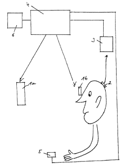
FIG. 1 shows a device with a stimulator 1 (la, lb) in
front of which a patient is seated. On the head of the patient a
sensor 2 is applied, the sensor 2 being connected by an isolating
amplifier 3 to a control unit 4. Tle device comprises a receiver
5, which also is connected to the ccntrol unit 4 and which can
register the reactions of the patieEt. In addition, the device
encompasses a means for monitoring the stimulation 6 which is
applied over a means for data processing and for displaying the
data so that the results can be visually and/or auditorially
delivered to the investigator. The control unit 4 is connected
with the means 6 for monitoring the stimulation. The sensor 2, the
receiver 5, the stimulator 1 and the means 6 for monitoring the
stimulation can also be in a contact-free connection with the
control unit 4, for example through transmitters and receivers.
FIG. 2 shows a schematic pattern of a pulse sequence for
a repetitive application. This pulse sequence has a periodic
succession of pulses and is followed by a desynchronization pulse
(last pulse). The frequency of the periodic pulse sequence is the
resonance frequency of the rhythm to be desynchronized. The
- 5
CA 02493305 2005-01-27
23158 Transl. of
PCT/DE2003/002250
purpose of the pulse sequence is to effect an entrainment which
controls the phase dynamic of the rhythm to be desynchronized.
After a constant time interval, the desynchronization pulse is
applied in the vulnerable phase of the neutral rhythm. The
abscissa is a time axis in any optionally selected unit while the
ordinate gives an intensity of the excitation also in any selected
unit.
FIG. 3a again has an absc:.ssa formed by a time axis in
any chosen units and an ordinate which gives an intensity of the
excitation also in selective units. The time segments Tl and T2 as
well as T4 and T5 correspond to the configuration in FIG. 2. In the
time segment T5, a periodic excitation sequence is supplied whose
frequency differs from the resonance! frequency of the neural
population to be desynchronized. Ix. the time segments Tl and T2 as
well as T4 and T5, the desynchronizing stimulation illustrated in
FIG. 2 are respectively carried out.
In FIG. 3b, the abscissa has a time axis which has the
same time units as in FIG. 3a. The ordinate indicates
schematically the amplitude as a function of time in a sliding time
window of the rhythm to be desynchronized in optional units. The
time segments T'k are identical with the time segments Tks whereby k
= 1, 2, 3, 4, 5. During the entrainment in time segment T1, apart
from a control of the phase dynamic there is additionally a
resonance-like amplification of the amplitude. The desynchronizing
individual excitation in the time segment T'2 encounters the
neuronal rhythm in its vulnerable phase and desynchronizes it so
that at the end of this stimulation the amplitude is minimal. In
- 6 -
CA 02493305 2005-01-27
23158 Transl. of
PCT/DE2003/002250
time segment T3 there is further sensor stimulus so that the
patient can accomplish his goals, for example, the detection of
special patterns in an on-going manner. To maintain the
suppression of the pathological rhythm as long a possible, in the
time segment T'3, an excitation is periodically applied at a
frequency different from the resonant frequency. As soon as the
amplitude of the desynchronized rhythm again exceeds a threshold
value, the desynchronization step iv carried out anew so that the
stimulation in the time segments T' and T'5 is identical with the
stimulation in the time segments T'l and T'2.
In FIG. 4a the abscissa ic the time axis in arbitrary
units and the ordinate gives the intensity of the stimulation also
in arbitrary units. FIG. 4a shows schematically the stimulation
used for the frequency scan. In this case a periodic excitation
sequence is applied whose frequency varies slowly and in this
example slowly increases.
In FIG. 4b, the abscissa is the same time axis with the
same units as in FIG. 4a. The ordirate indicates schematically in
a sliding time window the amplitude obtained with time of the
rhythm to be desynchronized, also ix. arbitrarily chosen units.
Corresponding to the excitation frequency which is illustrated by
the pulse sequence shown in FIG. 4a, a resonance frequency is
produced, i.e. a resonance is generated in which the amplitude of
the neuronal rhythm increases. FIG. 5 illustrates phase resetting
curves in which 0. over 03, is illustzated. 0. is the phase of the
neuronal activity determined directly following stimulation or at a
constant time delay after stimulaticn. 03, is the phase of the
- 7 -
CA 02493305 2005-01-27
23158 Trans].. of
PCT/DE2003/002250
neuronal activity determined either directly at the point in time
at which the stimulation commences r at a constant time interval
prior to the commencement of stimulation. The phases 0. and 0b are
given in radians. Each partial Figure a) - f) corresponds to a
series of test excitations with the same stimulus, that is an
excitation with constant intensity and excitation duration, applied
with different values of the starting phase 0b. The effect of the
excitation on the phase dynamics for the neuronal rhythm to be
desynchronized was evaluated by means of the phase resetting
curves. In the partial Figures a) to c), the mean gradient of the
curve was equal to 1, while in partial Figures d) to f), the mean
gradient of the phase resetting curves were equal to zero. By a
"mean gradient" the gradient obtained over a period of (6 is meant.
The transition between a phase resetting curve with a mean gradient
1 to a phase resetting curve with a mean gradient equal to zero is
found between partial FIGS. c and d in the region of the vertical
arrow with respect to the previously elevated phase 0b. This value
of the phase Pb is the vulnerable phase of the neuronal rhythm to be
desynchronized. The optimum value for the intensity lies between
the two intensity values of partial Figures c) and d). To obtain
this value one can either select vat iations approximating the
intensities of c) and d) or precisely generating still further
phase resetting curves with intensity values between those of c)
and d).
FIG. 6: flow diagram of the method of the invention.
- 8 -
CA 02493305 2005-01-27
23158 Transl. of
PCT/DE2003/002250
FIG. 6 shows a flow diagrim of the method of the
invention.
Initially there is a determination of the frequency
spectrum under spontaneous conditions (1), that is without
stimulation, whereby the patient is destressed and for example has
his or her eyes open for 5 minutes and the eyes closed over a
further period of 5 minutes. with open or closed eyes, respective
brain rhythms which are especially ntrong or especially weak are
obtained. For example the a rhythm is typically more strongly
expressed with closed eyes and more weakly expressed by contrast
with open eyes. A strong expression of a neuronal rhythm means
that this rhythm especially has a lcrge amplitude. In this manner
the point width of the expression of the physiological or
pathological rhythms which arise without stimulation can be
determined.
Next a frequency scan is carried out (evaluation of the
strength of the resonance by means et an amplitude determination of
the excited rhythm), possibly together with determination of the
quality of the entrainment over determination of the strength of
the phase synchronization between the excitation sequence and the
excited rhythm.
Depending upon the results from (1) and (2), either of
two different processes develop. In case the patient's natural and
nonpathological rhythmic activity is too weakly expressed or is
mainly not present, a need-controlled synchronization is carried
out in steps (3-5). In case the patent has a pathological rhythmic
- 9 -
CA 02493305 2005-01-27
23158 Transl. of
PCT/DE2003/002250
activity, a need-controlled desynch:mnization is carried out in
steps (6-9).
The need-controlled synch:mnization (3) can be carried
out in turn in two ways: in the context of a simple control
function, at the beginning of a senuory stimulation the excitation
frequency fA and the intensity are established and maintained
constant during the stimulation (4) In a preferred embodiment of
the invention the stimulation is corunenced by values suitable for
step (2) of the excitation frequency fA and the intensity (5). The
control unit 4 matches however in this mode the parameters
(especially the intensity) as controlled by need.
For the need-controlled desynchronization, initially the
quality of the entrainment evaluated (6) and then a determination
is made of the vulnerable phase (7), which - as described below -
is associated with a determination of the optimum excitation
intensity or excitation duration. The need-controlled
desynchronization can then be effected in two ways: either a
repetitive application of the sensory stimuli (8) is carried out or
a restraining application is carrieC out (9). During repetitive
application (8), the same desynchrmizing excitation sequence is
repetitively supplied whereas in the, pauses therebetween no
excitation is effected. During the continuous application (9) by
contrast, sensory stimuli are contiruously applied and upon
exceeding the threshold of the neurcual activity to be
desynchronized, the same desynchronizing excitation sequence is
always applied.
- 10 -
CA 02493305 2005-01-27
23158 Transl. of
PCT/DE2003/002250
In practically all of the steps, through the means for
visualization (FIGS. 1 and 6) a feedback to the investigator can
and should be provided.
Below the components of the device according to the
invention are deScribed in detail and their functions explained.
The stimulator 1 is an excitation pulse generator which
produces signals which can be conscLously or unconsciously
perceptible to the patient. Basica:Ay in this manner all signals
which can be sensorially processed by patients can be generated.
For example, visual excitation signals, acoustic excitation signals
or signals which excite the sense of taste or, less probably,
signals which evoke the pain sense can be mentioned. Visual
excitations can include images or patterns. The visual excitations
can be outputted, for example, through a special display screen la
or spectacles or glasses provided wi.th shutters lb. The display
screen can for example be a projecti.on screen which through a
shutter diaphragm with a projector which displays a continuous
image over time, provides the sensory response. The light-blocking
mechanism for the shutter glasses or spectacles and the shutter for
the projector screen can operate preferably either in accordance
with the LCD technique or FLC (ferroelectric liquid crystal)
technique. The images and patterns which are used to evoke the
visual responses can be those known to the artisan. They can be,
for example, Kanisza figures.
All tones or complex noises or sounds can be used as
acoustic stimuli, like for example i.terations of time-delayed broad
band noise or sounds in the audible frequency range which can be
- 11 -
CA 02493305 2005-01-27
23158 Transl. of
PCT/DE2003/002250
outputted by a loudspeaker lc or heed phones 1d. An excitation
stimulator which can excite the sounds of taste or pain sensitivity
can for example be a somatosensoric stimulation generator le or a
time-modulated laser if. An excitation generator in the sense of
the invention is thus a device for producing a visual, acoustic or
another sensory signal or stimulus. The stimulator 1 can output
the signals described in a time-based pattern either rhythmically
or arrythmically. This means that visual images or patterns can be
produced in a periodic sequence in time-spaced intervals of
preferably 1 to 100 Hz or 1 to 70 11.7, and/or in complex nonperiodic
time-based sequences although the application is not limited to
these frequencies. Furthermore the intensities or amplitudes of
the signals can also be varied. With visual excitations, not only
can the brightness be varied but the contrast can be varied as
well. Analogously tones can be applied in a periodic time-based
sequence of preferably 1 to 100 Hz end/or in complex nonperiodic
time-based sequences. In addition, the sound amplitude can be
varied. Analogously the same applies for the means for generating
the other sensory stimuli in which pressure and frequency can be
varied. The complex nonperiodic time-based sequence of individual
sensory excitations can, as described below, derive for example
from a combination of a periodic excitation sequence with
subsequent qualitatively individual excitations.
In health there is typicany rhythmic activity in certain
frequency bands and which arises in certain brain areas, for
example one can observe so-called a rhythm (ca. 10 Hz)
preferentially in the region of the visual cortex. In patients,
- 12 -
CA 02493305 2005-01-27
23158 Transl. of
PCT/DE2003/002250
these physiological rhythms on the Dile hand may be less expressed
or pronounced or on the other hand may have pathological rhythms
present in them and which are charaterized by nontypical, meaning
nonphysiological frequency bands. A pathological rhythm can also
be characterized by a normal frequency content but nontypical
anatomical localization. A pathological rhythm need not only be
limited to a single brain region but can also affect other
anatomically connected brain region a by feeding the pathologically
rhythmic activity thereto and affecting their functions.
The frequency content of the brain activity of the
patient has been characterized by the investigator, physiological
rhythms which are insufficiently divtinct can be excited or
excessively pronounced pathological rhythms can be suppressed or
weakened. If pathological rhythms axe weakly expressed or
pronounced, predominantly periodic ctimuli, which are outputted by
the stimulator 1, can excite these rhythms. In a further step,
through stimuli a desynchronization of the pathological rhythmic
activity can be effected. Then the signal sequences which are to
effect the desynchronization can differ from those which enable the
analysis or diagnosis in that these may tend to increase the
pathological rhythmic activity. Fox desynchronization at least one
desynchronizing pulse is produced.
The signals which are outputted by the stimulator 1
modulate rhythmic activity in certain brain areas in a manner which
can be detected by the sensor 2. The sensor 2 is in this sense a
means for detecting brain activity. As examples of them, scalp-EEG
electrodes are MEG sensors, that is SQUIDS, can be mentioned. The
- 13 -
CA 02493305 2005-01-27
23158
Transl. of PCT/DE2003/002250
apparatus is equipped according to :he invention with at least one
sensor which is connected with the =mntrol unit 4.
The control unit 4 procesms the signal obtained from the
sensor 2. The control unit 4 operaes through means for carrying
out the processed steps which have been described in the
application. This means can be especially a computer or an
electronic circuit together with a oomputer programmer a
programmable processor like, for example, a FPGA (field
programmable array) which is capable of carrying out the steps
according to the invention of signa:. collection and evaluation and
can control the stimulator 1 in the manner required by the
invention.
It is especially advantageous to be able to practice the
method with suitable processors. The term "processor" should not
however be understood to be limited in any sense. It can be for
example any optional unit suitable for carrying out computations.
It is possible for the processor to comprise a multiplicity of
individual processors which are advantageously assembled into an
appropriate processor unit.
In the sense of the present invention, in addition, any
circuitry suitable for computation can be used. Advantageously,
the circuit can be built into a computer or incorporated in a logic
component. The means of the descristion for carrying out the
method steps of the invention are ccmponents of the control unit 4
encompassing at least one component from the group comprised of a
computer, an electronic circuit, a computer program or a processor.
- 14
CA 02493305 2005-01-27
23158 Transl. of
PCT/DE2003/002250
The means for controlling the diffe:Nant method steps need not
however be provided in a single dev:Lce.
The control unit 4 determ.Lnes the degree of expression or
development of a pathological rhytlunic activity. If the
pathological activity is not present or is present only minimally,
the control unit 4 provides control signals to the stimulator which
can then output either no stimuli or other stimuli which differ
either in frequency or amplitude or in frequency and amplitude from
prior stimuli. In a diagnostic app:Acation the frequency and/or
amplitude of the stimuli are varied until the pathological reaction
is a maximum, that means that the rhythmic reaction of the
pathological brain area is the strongest. This has the advantage
that otherwise possibly nonnoticeab:.e pathological rhythms can be
recognized under certain conditions in case at the point in time of
the diagnostic investigation they might otherwise be too weak. In
this case the control unit operates through means capable of
calling up a maximum physiological end/or pathological brain
activity. This means operating for example through an electronic
circuit, a processor or a computer end associated software, ensures
that stimulation sequences are provided as described below. The
pathological rhythmic activity pattern is analyzed by the control
unit 4. The control unit 4 is adapted to provide another time-
based pattern of the stimulus which is targeted to modulate the
pathological activity and especially to suppress the pathological
activity pattern or to attenuate it. Thus, opposite to the first
effect, namely the promotion of the pathological activity, damping
and, especially preferably, a complete suppression of the
- 15 -
CA 02493305 2005-01-27
23158 Transl. of
PCT/DE2003/002250
pathological brain activity is effected. The sensor 2 continues to
detect the brain activity and with the control unit 4 analyzes the
new state of the brain. Through a number of cycles of this type
the control unit 4 is able to deternine the stimuli with which the
pathological conditions can be suppressed as completely as
possible.
The receiver 5, which serves for patient control is
connected with the control unit 4. The receiver 5, in the sense of
the invention can be for example a push button or a switch or lever
which is actuated by the patient. The patient is instructed to
actuate the receiver 5 in response to certain signals. In this
manner the ability of the patient tcl perceive the sensory stimuli
or the treatment effect and the reaction to the procedure can be
controlled. The signals from receiver 5 are computed or processed
in the control unit 4 and are transmitted to the means 6 for
monitoring the stimulation. Througt. these means 6 the investigator
can determine the quality of the stimulation and the result of its
application to the patient. The deice according to the invention
equipped with the receiver 5 and the means 6 for monitoring
stimulation constitutes thus a preferred embodiment of the
invention.
In the application of the apparatus, two cases A and B
can be distinguished and are exploited below by way of example.
A: For patients who naturallN have nonpathological rhythmic
activity which is expressed too weakly or primarily or is
usually not present.
- 16 -
CA 02493305 2011-11-24
52398-3
B: The patient presents with a pathological rhythmic
activity in at least one region of the brain.
In cases A and B, the control unit 4 operates in the
following manner:
Frequency Scan:
The frequency scan both in case A and in case B is
carried out initially. In the frequency scan, a periodic sensory
stimulation with an excitation frequency f, is carried out in which
IA varies slowly between preferably 1 and 100 Hz, especially
preferably between 1 to 60 Hz. In FIG. 4a this has been reproduced
by way of example with an increasing frequency of the applied
signal sequence. Sensor 2 measures the neuronal activity and
supplies it to the control unit 4 which determines in which
frequency range the neuronal' activity develops an excitation. This-
excitation can then be quantified by
(i) integrating the amplitude of the power spectrum over the
excited frequency range or, analogously thereto
(ii) determining the instantaneous amplitude of the frequency range
by means of the Hilbert transformation.
The device of the invention thus comprises means for
carrying out a frequency scan as well as means for carrying out the
step (i) and/or (ii).
- 17 -
=
CA 02493305 2005-01-27
23158 Transl. of
PCT/DE2003/002250
The electronic circuitry used for example for this
purpose or equivalent means in control unit 4, as well as a
computer program, which for example operates in accordance with the
methods (i) and (ii) can serve as the means for quantifying the
neuronal activity.
This frequency scan can bit carried out by the control 4
which activates the means for generating sensor stimuli 1 so that
the respective frequency is reproduced in the patient in the form
of a sensory stimulus. For this purpose the control unit 4 can act
through means for controlling the stimulator 2, for example a TTL
pulse generator. The control unit C recognizes the signals
captured by the sensor 2 or their amplitudes in the investigative
frequency range at which the excitation frequency produces a
maximum excitation. The device thuy comprises advantageously such
means which is capable of investigating in the signals measured by
the sensor 2 apart form the frequency range of the excitation
frequency also other frequency rangcs. This means can carry out
time-dependent frequency analysis based upon Fourier transformation
or wavelet analysis. For this purpcise the control unit 4 comprises
a means which is suitable for carrying out these steps. Such means
can be as has been described above Ily say of example, a computer,
an electronic circuit, a processor, a programmable electronic
circuit (FPGA) or a computer program. The frequency of the excited
activity can thus coincide with the excitation frequency or can
also not coincide therewith. Surprisingly it has been found that
the frequency of the periodic stimu3us sequence which serves for
entrainment follows the law given bclow:
- 18 -
CA 02493305 2005-01-27
23158 Transl. of
PCT/DE2003/002250
f ofit fA n m Formula 1
where fA = the excitation frequency, that is the frequency of the
periodic stimulus sequence serving for excitation
fR = the frequency of the exc:.ted neuronal activity
(resonance frequency)
whereby n and m are small whole num:Ders, that means < 10, (namely
1, 2, 3, 4, 5, 6, 7, 8, 9, 10) for ,?.xample, n/m = 1/1, 1/2, 2/3
etc.=
With the aid of the frequency scan, two aspects of the
excitation properties are explored.
1. A determination is made as to whether an excitation
will bring about a physiological rhythm in a frequency range
expected for this rhythm. With flicker light stimulation, these
frequency ranges can be for example in the region of 10 Hz, 20 Hz,
40 Hz and 80 Hz. In this case a determination is made whether a
physiological rhythm, which may be cd pathological origin or can
develop spontaneously, that is withcut stimulation, is too weakly
expressed, can be excited by periodic stimulation.
2. A determination is mad.e as to whether an excitation
will lead to a pathological rhythm. The latter is characterized by
a physiological response that does rot lie in a physiological
frequency range or is in a physiological frequency range but arises
at an untypical brain region. The physiological frequency ranges
are the frequency ranges at which neuronal rhythms naturally occur.
- 19 -
CA 02493305 2005-01-27
23158 Transl. of
FCT/DE2003/002250
For example, the a rhythm in the reaion of about 10 Hz and p rhythm
in the region of about 20 Hz can be mentioned. In this manner a
determination is made as to whether a pathologically generated
rhythm can be produced by a periodi: stimulation. Such
pathological rhythm is typically, although not necessarily already
present under spontaneous conditions, that is without stimulation.
After the frequency scan Is carried out as described
above, the application of the invention is effected in accordance
with cases A and B below.
A. Need-Controlled Synchronization:
The goal of the need-controlled synchronization is, with
patients who have one or more too woakly expressed physiological
rhythms to excite that by sensor st:Jauli during treatment. For
this purpose the stimulus treatment which is found to be required
because of the weakened physiologicul rhythm is improved or
enabled. For this purpose the sensor 2 registers the neuronal
activity of the brain area to be excited. The signals measured by
the sensor 2 are advantageously supplied to the control unit 4
through an isolating amplifier 3. The control unit 4 can then
control the senor stimulation in two different ways:
1.) In the framework of a, simple control function, at
the beginning of the sensory stimulation the sensory stimuli or
pulses are applied with an excitation frequency 12, and the
intensity according to the results of the frequency scan. These
- 20 -
CA 02493305 2005-01-27
23158 Transl. of
PCT/DE2003/002250
stimulation parameters remain constant during the sensory
excitation.
2.) As under (1), the exiAtation is commenced with an
excitation frequency fA and the intcnsities which are appropriate
from the results of the frequency soan. The control unit 4 matches
these parameters during the sensory excitation under need control.
That means that the control unit 4 :Naacts to a decrease in the
amplitude of the rhythm to be excitod with an increase in the
intensity of the exciting stimulus. In this case the control unit
4 acts through means for registering the change in the amplitude of
the rhythm which is to be excited to change the excitation
intensity. For this purpose as has been described by way of
example above, a computer, an electronic circuit, a processor, a
programmable electronic circuit (FPGA) or a computer program can be
used. The range of intensities used in this case is limited at its
upper level by safety consideration, this means an avoidance of the
triggering of an epileptic response.
During the sensory stimulation described under (1) or
(2), the patient is subjected to defined stimuli like, for example
Kanisza figures. The patient is previously instructed to look for
special features in these stimuli. By feedback over the push
button 5, the patient can control whether the recognition of the
sensory stimulus to which the patient is subjected is improved or
enabled by the excitation of the physiological rhythm. By at least
one and preferably three hiatuses in the reaction of the patient, a
suitable signal from the control unit 4 is provided to the means 6
for monitoring the stimulation and thus supplied to the
- 21 -
CA 02493305 2005-01-27
23158 Transl. of
PCT/DE2003/002250
investigator. This signal serves to let the investigator know when
the patient is not willing or is not in a position to process the
sensory stimulation in accordance with the predetermined
requirements as set out above.
B. Need-Controlled Dasynchronization:
The goal of the need-controlled desynchronization is to
damp or suppress one or more pathological rhythms which may be too
strongly active or expressed during the processing of sensory
stimuli. For this purpose, the stimalus processing which may be
destroyed by an excessively expressel neuronal rhythm should be
improved or enabled. This result is achieved with the device
according to the invention and especially with the control unit 4
or the above-described means forming part thereof and functioning
as described in the following.
The sensor 2 registers, for this purpose, the neuronal
activity of the brain area to be danved. The signals measured by
the sensor 2 are supplied to the con:rol unit 4, preferably through
the isolating amplifier. The control unit 4 operates, according to
the invention, in accordance with tha following principle:
A rhythmically active neur,mal population can be
desynchronized with a sensory stimulas when the stimulus or
excitation on the one hand has the cf=rect intensity and duration
and, on the other hand, is applied ia a critical phase of the
corrective oscillation of the neuronal population, the so-called
vulnerable phase. Because of the unavoidable variability of the
frequency of a neuronal population, It is difficult to determine
- 22 -
CA 02493305 2005-01-27
23158 Transl. of
PCT/DE2003/002250
precisely the vulnerable phase. Th43 problem is solved in
accordance with the invention by th13 use of complex stimuli. These
are comprised of two qualitatively different stimuli or excitations
or pulses:
The first stimulus contro:Ls the dynamics of the neuronal
population such that at the end of this stimulus the dynamic state
of the neuron population is known sc.th sufficient precision. For
this purpose an entrainment is carr:.ed out, that is, an entraining
periodic stimulus for excitation sequence is applied in order to
bring the dynamics of the neuron population into step with the
stimulus sequence. To this end, the device according to the
invention, through means for effecting an entrainment, that is a
periodic stimulation for the purpose of controlling the rhythm,
meaning the phase dynamic, can control the excitation of the
neuronal activity. This can be achieved as has been indicated in
greater detail by example above, with a computer, an electronic
circuit, a processor, a programmable. electronic circuit (FPGA) or a
computer program.
The second stimulus or excitation follows the first,
entraining stimulation (the stimulation sequence) with a
substantially constant time lag. It encounters the pathologically
synchronized, neuronal population iE a vulnerable state and gives
rise, in this way, to a desynchronization. The second stimulus or
excitation pulse is comprised advantageously of only a single
stimulus or excitation pulse, or a short periodic stimulus or pulse
sequence, which can be comprised of at least two individual stimuli
or pulses and advantageously not more than ten individual stimuli
- 23 -
CA 02493305 2005-01-27
23158 Transl. of
PCT/DE2003/002250
or excitation pulses. To this end, the device of the invention is
provided with means for desynchronillation. Such means, as has also
been indicated by example above, can be a computer, an electronic
circuit, a processor, a programmablo electronic circuit (FPGA) or a
computer program which is capable of carrying out the process steps
described below.
The stimulation parameters; required for the
desynchronization are determined in accordance with the invention
by the following standardization prcicedure.
1.) Monitoring the quality of the entrainment:
a stimulus for excitation pulse sequence comprised of k preferably
identical stimuli or excitation pulses are applied one time,
preferably ten to one hundred times. In this case, n small values
are varied as above until the entrainment is good enough. The
quality of the entrainment is then investigated in the following
manner or quantified:
the phase and the amplitude of the neuronal rhythm to be
desynchronized are determined preferably by the Hilbert
transformation. An alternative method can be the matching in a
sliding time window of the signal of the neuronal rhythm with a
slowly varied sine function. For this purpose, the device
according to the invention is provided with means for testing the
quality of the entrainment. Such means can be, as has been
indicated in an exemplary way above, a computer, an electronic
circuit, a processor, a programmable electronic circuit (FPGA) or a
computer program which carries out the described steps.
- 24 -
CA 02493305 2005-01-27
23158 Transl. of
PCT/DE2003/002250
The effect of the entrainment is that after the
entraining stimulation, the neuronal rhythm will always have the
same amplitude and above all always the same phase, independent of
the amplitude and the phase at the beginning of the stimulation.
To ensure that this will be the case, the phase or preferably the
phase and amplitude are evaluated by means for evaluating phase and
amplitude or in a less preferred embodiment of the invention,
exclusively by means for evaluating the phase of the neuronal
rhythm in the following manner. For this purpose as has been
described above by way of example, the described steps can be
carried out by a computer, an electronic circuit, a processor, a
programmable electronic circuit (FPGA) or a computer program.
For the first applied stimulus or excitation pulse
sequence, which is comprised of n individual stimuli or excitation
pulses, the means for carrying out a. phase resetting can produce a
so called phase-resetting curve. A phase resetting curve is a
phase response curve in which the plasm at the end of the
stimulation for all m applied excitation or stimuli sequences. A
perfect entrainment is obtained when a horizontal phase resetting
curves, that is a phase resetting curve which is independent of the
phase at the beginning of the stimulation always assumes the same
value as the phase at the end of the stimulation.
The phase resetting curve can be displayed to the
researcher for example by a display screen forming a means for
visualization 6. On the other hand, the phase resetting curve can
be used to evaluate the value of the phase at the end of the
stimulation, for example by a simple mathematical operation like
- 25
CA 02493305 2005-01-27
23158 Transl. of
PCT/DE2003/002250
determination of the standard deviation of the value of the phase,
or the quality of the match of a horizontal line to the phase
resetting curve by means for the quuntitative characterization of
the phase resetting curve. Such meuns, as has also been indicated
in an exemplary manner above, can bo a computer, an electronic
circuit, a processor, a programmable! electronic circuit (FPGA) or a
computer program which is designed to carry out the described
steps.
Preferably the quality of the entrainment is determined
exclusively visually by the investisator through the means for
monitoring the stimulation 6. The amplitude is determined in the
same way by means of amplitude resetting curves. The device of the
invention can then include means fox determining the amplitude and
for carrying out an amplitude resetting which operates in the
following manner. For this purpose the apparatus can include, as
was described above in an exemplary manner, a computer, an
electronic circuit, a processor, a programmable electronic circuit
(FPGA) or a computer program which can carry out the aforedescribed
steps. In the amplitude-resetting curves, that is amplitude
response curves, the amplitude at the end of the stimulation is
plotted against the amplitude at the beginning of the stimulation
for all m applied stimulation or excitation sequences. A perfect
entrainment leads to a horizontal amplitude resetting curve, that
means, independent from the amplitude, at the beginning of the
stimulation the amplitude at the end of the stimulation will assume
always the same value. The amplitude resetting curve is evaluated
- 26 -
CA 02493305 2005-01-27
23158 Transl. of
PCT/DE2003/002250
like the phase resetting curves quantitatively and/or and
preferably only visually.
The number of stimuli or excitation pulses following one
another in a pulse sequence k is in=eased until the entrainment is
sufficiently good in terms of ampli:ude and phase.
In an alternative and preEerred embodiment of the
invention the quality of the entrainment is examined in the
following manner and quantified. The goal of this alternative
procedure is to monitor the quality of the entrainment not only at
the end but during the application of the entire stimulus or
excitation sequence. This makes the determination of the quality
less dependent on fluctuation of the measured neuronal dynamic
which can be affected either by the measurement process or above
all by intrinsic neuronal noise. For this purpose, a stimulus
sequence comprised of k preferably identical stimuli or excitation
pulses is applied one time and prefcrably ten to one hundred times.
K is then varied by small values as described above until the
entrainment is good enough. The quality of the entrainment is
investigated or quantified in the fcllowing manner:
The signal representing tke excited neuronal activity
measured by the sensor two is filtered in a band pass filter which
completely contains the frequency peak fit (formula 1) of the
resonance frequency but does not contain other frequency peaks, the
harmonics, subharmonic or other physiological or pathological
rhythms. Using the Hilbert transformation, the phase 01, that is
the phase which is determined from the band pass filtered signal in
this manner. Apart from this, the phase 14:1A is determined, that is
- 27
CA 02493305 2005-01-27
23158 Transl. of
PCT/DE2003/002250
the phase of the excitation stimuluv sequence or excitation
sequence. This can be achieved in two ways: either a sine
function can be matched to the stimulus sequence such that the
maximum of the signed function will coincide with the point in time
at which the individual stimulus is applied. The phase 0A is then
the phase of the matched sine function. Alternatively, the signal
which represents the stimulus sequeLce and thus the sequence of
rectangular pulses which correspond to the excitation frequency fA
of formula 1 and the band pass filter is selected. The phase 0A is
then the phase determined by the Hilbert transformation of the band
pass filter signal of the stimulus Eequence. The band pass used
for this purpose must be so selected that it completely encompasses
the frequency peak fA in the spectrun of the signal of the
excitation sequence but contains no other frequency peak. Then the
n:m phase difference nos, - mob between the excitation stimulus
sequence and the excited neuronal activity is determined. The
strength of the entrainment is then preferably determined by means
of the n:m - entrainment index whicb is defined as follows: in the
time window used for determining the quality of the entrainment,
the distribution of the n:m phase difference is determined. The
entropy S of this distribution is tben determined according to the
formula 2
S = -E pk ln pk (Formula 2)
k=1
- 28 -
CA 02493305 2005-01-27
23158 Transl. of
PCT/DE2003/002250
whereby, pk is the relative probability that the value of the n:m
phase difference will be found in the k th case bin. The number of
the bins N is typically determined :L.n accordance with formula 3:
N= exp[0.626+0.4141(M-1)] (Formula 3)
whereby M is the number of measured values of the n:m phase
difference during a stimulus sequence.
The n:m entrainment index e is calculated in accordance
with formula 4
Sma, -s
e - _______________________________________________ (Formula 4)
whereby S is the entropy of an equilibrium, that is S = in
whereby the optimum number for deternining the distribution in
terms of equidistance partial intervsls or bins is given by the
formula 3. Through the means of fornula 4 a normalization is
carried out such that 0 s eno, s 1. = 0 means that no
entrainment is present while en,. = 1 corresponds to a perfect
entrainment. The larger the value oE en,,, the better is the
entrainment.
Values of en,. are obtained for each applied stimulus
sequence. From that value the mean 7alue
- 29 -
CA 02493305 2005-01-27
23158 Transl. of
PCT/DE2003/002250
En,m = -FE ecl) (Formula 5)
n vm
P4
is calculated whereby e is the j-.:11 stimulus sequence. The
relationship 0 s E s 1 applies. The number of the k stimuli or
excitation pulses in a stimulus sequence following one another is
increased until the entrainment is sufficiently good, that is until
E,. sufficiently approaches one.
2.) Determining the vulnerable phase:
The vulnerable phase depends upon the intensity and the
duration of the sensory stimulation. Advantageously the duration
of the sensory stimulus is held constant in the frame work of the
standardization procedure while the intensity and the vulnerable
phase are so varied, as described below, that the desynchronizing
effect of the stimulation is maximized.
The determination of the vulnerable phase is carried out
with means for determining the vulnerable phase. Such means, as
has been indicated above by way of emample previously, can be a
computer, an electronic circuit, a processor, a programmable
electronic circuit (FPGA) or a computer program which can carry out
the steps described in the following. In this case the device
according to the invention can operate in two different ways:
A) the time spacing between the last stimulus or
excitation pulse of the entraining stimulus or excitation pulse of
sequence and the desynchronizing pulse on the one hand and the
- 30
CA 02493305 2005-01-27
23158 Transl. of
PCT/DE2003/002250
intensity of the desynchronizing stimulus are varied by means for
varying the time spacing between th3 last stimulus of the
entrainment and the desynchronizing stimulus between preferably 0
and 2 period lengths of the mean froaquency of the frequency band
associated with the pathological rhythm in a systematic manner,
preferably in small equidistant steps. The means used for this
purpose as has been described by way of example above, can be a
computer, an electronic circuit, a processor, a programmable
electronic circuit (FPGA) or a computer program. This variation in
the time spacing is carried out systematically for different values
of the intensity by means of a meant; for varying the intensity.
Preferably the intensity is increased in small equidistant steps
and for each value of the intensity, the time spacing is determined
as described above between 0 and 2 period lengths. The variation
of the time spacing and the intensity is carried out preferably by
the control unit 4. The optimum value for the intensity of the
sensory stimulus and the time spacirg between the last stimulus of
the entrainment and the desynchronizing stimulus is the value at
which the strongest desynchronizaticn effect arises, that is the
amplitude at which the desynchronized rhythm after stimulation is
the smallest. The amplitude is preferably determined by band pass
filtration with subsequent Hilbert transformation.
Alternatively, the amplitude can be determined either by
a matching of a slowly varying sine function to the band pass
filtered signal of sensor two in a time window after stimulation or
by determining the integrated amplitude over the frequency band of
- 31 -
CA 02493305 2005-01-27
23158 Transl. of
PCT/DE2003/002250
the power spectrum of sensor two in a time window after
stimulation.
B) The time spacing is varied under A). Differing from
A), the intensity is not increased in equidistant steps but is
varied systematically in the following way: in this case phase
resetting curves are used with which the effect of the
desynchronizing stimulus on the phaae dynamics of the neuronal
activity to be desynchronized is in,restigated. The phase is
advantageously determined by means of band pass filtration and a
subsequent Hilbert transformation of the signals measured by the
sensor two. Alternatively to the uoe of the Hilbert
transformation, a slowly varying sine function in a sliding time
window can be matched to the band pcss filter signal of sensor two.
The limits of the pass band can then be the limits of the
frequency band of the pathological neuronal rhythm which is
determined at the outset. When reference is made to the phase
resetting curves, 0, over 01, are obtained by a means for applying 0,,
the phase of the neuronal activity after stimulation, over 016, the
phase of the neuronal activity at the beginning of the stimulation.
Such means can be a means for investigating the effect of the
desynchronizing stimulus on the phase dynamics of the neuronal
activity to be desynchronized. Such means can as has been
indicated above by way of example, be a computer, an electronic
circuit, a processor, a programmable electronic circuit (FPGA) or a
computer program.
0, is thus the phase of tho neuronal activity which is
determined either directly after stimulation or with a constant
- 32 -
CA 02493305 2005-01-27
23158 Transl. of
PCT/DE2003/002250
time delay following stimulation. 'Alio time delay should
preferably be smaller than one period length of the neuronal rhythm
to be desynchronized or better stil. is equal to 0. Since the
period length of the neuronal rhythn is varied with time, when the
reference is made above to period lengths, the period length
averaged over time is meant.
obis the phase of the neuronal activity which is
determined either directly at the point in time that the
stimulation commences or at a constant time interval prior to the
commencement of stimulation. The tine interval should be, by
analogy with the determination of 0., as small as possible or better
still equal to 0. The time interval in the determination of 0. or
0b should be as small as possible to ensure that time dependent
variation in the period length will not influence the quality of
the evaluation.
If the selected intensity of the sensory stimulus for
desynchronization is too small, the phase resetting curve will
typically have a mean rise of one. If, by contrast, the intensity
is too large, the phase resetting curve will typically have a mean
rise of 0.
The optimum intensity value and the optimum value for the
lag between the last entrainment pulse and the desynchronizing
pulse is determined with precision by the location in the phase
resetting curve at which the transition from a mean rise 1 to a
mean rise 0 occurs.
This has been shown in FIG. 5. FIG. 5a through 5f
respectfully show phase resetting cuxves, whereby in the individual
- 33 -
CA 02493305 2005-01-27
23158 Transl. of
PCT/DE2003/002250
partial figures, the intensity of Cie sensory stimulus is constant
but different from one of the partial figures to another and indeed
increases from the smallest value in FIG. 5a to the largest value
in FIG. 5f. The optimal stimulation parameter is thus found at the
transition from FIG. 5c through 5e at the location marked by the
arrow, (i) the mean value of the intensity belonging to FIGS. 5c
and 5d is optimum for the selected ntimulus duration at which the
desynchronization intensity is the ntrongest and (ii) the
inflection point indicated in FIG. !id with the arrow indicates the
phase 01, which is optimal for the selected stimulus duration to
which corresponds to the strongest desynchronizing time interval
between the last stimulus of the entrainment and the
desynchronizing stimulus. This timer interval can either be given
in absolute time values or, analogol,sly thereto as illustrated in
FIG. 5 in terms of the phase of the neuronal activity. With the
phase resetting curves it is possible to provide a x-axis
equivalent to 01, giving an absolute time interval between the last
excitation of the entrainment and tie desynchronizing excitation.
/f the experimental data are strongly affected by noise, the phase
resetting curve can be used to provide a pair of values comprised
of intensity and 01, by multiple measlarements and the mean value of
0, is then used.
The control unit 4 controls the sensory stimulation in
two different ways. The need-controlled desynchronization can
either be carried out repetitively or continuously. In both
functional methods, an entrainment is used for the effective
desynchronization. The frequency of the entrainment, that is the
- 34 -
CA 02493305 2005-01-27
23158 Transl. of
PCT/DE2003/002250
rate of the entraining sequence of sensory stimuli, is determined
in a previous frequency scan. In that frequency scan it is
determined which excitation frequency fA provides a maximum
amplitude of the pathological rhythz. If the excitation frequency
fA is identified or a plurality of mccitation frequencies are
identified, a desynchronization can be commenced. In the case that
a plurality of excitation frequencies are found which lead to
maxima in the amplitude of the pathological rhythm, the
desynchronzation is carried out for the one which has the strongest
entrainment effect, that is the strongest excitation
of the amplitude.
a) Repetitive Application:
In the repetitive application, the same desynchronizing
stimulus or excitation sequence is rApetitively applied. In the
pauses between these desynchronizing stimulus or excitation
sequences no stimulus or excitation Ls applied.
The patient is instructed l'efore commencement of the
need-control desynchronization by an investigator or by the device
itself. That means that the patient is either told by the
investigator how he or she should reapond to the repetitively
applied stimulation or excitation sequences or the device itself
can signal this to the patient by foE* example by visual or auditory
instruction: the patient hears or rends what he or she is to do.
For example, the patient must try, upon visual
stimulation with the repetitively applied visual stimulus pattern,
of certain objects or individual patterns, for example Kanisza
- 35 -
CA 02493305 2005-01-27
23158 Transl. of
PCT/DE2003/002250
figures to compare them with one another or count them. The
investigator controls, using the menns 6 for monitoring the
stimulation, preferably the effect of the stimulation of the brain
activity and the information procesning by the patient which is
determined by feedback via the push button 5. The patient must for
example, each time he recognizes a certain partial pattern, press
the push button 5. In this manner the investigator is able to
determine whether the applied sensory stimulus improves or enables
the damping or suppression of the pathological rhythm. If the
reaction of the patient is missed at least one time, an appropriate
signal is provided by the control uLit 4 to the means 6 for
monitoring the stimulation and thus is communicated to the
investigator. This signal serves tc inform the investigator that
the patient is not willing or is not capable of processing the
sensory stimulus in accordance with the predetermined conditions.
The control unit 4 controls the application of the
sensory stimuli in the following manner:
An entraining periodic sequence of sensory stimuli or
excitation is applied with the optimum excitation frequency fA.
The sensory stimuli or excitation here used can be identical
although they need not necessarily be identical. Preferably the
sensory stimuli used with respect to the following parameters are
identical in order to ensure an effective entrainment:
(i) They are of the same quality, that is that they deal
for example always with the same visual pattern.
- 36 -
= CA 02493305 2005-01-27
23156 Transl. of
PCT/DE2003/002250
(ii) They have the same intensity, that is for example the
same light or sound amplitude.
(iii) They have the same contrast, that is for example in the
case of visual stimuli, tie same light-dark contrast.
(iv) They have the same durati4m.
With a constant lag, there is thereafter effected an application of
the desynchronizing stimulus or exc:Ltation in a vulnerable phase
state of the pathological rhythm. !lie desynchronizing sensory
stimulus is preferably of the same nodality, that is, when the
entraining stimuli are visual stimuli, the desynchronizing stimulus
is also a visual stimulus and for example is without an auditory
stimulus.
The desynchronizing stimulus need not however be of the
same quality as the entraining stimuli. Preferably however it is
of the same quality, that is it uses: for example the same visual
pattern. The desynchronizing stimulus differs however preferably
from the stimuli of the entraining stimulus sequence by its
duration and/or its intensity and/or its contrast.
AS soon as the desynchronizing stimulus is applied, there
is a transitory period in which no stimulus is present. In
connection with this stimulus application, the patient
advantageously must signal via the rush button whether he has been
able to detect for example special cbjects or visual patterns
therein.
Following such a desynchronizing stimulus, there is a
pause whose duration can lie within a statistical distribution in a
- 37 -
CA 02493305 2005-01-27
23158 Transl. of
PCT/DE2003/002250
predetermined interval, preferably a uniform distribution. During
this pause no stimulus is applied. After this pause, there is a
repetition of the collective of desynchronizing stimuli, comprised
of another entraining stimulus sequence and an individual
desynchronizing stimulus.
In the framework of the repetitive application, the
control unit 4 determines whether the desynchronizing of the
pathologically active neuronal popuLation has been effected, that
is whether the damping of the patho2.ogical rhythm has been strong
enough. Should this be the case the repetition of the stimuli is
continued. If the damping of the pcthological rhythm is
insufficient at least once, restandardization must be carried out
with the above-described standardization procedure.
FIG. 2 shows an excitatior. with the resonance frequency
which is followed by a desynchronization pulse in the vulnerable
phase. In this Figure the x-axis represents the time and the y-
axis the intensity of the sensory stimulus.
b) Continuous Application:
By contrast with the repetitive application (a) in the
continuous application, there is a permanent sensory stimulus.
Whenever the neuronal activity exceeds a threshold of the above-
determined amplitude of the neuronal activity to be desynchronized,
a desynchronization is carried out. For this purpose an entraining
pulse sequence is followed by the application of at least one
single stimulus (FIG. 2). In the tine between the
- 38 -
= CA 02493305 2005-01-27
=
23158 Transl. of
PCT/DE2003/002250
desynchrizations, a continuous sensory stimulation applies. In
this case there are two possibilities:
I) In the time between the desynchronization,
stimulation is carried out with a poriodic sequence of sensory
stimuli or excitation pulses. This sequence is comprised of
identical individual stimuli which axe applied at a frequency
sufficiently different from the resonance frequency that no
resonance arises.
II) In the time between the desynchronization,
stimulation is applied with a random sequence of sensory stimuli or
excitation pulses. The individual stimuli of this sequence are
comprised of identical visual or auditory patterns in which the
following parameters are statistically varied from stimulus or
pulse to stimulus or pulse: with visual stimuli the contrast and/or
the brightness can be varied. With auditory stimuli the sound
volume can be varied. In addition, the pauses between the
individual stimuli and the duration of the individual stimuli can
be statistically varied. In the statistic variation, the
corresponding parameters can be varied between the normal
physiologically experimental limit or uniformly.
The purpose of the stimulation described above under I
and II is, 1. to continuously supply sensory stimuli to patients
which can be processed by them so th1t the patients can
continuously have available the required action, for example, the
- 39
= CA 02493305 2005-01-27
23158 Transl. of
PCT/DE2003/002250
detection of visual partial images, and 2. to prevent thereby a
resonance of the pathological rhytlui from developing.
Figure 3a shows a sequence of stimuli by way of example
for the means 1 for generating the sensory stimuli in the form of a
time-spaced application of patterns as the sensory stimuli whereby
variant I, that is a periodic stimu:Ation between the
desynchronization, is used. In Figure 3b the associated activity
pattern of the pathologically effected brain region has been given.
In Figures 3a and 3b the x-axis is the time axis in each. In FIG.
3a the intensity of the stimulus is plotted along the y- axis. In
Figure 3b the amplitude is a function of time in a sliding time
window for the neuronal activity to be desynchronized has been
plotted.
In Figures 3a and 3b, the time regions Tl and T11, T2 and
T'2, T3 and T'3, T4 and T'41, as well as T5 and T'5 are identical. In
the time regions Tl or T'1, the amplLtude of the pathological
rhythms because of resonance is a maximum. In the time regions T2
or T'2, a desynchronizing sensory stimulus is applied in the
vulnerable phase to either completely suppress the pathological
activity or at least reduce its intensity. This gives rise to a
drop in the amplitude in Figure 3b.
As has been described under I above in the time region T3
a periodic stimulus sequence is applied whose frequency differs
sufficiently from the resonance frequency in the time region Tl.
The effect in the time region T'3 is that in spite of the sensory
stimulation the pathological rhythms will recover only slowly.
- 40 -
CA 02493305 2005-01-27
23158
Transl. of PCT/DE2003/002250
In the second case II, in the time region T3, instead of
a periodic stimulus sequence a randtma or stochastic stimulus
sequence is used. With this feature the pathological rhythm is
suppressed as long as possible.
In Figure 3b this phase ia characterized by the segment
T'3 in which the curve of the brain activity to be suppressed
reaches its minimum value. As soon as the brain activity in the
time region T'3 again exceeds a threshold value, the need state for
desynchronization arises so that in the next time region T4 a new
desynchronization operation is carried out. For this purpose in
the time region T4 the same entrainment is effected as in the time
region Tl. Following the entrainment, in the time region T3, a
desynchronizing stimulus is applied as in the time region T2. For
this purpose the sensor 2 registers the increased activity of the
pathologically affected brain regior and reproduces the signal at
the control unit 4 which triggers tte next desynchronization. In
conjunction with the desynchronization effected in the time region
T3F a periodic stimulus sequence is applied anew as in the time
region T3, with a frequency sufficieatly different from the
resonance frequency. It corresponds to the above-described case I.
Alternatively thereto, also according to the above-described case
II, stochastic or random stimulus sequences can be used.
The invention comprises a computer program with program
code means for controlling a device which can carry out at least
one of the preceding method steps or optional combinations of at
least two of the method steps given in this description when the
program is run on a computer. The invention also encompasses a
- 41 -
=
CA 02493305 2005-01-27
23158 Trans'. of PCT/DE2003/002250
computer program product with program code means which is stored on
a computer-readable data carrier and permits the method to be
carried out as defined by that computer program. This computer-
program product can for example be u diskette. The invention also
comprises an electronic circuit which is suitable for carrying out
the instructions of the computer program or the computer program
product.
- 42