Note: Descriptions are shown in the official language in which they were submitted.
CA 02493655 2005-O1-19
TRANSGLUTAMINASE HAS INTRINSIC KINASE ACTIVITY
PRIOR APPLICATION INFORMATION
This application claims priority under 35 USC ~ 119(e) to
Provisional Patent Application Serial Number 60/539,594 filed on January 29,
2004.
FIELD OF THE INVENTION
The present invention relates generally to the field of biotechnology.
More specifically, the present invention relates to the field of biochemical
assays and
pharmaceutical identification.
BACKGROUND OF THE INVENTION
During the investigation of the mechanism of actions of insulin-like growth
factor binding protein-3 (IGFBP-3), we identified tissue transglutaminase
(TG2) as the
kinase responsible for the phosphorylation of IGFBP-3. IGFBP-3 is a
multifunctional
protein that not only functions to transport the insulin-like growth factors
(IGF-I and
IGF-II) and modulate the actions of these growth factors but also has IGF-
independent and anti-proliferative and proapoptotic effects (Jones and
Clemmons,
1995, Endoc. Rev., 16:3-34). It can both enhance and inhibit the effects of
IGF-I in
vivo and in vifro depending upon experimental conditions (DeMellow and Baxter,
1988, Biochem. Biophys. Res Commun., 156:199-204; Valentinis et al., 1996,
Mole.
Endocrinol., 9:361-367; Oh et al., 1995, Prog. Growth Factor Res., 6:205-212;
Lalou
et al., 1996, Endocrinology 137:3206-3212; Hong et al., 2002, J. Biol. Chem.,
277:10489-10497).
In addition to these IGF-dependent effects, emerging evidence suggests that
IGFBP-3 also functions directly to stimulate apoptosis (programmed cell death)
and
inhibits cellular proliferation of various cell lines including human breast
cancer cells
(Oh et al., 1995). However these IGF-independent effects are only apparent
under
conditions where the IGF-I dependent effects are not observed. For example,
studies
with mutant IGFBP-3 and IGFBP-3 fragments which have minimal affinity for IGF-
I
CA 02493655 2005-O1-19
2
(Lalou et al., 1996; Hong et al., 2002) and with cell lines devoid of IGF-I
receptors
(Valentinis et al., 1996). We believe that phosphorylation has some role in
the IGF-
independent effects of IGFBP-3.
In an attempt to further understand the mechanisms that allow for these
opposing effects of IGFBP-3 we have investigated the interaction of IGFBP-3
with
breast cancer cell membranes. In addition to proteolysis we have recently
reported
that IGFBP-3 is phosphorylated by breast cancer cells by a process that occurs
on the
cell membranes, does not require internalization and is inhibited by IGF-I
(Mishra and
Murphy, 2003, Endocrinology 144:4042-4050). Phosphorylation of IGFBP-3 by this
membrane-associated kinase enhanced the binding affinity of IGFBP-3 for IGF-I
(Mishra and Murphy, 2003, Endocrinology 144:4042-4050). Thus phosphorylation
of
IGFBP-3 at the membrane favors the interaction of IGF-I with IGFBP-3 rather
than the
IGF-I receptor. Furthermore, since formation of IGF-I/IGFBP-3 complexes
inhibits
binding of IGFBP-3 to the cell membrane, phosphorylation of IGFBP-3 may
modulate
its pro-apoptotic anti-proliferative effects. To further understand the role
of this kinase
in physiological regulation of IGFBP-3 action we purified this kinase activity
from
T47D breast cancer cells. We subsequently demonstrated that this kinase
activity is
attributed to TG2.
Tissue transglutaminase (TG2) is a ubiquitous enzyme that is involved in post-
translation modification and protein-protein interactions. It functions to
cross-link
glutamine residues with lysine residues resulting in protein polymerization,
cross-
linking of dissimilar proteins, and incorporation of diamines and polyamines
into
proteins. It has not previously been known to have kinase activity. In our
recent
report (Mishra and Murphy, 2003, Endocrinology 144:4042-4050) we demonstrated
that insulin-like growth factor binding protein-3 (IGFBP-3) was phosphorylated
by
breast cancer cell membranes and that this activity was due to TG2. Antiserum
to
TG2 and protein A-sepharose were used to immunoprecipitate TG2 from IGFBP-3
affinity purified membrane fractions. The immunoprecipitates retained IGFBP-3
kinase activity whereas immunoprecipitation deleted kinase activity in the
membrane
supernatant. The inhibitors of TG2, cystamine and monodansyl cadaverine,
CA 02493655 2005-O1-19
3
abolished the ability of the T47D cell membrane preparation to phosphorylate
IGFBP-
3. Both TG2 purified from guinea pig liver and recombinant human TG2 expressed
in
insect cells were able to phosphorylate IGFBP-3 in vitro. TG2 kinase activity
was
inhibited in a concentration dependent fashion by calcium, which has
previously been
shown to be important for the cross-linking activity of TG2. These data
provide
compelling evidence that TG2 has intrinsic kinase activity, a function that
has not
previously been ascribed to TG2. Furthermore we provide evidence that TG2 is a
major component of the IGFBP-3 kinase activity present on breast cancer cell
membranes.
Although TG2 contains a GTP binding domain and can hydrolyse both GTP
and ATP (Lai et al., 1998, J. Biol. Chem., 273:1776-1781 ) it has not
previously been
reported to have kinase activity. It has however been reported to be involved
in
apoptosis (Thomazy & Davies, 1999 Cell Death Differ., 6:146-154).
TG2 belongs to a family of nine evolutionary related genes that catalyze the
posttranscriptional modification of proteins by inserting an isopeptide bond
within or
between polypeptide chains. None of the these transglutaminase family members
have previously reported to have kinase activity. Although we initially
demonstarted
that the most abundant member of this family, namely TG2 has intrinsic kinase
activity
directed towards IGFBP-3 we have also shown that another member of the TG
family,
namely human coagulation factor Xllla has kinase activity directed against
IGFBP-3.
This makes it a reasonable prediction that all members of the TG family have
this
kinase activity. Furthermore we have shown that the kinase activity of TG2 is
not
restricted to IGFBP-3 but other substrates as well. TG2 also phosphorylated
the tumor
suppressor genes p53 and retinoblastoma protein (pRb) and histone H3 (Fig. 5).
These proteins have been shown to be critically important in cellular
proliferation and
disturbances in their expression and/or function is apparent in many cancers
and
disease states associated with increased cell proliferation.
Although the cross-linking activites of TG2 have been thought important in
apoptosis this has not been definitively demonstrated. We have shown that the
calcium, which stimulates the cross-linking activites of TG2 actually inhibits
the kinase
CA 02493655 2005-O1-19
4
activity and therefore we propose that calcium acts as a switch to change the
function
of TG2 from a kinase to a cross-linking enzyme.
SUMMARY OF THE INVENTION
According to a first aspect of the invention, there is provided a method of
identifying compounds capable of modulating TG2 kinase activity comprising:
adding
a test compound to a mixture comprising TG2 and a suitable TG2 kinase
substrate,
incubating the mixture under conditions promoting TG2 kinase activity, and
determining if the test compound activates TG2 kinase activity as indicated by
greater
than expected TG2-mediated phosphorylation of the TG2 kinase substrate or
inhibits
TG2 kinase activity as indicated by lower than expected TG2-mediated
phosphorylation of the TG2 kinase substrate as compared to a control
comprising
TG2 and a suitable TG2 kinase substrate.
According to a second aspect of the invention, there is provided a
pharmaceutical composition comprising a compound identified as described above
for
treating diseases characterized by excessive cell proliferation and/or
apoptosis.
BRIEF DESCRIPTION OF THE DRAWINGS
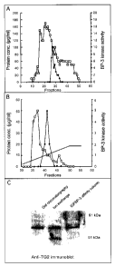
Figure 1. Purification of IGFBP-3 kinase from T-47D cells and identification
as
transglutaminase. Solubilized membranes were chromatographed on Sephracryl S-
100 (panel A) and fractions containing kinase activity (closed symbols) were
pooled
and analyzed on a High Q anion exchange column (panel B). The active fractions
(38
to 43) were pooled and applied to an IGFBP-3 affinity column. The fractions
from
each purification step were analyzed by immunoblot using TG2 antiserum (panel
C).
Figure 2. The effect of TG2 and ROCK2 inhibitors on phosphorylation of IGFBP-3
by
breast cancer cell monolayers (panel A) or cell membranes (Panel B and C). In
panel
A various concentrations of cystamine of the ROCK2 inhibitor, R-(+)trans-N-(4-
pyridyl)-4-(laminoethyl)-cyclohexanecarboxamide dihydrochloride, were pre-
incubated
with T47D cell monolayers prior to determining the ability of the cell
monolayer to
CA 02493655 2005-O1-19
phosphorylate IGFBP-3. In panel B, cystamine (20 NM), monodansyl cadaverine
(MDC, 200 pM) or the vehicle DMSO (0.005%) was added to MCF-7 and T47D cell
membranes and the ability of these membranes to phosphorylate IGFBP-3 was
determined. In panel C, the ability cystamine (10 and 20 pM) and the ROCK2
5 inhibitor, R-(+)trans-N-(4-pyridyl)-4-(laminoethyl)-cyclohexanecarboxamide
dihydrochloride (2 and 5 pM) to inhibit phosphorylation of IGFBP-3 by T47D
cell
membranes was compared.
Figure 3. Immunodepletion and immunoprecipitation of IGFBP-3 kinase activity
from
T47D solubilized membranes. In panel A, solubilized T47D cell membranes were
incubated with antibodies to TG2 or ROCK2. After the immunoprecipitates were
pelleted using protein-A agarose, the supernatants were tested for the ability
to
phosphorylate IGFBP-3. In panel B TG2 and ROCK2 were immunoprecipitated from
solubilized MCF-7 or T47D cell membranes and analyzed on SDS-PAGE. To
demonstrate the presence of these two proteins in the immunoprecipitates, the
membrane was immunoblotted with anti-TG2 or anti-ROCK2 antibodies. In panel C
the presence of IGFBP-3 kinase activity in the anti-TG2 immunoprecipitates was
demonstrated and ability of cystamine (20 NM) and the ROCK2 inhibitor (5 pM)
to
inhibit this kinase activity was assessed
Figure 4. Purified guinea pig (pg) liver TG2 and recombinant human TG2 have
IGFBP-3 kinase activity. In panel A, the effect of cystamine (cyst, 20 NM) and
MDC
(200 pM) on the IGFBP-3 kinase activity of Guinea pig and recombinant TG2 was
examined and compared to controls (cont.) or vehicle only (DMSO, 0.005%). In
panel
B, the ability of recombinant human TG2 to phosphorylate IGFBP-1, IGFBP-5,
fibronectin and fibronectin fragments was investigated. In panel C, the effect
of
calcium on recombinant TG2 kinase activity and cross-linking activity was
investigated.
Figure 5. Retinoblastoma protein, p53 and histone H3 are substrate for the
tissue
CA 02493655 2005-O1-19
6
transglutaminase kinase activity. Recombinant tissue transglutaminase was used
to
phosphorylate IGFBP-3, retinoblastoma proteins, p53 and histone H3 in vitro.
The
phosphorylated proteins were resolved electrophoresis on a polyacrylamide gel
and
visualized by autoradiography.
DESCRIPTION OF THE PREFERRED EMBODIMENTS
Unless defined otherwise, all technical and scientific terms used herein
have the same meaning as commonly understood by one of ordinary skill in the
art to
which the invention belongs. Although any methods and materials similar or
equivalent to those described herein can be used in the practice or testing of
the
present invention, the preferred methods and materials are now described. All
publications mentioned hereunder are incorporated herein by reference.
As used herein, the term "treating" in its various grammatical forms
refers to preventing, curing, reversing, attenuating, alleviating, minimizing,
suppressing or halting the deleterious effects of a disease state, disease
progression,
disease causitive agent other abnormal condition.
As used herein, "effective amount" refers to the administration of an amount
of
a given compound that achieves the desired effect.
As used herein, "TG" refers to any of a variety of transglutaminases. Herein,
TG2, or tissue transglutaminase, is the exemplary transglutaminase. Other
suitable
transglutaminases may also be used, for example, but by no means limited to,
human
factor Xllla (hFXllla), transglutaminse-1, transglutaminse-2, transglutaminse-
3,
transglutaminse-4, transglutaminse-5, transglutaminse-6, transglutaminse-7 and
epb42.
As used herein, "TG kinase substrate" refers to proteins, fragments thereof or
peptides that are phosphorylated by TG kinase activity. Examples include but
are by
no means limited to IGFBP-3, IGFBP-5, p52 tumor suppressor gene,
retinoblastoma
protein and histone H3.
In one aspect of the invention, there is provided a method of identifying
compounds capable of modulating TG kinase activity comprising: adding a test
CA 02493655 2005-O1-19
7
compound to a mixture comprising TG and a suitable TG kinase substrate,
incubating
the mixture under conditions promoting TG kinase activity, and determining if
the test
compound activates or modulates TG kinase activity as indicated by greater
than
expected TG-mediated phosphorylation of the TG kinase substrate or if the test
compound inhibits TG kinase activity as indicated by lower than expected TG-
mediated phosphorylation of the TG kinase substrate as compared to a control
comprising TG and a suitable TG kinase substrate incubated under conditions
promoting TG kinase activity.
As will be apparent to one of skill in the art, other suitable kinase assays
known
in the art may be used within the invention and these assays may be modified
such
that they may be used for high throughput assays.
Since regulation of p53 and pRb activity is critical to cell cycle control and
unregulated p53 and pRb activity is the hallmark of many cancerous cells,
identification of TG kinase modulators may have potential benefit in the
treatment of
disease states. Phosphorylation of p53 and pRb plays a very important role in
the
interaction of these two proteins with other proteins involved in regulation
of cell cycle
progression and apoptosis. Phosphorylation of these two proteins (p53 and pRb)
by
TG may be relevant to the role of TG in cell proliferation and/or apoptosis.
Therefore,
it is likely that modulation of kinase activity of TG2 or related TG family
member could
be used in the treatment of diseases characterized by cell proliferation
and/or
apoptosis.
As will be appreciated by one of skill in the art, compounds identified as
useful
in modulating transglutaminase activity using one of the above-described
methods or,
as discussed below, pharmaceutical compositions prepared therefrom, may be
useful
in treating or preventing diseases characterized by cell proliferation, for
example but
by no means limited to various forms of cancer, including but not limited to
breast,
prostate and colon cancer, or diseases characterized by impaired apoptosis,
for
example, but by no means limited to psoriasis, and chronic leukemias.
It is of note that the pharmaceutical compositions may be combined with other
components known in the art, for example, targeting molecules or permeation
CA 02493655 2005-O1-19
enhancers, or may be combined with other treatments known in the art.
As will be appreciated by one of skill in the art, the test compound or test
agent
may comprise a small molecule, chemical compound, peptide, antibody or
antibody
fragment or other such compound.
The invention is also directed to compounds isolated by these methods and the
use thereof to prepare pharmaceutical compositions. In these embodiments, the
test
compound may be combined with a pharmaceutically or pharmacologically
acceptable
carrier, excipient or diluent, either biodegradable or non-biodegradable.
Exemplary
examples of carriers include, but are by no means limited to, for example,
polyethylene-vinyl acetate), copolymers of lactic acid and glycolic acid,
poly(lactic
acid), gelatin, collagen matrices, polysaccharides, poly(D,L lactide),
poly(malic acid),
poly(caprolactone), celluloses, albumin, starch, casein, dextran, polyesters,
ethanol,
mathacrylate, polyurethane, polyethylene, vinyl polymers, glycols, mixtures
thereof
and the like. Standard excipients include gelatin, casein, lecithin, gum
acacia,
cholesterol, tragacanth, stearic acid, benzalkonium chloride, calcium
stearate, glyceryl
monostearate, cetostearyl alcohol, cetomacrogol emulsifying wax, sorbitan
esters,
polyoxyethylene alkyl ethers, polyoxyethylene castor oil derivatives,
polyoxyethylene
sorbitan fatty acid esters, polyethylene glycols, polyoxyethylene stearates,
colloidol
silicon dioxide, phosphates, sodium dodecylsulfate, carboxymethylcellulose
calcium,
carboxymethylcellulose sodium, methylcellulose, hydroxyethylcellulose,
hydroxypropylcellulose, hydroxypropylmethycellulose phthalate, noncrystalline
cellulose, magnesium aluminum silicate, triethanolamine, polyvinyl alcohol,
polyvinylpyrrolidone, sugars and starches. See, for example, Remington: The
Science
and Practice of Pharmacy, 2000, Gennaro, AR ed., Eaton, PA: Mack Publishing
Co.
As will be apparent to one knowledgeable in the art, specific carriers and
carrier combinations known in the art may be selected based on their
properties and
release characteristics in view of the intended use. Specifically, the carrier
may be
pH-sensitive, thermo-sensitive, thermo-gelling, arranged for sustained release
or a
quick burst. In some embodiments, carriers of different classes may be used in
combination for multiple effects, for example, a quick burst followed by
sustained
CA 02493655 2005-O1-19
9
release.
The invention will now be described by way of examples. However, the
examples are for illustrative purposes and the invention is not in any way
limited by
the examples.
Biotinylation of IGF8P-3
Non-glycosylated E. coli derived IGFBP-3 was biotinylated using p-biotinoyl-
aminocaproic acid-N-hydroxy-succinamide ester (Roche Molecular Biochemicals,
Mannheim, Germany) as previously described (Mishra and Murphy, 2003).
Purification of IGF8P-3 kinase activity
Solubilized T47D cell membranes were prepared using membrane preparation
kit (Pierce, Rockford, IL) according to the manufacturer's instructions in the
presence
of protease inhibitors (0.1 mM PMSF, 10 mM aprotinin and 10 pg/ml leupeptin).
3 ml
of solubilized membranes was filtered through 0.22 Nm filter and loaded on
16/60
Sephacryl S-100 gel filtration column which had been equilibrated with 20 mM
Tris/HCI, 0.02% NaN3, and pH 7.5. The eluate was monitored for absorbance at
280
nm through a Pharmacia UV-1 single path monitor. 1 ml fractions were collected
at a
flow rate of 1 ml/min and stored at -70°C. The activity was
consistently found in
fractions within molecular weight range of 65 to 85 kDa. A 20 pl aliquot of
each
fraction was assayed for IGFBP-3 kinase activities. Active fractions were
pooled and
concentrated with Amicon Centricon 30 filter. Buffer-exchanged sample was
passed
through High Q anion exchange column (Bio-Rad, CA) which had been equilibrated
with 50 mM Tris/HCI pH 8.0 containing 0.05 M NaCI, 0.02% NaN3. Separation was
performed in a linear gradient from 0.05 to 0.5 M NaCI over 50 min at flow
rate of
1 ml/min and 1 ml fractions were collected. Fractions containing kinase
activity were
concentrated using Amicon Centricon filter, desalted and buffer was exchanged
using
Micro Bio-Spin chromatography columns (BioRad, CA) and loaded on to an IGFBP-3-
Sepharose 4B affinity column (2 ml bed volume). Bound proteins were first
eluded
CA 02493655 2005-O1-19
with 0.05 M sodium phosphate containing 0.15 M NaCI (pH 7.2 ) followed by 0.1
M
acetate buffer containing 0.5M NaCI (pH 4.0). Eluted fractions were desalted,
concentrated and used for IGFBP-3 kinase assay. Fraction with IGFBP-3 kinase
activity was processed for liquid chromatography mass spectroscopy (LC-MS).
5 For LC-MC analysis, 100 pl of desalted affinity fraction was digested with
sequencing grade trypsin. The peptide mixture was lyophilized and resuspended
in
10 pl of 0.05% TFA and used for pHPLC-MALDI-QqTOF analysis. Chromatographic
separation was performed using an Agilent 1100 Series system. Sample (5 pl)
were
injected into 150 pm X 150 mm column (Vydac 218 TP C18, 5p) and eluted with 1-
10 80% acetonitrile (0.1 % TFA) in 60 min. Major ion peaks of the total ion
chromatogram
were analyzed by mass spectrometry (MS) in Manitoba/ Sciex prototype
quadrupole/time of flight mass spectrometer. In this instrument ions are
produced by
irradiation of the target using proton pulses from a 20-Hz nitrogen laser and
the mass
accuracy is within a few mDa in TOF spectra. Identification of the tryptic
peptides was
done by searching database against the peptide fingerprints using Mascot
search
engine (http://www.matrixscience.com)
Phosphorylation of IGF8P-3
Polystyrene tube were coated with streptavidin and blocked with bovine serum
albumin, washed in saline and stored at -20°C until used. Biotinylated
IGFBP-3 (500
ng) was added in streptavidin-coated tubes for 2 h on ice. At the end of
incubation,
excess, unbound IGFBP-3 was removed. Tubes were placed on ice and
phosphorylation reaction mixture containing 20 mM Tris buffered saline pH 7.5,
10
mM Mg/ATP, 60pCi/ml 32P -ATP was added. Reaction was initiated by the addition
of
membrane fraction and allowed to proceed for 30 min at 30°C. Reaction
was stopped
by addition of SDS-PAGE sample buffer, boiled for 7-10 min and analyzed on 11
gel. Subsequently gels were dried and processed for autoradiography. In some
cases 2 NU of pure TG2 (Sigma-Aldrich, MO) or histidine-tagged full length
human
TG2 expressed using baculovirus expression system in insect cells and purified
by
Ni(II)-nitroacetate agarose chromatography was used to phosphorylate IGFBP-3
in
CA 02493655 2005-O1-19
11
presence or absence of TG2 specific inhibitors. In experiments where cell
monolayers were used to phosphorylate IGFBP-3, cells were grown in 24-well
culture
plate to near confluence and washed with PBS to remove residual media and
serum.
Phosphorylation was then performed in 100 pl reaction mixture as above,
containing 1
pg IGFBP-3 for 10 min at 37°C. At the end of incubation, reaction
mixture was
aspirated, reaction was stopped by addition of sample buffer and analyzed on
11
gel. In some experiments, cell monolayer were treated with TG2 inhibitor for
30 min
prior to the phosphorylation reaction.
Immunoprecipitation
To 200 pl of solubilized membranes from T47D of MCF-7 cells, 10 pl of anti-
TG2 goat polyclonal antiserum (Upstate Biotechnology, Lake Placid, NY) and
incubated for 1 h at 4°C. 20 NI of protein A-agarose (Pierce) was added
and further
incubated on a rotating device overnight at 4°C. At the end of
incubation the pellet
was washed four times in ice cold PBS. The supernatant was discarded and the
pellet was resuspended in 50 pl of kinase buffer. 10 pl of the resuspended
sample
was used for phosphorylation of 500 ng of IGFBP-3. The samples were analyzed
by
SDS-PAGE, autoradiography and immunoblotting with TG2 antiserum.
Western blotting
Various column fractions that had IGFBP-3 kinase activity were analyzed on
10% SDS-PAGE gel and transferred to nitrocellulose membrane. Membranes were
blocked in 5% milk, incubated with TG2 antiserum diluted to 1:1000, washed
three
times, (5 min each) in TBST (10 mM Tris/HCI, 150 mM NaCI, 0.05% Tween-20, pH
8.0) and incubated with HRP-conjugated anti-goat (Santa Cruz Biotechnology,
CA)
secondary antibody (1:3000 dilution) for 1 h at room temperature. Membranes
were
washed three times in TBST and subsequently analyzed with ECL.
Purification of IGFBP-3 kinase activity from T 47D cell membranes
The IGFBP-3 kinase activity was purified from solubilized T-47D cell
CA 02493655 2005-O1-19
12
membranes using immobilized biotinylated IGFBP-3 as a substrate. A three step
procedure was used involving gel permeation, ion exchange and IGFBP-3 affinity
chromatography (Fig.1 ). Fractions eluted from the IGFBP-3 affinity column
under
acidic conditions which contained IGFBP-3 kinase activity were further
analyzed by
HPLC and tandem mass spectroscopy. Using the Mascot search engine a variety of
proteins were identified which had significant scores (Table 1). Of these, TG2
had the
highest score and there was wide coverage over the entire TG2 molecule with
peptides from various regions of the molecule identified. Tandem mass
spectroscopy
was used to confirm the sequence of various peptide fragments (Table 2).
Fractions containing peak IGFBP-3 kinase activity from the various
purification
steps were analyzed by immunoblot using TG2 antiserum. Immunoreactive TG2 was
present in all three samples (Fig. 1 C). ROCK2 was also detectable in T47D
cell
membrane fractions.
We assessed the effect of the TG2 and ROCK2 inhibitors on phosphorylation
of IGFBP-3 by cell monolayers. We have previously shown that intact washed
cells
were able to phosphorylate IGFBP-3 immobilized on polystyrene tubes (Mishra
and
Murphy, 2003). T47D cell monolayers were incubated for 30 minutes in the
presence
of various concentrations of cystamine, an inhibitor of TG2. The washed cell
monolayers were incubated with IGFBP-3 in the presence of 32P-ATP for 10
minutes
at 37°C. After termination of the incubation, the IGFBP-3 was analyzed
by SDS-
PAGE and autoradiography (Fig. 2A). Inhibition was seen with as little as 20
NM and
complete inhibition was apparent with 50 pM of cystamine. Similar experiments
were
undertaken utilizing membrane preparations from both T47D and MCF-7 cells. As
reported previously both T47D and MCF-7 cells were able to phosphorylate IGFBP-
3.
This process was inhibited by cystamine and MDC in both cells lines (Fig. 2B).
DMSO the vehicle in which MDC was dissolved had no effect. Since TG2 has been
found in association with ROCK2, a Rho-kinase (Singh et al., 2001, EM80 J.,
20:2413-2423), and ROCK2 was present in the affinity purified cell membrane
fractions, we investigated the effect of the ROCK2 inhibitor, R (+)trans-N-(4-
pyridyl)-4-
(1aminoethyl)-cyclohexanecarboxamide dihydrochloride. The ROCK2 inhibitor had
CA 02493655 2005-O1-19
13
no effect on IGFBP-3 phosphorylation (Fig. 2C).
We next examined the effect of TG2 and ROCK2 antisera on IGFBP-3 kinase
activity present in cell membrane preparations. Antiserum against TG2 but not
ROCK2 antiserum was able to immunodeplete IGFBP-3 kinase activity from
membrane preparations (Fig. 3A). When the immunoprecipitates were analyzed for
kinase activity, the precipitates obtained with TG2 antiserum but not those
obtained
with ROCK2 antiserum had kinase activity. Furthermore, the IGFBP-3 kinase
activity
present in the immunoprecipitates was inhibited by cystamine but not by the
ROCK2
inhibitor.
Purified guinea pig liver TG2 and human recombinant TG2 both were able to
phosphorylate IGFBP-3 (Fig. 4A). This process was inhibited cystamine and MDC.
The related binding protein IGFBP-5 was also phosphorylated by TG2 whereas
IGFBP-1 was not phosphorylated by TG2 (Fig. 4B). Consistent with a previous
report
(Sakai et al., 2001, J.Biol. Chem., 276:8740-8745), an increase in the
molecular mass
of IGFBP-1 was observed suggesting that TG2 can polymerase IGFBP-1.
Fibronectin, another reported substrate for TG2 (Akimov and Belkin, 2001, J.
Cell
Sci., 114:2989-3000), was not phosphorylated by this enzyme under the
conditions
we used to phosphorylate IGFBP-3 (Fig. 4B). Since calcium is necessary for the
cross linking activity of TG2 (Kang et al., 2002, Biochem. Biophys. Res.
Commun.,
293:383-390), we examined the effect of increasing concentration of calcium on
the
kinase activity of TG2. As the calcium concentration was increased we observed
a
decrease in the kinase activity of TG2. Concomitantly there was an increase in
polymerization of IGFBP-3 observed (Fig. 4C).
Analysis of purified fractions from breast cancer cells containing IGFBP-3
kinase activity identified a number of potentially interesting proteins. Of
these only
ROCK2 was previously known to have kinase activity and we assumed that this
was
responsible for phosphorylation of IGFBP-3. However a specific inhibitor of
ROCK2
kinase activity had no effect on the IGFBP-3 kinase activity of breast cancer
cell
monolayers or purified membrane preparations. Furthermore, immunoprecipitation
of
CA 02493655 2005-O1-19
14
ROCK2 from membrane preparation did not deplete the IGFBP-3 kinase activity
whereas this activity could be completely removed by immunoprecipitation with
TG2
antiserum. These data, together with the demonstration that both guinea pig
liver
TG2 and recombinant human TG2 could phosphorylate IGFBP-3 provided convincing
evidence that TG2 can function as ectokinase in breast cancer cells.
Furthermore it
appears to account for virtually all the IGFBP-3 kinase activity present on
the
membrane of these cells since very little residual activity was apparent after
immunoprecipitation of TG2 from breast cancer membrane preparations. We have
previously shown that IGFBP-3 can also be phosphorylated by an ecto-kinase
present
on COS cells (Mishra and Murphy, 2003) and human umbilical vein endothelail
cells.
The latter is particularly relevant since endothelial cells are know to
express high
levels of TG2 on their plasma membranes (Fesus and Piacentini, 2002, Trends
Biochem. Sci., 27:534-539).
TG2 is a ubiquitous enzyme that has been implicated in a variety of biological
processes. It is important in post-translational protein modification and
protein-protein
interactions. It functions as a calcium-dependent transamidating
acytransferase that
crosslinks glutamine residues with lysine residues in the same proteins
resulting in
polymerization or with lysine residues in other proteins resulting in protein
cross-
linking (Fesus and Piacentini, 2002). In addition to adding diamines and
polyamines
to proteins it can also deamidate glutamine residues to glutamic acid which
introduces
a negative charges and changes the pl of the protein. Recently it has been
reported
to also function as a protein disulfide isomerase (Hassegawa et al., 2003,
Biochem.
J., 373:793-803). However this latter function unlike other functions
described for
TG2 was not calcium dependent and was not inhibited by GTP. TG2 has also been
reported to function as novel G protein couple membrane receptor (Nakaoka et
al.,
1994, Science 264:1593-1596) and has been shown to have a role in transmitting
signals from classical seven-transmembrane helix G-coupled receptors such as
the
b~B-adrenergic receptor (Chen et al., 1996, J. Biol. Chem., 271, 32385-32391).
Here
we report that TG2 has another novel enzymatic function namely kinase
activity. It is
likely to phosphorylate a variety of other proteins.
CA 02493655 2005-O1-19
Interestingly the kinase activity of TG2 was inhibited by increasing calcium
and
consistent with previous reports (Kang et al., 2002) increasing the calcium
concentration enhanced the cross-linking activity of TG2. In the case of TG2
activity
directed against IGFBP-3, calcium appeared to act as switch, inhibiting kinase
activity
5 and enhancing cross-linking activity.
TG2 has been implicated in a variety of processes where phosphorylation is
important. These include activation of RhoA and M kinase pathways (Singh et
al.,
2003, J. Biol. Chem. 278:391-399), activation of CREB (Tucholski and Johnson,
2003,
J. Biol. Chem., 278:26838-26843) and activation of phospholipase C (Nakaoka et
al.,
10 1994).
In most cell types TG2 is predominantly localized in the cytoplasm and the
nucleus (Fesus and Piacentini, 2002) but it is also localized to the cell
membrane
(Gaudry et al., 1999, J. Biol. Chem., 274:30707-30714). It ca be released from
various cell types under certain circumstances such as inflammation and during
15 apoptotic cell death (Griffin and Verderio, 2000, in Tissue
transglutaminase in cell
death in programmed Cell Death in Animals and Plants, eds. Bryant, J. A.,
Hughes, S.
G. & Garland, J. M. (BIOS Scientific Publishers Ltd. Oxford), pp. 223-240). In
the
latter case it appears to be important in the latter stages of the process and
may
function to prepare dying cells for phagocytosis by macrophages (Griffin and
Verderio,
2000, in Tissue transglutaminase in cell death in programmed Cell Death in
Animals
and Plants, eds. Bryant, J. A., Hughes, S. G. & Garland, J. M. (BIOS
Scientific
Publishers Ltd. Oxford), pp. 223-240). However, TG2 gene expression is
activated
early in apoptosis, particularly morphogenic apoptosis, in developing
embryonic limbs
(Thomazy and Davies, 1999, Cell Death Differ., 6:146-154) and retinoid-induced
apoptosis (Kochhar et al., 1993, Prog. Clin. Biol. Res., 383B, 815-825).
Interestingly, IGFBP-3 has been shown to be pro-apoptotic in a variety of cell
lines (Oh et al., 1995; Hong et al., 2002 ; Longobardi et al., 2003,
Endocrinology
144:1695-1702). This process is thought to be an IGF-independent effect of
IGFBP-3
mediated by binding of IGFBP-3 to a surface receptor (Oh et al., 1993, J.
Biol. Chem.
268: 26045-26048). The presence of IGF-I inhibits the interaction of IGFBP-3
with
CA 02493655 2005-O1-19
16
binding sites present on breast cancer cells (Yamanaka et al., 1999,
Endocrinology
140:1319-1328) and thus would potentially inhibit the pro-apoptotic IGF-
independent
effects of IGFBP-3. We have previously shown that phosphorylation of IGFBP-3
by
TG2 enhances the affinity of this binding protein for IGF-I. Thus
phosphorylation of
IGFBP-3 by TG2 could serve to attenuate the pro-apoptotic effects of IGFBP-3
and
the proliferative effect of IGF-I by enhancing formations of IGFBP-3/IGF-I
binary
complexes and reducing the interaction of IGF-I and IGFBP-3 with their cognate
membrane binding sites.
In summary we have identified a novel kinase function for TG2. We provide
compelling evidence that TG2 is the major IGFBP-3 kinase present on breast
cancer
cell membranes. The observation that TG2 has kinase activity should serve as a
stimulus to re-examine the role of the TG2 kinase activity in other biological
processes
where TG2 kinase activity could be important.
While the preferred embodiments of the invention have been described
above, it will be recognized and understood that various modifications may be
made
therein, and the appended claims are intended to cover all such modifications
which
may fall within the spirit and scope of the invention.
CA 02493655 2005-O1-19
17
Table 1. Proteins whose tryptic peptide fragments were identified by
HPLC/tandem
spectroscopy in IGFBP-3 affinity column fraction containing kinase activity.
Protein Score* Accession number
Transglutaminase 50 P21980
ROCK2 48 NP-004841
WW domain containing adaptor isoform37 NP-057712
I
Glutamate receptor 36 AAD15616
G protein coupled receptor GPR 44 35 AAD21055
KIAA0322 33 BAA20780
* Score determined as -10Log(P), where P is the probability that the observed
match
is a random event. A score >40 indicates indentity or extensive homology, p <
0.05.
Table 2. Transglutaminase peptide fragment identified by tandem mass
spectroscopy
in IGFBP-3 affinity column fraction containing kinase activity.
TG2 residues Sequence
31-35 LVVRR
223-240 VWSGMVNCNDDQGVLLGR
422-433 VGLKISTKSVGR
553-562 DCLTESNLIK