Note: Descriptions are shown in the official language in which they were submitted.
CA 02493675 2005-O1-14
WO 2004/007723 PCT/GB2003/003093
GENES
FIELD
The present invention relates to the fields of development, molecular biology
and
genetics. More particularly, the invention relates to genes which are
expressed
exclusively in the earliest populations of primordial germ cells (PGCs) and
the use of
such genes and the products thereof in identification of pluripotent and
multipotent cells
such as PGCs, pluripotent embryonic stem cells (ES) and pluripotent embryonic
germ
cells (EG), in cell populations. They are also markers for a change in the
state of cells
from being non pluripotent to becoming pluripotent, and in being able to
confer this state
on a non pluripotent cell.
INTRODUCTION
Post fertilisation, the early mammalian embryo undergoes four rounds of
cleavage
to form a morula of 16 cells. These cells, following further rounds of
division, develop
into a blastocyst in which the cells can be divided into two distinct regions;
the inner cell
mass, which will form the embryo, and the trophectoderm, which will form extra-
embryonic tissue, such as the placenta.
The cells that form part of the embryo up until the formation of the
blastocyst are
totipotent; in other words, each of the cells has the ability to give rise to
a complete
individual embryo, and to all the extra-embryonic tissues required for its
development.
After blastocyst formation, the cells of the inner cell mass are no longer
totipotent, but are
pluripotent, in that they can give rise to a range of different tissues. A
known marker for
such cells is the expression of the enzyme alkaline phosphatase and Oct4.
Primordial germ cells (PGCs) are pluripotent cells that have the ability to
differentiate into all three primary germ layers. In mammals, the PGCs migrate
from the
base of the allantois, through the hindgut epithelium and dorsal mesentery, to
colonise the
gonadal anlague. The PGC-derived cells have a characteristically low
cytoplasmlnucleus
ratio, usually with prominent nucleoli. PGCs may be isolated from the embryos
by
CA 02493675 2005-O1-14
WO 2004/007723 PCT/GB2003/003093
removing the genital ridge of the embryo, dissociating the PGCs from the
gonadal
anlague, and collecting the PGCs. The earliest PGC population is reported to
consist of a
cluster of some 45 (forty-five) alkaline phosphatase positive cells, found at
the base of the
emerging allantois, 7.25 days post-fertilisation (Ginsburg et al., (1990)
Development
110:521-528).
PGCs have many applications in modern biotechnology and molecular biology.
They are useful in the production of transgenic animals, where embryonic germ
(EG)
cells derived from PGCs may be used in much the same manner as embryonic stem
(ES)
cells (Labosky et al., (1994) Development 120:3197-3204). Moreover, they are
useful in
the study of foetal development and the provision of pluripotent stem cells
for tissue
regeneration in the therapy of degenerative diseases and repopulation of
damaged tissue
following trauma. Above all, PGCs while having some specialised properties,
retain an
underlying pluripotency, which is lost from the neighbouring cells that
surround the
founder population of PGCs that acquire a somatic cell fate. PGCs and the
surrounding
somatic cells share a common ancestry. However, the founder PGCs are few in
number
and difficult to isolate from embryonic tissue and the surrounding somatic
cells, which
complicates their study and the development of techniques which make use
thereof.
Little is known in the art about the expression of genes in the founder
population
of PGCs and the relationship between PGC-specific gene expression and the
retention of
pluripotency in these cells. Certain markers for PGCs are known - for example,
the
expression of tissue non-specific alkaline phosphatase (TNAP) has been used as
a marker
fox early PGCs (Ginsburg et al., (1990) Development 110:521-528). Oct4 is
known to be
expressed in PGCs, but not somatic cells (Poem et al., (1996) Development
122:881-
894). Other markers, such as BMP4, are known to be expressed primarily in
somatic
tissues (Lawson et al., (1999) Genes & Dev. 13:424-436). However, none of
these genes
is specific for PGCs, since they are also expressed in other tissue types.
There is therefore
a need in the art for the identification of genes which may be used as markers
for PGCs
and which may provide an insight into the biology of germ cell development and
the
nature of the pluripotent state.
CA 02493675 2005-O1-14
WO 2004/007723 PCT/GB2003/003093
Our unpublished International Patent Application Number PCT/GB02/0021 S, the
contents of which are hereby incorporated by reference, discloses sequences
and uses of
two rodent genes, Stella and Fragilis. That document shows that Stella and
Fragilis are
expressed by PGCs and other pluripotent cells. The sequences of mouse Stella
and
Fragilis are also set out here as SEQ ID NO: 2 and SEQ ID NO: 1 respectively.
SUMMARY
We now disclose human sequences of Stella and Fragilis, as well as sequences
of
novel genes Fragilis 9-27, Fragilis 2, Fragilis 3, Fragilis 4, Fragilis 5 and
Fragilis 6
comprising members of the Fragilis family of genes. We also disclose sequences
of
human Fragilis homologues, Ifitm l, Ifitm 2, Ifitm 3 and Human ENSG142056. The
genes and polypeptides described here, including associated products such as
antibodies,
are useful in a number of ways, for example, for diagnosis, treatment and/or
prevention of
diseases such as cancer.
According to a first aspect of the present invention, we provide a polypeptide
comprising a human Stella amino acid sequence, preferably a sequence as shown
in SEQ
ID NO: 4, or a fragment, homologue, variant or derivative thereof.
There is provided, according to a second aspect of the present invention, a
polypeptide comprising a human Fragilis amino acid sequence, preferably a
sequence as
shown in SEQ ID NO: 6, or a fragment, homologue, variant or derivative
thereof.
We provide, according to a third aspect of the present invention, a
polypeptide
comprising an amino acid sequence selected from the group consisting of:
Fragilis 9-27
(preferably comprising a sequence as shown in SEQ ID NO: 8), Fragilis 2
(preferably
comprising a sequence as shown in SEQ ID NO: 10), Fragilis 3 (preferably
comprising a
sequence as shown in SEQ TD NO: 12), Fragilis 4 (preferably comprising a
sequence as
shown in SEQ ID NO: 14), Fragilis 5 (preferably comprising a sequence as shown
in SEQ
ID NO: 16) and Fragilis 6 (preferably comprising a sequence as shown in SEQ ID
NO:
18), or a fragment, homologue, variant or derivative thereof.
CA 02493675 2005-O1-14
WO 2004/007723 PCT/GB2003/003093
As a fourth aspect of the present invention, there is provided a polypeptide
consisting essentially of a sequence selected from the group consisting of:
(a) the
sequence of human Stella shown in SEQ ID NO: 4; (b) the sequence of human
Fragilis
shown in SEQ ID NO: 6; (c) the sequence of human Fragilis 9-27 shown in SEQ ID
NO:
8; (d) the sequence of mouse Fragilis 2 shown in SEQ ID NO: 10; (e) the
sequence of
mouse Fragilis 3 shown in SEQ ID NO: 12; (f) the sequence of mouse Fragilis 4
shown in
SEQ ID NO: 14; (g) the sequence of mouse Fragilis 5 shown in SEQ ID NO: 16;
and (h)
the sequence of mouse Fragilis 6 shown in SEQ ID NO: 18.
We provide, according to a fifth aspect of the present invention, a
polypeptide
which has at least 50%, 60%, 70%, 80%, 90% or 95% homology to a polypeptide
according to any of the preceding aspects.
The present invention, in a sixth aspect, provides a polypeptide comprising a
sequence of 15 or fewer contiguous amino acids, preferably a sequence of fewer
than 10
contiguous amino acids, of a nucleic acid according to any of the preceding
aspects.
1 S In a seventh aspect of the present invention, there is provided a nucleic
acid
encoding a polypeptide according to any preceding aspect.
According to an eighth aspect of the present invention, we provide a nucleic
acid
sequence comprising a human Stella nucleic acid sequence, preferably a
sequence as
shown in SEQ ID NO: 3, or a fragment, homologue, vaxiant or derivative
thereof.
We provide, according to a ninth aspect of the invention, a nucleic acid
sequence
comprising a human Fragilis nucleic acid sequence, preferably a sequence as
shown in
SEQ ID NO: 5, or a fragment, homologue, vaxiant or derivative thereof.
There is provided, in accordance with a tenth aspect of the present invention,
a
nucleic acid sequence comprising an Fragilis nucleic acid sequence acid
sequence
selected from the group consisting of: Fragilis 9-27 (preferably a sequence as
shown in
SEQ ID NO: 7), Fragilis 2 (preferably a sequence as shown in SEQ ID NO: 9),
Fragilis 3
(preferably a sequence as shown in SEQ ID NO: 11), Fragilis 4 (preferably a
sequence as
CA 02493675 2005-O1-14
WO 2004/007723 PCT/GB2003/003093
shown in SEQ ID NO: 13), Fragilis 5 (preferably a sequence as shown in SEQ ID
NO:
15) and Fragilis 6 (preferably a sequence as shown in SEQ ID NO: 17), or a
fragment,
homologue, variant or derivative thereof.
As an eleventh aspect of the invention, we provide a nucleic acid sequence
consisting essentially of a sequence selected from the group consisting of:
(a) the
sequence of human Stella shown in SEQ ID NO: 3; (b) the sequence of human
Fragilis
shown in SEQ ID NO: 5; (c) the sequence of human Fragilis 9-27 shown in SEQ ID
NO:
7; (d) the sequence of mouse Fragilis 2 shown in SEQ ID NO: 9; (e) the
sequence of
mouse Fragilis 3 shown in SEQ ID NO: 11; (f) the sequence of mouse Fragilis 4
shown in
SEQ ID NO: 13; (g) the sequence of mouse Fragilis 5 shown in SEQ ID NO: 15;
and (h)
the sequence of mouse Fragilis 6 shown in SEQ ID NO: 17.
We provide, according to a twelfth aspect of the invention, there is provided
a
nucleic acid sequence which has at least 50%, 60%, 70%, 80%, 90% or 95%
homology to
a nucleic acid sequence as disclosed.
According to a thirteenth aspect of the present invention, we provide a
nucleic
acid comprising a sequence of 25 or fewer contiguous nucleotides, preferably a
sequence
of 15 contiguous nucleotides, of a nucleic acid as disclosed.
There is provided, according to a fourteenth aspect of the present invention,
a
complement of a nucleic acid sequence as disclosed.
We provide, according to a fifteenth aspect of the present invention, a
nucleic acid
comprising a nucleic acid sequence as disclosed, together with one or more
nucleotide
substitutions, in which the one or more substitutions do not alter the coding
specificity of
said nucleic acid as a result of the degeneracy of the genetic code.
According to a sixteenth aspect of the present invention, we provide a vector
comprising such a nucleic acid sequence.
CA 02493675 2005-O1-14
WO 2004/007723 PCT/GB2003/003093
According to a seventeenth aspect of the present invention, we provide a host
cell
comprising a nucleic acid sequence as disclosed.
There is provided, according to a eighteenth aspect of the present invention,
a
method of producing a polypeptide as disclosed, the method comprising
providing a
nucleic acid as disclosed, a vector as disclosed, or a host cell as disclosed,
and enabling
the expression of a nucleic acid encoding the polypeptide.
We provide, according to a nineteenth aspect of tile invention, a method for
identifying a pluripotent cell, comprising detecting such a polypeptide, or
such a nucleic
acid in the cell.
Preferably, the method comprises the steps of amplifying nucleic acids from a
putative pluripotent cell using 5' and 3' primers specific for Stella and/or
Fragilis, and
detecting amplified nucleic acid thus produced. Preferably, the nucleic acid
sequence is
detected by i~c situ hybridisation. Preferably, the nucleic acid sequence is
detected by
detecting a protein product encoded by the nucleic acid. Preferably, the
polypeptide is
detected by immunostaining.
There is provided, according to a twentieth aspect of the present invention,
an
antibody specific for a polypeptide as disclosed. Preferably, the antibody is
capable of
specifically binding to an extracellular domain of Fragilis.
We provide, according to a twenty-first aspect of the present invention, use
of
such an antibody fox the identification and/ or isolation of a pluripotent
cell.
As a twenty-second aspect of the present invention, there is provided
pluripotent
cell identified by a method as disclosed.
We provide, according to a twenty-third aspect of the present invention, a
method
of treatment or pxophylaxis of a disease in an individual, the method
comprising
modulating the expression and/or the activity of Stella and/or Fragilis in a
cell of the
individual.
CA 02493675 2005-O1-14
WO 2004/007723 PCT/GB2003/003093
Preferably, the method comprises up-regulating the expression and/or the
activity
of Stella and/or Fragilis.
The present invention, in a twenty-fourth aspect, provides a Stella or
Fragilis
polypeptide or nucleic acid, or an antibody against Stella or Fragilis, for
use in a method
of treatment or prophylaxis of a disease in an individual.
In a twenty-fifth aspect of the present invention, there is provided use of a
Stella
or Fragilis polypeptide or nucleic acid, or an antibody against SteIIa or
Fragilis, for the
preparation of a pharmaceutical composition for the treatment of a disease.
According to an twenty-sixth aspect of the present invention, we provide a
method
of diagnosis of a disease, the method comprising detecting a modulation of
expression
and/or activity of Stella and/or Fragilis in a cell of an individual.
In preferred embodiments, the disease is selected from the group consisting
of:
testis tumor, colon tumor, stomach, germ cell tumors, choriocarcinoma, Lung,
large cell
carcinoma, uterus, and leiomyosarcoma. Stella is therefore suitable for
treatment or
diagnosis of any disease of or associated with such tissues, in particular,
cancer or
tumours of such tissues.
We provide, according to a twenty-seventh aspect of the invention, a method of
identifying a molecule capable of binding to Stella or Fragilis, the method
comprising the
steps of: (a) providing a Stella or Fragilis polypeptide; (b) contacting the
Stella or Fragilis
polypeptide with a candidate molecule; and (c) detecting binding between the
candidate
molecule and the Stella or Fragilis polypeptide, as the case may be.
There is provided, according to a twenty-eighth aspect of the present
invention, a
method of identifying an agonist or antagonist of Stella or Fragilis, the
method
comprising the steps of: (a) providing a Stella or Fragilis polypeptide; (b)
contacting the
Stella or Fragilis polypeptide with a candidate molecule; and (c) detecting
modulation of
an activity of the Stella or Fragilis polypeptide.
CA 02493675 2005-O1-14
WO 2004/007723 PCT/GB2003/003093
Preferably, the polypeptide is contacted with a plurality of candidate
molecules,
preferably in the form of a library.
We provide, according to a twenty-ninth aspect of the present invention, use
of an
antagonist of Stella or Fragilis, preferably as identified by a method
according to any of
the previous aspects, in a method of treatment or prophylaxis of a disease in
an
individual.
BRIEF DESCRIPTION OF THE FIGURES
Figure 1: Nucleotide and deduced amino acid sequence of mouse Fragilis (SEQ
ID NO: 1). Predicted positions of the two transmembrane domains (TM I and TM
II) are
underlined and indicated by bold letters. The poly(A) signal is undeflined.
Figure 2: Nucleotide and deduced amino acid sequence of mouse Stella (SEQ ID
NO: 2). Three nuclear localization signals are underlined. A potential nuclear
export
signal is underlined twice, and the hydrophobic residues are indicated in
bold. Helical
structures in a motif with similarity to SAP domain (a.a.28 to a.a.63) are
underlined in
red, and the conserved residues are indicated by blue. A splicing factor-like
motif is
underlined and the conserved residues are indicated in green. Poly(A) signals
are also
underlined.
Figure 3: Expression of mouse Fragilis in embryonic stem (ES) cells. ES cells
are
fixed in 4% paraformaldehyde in PBS for l Omin. at room temperature and
processed for
immunohistochemistry as described by Saitou et al., (1998). J Cell Biol 141,
397-408.
(1998). Fragilis expression is similarly detected in E6.5 proximal epiblast
cells, which are
germ cell competent cells, and in newly specified germ cells. The expression
declines
after E8.5 following completion of the specification of germ cells fate.
Figure 4: Expression of mouse Stella in PGCs. PGCs from E12.5 genital ridges
are fixed in 4% paraformaldehyde in PBS for l Omin. at room temperature and
processed
for immunohistochemistry as described by Saitou et al., (1998). JCell Biol
141, 397-408.
(1998). Stella is detected in PGCs from E 7.25-13.5, as well as in pluripotent
ES cells and
CA 02493675 2005-O1-14
WO 2004/007723 PCT/GB2003/003093
in EG cells. Stella is also detected in the totipotent oocyte, zygote and in
the totipotent
and pluripotent blastomeres during preimplantation development and in
developing
gametes. When EG cells are derived from PGCs (Labosky et al., (1994)
Development
120:3197-3204). Fragilis expression is again detected in the pluripotent EG
cells as it is in
ES cells. Therefore, Fragilis and Stella are also markers for the pluripotent
stem cells.
Figure 5. Mouse Fragilis expression by whole-mount in situ hybridization in
E7.2
mouse embryos.
Figure 6. Mouse Stella expression by whole mount ivy situ hybridisation in E
7.2
mouse embryos.
Figure 7. Mouse Stella expression in PGCs in the process of migration into the
gonads in E9.0 embryos.
Figure 8a and 8b. Expression of mouse Fragilis and mouse Stella in single
cells
detected by PCR analysis of single cell cDNAs. Numbers marked by symbol* in 8b
are
the PGCs. Note that there are more single cells showing expression of Fragilis
compared
to those showing expression of Stella. Only cells with the highest levels of
Fragilis
expression were found to express Stella and acquire the germ cell fate. Cells
that express
Stella were found not to show expression of Hoxbl. Cells that express lower
levels of
Fragilis and no Stella become somatic cells and showed expression of Hoxbl.
The
founder population of PGCs also show high levels of Tnap. Both the founder
PGCs and
the somatic cells show expression of Oct4, T(Brachyury), and FgfB.
Figure 9. The Fragilis family cluster on mouse Chr7, and the human homologues
in the syntenic region on Chrl 1. In the mouse, the five Fragilis genes are
clustered within
a 70kb region. All genes are encoded by two exons, and apart from f~agilis2,
they are
located on the minus strand. In human, the four homologous genes, ENSG142056
and
Ifitml (9-2~, If tm2 (1-8D) and Ifitm3 (1-BIB, are clustered within a 25kb
stretch. The
four human homologues are each encoded by two exons, but the length of the
intronic
sequence for Ifit~zl and Iftm3 is not known. Apart Ifitr~~2, all human genes
are encoded on
the minus strand. The green circles represent ISRE consensus sequences.
CA 02493675 2005-O1-14
WO 2004/007723 PCT/GB2003/003093
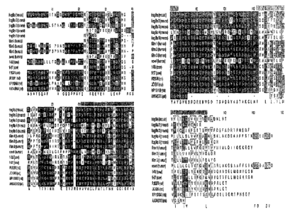
Figure I0. Protein alignment of the Fragilis family and their homologues in
human, cow and rat. Green baxs indicate the location of the two predicted
transmembrane
domains, of which the first as well as the inter-domain stretch appear to be
highly
conserved throughout the four mammalian species. Identical amino acids are
highlighted
5 in dark grey, similar amino acids in Iight grey. The alignment was done
using ClustlW.
Figure 11. Expression analysis of fragilis (a-f), fi~agilis2 (g-1) and
f~agilis3 (m-r)
by whole mount in situ hybridisation. Pictures are taken as lateral view
unless otherwise
stated, with anterior to the left and posterior to the right. f~agilis is
expressed throughout
the epiblast in E5.5 embryos (a) and in the region of germ cell specification
at the base of
10 the incipient and early allantoic bud at E7.5 (b, b' posterior view, c). At
E8.5, signal is
detected at the base and in the proximal third of the allantois as well as in
the latero-
anterior aspects of the brain (d superior view, a anterior view). At E9.5,
fragilis appears
expressed in a population of cells at the beginning of the invaginating
hindgut (arrow in
fJ, as well as in the pharyngeal arches (f). f~agilis2 is detected throughout
the epiblast at
E5.5 (g). Expression seems thereafter downregulated but becomes again
detectable in the
posterior mesoderm and at the base of the incipient and growing allantoic bud
in E7.0 and
E7.5 embryos (h, i, i' posterior view). At E8.5, expression is seen in caudal
mesoderm (j,
k posterior view), while at E9.5 expression is seen in the tailbud, the
mesoderm caudal to
the 12a' somite and the lung primordia (arrow, l). fi°agilis3 is
expressed throughout the
epiblast at E6.5 (rn) and around E7.5 additionally in the region of PGC
specification (n, n'
posterior view, o). At E8.5, fragilis2 expression is seen throughout the
embryo, with
exception of the developing heart, and appears intense in single cells (arrow
in q posterior
view) at the base and within the proximal region of the allantois (p posterior
view, q, r).
asterix: allantois; black arrowhead: allantoic bud; white arrowhead:
developing heart;
scale bars: 100~,m (a, b, g-i, m, n); 200~,m (c-e, o-q); 400~.m (f, j-1, r).
Figure 12. Expression analysis of fragilis2 by in situ hybridisation on
sections. (a-d)
transverse sections through the caudal region of an embryo at E9.5 (approx. 25
somites)
at progressively rostral levels. At most caudal levels, fragilis2 expression
is seen in cells of
the neural tube, in the presomitic mesoderm, in single cells within the
hindgut
(arrowhead) and in the body wall. (b) staining at approx. 23ra somite Ievel is
present
within the forming somite, the body wall mesoderm and cells within the hindgut
as well
CA 02493675 2005-O1-14
WO 2004/007723 PCT/GB2003/003093
11
as the floorplate. (c) at approx. 2lstsomite level, expression in the
differentiating somites
is reduced, while cells in the floor plate and within the hindgut remain
fragilis2 mRNA
positive. (d) at approx. the l3tn somite level, fragilis2 expression is absent
from the somatic
mesoderm as well as the neural tube. (e) sagittal section through an E10.5
embryo shows
fragilis2 expression in developing lung tissue (asterix; higher magnification
in f) and
migrating cells along the hindgut anterior to the dorsal aorta (arrow). (g)
shows a
magnified view of fragilis2 mRNA expressing, migrating cells. da: dorsal
aorta; fp: floor
plate; g: gut; h: developing heart; nt: neural tube; s: somite; bw: body wall;
scale bars:
150,cam (a-d); 1 mm (e); 400,u m (f, g).
Figure 13. Expression analysis of the Fragilis family genes in single cells
from the
region of germ cell specification of E7.5 embryos. (a) shows PCR analysis of
cDNAs
from three nascent, stella positive PGCs and three surrounding, stella
negative somatic
cells. Note that f~agilis, f~agilis2 and fragilis3 are expressed in PGCs and
somatic cells,
while fragilis4 and fi~agilis5 are not detected in any of the cells. (b) shows
expression of
fi~agilis, fragilis2 and fragilis3 in single cell cDNAs using Southernblot
analysis. GAPDH
was used as blotting control. (c) Semi-quantitative expression analysis of the
Southernblot
data shows that all three Fragilis genes are predominantly expressed in
nascent PGCs
compared to the somatic cells within the region.
Figure 14. Expression analysis of fragilis, f~agilis2 and fragilis3 at
E11.5/E12.5 in
single cells from the genital ridge and by in situ hybridisation. (a) shows
PCR analysis of
cDNAs from three gonadal stella-positive germ cells and three surrounding,
stella-
negative somatic cells. While fi°agilis is detected only in the three
germ cell clones,
fiagilis2 and fi~agilis3 are expressed in the germ cells as well as the
somatic cells. (b)
shows in situ hybridisation of urogenital ridges of El 1.5/E12.5 embryos.
While fragilis3
is expressed in the mesonephros as well as the genital ridge, fragilis and
fi~agilis2 are
restricted to the genital ridge. The staining pattern for fi~agilis appears
punctuate and
restricted to single cells mimicking the pattern seen for the germ cell-
specific stella gene.
asterix: genital ridge; black arrowhead: mesonephros; scale bars: 400~,m.
Figure 15. Stella expression during preimplantation development and
evolutionary
conservation. a-l, Confocal sections of anti-stella (a, d,gj) and propidium
iodide (b, e, h, k)
CA 02493675 2005-O1-14
WO 2004/007723 PCT/GB2003/003093
12
stained embryos (c, f, i, l merged images). Maternal stella is stored in the
unfertilised egg
(a-c) (arrow, exclusion of Stella from condensed metaphase chromosomes) and
localizes
both to the cytoplasm and pronuclei (PN) after fertilisation (d f PB, polar
body). Also
during later stages (2-cell, g-i; 4-cell, j-l) it can be seen both in the
cytoplasm and the
nucleus. Scale bar = 20 ~.m. Synteny (~z) of the stella gene in mouse, rat and
human and
close up view (h) of stella and its neighbouring genes in mouse and human.
Arrows
indicate the direction of transcription. o, Alignment of Stella protein
sequences. Identical
amino acids have a black background and similar amino acids a grey one.
Putative
nuclear export and localisation signals are marked by red and black lines,
respectively.
The red stars indicate conserved hydrophobic amino acids, which are typical
for nuclear
export signals27. p, RT-PCR analysis of STELLA-expression in human pluripotent
cells
and reproductive organs. RPL32 was used as control. ES, embryonic stem cells;
EC,
embryonic carcinoma cells (nTera2); tet, testis tumor; te, normal testis; ov,
normal ovary;
-Rt, without reverse transcriptase; 0, water control.
Figure 16. Knockout strategy of stella and confirmation of correct targeting
by
Southern-blot and RT-PCR. a, The targeting vector was designed to delete exon
2 and
replace it with an IRES-Lac2 / MC-neo reporter-selection cassette. HS'V-TK.
was used for
negative selection against non-homologous recombination. 5', 3' and neo-probes
were
used to confirm correct targeting of ES-cells. b, Southern blot analysis of
genomic DNA
derived from littermate mice born from a stella+~ intercross. The example
shows a NcoI
digest hybridised with the 3' probe, indicating the absence of the wild-type
allele in stella
mice. c, RT-PCR of testis (te) or ovary (ov) RNA from male or female mice,
respectively using exon 2-specific primers. The wild-type stella transcript is
reduced in
stella+~ mice compared to stella+~~ mice and absent in stella ~ mice. Gapdh
was used as a
control for equivalent quality and amount of RNA. -Rt, without reverse
transcriptase; 0,
water control.
Figure 17. Germ cell development in stella knockout mice. a, Numbers of PGCs
in wild-type (wt, n=9), stella~~ (n=14) and stella'~ (n=7) embryos are not
significantly
different at E8.5 (0-8 somites). The results are presented as means ~SEM. b-g,
Gonadal
PGCs (E11.5) stained with anti-stella (b, e) and anti-SSEA1 (c, ~ antibodies
(d, g merge
including Toto3 (blue) as DNA stain). The PGC-marker SSEAlI7 is coexpressed
with
CA 02493675 2005-O1-14
WO 2004/007723 PCT/GB2003/003093
13
stella in wild-type PGCs (b-d) and also detectable in stella ~ animals (e-g),
showing that
PGCs are present in knockout mice. Scale bar = 10 ~,m. Sections of testes (Iz
j) and
ovaries (k-r~z) of adult wild-type (h, k), stella'~~ (i, l) and stella ~ (j,
fn) mice. Knockout
males show normal development of sperm (arrowheads) and knockout females
normal
ovary morphology with follicles containing oocytes of different stages
(avows). Scale
bars in j (for h j), ~a (for k fn) = 100 Vim.
Figure I 8. Maternal effect of the stella knockout and onset of paternal
expression
of stella during preimplantation development. a, 80% of coatings with wild-
type males
resulted in pregnancies of wild-type females, while in only 24% of the plugs
stella
females became pregnant. b, Fxom these pregnancies, the littersize was
strongly reduced
in knockouts compared to wild-type females. c-i, A stella-GFP reporter
construct (c) was
used to determine, when the paternal allele of stella starts to be expressed.
Zygotic
expression of the stella-GFP transgene begins at the 2-cell stage (E1.5; e, h)
and
continues during later stages (E2.5, 4-8 cell; f, i). d--f, GFP-fluorescence;
g-i, brightfield
I S merged with GFP-image; arrowheads, non-transgenic embryos; arrows,
transgenic
embryos. Scale bar in d (for d i) = 100 ~,m. j-l, Confocal section through a
morula (E3.5)
derived from a mating of a wild-type male with a stella ~ female stained with
anti-stella
antibody (j) and propidium iodide (k) (l, merge). Stella protein is made from
the paternal
allele, but not sufficient to rescue the observed phenotype. Scale bar in Z
(for j-l) = 20 ~,m
Figure 19. Preimplantation development is perturbed without Stella. cz, The
percentage of embryos developing i~ vivo to the various stages are given for
stella
(white bars) and wild-type or stella+~ (black bars) mothers, respectively.
Total numbers of
embryos examined at each timepoint are given in parentheses. Development of
knockout-
derived embryos starts to be affected from E1.5 onwards (2-cell stage) and
only a low
percentage reach the blastocyst stage by E3.5 (b) compared to wild-type-
derived embryos
(c). d--f, Distribution of stages of embryos cultured in vitro from E1.5 until
E4.5
(timepoint of implantation). Similar as in vivo, most embryos from wild-type
mothers
(blaclc bars) develop to blastocysts (f), while many embryos of stella
knoclcout mothers
(white bars) are delayed or show abnormal morphology (e). Total number of
embryos
examined in d: -/- mothers: 41, wt or +/-mothers: 36. Scale bar = 100 ~xn.
CA 02493675 2005-O1-14
WO 2004/007723 PCT/GB2003/003093
14
SEpUENCE LISTINGS
SEQ ID NO: 1 shows the nucleic acid sequence of mouse Fragilis while SEQ ID
NO: 2 shows the nucleic acid sequence of mouse Stella.
SEQ ID NO: 3 shows the nucleic acid sequence of human Stellar SEQ ID NO: 4
shows the amino acid sequence of human Stellar SEQ ID NO: 5 shows the nucleic
acid
sequence of human Fragilis 1-8D; SEQ ID NO: 6 shows the amino acid sequence of
human Fragilis 1-8D; SEQ ID NO: 7 shows the nucleic acid sequence of human
Fragilis
9-27 / Leul3; SEQ ID NO: 8 shows the amino acid sequence of human Fragilis 9-
27 /
Leul3; SEQ ID NO: 9 shows the nucleic acid sequence of mouse Fragilis 2; SEQ
ID
NO: 10 shows the amino acid sequence of mouse Fragilis 2; SEQ ID NO: 11 shows
the
nucleic acid sequence of mouse Fragilis 3; SEQ ID NO: 12 shows the amino acid
sequence of mouse Fragilis 3; SEQ ID NO: 13 shows the nucleic acid sequence of
mouse
Fragilis 4; SEQ ID NO: 14 shows the amino acid sequence of mouse Fragilis 4;
SEQ ID
NO: 15 shows the nucleic acid sequence of mouse Fragilis 5; SEQ ID NO: 16
shows the
amino acid sequence of mouse Fragilis 5; SEQ ID NO: 17 shows the nucleic acid
sequence of mouse Fragilis 6; SEQ ID NO: 18 shows the amino acid sequence of
mouse
Fragilis 6.
DETAILED DESCRIPTION
The practice of the present invention will employ, unless otherwise indicated,
conventional techniques of chemistry, molecular biology, microbiology,
recombinant
DNA and immunology, which are within the capabilities of a person of ordinary
skill in
the art. Such techniques are explained in the literature. See, for example, J.
Sambrook, E.
F. Fritsch, and T. Maniatis, 1989, Molecular Cloning: A Labor'ator'y Manual,
Second
Edition, Books 1-3, Cold Spring Harbor Laboratory Press; Ausubel, F. M. et al.
(1995 and
periodic supplements; Cm°rent Protocols in Molecular Biology, ch. 9,
13, and 16, John
Wiley & Sons, New York, N.Y.); B. Roe, J. Crabtree, and A. Kahn, 1996, DNA
Isolation
and Sequencing: Essential Techniques, John Wiley & Sons; J. M. Polalc and
James O'D.
McGee, 1990, Its Situ Hyby~idization: Principles and Practice; Oxford
'University Press;
M. J. Gait (Editor), 1984, Oligonucleotide Synthesis: A Practical Apps~oach,
Irl Press; D.
CA 02493675 2005-O1-14
WO 2004/007723 PCT/GB2003/003093
M. J. Lilley and J. E. Dahlberg, 1992, Methods of Ehzymology: DNA St~~ucture
Past A:
Synthesis and Physical Analysis of DNA Methods in Enzymology, Academic Press;
Using
Antibodies : A Laboratory Manual : Portable Protocol NO. I by Edward Harlow,
David
Lane, Ed. Harlow (1999, Cold Spring Harbor Laboratory Press, ISBN 0-87969-544-
7);
5 Antibodies : A Laboratory Manual by Ed Harlow (Editor), David Lane (Editor)
(1988,
Cold Spring Harbor Laboratory Press, ISBN 0-87969-314-2), 1855. Handbook of
Drug
Screening, edited by Ramakrishna Seethala, Prabhavathi B. Fernandes (2001, New
York,
NY, Marcel Dekker, ISBN 0-8247-0562-9); and Lab Ref: A Handbook of Recipes,
Reagents, and Other Reference Tools for Use at the Bench, Edited Jane Roskams
and
10 Linda Rodgers, 2002, Cold Spring Harbor Laboratory, ISBN 0-87969-630-3.
Each of
these general texts is herein incorporated by reference.
STELLA AND FRAGILIS
The disclosure provides generally for Fragilis and Stella nucleic acids,
polypeptides, as well as fragments, homologues, variants and derivatives
thereof. In
15 particular, we provide for human Stella and Fragilis nucleic acids,
polypeptides, as well
as fragments, etc.
The names "Fragilis" and "GCR1" should be understood as synonymous with
each other, and likewise, "Stella" and "GCR2" should be considexed synonyms.
In
general, unless the context requires otherwise, the term "Fragilis" should be
taken to refer
to any member of the Fragilis family, including Fragilis itself, Fragilis 9-
27, Fragilis 2,
Fragilis 3, Fragilis 4, Fragilis 5 and Fragilis 6. In addition, Ifitml,
Ifitm2, Ifitm3 and
ENSG142056 are preferably included within the term "Fragilis". Preferably, the
SteIIa
and Fragilis sequences disclosed are derived fxom higher animals, for example
primates,
in particular, Homo sapiefzs (man).
In preferred embodiments, therefore, Fragilis should be taken to refer to the
human nucleic acid sequence shown in SEQ ID NO: 5, or the human amino acid
sequence
shown in SEQ ID NO: 6, as the context requires. Furthermore, in preferred
embodiments,
Stella should be talcen to refer to the human nucleic acid sequence shown in
SEQ ID NO:
3, or the human amino acid sequence shown in SEQ ID NO: 4, as the context
requires.
CA 02493675 2005-O1-14
WO 2004/007723 PCT/GB2003/003093
16
Human Stella and Fragilis are related by homology and function to the mouse
Stella and Fragilis sequences disclosed in PCT/GB02/00215, and also set out as
SEQ ID
NO: 1 and SEQ ID NO: 2 in this document. Fragilis and Stella axe PGC-specific
transcripts. Fragilis is upregulated during the process of lineage commitment
of PGCs,
while Stella is upregulated after Fragilis, and marks commitment to the PGC
fate.
Human Fragilis (also known as Germ cell restricted-l, GCR1), encodes a 132
amino acid protein with a predicted molecular weight of l4.SkD. A best fit
model of the
EMBL program PredictProtein predicts two transmembrane domains, both N and C
terminus ends being located outside. A BLASTP search revealed that Fragilis is
a novel
member of the interferon-inducible protein family. One prototype member, human
9-27
(identical to Leu-13 antigen), is inducible by interferon in leukocytes and
endothelial
cells, and is located at the cell surface as a component of a multimeric
complex involved
in the transduction of antiproliferative and homotypic adhesion signals
(Deblandre, 1995).
The BLASTN search revealed that the Fragilis sequence was found in ESTs
derived from
many different tissues both from embryos and adults, indicating that Fragilis
may play a
common role in different developmental and cell biological contexts, including
cancer;
this is described in further detail below. Database searches reveal a sequence
match with
the rat interferon-inducible protein (sp:INIB RAT, pir:JC1241) with unknown
function.
The Fragilis sequence appears several times in our screen, indicating high
level
expression in PGCs.
The second gene, human Stella, (Stella) encodes a 159 amino acid protein, of
l7.SkD. It has no sequence homology with any known protein, apart from murine
and
rodent Stella, contains several nuclear localisation consensus sequences and
is highly
basic pI, indicating a possible affinity to DNA. Furthermore a potential
nuclear export
signal is identified, indicating that Stella may shuttle between the nucleus
and the
cytoplasm. BLASTN analysis reveals that the Stella sequence is found only in
the
preimplantation embryo and germ line (newborn ovary, female 12.5 mesonephros
and
gonad etc.) ESTs indicating its predominant expression in totipotent and
pluripotent cells.
Interestingly, we found that Stella contains in its N terminus a modular
domain which has
some sequence similarity with the SAP motif. This motif is a putative DNA-
binding
domain involved in chromosomal orgainisation. Furthermore, the SMART program
CA 02493675 2005-O1-14
WO 2004/007723 PCT/GB2003/003093
17
reveals the presence of a splicing factor motif like structure in its C-
terminus, These
findings indicate a possible involvement of Stella in chromosomal organisation
and RNA
processing.
Antibodies may be raised against the Fragilis andlor Stella polypeptides. In
particular, antibodies may be raised against the extracellular domain of
Fragilis, which is
a transmembrane polypeptide.
Antibodies, polypeptides and nucleic acids disclosed here are useful for the
identification of PGCs in cell populations. The methods and compositions
described here
therefore provide a means to isolate PGCs, useful for example for the study of
germ tissue
development and the generation of transgenic animals, and PGCs when isolated
by a
method described here.
Homologues of Fragilis and Stella may also be used to identify PGCs and other
pluripotent cells, such as ES or EG cells.
Furthermore, the methods and compositions described here are useful for
treating
or preventing cancer, in view of the role of Stella and Fragilis in
development and cell
fate control.
HUMAN STELLA ANI? FRAGILIS
The murine Stella and Fragilis genes are isolated by the methods shown in the
Examples; these are disclosed in detail in our International Application
PCT/GB02/00215. The murine Fragilis sequence isolated is shown as SEQ ID NO:
1,
while the mouse Stella sequence is shown as SEQ ID NO: 2.
Human homologues of Stella and Fragilis are described in further detail in the
following sections, and may (if necessary) be identified and cloned by
standard
techniques.
CA 02493675 2005-O1-14
WO 2004/007723 PCT/GB2003/003093
18
HUMAN STELLA
Human Stella Nucleic Acid Sequence
AGCAATTTGAGGCTCTGTCATCAGTTTCTGCTACGTTTCAAAGATCCTGGAGAAGCCTAGTGTTG
TGTCAAGACGCCGATGGACCCATCACAGTTTAATCCAACCTACATCCCAGGGTCTCCACAAATGC
S TCACCGAAGAAAATTCCCGGGACGATTCAGGGGCCTCTCAAATCTCCTCCGAGACGTTGATAAAG
AACCTTAGTAACTTGACTATCAACGCTAGTAGCGAATCTGTTTCCCCTCTATCGGAAGCTTTACT
CCGTCGAGAGTCTGTAGGAGCAGCAGTCCTCAGGGAAATCGAAGATGAGTGGCTTTACAGCAGGA
GAGGAGTAAGAACATTGCTGTCTGTGCAGAGAGAAAAGATGGCAAGATTGAGATACATGTTACTC
GGCGGAGTTCGTACGCATGAAAGAAGACCAACAAACAAGGAGCCTAAGGGAGTTAAGAAGGAATC
lO AAGACCATTCAAATGTCCCTGCAGTTTCTGCGTGTCTAATGGATGGGATCCTTCTGAGAATGCTA
GAATAGGGAATCAAGACACCAAGCCACTTCAGCCATAAATCTTATTCTTGCACCTTTTTTTCTTG
GTAGTAATTTTATATAGCAGGTTGAGAAAGCTACTCTATGCTAGTATAGACTATACACCAATAAT
TTTGATAATGAGTTCTAGGATGTATTTTTCTTGTATCTTTTTCTTCCTACTATGATACTAGTAAT
TCATAAGGGATCTGTGTAATCTGAATGTATTTGAATAACTTTAGCTCTACTGTTTGATTTGACCC
1S AA.AGAAGCCAAGATGATATAAGTATTCCCATGTGTCTTAGAAGCCCAAAGTCAGTGAGATGAAAC
CCAACATCAAGAAATTGAAGCAAAGTTACTTATGGATAAAGAAAGCATTAGGTAGTTGGGCTATA
GCATAATTAGATTTTCTGGCTTTCAA.AA.ATTTGGATTGCAATCACAGCAAACTTTGTTATTTTTA
CAGTTTTCAGTACAAAAGTGTTTATATAGAAACAATAAAGTTGACATTTGAGTACCTTTTAAAA.A
Huf~zan Stella Amivco Acid Sequence
20 MDPSQFNPTYIPGSPQMLTEENSRDDSGASQISSETLIKNLSNLTINASSESVSPLSEALLRRES
VGAAVLREIEDEWLYSRRGVRTLLSVQREKMARLRYMLLGGVRTHERRPTNKEPKGVKKESRPFK
CPCSFCVSNGWDPSENARIGNQDTKPLQP
Human Stella Expression
Human Stella is expressed in a number of tissues, includingaestis, colon,
lung,
2S uterus, and germinal center B cell.
Human Stella 1?iseases
Diseases associated with abnormal expression of human Stella include: testis
tumor, colon tumor, stomach, germ cell tumors, choriocarcinoma, lung, large
cell
carcinoma, uterus, and leiomyosarcoma.
30 Human Stella is therefore suitable for treatment or diagnosis of any
disease of or
associated with such tissues, in particular, cancer or tumours of such
tissues.
HUMAN FRAGILIS
Human Fragilis is also referred to as Fragilis 1-8D.
CA 02493675 2005-O1-14
WO 2004/007723 PCT/GB2003/003093
19
Human Fragilis Nucleic Acid Sequence
TCCCGGTAACCCGATCACCGCTGGTCACCATGAACCACATTGTGCAAACCTTCTCTCCTGTCAAC
AGCGGCCAGCCTCCCAACTACGAGATGCTCAAGGAGGAGCAGGAAGTGGCTATGCTGGGGGTGCC
CCACAACCCTGCTCCCCCGATGTCCACCGTGATCCACATCCGCAGCGAGACCTCCGTGCCTGACC
S ATGTGGTCTGGTCCCTGTTCAACACCCTCTTCATGAACACCTGCTGCCTGGGCTTCATAGCATTC
GCGTACTCCGTGAAGTCTAGGGACAGGAAGATGGTTGGCGACGTGACCGGGGCCCAGGCCTATGC
CTCCACCGCCAAGTGCCTGAACATCTGGGCCCTGATTTTGGGCATCTTCATGACCATTCTGCTCA
TCATCATCCCAGTGTTGGTCGTCCAGGCCCAGCGATAGATCAGGAGGCATCATTGAGGCCAGGAG
CTCTGCCCGTGACCTGTATCCCACGTACTCTATCTTCCATTCCTCGCCCTGCCCCCAGAGGCCAG
lO GAGCTCTGCCCTTGACCTGTATTCCACTTACTCCACCTCCCATTCCTCGCCCTGTCCCCACAGCC
GAGTCCTGCATCAGCCCTTTATCCTCACACGCTTTTCTACAATGGCATTCAATAAAGTGTATATG
TTT
Human Fragilis Amino Acid Sequence
MNHIVQTFSPVNSGQPPNYEMLKEEQEVAMLGVPHNPAPPMSTVIHIRSETSVPDHVVWSLFNTL
15 FMNTCCLGFIAFAYSVKSRDRKMVGDVTGAQAYASTAKCLNIWALILGIFMTILLIIIPVLVVQA
QR
Human F~~agilis Genomic Location
Human Fragilis is located on chromosome 11, at chromosomal location 11p15.5.
Human F~~agilis Diseases
20 Human Fragilis 1-8D is expressed in a number of tissues, including:
stomach;
adenocarcinoma; adenocarcinoma cell line; adipose; adrenal cortico adenoma for
cushing's syndrome; adrenal gland; aorta; ascites; bone; bone marrow; brain;
breast;
breast normal; cartilage; cervical carcinoma cell line ; colon; colon ins;
colonic mucosa
from 3 patients with crohn's disease; cord blood; corresponding non cancerous
liver
25 tissue; embryonal carcinoma; embryonal carcinoma, cell line; endometrium,
adenocarcinoma cell line; epithelioid carcinoma; epithelioid carcinoma cell
line; eye;
foreskin; four pooled poorly-differentiated adenocarcinomas; from acute
myelogenous
leukemia; from chronic myelogenous leukemia; gall bladder; head neck; heart;
hepatocellular carcinoma, cell line; human retina; human skeletal muscle;
30 hypernephroma; hypernephroma, cell line; hypothalamus, cell line;
insulinoma; iris; islets
of langerhans; kidney; kidney tumor; large cell carcinoma; large cell
carcinoma,
undifferentiated; leiomyosarcoma; leiomyosarcoma cell line; lens; leukocyte;
liver; lung;
lung focal fibrosis; lung normal; lung tumor; lymph; marrow; metastatic
chondrosarcoma; mixed (pool of 40 rnas); mucoepidermoid carcinoma; muscle;
muscle
35 (skeletal); neuroblastoma cells; normal epithelium; normal head/necle
tissue; optic nerve;
osteosarcoma, cell line; ovary; ovary (pool of 3); pancreas; papillary serous
ovarian
CA 02493675 2005-O1-14
WO 2004/007723 PCT/GB2003/003093
metastasis; parathyroid; pheochromocytoma; pituitary; placenta; placenta
normal; pool;
pooled brain, lung, testis; pooled colon, kidney, stomach; pooled lung and
spleen; pooled
pancreas and spleen; primary b-cells from tonsils (cell line); primary lung
epithelial cells;
primitive neuroectoderm; prostate; prostate normal; prostate tumor; purified
pancreatic
5 islet; renal cell adenocarcinoma; rpe and choroid; serous papillary tumor;
skin; spleen;
squamous cell carcinoma, poorly differentiated (4 pooled tumors, including
primary and
metastatic); stomach; subchondral bone; testis; testis normal; thyroid;
tonsil; two pooled
squamous cell carcinomas; uterus; uterus tumor; whole embryo.
Human Fragilis is therefore suitable for treatment or diagnosis of any disease
of or
10 associated with such tissues, in particular, cancer or tumours of such
tissues.
HUMAN FIZAGILIS 9-27 / LEU13
A sequence related to human Fragilis, i.e., human Fragilis 9-27 (also known as
Leul3) is also identified. This sequence comprises a member of the Fragilis
family of
proteins.
15 Human F~agilis 9-27 /Leul3 Nucleic Acid Sequence
tgagaaactgaaacgacaggggaaaggAggtctcActgagcaccgtcccagcatccggacaccac
agcggcccttcgctccacgcagaaaaccacacttctcaaaccttcActcaacacttccttcccca
aagccagaagatgcacaaggaggaacatgaggtggctgtgctgggggcaccccccagcaccatcc
ttccaaggtccaccgtgatcaacatccacagcgagacctccgtgcccgaccatgtcgtctggtcc
20 ctgttcaacaccctcttcttgaactggtgctgtctgggcttcatagcattcgcctactccgtgaa
gtctagggacaggaagatggttggcgacgtgaccggggcccaggcctatgcctccaccgccaagt
gcctgaacatctgggccctgattctgggcatcctcatgaccattggattcatcctgtcactggta
ttcggctctgtgacagtctaccatattatgttacagataatacaggaaaaacggggttactagta
gccgcccatagcctgcaacctttgcactccactgtgcaatgctggccctgcacgctggggctgtt
gcccctgcccccttggtcctgcccctagatacagcagtttatacccacacacctgtctacagtgt
cattcaataaagtgcacgtgcttgtga
Human Fr~agilis 9-27 / Leul3 Amino Acid Sequence
MHKEEHEVAVLGAPPSTILPRSTVINIHSETSVPDHVVWSLFNTLFLNWCCLGFIAFAYSVKSRD
RKMVGDVTGAQAYASTAKCLNIWALILGILMTIGFILSLVFGSVTVYHIMLQIIQEKRGY
Human Fragilis 9-~7/Leul3 Genomic Location
Human Fragilis 9-27 l Leul3 is located on chromosome 11.
CA 02493675 2005-O1-14
WO 2004/007723 PCT/GB2003/003093
21
Human F~agilis 9-27/Leul3 Diseases
Human-Fragilis 9-27 / Leul3 is expressed in a number of tissues, including:
Stomach; adenocarcinoma; adenocarcinoma, cell line; adipose; adrenal cortico
adenoma
for cushing's syndrome; amnion normal; ascites; bone; bone marrow; brain;
breast;
cervical carcinoma cell line ; chondrosarcoma; colon; colon ins; cord blood;
ear;
embryonal carcinoma; epid tumor; epithelioid carcinoma; eye; eye anterior
segment; from
acute myelogenous leukemia; from chronic myelogenous leukemia; germ cell; head
neck;
head normal; heart; human retina; hypothalamus, cell line; insulinoma; kidney;
kidney
tumor; large cell carcinoma; large cell carcinoma, undifferentiated; leiomios;
leiomyosarcoma; leukocyte; liver; lung; lung normal; lymph; marrow; mixed
(pool of 40
rnas); mucoepidermoid carcinoma; muscle; muscle (skeletal); nervous tumor;
normal
head/neck tissue; nose; optic nerve; osteoarthritic cartilage; ovary;
pancreas; papillary
serous ovarian metastasis; parathyroid; pheochromocytoma; pituitary; placenta;
placenta
normal; pool; pooled; pooled brain, lung, testis; pooled pancreas and spleen;
primary b-
cells from tonsils (cell line); primitive neuroectoderm; prostate; prostate
normal; purified
pancreatic islet; retinal pigment epithelium sheets; rpe and choroid; skin;
spleen; stomach;
testis, cell line; testis normal; tonsil; trabecular meshwork; uterus; uterus
tumor; whole
embryo.
Human Fragilis 9-27 / Leul3 is therefore suitable for treatment or diagnosis
of
any disease of or associated with such tissues, in particular, cancer or
tumours of such
tissues.
MOUSE FRAGILIS 2
Furthermore, we have identified several mouse sequences (namely, Fragilis 2,
Fragilis 3, Fragilis 4, Fragilis 5 and Fragilis 6) which are related to
Fragilis and comprise
Fragilis family members.
Mouse Fragilis 2 Nucleic Acid Sequence
GCGGGTCTACAGAACCAGGATAGCAGCAGCCATCCTCCAGACGGGGCGATTGTTCCAGAGTCAGT
ACCATGAGCCACAATTCTCAAGCCTTCTTGTCCACCAATGCCGGGCTTCCTCCAAGCTATGAGAC
AATCAAAGAGGAGTACGGGGTGACTGAGCTGGGGGAACCCAGCAACTCAGCTGTTGTGAGGACCA
3O CCGTGATCAACATGCCCAGAGAGGTGTCGGTGCCTGACCATGTGGTCTGGTCCCTGTTCAATACA
CTCTTCTTCAACGCCTGCTGCCTGGGCTTCGTTGCCTATGCCTACTCTGTGAAGTCTAGGGACAG
CA 02493675 2005-O1-14
WO 2004/007723 PCT/GB2003/003093
22
GAAGATGGTGGGCGATGTGGTTGGAGCCCAGGCCTACGCCTCCACTGCCAAGTGCCTGAATATCA
GCTCCCTGATCTTCAGCATCCTTATGGTCATTATCTGCATCATTATTTTCTCTACCACCTCTGTG
GTAGTCTTTCAGTCTTTTGCACAAAGAACACCCCATTCTGGATTCTAGCTGCCCTGTGCTCCACG
GTCCACATCTGCCCCGCCCCTGCCCCGCCCCCAGGCTCAAGCCTCGACCCTTTACCCTACGCGTA
S TGCAAATGTTACCTTCACCTATCTGTCCACAGTGGATTCAATAAAGTGCACGGGGTGGCAACTCT
G
Mouse Fragilis 2 Amino Acid Sequence
MSHNSQAFLSTNAGLPPSYETIKEEYGVTELGEPSNSAVVRTTVINMPREVSVPDHVVWSLFNTL
FFNACCLGFVAYAYSVKSRDRKMVGDVVGAQAYASTAKCLNISSLIFSILMVIICIIIFSTTSVV
lO VFQSFAQRTPHSGF
Mouse Fragilis 2 Diseases
Mouse Fragilis 2 is expressed in a number of tissues, including: adult;
branchial
arches; colon; embryo; embryonal carcinoma; head; heart; hippocampus; liver;
lymph;
macrophage; mammary gland; mandible; muscle; pancreas; pool; pooled lung
tumors;
1 S pooled mammary gland tumors; salivary gland; skin; small intestine;
spleen; spontaneous
tumor, metastatic to mammary. stem cell origin.; stomach; subfornical organ
and
postrema; t cell; testis; thymus; tumor, biopsy sample; tumor, gross tissue;
tumor,
metastatic to mammary; uterus; whole embryo.
Mouse Fragilis 2, as well as its human homologue, is therefore suitable for
20 treatment or diagnosis of any disease of or associated with such tissues,
in particular,
cancer or tumours of such tissues.
MOUSE FRAGILIS 3
Mouse F~ragilis 3 Nucleic Acid Sequence
TGGAGAAAAGGCCACTGCGCAAAGGGCTCTGGACTTCTCAGCTTGTACCACCATTCTCATTCCTT
2S CCTTATTCTCAACTCTTCCAGCCTCAAAAACCAAGAGATGCCTAAGGATCAGCATGAGGTGGTTG
TAATGGGGACACCCCACACCTCAACTTCTTCGACAACCACCATAATCACCATGCCTGAGATCTCC
AAGCCTGATTATGTGGTCTGGTCTCTGTTCAATACACTCTTCATGAACTTCTGCTGCCTGGGTTT
CATAGCCTATGCCTACTCTGTGAAGTCTAGGGACAGGAAGATGGTGGGTGATATGACTGGGGCCC
AGGCCTTCGCCTCCACTGCCAGGTGCCTGAACATCAGCTGCCTGATCCTCTCCGTCGTCATGGTC
3O ATCCTCTTCATCACTTTCTTTGCCACTAGAAGGTAGCCATCTTGTAGCATCTCACAGTAGATAAC
AGATTCTGGGGCCTTCCGGGCTTGCTATGTGTTCTATTGTCTATCGCTGTCCCAAACCCTAGTCT
TAGTCCTGACCATTTACCCCATACATATGCAAATGTTACACTTGCATATCTGTTCATTCAATAAA
GTGCA
Mouse F~~agilis 3 Amino Acid Sequence
3S MPKDQHEVVVMGTPHTSTSSTTTIITMPEISKPDYVVWSLFNTLFMNFCCLGFIAYAYSVKSRDR
KMVGDMTGAQAFASTARCLNISCLILSVVMVILFITFFATRR '
CA 02493675 2005-O1-14
WO 2004/007723 PCT/GB2003/003093
23
Mouse Fragilis 3 Genomic Location
Mouse Fragilis 3 is located on chromosome 16.
Mouse Fragilis 3 Diseases
Mouse Fragilis 3 is expressed in a number of tissues, including: adult;
branchial
round spermatids, pooled from multiple mice; testis.
Mouse Fragilis 3, as well as its human homologue, is therefore suitable for
treatment or diagnosis of any disease of or associated with such tissues, in
particular,
cancer or tumours of such tissues.
MOUSE FRAGILIS 4
Mouse F~agilis 4 Nucleic Aeid Sequence
GATTCCTTCCTTATTCTCACTCTGCAGCTTCAAAAGCCGAGAGATGCCTAAGGAGCAGCAAGAGG
TGGTTGTACTGGGGTCACCCCACATCTCAACTTCTGCGACAGCCACCACAATCAACATGCCTGAG
ATCTCCACGCCTGACCATGTGGTCTGGTCCCTGTTCAATACACTCTTCATGAACTTCTGCTGCCT
GGGCTTCGTAGCCTATGCCTACTCCGTGAAGTCTAGGGACAGGAAGATGGTGGGTGATACGACTG
GGGCCCAGGCCTTCGCCTCCACCGCCAAGTGCCTGAACATCAGCTCCCTGTTCTTCACCATCCTC
ACGGCCATCGTCGTCATCGTTGTCTGTGCCATTAGATGATGTGAGATGTCTTGCAACATCTCACA
GTAGATAACAGATTCTGGGGCCTCCCAGGCTTGCTATGTGTTTCCTTGTCTATCGCTGCCCCAAA
CCCTAGACTTAGTCCTGACCATTTGCCCCATACATATGCAAATGTGACACTCACAAATCTGTCCA
TGGTGGACTCAATAAAGTGCACGTGCTGTG
Mouse Fr~agilis 4 Amifzo Acid Sequence
MPKEQQEVVVLGSPHISTSATATTINMPEISTPDHVVWSLFNTLFMNFCCLGFVAYAYSVKSRDR
KMVGDTTGAQAFASTAKCLNISSLFFTILTAIVVIVVCAIR
Mouse Fragilis 4 Diseases
Mouse Fragilis 4 is expressed in a number of tissues, including: bowel;
cerebellum; colon; embryo; head; heart; lcidney; liver; lung; lymph;
macrophage;
mammary gland; placenta; spleen; spontaneous tumor, metastatic to mammary.
stem cell
origin.; stomach; t cell; thymus; tlunor, biopsy sample; tumor, gross tissue;
tumor,
metastatic to mammary; vagina.
Mouse Fragilis 4, as well as its human homologue, is therefore suitable for
treatment or diagnosis of any disease of or associated with such tissues, in
particular,
cancer or tumours of such tissues.
CA 02493675 2005-O1-14
WO 2004/007723 PCT/GB2003/003093
24
MOUSE FRAGILIS S
Mouse Fragilis 5 Nucleic Acid Sequence
CTCAGCTAGGAAGACACGGCGCTGGAACCCATGGACACTTCATATCCCCGTGAGGACCCCCGGGC
TCCATCATCCCGCAAGGCTGATGCTGCAGCCCACACAGCCCTCTCCATGGGAACACCTGGCCCTA
S CACCACGAGATCACATGCTCTGGTCTGTCTTCAGCACGATGTACCTGAATCTGTGCTGCCTTGGA
TTCCTGGCGCTGGTCCACTCTGTCAAGGCCCGAGACCAGAAGATGGCTGGGAACTTGGAGGCTGC
AAGGCAGTATGGCTCCAAAGCCAAGTGCTACAACATCCTGGCTGCAATGTGGACATTGGTGCCCC
CATTGCTGCTCCTGGGACTGGTGGTGACTGGCGCCTTGCACCTGTCCAAGTTAGCCAAAGACTCT
GCGGCTTTCTTCAGCACCAAGTTTGATGAGGAGGACTATAACTAAGAGTTCCGAGCCTGTCCCTG
AACCGAGGACAACCGGGCTAGAGCGGCCGCCACCGCGGTGGAGC
Mouse Fragilis 5 Amino Acid Sequence
MDTSYPREDPRAPSSRKADAAAHTALSMGTPGPTPRDHMLWSVFSTMYLNLCCLGFLALVHSVKA
RDQKMAGNLEAARQYGSKAKCYNILAAMWTLVPPLLLLGLVVTGALHLSKLAKDSAAFFSTKFDE
EDYN
1 S Mouse F~agilis 5 Genomic Location
Mouse Fragilis 5 is located on chromosome 6.
Mouse F~agilis 5 Diseases
Mouse Fragilis S is expressed in a number of tissues, including: embryo;
tumor,
gross tissue.
Mouse Fragilis S, as well as its human homologue, is therefore suitable for
treatment or diagnosis of any disease of or associated with such tissues, in
particular,
cancer or tumours of such tissues.
MOUSE FRAGILIS 6
Mouse Fragilis 6 Nucleic Acid Sequence
2S TGAACTTCCTTGAAACAAGAGCTTCCTTGCTTCCTTTAAGCACAAAAACATGGTTAAGAGGGATC
CTGACTCAGCTCCAGTGCCATCCACTGTGGTTTGCATCAACAGTGATGTTATCCAGCCGGATCAC
ATTACCTGGTCTACATTTAACACAGTGTTCATGAATGGCTGCTGCCTGGGTTTCATTGCCTACAT
CTACTCGGTGAAGTCCAGGGACCGGAAGATGGTGGGCGACATGACTGGGGCCCAATCCCATGCTT
CAACCGCCAAGATTCTGAACATCCTTGCTCTGGTCATCTCCCTCATCTTCTACATCATGCTTATC
GTTTTATACAGCTTTAACTTACTAGGTAACCAAAGATAATAGAACCACTAGTTAGGTACTAACTA
GTTAGTTAGCTAATTATTAATTAACTAAACTAGTACCGAATTTAGTATCTTTAGT
Mouse Fragilis 6 Amino Acid Sequence
MVKRDPDSAPVPSTVVCINSDVIQPDHITWSTFNTVFMNGCCLGFIAYIYSVKSRDRKMVGDMTG
AQSHASTAKILNILALVISLIFYIMLIVLYSFNLLGNQR
CA 02493675 2005-O1-14
WO 2004/007723 PCT/GB2003/003093
Mouse Fragilis 6 Gehomic Location
Mouse Fragilis 6 is located on chromosome 7.
Mouse Fragilis 6 Diseases
Mouse Fragilis 6 is expressed in a number of tissues, including: dbEST Library
Tissue Type restricted to Spleen; cDNA sources: in vitro fertilized eggs;
lymph; spinal
cord; spleen.
Mouse Fragilis 6, as well as its human homologue, is therefore suitable for
treatment or diagnosis of any disease of or associated with such tissues, in
particular,
cancer or tumours of such tissues.
1 O HUMAN IFITMl, IFITM2 IFITM3 AND ENSG142056
Human Ifitml, Ifitm2 Ifitm3
We disclose four homologues of Fragilis genes, located on Chromosome 11
(p15.5). This region is syntenic to the Fragilis family locus on mouse Chr 7
(Figure 9).
Nucleic acids encoding each of these proteins are also disclosed.
15 Three of these genes, Ifitml (9-2~, Ifitm2 (1-8D) and Ifitm3 (1-8~, share
58-65%
similarity to the fragilis gene cluster and are located within an 181cb
genomic stretch [AR
Lewin, LE Reid, M McMahon, GR Stark, IM Kerr: Molecular analysis of a human
interferon-inducible gene family. Eu~ JBiochenZ 1991, 199: 417-423.
They are responsive to type 1 /2 interferons and code for interferon induced
20 transmembrane (Ifitm) proteins, involved in antiproliferative signalling
and homotypic
cell adhesion. See RL Friedman, SP Manley, M Mcahon, IM Kerr, GR Starlc:
Transcriptional and posttranscriptional regulation of interferon-induced gene
expression
in human cells. Cell 1984, 38: 745-755; JM Kelly, CS Gilbert, GR Stark, IM
Kerr:
Differential regulation of interferon-induced mRNAs and c-myc mRNA by alpha-
and
25 gamma- interferons. Eur JBiochem 1985, 153: 367-371; SS Evans, DB Lee, T
Han, TB
Tomasi, RL Evans: Monoclonal antibody to the interferon-inducible protein Leu-
13
CA 02493675 2005-O1-14
WO 2004/007723 PCT/GB2003/003093
26
triggers aggregation and inhibits proliferation of leukemic B cells. Blood
1990, 76 (12):
2583-2593; and SS Evans, RP Collea, JA Leasure, DB Lee: IFN-a induces
homotypic
adhesion and Leu-13 expression in human B lymphoid cells. Jlmmuhol 1993, 150:
736-
747.
Interferon stimulable response elements (ISREs, GGAAAN(N)GAAAC) within
the human Ifitm locus confer the responsiveness of the three human Ifitm genes
to
interferons (AR Lewin, LE Reid, M McMahon, GR Stark, IM Kerr: Molecular
analysis of
a human interferon-inducible gene family. Eur JBiochem 1991, 199: 417-423; LE
Reid,
AH Brasnett, CS Gilbert, ACG Porter, DR Gewert, GR Stark, IM Kerr: A single
DNA
response elemnt can confer inducibility by both alpha- and gamma-interferons.
Proc Natl
Acad Sci USA 1989, 86: 840-844). Similar ISRE consensus sequences are also
found
within the Fragilis family cluster in the mouse, associated in particular with
fragilis,
fragilis 2 and fragilis5 (Figure 9).
Human ENSG142056
We further disclose the fourth gene, ENSG142056, a novel gene with two exons,
is highly similar to mouse fragilis4 (83% DNA sequence similarity) and
neighbours
Ifitm2. The human Fragilis family homologues hence form a similar genomic
cluster as
the five Fragilis genes in the mouse.
Other Fragilis Homologues
We also identified two Fragilis family-like genes in cow (bovine 1-~U, bovine
9-
2~ and four genes in rat (P26376, JC1241, NP110460, AAD4~010). While the rat
genes
have been annotated as putative interferon inducible, the two bovine genes
that are similar
to the human Ifitm genes, have been reported to respond to interferon
signalling (DJ
Hayzer, E Brinson, MS Runge: A rat beta-interferon-induced mRNA: sequence
characterization. Gene 1992, 117 (2): 227-228; 17. JK Pru, KJ Austin, AL Haas,
TR
Hansen: Pregnancy and interferon-tau upregulate gene expression of members of
the 1-8
family in the bovine uterus. Biol Reps°od 2001, 65 (5): 1471-1480).
CA 02493675 2005-O1-14
WO 2004/007723 PCT/GB2003/003093
27
FUNCTIONS OF HUMAN ANA STELLA
The functions of human Stella and Fragilis are disclosed throughout this
document. It is also expected that human Stella and Fragilis will broadly
display the
properties of their mouse counterparts. The functional homology of the human
sequences
disclosed here to their mouse Stella and Fragilis counterparts may be verified
by a
complementation test. An example of such a complementation test is set out in
the
following paragraphs.
Mouse mutants for Stella and Fragilis are created by gene knockout technology
using ES cells, using standard techniques. Further details are provided in the
Examples,
particularly Examples 11 to 15. Human STELLA and FRAGILIS clones may be
isolated
by screening human genomic libraries, such as BAC libraries, or by nucleotide
synthesis
or PCR of a human cDNA library using the sequences disclosed here.
These BAC clones may then be introduced into the mouse genome by
microinjection of the genomic clones into the oocyte or by transfection,
lipofection or by
using viral vectors, into ES cells. Transgenic mice carrying human homologues
of
STELLA and FRAGILIS are generated and crossed with mouse mutants for Stella or
F~agilis. If the human homologue contains appropriate regulatory sequences
a,nd is
therefore is expressed similarly to the mouse gene, it will overcome
phenotypic
def ciencies in mutant mice, if it is a true homologue. The wild type
phenotype will be
restored to the transgenic knockout mouse.
BAC clones may be modified to contain reporter genes such as GFP. When
introduced into the mouse genome to generate transgenic mice, the reporter
constructs
can be used to verify whether the human homologue shows appropriate temporal
and
spatial patterns of expression. This expression analysis combined with the
phenotypic
rescue of mutants described above would verify if the human genes are the
homologues
of marine genes.
There are other ways in which marine and human genes can be tested for their
effects on marine tumours. If the tumours are the result of gain of function,
it would be
CA 02493675 2005-O1-14
WO 2004/007723 PCT/GB2003/003093
28
possible to use RNAi or other antisense approaches to see if repression of
modifies the
phenotype. If tumours result from loss of function, transgenes (as BACs or
under the
control of appropriate tissue-specific regulatory sequences) can be introduced
into cells or
mice to check if they have an effect on tumours.
The procedure for BAC modifications and the generation of transgenic mice may
employ the technique described in Developmental Biology, volume 236, 2001 by
John, R
M et al, or airy other suitable technique.
STELLA DISEASES AND FRAGILIS ASSOCIATED DISEASES
Human Stella and Fragilis are expressed in a number of tissues and cells, and
in
particular in a number of tumour or cancerous cells and tissues, as described
in further
detail below. Accordingly, the Stella and/or Fragilis polypeptides, nucleic
acids,
antibodies, peptides, etc described here may be used in the diagnosis and / or
treatment or
prevention of any diseases associated with such tissues and cells, for
example, tumours or
cancers of the tissues and cells.
Examples of diseases associated with Stella and Fragilis are set out in the
sections
above relating to individual Stella and Fragilis sequences.
Particular further examples of such tumours and cancers include, but are not
limited to, testis tumor, colon tumor, stomach, germ cell tumors,
choriocarcinoma, lung,
large cell carcinoma, uterus, and leiomyosarcoma. Alternatively or in
addition, Stella
and/or Fragilis associated diseases include any one or more of diseases of the
following
tissues, including: adenocarcinoma;; adrenal cortico adenoma for cushing's
syndrome;
cervical carcinoma; Crohn's disease; embryonal carcinoma; endometrium
adenocarcinoma; epithelioid carcinoma; poorly-differentiated adenocarcinomas;
acute
myelogenous leukemia; chronic myelogenous leukemia; hepatocellular carcinoma;
hypernephroma; insulinoma; iris; kidney tumor; large cell carcinoma; large
cell
carcinoma, undifferentiated; leiomyosarcoma; lung focal fibrosis; metastatic
chondrosarcoma; mucoepidermoid carcinoma; neuroblastoma; papillary serous
ovarian
CA 02493675 2005-O1-14
WO 2004/007723 PCT/GB2003/003093
29
metastasis; parathyroid; pheochromocytoma; prostate tumor; serous papillary
tumor;
squamous cell carcinoma, preferably poorly differentiated.
STELLA AND FRAGILIS POLYPEPTIDES
It will be understood that polypeptide sequences disclosed here are not
limited to
the particular sequences set forth in the sequence listing, or fragments
thereof, or
sequences obtained from Fragilis or Stella protein, but also include
homologous
sequences obtained from any source, for example related cellular homologues,
homologues from other species and variants or derivatives thereof, provided
that they
have at least one of the biological activities of Stella or Fragilis
(including related
sequences such as Fragilis 2, 3, 4, 5 and 6), as the case may be.
This disclosure therefore encompasses variants, homologues or derivatives of
the
amino acid sequences set forth in this description, including in the sequence
listings, as
well as variants, homologues or derivatives of the amino acid sequences
encoded by the
nucleotide sequences disclosed here. In a preferred embodiment, the homologues
and
variants of Stella and Fragilis do not include the sequences disclosed in
PCT/GB02/00215, in particular do not include the mouse Stella and Fragilis
sequences
set out in SEQ ID NO: 2 and 1 respectively. Furthermore, they preferably do
not include
any sequence portion of these genes previously disclosed without any
indication of
function, for example, AC006927.26.74831.117191 (Chromosome 12, p12.3). In
particular, preferred embodiments do not comprise any EST (Expressed Sequence
Tag)
which may have been disclosed, for example, any one or more of the following
ESTs:
gi~1953020~gb~AA300687.1~AA300687 EST13535 Testis tumor cDNA 5' end;
gi~8149136~gb~AW959452.1~AW959452 EST371522 MACE resequences, MAGF cDNA
(colon tumor); gi~10896747~gb~BF091037.1~BF091037 MR3-SN0036-120900-007-h05
SN0036 cDNA (stomach normal); and gi~3367222~gb~AI066520.1~AI066520 ovl7hOl.xl
NCI CGAP GC3 cDNA clone IMAGE:1637617 3' (pooled germ cell tumors)
In further preferred embodiments, the variants, homologues, etc of Stella and
Fragilis do not include any one or more of the following ESTs:
gi~2335869~gb~AA564230.1~AA564230 n1c43hOl.s1 NCI CGAP_GC2 cDNA clone
CA 02493675 2005-O1-14
WO 2004/007723 PCT/GB2003/003093
IMAGE:1016305 3' (germ cell tumor); gi~3076239~gb~AA927342.1~AA927342
om69e05.s1 NCI CGAP_GC4 cDNA clone IMAGE:1552448 3' (pooled germ cell
tumors); gi~2959237~gb~AA864924.1~AA864924 oh44h08.s1 NCI CGAP_GC4 cDNA
clone IMAGE:1469535 3' (pooled germ cell tumors);
5 gi~9512276~gb~BE466414.1~BE466414 hz21d1l.xl NCI CGAP_GC6 cDNA clone
IMAGE:3208629 3' (pooled germ cell tumors); gi~5837653~gb~AI990772.1~AI990772
ws23f02.x1 NCI CGAP GC6 cDNA clone IMAGE:2498043 3' (pooled germ cell
tumors); gi~5741162~gb~AI948852.1~AI948852 wq37c09.x1 NCI CGAP_GC6 cDNA
clone IMAGE:2473456 3' (pooled germ cell tumors);
10 gi~4734510~gb~AI650531.1~AI650531 wa92b04.x1 NCI CGAP_GC6 cDNA clone
IMAGE:2303599 3' (pooled germ cell tumors); gi~5849349~gb~AW002433.1~AW002433
wu61h07.x1 NCI CGAP_GC6 cDNA clone IMAGE:2524573 3' (pooled germ cell
tumors); gi~5746921 ~gb~AI954611.1 ~AI954611 wq34bO5.x1 NCI CGAP_GC6 cDNA
clone IMAGE:2473137 3' (pooled germ cell tumors);
15 gi~4735956~gb~AI651977.1~AI651977 wb64h02.x1 NCI CGAP_GC6 cDNA clone
IMAGE:2310483 3' (pooled germ cell tumors); gi~19763950~gb~BQ028671.1~BQ028671
UI-1-EEO-ayz-b-02-0-ULsl ; NCI CGAP P17 cDNA clone UI-1-EEO-ayz-b-02-0-UI 3'
(choriocarcinoma)gi~9890324~gb~BE619386.1~BE619386 601473204F1 NIH MGC 68
cDNA clone IMAGE:3 876145 5' (lung, large cell carcinoma);
20 gi~10344247~gb~BE888191.1~BE888191 601511709F1 NIH MGC_71 cDNA clone
IMAGE:3912866 5' (uterus, leiomyosarcoma); and
gi~1933621~gb~AA286758.1~AA286758 zs5lall.rl NCI CGAP_GCB1 cDNA clone
IMAGE:700988 5' (germinal center B cell).
HOMOLOGUES
25 The polypeptides disclosed include homologous sequences obtained from any
source, for example related viral/bacterial proteins, cellular homologues and
synthetic
peptides, as well as variants or derivatives thereof. Thus polypeptides also
include those
encoding homologues of Fragilis and/or Stella from other species including
animals such
as mammals (e.g. mice, rats or rabbits), especially primates, more especially
humans.
30 More specifically, homologues include human homologues.
CA 02493675 2005-O1-14
WO 2004/007723 PCT/GB2003/003093
31
In the context of the present document, a homologous sequence or homologue is
taken to include an amino acid sequence which is at least 60, 70, 80 or 90%
identical,
preferably at least 95 or 98% identical at the amino acid level over at least
30, preferably
50, 70, 90 or 100 amino acids with Fragilis or Stella, for example as shown in
the
sequence listing herein. In the context of this document, a homologous
sequence is taken
to include an amino acid sequence which is at least 15, 20, 25, 30, 40, 50,
60, 70, 80 or
90% identical, preferably at least 95 or 98% identical at the amino acid
level, preferably
over at least 50 or 100, preferably 200, 300, 400 or 500 amino acids with the
sequence of
Fragilis or Stella, for example Fragilis (SEQ ID NO: 6) and Stella (SEQ ID NO:
4), or the
Fragilis family member sequences Fragilis 9-27 (preferably comprising a
sequence as
shown in SEQ ID NO: 8), Fragilis 2 (preferably comprising a sequence as shown
in SEQ
ID NO: 10), Fragilis 3 (preferably comprising a sequence as shown in SEQ ID
NO: 12),
Fragilis 4 (preferably comprising a sequence as shown in SEQ ID NO: 14),
Fragilis 5
(preferably comprising a sequence as shown in SEQ ID NO: 16) and Fragilis 6
(preferably comprising a sequence as shown in SEQ ID NO: 18).
Although homology can also be considered in terms of similarity (i.e. amino
acid
residues having similar chemical properties/functions), in the context of the
present
document it is preferred to express homology in terms of sequence identity.
Homology comparisons can be conducted by eye, or more usually, with the aid of
readily available sequence comparison programs. These commercially available
computer
programs can calculate % homology between two or more sequences.
homology may be calculated over contiguous sequences, i.e. one sequence is
aligned with the other sequence and each amino acid in one sequence directly
compared
with the corresponding amino acid in the other sequence, one residue at a
time. This is called
an "ungapped" alignment. Typically, such ungapped alignments are performed
only over a
relatively short number of residues (for example less than 50 contiguous amino
acids).
Although this is a very simple and consistent method, it fails to take into
consideration that, for example, in an otherwise identical pair of sequences,
one insertion or
deletion will cause the following amino acid residues to be put out of
alignment, thus
CA 02493675 2005-O1-14
WO 2004/007723 PCT/GB2003/003093
32
potentially resulting in a large reduction in % homology when a global
alignment is
performed. Consequently, most sequence comparison methods are designed to
produce
optimal alignments that take into consideration possible insertions and
deletions without
penalising unduly the overall homology score. This is achieved by inserting
"gaps" in the
sequence alignment to try to maximise local homology.
However, these more complex methods assign "gap penalties" to each gap that
occurs in the alignment so that, for the same number of identical amino acids,
a sequence
alignment with as few gaps as possible - reflecting higher relatedness between
the two
compared sequences - will achieve a higher score than one with many gaps.
"Affine gap
costs" are typically used that charge a relatively high cost for the existence
of a gap and a
smaller penalty for each subsequent residue in the gap. This is the most
commonly used gap
scoring system. High gap penalties will of course produce optimised alignments
with fewer
gaps. Most aligmnent programs allow the gap penalties to be modified. However,
it is
preferred to use the default values when using such software for sequence
comparisons. For
example when using the GCG Wisconsin Bestfit package (see below) the default
gap
penalty for amino acid sequences is -12 for a gap and -4 for each extension.
Calculation of maximum % homology therefore firstly requires the production of
an
optimal alignment, taking into consideration gap penalties. A suitable
computer program for
carrying out such an alignment is the GCG Wisconsin Bestfit package
(University of
Wisconsin, U.S.A.; Devereux et al., 1984, Nucleic Acids Research 12:387).
Examples of
other software than can perform sequence comparisons include, but are not
limited to, the
BLAST package (see Ausubel et al., 1999 abid - Chapter 18), FASTA (Atschul et
al.,
1990, J. Mol. Biol., 403-410) and the GENEWORKS suite of comparison tools.
Both
BLAST and FASTA are available for offline and online searching (see Ausubel et
al.,
1999 ibid, pages 7-58 to 7-60). However it is preferred to use the GCG Bestfit
program.
Although the final % homology can be measured in terms of identity, the
alignment process itself is typically not based on an all-or-nothing pair
comparison.
Tnstead, a scaled similarity score matrix is generally used that assigns
scores to each
pairwise comparison based on chemical similarity or evolutionary distance. An
example
of such a matrix commonly used is the BLOSUM62 matrix - the default matrix for
the
CA 02493675 2005-O1-14
WO 2004/007723 PCT/GB2003/003093
33
BLAST suite of programs. GCG Wisconsin programs generally use either the
public
default values or a custom symbol comparison table if supplied (see user
manual for
further details). It is preferred to use the public default values for the GCG
package, or in
the case of other software, the default matrix, such as BLOSUM62.
Onee the software has produced an optimal alignment, it is possible to
calculate
homology, preferably % sequence identity. The software typically does this as
part of the
sequence comparison and generates a numerical result.
VARIANTS AND DERIVATIVES
The terms "variant" or "derivative" in relation to the amino acid sequences as
described here includes any substitution of, variation of, modification of,
replacement of,
deletion of or addition of one (or more) amino acids from or to the sequence.
Preferably,
the resultant amino acid sequence retains substantially the same activity as
the
unmodified sequence, preferably having at least the same activity as the
Fragilis and/or
Stella polypeptides shown in the sequence listings. Thus, the key feature of
the sequences
- namely that they are specific for PGCs and other pluripotent cells, such as
ES or EG
cells, and can serve as a marker for these cells in a cell population - is
preferably retained.
Polypeptides having the amino acid sequence shown in the Examples, or
fragments or homologues thereof may be modified for use in the methods and
compositions described here. Typically, modifications are made that maintain
the
biological activity of the sequence. Amino acid substitutions may be made, for
example
from 1, 2 or 3 to 10, 20 or 30 substitutions provided that the modified
sequence retains
the biological activity of the unmodified sequence. Amino acid substitutions
may include
the use of non-naturally occurring analogues, for example to increase blood
plasma half
life of a therapeutically administered polypeptide.
Natural variants of Fragilis and Stella are likely to comprise conservative
amino
acid substitutions. Conservative substitutions may be defined, for example
according to
the Table below. Amino acids in the same block in the second column and
preferably in
the same line in the third column may be substituted for each other:
CA 02493675 2005-O1-14
WO 2004/007723 PCT/GB2003/003093
34
ALIPHATIC Non-polar G A P
ILV
Polar - uncharged C S T M
NQ
Polar - charged D E
KR
AROMATIC H F W Y
FRAGMENTS
Polypeptides disclosed here and useful as markers also include fragments of
the
above mentioned full length polypeptides and variants thereof, including
fragments of the
sequences set out in SEQ ID NO: 4, SEQ ID NO: 6, SEQ ID NO: 8, SEQ ID NO: 10,
SEQ ID NO: 12, SEQ ID NO: 14, SEQ ID NO: 16 and SEQ ID NO: 18.
Polypeptides also include fragments of the full length sequence of any of the
Fragilis and/or Stella polypeptides. Preferably fragments comprise at least
one epitope.
Methods of identifying epitopes are well known in the art. Fragments will
typically
comprise at least 6 amino acids, more preferably at least 10, 20, 30, 50 or
100 amino
acids.
Included are fragments comprising, preferably consisting of, 5, 6, 7, 8, 9,
10, 1 l,
12, 13, 14, 15, 16, 17, 18, 19, 20, 21, 22, 23, 24, 25, 26, 27, 28, 29, 30,
31, 32, 33, 34, 35,
36, 37, 38, 39, 40, 41, 42, 43, 44, 45, 46, 47, 48, 49, 50, 51, 52, 53, 54,
55, 56, 57, 58, 59,
60, 61, 62, 63, 64, 65, 66, 67, 68, 69, 70, 71, 72, 73, 74, 75, 76, 77, 78,
79, 80, 81, 82, 83,
84, 85, 86, 87, 88, 89, 90, 91, 92, 93, 94, 95, 96, 97, 98, 99, 100, 105, 110,
115, 120, 125,
130, 135, 140, 145 or 150, or more residues from a Fragilis and/or Stella
amino acid
sequence.
Polypeptide fragments of the Stella/Fragilis proteins and allelic and species
variants
thereof may contain one or more (e.g. 5, 10, 15, or 20) substitutions,
deletions or insertions,
including conserved substitutions. Where substitutions, deletion and/or
insertions occur, for
CA 02493675 2005-O1-14
WO 2004/007723 PCT/GB2003/003093
example in different species, preferably less than 50%, 40% or 20% of the
amino acid
residues depicted in the sequence listings are altered.
Fragilis and Stella, and their fragments, homologues, variants and
derivatives,
may be made by recombinant means. However, they may also be made by synthetic
5 means using techniques well known to skilled persons such as solid phase
synthesis. The
proteins may also be produced as fusion proteins, for example to aid in
extraction and
purification. Examples of fusion protein partners include glutathione-S-
transferase (GST),
6xHis, GAL4 (DNA binding and/or transcriptional activation domains) and (3-
galactosidase. It may also be convenient to include a proteolytic cleavage
site between the
10 fusion protein partner and the protein sequence of interest to allow
removal of fusion
protein sequences. Preferably the fusion protein will not hinder the function
of the protein
of interest sequence. Proteins may also be obtained by purification of cell
extracts from
animal cells.
The Fragilis and/or Stella polypeptides, variants, homologues, fragments and
15 derivatives disclosed here may be in a substantially isolated form. It will
be understood
that such polypeptides may be mixed with carriers or diluents which will not
interfere
with the intended purpose of the protein and still be regarded as
substantially isolated. A
Fragilis/Stella variant, homologue, fragment or derivative may also be in a
substantially
purified form, in which case it will generally comprise the protein in a
preparation in
20 which more than 90%, e.g. 95%, 9~% or 99% of the protein in the preparation
is a
protein.
The Fragilis/Stella polypeptides, variants, homologues, fragments and
derivatives
disclosed here may be labelled with a revealing label. The revealing label may
be any
suitable label which allows the polypeptide , etc to be detected. Suitable
labels include
25 radioisotopes, e.g. lash enzymes, antibodies, polynucleotides and linkers
such as biotin.
Labelled polypeptides may be used in diagnostic procedures such as
immunoassays to
determine the amount of a polypeptide in a sample. Polypeptides or labelled
polypeptides
may also be used in serological or cell-mediated immune assays for the
detection of immune
reactivity to said polypeptides in animals and humans using standard
protocols.
CA 02493675 2005-O1-14
WO 2004/007723 PCT/GB2003/003093
36
Fragilis/Stella polypeptides, variants, homologues, fragments and derivatives
disclosed here, optionally labelled, my also be fixed to a solid phase, for
example the
surface of an immunoassay well or dipstick. Such labelled and/or immobilised
polypeptides
may be packaged into kits in a suitable container along with suitable
reagents, controls,
instructions and the like. Such polypeptides and kits may be used in methods
of detection of
antibodies to the polypeptides or their allelic or species variants by
immunoassay.
Immunoassay methods are well known in the art and will generally comprise: (a)
providing a polypeptide comprising an epitope bindable by an antibody against
said
protein; (b) incubating a biological sample with said polypeptide under
conditions which
allow for the formation of an antibody-antigen complex; and (c) determining
whether
antibody-antigen complex comprising said polypeptide is formed.
The Fragilis/Stella polypeptides, variants, homologues, fragments and
derivatives
disclosed here may be used in in vitro or in vivo cell culture systems to
study the role of
their corresponding genes and homologues thereof in cell function, including
their
function in disease. For example, truncated or modified polypeptides may be
introduced
into a cell to disrupt the normal functions which occur in the cell. The
polypeptides may
be introduced into the cell by i~ situ expression of the polypeptide from a
recombinant
expression vector (see below). The expression vector optionally carries an
inducible
promoter to control the expression of the polypeptide.
The use of appropriate host cells, such as insect cells or mammalian cells, is
expected to provide for such post-translational modifications (e.g.
myristolation,
glycosylation, truncation, lapidation and tyrosine, serine or threonine
phosphorylation) as
may be needed to confer optimal biological activity on recombinant expression
products.
Such cell culture systems in which the Fragilis/Stella polypeptides, variants,
homologues,
fragments and derivatives disclosed here are expressed may be used in assay
systems to
identify candidate substances which interfere with or enhance the functions of
the
polypeptides in the cell.
CA 02493675 2005-O1-14
WO 2004/007723 PCT/GB2003/003093
37
FRAGILIS/STELLA NUCLEIC ACIDS
We provide generally for a number of Fragilis and Stella nucleic acids,
together
with fragments, homologues, variants and derivatives thereof. These nucleic
acid
sequences preferably encode the polypeptide sequences disclosed here, and
particularly in
the sequence listings. Preferably, the polynucleotides comprise Stella and/or
Fragilis
nucleic acids, preferably selected from the group consisting of: SEQ ID NO: 3,
SEQ ID
NO: 5, SEQ ID NO: 7, SEQ ID NO: 9, SEQ ID NO: 11, SEQ ID NO: 13, SEQ ID NO: 15
and SEQ ID NO: 17 or fragments, homologues, variants and derivatives thereof.
In particular, we provide for nucleic acids which encode any of the Fragilis
and/or
Stella polypeptides disclosed here. Thus, the terms "GCR nucleic acid",
"Fragilis nucleic
acid" and "Stella nucleic acid" should be construed accordingly. Preferably,
however,
such nucleic acids comprise any of the sequences set out as SEQ ID NO: 3, SEQ
ID NO:
5, SEQ ID NO: 7, SEQ ID NO: 9, SEQ ID NO: 11, SEQ ID NO: 13, SEQ ID NO: 15 and
SEQ ID NO: 17 or a sequence encoding any of the corresponding polypeptides,
and a
fragment, homologue, variant or derivative of such a nucleic acid. The above
terms
therefore preferably should be taken to refer to these sequences.
In preferred embodiments, the Stella nucleic acid comprises a nucleic acid
having
a sequence set out in SEQ ID NO: 3 and the Fragilis nucleic acid comprises a
nucleic acid
having a sequence set out in SEQ ID NO: 5. However, also included are nucleic
acids
encoding any Fragilis family member, for example: Fragilis 9-27 (preferably a
sequence
as shown in SEQ ID NO: 7), Fragilis 2 (preferably a sequence as shown in SEQ
ID NO:
9), Fragilis 3 (preferably a sequence as shown in SEQ ID NO: 11), Fragilis 4
(preferably a
sequence as shown in SEQ ID NO: 13), Fragilis 5 (preferably a sequence as
shown in
SEQ ID NO: 15) and Fragilis 6 (preferably a sequence as shown in SEQ ID NO:
17).
As used here in this document, the terms "polynucleotide", "nucleotide", and.
nucleic acid are intended to be synonymous with each other. "Polynucleotide"
generally
refers to any polyribonucleotide or polydeoxribonucleotide, which may be
unmodified
RNA or DNA or modified RNA or DNA. "Polynucleotides" include, without
limitation
single- and double-stranded DNA, DNA that is a mixture of single- and double-
stranded
CA 02493675 2005-O1-14
WO 2004/007723 PCT/GB2003/003093
38
regions, single- and double-stranded RNA, and RNA that is mixture of single-
and
double-stranded regions, hybrid molecules comprising DNA and RNA that may be
single-stranded or, more typically, double-stranded or a mixture of single-
and double-
stranded regions. In addition, "polynucleotide" refers to triple-stranded
regions
comprising RNA or DNA or both RNA and DNA. The term polynucleotide also
includes
DNAs or RNAs containing one or more modified bases and DNAs or RNAs with
backbones modified for stability or for other reasons. "Modified" bases
include, for
example, tritylated bases and unusual bases such as inosine. A variety of
modifications
has been made to DNA and RNA; thus, "polynucleotide" embraces chemically,
enzymatically or metabolically modified forms of polynucleotides as typically
found in
nature, as well as the chemical forms of DNA and RNA characteristic of viruses
and
cells. "Polynucleotide" also embraces relatively short polynucleotides, often
referred to as
oligonucleotides.
It will be understood by a skilled person that numerous different
polynucleotides
and nucleic acids can encode the same polypeptide as a result of the
degeneracy of the
genetic code. In addition, it is to be understood that skilled persons may,
using routine
techniques, make nucleotide substitutions that do not affect the polypeptide
sequence
encoded by the polynucleotides described here to reflect the codon usage of
any particular
host organism in which the polypeptides are to be expressed.
VARIANTS, DERIVATIVES AND HOMOLOGUES
The polynucleotides described here may comprise DNA or RNA. They may be
single-stranded or double-stranded. They may also be polynucleotides which
include
within them synthetic or modified nucleotides. A number of different types of
modification to oligonucleotides are known in the art. These include
methylphosphonate
and phosphorothioate backbones, addition of acridine or polylysine chains at
the 3' and/or
5' ends of the molecule. For the purposes of the present document, it is to be
understood
that the polynucleotides described herein may be modified by any method
available in the
art. Such modifications may be carried out in order to enhance the ivy vivo
activity or life
span of polynucleotides.
CA 02493675 2005-O1-14
WO 2004/007723 PCT/GB2003/003093
39
Where the polynucleotide is double-stranded, both strands of the duplex,
either
individually or in combination, are encompassed by the methods and
compositions
described here. Where the polynucleotide is single-stranded, it is to be
understood that the
complementary sequence of that polynucleotide is also included.
The terms "variant", "homologue" or "derivative" in relation to a nucleotide
sequence include any substitution of, variation of, modification of,
replacement of, deletion
of or addition of one (or more) nucleotides from or to the sequence providing
the resultant
nucleotide sequence is specific for pluripotent cells, preferably specific for
PGCs, ES cells or
EG cells. Most preferably, the resultant nucleotide sequence is specific for
PGCs.
As indicated above, with respect to sequence identity, a "homologue" has
preferably at least 5% identity, at least 10% identity, at least 15% identity,
at least 20%
identity, at least 25% identity, at least 30% identity, at least 35% identity,
at least 40%
identity, at least 45% identity, at least 50% identity, at least 55% identity,
at least 60%
identity, at least 65% identity, at least 70% identity, at least 75% identity,
at least 80%
identity, at least 85% identity, at least 90% identity, or at least 95%
identity to the
relevant sequence shown in the sequence listings.
More preferably there is at least 95% identity, more preferably at least 96%
identity, more preferably at least 97% identity, more preferably at least 98%
identity,
more preferably at least 99% identity. Nucleotide homology comparisons may be
conducted as described above. A preferred sequence comparison program is the
GCG
Wisconsin Bestfit program described above. The default scoring matrix has a
match value of
10 for each identical nucleotide and -9 for each mismatch. The default gap
creation penalty
is -50 and the default gap extension penalty is -3 for each nucleotide.
HYBRIDISATION
We further describe nucleotide sequences that are capable of hybridising
selectively to any of the sequences presented herein, or any variant, fragment
or
derivative thereof, or to the complement of any of the above. Nucleotide
sequences are
CA 02493675 2005-O1-14
WO 2004/007723 PCT/GB2003/003093
preferably at least 15 nucleotides in length, more preferably at least 20, 30,
40 or 50
nucleotides in length.
The term "hybridisation" as used herein shall include "the process by which a
strand of nucleic acid joins with a complementary strand through base pairing"
as well as
5 the process of amplification as carried out in polymerase chain reaction
technologies.
Polynucleotides capable of selectively hybridising to the nucleotide sequences
presented herein, or to their complement, will be generally at least 70%,
preferably at least
80 or 90% and more preferably at least 95% or 98% homologous to the
corresponding
nucleotide sequences presented herein over a region of at least 20, preferably
at least 25 or
10 30, for instance at least 40, 60 or 100 or more contiguous nucleotides.
The term "selectively hybridisable" means that the polynucleotide used as a
probe is
used under conditions where a target polynucleotide is found to hybridize to
the probe at a
level significantly above background. The background hybridization may occur
because of
other polynucleotides present, for example, in the cDNA or genomic DNA library
being
15 screened. In this event, background implies a level of signal generated by
interaction
between the probe and a non-specific DNA member of the library which is less
than 10 fold,
preferably less than 100 fold as intense as the specific interaction observed
with the target
DNA. The intensity of interaction may be measured, for example, by
radiolabelling the
probe, e.g. with 32P.
20 Hybridisation conditions are based on the melting temperature (Tm) of the
nucleic
acid binding complex, as taught in Berger and Kimmel (1987, Guide to Molecular
Cloning Techniques, Methods in Enzymology, Vol 152, Academic Press, San Diego
CA),
and confer a defined "stringency" as explained below.
Maximum stringency typically occurs at about Tm-5°C (5°C below
the Tm of the
25 probe); high stringency at about 5°C to 10°C below Tm;
intermediate stringency at about
10°C to 20°C below Tm; and low stringency at about 20°C
to 25°C below Tm. As will be
understood by those of skill in the art, a maximum stringency hybridisation
can be used to
identify or detect identical polynucleotide sequences while an intermediate
(or low)
CA 02493675 2005-O1-14
WO 2004/007723 PCT/GB2003/003093
41
stringency hybridisation can be used to identify or detect similar or related
polynucleotide
sequences.
In a preferred aspect, we disclose nucleotide sequences that can hybridise to
a
FragilislStella nucleic acid, or a fragment, homologue, variant or derivative
thereof, under
stringent conditions (e.g. 65°C and O.IxSSC { lxSSC = 0.15 M NaCI,
0.015 M Na3 Citrate
pH 7.0}).
Where a polynucleotide is double-stranded, both strands of the duplex, either
individually or in combination, are encompassed by the present disclosure.
Where the
polynucleotide is single-stranded, it is to be understood that the
complementary sequence of
that polynucleotide is also disclosed and encompassed.
Polynucleotides which are not 100% homologous to the sequences 'disclosed here
but
fall within the disclosure can be obtained in a number of ways. Other variants
of the
sequences described herein may be obtained for example by probing DNA
libraries made
from a range of individuals, for example individuals from different
populations. In addition,
other viral/bacterial, or cellular homologues particularly cellular homologues
found in
mammalian cells (e.g. rat, mouse, bovine and primate cells, including human
cells), may be
obtained and such homologues and fragments thereof in general will be capable
of
selectively hybridising to the sequences shown in the sequence listing herein.
Such
sequences may be obtained by probing cDNA libraries made from or genomic DNA
libraries from other animal species, and probing such libraries with probes
comprising all or
part of SEQ ID NOs: 1 or 3 under conditions of medium to high stringency.
Similar
considerations apply to obtaining species homologues and allelic variants of
Fragilis and
Stella.
The polynucleotides described here may be used to produce a primer, e.g. a PCR
primer, a primer for an alternative amplification reaction, a probe e.g.
labelled with a
revealing label by conventional means using radioactive or non-radioactive
labels, or the
polynucleotides may be cloned into vectors. Such primers, probes and other
fragments will
be at least 15, preferably at least 20, for example at least 2S, 30 or 40
nucleotides in length,
CA 02493675 2005-O1-14
WO 2004/007723 PCT/GB2003/003093
42
and are also encompassed by the term polynucleotides as used herein. Preferred
fragments
are less than 500, 200, 100, 50 or 20 nucleotides in length.
Polynucleotides such as a DNA polynucleotides and probes may be produced
recombinantly, synthetically, or by any means available to those of skill in
the art. They may
also be cloned by standard techniques.
In general, primers will be produced by synthetic means, involving a step wise
manufacture of the desired nucleic acid sequence one nucleotide at a time.
Techniques for
accomplishing this using automated techniques are readily available in the
art.
Longer polynucleotides will generally be produced using recombinant means, for
example using PCR (polymerase chain reaction) cloning techniques. This will
involve
making a pair of primers (e.g. of about 15 to 30 nucleotides) flanking a
region of the
sequence which it is desired to clone, bringing the primers into contact with
mRNA or
cDNA obtained from an animal or human cell, performing a polymerase chain
reaction
under conditions which bring about amplification of the desired region,
isolating the
amplified fragment (e.g. by purifying the reaction mixture on an agarose gel)
and recovering
the amplified DNA. The primers may be designed to contain suitable restriction
enzyme
recognition sites so that the amplified DNA can be cloned into a suitable
cloning vector
NUCLEOTIDE VECTORS
The polynucleotides can be incorporated into a recombinant replicable vector.
The
vector may be used to replicate the nucleic acid in a compatible host cell.
Thus in a
further embodiment, we provide a method of making polynucleotides by
introducing a
polynucleotide into a replicable vector, introducing the vector into a
compatible host cell,
and growing the host cell under conditions which bring about replication of
the vector.
The vector rnay be recovered from the host cell. Suitable host cells include
bacteria such
as E. coli, yeast, mammalian cell lines and other eukaxyotic cell lines, for
example insect
S~ cells.
CA 02493675 2005-O1-14
WO 2004/007723 PCT/GB2003/003093
43
Preferably, a polynucleotide in a vector is operably linked to a control
sequence
that is capable of providing for the expression of the coding sequence by the
host cell, i.e.
the vector is an expression vector. The term "operably linked" means that the
components
described are in a relationship permitting them to function in their intended
manner. A
regulatory sequence "operably linked" to a coding sequence is ligated in such
a way that
expression of the coding sequence is achieved under condition compatible with
the
control sequences.
The control sequences may be modified, for example by the addition of fuxther
transcriptional regulatory elements to make the level of transcription
directed by the
control sequences more responsive to transcriptional modulators.
Vectors may be transformed or transfected into a suitable host cell as
described
below to provide for expression of a protein. This process may comprise
culturing a host
cell transformed with an expression vector as described above under conditions
to provide
for expression by the vector of a coding sequence encoding the protein, and
optionally
recovering the expressed protein.
The vectors may be for example, plasmid or virus vectors provided with an
origin
of replication, optionally a promoter for the expression of the said
polynucleotide and
optionally a regulator of the promoter. The vectors may contain one or more
selectable
marker genes, for example an ampicillin resistance gene in the case of a
bacterial plasmid
or a neomycin resistance gene for a mammalian vector. Vectors may be used, for
example, to transfect or transform a host cell.
Control sequences operably linked to sequences encoding the protein include
promoters/enhancers and other expression regulation signals. These control
sequences
may be selected to be compatible with the host cell for which the expression
vector is
designed to be used in. The term "promoter" is well-known in the art and
encompasses
nucleic acid regions ranging in size and complexity from minimal promoters to
promoters
including upstream elements and enhancers.
CA 02493675 2005-O1-14
WO 2004/007723 PCT/GB2003/003093
44
The promoter is typically selected from promoters which are functional in
mammalian cells, although prokaryotic promoters and promoters functional in
other
eukaryotic cells may be used. The promoter is typically derived from promoter
sequences
of viral or eukaryotic genes. For example, it may be a promoter derived from
the genome
of a cell in which expression is to occur. With respect to eukaryotic
promoters, they may
be promoters that function in a ubiquitous manner (such as promoters of a-
actin, (3-actin,
tubulin) or, alternatively, a tissue-specific manner (such as promoters of the
genes for
pyruvate kinase). They may also be promoters that respond to specific stimuli,
for
example promoters that bind steroid hormone receptors. Viral promoters may
also be
used, for example the Moloney murine leukaemia virus long terminal repeat
(MMLV
LTR) promoter, the Rous sarcoma virus (RSV) LTR promoter or the human
cytomegalovirus (CMV) IE promoter.
It may also be advantageous for the promoters to be inducible so that the
levels of
expression of the heterologous gene can be regulated during the life-time of
the cell.
Inducible means that the levels of expression obtained using the promoter can
be
regulated.
In addition, any of these promoters may be modified by the addition of further
regulatory sequences, for example enhancer sequences. Chimeric promoters may
also be
used comprising sequence elements from two or more different promoters
described
above.
EXPRESSION OF STELLA AND/OR FRAGILIS POLYPEPTIDES
In order to express a biologically active Stella and/or Fragilis, the
nucleotide
sequences encoding Stella and/or Fragilis or homologues, variants, or
derivatives thereof
are inserted into appropriate expression vector, i.e., a vector which contains
the necessary
elements for the transcription and translation of the inserted coding
sequence.
Methods which are well ltnown to those slcilled in the art are used to
construct
expression vectors containing sequences encoding Stella and/or Fragilis and
appropriate
transcriptional and translational control elements. These methods include i~
vitro
CA 02493675 2005-O1-14
WO 2004/007723 PCT/GB2003/003093
recombinant DNA techniques, synthetic techniques, and in vivo genetic
recombination.
Such techniques are described in Sambrook, J. et al. (1989; Molecular Cloning,
A
Laboratory Manual, ch. 4, 8, and I6-I7, Cold Spring Harbor Press, Plainview,
N.Y.) and
Ausubel, F. M. et al. (1995 and periodic supplements; Current Protocols in
Molecular
5 Biology, ch. 9, 13, and 16, John Wiley & Sons, New York, N.Y.).
A variety of expression vector/host systems may be utilized to contain and
express
sequences encoding Stella and/or Fragilis. These include, but are not limited
to,
microorganisms such as bacteria transformed with recombinant bacteriophage,
plasmid,
or cosmid DNA expression vectors; yeast transformed with yeast expression
vectors;
10 insect cell systems infected with virus expression vectors (e.g.,
baculovirus); plant cell
systems transformed with virus expression vectors (e.g., cauliflower mosaic
virus
(CaMV) or tobacco mosaic virus (TMV)) or with bacterial expression vectors
(e.g., Ti or
pBR322 plasmids); or animal cell systems. Any suitable host cell may be
employed.
The "control elements" or "regulatory sequences" are those non-translated
regions
15 of the vector (i.e., enhancers, promoters, and 5' and 3' untranslated
regions) which
interact with host cellular proteins to carry out transcription and
translation. Such
elements may vary in their strength and specificity. Depending on the vector
system and
host utilized, any number of suitable transcription and translation elements,
including
constitutive and inducible promoters, may be used. For example, when cloning
in
20 bacterial systems, inducible promoters such as the hybrid lacZ promoter of
the
BLUESCRIPT phagemid (Stratagene, La JoIla, Calif.) or PSPORT1 plasmid
(GIBCO/BRL), and the like, may be used. The baculovirus polyhedrin promoter
may be
used in insect cells. Promoters or enhancers derived from the genomes of plant
cells (e.g.,
heat shock, RUBISCO, and storage protein genes) or from plant viruses (e.g.,
viral
25 promoters or leader sequences) may be cloned into the vector. In mammalian
cell
systems, promoters from mammalian genes or from mammalian viruses axe
preferable. If
it is necessary to generate a cell Iine that contains multiple copies of the
sequence
encoding Stella and/or Fragilis, vectors based on SV40 or EBV may be used with
an
appropriate selectable marker.
CA 02493675 2005-O1-14
WO 2004/007723 PCT/GB2003/003093
46
In bacterial systems, a number of expression vectors may be selected depending
upon the use intended for Stella and/or Fragilis. For example, when large
quantities of
Stella and/or Fragilis are needed for the induction of antibodies, vectors
which direct high
level expression of fusion proteins that are readily purified may be used.
Such vectors
include, but axe not limited to, multifunctional E. coli cloning and
expression vectors such
as BLUESCRIPT (Stratagene), in which the sequence encoding Stella and/or
Fragilis may
be Iigated into the vector in frame with sequences for the amino-terminal Met
and the
subsequent 7 residues of (3-galactosidase so that a hybrid protein is
produced, pIN vectors
(Van Heeke, G. and S. M. Schuster (1989) J. Biol. Chem. 264:5503-5509), and
the Like.
pGEX vectors (Promega, Madison, Wis.) may also be used to express foxeign
polypeptides as fusion proteins with glutathione S-transferase (GST). In
general, such
fusion proteins are soluble and can easily be purified from lysed cells by
adsorption to
glutathione-agarose beads followed by elution in the presence of free
glutathione.
Proteins made in such systems may be designed to include heparin, thxombin, or
factor
XA protease cleavage sites so that the cloned polypeptide of interest can be
released from
the GST moiety at will.
In the yeast Saccha~onayces ce~evisiae, a number of vectors containing
constitutive or inducible promoters, such as alpha factor, alcohol oxidase,
and PGH, may
be used. For reviews, see Ausubel (supra) and Grant et al. (1987; Methods
Enzymol.
153 :516-544).
In cases where plant expression vectors are used, the expression of sequences
encoding Stella and/or Fragilis may be driven by any of a number of promoters.
For
example, viral promoters such as the 35S and 19S promoters of CaMV may be used
alone
or in combination with the omega leader sequence from TMV. (Takamatsu, N.
(1987)
EMBO J. 6:307-311.) Alternatively, plant promoters such as the small subunit
of
RUBTSCO or heat shock promoters may be used. (Coruzzi, G. et al. (1984) EMBO
J.
3:1671-1680; Broglie, R. et a1. (I984) Science 224:838-843; and Winter, J. et
al. (1991)
Results Probl. Cell Differ. 17:85-105.) These constructs can be introduced
into plant cells
by direct DNA transformation or pathogen-mediated transfection. Such
techniques are
described in a number of generally available reviews. (See, for example,
Hobbs, S. or
CA 02493675 2005-O1-14
WO 2004/007723 PCT/GB2003/003093
47
Murry, L. E. in McGraw Hill Yearbook of Science and Technology (1992) McGraw
Hill,
New York, N.Y.; pp. 191-196.).
An insect system may also be used to express Stella and/or Fragilis. For
example,
in one such system, Autographa califo~°v~ica nuclear polyhedrosis virus
(AcNPV) is used
as a vector to express foreign genes in Spodopte~a frugipe~da cells or in
Trichoplusia
larvae. The sequences encoding Stella and/or Fragilis may be cloned into a non-
essential
region of the virus, such as the polyhedrin gene, and placed under control of
the
polyhedrin promoter. Successful insertion of Stella and/or Fragilis will
render the
polyhedrin gene inactive and produce recombinant virus lacking coat protein.
The
recombinant viruses may then be used to infect, for example, S. frugipe~da
cells or
Trichoplusia larvae in which Stella and/or Fragilis may be expressed.
(Engelhard, E. I~. et
al. (1994) Proc. Nat. Acad. Sci. 91:3224-3227.)
In mammalian host cells, a number of viral-based expression systems may be
utilized. In cases where an adenovirus is used as an expression vector,
sequences
encoding Stella and/or Fragilis may be ligated into an adenovirus
transcription/translation
complex consisting of the late promoter and tripartite leader sequence.
Insertion in a non-
essential E1 or E3 region of the viral genome may be used to obtain a viable
virus which
is capable of expressing Stella and/or Fragilis in infected host cells.
(Logan, J. and T.
Shenk (1984) Proc. Natl. Acad. Sci. 81:3655-3659.) In addition, transcription
enhancers,
such as the Rous sarcoma virus (RSV) enhancer, may be used to increase
expression in
mammalian host cells.
Thus, for example, the Stella and/or Fragilis proteins are expressed in either
human embryonic kidney 293 (HEK293) cells or adherent dhfr CHO cells. To
maximize
receptor expression, typically all 5' and 3' untranslated regions (UTRs) are
removed from
the receptor cDNA prior to insertion into a pCDN or pCDNA3 vector. The cells
are
transfected with individual receptor cDNAs by lipofectin and selected in the
presence of
400 mg/ml 6418. After 3 weeks of selection, individual clones are piclced and
expanded
for further analysis. HEI~293 or CHO cells transfected with the vector alone
serve as .
negative controls. To isolate cell lines stably expressing the individual
receptors, about 24
CA 02493675 2005-O1-14
WO 2004/007723 PCT/GB2003/003093
48
clones are typically selected and analyzed by Northern blot analysis. Receptor
mRNAs
are generally detectable in about 50% of the G41 ~-resistant clones analyzed.
Human artificial chromosomes (HACs) may also be employed to deliver larger
fragments of DNA than can be contained and expressed in a plasmid. HACs of
about 6 kb
to 10 Mb are constructed and delivered via conventional delivery methods
(liposomes,
polycationic amino polymers, or vesicles) for therapeutic purposes.
Specific initiation signals may also be used to achieve more efficient
translation of
sequences encoding Stella and/or Fragilis. Such signals include the ATG
initiation codon
and adj acent sequences. In cases where sequences encoding Stella and/or
Fragilis and its
initiation codon and upstream sequences are inserted into the appropriate
expression
vector, no additional transcriptional or translational control signals may be
needed.
However, in cases where only coding sequence, or a fragment thereof, is
inserted,
exogenous translational control signals including the ATG initiation codon
should be
provided. Furthermore, the initiation codon should be in the correct reading
frame to
ensure translation of the entire insert. Exogenous translational elements and
initiation
codons may be of various origins, both natural and synthetic. The efficiency
of expression
may be enhanced by the inclusion of enhancers appropriate for the particular
cell system
used, such as those described in the literature. (Scharf, D. et al. (1994)
Results Probl. Cell
Differ. 20:125-162.)
In addition, a host cell strain may be chosen for its ability to modulate
expression
of the inserted sequences or to process the expressed protein in the desired
fashion. Such
modifications of the polypeptide include, but are not limited to, acetylation,
carboxylation, glycosylation, phosphorylation, lipidation, and acylation. Post-
translational
processing which cleaves a "prepro" form of the protein may also be used to
facilitate
correct insertion, folding, and/or function. Different host cells which have
specific
cellular machinery and characteristic mechanisms for post-translational
activities (e.g.,
CHO, HeLa, MDCK, HEK293, and WI3~), are available from the American Type
Culture Collection (ATCC, Bethesda, Md.) and may be chosen to ensure the
correct
modification and processing of the foreign protein.
CA 02493675 2005-O1-14
WO 2004/007723 PCT/GB2003/003093
49
For long term, high yield production of recombinant proteins, stable
expression is
preferred. For example, cell lines capable of stably expressing Stella and/or
Fragilis can
be transformed using expression vectors which may contain viral origins of
replication
and/or endogenous expression elements and a selectable marker gene on the same
or on a
separate vector. Following the introduction of the vector, cells may be
allowed to grow
for about 1 to 2 days in enriched media before being switched to selective
media. The
purpose of the selectable marker is to confer resistance to selection, and its
presence
allows growth and recovery of cells which successfully express the introduced
sequences.
Resistant clones of stably transformed cells may be proliferated using tissue
culture
techniques appropriate to the cell type.
Any number of selection systems may be used to recover transformed cell lines.
These include, but are not limited to, the herpes simplex virus thymidine
kinase genes
(Wigler, M. et al. (1977) Cell 11:223-32) and adenine
phosphoribosyltransferase genes
(Lowy, I. et al. (1980) Cell 22:817-23), which can be employed in tk- or apr
cells,
respectively. Also, antimetabolite, antibiotic, or herbicide resistance can be
used as the
basis for selection. For example, dhfr confers resistance to methotrexate
(Wigler, M. et al.
(1980) Proc. Natl. Acad. Sci. 77:3567-70); npt confers resistance to the
aminoglycosides
neomycin and G-418 (Colbere-Garapin, F. et al (1981) J. Mol. Biol. 150:1-14);
and als or
pat confer resistance to chlorsulfuron and phosphinotricin acetyltrazlsferase,
respectively
(Marry, supra). Additional selectable genes have been described, for example,
trpB,
which allows cells to utilize indole in place of tryptophan, or hisD, which
allows cells to
utilize histinol in place of histidine. (Hartman, S. C. and R. C. Mulligan
(1988) Proc.
Natl. Acad. Sci. 85:8047-51.) Recently, the use of visible markers has gained
popularity
with such markers as anthocyanins, .[3-glucuronidase and its substrate GUS,
and
luciferase and its substrate luciferin. These markers can be used not only to
identify
transformants, but also to quantify the amount of transient or stable protein
expression
attributable to a specific vector system. (Rhodes, C. A. et al. (1995) Methods
Mol. Biol.
55:121-131.)
Although the presence/absence of marlcer gene expression suggests that the
gene
of interest is also present, the presence and expression of the gene may need
to be
confirmed. For example, if the sequence encoding Stella and/or Fragilis is
inserted within
CA 02493675 2005-O1-14
WO 2004/007723 PCT/GB2003/003093
a marker gene sequence, transformed cells containing sequences encoding Stella
and/or
Fragilis can be identified by the absence of marker gene function.
Alternatively, a markex
gene can be placed in tandem with a sequence encoding SteIIa and/or Fragilis
under the
contxol of a single promoter. Expression of the marker gene in response to
induction or
5 selection usually indicates expression of the tandem gene as well.
Alternatively, host cells which contain the nucleic acid sequence encoding
Stella
and/or Fragilis and express Stella and/or Fragilis may be identified by a
variety of
procedures known to those of skill in the art. These procedures include, but
are not
limited to, DNA--DNA or DNA-RNA hybridizations and protein bioassay or
10 immunoassay techniques which include membrane, solution, or chip based
technologies
for the detection and/or quantification of nucleic acid or protein sequences.
The presence of polynucleotide sequences encoding Stella and/or Fragilis can
be
detected by DNA--DNA or DNA-RNA hybridization or amplification using probes or
fragments or fragments of polynucleotides encoding Stella and/or Fragilis.
Nucleic acid
15 amplification based assays involve the use of oligonucleotides or oligomers
based on the
sequences encoding SteIIa and/or Fragilis to detect transformants containing
DNA or
RNA encoding Stella and/or Fragilis.
A variety of protocols for detecting and measuring the expression of Stella
and/or
Fragilis, using either polyclonal or monoclonal antibodies specific for the
protein, are
20 known in the art. Examples of such techniques include enzyme-linked
immunosorbent
assays (ELISAs), radioimmunoassays (RIAs), and fluorescence activated cell
sorting
(FACE). A two-site, monoclonal-based immunoassay utilizing monoclonal
antibodies
reactive to two non-interfering epitopes on Stella and/or Fragilis is
preferred, but a
competitive binding assay may be employed. These and other assays are well
described in
25 the art, for example, in Hampton, R. et al. (1990; Serological Methods, a
Laboratoxy
Manual, Section IV, APS Press, St Paul, Minn.) and in Maddox, D. E. et al.
(1983; J. Exp.
Med. 158:1211-1216).
A wide variety of labels and conjugation techniques are known by those skilled
in
the art and may be used in various nucleic acid and amino acid assays. Means
for
CA 02493675 2005-O1-14
WO 2004/007723 PCT/GB2003/003093
51
producing labeled hybridization or PCR probes for detecting sequences related
to
polynucleotides encoding Stella and/or Fragilis include oligolabeling, nick
translation,
end-labeling, or PCR amplification using a labeled nucleotide. Alternatively,
the
sequences encoding Stella and/or Fragilis, or any fragments thereof, may be
cloned into a
vector for the production of an mRNA probe. Such vectors are known in the art,
are
commercially available, and may be used to synthesize RNA probes in vitro by
addition
of an appropriate RNA polymerase such as T7, T3, or SP6 and labeled
nucleotides. These
procedures may be conducted using a variety of commercially available kits,
such as
those provided by Pharmacia & Upjohn (Kalamazoo, Mich.), Promega (Madison,
Wis.),
and U.S. Biochemical Corp. (Cleveland, Ohio). Suitable reporter molecules or
labels
which may be used for ease of detection include radionuclides, enzymes,
fluorescent,
chemiluminescent, or chromogenic agents, as well as substrates, cofactors,
inhibitors,
magnetic particles, and the like.
Host cells transformed with nucleotide sequences encoding Stella and/or
Fragilis
may be cultured under conditions suitable for the expression and recovery of
the protein
from cell culture. The protein produced by a transformed cell may be located
in the cell
membrane, secreted or contained intracellularly depending on the sequence
and/or the
vector used. As will be understood by those of skill in the art, expression
vectors
containing polynucleotides which encode Stella and/or Fragilis may be designed
to
contain signal sequences which direct secretion of Stella and/or Fragilis
through a
prokaryotic or eukaryotic cell membrane. Other constructions may be used to
join
sequences encoding Stella and/or Fragilis to nucleotide sequences encoding a
polypeptide
domain which will facilitate purification of soluble proteins. Such
purification facilitating
domains include, but are not limited to, metal chelating peptides such as
histidine-
tryptophan modules that allow purification on immobilized metals, protein A
domains
that allow purification on immobilized immunoglobulin, and the domain utilized
in the
FLAGS extension/affinity purification system (Immunex Corp., Seattle, Wash.).
The
inclusion of cleavable linker sequences, such as those specific for Factor XA
or
enterolcinase (Invitrogen, San Diego, Calif.), between the purification domain
and the
Stella and/or Fragilis encoding sequence may be used to facilitate
purification. One such
expression vector provides for expression of a fusion protein containing
Stella and/or
Fragilis and a nucleic acid encoding 6 histidine residues preceding a
thioredoxin or an
CA 02493675 2005-O1-14
WO 2004/007723 PCT/GB2003/003093
52
enterokinase cleavage site. The histidine residues facilitate purification on
immobilized
metal ion affinity chromatography (IMIAC; described in Porath, J. et al.
(1992) Prot. Exp.
Purif. 3: 263-281), while the enterokinase cleavage site provides a means for
purifying
Stella and/or Fragilis from the fusion protein. A discussion of vectors which
contain
fusion proteins is provided in Kroll, D. J. et al. (1993; DNA Cell Biol.
12:441-453).
Fragments of Stella and/or Fragilis may be produced not only by recombinant
production, but also by direct peptide synthesis using solid-phase techniques.
(Merrifield
J. (1963) J. Am. Chem. Soc. 85:2149-2154.) Protein synthesis maybe performed
by
manual techniques or by automation. Automated synthesis may be achieved, for
example,
using the Applied Biosystems 431A peptide synthesizer (Perkin Elmer). Various
fragments of Stella and/or Fragilis may be synthesized separately and then
combined to
produce the full length molecule.
RECOMBINANT STELLA AND FRAGILIS PROTEINS
We also provide for expression of Stella and Fragilis proteins, for example,
recombinant proteins. These may be expressed by a number of methods as
generally
known in the art; the following provides an example of how this may be
achieved.
Nucleotide sequences of Stella and Fragilis are cloned into a TRI-system
vector
(Qiagen). Stella sequence comprising the second codon onwards (i.e., an N
terminal
fragment of Stella without the first ATG codon) is cloned into a pQE vector
using
appropriate restriction enzyme sites, and according to the manufacturers
instructions.
QIAexpress pQE vectors enable high-level expression of 6xHis-tagged proteins
in E. coli.
A His tag is placed in the N terminal portion of the Stella gene. Recombinant
protein is purified by affinity chromatography on a Ni-NTA column, according
to
manufacturer's instructions. The His tag is cleaved using a suitable protease.
Recombinantly expressed Stella and Fragilis protein are found to be
biologically
active.
CA 02493675 2005-O1-14
WO 2004/007723 PCT/GB2003/003093
53
MODULATORS, AGONISTS AND ANTAGONISTS
The methods and compositions described here rely, in some embodiments, on
blocking, reducing, or increasing the activity of proteins such as Stella and
Fragilis, for
example, in methods of treating or preventing a disease such as cancer. In
general, the
methods employ modulators of Stella and/or Fragilis activity or expression.
Agents which are capable of increasing the activity of a the Stella and / or
Fragilis
protein are referred to as agonists of that activity. Similarly, antagonists
reduce the
activity of the relevant protein.
The term "antagonist", as used in the art, is generally taken to refer to a
compound
which binds to an enzyme and inhibits the activity of the enzyme. The term as
used here,
however, is intended to refer broadly to any agent which inhibits the activity
of a
molecule, not necessarily by binding to it. Accordingly, it includes agents
which affect
the expression of a protein such as Stella or Fragilis, or the biosynthesis of
a regulatory
molecule, or the expression of modulators of the activity of the Stella or
Fragilis. The
specific activity which is inhibited may be any activity which is exhibited
by, or
characteristic of, the enzyme or molecule, for example, any activity of Stella
and /
Fragilis as the case may be, for example, a signal transduction activity or
PGC
specification activity.
The antagonist may bind to and compete for one or more sites on the relevant
molecule preferably, the catalytic site of the enzyme. Preferably, such
binding blocks the
interaction between the molecule and another entity (for example, the
interaction between
a enzyme and its substrate). However, the antagonist need not necessarily bind
directly to
a catalytic site, and may bind for example to an adjacent site, another
protein (for
example, a protein which is complexed with the enzyme) or other entity on or
in the cell,
so long as its binding reduces the activity of the enzyme or molecule.
Where antagonists of a enzyme such as a enzyme are concerned, an antagonist
may include a substrate of the enzyme, or a fragment of this which is capable
of binding
to the enzyme. In addition, whole or fragments of a substrate generated
natively or by
CA 02493675 2005-O1-14
WO 2004/007723 PCT/GB2003/003093
54
peptide synthesis may be used to compete with the substrate for binding sites
on the
enzyme. Alternatively, or in addition, an immunoglobulin (for example, a
monoclonal or
polyclonal antibody) capable of binding to the enzyme may be used. The
antagonist may
also include a peptide or other small molecule which is capable of interfering
with the
binding interaction. Other examples of antagonists are set forth in greater
detail below,
and will also be apparent to the skilled person.
Blocking the activity of an inhibitor of the relevant Stella and/or Fragilis
proteinmay also be achieved by reducing the level of expression of the protein
or an
inhibitor in the cell. For example, the cell may be treated with antisense
compounds, for
example oligonucleotides having sequences specific to the Stella and/or
Fragilis mRNA.
The level of expression of pathogenic forms of adhesion proteins may also be
regulated
this way.
In general, agaonists, antagonists and modulators comprise agents such as an
atom
or molecule, wherein a molecule may be inorganic or organic, a biological
effector
molecule and/or a nucleic acid encoding an agent such as a biological effector
molecule, a
protein, a polypeptide, a peptide, a nucleic acid, a peptide nucleic acid
(PNA), a virus, a
virus-like particle, a nucleotide, a ribonucleotide, a synthetic analogue of a
nucleotide, a
synthetic analogue of a ribonucleotide, a modified nucleotide, a modified
ribonucleotide,
an amino acid, an amino acid analogue, a modified amino acid, a modified amino
acid
analogue, a steroid, a proteoglycan, a lipid, a fatty acid and a carbohydrate.
An agent may
be in solution or in suspension (e.g., in crystalline, colloidal or other
particulate form).
The agent may be in the form of a monomer, dimer, oligomer, etc, or otherwise
in a
complex.
The terms "modulator", "antagonist" and "agent" are also intended to include,
a
protein, polypeptide or peptide including, but not limited to, a structural
protein, an
enzyme, a cytokine (such as an interferon and/or an interleulcin) an
antibiotic, a
polyclonal or monoclonal antibody, or an effective part thereof, such as an Fv
fragment,
which antibody or part thereof may be natural, synthetic or humanised, a
peptide
hormone, a receptor, a signalling molecule or other protein; a nucleic acid,
as defined
below, including, but not limited to, an oligonucleotide or modified
oligonucleotide, an
CA 02493675 2005-O1-14
WO 2004/007723 PCT/GB2003/003093
antisense oligonucleotide or modified antisense oligonucleotide, cDNA, genomic
DNA,
an artificial or natural chromosome (e.g. a yeast artificial chromosome) or a
part thereof,
RNA, including mRNA, tRNA, rRNA or a ribozyme, or a peptide nucleic acid
(PNA); a
virus or virus-like particles; a nucleotide or ribonucleotide or synthetic
analogue thereof,
5 which may be modified or unmodified; an amino acid or analogue thereof,
which may be
modified or unmodified; a non-peptide (e.g., steroid) hormone; a proteoglycan;
a lipid; or
a carbohydrate. Small molecules, including inorganic and organic chemicals,
which bind
to and occupy the active site of the polypeptide thereby making the catalytic
site
inaccessible to substrate such that normal biological activity is prevented,
are also
10 included. Examples of small molecules include but are not limited to small
peptides or
peptide-like molecules.
In a particular embodiment, the technique of RNA interference (RNAi) may be
used to abolish or knock out or reduce gene activity, for example, Stella
and/or Fragilis
activity. The overall strategy is to prepare double stranded RNA (dsRNA)
specific to each
15 gene of interest and to transfect this into a cell of interest to inhibit
the expression of the
particular gene.
The following protocol may be used: a sample of PCR product is analysed by
horizontal gel electrophoresis and the DNA purified using a Qiagen QiaQuick
PCR
purification kit. 1 ~.g of DNA is used as the template in the preparation of
gene specific
20 single stranded RNA using the Ambion T7 Megascript kit. Single stranded RNA
is
produced from both strands of the template and is purified and immediately
annealed by
heating to 90 degrees C for 15 mins followed by gradual cooling to room
temperature
overnight. A sample of the dsRNA is analysed by horizontal gel
electrophoresis, and
introduced into the relevant cell by conventional means.
25 IDENTIFYING MODULATORS, AGONISTS AND ANTAGONISTS
Modulators, agonists and antagonists of Stella and/or Fragilis activity or
expression may be identified by any means known in the art. Putative such
molecules
may be identified by their binding to Stella and/or Fragilis, in an assay
which detects
binding between Stella (or Fragilis as the case may be) and the putative
molecule.
CA 02493675 2005-O1-14
WO 2004/007723 PCT/GB2003/003093
56
Assays to detect modulators, agonists or antagonists typically involve
detecting
modulation of any activity of Stella and/or Fragilis in the presence,
optionally together
with detection of modulation of activity in the absence, of a candidate
molecule. The
assays involve contacting a candidate molecule with Stella or Fragilis,
whether in the
form of a polypeptide, a nucleic acid encoding the polypeptide, or a cell,
organelle,
extract, or other material comprising such, with a candidate modulator. The
relevant
activity of Stella or Fragilis (as described below) may be detected, to
establish whether .
the presence of the candidate modulator has any effect. Promoter binding
assays to detect
candidate modulators which bind to and/or affect the transcription or
expression of SteIIa
and/or Fragilis may also be used. Candidate modulators may then be chosen for
f~u-ther
study, or isolated for use. Details of such screening procedures are well
known in the art,
and are for example described in, Handbook of Drug Screening, edited by
Ramakrishna
Seethala, Prabhavathi B. Fernandes (2001, New York, NY, Marcel Dekker, ISBN 0-
8247-0562-9).
The screening methods described here preferably employ i~ vivo assays,
although
they may be configured for in vitro use. Ih vivo assays generally involve
exposing a cell
comprising Stella and/or Fragilis to the candidate molecule. In in vitro
assays, Stella
and/or Fragilis is exposed to the candidate molecule, optionally in the
presence of other
components, such as crude or semi-purified cell extract, or purified proteins.
Where in
vitro assays axe conducted, these preferably employ arrays of candidate
molecules (for
example, an arrayed library). Ih vivo assays are preferred. Preferably,
therefore, the Stella
and/or Fragilis is comprised in a cell, preferably heterologously. Such a cell
is preferably
a transgenic cell, which has been engineered to express Stella and/or
Fragilis.
It will be appreciated that any component of a cell comprising Stella and/or
Fragilis may be employed, such as an organelle. A preferred embodiment
utilises a
nuclear preparation, e.g., comprising a cell nucleus which comprises Stella
and/or Fragilis
as described. The nuclear preparation may comprise one or more nuclei, which
may be
permeabilised or semi-permeabilised, by detergent treatment, for example.
Thus, in a specific embodiment, an assay format may include the following: a
multiwell microtitre plate is set up to include one or more cells expressing
Stella and/or
CA 02493675 2005-O1-14
WO 2004/007723 PCT/GB2003/003093
57
Fragilis in each well; individual candidate molecules, or pools of candidate
molecules,
may be added to individual wells and modulation of Stella and/or Fragilis
activity
measured. Where pools are used, these may be subdivided in to further pools
and tested in
the same manner.
Alternatively or in addition to the assay methods described above,
"subtractive"
procedures may also be used to identify modulators, agonists or antagonists of
Stella
and/or Fragilis. Under such "subtractive" procedures, a plurality of molecules
is provided,
which comprises one or more candidate molecules capable of functioning as a
modulator
(e.g., cell extract, nuclear extract, library of molecules, etc), and one or
more components
is removed, depleted or subtracted from the plurality of molecules. The
"subtracted"
extract, etc, is then assayed for activity, by exposure to a cell comprising
Stella and/or
Fragilis (or a component thereof) as described.
Thus, for example, an 'irnmunodepletion' assay may be conducted to identify
such modulators as follows. A nuclear extract may be prepared from a
pluripotent cell, for
example, a pluripotent EG/ES cell. The extract may be depleted or fractionated
to remove
putative modulators, such as by use of immunodepletion with appropriate
antibodies. If
the extract is depleted of a modulator, it will lose the ability to affect
Stella and/or Fragilis
function or activity or expression. A series of subtractions and/or depletions
rnay be
required to identify the modulators, agonists or antagonists.
It will also be appreciated that the above "depletion" or "subtraction" assay
may
be used as a preliminary step to identify putative modulatory factors for
further screening.
Furthermore, or alternatively, the "depletion" or "subtraction" assay may be
used to
confirm the modulatory activity of a molecule identified by other means (for
example, a
"positive" screen as described elsewhere in this document) as a putative
modulator.
Candidate molecules subjected to the assay and which are found to be
ofinterest
may be isolated and further studied. Methods of isolation of molecules of
interest will
depend on the type of molecule employed, whether it is in the form of a
library, how
many candidate molecules are being tested at any one time, whether a batch
procedure is
being followed, etc.
CA 02493675 2005-O1-14
WO 2004/007723 PCT/GB2003/003093
58
The candidate molecules may be provided in the form of a library. In a
preferred
embodiment of the invention, more than one candidate molecule is screened
simultaneously. A library of candidate molecules may be generated, for
example, a
polypeptide library, a nucleic acid library, a library of compounds (such as a
combinatorial library), a library of antisense molecules such as antisense DNA
or
antisense RNA, an antibody library etc, by means known in the art. Such
libraries are
suitable for high-throughput screening. Different cells comprising Stella
and/or Fragilis
may be exposed to individual members of the library, and expression of the
reporter or
reporters detected. Array technology may be employed for this purpose. The
cells may be
spatially separated, for example, in wells of a microtitre plate.
In a preferred embodiment, a small molecule library is employed. By this term,
we
refer to a library of molecules, whose molecular weights are individually less
than about
50 kDa. In particular embodiments, small molecule libraries comprise molecules
having
molecular weights preferably less than about 30 kDa, more preferably less than
about 15
kDa, most preferably less than 10 kDa or so. Such "small molecule" libraries
may contain
polypeptides, small peptides, for example, peptides of 20 amino acids or
fewer, for
example, 15, 10 or 5 amino acids, simple compounds, etc.
Alternatively or in addition, a combinatorial library, as described in further
detail
below, may be screened for modulators, antagonists or agonists of Stella
and/or Fragilis.
Any of the activities of Stella and/or Fragilis may be used as the basis of
the
assay. In particular, cellular activities mediated by Stella and/or Fragilis
may be assayed
to identify antagonists. For example, Fragilis family members are responsible
for
homotypic adhesion between cells, and effects of the putative antagonist or
agonist on
adhesion activity mediated by Fragilis family members may be assayed using for
example
cell adhesion assays as known in the art. Furthermore, we show that Fragilis
family
members are capable of lengthening cell cycle times; accordingly, the cell
cycle period
may be assayed in the presence and absence of a candidate molecule to identify
antagonists or agonists of Fragilis activity.
CA 02493675 2005-O1-14
WO 2004/007723 PCT/GB2003/003093
59
LIBRARIES
Libraries of candidate molecules, such as libraries of polypeptides or nucleic
acids, may be employed in the methods and compositions described here. Such
libraries
axe exposed to the nucleic acid encoding the reporter (or cell comprising such
a nucleic
acid), for the detection of expression of the reporter.
Selection protocols for isolating desired members of large libraries are known
in
the art, as typified by phage display techniques. Such systems, in which
diverse peptide
sequences are displayed on the surface of filamentous bacteriophage (Scott and
Smith
(1990 supra), have proven useful for creating libraries of antibody fragments
(and the
nucleotide sequences that encoding them) for the in vitro selection and
amplification of
specific antibody fragments that bind a target antigen. The nucleotide
sequences encoding
the VH and VL regions are linked to gene fragments which encode leader signals
that
direct them to the periplasmic space of E. coli and as a result the resultant
antibody
fragments are displayed on the surface of the bacteriophage, typically as
fusions to
bacteriophage coat proteins (e.g., pIII or pVIII). Alternatively, antibody
fragments are
displayed externally on lambda phage capsids (phagebodies). An advantage of
phage-
based display systems is that, because they are biological systems, selected
library
members can be amplified simply by growing the phage containing the selected
library
member in bacterial cells. Furthermore, since the nucleotide sequence that
encodes the
polypeptide library member is contained on a phage or phagemid vector,
sequencing,
expression and subsequent genetic manipulation is relatively straightforward.
Methods for the construction of bacteriophage antibody display libraries and
lambda phage expression libraries are well known in the art (McCafferty et al.
(1990)
supra; Kang et al. (1991) Pf~oc. Natl. Acad. Sci. U.SA., 88: 4363; Clackson et
al. (I991)
Nature, 352: 624; Lowman et al. (1991) Biochemistry, 30: 10832; Burton et al.
(1991)
P~oc. Natl. Acad. Sci U.SA., 88: 10134; Hoogenboom et al. (1991) Nucleic Acids
Res.,
19: 4133; Chang et al. (1991) J. Immunol., 147: 3610; Breitling et al. (1991)
Gene, 104:
147; Marks et al. (1991) supra; Barbas et al. (1992) supra; Hawkins and Winter
(1992) J.
Immur~ol., 22: 867; Marlcs et al., 1992, J. Biol. Chem., 267: 16007; Lerner et
al. (1992)
CA 02493675 2005-O1-14
WO 2004/007723 PCT/GB2003/003093
Science, 258: 1313, incorporated herein by reference). Such techniques may be
modified
if necessary for the expression generally of polypeptide libraries.
One particularly advantageous approach has been the use of scFv phage-
libraries
(Bird, R.E., et al. (1988) Sciefzce 242: 423-6, Huston et al., 1988, Proc.
Natl. Acad. Sci
5 U.S.A., 85: 5879-5883; Chaudhary et al. (1990) Proc. Natl. Acad. Sci U.S.A.,
87: 1066-
1070; McCafferty et al. (1990) supra; Clackson et al. (I99I) supra; Marks et
ul. (I99I)
supra; Chiswell et al. (1992) Trends Biotech., 10: 80; Marks et al. (1992)
supra). Various
embodiments of scFv libraries displayed on bacteriophage coat proteins have
been
described. Refinements of phage display approaches are also known, for example
as
10 described in W096/06213 and W092/01047 (Medical Research Council et aZ.)
and
W097/08320 (Morphosys, supra), which are incorporated herein by reference.
Alternative library selection technologies include bacteriophage lambda
expression systems, which may be screened directly as bacteriophage plaques or
as
colonies of Iysogens, both as previously described (Huse et al. (1989)
Science, 246: 1275;
15 Caton and Koprowski (1990) P~oc. Natl. Acad. Sci. U.SA., 87; Mullinax et
al. (I990)
P~oc. Natl. Acad. Sci. U.S.A., 87: 8095; Persson et al. (1991) Proc. Natl.
Acad. Sci.
U.S.A., 88: 2432) and are of use in the invention. These expression systems
may be used
to screen a large number of different members of a library, in the order of
about 106 or
even more. Other screening systems rely, for example, on direct chemical
synthesis of
20 library members. One early method involves the synthesis of peptides on a
set of pins or
rods, such as described in W084/03564. A similar method involving peptide
synthesis on
beads, which forms a peptide library in which each bead is an individual
library member,
is described in U.S. Patent No. 4,631,21 l and a related method is described
in
W092/00091. A significant improvement of the bead-based methods involves
tagging
25 each bead with a unique identifier tag, such as an oligonucleotide, so as
to facilitate
identification of the amino acid sequence of each library member. These
improved bead-
based methods are described in W093/0612I.
Another chemical synthesis method involves the synthesis of arrays of peptides
(or peptidomimetics) on a surface in a manner that places each distinct
library member
30 (e.g., unique peptide sequence) at a discrete, predefined location in the
array. The identity
CA 02493675 2005-O1-14
WO 2004/007723 PCT/GB2003/003093
61
of each library member is determined by its spatial location in the array. The
locations in
the array where binding interactions between a predetermined molecule (e.g., a
receptor)
and reactive library members occur is determined, thereby identifying the
sequences of
the reactive library members on the basis of spatial location. These methods
are described
in U.S. Patent No. 5,143,854; W090/15070 and W092/10092; Fodor et al. (1991)
Science, 251: 767; Dower and Fodor (1991) Ash. Rep. Med. Chem., 26: 271.
Other systems for generating libraries of polypeptides or nucleotides involve
the
use of cell-free enzymatic machinery for the ih vitro synthesis of the library
members. In
one method, RNA molecules are selected by alternate rounds of selection
against a target
ligand and PCR amplification (Tuerk and Gold (1990) Science, 249: 505;
Ellington and
Szostak (1990) Nature, 346: 818). A similar technique may be used to identify
DNA
sequences which bind a predetermined human transcription factor (Thiesen and
Bach
(1990) Nucleic Acids Res., 18: 3203; Beaudry and Joyce (1992) Science, 257:
635;
W092/05258 and W092/14843). In a similar way, i~ vitro translation can be used
to
synthesise polypeptides as a method for generating large libraries. These
methods which
generally comprise stabilised polysome complexes, are described further in
W088/08453,
W090/05785, W090/07003, W091/02076, WO91/05058, and WO92/02536. Alternative
display systems which are not phage-based, such as those disclosed in
W095/22625 and
WO95/11922 (Affymax) use the polysomes to display polypeptides for selection.
These
and all the foregoing documents also are incorporated herein by reference.
The library may in particular comprise a library of zinc fingers; zinc fingers
are
ltnown in the art and act as transcription factors. Suitable zinc finger
libraries are
disclosed in, for example, WO 96/06166 and WO 98/53057. Construction of zinc
finger
libraries may utilise rules for determining interaction with specific DNA
sequences, as
disclosed in for example WO 98/53058 and WO 98/53060. Zinc fingers capable of
interacting specifically with methylated DNA are disclosed in WO 99/47656. The
above
zinc finger libraries may be immobilised in the form of an array, for example
as disclosed
in WO 01/25417. Accordingly, preferred molecules capable of altering the
potency of a
cell include zinc fingers.
CA 02493675 2005-O1-14
WO 2004/007723 PCT/GB2003/003093
62
COMBINATORIAL LIBRARIES
Libraries, in particular, libraries of candidate molecules, may suitably be in
the
form of combinatorial libraries (also known as combinatorial chemical
libraries).
A "combinatorial library", as the term is used in this document, is a
collection of
S multiple species of chemical compounds that consist of randomly selected
subunits.
Combinatorial libraries may be screened for molecules which are capable of
causing
expression of the reporter, and optionally of altering the potency of the
cell.
Various combinatorial libraries of chemical compounds are currently available,
including libraries active against proteolytic and non-proteolytic enzymes,
libraries of
agonists and antagonists of G-protein coupled receptors (GPCRs), libraries
active against
non-GPCR targets (e.g., integrins, ion channels, domain interactions, nuclear
receptors,
and transcription factors) and libraries of whole-cell oncology and anti-
infective targets,
among others. A comprehensive review of combinatorial libraries, in particular
their
construction and uses is provided in Dolle and Nelson (1999), Journal of
Combinatorial
Chemistry, Vol 1 No 4, 235-282. Reference is also made to
Combiv~atof°ial peptide libYa~y
protocols (edited by Shmuel Cabilly, Totowa, N.J. : Humana Press, c1998.
Methods ih
Molecular Biology ; v. 87). Specific combinatorial libraries and methods for
their
construction are disclosed in United States Patent 6,168,914 (Campbell , et
al), as well as
in Baldwin et al. (1995), "Synthesis of a Small Molecule Library Encoded with
Molecular Tags," J. Am. Chem. Soc. 117:5588-5589, and in the references
mentioned in
those documents.
In a preferred embodiment, the combinatorial library which is screened is one
which is designed to potentially include molecules which interact with a
component of
the cell to influence gene expression. For example, combinatorial libraries
against
chromatin structural proteins may be screened. Other libraries which are
useful for this
embodiment include combinatorial libraries against histone modification
enzymes (e.g.,
histone acetylation or histone metylation enzymes), or DNA modification, for
example,
DNA methylation or demethylation.
CA 02493675 2005-O1-14
WO 2004/007723 PCT/GB2003/003093
63
Further references describing chemical combinatorial libraries, their
production
and use include those available from the URL
http://www.netsci.org/Science/Combichem/, including The Chemical Generation of
Molecular Diversity. Michael R. Pavia, Sphinx Pharmaceuticals, A Division of
Eli Lilly
(Published July, 1995); Combinatorial Chemistry: A Strategy for the Future -
MDL
Information Systems discusses the role its Project Library plays in managing
diversity
libraries (Published July, 1995); Solid Support Combinatorial Chemistry in
Lead
Discovery and SAR Optimization, Adnan M. M. Mjalli and Barry E. Toyonaga,
Ontogen
Corporation (Published July, 1995); Non-Peptidic Bradykinin Receptor
Antagonists From
a Structurally Directed Non-Peptide Library. Sarvajit Chakravarty, Babu J.
Mavunkel,
Robin Andy, Donald J. Kyle*, Scios Nova Inc. (Published July, 1995);
Combinatorial
Chemistry Library Design using Pharmacophore Diversity Keith Davies and Clive
Briant,
Chemical Design Ltd. (Published July, 1995); A Database System for
Combinatorial
Synthesis Experiments - Craig James and David Weininger, Daylight Chemical
Information Systems, Inc. (Published July, 1995); An Information Management
Architecture for Combinatorial Chemistry, Keith Davies and Catherine White,
Chemical
Design Ltd. (Published July, 1995); Novel Software Tools for Addressing
Chemical
Diversity, R. S. Pearlman, Laboratory for Molecular Graphics and Theoretical
Modeling,
College of Pharmacy, University of Texas (Published June/July, 1996);
Opportunities for
Computational Chemists Afforded by the New Strategies in Drug Discovery: An
Opinion,
Yvonne Connolly Martin, Computer Assisted Molecular Design Project, Abbott
Laboratories (Published June/July, 1996); Combinatorial Chemistry and
Molecular
Diversity Course at the University of Louisville: A Description, Arno F.
Spatula,
Department of Chemistry, University of Louisville (Published June/July, 1996);
Chemically Generated Screening Libraries: Present and Future. Michael R.
Pavia, Sphinx
Pharmaceuticals, A Division of Eli Lilly (Published June/July, 1996); Chemical
Strategies
For Introducing Carbohydrate Molecular Diversity Into The Drug Discovery
Process..
Michael J. Sofia, Transcell Technologies Inc. (Published June/July, 1996);
Data
Management for Combinatorial Chemistry. Maryjo Zaborowski, Chiron Corporation
and
Sheila H. DeWitt, Parlce-Davis Pharmaceutical Research, Division of Warner-
Lambent
Company (Published November, 1995); and The Impact of High Throughput Organic
Synthesis on R&D in Bio-Based Industries, John P. Devlin (Published March,
1996).
CA 02493675 2005-O1-14
WO 2004/007723 PCT/GB2003/003093
64
Techniques in combinatorial chemistry are gaining wide acceptance among
modern methods for the generation of new pharmaceutical leads (Gallop, M. A.
et al.,
1994, J. Med. Chem. 37:1233-1251; Gordon, E. M. et al., 1994, J. Med. Chem.
37:1385-
1401.). One combinatorial approach in use is based on a strategy involving the
synthesis
of libraries containing a different structure on each particle of the solid
phase support,
interaction of the library with a soluble receptor, identification of the
'bead' which
interacts with the macromolecular target, and determination of the structure
carried by the
identified 'bead' (Lam, K. S. et al., 1991, Nature 354:82-84). An alternative
to this
approach is the sequential release of defined aliquots of the compounds from
the solid
support, with subsequent determination of activity in solution, identification
of the
particle from which the active compound was released, and elucidation of its
structure by
direct sequencing (Salmon, S. E. et al., 1993, Proc.Natl.Acad.Sci.USA 90:11708-
11712),
or by reading its code (Kerr, J. M. et al., 1993, J.Am.Chem.Soc. 115:2529-
2531;
Nikolaiev, V. et al., 1993, Pept. Res. 6:161-170; Ohlmeyer, M. H. J. et al.,
1993,
Proc.Natl.Acad.Sci.USA 90:10922-10926).
Soluble random combinatorial libraries may be synthesized using a simple
principle for the generation of equimolar mixtures of peptides which was first
described
by Furka (Furka, A. et al., 1988, Xth International Symposium on Medicinal
Chemistry,
Budapest 1988; Furka, A. et al., 1988, 14th International Congress of
Biochemistry,
Prague 1988; Furka, A. et al., 1991, Int. J. Peptide Protein Res. 37:487-493).
The
construction of soluble libraries for iterative screening has also been
described
(Houghten, R. A. et a1.1991, Nature 354:84-86). K. S. Lam disclosed the novel
and
unexpectedly powerful technique of using insoluble random combinatorial
libraries. Lam
synthesized random combinatorial libraries on solid phase supports, so that
each support
had a test compound of uniform molecular structure, and screened the libraries
without
prior removal of the test compounds from the support by solid phase binding
protocols
(Lam, K. S. et al., 1991, Nature 354:82-84).
Thus, a library of candidate molecules may be a synthetic combinatorial
library
(e.g., a combinatorial chemical library), a cellular extract, a bodily fluid
(e.g., urine,
blood, tears, sweat, or saliva), or other mixture of synthetic or natural
products (e.g., a
library of small molecules or a fermentation mixture).
CA 02493675 2005-O1-14
WO 2004/007723 PCT/GB2003/003093
A library of molecules may include, for example, amino acids, oligopeptides,
polypeptides, proteins, or fragments of peptides or proteins; nucleic acids
(e.g., antisense;
DNA; RNA; or peptide nucleic acids, PNA); aptamers; or carbohydrates or
polysaccharides. Each member of the library can be singular or can be a part
of a mixture
5 (e.g., a compressed library). The library may contain purified compounds or
can be
"dirty" (i.e., containing a significant quantity of impurities).
Commercially available libraries (e.g., from Affymetrix, ArQule, Neose
Technologies, Sarco, Ciddco, Oxford Asymmetry, Maybridge, Aldrich, Panlabs,
Pharmacopoeia, Sigma, or Tripose) may also be used with the methods described
here.
10 In addition to libraries as described above, special libraries called
diversity files
can be used to assess the specificity, reliability, or reproducibility of the
new methods.
Diversity files contain a large number of compounds (e.g., 1000 or more small
molecules)
representative of many classes of compounds that could potentially result in
nonspecific
detection in an assay. Diversity files are commercially available or can also
be assembled
15 from individual compounds commercially available from the vendors listed
above.
ANTIBODIES
Specific antagonists of Stella and/or Fragilis, which may be used to regulate
the
activity of these proteins (for example, for methods of treating or preventing
diseases
such as cancer) may include antibodies against the protein(s).
20 Antibodies, as used herein, refers to complete antibodies or antibody
fragments
capable of binding to a selected target, and including Fv, ScFv, Fab' and
F(ab')2,
monoclonal and polyclonal antibodies, engineered antibodies including
chimeric, CDR-
grafted and humanised antibodies, and artificially selected antibodies
produced using
phage display or alternative techniques. Small fragments, such as Fv and ScFv,
possess
25 advantageous properties for diagnostic and therapeutic applications on
account of their
small size and consequent superior tissue distribution.
CA 02493675 2005-O1-14
WO 2004/007723 PCT/GB2003/003093
66
The antibodies according described here are especially indicated for the
detection
of PGCs and other pluripotent cells, such as ES or EG cells. Accordingly, they
may be
altered antibodies comprising an effector protein such as a label. Especially
preferred are
labels which allow the imaging of the distribution of the antibody in vivo or
in vitro. Such
labels may be radioactive labels or radioopaque labels, such as metal
particles, which are
readily visualisable within an embryo or a cell mass. Moreover, they may be
fluorescent
labels or other labels which are visualisable on tissue samples.
Recombinant DNA technology may be used to improve the antibodies as
described here. Thus, chimeric antibodies may be constructed in order to
decrease the
immunogenicity thereof in diagnostic or therapeutic applications. Moreover,
immunogenicity may be minimised by humanising the antibodies by CDR grafting
[see
European Patent Application 0 239 400 (Winter)] and, optionally, framework
modification [EP 0 239 400].
Antibodies may be obtained from animal serum, or, in the case of monoclonal
antibodies or fragments thereof, produced in cell culture. Recombinant DNA
technology
may be used to produce the antibodies according to established procedure, in
bacterial or
preferably mammalian cell culture. The selected cell culture system preferably
secretes
the antibody product.
Therefore, we disclose a process for the production of an antibody comprising
culturing a host, e.g. E. coli or a mammalian cell, which has been transformed
with a
hybrid vector comprising an expression cassette comprising a promoter operably
linked to
a first DNA sequence encoding a signal peptide linked in the proper reading
frame to a
second DNA sequence encoding said antibody protein, and isolating said
protein.
Multiplication of hybridoma cells or mammalian host cells in vitro is carried
out
in suitable culture media, which are the customary standard culture media, for
example
Dulbecco's Modified Eagle Medium (DMEM) or RPMI 1640 medium, optionally
replenished by a mammalian serum, e.g. foetal calf serum, or trace elements
and growth
sustaining supplements, e.g. feeder cells such as normal mouse peritoneal
exudate cells,
spleen cells, bone marrow macrophages, 2-aminoethanol, insulin, transferrin,
low density
CA 02493675 2005-O1-14
WO 2004/007723 PCT/GB2003/003093
67
lipoprotein, oleic acid, or the like. Multiplication of host cells which are
bacterial cells or
yeast cells is likewise carried out in suitable culture media known in the
art, for example
for bacteria in medium LB, NZCYM, NZYM, NZM, Terrific Broth, SOB, SOC, 2 x YT,
or M9 Minimal Medium, and for yeast in medium YPD, YEPD, Minimal Medium, or
Complete Minimal Dropout Medium.
In vitro production provides relatively pure antibody preparations and allows
scale-up to give large amounts of the desired antibodies. Techniques for
bacterial cell,
yeast or mammalian cell cultivation are known in the art and include
homogeneous
suspension culture, e.g. in an airlift reactor or in a continuous stirrer
reactor, or
immobilised or entrapped cell culture, e.g. in hollow fibres, microcapsules,
on agarose
microbeads or ceramic cartridges.
Large quantities of the desired antibodies can also be obtained by multiplying
mammalian cells in vivo. For this purpose, hybridoma cells producing the
desired
antibodies are injected into histocompatible mammals to cause growth of
antibody-
producing tumours. Optionally, the animals are primed with a hydrocarbon,
especially
mineral oils such as pristane (tetramethyl-pentadecane), prior to the
injection. After one to
three weeks, the antibodies are isolated from the body fluids of those
mammals. For
example, hybridoma cells obtained by fusion of suitable myeloma cells with
antibody-
producing spleen cells from Balb/c mice, or transfected cells derived from
hybridoma cell
line Sp2/0 that produce the desired antibodies are injected intraperitoneally
into Balblc
mice optionally pre-treated with pristane, and, after one to two weeks,
ascitic fluid is
taken from the animals.
The foregoing, and other, techniques are discussed in, for example, I~ohler
and
Milstein, (1975) Nature 256:495-497; US 4,376,110; Harlow and Lane,
Antibodies: a
Laboratory Manual, (1988) Cold Spring Harbor, incorporated herein by
reference.
Techniques for the preparation of recombinant antibody molecules is described
in the
above references and also in, for example, EP 0623679; EP 0368684 and EP
0436597,
which are incorporated herein by reference.
CA 02493675 2005-O1-14
WO 2004/007723 PCT/GB2003/003093
68
The cell culture supernatants are screened for the desired antibodies,
preferentially by immunofluorescent staining of PGCs or other pluripotent
cells, such as
ES or EG cells, by immunoblotting, by an enzyme immunoassay, e.g. a sandwich
assay or
a dot-assay, or a radioimmunoassay.
For isolation of the antibodies, the immunoglobulins in the culture
supernatants
or in the ascitic fluid may be concentrated, e.g. by precipitation with
ammonium sulphate,
dialysis against hygroscopic material such as polyethylene glycol, filtration
through
selective membranes, or the like. If necessary and/or desired, the antibodies
are purified
by the customary chromatography methods, for example gel filtration, ion-
exchange
chromatography, chromatography over DEAE-cellulose and/or (immuno-) affinity
chromatography, e.g. affinity chromatography with Fragilis or Stella, or
fragments
thereof, or with Protein-A.
Hybridorria cells secreting the monoclonal antibodies are also provided.
Preferred
hybridoma cells are genetically stable, secrete monoclonal antibodies of the
desired
specificity and can be activated from deep-frozen cultures by thawing and
recloning.
Also included is a process for the preparation of a hybridoma cell line
secreting
monoclonal antibodies directed to Fragilis and/or Stella, characterised in
that a suitable
mammal, for example a Balb/c mouse, is immunised with a one or more Fragilis
or Stella
polypeptides, or antigenic fragments thereof; antibody-producing cells of the
immunised
mammal are fused with cells of a suitable myeloma cell line, the hybrid cells
obtained in
the fusion are cloned, and cell clones secreting the desired antibodies are
selected. For
example spleen cells of Balb/c mice immunised with Fragilis and/or Stella are
fused with
cells of the myeloma cell line PAI or the myeloma cell line Sp2/0-Agl4, the
obtained
hybrid cells are screened for secretion of the desired antibodies, and
positive hybridoma
cells are cloned.
Preferred is a process for the preparation of a hybridoma cell line,
characterised in
that Balb/c mice are immunised by injecting subcutaneously and/or
intraperitoneally
between 10 and 10~ and 108 cells expressing Fragilis and/or Stella and a
suitable adjuvant
several times, e.g. four to six times, over several months, e.g. between two
and four
CA 02493675 2005-O1-14
WO 2004/007723 PCT/GB2003/003093
69
months, and spleen cells from the immunised mice are taken two to four days
after the
last injection and fused with cells of the myeloma cell line PAI in the
presence of a fusion
promoter, preferably polyethylene glycol. Preferably the myeloma cells are
fused with a
three- to twentyfold excess of spleen cells from the immunised mice in a
solution
containing about 30 % to about 50 % polyethylene glycol of a molecular weight
around
4000. After the fusion the cells are expanded in suitable culture media as
described
hereinbefore, supplemented with a selection medium, for example HAT medium, at
regular intervals in order to prevent normal myeloma cells from overgrowing
the desired
hybridoma cells.
Recombinant DNAs comprising an insert coding for a heavy chain variable
domain and/or for a light chain variable domain of antibodies directed to
Fragilis and/or
Stella as described hereinbefore are also disclosed. By definition such DNAs
comprise
coding single stranded DNAs, double stranded DNAs consisting of said coding
DNAs
and of complementary DNAs thereto, or these complementary (single stranded)
DNAs
themselves.
Furthermore, DNA encoding a heavy chain variable domain and/or for a light
chain variable domain of antibodies directed to Fragilis andlor Stella can be
enzymatically or chemically synthesised DNA having the authentic DNA sequence
coding for a heavy chain variable domain and/or for the light chain variable
domain, or a
mutant thereof. A mutant of the authentic DNA is a DNA encoding a heavy chain
variable domain and/or a light chain variable domain of the above-mentioned
antibodies
in which one or more amino acids are deleted or exchanged with one or more
other amino
acids. Preferably said modifications) are outside the CDRs of the heavy chain
variable
domain and/or of the light chain variable domain of the antibody. Such a
mutant DNA is
also intended to be a silent mutant wherein one or more nucleotides are
replaced by other
nucleotides with the new codons coding for the same amino acid(s). Such a
mutant
sequence is also a degenerated sequence. Degenerated sequences are degenerated
within
the meaning of the genetic code in that an unlimited number of nucleotides are
replaced
by other nucleotides without resulting in a change of the amino acid sequence
originally
encoded. Such degenerated sequences may be useful due to their different
restriction sites
and/or frequency of particular codons which are preferred by the specific
host,
CA 02493675 2005-O1-14
WO 2004/007723 PCT/GB2003/003093
particularly E. coli, to obtain an optimal expression of the heavy chain
marine variable
domain and/or a light chain marine variable domain.
The term mutant is intended to include a DNA mutant obtained by in vitro
mutagenesis of the authentic DNA according to methods known in the art.
5 For the assembly of complete tetrameric immunoglobulin molecules and the
expression of chimeric antibodies, the recombinant DNA inserts coding for
heavy and
light chain variable domains axe fused with the corresponding DNAs coding for
heavy
and light chain constant domains, then transferred into appropriate host
cells, for example
after incorporation into hybrid vectors.
10 Also disclosed are recombinant DNAs comprising an insert coding for a heavy
chain marine variable domain of an antibody directed to Fragilis and/or Stella
fused to a
human constant domain g, for example yl, ~2, y3 or y4, preferably yl or y4.
Likewise
recombinant DNAs comprising an insert coding for a light chain marine variable
domain
of an antibody directed to Fragilis and/or Stella fused to a human constant
domain ~c or ~,,
15 preferably K are also disclosed.
In another embodiment, we disclose recombinant DNAs coding for a recombinant
polypeptide wherein the heavy chain variable domain and the light chain
variable domain
are linked by way of a spacer group, optionally comprising a signal sequence
facilitating
the processing of the antibody in the host cell and/or a DNA coding for a
peptide
20 facilitating the purification of the antibody and/or a cleavage site and/or
a peptide spacer
and/or an effector molecule.
The DNA coding for an effector molecule is intended to be a DNA coding for the
effector molecules useful in diagnostic or therapeutic applications. Thus,
effector
molecules which are toxins or enzymes, especially enzymes capable of
catalysing the
25 activation of prodrugs, are particularly indicated. The DNA encoding such
an effector
molecule has the sequence of a naturally occurring enzyme or toxin encoding
DNA, or a
mutant thereof, and can be prepared by methods well known in the art.
CA 02493675 2005-O1-14
WO 2004/007723 PCT/GB2003/003093
71
ANTI-PEPTIDE STELLA AND FRAGILIS ANTIBODIES
Anti-peptide antibodies may be produced against Stella and Fragilis peptide
sequences. The sequences chosen may be based on the mouse sequences as follow:
Fragilis: ASGGQPPNYERII~EEYE and RDRKMVGDVTGAQAYA
Stella: MEEPSEKVDPMKDPET and CHYQRWDPSENAKIGKN
Corresponding sequences from human Stella and Fragilis may be chosen for use
in eliciting anti-peptide antibodies from immunised animals. Antibodies may be
produced
by injection into rabbits, and other conventional means, as described in for
example,
Harlow and Lane (supra).
Antibodies are checked by Elisa assay and by Western blotting, and used for
immunostaining as described in the Examples.
THERAPEUTIC PEPTIDES
The polypeptides disclosed here, for example, Stella and Fragilis
polypeptides,
may be used therapeutically for treatment of various diseases, including
cancer, in the
form of peptides comprising any portion of their sequence.
Where such Stella and/or Fragilis peptides are used therapeutically, it is
preferred
to use peptides that do not consist solely of naturally-occurring amino acids
but which
have been modified, for example to reduce immunogenicity, to increase
circulatory half
life in the body of the patient, to enhance bio-availability and/or to enhance
efficacy
and/or specificity.
A number of approaches have been used to modify peptides for therapeutic
application. One approach is to link the peptides or proteins to a variety of
polymers, such
as polyethylene glycol (PEG) and polypropylene glycol (PPG) - see for example
U.S.
PatentNos. 5,091,176, 5,214,131 and US 5,264,209.
CA 02493675 2005-O1-14
WO 2004/007723 PCT/GB2003/003093
72
Replacement of naturally-occurring amino acids with a variety of uncoded or
modified amino acids such as D-amino acids and N-methyl amino acids may also
be used
to modify the Stella and/or Fragilis peptides.
Another approach is to use bi-functional crosslinkers, such as N-succinimidyl
3-(2
pyridyldithio) propionate, succinimidyl 6-[3-(2 pyridyldithio) propionamido]
hexanoate,
and sulfosuccinimidyl 6-[3-(2 pyridyldithio) propionamido]hexanoate (see US
Patent
5,580,853).
It may be desirable to use derivatives of the peptides disclosed here which
are
conformationally constrained. Conformational constraint refers to the
stability and
preferred conformation of the three-dimensional shape assumed by a peptide.
Conformational constraints include local constraints, involving restricting
the
conformational mobility of a single residue in a peptide; regional
constraints, involving
restricting the conformational mobility of a group of residues, which residues
may form
some secondary structural unit; and global constraints, involving the entire
peptide
structure.
The active conformation of the peptide may be stabilised by a covalent
modification, such as cyclization or by incorporation of y-lactam or other
types of
bridges. For example, side chains can be cyclized to the backbone so as create
a L-y-
lactam moiety on each side of the interaction site. See, generally, Hruby et
al.,
"Applications of Synthetic Peptides," in Synthetic Peptides: A User's Guide:
259-345 (W.
H. Freeman & Co. 1992). Cyclization also can be achieved, for example, by
formation of
cystine bridges, coupling of amino and carboxy terminal groups of respective
terminal
amino acids, or coupling of the amino group of a Lys residue or a related
homologue with
a carboxy group of Asp, Glu or a related homologue. Coupling of the .alpha-
amino group
of a polypeptide with the epsilon-amino group of a lysine residue, using
iodoacetic
anhydride, can be also undertaken. See Wood and Wetzel, 1992, hct'l J. Peptide
Protein
Res. 39, 533-39.
CA 02493675 2005-O1-14
WO 2004/007723 PCT/GB2003/003093
73
Another approach described in US 5,891,418 is to include a metal-ion
complexing
backbone in the peptide structure. Typically, the preferred metal-peptide
backbone is
based on the requisite number of particular co-ordinating groups required by
the co-
ordination sphere of a given complexing metal ion. In general, most of the
metal ions that
may prove useful have a co-ordination number of four to six. The nature of the
co-
ordinating groups in the peptide chain includes nitrogen atoms with amine,
amide,
imidazole, or guanidino functionalities; sulfur atoms of thiols or disulfides;
and oxygen
atoms of hydroxy, phenolic, carbonyl, or carboxyl functionalities. In
addition, the peptide
chain or individual amino acids can be chemically altered to include a co-
ordinating
group, such as for example oxime, hydrazino, sulfliydryl, phosphate, cyano,
pyridino,
piperidino, or morpholino. The peptide construct can be either linear or
cyclic, however a
linear construct is typically preferred. One example of a small linear peptide
is Gly-Gly-
Gly-Gly which has four nitrogen atoms (an N4 complexation system) in the back
bone
that can complex to a metal ion with a co-ordination number of four.
A further technique for improving the properties of therapeutic peptides is to
use
non-peptide peptidomimetics. A wide variety of useful techniques may be used
to
elucidating the precise structure of a peptide. These techniques include amino
acid
sequencing, x-ray crystallography, mass spectroscopy, nuclear magnetic
resonance
spectroscopy, computer-assisted molecular modelling, peptide mapping, and
combinations thereof. Structural analysis of a peptide generally provides a
Large body of
data which comprise the amino acid sequence of the peptide as well as the
three-
dimensional positioning of its atomic components. From this information, non-
peptide
peptidomimetics may be designed that have the required chemical
functionalities for
therapeutic activity but are more stable, for example less susceptible to
biological
degradation. An example of this approach is provided in US 5,811,512.
Techniques for chemically synthesising therapeutic peptides are described in
the
above references and also reviewed by Borgia and Fields, 2000, TibTech 18, 243-
251 and
described in detail in the references contained therein.
CA 02493675 2005-O1-14
WO 2004/007723 PCT/GB2003/003093
74
TRANSGENIC ANIMALS
We further describe transgenic animals capable of expressing natural or
recombinant Stella and/or Fragilis, or a homologue, variant or derivative, at
elevated or
reduced levels compared to the normal expression Ievel. Included are
transgenic animals
("Stella knockout"s or "Fragilis knockout"s) which do not express functional
Stella
and/or Fragilis, as the case may be. The Stella and Fragilis knockouts may
arise as a result
of functional disruption of the Stella and/or Fragilis gene or any portion of
that gene,
including one or more loss of function mutations, including a deletion or
replacement, of
the Stella and/or Fragilis gene. The mutations include single point mutations,
and may
target coding or non-coding regions of Stella and/or Fragilis.
Preferably, such a transgenic animal is a non-human mammal, such as a pig, a
sheep or a rodent. Most preferably the transgenic animal is a mouse or a rat.
Such
transgenic animals may be used in screening procedures to identify agonists
and/or
antagonists of Stella and/or Fragilis, as well as to test fox their efficacy
as treatments for
diseasesih vivo.
Mice which are null for Stella and/or Fragilis may be used for various
purposes.
For example, transgenic animals that have been engineered to be deficient in
the
production of Stella and/or Fragilis may be used in assays to identify
agonists and/or
antagonists of Stella and/or Fragilis. One assay is designed to evaluate a
potential drug
(aa candidate ligand or compound) to determine if it produces a physiological
response in
the absence Stella and/or Fragilis. This may be accomplished by administering
the drug to
a transgenic animal as discussed above, and then assaying the animal for a
particular
response.
Tissues derived from the Stella and/or Fragilis knockout animals may be used
in
binding assays to determine whether the potential drug (a candidate ligand or
compound)
binds to Stella or Fragilis, as the case may be. Such assays can be conducted
by obtaining
a first Stella and/or Fragilis preparation from the transgenic animal
engineered to be
deficient in Stella and/or Fragilis production and a second Stella and/or
Fragilis
preparation from a source known to bind any identified ligands or compounds.
In general,
CA 02493675 2005-O1-14
WO 2004/007723 PCT/GB2003/003093
the first and second preparations will be similar in all respects except for
the source from
which they are obtained. For example, if brain tissue from a transgenic animal
(such as
described above and below) is used in an assay, comparable brain tissue from a
normal
(wild type) animal is used as the source of the second preparation. Each of
the
5 preparations is incubated with a Iigand known to bind to Stella and/or
Fragilis, both alone
and in the presence of the candidate ligand or compound. Preferably, the
candidate ligand
or compound will be examined at several different concentrations.
The extent to which binding by the known Iigand is displaced by the test
compound is determined for both the first and second preparations. Tissues
derived from
10 transgenic animals may be used in assays directly or the tissues may be
processed to
isolate Stella and/or Fragilis pxoteins, which are themselves used in the
assays. A
preferred transgenic animal is the mouse. The ligand may be labeled using any
means
compatible with binding assays. This would include, without limitation,
radioactive,
enzymatic, fluorescent or chemiluminescent labeling (as well as other
labelling
15 techniques as described in further detail above).
Furthermore, antagonists of Stella and/or Fragilis may be identified by
administering candidate compounds, etc, to wild type animals expressing
functional Stella
and/or Fragilis, and animals identified which exhibit any of the phenotypic
characteristics
associated with reduced or abolished expression of Stella and/or Fragilis
function.
20 Methods for generating non-human transgenic animal are known in the art,
and
are described in further detail in the Examples below. Transgenic gene
constructs can be
introduced into the germ line of an animal to make a transgenic mammal. For
example,
one or several copies of the construct may be incorporated into the genome of
a
mammalian embryo by standard transgenic techniques.
25 In an exemplary embodiment, the transgenic non-human animals described here
are produced by introducing transgenes into the germline of the non-human
animal.
Embryonal target cells at various developmental stages can be used to
introduce
transgenes. Different methods are used depending on the stage of development
of the
embryonal target cell. The specific lines) of any animal used to produce
transgenic
CA 02493675 2005-O1-14
WO 2004/007723 PCT/GB2003/003093
76
animals are selected for general good health, good embryo yields, good
pronuclear
visibility in the embryo, and good reproductive fitness. In addition, the
haplotype is a
significant factor.
Introduction of the transgene into the embryo can be accomplished by any means
known in the art such as, fox example, microinjection, electroporation, or
lipofection. For
example, the Stella or Fragilis transgene can be introduced into a mammal by
microinjection of the construct into the pronuclei of the fertilized mammalian
eggs) to
cause one or more copies of the construct to be retained in the cells of the
developing
mammal(s). Following introduction of the transgene construct into the
fertilized egg, the
egg may be incubated in vitro for varying amounts of time, or reimplanted into
the
surrogate host, or both. Ih vitro incubation to maturity is also included. One
common
method in to incubate the embryos in vitro for about 1-7 days, depending on
the species,
and then reimplant them into the surrogate host.
The progeny of the transgenically manipulated embryos can be tested for the
presence of the construct by Southern blot analysis of the segment of tissue.
If one or
mare copies of the exogenous cloned construct remains stably integrated into
the genome
of such transgenic embryos, it is possible to establish permanent transgenic
mammal lines
carrying the transgenically added construct.
The litters of transgenically altered mammals can be assayed after birth for
the
incorporation of the construct into the genome of the offspring. Preferably,
this assay is
accomplished by hybridizing a probe corresponding to the DNA sequence coding
for the
desired recombinant protein product or a segment thereof onto chromosomal
material
from the progeny. Those mammalian progeny found to contain at least one copy
of the
construct in their genome are grown to maturity.
For the purposes of this document, a zygote is essentially the formation of a
diploid cell which is capable of developing into a complete organism.
Generally, the
zygote will be comprised of an egg containing a nucleus formed, either
naturally ox
artif cially, by the fusion of two haploid nuclei from a gamete or gametes.
Thus, the
gamete nuclei must be ones which are naturally compatible, i.e., ones which
result in a
CA 02493675 2005-O1-14
WO 2004/007723 PCT/GB2003/003093
77
viable zygote capable of undergoing differentiation and developing into a
functioning
organism. Generally, a euploid zygote is preferred. If an aneuploid zygote is
obtained,
then the number of chromosomes should not vary by more than one with respect
to the
euploid number of the organism from which either gamete originated.
In addition to similar biological considerations, physical ones also govern
the
amount (e.g., volume) of exogenous genetic material which can be added to the
nucleus
of the zygote or to the genetic material which forms a part of the zygote
nucleus. If no
genetic material is removed, then the amount of exogenous genetic material
which can be
added is limited by the amount which will be absorbed without being physically
disruptive. Generally, the volume of exogenous genetic material inserted will
not exceed
about 10 picoliters. The physical effects of addition must not be so great as
to physically
destroy the viability of the zygote. The biological limit of the number and
variety of DNA
sequences will vary depending upon the particular zygote and functions of the
exogenous
genetic material and will be readily apparent to one skilled in the art,
because the genetic
material, including the exogenous genetic material, of the resulting zygote
must be
biologically capable of initiating and maintaining the differentiation and
development of
the zygote into a functional organism.
The number of copies of the transgene constructs which are added to the zygote
is
dependent upon the total amount of exogenous genetic material added and will
be the
amount which enables the genetic transformation to occur. Theoretically only
one copy is
required; however, generally, numerous copies are utilized, for example, 1,000-
20,000
copies of the transgene construct, in order to insure that one copy is
functional. There will
often be an advantage to having more than one functioning copy of each of the
inserted
exogenous DNA sequences to enhance the phenotypic expression of the exogenous
DNA
sequences.
Any technique which allows for the addition of the exogenous genetic material
into nucleic genetic material can be utilized so long as it is not destructive
to the cell,
nuclear membrane or other existing cellular or genetic structures. The
exogenous genetic
material is preferentially inserted into the nucleic genetic material by
microinjection.
Microinjection of cells and cellular structures is known and is used in the
art.
CA 02493675 2005-O1-14
WO 2004/007723 PCT/GB2003/003093
78
Reimplantation is accomplished using standard methods. Usually, the surrogate
host is anesthetized, and the embryos are inserted into the oviduct. The
number of
embryos implanted into a particular host will vary by species, but will
usually be
comparable to the number of off spring the species naturally produces.
Transgenic offspring of the surrogate host may be screened for the presence
and/or expression of the transgene by any suitable method. Screening is often
accomplished by Southern blot or Northern blot analysis, using a probe that is
complementary to at least a portion of the transgene. Western blot analysis
using an
antibody against the protein encoded by the transgene may be employed as an
alternative
or additional method for screening for the presence of the transgene product.
Typically,
DNA is prepared from tail tissue and analyzed by Southern analysis or PCR for
the
transgene. Alternatively, the tissues or cells believed to express the
transgene at the
highest levels are tested for the presence and expression of the transgene
using Southern
analysis or PCR, although any tissues or cell types may be used for this
analysis.
Alternative or additional methods for evaluating the presence of the transgene
include, without limitation, suitable biochemical assays such as enzyme and/or
immunological assays, histological stains for particular marker or enzyme
activities, flow
cytometric analysis, and the like. Analysis of the blood may also be useful to
detect the
presence of the transgene product in the blood, as well as to evaluate the
effect of the
transgene on the levels of various types of blood cells and other blood
constituents.
Progeny of the transgenic animals may be obtained by mating the transgenic
animal with a suitable partner, or by in vitro fertilization of eggs and/or
sperm obtained
from the transgenic animal. Where mating with a partner is to be performed,
the partner
may or may not be transgenic and/or a knockout; where it is transgenic, it may
contain the
same or a different transgene, or both. Alternatively, the partner may be a
parental line.
Where in vitro fertilization is used, the fertilized embryo may be implanted
into a
surrogate host or incubated in vitro, or both. Using either method, the
progeny may be
evaluated for the presence of the transgene using methods described above, or
other
appropriate methods.
CA 02493675 2005-O1-14
WO 2004/007723 PCT/GB2003/003093
79
The txansgenic animals produced in accordance the methods described here will
include exogenous genetic material. As set out above, the exogenous genetic
material
will, in certain embodiments, be a DNA sequence which results in the
production of a
Stella and/or Fragilis protein. Further, in such embodiments the sequence will
be attached
to a transcriptional control element, e.g., a promoter, which preferably
allows the
expression of the transgene product in a specific type of cell.
Tt will be appreciated that it is possible to manipulate the control elements
(promoters or enhancers) to regulate the spatial or temporal expression, or
both, of Stella
or Fragilis (as the case may be). For example, specific control elements may
be deleted
from the endogenous Stella and/or Fragilis locus so that expression is
restricted to only
certain tissues. Alternatively, it is possible to prepare transgenes which
only contain one,
some, or more, of the control elements. Transgenic animals made this way for
Stella
and/or Fragilis and having properties of ectopic expression, temporally or
spatially, or
both, will be useful for investigation of Stella and/or Fragilis gene
function.
Retroviral infection can also be used to introduce transgene into a non-human
animal. The developing non-human embryo can be cultured in vitro to the
blastocyst
stage. During this time, the blastomeres can be targets for retroviral
infection (Jaenich, R.
(1976) PNAS 73:1260-1264). Efficient infection ofthe blastomeres is obtained
by
enzymatic treatment to remove the zona pellucida (Manipulating the Mouse
Embryo,
Hogan eds. (Cold Spring Harbor Laboratory Press, Cold Spring Harbor, 1986).
The viral
vector system used to introduce the transgene is typically a replication-
defective
retrovirus carrying the transgene (Jahner et al. (1985) PNAS 82:6927-6931; Van
der
Putten et al. (1985) PNAS 82:6148-6152). Transfection is easily and
efficiently obtained
by culturing the blastomeres on a monolayer of virus-producing cells (Van der
Putten,
supra; Stewart et al. (1987) EMBO J. 6:383-388). Alternatively, infection can
be
performed at a later stage. Virus or virus-producing cells can be injected
into the
blastocoele (Jahner et al. (1982) Nature 298:623-628). Most of the founders
will be
mosaic for the txansgene since incorporation occurs only in a subset of the
cells which
formed the transgenic non-human animal. Further, the founder may contain
various
retroviral insertions of the transgene at different positions in the genom.e
which generally
will segregate in the offspring. In addition, it is also possible to introduce
transgenes into
CA 02493675 2005-O1-14
WO 2004/007723 PCT/GB2003/003093
the germ line by intrauterine retroviral infection of the midgestation embryo
(Jahner et al.
(1982) supra).
A third type of target cell for transgene introduction is the embryonal stem
cell
(ES). ES cells are obtained from pre-implantation embryos cultured in vitro
and fused
5 with embryos (Evens et al. (1981) Nature 292:154-156; Bradley et al. (1984)
Nature
309:255-258; Gossler et al. (1986) PNAS 83: 9065-9069; and Robertson et al.
(1986)
Nature 322:445-448). Transgenes can be efficiently introduced into the ES
cells by DNA
transfection or by retrovirus-mediated transduction. Such transformed ES cells
can
thereafter be combined with blastocysts from a non-human animal. The ES cells
10 thereafter colonize the embryo and contribute to the germ line of the
resulting chimeric
animal. For review see Jaenisch, R. (1988) Science 240:1468-1474.
We also provide non-human transgenic animals, where the transgenic animal is
characterized by having an altered Stella and/or Fragilis gene, preferably as
described
above, as models for Stella or Fragilis function, as the case may be.
Alterations to the
15 gene include deletions or other loss of function mutations, introduction of
an exogenous
gene having a nucleotide sequence with targeted or random mutations,
introduction of an
exogenous gene from another species, or a combination thereof. The transgenic
animals
may be either homozygous or heterozygous for the alteration. The animals and
cells
derived therefrom are useful for screening biologically active agents that may
modulate
20 .Stella and/or Fragilis function. The screening methods are of particular
use for
determining the specificity and action of potential therapies for Stella
and/or Fragilis
associated diseases, as described above. The animals are useful as a model to
investigate
the role of Stella and/or Fragilis proteins in the body.
Another aspect pertains to a transgenic animal having a functionally disrupted
25 endogenous Stella or Fragilis gene, or both, but which also carries in its
genome, and
expresses, a transgene encoding a heterologous Stella and/or Fragilis protein
(i.e., a Stella
and/or Fragilis gene from another species). Preferably, the animal is a mouse
and the
heterologous Stella or Fragilis is a human Stella or Fragilis. An animal, or
cell lines
derived from such an animal, which has been reconstituted with human Stella
and/or
30 Fragilis, can be used to identify agents that inhibit human Stella and/or
Fragilis i~ vivo
CA 02493675 2005-O1-14
WO 2004/007723 PCT/GB2003/003093
81
and i~c vitro. For example, a stimulus that induces signalling through human
Stella and/or
Fragilis can be administered to the animal, or cell line, in the presence and
absence of an
agent to be tested and the response in the animal, or cell line, can be
measured. An agent
that inhibits human Stella and/or Fragilis ih vivo or i~ vitro can be
identified based upon a
decreased response in the presence of the agent compared to the response in
the absence
of the agent.
We also provide for a Stella and/or Fragilis deficient transgenic non-human
animal (a "StellalFragilis knock-out" or a "Stella/Fragilis null"). Such an
animal is one
which expresses lowered or no Stella/Fragilis activity, preferably as a result
of an
endogenous Stella or Fragilis (as the case may be) genomic sequence being
disrupted or
deleted. The endogenous Stella or Fragilis genomic sequence may be replaced by
a null
allele, which may comprise non-functional portions of the wild-type
Stella/Fragilis
sequence. For example, the endogenous Stella/Fragilis genomic sequence may be
replaced by an allele of StellalFragilis comprising a disrupting sequence
which may
comprise heterologous sequences, for example, reporter sequences and/or
selectable
markers. Preferably, the endogenous Stella/Fragilis genomic sequence in a
Stella/Fragilis
knock-out mouse is replaced by an allele of Stella or Fragilis in which one or
more,
preferably all, of the coding sequences is replaced by such a disrupting
sequence,
preferably a lacZ sequence and a neomycin resistance sequence. Preferably, the
genomic
Stella/Fragilis sequence which is functionally disrupted comprises a mouse
Stella/Fragilis
genomic sequence.
Preferably, such an animal expresses no Stella or Fragilis activity, or both.
More
preferably, the animal expresses no activity of the Stella or Fragilis
proteins shown in the
sequence listings. StellalFragilis knock-outs may be generated by various
means known
in the art, as described in further detail below. A specific description of
the construction
of a Stella knock-out mouse is disclosed in Example 20 et seq below.
We further disclose a nucleic acid construct for functionally disrupting a
StellalFragilis gene in a host cell. The nucleic acid construct comprises: a)
a non-
homologous replacement portion; b) a first homology region located upstream of
the non-
homologous replacement portion, the first homology region having a nucleotide
sequence
CA 02493675 2005-O1-14
WO 2004/007723 PCT/GB2003/003093
82
with substantial identity to a first Stella/Fragilis gene sequence; and c) a
second
homology region located downstream of the non-homologous replacement portion,
the
second homology region having a nucleotide sequence with substantial identity
to a
second Stella/Fragilis gene sequence, the second Stella/Fragilis gene sequence
having a
location downstream of the first StellalFragilis gene sequence in a naturally
occurring
endogenous Stella/Fragilis gene. Additionally, the first and second homology
regions are
of sufficient length for homologous recombination between the nucleic acid
construct and
an endogenous Stella/Fragilis gene in a host cell when the nucleic acid
molecule is
introduced into the host cell. In a preferred embodiment, the non-homologous
replacement portion comprises an expression reporter, preferably including
lacZ and a
positive selection expression cassette, preferably including a neomycin
phosphotransferase gene operatively linked to a regulatory element(s).
Another aspect pertains to recombinant vectors into which the nucleic acid
construct described above has been incorporated. Yet another aspect pertains
to host cells
into which the nucleic acid construct has been introduced to thereby allow
homologous
recombination between the nucleic acid construct and an endogenous
Stella/Fragilis gene
of the host cell, resulting in functional disruption of the endogenous
Stella/Fragilis gene.
The host cell can be a mammalian cell that normally expresses Stella/Fragilis
from the
liver, brain, spleen or heart, or a pluripotent cell, such as a mouse
embryonic stem cell.
Further development of an embryonic stem cell into which the nucleic acid
construct has
been introduced and homologously recombined with the endogenous
Stella/Fragilis gene
produces a transgenic nonhuman animal having cells that are descendant from
the
embryonic stem cell and thus carry the Stella/Fragilis gene disruption in
their genome.
Animals that carry the Stella/Fragilis gene disruption in their germline can
then be
selected and bred to produce animals having the Stella/Fragilis gene
disruption in all
somatic and germ cells. Such mice can then be bred to homozygosity for the
Stella/Fragilis gene disruption.
DETECTION OF PLURIPOTENT CELLS IN CELL POPULATIONS
Polynucleotide probes or antibodies as described here may be used for the
detection of pluripotent cells such as primordial germ cells (PGCs), stem
cells such as
CA 02493675 2005-O1-14
WO 2004/007723 PCT/GB2003/003093
83
embryonic stem (ES) and embryonic germ (EG) cells in cell populations. As used
herein,
a "cell population" is any collection of cells which may contain one or more
PGCs, ES or
EG cells. Preferably, the collection of cells does not consist solely of PGCs,
but
comprises at least one other cell type.
Cell populations comprise embryos and embryo tissue, but also adult tissues
and
tissues grown in culture and cell preparations derived from any of the
foregoing.
Polynucleotides as described here may be used for detection of Fragilis and
Stella
transcripts in PGCs or other pluripotent cells, such as ES or EG cells, by
nucleic acid
hybridisation techniques. Such techniques include PCR, in which primers are
hybridised
to Fragilis and/or Stella transcripts and used to amplify the transcripts, to
provide a
detectable signal; and hybridisation of labelled probes, in which probes
specific for an
unique sequence in the Fragilis and/or Stella transcript axe used to detect
the transcript in
the target cells.
As noted hereinbefore, probes may be labelled with radioactive, radioopaque,
~ 5 fluorescent or other labels, as is known in the art.
The antibodies may also be used to detect Fragilis and/or Stella. Fxagilis, in
particular, possesses an extracellular domain which may be targeted by an anti-
Fragilis
antibody and detected at the cell surface. Alternatively, intracellular scFv
may be used to
detect Fragilis and/or Stella within the cell.
Particularly indicated are imrnunostaining and FACS techniques. Suitable
fluorophores are known in the art, and include chemical fluorophores and
fluorescent
polypeptides, such as GFP and mutants thereof (see WO 97/28261). Chemical
fluorophores may be attached to immunoglobulin molecules by incorporating
binding
sites therefor into the immunoglobulin molecule during the synthesis thereof.
Preferably, the fluorophore is a fluorescent protein, which is advantageously
GFP
or a mutant thereof. GFP and its mutants may be synthesised together with the
immunoglobulin or target molecule by expression therewith as a fusion
polypeptide,
CA 02493675 2005-O1-14
WO 2004/007723 PCT/GB2003/003093
84
according to methods well known in the art. For example, a transcription unit
may be
constructed as an in-frame fusion of the desired GFP and the immunoglobulin or
target,
and inserted into a vector as described above, using conventional PCR cloning
and
ligation techniques.
Antibodies may be labelled with any label capable of generating a signal. The
signal may be any detectable signal, such as the induction of the expression
of a
detectable gene product. Examples of detectable gene products include
bioluminescent
polypeptides, such as luciferase and GFP, polypeptides detectable by specific
assays, such
as (3-galactosidase and CAT, and polypeptides which modulate the growth
characteristics
of the host cell, such as enzymes required for metabolism such as HIS3, or
antibiotic
resistance genes such as 6418. In a preferred aspect, the signal is detectable
at the cell
surface. For example, the signal may be a luminescent or fluorescent signal,
which is
detectable from outside the cell and allows cell sorting by FAGS or other
optical sorting
techniques.
Preferred is the use of optical immunosensor technology, based on optical
detection of fluorescently-labelled antibodies. Immunosensors are biochemical
detectors
comprising an antigen or antibody species coupled to a signal transducer which
detects
the binding of the complementary species (Rabbany et al., 1994 Crit Rev Biomed
Eng
22:307-346; Morgan et al., 1996 Clin Chezn 42:193-209). Examples of such
complementary species include the antigen Zif 268 and the anti-Zif 268
antibody.
Immunosensors produce a quantitative measure of the amount of antibody,
antigen or
hapten present in a complex sample such as serum or whole blood (Robinson 1991
Biosens Bioelectron 6:183-191). The sensitivity of immunosensors makes them
ideal for
situations requiring speed and accuracy (Rabbany et al., 1994 Cnit Rev Biomed
Eng
22:307-346).
Detection techniques employed by immunosensors include electrochemical,
piezoelectric or optical detection of the irnmunointeraction (Ghindilis et
al., 1998 Biosens
Bioelectron 1:113-131). An indirect immunosensor uses a separate labelled
species that is
detected after binding by, for example, fluorescence or luminescence (Morgan
et al., 1996
Clin Clzenz 42:193-209). Direct immunosensors detect the binding by a change
in
CA 02493675 2005-O1-14
WO 2004/007723 PCT/GB2003/003093
potential difference, current, resistance, mass, heat or optical properties
(Morgan et ~cl.,
1996 Clih Chem 42:193-209). Indirect immunosensors may encounter fewer
problems
due to non-specific binding (Attridge et al., 1991 Biosens Bioelectozz 6:201-
214; Morgan
et al., 1996 Clifz Chezn 42:193-209).
S PROPHYLACTIC AND THERAPEUTIC METHODS
We provide methods of treating an abnormal conditions related to both an
excess
of and insufficient amounts of Stella and/or Fragilis activity. Examples of
these include
the Stella associated diseases and Fragilis associated diseases disclosed
above.
If the activity of Stella and/or Fragilis is in excess, several approaches are
10 available. One approach comprises administering to a subject an inhibitor
compound
(antagonist) as described along with a pharmaceutically acceptable carrier in
an amount
effective to inhibit activation by blocking binding of a relevant molecule to
the Stella
and/or Fragilis, or by inhibiting a second signal, and thereby alleviating the
abnormal
condition.
1 S In another approach, where Stella and/or Fragilis act by binding a ligand.
soluble
forms of Stella and/or Fragilis polypeptides still capable of binding the
Iigand in
competition with endogenous Stella and/or Fragilis may be administered.
Typical
embodiments of such competitors comprise fragments of the Stella and/or
Fragilis
polypeptide.
20 In still another approach, expression of the gene encoding endogenous
Stella
and/or Fragilis can be inhibited using expression blocking techniques. Known
such
techniques involve the use of antisense sequences, either internally generated
or
separately administered. See, for example, O'Connor, JNeu>~ochem (1991) S6:S60
in
Oligodeoxvnucleotides as Antisense Inhibitors of Gene Expression, CRC Press,
Boca
2S Raton, Fla. (1988). Alternatively, oligonucleotides which form triple
helices with the
gene can be supplied. See, for example, Lee et al., Nucleic Acids Res (1979)
6:3073;
Cooney et al., Science (1988) 241:456; Dervan et al., Science (1991) 251:1360.
These
oligomers can be administered per se or the relevant oligomers can be
expressed in vivo.
CA 02493675 2005-O1-14
WO 2004/007723 PCT/GB2003/003093
86
For treating abnormal conditions related to an under-expression of Stella
and/or
Fragilis and its activity, several approaches are also available. One approach
comprises
administering to a subject a therapeutically effective amount of a compound
which
activates Stella and/or Fragilis, i.e., an agonist as described above, in
combination with a
pharmaceutically acceptable carrier, to thereby alleviate the abnormal
condition.
Alternatively, gene therapy may be employed to effect the endogenous
production of
Stella and/or Fragilis by the relevant cells in the subject.
For example, a polynucleotide as described in this document may be engineered
for expression in a replication defective retroviral vector. The retroviral
expression
construct may then be isolated and introduced into a packaging cell transduced
with a
retroviral plasmid vector containing RNA encoding a Stella and/or Fragilis
polypeptide
such that the packaging cell now produces infectious viral particles
containing the gene of
interest. These producer cells may be administered to a subject for
engineering cells in
vivo and expression of the polypeptide in vivo. For overview of gene therapy,
see Chapter
20, Gene Therapy and other Molecular Genetic-based Therapeutic Approaches,
(and
references cited therein) in Human Molecular Genetics, T Strachan and A P
Read, BIOS
Scientific Publishers Ltd (1996).
FORMULATION AND ADMINISTRATION
Peptides and polypeptides, such as the Stella and/or Fragilis peptides and
polypeptides, and agonists and antagonist peptides or small molecules, may be
formulated
in combination with a suitable pharmaceutical carrier. Such formulations
comprise a
therapeutically effective amount of the polypeptide or compound, and a
pharmaceutically
acceptable carrier or excipient. Such carriers include but are not limited to,
saline,
buffered saline, dextrose, water, glycerol, ethanol, and combinations thereof.
Formulation
should suit the mode of administration, and is well within the skill of the
art. We fzu-ther
describe pharmaceutical packs and lcits comprising one or more containers
filled with one
or more of the ingredients of the aforementioned compositions.
Polypeptides and other compounds may be employed alone or in conjunction with
other compounds, such as therapeutic compounds.
CA 02493675 2005-O1-14
WO 2004/007723 PCT/GB2003/003093
87
Preferred forms of systemic administration of the pharmaceutical compositions
include injection, typically by intravenous injection. Other injection routes,
such as
subcutaneous, intramuscular, or intraperitoneal, can be used. Alternative
means for
systemic administration include transmucosal and transdermal administration
using
penetrants such as bile salts or fusidic acids or other detergents. In
addition, if properly
formulated in enteric or encapsulated formulations, oral administration may
also be
possible. Administration of these compounds may also be topical and/or
localize, in the
form of salves, pastes, gels and the like.
The dosage range required depends on the choice of peptide, the route of
administration, the nature of the formulation, the nature of the subject's
condition, and the
judgment of the attending practitioner. Suitable dosages, however, are in the
range of 0.1-
100 ~,g/kg of subject. Wide variations in the needed dosage, however, are to
be expected
in view of the variety of compounds available and the differing efficiencies
of various
routes of administration. For example, oral administration would be expected
to require
higher dosages than administration by intravenous injection. Variations in
these dosage
levels can be adjusted using standard empirical routines for optimization, as
is well
understood in the art.
Polypeptides used in treatment can also be generated endogenously in the
subject,
in treatment modalities often referred to as "gene therapy" as described
above. Thus, for
example, cells from a subject may be engineered with a polynucleotide, such as
a DNA or
RNA, to encode a polypeptide ex vivo, and for example, by the use of a
retroviral plasmid
vector. The cells are then introduced into the subject.
PHARMACEUTICAL COMPOSITIONS
We also provide a pharmaceutical composition comprising administering a
therapeutically effective amount of the polypeptide, polynucleotide, peptide,
vector or
antibody (such as a Stella and/or Fragilis polypeptide, etc) and optionally a
pharmaceutically acceptable carrier, diluent or excipients (including
combinations
thereof).
CA 02493675 2005-O1-14
WO 2004/007723 PCT/GB2003/003093
88
The pharmaceutical compositions may be for human or animal usage in human
and veterinary medicine and will typically comprise any one or more of a
pharmaceutically acceptable diluent, carrier, or excipient. Acceptable
carriers or diluents
for therapeutic use are well known in the pharmaceutical art, and are
described, for
example, in Remington's Pharmaceutical Sciences, Mack Publishing Co. (A. R.
Gennaro
edit. 1985). The choice of pharmaceutical carrier, excipient or diluent can be
selected
with regard to the intended route of administration and standard
pharmaceutical practice.
The pharmaceutical compositions may comprise as - or in addition to - the
carrier,
excipient or diluent any suitable binder(s), lubricant(s), suspending
agent(s), coating
agent(s), solubilising agent(s).
Preservatives, stabilizers, dyes and even flavoring agents may be provided in
the
pharmaceutical composition. Examples of preservatives include sodium benzoate,
sorbic
acid and esters of p-hydroxybenzoic acid. Antioxidants and suspending agents
may be
also used.
There may be different composition/formulation requirements dependent on the
different delivery systems. By way of example, the pharmaceutical composition
as
described here may be formulated to be delivered using a mini-pump or by a
mucosal
route, for example, as a nasal spray or aerosol for inhalation or ingestable
solution, or
parenterally in which the composition is formulated by an injectable form, for
delivery,
by, for example, an intravenous, intramuscular or subcutaneous route.
Alternatively, the
formulation may be designed to be delivered by both routes.
Where the agent is to be delivered mucosally through the gastrointestinal
mucosa,
it should be able to remain stable during transit though the gastrointestinal
tract; for
example, it should be resistant to proteolytic degradation, stable at acid pH
and resistant
to the detergent effects of bile.
Where appropriate, the pharmaceutical compositions can be administered by
inhalation, in the form of a suppository or pessary, topically in the form of
a lotion,
solution, cream, ointment or dusting powder, by use of a skin patch, orally in
the form of
tablets containing excipients such as starch or lactose, or in capsules or
ovules either
CA 02493675 2005-O1-14
WO 2004/007723 PCT/GB2003/003093
89
alone or in admixture with excipients, or in the form of elixirs, solutions or
suspensions
containing flavouring or colouring agents, or they can be injected
parenterally, for
example intravenously, intramuscularly or subcutaneously. For parenteral
administration,
the compositions may be best used in the form of a sterile aqueous solution
which may
contain other substances, for example enough salts or monosaccharides to make
the
solution isotonic with blood. For buccal or sublingual administration the
compositions
may be administered in the form of tablets or lozenges which can be formulated
in a
conventional manner.
VACCINES
Another embodiment relates to a method for inducing an immunological response
in a mammal which comprises inoculating the mammal with the Stella and/or
Fragilis
polypeptide, or a fragment thereof, adequate to produce antibody and/or T cell
immune
response to protect said animal from Stella and/or Fragilis associated
disease.
Yet another embodiment relates to a method of inducing immunological response
in a mammal which comprises delivering a Stella and/or Fragilis polypeptide
via a vector
directing expression of a Stella and/or Fragilis polynucleotide in vivo in
order to induce
such an immunological response to produce antibody to protect said animal from
diseases.
A further embodiment relates to an immunological/vaccine formulation
(composition) which, when introduced into a mammalian host, induces an
immunological
response in that mammal to a Stella and/or Fragilis polypeptide wherein the
composition
comprises a Stella and/or Fragilis polypeptide or Stella and/or Fragilis gene.
The vaccine
formulation may further comprise a suitable carrier.
Since the Stella and/or Fragilis polypeptide may be broken down in the
stomach, it
is preferably administered parenterally (including subcutaneous,
intramuscular,
intravenous, intradermal etc. injection). Formulations suitable for parenteral
administration include aqueous and non-aqueous sterile injection solutions
which may
contain anti-oxidants, buffers, bacteriostats and solutes which render the
formulation
CA 02493675 2005-O1-14
WO 2004/007723 PCT/GB2003/003093
instonic with the blood of the recipient; and aqueous and non-aqueous sterile
suspensions
which may include suspending agents or thickening agents. The formulations may
be
presented in unit-dose or mufti-dose containers, for example, sealed ampoules
and vials
and may be stored in a freeze-dried condition requiring only the addition of
the sterile
5 liquid carrier immediately prior to use. The vaccine formulation may also
include
adjuvant systems for enhancing the immunogenicity of the formulation, such as
oil-in
water systems and other systems known in the art. The dosage will depend on
the specific
activity of the vaccine and can be readily determined by routine
experimentation.
Vaccines may be prepared from one or more polypeptides or peptides as
described
10 here.
The preparation of vaccines which contain an immunogenic polypeptide(s) or
peptides) as active ingredient(s), is known to one skilled in the art.
Typically, such
vaccines are prepared as injectables, either as liquid solutions or
suspensions; solid forms
suitable for solution in, or suspension.in, liquid prior to injection may also
be prepared.
15 The preparation may also be emulsified, or the protein encapsulated in
liposomes. The
active immunogenic ingredients are often mixed with excipients which are
pharmaceutically acceptable and compatible with the active ingredient.
Suitable
excipients are, for example, water, saline, dextrose, glycerol, ethanol, or
the like and
combinations thereof.
20 In addition, if desired, the vaccine may contain minor amounts of auxiliary
substances such as wetting or emulsifying agents, pH buffering agents, and/or
adjuvants
which enhance the effectiveness of the vaccine. Examples of adjuvants which
may be
effective include but are not limited to: aluminum hydroxide, N-acetyl-muramyl-
L-
threonyl-D-isoglutamine (thr-MDP), N-acetyl-nor-muramyl-L-alanyl-D-
isoglutamine
25 (CGP 11637, referred to as nor-MDP), N-acetylmuramyl-L-alanyl-D-
isoglutaminyl-L-
alanine-2-(1'-2'-dipalmitoyl-sn-glycero-3-hydroxyphosphoryloxy)-ethylamine
(CGP
19835A, referred to as MTP-PE), and RIBI, which contains three components
extracted
from bacteria, monophosphoryl lipid A, trehalose dimycolate and cell wall
skeleton
(MPL+TDM+CWS) in a 2% squalene/Tween 80 emulsion.
CA 02493675 2005-O1-14
WO 2004/007723 PCT/GB2003/003093
91
Further examples of adjuvants and other agents include aluminum hydroxide,
aluminum phosphate, aluminum potassium sulfate (alum), beryllium sulfate,
silica,
kaolin, carbon, water-in-oil emulsions, oil-in-water emulsions, muramyl
dipeptide,
bacterial endotoxin, lipid X, Co~yhebacterium pa~vum (P~opionobacte~ium
aches),
Bo~detella pertussis, polyribonucleotides, sodium alginate, lanolin,
lysolecithin, vitamin
A, saponin, liposomes, levamisole, DEAE-dextran, blocked copolymers or other
synthetic
adjuvants. Such adjuvants are available commercially from various sources, for
example,
Merck Adjuvant 65 (Merck and Compamy, Inc., Rahway, N.J.) or Freund's
Incomplete
Adjuvant and Complete Adjuvant (Difco Laboratories, Detroit, Michigan).
Typically, adjuvants such as Amphigen (oil-in-water), Alhydrogel (aluminum
hydroxide), or a mixture of Amphigen and Alhydrogel are used. Only aluminum
hydroxide is approved for human use.
The proportion of immunogen and adjuvant can be varied over a broad range so
long as both are present in effective amounts. For example, aluminum hydroxide
can be
present in an amount of about 0.5% of the vaccine mixture (A1203 basis).
Conveniently,
the vaccines are formulated to contain a final concentration of immunogen in
the range of
from 0.2 to 200 ~,g/ml, preferably 5 to 50 ~.g/ml, most preferably 15 ~.g/ml.
After formulation, the vaccine may be incorporated into a sterile container
which
is then sealed and stored at a low temperature, for example 4°C, or it
may be freeze-dried.
Lyophilisation permits long-term storage in a stabilised form.
The vaccines are conventionally administered parenterally, by inj ection, for
example, either subcutaneously or intramuscularly. Additional formulations
which are
suitable for other modes of administration include suppositories and, in some
cases, oral
formulations. For suppositories, traditional binders and carriers may include,
for example,
polyalkylene glycols or triglycerides; such suppositories may be formed from
mixtures
containing the active ingredient in the range of 0.5% to 10%, preferably 1% to
2%. Oral
formulations include such normally employed excipients as, for example,
pharmaceutical
grades of mannitol, lactose, starch, magnesium stearate, sodium saccharine,
cellulose,
magnesium carbonate, and the like. These compositions take the form of
solutions,
CA 02493675 2005-O1-14
WO 2004/007723 PCT/GB2003/003093
92
suspensions, tablets, pills, capsules, sustained release formulations or
powders and
contain 10% to 95% of active ingredient, preferably 25% to 70%. Where the
vaccine
composition is lyophilised, the lyophilised material may be reconstituted
prior to
administration, e.g. as a suspension. Reconstitution is preferably effected in
buffer
Capsules, tablets and pills for oral administration to a patient may be
provided
with an enteric coating comprising, for example, Eudragit "S", Eudragit "L",
cellulose
acetate, cellulose acetate phthalate or hydroxypropylmethyl cellulose.
The polypeptides described here may be formulated into the vaccine as neutral
or
salt forms. Pharmaceutically acceptable salts include the acid addition salts
(formed with
free amino groups of the peptide) and which are formed with inorganic acids
such as, for
example, hydrochloric or phosphoric acids, or such organic acids such as
acetic, oxalic,
tartaric and malefic. Salts formed with the free carboxyl groups may also be
derived from
inorganic bases such as, for example, sodium, potassium, ammonium, calcium, or
ferric
hydroxides, and such organic bases as isopropylamine, trimethylamine, 2-
ethylamino
I S ethanol, histidine and procaine.
ADMINISTRATION
Typically, a physician will determine the actual dosage which will be most
suitable for an individual subject and it will vary with the age, weight and
response of the
particular patient. The dosages below are exemplary of the average case. There
can, of
course, be individual instances where higher or lower dosage ranges are
merited.
The pharmaceutical and vaccine compositions as disclosed here may be
administered by direct injection. The composition may be formulated for
parenteral,
mucosal, intramuscular, intravenous, subcutaneous, intraocular or transdermal
administration. Typically, each protein may be administered at a dose of from
0.01 to 30
mg/kg body weight, preferably from 0.1 to 10 mg/kg, more preferably from 0.1
to 1
mg/kg body weight.
CA 02493675 2005-O1-14
WO 2004/007723 PCT/GB2003/003093
93
The term "administered" includes delivery by viral or non-viral techniques.
Viral
delivery mechanisms include but are not limited to adenoviral vectors, adeno-
associated
viral (AAV) vectos, herpes viral vectors, retroviral vectors, lentiviral
vectors, and
baculoviral vectors. Non-viral delivery mechanisms include lipid mediated
transfection,
liposomes, immunoliposomes, lipofectin, cationic facial amphiphiles (CFAs) and
combinations thereof. The routes for such delivery mechanisms include but are
not limited
to mucosal, nasal, oral, parenteral, gastrointestinal, topical, or sublingual
routes.
The term "administered" includes but is not limited to delivery by a mucosal
route, for example, as a nasal spray or aerosol for inhalation or as an
ingestable solution; a
parenteral route where delivery is by an injectable form, such as, for
example, an
intravenous, intramusculax or subcutaneous route.
The term "co-administered" means that the site and time of administration of
each
of for example, the polypeptide and an additional entity such as adjuvant are
such that the
necessary modulation of the immune system is achieved. Thus, whilst the
polypeptide and
the adjuvant may be administered at the same moment in time and at the same
site, there
may be advantages in administering the polypeptide at a different time and to
a different
site from the adjuvant. The polypeptide and adjuvant may even be delivered in
the same
delivery vehicle - and the polypeptide and the antigen may be coupled and/or
uncoupled
and/or genetically coupled and/or uncoupled.
The Stella and/or Fragilis polypeptide, polynucleotide, peptide, nucleotide,
antibody etc and optionally an adjuvant may be administered separately or co-
administered to the host subject as a single dose or in multiple doses.
The vaccine composition and pharmaceutical compositions described here may be
administered by a number of different routes such as injection (which includes
parenteral,
subcutaneous and intramuscular injection) intranasal, mucosal, oral, infra-
vaginal,
urethral or ocular administration.
The vaccines and pharmaceutical compositions described here may be
conventionally administered parenterally, by injection, for example, either
CA 02493675 2005-O1-14
WO 2004/007723 PCT/GB2003/003093
94
subcutaneously or intramuscularly. Additional formulations which are suitable
for other
modes of administration include suppositories and, in some cases, oral
formulations. For
suppositories, traditional binders and carriers may include, for example,
polyalkylene
glycols or triglycerides; such suppositories may be formed from mixtures
containing the
active ingredient in the range of 0.5% to 10%, may be 1% to 2%. Oral
formulations
include such normally employed excipients as, for example, pharmaceutical
grades of
mannitol, lactose, starch, magnesium stearate, sodium saccharine, cellulose,
magnesium
carbonate, and the like. These compositions take the form of solutions,
suspensions,
tablets, pills, capsules, sustained release formulations or powders and
contain 10% to
95% of active ingredient, preferably 25% to 70%. Where the vaccine composition
is
lyophilised, the lyophilised material may be reconstituted prior to
administration, e.g. as a
suspension. Reconstitution is preferably effected in buffer.
DIAGNOSTIC ASSAYS
We describe the use of Stella and/or Fragilis polynucleotides and polypeptides
(as
well as homologues, variants and derivatives thereof) for use in diagnosis as
diagnostic
reagents or in genetic analysis. Nucleic acids complementary to or capable of
hybridising
to Stella and/or Fragilis nucleic acids (including homologues, variants and
derivatives), as
well as antibodies against Stella and/or Fragilis polypeptides are also useful
in such
assays.
We provide for a natural variant of Stella and/or Fragilis polypeptide or
nucleic
acid, and the use of such a natural variant in diagnosis of Stella and/or
Fragilis associated
disease. Stella and/or Fragilis polymorphisms may include differences at the
nucleic acid
level, which may or may not reflect differences in the amino acid level.
Preferably, such
Stella and/or Fragilis variants or mutants are such that they include changes
in the amino
acid level. However, we also disclose Stella and/or Fragilis polymorphisms
which occur
in non-coding regions, for example, expression control regions such as
promoters and
enhancers.
CA 02493675 2005-O1-14
WO 2004/007723 PCT/GB2003/003093
Polymorphisms in Stella and/or Fragilis include deletions of one or more
nucleic
acids, insertions of one or more nucleic acids, inversions, etc. Preferably,
Stella and/or
Fragilis polymorphisms comprise single nucleotide polymorphisms.
Polymorphisms in Stella and/or Fragilis may be identified by comparing
5 sequences at the appropriate level (whether nucleic acid or protein) between
individuals
in a population. Differences in sequences may be reflected in different
physical
properties, and techniques for detecting these may rely on detection of
changes in
physical properties. For example, single nucleotide polymorphisms may be
detected as
restriction fragment length polymorphisms (i.e., difference in susceptibility
to digestion
10 by a restriction enzyme). Furthermore, SNPS may affect the migration or
mobility of a
nucleic acid fragment or protein fragment in a gel.
Non-coding polymorphisms in Stella and/or Fragilis may be identified by
sequencing non-coding regions of Stella and/or Fragilis. For example, control
regions of
the Stella and/or Fragilis gene, such as enhancers and promoters may be
sequenced to
15 identify polymorphisms. The effect of such non-coding polymorphisms on the
expression
level of Stella and/or Fragilis may be determined by constructing transgenic
mice (as
described below) comprising the mutant Stella and/or Fragilis sequences, or by
generating
expression constructs and transfection into cell lines. In each case, the
expression level of
Stella and/or Fragilis is detected, by RT-PCR or antibody Western staining, to
determine
20 the effect of the mutation in the control of expression of Stella and/or
Fragilis. Useful
Stella and/or Fragilis polymorphisms are those which modulate the level of
expression,
wiether by up-regulation or down-regulation of Stella and/or Fragilis levels.
Accordingly, we provide for a variant or mutant or polymorphism in a non-
coding
region of Stella and/or Fragilis, preferably in a control region of Stella
and/or Fragilis,
25 preferably in a promoter and/or enhancer of Stella and/or Fragilis, which
is capable of
modulating the level of expression of Stella and/or Fragilis in an organism.
We also
provide for a set of two or more of such mutants or variants or polymorphisms,
preferably
non-coding polymorphisms. We also provide for the use of such variants or
polymorphisms or sets of variants to identify nucleic acid and/or amino acid
positions, in
30 which changes to such positions affect the level of expression of Stella
and/or Fragilis.
CA 02493675 2005-O1-14
WO 2004/007723 PCT/GB2003/003093
96
We also provide for a transgenic animal comprising a variant or mutant or
polymorphism
of Stella and/or Fragilis, preferably, a non-coding polymorphism.
Detection of a mutated form of the Stella and/or Fragilis gene associated with
a
dysfunction will provide a diagnostic tool that can add to or define a
diagnosis of a
disease or susceptibility to a disease which results from under-expression,
over-
expression or altered expression of Stella and/or Fragilis . Individuals
carrying mutations
in the Stella and/or Fragilis gene (including control sequences) may be
detected at the
DNA level by a variety of techniques.
For example, DNA may be isolated from a patient and the DNA polymorphism
pattern of Stella and/or Fragilis determined. The identified pattern is
compared to controls
of patients known to be suffering from a disease associated with over-, under-
or
abnormal expression of Stella and/or Fragilis. Patients expressing a genetic
polymorphism pattern associated with Stella and/or Fragilis associated disease
may then
be identified. Genetic analysis of the Stella and/or Fragilis gene may be
conducted by any
technique known in the art. For example, individuals may be screened by
determining
DNA sequence of a Stella and/or Fragilis allele, by RFLP or SNP analysis, etc.
Patients
may be identified as having a genetic predisposition for a disease associated
with the
over-, under-, or abnormal expression of Stella and/or Fragilis by detecting
the presence
of a DNA polymorphism in the gene sequence for Stella and/or Fragilis or any
sequence
controlling its expression.
Patients so identified can then be treated to prevent the occurrence of Stella
and/or
Fragilis associated disease, or more aggressively in the early stages of
Stella and/or
Fragilis associated disease to prevent the further occurrence or development
of the
disease. Stella and/or Fragilis associated diseases include any cancer, for
example, as
described above.
We further disclose a kit for the identification of a patient's genetic
polymorphism
pattern associated with Stella and/or Fragilis associated disease. The lcit
includes DNA
sample collecting means and means for determining a genetic polymorphism
pattern,
which is then compared to control samples to determine a patient's
susceptibility to Stella
CA 02493675 2005-O1-14
WO 2004/007723 PCT/GB2003/003093
97
and/or Fragilis associated disease. Kits for diagnosis of a Stella and/or
Fragilis associated
disease comprising Stella and/or Fragilis polypeptide and/or an antibody
against such a
polypeptide (or fragment of it) are also provided.
Nucleic acids for diagnosis may be obtained from a subject's cells, such as
from
blood, urine, saliva, tissue biopsy or autopsy material. In a preferred
embodiment, the
DNA is obtained from blood cells obtained from a finger prick of the patient
with the
blood collected on absorbent paper. In a further preferred embodiment, the
blood is
collected on an AmpliCard.TM. (University of Sheffield, Department of Medicine
and
Pharmacology, Royal Hallamshire Hospital, Sheffield, England S 10 2JF).
The DNA may be used directly for detection or may be amplified enzymatically
by using PCR or other amplification techniques prior to analysis.
Oligonucleotide DNA
primers that target the specific polymorphic DNA region within the genes of
interest may
be prepared so that in the PCR reaction amplification of the target sequences
is achieved.
RNA or cDNA may also be used as templates in similar fashion. The amplified
DNA
sequences from the template DNA may than be analyzed using restriction enzymes
to
determine the genetic polymorphisms present in the amplified sequences and
thereby
provide a genetic polymorphism profile of the patient. Restriction fragments
lengths may
be identif ed by gel analysis. Alternatively, or in conjunction, techniques
such as SNP
(single nucleotide polyrnorphisms) analysis may be employed.
Deletions and insertions can be detected by a change in size of the amplified
product in comparison to the normal genotype. Point mutations can be
identified by
hybridizing amplified DNA to labeled Stella and/or Fragilis nucleotide
sequences.
Perfectly matched sequences can be distinguished from mismatched duplexes by
RNase
digestion or by differences in melting temperatures. DNA sequence differences
may also
be detected by alterations in electrophoretic mobility of DNA fragments in
gels, with or
without denaturing agents, or by direct DNA sequencing. See, eg., Myers et al,
Science
(1985)230:1242. Sequence changes at specific locations may also be revealed by
nuclease
protection assays, such as RNase and S lprotection or the chemical cleavage
method. See
Cotton et al., Py~oc Natl Acad Sci USA (1985) 85: 4397-4401. In another
embodiment, an
array of oligonucleotides probes comprising the SteIIa and/or Fragilis
nucleotide sequence
CA 02493675 2005-O1-14
WO 2004/007723 PCT/GB2003/003093
98
or fragments thereof can be constructed to conduct efficient screening of
e.g., genetic
mutations. Array technology methods are well known and have general
applicability and
can be used to address a variety of questions in molecular genetics including
gene
expression, genetic linkage, and genetic variability. (See for example: M.Chee
et al.,
Science, Vol 274, pp 610-613 (1996)).
Single strand conformation polymorphism (SSCP) may be used to detect
differences in electrophoretic mobility between mutant and wild type nucleic
acids (Orita
et al. (1989) Proc Natl. Acad. Sci USA: 86:2766, see also Cotton (1993) Mutat
Res
285:125-144; and Hayashi (1992) Genet Anal Tech Appl 9:73-79). Single-stranded
DNA
fragments of sample and control Stella and/or Fragilis nucleic acids may be
denatured and
allowed to renature. The secondary structure of single-stranded nucleic acids
varies
according to sequence, the resulting alteration in electrophoretic mobility
enables the
detection of even a single base change. The DNA fragments may be labeled or
detected
with labeled probes. The sensitivity of the assay may be enhanced by using RNA
(rather
than DNA), in which the secondary structure is more sensitive to a change in
sequence. In
a preferred embodiment, the subject method utilizes heteroduplex analysis to
separate
double stranded heteroduplex molecules on the basis of changes in
electrophoretic
mobility (Keen et al. (1991) Trends Genet 7:5).
The diagnostic assays offer a process for diagnosing or determining a
susceptibility to Stella and/or Fragilis associated diseases, for example, as
described
above.
The presence of Stella and/or Fragilis polypeptides and nucleic acids may be
detected in a sample. Thus, infections and diseases as listed above can be
diagnosed by
methods comprising determining from a sample derived from a subject an
abnormally
decreased or increased level of the Stella and/or Fragilis polypeptide or
Stella and/or
Fragilis mRNA. The sample may comprise a cell or tissue sample from an
organism
suffering or suspected to be suffering from a disease associated with
increased, reduced or
otherwise abnormal Stella and/or Fragilis expression, including spatial or
temporal
changes in level or pattern of expression. The level or pattern of expression
of Stella
and/or Fragilis in an organism suffering from or suspected to be suffering
from such a
CA 02493675 2005-O1-14
WO 2004/007723 PCT/GB2003/003093
99
disease may be usefully compared with the level or pattern of expression in a
normal
organism as a means of diagnosis of disease.
In general therefore, we describe a method of detecting the presence of a
nucleic
acid comprising a Stella and/or Fragilis nucleic acid in a sample, by
contacting the sample
with at least one nucleic acid probe which is specific for said nucleic acid
and monitoring
said sample for the presence of the nucleic acid. For example, the nucleic
acid probe may
specifically bind to the Stella and/or Fragilis nucleic acid, or a portion of
it, and binding
between the two detected; the presence of the complex itself may also be
detected.
Furthermore, we disclose a method of detecting the presence of a Stella and/or
Fragilis
polypeptide by contacting a cell sample with an antibody capable of binding
the
polypeptide and monitoring said sample for the presence of the polypeptide.
This may
conveniently be achieved by monitoring the presence of a complex formed
between the
antibody and the polypeptide, or monitoring the binding between the
polypeptide and the
antibody. Methods of detecting binding between two entities are known in the
art, and
include FRET (fluorescence resonance energy transfer), surface plasmon
resonance, etc.
Decreased or increased expression can be measured at the RNA level using any
of the methods well known in the art for the quantitation of polynucleotides,
such as, for
example, PCR, RT-PCR, RNase protection, Northern blotting and other
hybridization
methods. Assay techniques that can be used to determine levels of a protein,
such as a
Stella and/or Fragilis , in a sample derived from a host are well-known to
those of skill in
the art. Such assay methods include radioimmunoassays, competitive-binding
assays,
Western Blot analysis and ELISA assays.
We also describe a diagnostic kit for a disease or susceptibility to a Stella
and/or
Fragilis associated disease (including an infection). The diagnostic kit
comprises a Stella
and/or Fragilis polynucleotide or a fragment thereof; a complementary
nucleotide
sequence; a Stella and/or Fragilis polypeptide or a fragment thereof, or an
antibody to a
Stella and/or Fragilis polypeptide.
CA 02493675 2005-O1-14
WO 2004/007723 PCT/GB2003/003093
100
FURTHER ASPECTS OF THE INVENTION
We provide a nucleic acid molecule which is at least 90% homologous to SEQ ID
NO: 3 and a nucleic acid molecule which is at least 75% homologous to SEQ ID
NO: No.
5.
We disclose polynucleotides which comprise a contiguous stretch of nucleotides
from SEQ ID NO: 3 or SEQ ID NO: 5, or any of SEQ ID NOs: 7, 9, 11, 13, 15 and
17, or
of a sequence at least 90% homologous thereto. Advantageously, this stretch of
contiguous nucleotides is 50 nucleotides in length, preferably 40, 35, 30, 25,
20, 15 or 10
nucleotides in length.
The human genes Fragilis and Stella encode novel polypeptides, the sequences
of
which are set forth in SEQ ID NO: 4 and SEQ ID NO: 6. Other Fragilis genes are
set out
in SEQ ID NOs: 8, 10, 12, 14, 16 and I8. We therefore disclose polypeptides
encoded by
the nucleic acids described here, as well as nucleic acids encoding any of the
polypeptides
disclosed here. Preferably, the polypeptides have the sequences set forth in
SEQ ID NO: 4
I 5 and SEQ ID NO: 6.
EXAMPLES
The following Examples 1 to 9 relate to cloning and characterisation of rodent
Fragilis and Stella genes, and axe included here for reference.
Example 1. Identification of Murine Genes Specific to the Earliest Population
of
Primordial Germ Cells (PGCs) by Single Cell cDNA Differential Screening
A method for single cell analysis is developed to identify genes that are
involved
in the specification of the germ cell lineage, which results in the
establishment of a
founder population of Primordial Gerrn Cells (PGCs). It is determined that the
lineage
specification of PGCs accompanies the expression of a unique set of genes,
which are not
expressed in somatic cells.
CA 02493675 2005-O1-14
WO 2004/007723 PCT/GB2003/003093
101
The method for the identification of the genes is mainly based on the
differential
screening of the libraries made from single cells from day 7.25 mouse
embryonic
fragments that contain PGCs. The single cell cDNA differential screen was
originally
described by Brady and Iscove (1993), and subsequently modified by Cathaline
Dulac
and Richard Axel which resulted in the successful identification of the
pheromone
receptor genes from rat (Dulac, C. and Axel, 1995). The method of Axel's group
is
employed, with slight modifications as described.
Const~uetion of single cell cDNAs from embryonic fi°agment bea~~ivcg
the earliest
population of PGCs
In the mouse, the earliest population of the PGCs is reported to consist of
alkaline
phosphatase positive cluster of some 40 cells, at the base of the emerging
allantois at day
7.25 of gestation (Ginsburg, M., Snow, M.H.L., and McLaren, A. (1990)). The
precise
location of the PGC cluster in the inbred 129Sv and C57BL/6 strain is
determined by
microscopy using both whole-mount alkaline phosphatase staining and semi-thin
sections
stained by methylene blue. The earliest stage at which a cluster of PGCs can
be detected
is at the Late Streak stage (Downs, K.M., and Davies, T. (1993)), when a
distinctively
stained population of cells is found just beneath an epithelial lining from
which the
allantoic bud appears. This region is at the border between the extraembryonic
and
embryonic tissues just posterior to and above the most proximal part of the
primitive
streak. The cluster persists at this position at least until Early/Mid Bud
stage. In the inbred
129Sv strain, the PGC cluster is found to contain a slightly larger number of
the cells,
which are more tightly packaged than in the C57BL/6 strain. The 129Sv strain
is used for
subsequent experiments, as a better recovery of the earliest PGCs is obtained.
129Sv embryos are isolated at E7.5 in DMEM plus 10% FCS buffered with
25mM HEPES at room temperature and the developmental stage of each embryo is
determined under a dissection microscope. The precise developmental stage can
differ
substantially even amongst embryos within the same litter. Embryos that are at
the no bud
or early bud (allantoic) stage are chosen for further dissection, which in
part is dictated by
the ease of identification of the region containing PGCs as seen under the
dissection
microscope. The fragment that is expected to contain the PGC cluster is cut
out very
precisely by means of solid glass needles. This region is dissociated it into
single cells
CA 02493675 2005-O1-14
WO 2004/007723 PCT/GB2003/003093
102
using 0.25% trypsin-1mM EGTA/PBS treatment at 37°C for 10 min, followed
by gentle
pipetting with a mouth pipette. The dissected fragment usually contained
between 250-
300 cells. The procedure for cell dispersal with this gentle procedure Left
the visceral
endoderm layer remained as an intact cellular sheet.
We picked single cells randomly from the cell suspension by a mouth pipette
and
put individual single cells (but avoiding generating air bubbles), into a thin-
walled PCR
tube containing 4~.1 of ice-cold cell lysis buffer (SOmM Tris-HCl pH8.3, 75mM
KCI,
3mM MgCl2, 0.5% NP-40, containing 80ng/ml pd(T)24, S~,g/ml prime RNase
inhibitor,
324U/ml RNA guard, and l OmM each of dATP, dCTP, dGTP, and dTTP). The volume
of
medium carried with the single cell is less than 0.5,1. The tube is briefly
centrifuged to
ensure that the cell is indeed in the lysis buffer. During each separate
experiment, we
picked a total of 19 single cells, and left one tube without a cell, to serve
as a negative
control for the PCR amplification procedure. All the cells that are collected
in tubes are
kept on ice before starting the subsequent procedure.
The cells are lysed by incubating the tubes at 65°C for lmin, and then
kept at
room temperature for 1-2 min to allow the oligo dT to anneal the to RNA. First-
strand
cDNA synthesis is initiated by adding SOU of Moloney marine leukaemia virus
(MMLV)
and O.SU of avian myeloblastosis virus (AMV) reverse transcriptase followed by
incubation for l5min at 37°C. The reverse transcriptases are
inactivated for I Omin at
65°C. This reverse transcription reaction is restricted to I S min,
which allows the
synthesis of relatively uniform size cDNAs of between 500 base -1000 bases in
length
from the C termini. This enables the subsequent PCR amplification to be fairly
representative.
Next, in order to add the poly A tail to the 5 prime end of the synthesised
first-
strand cDNA, 4.5,1 of 2X tailing buffer (200mM potassium cacodylate pH7.2, 4mM
CoCl2, 0.4mM DTT, 200mM dATP containing 1 OU of terminal transferase) is added
to
the reaction followed by incubation for l5min at 37 °C. The samples are
heat inactivated
for 10 min at 65°C. The reaction now contained synthesised cDNAs
bearing poly T tail at
their C termini and poly A stretch at their N termini, ready for the
amplification by the
PCR using the specific primer.
CA 02493675 2005-O1-14
WO 2004/007723 PCT/GB2003/003093
103
The contents of each tube is brought to 100,1 with a solution made of l OmM
Tris-
HCl pH8.3, 50mM KCI, 2.5mM MgCl2, 100~g/ml bovine serum albumin, 0.05% Triton-
X 100, 1mM of dATP, dCTP, dGTP, dTTP, l0U of Taq polymerase, and 5~,g of the
AL1
primer. The AL1 sequence is ATT GGA TCC AGG CCG CTC TGG ACA AAA TAT
GAA TCC (T)24. The PCR amplification is performed according to the following
schedule: 94°C for 1 min, 42°C for 2 min, and 72°C for 6
min with 10 s extension per
cycle for 25 cycles. Five additional units of Taq polymerase are added before
performing
25 more cycles with the same programme but without the extension time. Each
tube at
this point contains amplified cDNA products derived from a single cell. The
protein
contents of the solution are extracted by phenol/chloroform treatment, and the
amplified
cDNAs are precipitated by ethanol and eventually suspended in 1001 of TE
pH8Ø 5~,1
of the cDNA solution is run on a 1.5% agarose gel to check the success of the
amplification. Most of the samples show a very intense 'smeared' band ranging
mainly
between 500bp to 1200bp, indicating the efficient amplification of the single
cell cDNA.
Only the successfully amplified samples are used for the subsequent 'cell
typing'
analysis.
Example 2. Identification of PGCs by Examination of the Expression of Marker
Genes
The embryonic fragment which is excised theoretically contains three major
components: the allantoic mesoderm, PGCs, and extraembryonic mesoderm
surrounding
PGCs. In order to identify the single cell cDNA of PGC origin amongst these
samples,
positive and negative selection of the constructed cDNAs is performed, by
examining the
expression of four marker genes (BMP4, TNAP, Hoxbl, and Oct4), which are known
to
be either expressed or repressed in various cell types in this region.
At the NolEarly Bud stage, BMP4 is reported to be expressed in the emerging
allantois and mesodermal components of the developing amnion, chorion, and
visceral
yolk sac (Lawson, K.A., Dunn, N.R., Roelen, B.A.J., Zeinstra, L.M., Davis,
A.M.,
Wright, C.V.E., Korving, J.P.W.F.M., and Hogan, B.L.M. (1999)). The boundary
of
BMP4 expression is very sharp, and the expression is completely excluded in
the
mesodermal region beneath the epithelial lining continuous from the amnionic
mesoderm
CA 02493675 2005-O1-14
WO 2004/007723 PCT/GB2003/003093
104
where the putative PGCs are determined. Therefore, BMP4 is used as a negative
marker
for the selection. Primer pairs are designed for amplifying the C terminal
portion of
BMP4 (5': GCC ATA CCT TGA CCC GCA GAA G, 3': AAA TGG CAC TCA GTT
CAG TGG G). The PCR amplification is performed using 0.51 of the cDNA solution
as
a template according to the following schedule: 95°C for 1 min,
55°C for 1 min, and 72°C
for 1 min for 20 cycles. Among 83 samples tested, 57 samples show the expected
size of
balids, indicating expression of BMP4 these single cells. These samples are
considered to
be of allantoic mesodermal origin, and therefore excluded from amongst the
candidates
representing cells of PGC origin.
The expression of tissue non-specific alkaline phosphatase (TNAP), which has
long been used as an early marker for PGCs (Ginsburg, M., Snow, M.H.L., and
McLaren,
A. (1990)), is then examined. Primer pairs are designed (5': CCC AAA GCA CCT
TAT
TTT TCT ACC, 3': TTG GCG AGT CTC TGC AAT TGG) and the same PCR reaction
as above is performed. Amongst the 26 samples, 22 samples are judged to be
positive for
1 S TNAP. From the alkaline phosphatase staining of the sectioned embryos, it
is known that
the somatic cells surrounding PGCs also express some amount of TNAP, although
the
level of expression is slightly lower than that in PGCs. Therefore, amongst
these 22
positive samples there should be still be cells destined to become somatic
cells as well as
PGCs.
One of the genes known to be expressed in the totipotent PGCs but not in
somatic
cells is Oct4 (Poem, Y.IL, Fuhrmann, G., Ovitt, C.E., Brehm, A., Ohbo, K.,
Gross, M.,
Hubner, K., and Scholer, H.R. (1996)). To examine the possibility that Oct4
can be used
as a marker to distinguish PGCs from somatic cells at this stage, Oct4
expression is
checked in the 22 samples by PCR (5' : CAC TCT ACT CAG TCC CTT TTC, 3' : TGT
GTC CCA GTC TTT ATT TAA G). All the 22 samples express Oct4 at comparable
levels, indicating that the somatic cells at this stage are still actively
transcribing Oct4
RNA.
The amount of expression of TNAP is quantitated in 22 samples by Southern blot
analysis (reverse northern blot analysis). Given the fairly representative
amplification of
the single cell method, confirmed by amplifying single ES cell cDNA, Southern
blot
CA 02493675 2005-O1-14
WO 2004/007723 PCT/GB2003/003093
105
analysis allows semi-quantitative measurement of the amount of the genes
expressed in
the original single cells, although it does not serve as a perfect indicator
of cell identity.
However, as a result of this TNAP analysis, 10 samples out of 22 show
relatively stronger
bands at an equivalent level, while the remaining 12 samples exhibit weaker
signals.
These results indicate that these 22 samples can be divided at least into two
groups, one
with stronger TNAP expression (therefore from putative PGCs) and the other
with weaker
TNAP.
The possibility that somatic cells surrounding PGCs start to express Hoxbl,
while
PGCs do not (personal communication from Dr. I~irstie Lawson) is also
examined.
Primer pairs are designed (5' : AAC TCA TCA GAG GTC GAA GGA, 3' : CGG TGC
TAT TGT AAG GTC TGC) and the same PCR reaction as above is performed. Among
the 22 samples tested, 12 are positive, and more importantly, these 12 samples
perfectly
match the ones which show weaker TNAP signals, by Southern blot analysis.
Taking all these results into consideration, it is concluded that 10 samples
out of
83, which are Oct4 (+), TNAP (++), BMP4 (-), and Hoxb1(-),are of PGC origin.
This
ratio (10/83) is reasonable, considering the number of the founding population
of PGCs as
40 and the number of cells in the fragment as 250-300.
Example 3. Differential Screening of Single Cell cDNA Libraries
As the efficiency of the amplification of cDNA differs in each tube, it is
very
important to select the samples with the most efficiently amplified cDNA for
the
construction of libraries. The amplification of six different genes (ribosomal
protein S 12,
intermediate filament protein vimentin, [3 tubulin-5, a actin, Oct4, E-
cadherin) is
examined in the 10 PGC candidate samples, by Southern blot analysis. Judging
from the
overall profile of the amplification of all these six genes, three cDNA
preparations are
selected for the construction of libraries.
To obtain the maximum amount of double strand cDNA, an extension step is
performed with Sp,l of cell cDNA in 100,1 of the PCR buffer described as above
(including 1 ~,l of Amplitaq) according to the following schedule: 94°C
for Smin, 42°C for
CA 02493675 2005-O1-14
WO 2004/007723 PCT/GB2003/003093
106
Smin, 72°C for 30min. The solution is extracted by phenol/chloroform
treatment, and the
amplified cDNAs are precipitated by ethanol, suspended in TE, and completely
digested
with EcoRI. The PCR primer and excess amount of dNTPs are removed by QIAGEN
PCR Purification Kit, and all the purified cDNAs are run on a 2% low melting
agarose
gel. cDNAs above SOObp are cut and purified by QIAGEN Gel Purification Kit.
The
purified cDNAs are precipitated by ethanol and suspended in TE and ligated
into ~, ZAP
II vector arms. The ligated vector is packaged, titered and the ratio of the
successfully
ligated clones is monitored by amplifying the inserts with T3 and T7 primers
from 20
plaques. More than 95% of the phage are found to contain inserts.
The representation of the three genes, ribosomal protein S 12, (3 tubulin-5,
Oct4, is
quantitated by screening 5000 plaques, and the library of the best quality
among the three
(S12 0.62%, [3 tubulin 0.4%, Oct4 0.5%) is used for the differential
screening. As a
comparison partner with the PGC probe, one of the most efficiently amplif ed
surrounding somatic cell cDNA (Oct4 (+), TNAP(+/-), BMP(-), and Hoxbl(+)) is
selected by the similar Southern blot analysis.
The library is plated at a density of 1000 plaques per 1 Scm dish to obtain
Large
plaques (2mm diameter) and two duplicate lifts are taken using Hybond N+
filters from
Amersham. The filters are prehybridized at 65°C in O.SM sodium
phosphate buffer
(pH7.3) containing 1% bovine serum albumin and 4% SDS. We prepared the cell
cDNA
probes by reamplifying for 10 cycles 1 ~,1 of the original cell cDNA into 50.1
of total
reaction with the AL1 primer, in the absence of cold dCTP and with 100~,Ci of
newly
received 32PdCTP, followed by the purification using Amersham NickTi''1 Spin
Column.
The filters are hybridised for at least 16 hrs with 1.OX107cpm/ml (The first
filter is
hybridised with somatic cell probe and the second filter is hybridised with
the PGC
probe). After the hybridisation, the filters are washed three times at
65°C in O.SX SSC,
0.5% SDS and exposed to X ray films until the appropriate signal is obtained
(usually one
to two days).
The positive plaques in the two duplicate filters are compared very carefully.
Among 5000 plaques screened, 280 are picked as candidates representing the
differentially expressed genes. The inserts of all the 280 plaques are
amplified with T3
CA 02493675 2005-O1-14
WO 2004/007723 PCT/GB2003/003093
107
and T7 primers, run on 1.5% gels, and double sandwich Southern blotted. Each
membrane is hybridised with the PGC and somatic cell probe, respectively,
using the
same conditions as the screening. 38 clones amongst the 280 are selected as
differentially
expressed genes. These clones are next hybridised with the second PGC and
somatic cell
cDNA probes, which resulted in 20 clones out of 38 to be common in both PGC
cDNAs
but they are either not included or less abundant in both somatic cell cDNAs.
The
sequences of all the 20 clones are determined.
Genes highly specific to the earliest population of PGCs
The 20 clones represent 11 different genes (two clones appear two times, one
clone appears three times, and one clone appears 6 times). To further
stringently check
the specificity of expression, primer pairs are designed for these 11 clones
and their
expression checked in 10 different single PGC-candidate cDNAs and 10 different
single
somatic cell cDNAs by PCR. Two of them show highly specific expression to PGC
cDNAs.
The first gene, Fragilis (Germ cell restricted-1, Fragilis), encodes a 137
amino
acid protein with a predicted molecular weight of lS.OkD. The nucleic acid
sequence of
mouse Fragilis is set out in SEQ ID NO: 2.
Fragilis, comprises two transmembrane domains, the amino and carboxy terminus
ends being located outside the cell. Fragilis is a newly discovered member of
the
interferon (IFN)- inducible transmembrane protein family (Deblandre, G. A. et
al.
Expression cloning of an interferon-inducible 17-kDa membrane protein
implicated in the
control of cell growth. J. Biol. Chem. 270, 23860-23866 (1995); Friedman, R.
L., Manly,
S. P., McMahon, M., Kerr, I. M. & Stark, G. R. Transcriptional and
posttranscriptional
regulation of interferon-induced gene expression in human cells. Cell 38, 745-
755 (1984)
and is detected in expressed sequence tags (ESTs) derived from many different
embryonic and adult tissues, suggesting that it may have a common role in
different
developmental contexts. One prototype member is the IFN-inducible human 9-27
(identical to the Leu-13 antigen) protein in leukocytes and endothelial cells,
a cell surface
component of a multimeric complex involved in homotypic adhesion and
transduction of
antiproliferative signals (Deblandre, G. A. et al. Expression cloning of an
interferon-
CA 02493675 2005-O1-14
WO 2004/007723 PCT/GB2003/003093
108
inducible 17-kDa membrane protein implicated in the control of cell growth. J,
Biol.
Chem. 270, 23860 23866 (1995); Evans, S. S., Collea, R. P., Leasure, J. A. ~
Lee, D. B.
IFN-a induces homotypic adhesion and Leu-13 expression in human B lymphoid
cells. J.
Immunol. 150, 736-747 (1993); Evans, S. S., Lee, D. B., Han, T., Tomasi, T. B.
& Evans,
R. L. Monoclonal antibody to the interferoninducible protein Leu-13 triggers
aggregation
and inhibits proliferation of leukemic B cells. Blood 76, 2583-2593 (1990)).
One prototype Fragilis family member, human 9-27 (identical to Leu-13
antigen),
is inducible by interferon in leukocytes and endothelial cells, and is located
at the cell
surface as a component of a multimeric complex involved in the transduction of
antiproliferative and homotypic adhesion signals (Deblandre, 1995).
The BLASTN search revealed that the Fragilis sequence was found in ESTs
derived from many different tissues both from embryos and adults, indicating
that Fragilis
may play a common role in different developmental and cell biological
contexts.
Database searches reveal a sequence match with the rat interferon-inducible
protein
(sp:INIB RAT, pir:JC1241) with unknown function. The Fragilis sequence appears
six
times in our screen, indicating high level expression in PGCs.
The second gene, Stella, encodes a 150 amino acid protein, of l8kD. The
nucleic
acid sequence of mouse Stella is set out in SEQ ID NO: 1.
It has no sequence homology with any known protein, contains several nuclear
localisation consensus sequences and is highly basic pI (pI=9.67, the content
of basic
residues=23.3%), indicating a possible affinity to DNA. Furthermore a
potential nuclear
export signal was identified, indicating that Stella may shuttle between the
nucleus and
the cytoplasm. BLASTN analysis revealed that the Stella sequence was found
only in the
preimplantation embryo and germ line (newborn ovary, female 12.5 mesonephros
and
gonad etc.) ESTs indicating its predominant expression in totipotent and
pluripotent cells.
Interestingly, we found that Stella contains in its N terminus a modular
domain which has
some sequence similarity with the SAP motif. This motif is a putative DNA-
binding
domain involved in chromosomal organisation. There is also apparently a
splicing factor
CA 02493675 2005-O1-14
WO 2004/007723 PCT/GB2003/003093
109
motif like structure in its C terminus. These findings suggest a possible
involvement of
Stella in chromosomal organization and RNA processing.
Example 4. Identification of PGCs by Screening for Fragilis and Stella
Expression
Although PGCs are identified in Example 2 by analysis of BMP4, TNAP, Hoxbl,
and Oct4, no single one of these genes can be taken as a marker for the PGC
state.
However, both Fragilis and Stella may be used as such.
The expression of Fragilis is examined. Primer pairs are designed (5'
CTACTCCGTGAAGTCTAGG, 3': AATGAGTGTTACACCTGCGTG) and the same
PCR reaction as above is performed. Fragilis expression was detected in germ
cell
competent cells. The definitive PGCs were recruited from amongst this group of
cells
showing expression of Fragilis.
The boundary of Stella expression in particular is well-defined, and the
expression
is substantially limited to PGCs. Therefore, Stella is used as a positive
marker for the
selection of PGCs. Primer pairs are designed for amplifying the C terminal
portion of
Stella (5': GCCATTCAGATGTCTCTGCAC, 3': CTCACAGCTTGAGGCTTCTAA).
The PCR amplification is performed using 0.51 of the cDNA solution obtained
from
PGCs in Example 1 as a template according to the following schedule:
95°C for 1 min,
55°C for 1 min, and 72°C for 1 min for 20 cycles. Among 83
samples tested, only those
taken from PGCs show expression of Stella. Hence, Stella is a positive marker
for the
PGC fate.
Antibodies against Fragilis and Stella can be similarly used to detect
pluripotent
cells. Preferably, antibodies against Fragilis are used to detect germ cell
competent cells,
and antibodies against Stella are used to detect PGCs.
Accordingly, both Fragilis and Stella are positive markers for the PGC fate
which
can be used to positively identify PGC.
CA 02493675 2005-O1-14
WO 2004/007723 PCT/GB2003/003093
110
Identification of PGC by ISH
The in vivo expression of the two genes is examined by iu situ hybridisation.
The
expression of Fragilis starts very weakly in the entire epiblast at E6.0-E6.5
(PreStreak
stage) and becomes strong in the few cell layers of the proximal rim of the
epiblast.
BMP4 that is expressed in the extraembryonic ectoderm is one signalling
molecule that is
important for the induction of germ cell competence and expression of
Fragilis. Other
signals, such as interferons are likely to be involved in the induction of
Fragilis. The
expression becomes more intense at the proximo-posterior end of the developing
primitive streak at the Early/Mid Streak stage and becomes very strong at this
position
from Late Streak stage onward. The expression persists until Early Head Fold
stage and
eventually disappears gradually. No expression is detected in the migrating
PGCs at E8.5.
The expression of Stella starts at the proximo-posterior end of the developing
primitive streak at Mid/Late Streak stage and becomes gradually strong at the
same
position from the later stage onward. The expression is specific and
individual single cells
stained in a dotted manner can be seen in the region where PGCs are considered
to start
differentiating as a cluster of cells. At Late Bud/Early Head Fold stage, some
cells
considered to be migrating from the initial cluster are stained as well as
cells in the
cluster. At E8.5 and E9.5, a group of cells considered to be the migrating
PGCs are very
specifically stained.
From these results, it is concluded that Fragilis is a gene which is
upregulated
during the process of lineage specification and germ cell competence, and
subsequently
of PGCs, when Stella is turned on after Fragilis to fix the PGC fate.
Accordingly, expression of Fragilis may be detected in a method of detecting
lineage specification, and/or pluripotency, such as germ cell competence.
Similarly,
expression of Stella may be detected to detect commitment to cell fate, for
example,
commitment to fate as a primordial germ cell.
CA 02493675 2005-O1-14
WO 2004/007723 PCT/GB2003/003093
111
Example 5. Expression of Fragilis and Stella During Germ Line Development
Antibodies against Stella and Fragilis are used to detect expression of these
genes
in early embryos. It is found that each of these genes is expressed in
primordial germ
cells. In particular, we find that Fragilis is the first gene to mark PGC
competent cells at
the time of germ cell allocation. Stella is expressed only in the lineage-
restricted founder
PGCs and thereafter in the germ cell lineage.
Figure 3 shows expression of mouse Fragilis in embryonic stem (ES) cells.
Fragilis is expressed in pluripotent ES and EG cells. During the derivation of
EG
cells from PGCs, it is found that Fragilis expression re-appears on EG cells.
Late PGCs
are negative for Fragilis after specification of these cells is completed.
Figure 5 shows expression of mouse Fragilis as detected by whole-mount in situ
hybridization in E7.2 mouse embryos.
There is strong Fragilis expression at the base of incipient allantois where
the
founder PGC population differentiates in the E7.25 embryos. Fragilis
expression persisted
until E7.5, but it was not detected in migrating PGCs at E8.5. Fragilis is
first detected in
germ cell competent proximal epiblast cells. Fragilis expression can be
induced in the
epiblast cells when combined with the tissues extraembryonic ectoderm tissues,
which is
the source of BMP4. In the BMP4 mutant mice, there is no expression of
Fragilis,
consistent with the absence of PGCs in these embryos (Lawson et al., 1999).
Figure 4 shows expression of mouse Stella in PGCs.
Stella expression which is strong in PGCs is downregulated in EG cells. There
is
also low level expression of Stella in ES cells. Stella and Fragilis are
detectable in ES and
EG cells by Northern blot analysis. Stella is first detected at E7.0 in single
cells within the
distinctive cluster of lineage-restricted PGCs, and thereafter in migrating
PGCs and
subsequently when they enter the gonads. Figure 7 shows Stella expression in
PGCs in
CA 02493675 2005-O1-14
WO 2004/007723 PCT/GB2003/003093
112
the process of migration into the gonads in E9.0 embryos. Stella is the only
gene so far
known to be a definitive marker for the founder population of PGCs.
Figure 6 shows expression of mouse Stella as detected by whole-mount ih situ
hybridization in E7.2 mouse embryos.
Figure 8. Expression of mouse Fragilis and Stella in single cells detected by
PCR
analysis of single cell cDNAs. Note that there are more single cells showing
expression of
Fragilis compared to those showing expression of Stella. Only cells with the
highest
levels of Fragilis expression are found to express Stella and acquire the germ
cell fate.
Cells that express Stella were found not to show expression of Hoxbl. Cells
that express
lower levels of Fragilis and no Stella become somatic cells and show
expression of
Hoxbl. The founder population of PGCs also show high levels of Tnap. Both the
founder
PGCs and the somatic cells show expression of Oct4, T(Brachyury), and Fgf~.
Example 6. Expression of Fragilis and Stella in Individual Cells
Intracellular localisation of Stella and Fragilis is also determined. Fragilis
localised to a single cytoplasmic spot at the Golgi apparatus, as well as in
the plasma
membrane. Stella comprises a putative nuclear localisation signal and nuclear
export
signal, and is localised in both the cytoplasm and nucleus.
Fragilis is observed in the Golgi apparatus as well as in the plasma membrane
of
PGCs. The cell surface localization of Fragilis is expected as a member of the
interferon
inducible gene family [Deblandre, 1995]. Expression of Fragilis in the
proximal rim of
the epiblast marks the onset of germ cell competence. F~~agilis has an IFN
response
element upstream of its exon 1, so it is very likely to be induced by IFN
after initial
priming by BMP4 of the proximal epiblast cells. These IFN inducible proteins
can from a
multimeric complex with other proteins such as TAPAl, which is capable of
transduction
of antiproliferative signals, which may be why the cell cycle time in founder
PGCs
increases from 6 to l6hr, while the somatic cells continue to divide rapidly.
CA 02493675 2005-O1-14
WO 2004/007723 PCT/GB2003/003093
113
Stella, which has the putative nuclear localization signal and a nuclear
export
signal, was observed in both the cytoplasm and the nucleus. The onset of
Stella is
followed by the loss of Fragilis expression by E8.5. Therefore, Fragilis
expresiion marks
the onset of germ cell competence and Stella expression marks the end of this
specification process. Expression of Stella in the founder PGCs marks an
escape from the
somatic cell fate and consistent with their pluripotent state. These studies
indicate that
specific set of genes are required to impose a germ line fate on cells that
may otherwise
become somatic cells. Stella, with its potential to shuttle between the
nucleus and
cytoplasm, could have a role in transcriptional and translational regulation,
since many
organisms possess elaborate transcriptional mechanisms to prevent germ cells
from
becoming somatic cells. Expression of Stella in the oocyte and preimplantation
embryos
indicates that it has a wider role in totipotency and pluripotency.
Example 7. The Link Between Fragilis and Stella
Only some of the cells that express Fragilis, ended up showing expression of
Stella. Only those cells with the higest levels of Fragilis expression become
PGCs and
began to express Stella. Furthermore, Stella positive PGCs never show
expression of
Hoxbl. More importantly, only somatic cells with lower levels of Fragilis
expression,
show Hoxb 1 expression. Furthermore, only the somatic cells show expression of
two
other homeobox-containing genes, Liml and Evx-1. Therefore lack of expression
of
Hoxbl, Evx-l and Liml, appears to be important for the specification of germ
cell fate.
Figures 8a and 8b show expression of various genes in single cell PGCs and
somatic cells by PCR analysis.
Our experiments also show that Oct4 is not a definitive marker of PGC,
Previously, Oct4 expression is demonstrated in totipiotent and pluripotent
cells [Nichols,
199, Pesce, 1998; Yeom, 1996. However, we find that Oct4 is expressed to the
same
extent in all PGCs and somatic cells. We do however find expression of T
(Brachyuri)
and Fgf 8 in PGCs indicating that PGCs are recruited from amongst embryonic
cells that
are initially destined to become mesodermal cells.
CA 02493675 2005-O1-14
WO 2004/007723 PCT/GB2003/003093
114
Example 8 PGC Specification
The founder PGCs and their somatic neighbours share common origin from the
proximal epiblast cells. By analysing the founder PGC and the somatic
neighbour, a
systematic screen for critical genes for the specification of germ cell fate
has been
established. Fragilis is an interferon (IFN) inducible gene that can promote
germ cell
competence and homotypic association to demarcate putative germ cells from
their
somatic neighbours, and such an example may apply to other situation during
development. Expression of Stella occurs in cells with high expression of
Fragilis.
Fragilis is no longer required once germ cell specification is complete, but
Stella
expression continues in the germ cell lineage. Stella may also be important
throughout in
the totipotent/pluripotent cells since it is also expressed in oocytes and
early
preimplantion development embryos.
Example 9 Germ Line and Pluripotent Stem Cells
PGCs can be used to derive pluripotent embryonic germ (EG) cells. However,
unlike EG cells, PGCs do not participate in development if introduced into
blastocysts.
They either cannot respond to signalling molecules, or that they are
transcriptionally
repressed. PGCs once specified do not express Fragilis on their cell surface.
However, EG
cells clearly show expression of Fragilis on their cell surface as do ES
cells. Both EG and
ES cells express Stella as judged by Northern analysis, although Stella is
expressed at a
lower level in ES and EG cells than in PGCs. Fragilis and Stella therefore
have a role in
pluripotent stem cells. These genes are therefore markers of these pluripotent
stem cells,
where they may also have a role in conferring pluripotency on these stem
cells.
Example 10 Proposed Roles of Fragilis and Stella in PGC Specification
Fragilis as a typical IFN-inducible cell surface protein, probably shares
certain
properties common to all of these family members (Deblandre, G. A. et al.
Expression
cloning of an interferon-inducible 17-lcDa membrane protein implicated in the
control of
cell growth. J. Biol. Chem. 270, 23860-23866 (1995); Evans, S. S., Collea, R.
P.,
Leasure, J. A. & Lee, D. B. IFN-a induces homotypic adhesion and Leu-13
expression in
CA 02493675 2005-O1-14
WO 2004/007723 PCT/GB2003/003093
115
human B lymphoid cells. J. Immunol. 150, 736-747 (1993); Evans, S. S., Lee, D.
B.,
Han, T., Tomasi, T. B. & Evans, R. L. Monoclonal antibody to the
interferoninducible
protein Leu-13 triggers aggregation and inhibits proliferation of leukemic B
cells. Blood
76, 2583-2593 (1990)).
The acute but transient expression of fragilis is itself consistent with the
kinetics
of IFN-inducible genes that can increase by up to 40-fold within 1 h, and
decline quickly
after IFN withdrawal (Friedman, R. L., Manly, S. P., McMahon, M., I~err, I. M.
& Stark,
G. R. Transcriptional and posttranscriptional regulation of interferon-induced
gene
expression in human cells. Cell 38, 745-755 (1984)). This Fragilis positive
assembly of
cells could correspond to about 100 TNAP positive cells (Lawson, I~. A. &
Hage,W. J.
Clonal analysis of the origin of primordial germ cells in the mouse. Ciba
Found. Symp.
182, 68-84 (1994); Ginsburg, M., Snow, M. H. & McLaren, A. Primordial germ
cells in
the mouse embryo during gastrulation. Development 110, 521-528 (1990)), which
is
larger than the number of stella positive cells.
According to our estimates, the stella positive cluster in the 129/SvEv mouse
strain consists of approximately 36- 43 cells, which is close to the expected
45 nascent
PGCs. The fragilis positive cells probably form a community of cells through
homotypic
adhesion (Evans, S. S., Collea, R. P., Leasure, J. A. ~ Lee, D. B. IFN-a
induces
homotypic adhesion and Leu-13 expression in human B lymphoid cells. J.
Immunol. 150,
736-747 (1993); Evans, S. S., Lee, D. B., Han, T., Tomasi, T. B. & Evans, R.
L.
Monoclonal antibody to the interferoninducible protein Leu-13 triggers
aggregation and
inhibits proliferation of leukemic B cells. Blood 76, 2583-2593 (1990)), from
which the
founder PGCs are recruited, thus demarcating them from most of the cells
destined for
somatic tissues. These IFN-inducible cell surface proteins are capable of
transduction of
antiproliferative signals (Deblandre, G. A. et al. Expression cloning of an
interferon-
inducible 17-kDa membrane protein implicated in the control of cell growth. J.
Biol.
Chem. 270, 23860-23866 (1995)), which is a probable mechanism by which the
cell
cycle time in the nascent PGCs increases from 6 to 16 h, while the somatic
cells continue
to divide rapidly.
CA 02493675 2005-O1-14
WO 2004/007723 PCT/GB2003/003093
116
The induction of fragilis in epiblast cells may not by itself be sufficient
for the
expression of stella, as shown by our in vitro studies-induction may require a
specific
signal thought to be within the niche, for PGC specification in vivo (Lawson,
K. A. et al.
Bmp4 is required for the generation of primordial germ cells in the mouse
embryo. Genes
Dev. 13, 424-436 (1999); McLaren, A. Signaling for germ cells. Genes Dev. 13,
373-376
(1999)). This signal could be a specific ligand that binds to fragilis during
the
specification of germ cell fate. Once nascent PGCs are established, expression
of fragilis
is diminished by E~.O, thus freeing the PGCs from homotypic adhesion for their
migration into the genital ridge (Wylie, C. Germ cells. Cell 96, 165-174
(1999);
Gomperts, M., Garcia-Castro, M.,Wylie, C. ~Heasman, J. Interactions between
primordial germ cells play a role in their migration in mouse embryos.
Development 120,
135-141 (1994)). fragilis must have other functions, as it is apparently
expressed
elsewhere in developing embryos. In this context, we also note fragilis
expression in
pluripotent ES and embryonic germ cells (data not shown), where it may have a
role in
the propagation of the pluripotent state.
The role of stella may in part be regulated by its potential to shuttle
between the
nucleus and cytoplasm. We have observed, for example, that overexpression of
stella in
somatic cells causes the protein to be retained in the cytoplasm and not in
the nucleus, as
is predominantly the case in PGCs (data not shown). A particularly critical
event involved
in the specification of PGCs is repression of the region-specific homeobox
genes, by
which nascent PGCs escape from the somatic cell fate. As the expression of
stella is most
intimately connected with the generation of PGCs, this gene is a chief
candidate for either
initiating or maintaining repression of Hox genes in PGCs. The detection of
stella in the
oocyte and through pre-implantation development (B. Payer et al., unpublished
data;
Sato, M. et al. Identification of PGC7, a new gene expressed specifically in
preimplantation embryos and germ cells. Mech. Dev. 113, 91-94 (2002)) suggests
that it
may serve a critical role during all the phases of totipotentlpluripotent
states in mice.
Example 11. Fragilis 2, Fragilis 3, Fragilis 4 and Fragilis 5
Specification of primordial germ cells in mice depends on instructive
signalling
events, which act first to confer germ cell competence on epiblast cells, and
second, to
CA 02493675 2005-O1-14
WO 2004/007723 PCT/GB2003/003093
117
impose a germ cell fate upon competent precursors. fi~agilis, an interferon-
inducible gene
coding for a transmembrane protein, is the first gene to be implicated in the
acquisition of
germ cell competence.
In this and the following Examples (Examples 11 to 20), we describe four
additional f~agilis-related genes, fi~agilis2-5, which are clustered within a
70kb region in
the vicinity of the fragilis locus on Chr 7. These genes exist in a number of
mammalian
species, which in the human are also clustered on the syntenic region on Chr
11. In the
mouse, fi~agilis2 and f~agilis3, which are proximate to fi°agilis,
exhibit expression that
overlaps with the latter in the region of specification of primordial germ
cells. Using
single cell analysis, we confirm that all these three fi°agilis-related
genes are predominant
in nascent primordial germ cells, as well as in gonadal germ cells.
The Fragilis family of interferon-inducible genes is tightly associated with
germ
cell specification in mice. Furthermore, its evolutionary conservation
suggests that it
probably plays a critical role in all mammals. Detailed analysis of these
genes may also
elucidate the role of interferons as signalling molecular during development.
Example 12. Background to Examples
Germ line determination in the mouse is thought to occur through instructive
signalling in the gastrulating post-implantation embryo [1, 2]. First,
proximal epiblast
cells acquire germ cell competence at E6.5, partly in response to
extraembryonic
ectoderm-derived signalling molecules. A subset of these competent cells then
acquire a
primordial germ cell (PGC) fate and a population of approximately 45 founder
germ cells
are detected in the posterior proximal region of the embryo at the base of the
incipient
allantoic bud on E 7.5 [1, 2]. The secreted signalling molecules, BMP4, BMPBb
and
BMP2 as well as components of the BMP signal transduction pathway, including
Smadl
and SmadS, appear to be involved in the specification of PGCs [3-7]. However,
ih vit~~o
culture studies and analysis of BMP4-deficient mice suggest that an additional
signal may
also be required for the acquisition of PGC fate, but its identity is yet
unknown [2, 3].
CA 02493675 2005-O1-14
WO 2004/007723 PCT/GB2003/003093
118
We have identified fi°agilis, a putative interferon-inducible gene,
which codes for
a transmembrane protein that is apparently associated with the acquisition of
germ cell
competence by epiblast cells [8]. Extraembryonic ectoderm is able to induce
fragilis
expression in epiblast tissue, and BMP4 is required for this induction [8].
f~agilis is
expressed in proximal epiblast at E6.5, the region in which PGC-competent
cells reside
according to clonal analysis [1]. As these proximal cells move to the
posterior proximal
region during gastrulation, fi°agilis expression increases within a
community of cells at
the base of the incipient allantoic bud. Cells with the highest expression of
fi°agilis initiate
the germ cell-characteristic expression of TNAP and stellalPGC-7 [8, 9, 10].
These
nascent PGCs with high expression of fi°agilis also show repression of
Hox genes,
including Hoxbl in nascent PGCs [8].
In view of the strong association of fi°agilis with PGC specification,
we have
started to investigate further how this gene may be regulated and what precise
function it
serves during germ cell development. Towards this objective, we now report
that fYagilis
belongs to a novel murine gene family, comprising five members, which code for
five
highly similar transmembrane proteins. More importantly, the genes are
clustered within
a 70kb genomic region. As we found several homologues of the Fragilis family
in human,
cow and rat, they seem to be evolutionarily conserved amongst mammalian
species. Most
if not all homologous genes have been reported to be responsive to interferon
signalling,
which is in agreement with the presence of conserved interferon stimulable
response
elements (ISREs) within at least the murine and human loci. Furthermore, our
in situ
hybridisation and single cell expression analysis reveal that the two members
located
close to,fragilis, f~agilis2 and fragilis3, are also expressed in nascent
PGCs, although
their overall expression pattern in post-implantation embryos in other
respects is distinct.
Studies on the Fragilis family of genes could therefore be crucial for our
understanding of
PGC specification, especially since their homologues have been implicated in
mediating
homotypic cell adhesion and lengthening of the cell cycle time [14, 15]. These
studies
may also show how interferons act as signalling molecules, which has hitherto
not been
considered in the context of embryonic development.
CA 02493675 2005-O1-14
WO 2004/007723 PCT/GB2003/003093
119
Example 13. Materials and Methods: Database searches and animals
Ensembl and NCBI genome browsers are used for data retrieval.
Embryos and genital ridges used for in situ hybridisation experiments came
from
129x129 or FlxGoFl mothers, respectively. Embryos and genital ridges used for
single
cell analysis came from 129xSvEv or Oct4GFP(129)xMFl mothers, respectively.
The
day of the vaginal plug was designated as E0.5. Embryos were staged according
to
Downs and Davies [22].
Example 14. Materials and Methods: In situ hybridisation
3'-fragments of f~agilis and fragilis2-5 cDNAs were PCR amplified using the
primers described below, and cloned into pGEMT vector (Promega). DIG-labelled
antisense RNA probes were synthesized using DIG RNA labelling kit (Sp6/T7;
Roche).
In situ hybridisation on embryos and urogenital ridges was performed as
described [23,
24]. Hybridisation was carried out using l~,g/ml DIG-labelled RNA probe in
hybridisation buffer (50% formamide, 1.3x SSC (pH 5), SmM EDTA (pH 8),
SO~.g/ml
yeast RNA, 0.2% Tween-20, 0.5% CHAPS, 100~,g/ml heparin in DEPC treated H20)
at
70°C over night. Hybridised probe was detected using alkaline
phosphatase conjugated
anti-DIG Fab fragments (Roche) and BM Purple alkaline phosphatase substrate
(Roche).
Example 15. Materials and Methods: Preparation, PCR and Southern blot analysis
of single cell cDNAs
Early bud stage embryos (E 7.5) and genital ridges (E 11.5) were isolated in
DMEM/10% fetal calf serum/25mM HEPES (pH 7.4). Fragments bearing primordial
and
gonadal germ cells, respectively, were dissected out and dissociated
into~single cells. The
latter were picked using mouth pipettes and their cDNAs were amplified as
described
previously [25]. The following primers were used in order to PCR amplify
stella cDNA
and 3'-fragments offi~agilis and fi°agilis2-5 cDNAs (25 cycles of
amplification): stella:
5'CTCACAGCTTGAGGCTTCTAA3', 5'GCGATTCAGATGTCTCTGCAC3', f~agilis:
5'GTTATCACCATTGTTAGTGTCATC3', 5'AATGAGTGTTACACCTGCGTG3';
CA 02493675 2005-O1-14
WO 2004/007723 PCT/GB2003/003093
120
fi°agilis3: 5' GATCTTCAGCATCCTTATGGTC3',
5'GAAGGTAACATTTGCATACGCG3'; ff~agilis2:
5'CCTTCCTTATTCTCACTCTG3', 5'GTTGCAAGACATCTCACATC3'; fi°agilis4:
5'AACTTGGAGGCTGCAAGGCAG3', 5'CTCGGAACTCTTAGTTATAGTC3';
fi°agilis5: 5'TGCTCTGGTCATCTCCCTCA3', 5'CAGGATAAGGGGCAACTCTG3'.
PCR products were run on 1.5% agarose/TBE electrophoresis gels. For
Southernblot
analysis, single cell cDNAs were blotted onto Hybond-N+ membranes (Amersham)
and
probed with 32aP dCTP-labelled DNA probes comprising the 3' regions of
fragilis,
fiAagilis2 and f~agilis3 cDNAs and full length stella cDNA. GAPDHwas used as
loading
control. Blotting signal was detected using a Fuji film FLA 5000 scanner.
Signal strength
was quantified in relation to GAPDH signal, whereby relative gene expression
was
calculated as ratio of gene signal to GAPDH signal and this ratio was
subsequently
normalized by division through the highest hybridisation signal per blot. For
dotblot
analysis, full length fragilis cDNAs were blotted and probed with 32ocP dCTP-
labelled 3'
probes.
Example 16. The Fragilis gene family
Using the cDNA sequence of fi~agilis as a template to search the ensembl
genome
browser (www.ensembl.org), we identified eight mouse genes with moderate to
high
DNA sequence similarity to fi°agilis (45-74%). ESTs from a variety of
embryonic and
adult tissues have been reported for five of these genes, of which four
possess a two-exon
structure similar to fragilis. Analysis of the genomic location of the latter
revealed that
the four genes cluster around the fragilis locus within a 70kb region on the
distal tip of
mouse Chr 7 (FS). We therefore named the four novel genes fragilis2-5,
reflecting their
genomic location, similarity to ff~agilis and germ cell associated expression
pattern (see
below; Figure 9). The four remaining putative genes that we detected have few
or mostly
no reported ESTs and are coded by a single exon unlike fi°agilis. We
therefore consider
them to be pseudogenes.
To determine whether the Fragilis genes are evolutionary conserved, we have
identified four homologues of mouse Fragilis in the human genome on Chr 11
(p15.5), a
region which is indeed syntenic to the Fragilis family locus on mouse Chr 7
(Figure 9).
CA 02493675 2005-O1-14
WO 2004/007723 PCT/GB2003/003093
121
Three of these genes, Ifitml (9-2~, Ifitm2 (1-8D) and Ifitm3 (1-8U), share S8-
65%
similarity to the fragilis gene cluster and are located within an l8kb genomic
stretch [11].
They are responsive to typel/2 interferons and code for interferon induced
transmembrane (Ifitm) proteins, involved in antiproliferative signalling and
homotypic
cell adhesion [12-IS]. The fourth gene, ENSGI42056, a novel gene with two
exons, is
highly similar to mouse fragilis4 (83% DNA sequence similarity) and neighbours
Ifitm2.
The human Fragilis family homologues hence form a similar genomic cluster as
the five
Fragilis genes in the mouse. Phylogenetic tree analysis suggests however, that
only two
Fragilis genes, fragilis4 and either f~agilis, fragilis2 or fi~agilis3, have
been conserved
from mouse to human (data not shown). Subsequent gene duplications may
therefore
have occurred independently in both species. We also identified two Fragilis
family-like
genes in cow (bovine 1-8U, bovine 9-2~ and four genes in rat (P26376, JC1241,
NP110460, AAD48010). While the rat genes have been annotated as putative
interferon
inducible, the two bovine genes that are similar to the human Ifitm genes,
have been
reported to respond to interferon signalling [16,17j. Due to limited mapping
information
of the cow and rat genomes, we cannot, at this stage, deduce whether these
homologous
genes are also organised in a cluster. Interferon stimulable response elements
(ISREs,
GGAAAN(N)GAAAC) within the human Ifitm locus confer the responsiveness of the
three human Ifitm genes to interferons [11, 18j. Similar ISRE consensus
sequences are
also found within the Fragilis family cluster in the mouse, associated in
particular with
fi~agilis, f~agilis 2 and f~agilis5 (Figure 9).
The marine family of fragilis and related genes code for five highly similar
transcripts of 104 to 144 amino acids, each containing two predicted
transmembrane
domains (Figure 10). The sequence similarity to human, cow and rat
fi°agilis-like genes is
equally high (overall 68% amino acid similarity). It should be noted, that the
first
transmembrane domain as well as the following stretch to the beginning of the
second
transmembrane domain constitute the regions of highest intra- and inter-
species
conservation.
CA 02493675 2005-O1-14
WO 2004/007723 PCT/GB2003/003093
122
Example 17. fragilis, fragilis2 and fragilis3 are expressed during early post-
implantation development
We analysed the expression pattern of the five Fragilis family genes by whole
mount ih situ hybridisation using probes that span the 3' region (150-200bp)
of the
corresponding mRNAs. These probes show no significant cross-hybridization
between
members of the Fragilis family as judged by dotblot analysis (data not shown).
As
reported, we saw expression of fragilis restricted to the epiblast at E5.5 and
E6.5. More
importantly, around E7.5, expression of fi°agilis is intense within a
population of cells at
the base of the allantois in the region where PGC specification occurs (Figure
1 la-c) [8].
fragilis~ and fragilis3 are also expressed within the epiblast of E5.5 embryos
(Figure 11 g,
data not shown). While expression of fragilis2 is thereafter significantly
downregulated,
fragilis3 remains expressed at a similar level in the embryonic tissues. At
E7.5, fragilis2
is detected in the posterior mesoderm, while fragilis3 expression is seen
throughout the
epiblast. More significantly, like fragilis, both fi~agilis2 and fragilis3
show high
expression in the region where the cluster of nascent PGCs originates (Figure
11 i/i',n/n')
Thus, these three members of the Fragilis family show significant expression
at the time
and site of PGC specification.
At E8.5, fi°agilis expression is seen in cells at the base and within
the proximal
third of the allantois (Figure 11 d). Additionally, a signal is detected in
the latero-anterior
aspects of the developing brain (Figure 1 le). At this stage, fragilis2 is
expressed in the
mesoderm in the caudal half of the embryo (Figure l lj,k), whereas fragilis3
appears
present throughout the entire embryo with the exception of the developing
heart (Figure
l lp-r). It is noteworthy, that expression seems significantly stronger in
single cells at the
base and within the proximal third of the allantois at this stage (Figure
11c~. At E9.5,
when PGCs have started to migrate along the hindgut, fragilis signal is seen
in a
population of cells located at the beginning of the invaginated hindgut. In
addition, the
signal appears enhanced in the pharyngeal arches (Figure 11f). At this stage,
fi~agilis2
expression appears restricted to the tailbud, the mesoderm caudal to the 12th
somite and
the lung primordium (Figure 111).
CA 02493675 2005-O1-14
WO 2004/007723 PCT/GB2003/003093
123
In contrast to the first three members of the family, neither f~agilis4 nor
fragilis5
showed expression at early post-implantation stages (E7.0-E8.5, data not
shown).
Consequently, only the three genes at the centre of the family cluster, that
is fragilis,
fragilis2 and fi°agilis3 are expressed in the embryo between E5.5 and
E9.5. While their
expression pattern is distinct, there is a striking overlap within the region
where founder
germ cells are located. This suggests that the three neighbouring genes,
fi°agilis, f~agilis2
and fragilis3, may share regulatory elements that are likely to be present
within the
cluster. These regulatory elements may also be responsible for the genes'
overlapping
expression pattern specifically around the region of nascent PGCs.
Example 18. Single cell analysis of fragilis, fragilis2 and fragilis3 in PGCs
and
somatic neighbours
To obtain more precise information on the expression of the new Fragilis
family
members in the context of germ cell specification, we tested single cell cDNAs
from
PGCs and surrounding somatic cells sited at the base of the incipient
allantoic bud in E7.5
embryos. Both fiagilis2 and fragilis3 were expressed in nascent PGCs, which
show
transcription of the germ cell marker stellalPGC7 (Figure 13a) [8,10]. The two
Fragilis
family members were also detected in surrounding somatic cells that lack
expression of
stellalPGC7 [8]. Importantly, semi-quantitative analysis using
Southernblotting showed
that fragilis2 and fi°agilis3 are expressed predominantly and at higher
levels in nascent
PGCs compared to the neighbouring somatic cells (Figure l3b,c). This mimics
the pattern
seen for fi°agilis, although expression of the latter is more specific
to germ cells.
Combined with the in situ hybridisation data, these observations further
support the
notion that certain common control elements may be involved in the upregulated
expression of the three Fragilis genes in the founder PGCs.
During the developmental stages directly subsequent to PGC specification, all
three Fragilis family genes are expressed in a population of cells associated
with the
allantois and in a location where premigrating PGCs are thought to reside
(Figure
1 ld,k,q). The precise gene expression during migration of PGCs is not clear
at this stage
from our analysis. However, using in situ hybridisation and PCR analysis of
cDNAs from
single cells within the genital ridge, we found clear expression of fi~agilis,
f~agilis2 and
CA 02493675 2005-O1-14
WO 2004/007723 PCT/GB2003/003093
124
f~agilis3 in the gonadal germ cells at El 1.5-12.5 (Figure 14). While
fi~agilis3 expression
extends to the mesonephros, fy~agilis and fragilis2 signal was restricted to
the genital
ridge. A punctuate staining pattern was seen for fragilis, mimicking the germ
cell
restricted expression of stellalPGC7 (Figure 14b). This pattern in addition to
the PCR
analysis suggests that fragilis is expressed predominantly if not solely in
germ cells at
E11.5. As was the case in earlier embryos, neitherf~agilis4
nor,f°agilis5 were detected in
gonadal germ cells (data not shown).
Example 19. Discussion
In this study we describe the identification of the murine Fragilis gene
family,
which appears to be conserved amongst mammalian species, and whose members
code
for five highly similar transmembrane proteins. Three members of the Fragilis
family,
fragilis, fi°agilis2 and fi°agilis3, exhibit expression, which
is associated with germ cell
specification and development. Located at the cell membrane, the Fragilis
proteins may
be crucial for mediating interactions amongst germ cells and their surrounding
neighbours. While the three genes are expressed earlier at E5.5 and thereafter
to a varying
extent, they all show upregulation of expression within nascent PGCs. It is
likely that a
cis control element exists within the locus that is required for this
expression, which
continues within gonadal PGCs. Future studies will elucidate where these
control
elements are located and how they regulate expression of the fi°agilis-
related genes.
Although the five Fragilis family members are clustered within a small genomic
region, it appears that neither fi~agilis4 or fi°agilis5 show
expression in early embryos or
embryonic germ cells. It is striking that these two members are located at the
periphery of
the cluster in contrast to the centrally located f~agilis, fi~agilis2 and
fi~agilis3 genes. This
lack of expression may be due to the presence of boundary elements, which
might restrict
the action of control elements to genes present within the centre of the
cluster. Since
sequence comparison suggests that gene duplications may have occurred
independently in
the two species, it appears that a certain evolutionary constrain may exist on
duplication
and maintenance of the duplicated genes within immediate neighbourhood. Since
the four
human homologues of the Fragilis family in the syntenic region are also
arranged in a
CA 02493675 2005-O1-14
WO 2004/007723 PCT/GB2003/003093
125
genomic cluster and are highly similar to the family genes, it is tempting to
suggest that
they may also serve similar functions as in the mouse.
The presence of several interferon stimulable response element (ISRE)
consensus
sequences within the Fragilis locus, together with the similarity of the genes
to their
interferon-inducible human and bovine counterparts, suggest very strongly that
fi°agilis
and the fragilis-related genes are responsive to interferons. Indeed, the ISRE
tandem
repeat present in the 5' flanking region of human Ifitml, Ifitm~ and Ifitm3
genes is also
present in the 5' flanking region of fragilis exon 1 [11]. Interferons, as
secreted signalling
molecules, have so far been implicated mainly in the process of immune
response, the
inhibition of cellular growth and the control of apoptosis [19]. Although
interferons are
expressed in the post-implantation embryo, their role during development has
not been
addressed in detail [20, 21]. Our studies have pointed to a possible
involvement of
interferons in gene cell development. Future work will determine whether the
Fragilis
genes respond to interferon signals in all or some instances where the genes
are
expressed, which we expect in view of the presence of conserved TSRE elements
in the
mouse and human loci.
Example 20. Conclusion
We have identified the Fragilis family of interferon inducible genes, which
code
for transmembrane proteins. The five members are arranged in a cluster within
a genomic
region of 70kb in the mouse that also contains ISRE elements. The centrally
located
fragilis, fiagilis2 and, f ~agilis3 genes are of particular interest, because
they are expressed
in the region where germ cell specification occurs. The family is evolutionary
conserved
amongst mammalian species where it may serve similar functions. Detailed
studies of the
Fragilis family may also show what role interferons have in embryonic
development.
Example 21. Stella is a maternal effect gene required for normal early
development
in mice
In this and the following Examples (Examples 21 to 25), we have investigated
the
effects of a targeted mutation of stella in mice. Maternal inheritance in
mammalian
CA 02493675 2005-O1-14
WO 2004/007723 PCT/GB2003/003093
126
oocytes includes proteins important for totipotency and epigenetic
modificationsl, as well
as factors crucial for early development, which are transcribed from so called
maternal
effect genes2-7.
Amongst these maternally inherited proteins is Stella, which is also expressed
in
S preimplantation embryos; primordial germ cells, and pluripotent cellsg'9. We
show that
while matings between heterozygous animals resulted in the birth of apparently
normal
stella-null offspring, stella-deficient females showed severely reduced
fertility, which is
due to a lack of maternally inherited Stella in their oocytes.
Stella is a maternal effect gene, as the phenotypic effect on embryonic
development is a consequence of the maternal stella mutant genotype. Indeed,
we
demonstrate that embryos lacking Stella-protein are compromised in
preimplantation
development and rarely reach the blastocyst stage. Furthermore, we show that
STELLA
that is expressed in human oocytesl° is also expressed in human
pluripotent cells and in
germ cell tumours. Interestingly, human chromosome 12p, which harbours STELLA
is
consistently overrepresented in these tumoursll. These findings suggest a
similar role for
STELLA during early human development as in mice and a potential involvement
in germ
cell tumours.
The aim of this study was to determine the role of stella by loss of function
analysis in mice. In our previous work, we have shown that expression of
stella (also
called PGC7) is activated during the process of germ cell specification at
E7.25
specifically in the founder population of lineage restricted primordial germ
cells
(PGCs)8'9. Thereafter it is expressed in the germ line until about E15.5 in
male and E13.5
in female gonads. Expression of stella resumes in the immature oocytes in
newborn
ovaries, and it is subsequently detected in maturing oocytes and in
preimplantation
embryos (Figure 15a-~8. Soon after the formation of the zygote, Stella
accumulates in the
pronuclei, although it is also detected in the cytoplasm (Figure 15d f). Both
cytoplasmic
and nuclear staining continues during cleavage stages until the blastocyst
stage, after
which Stella is downregulated (Figure 15g-l and data not shown)8, until its re-
appearance
in the nascent PGCsg'9
CA 02493675 2005-O1-14
WO 2004/007723 PCT/GB2003/003093
127
Example 22. Materials and Methods
Immunofluorescence
Embryos were fixed in 4% paraformaldehyde fox 15 minutes, washed 3 times with
PBS and permeabilised in AB-buffer (1% Triton-X100, 0.2% SDS, 10 mg/ml BSA in
PBS), which was also used for the following antibody incubations and washes.
They were
then incubated in primary antibody (anti-Stella9 1:200, anti-PGC78 1:2000)
overnight at
4°C, washed 3 times and incubated with secondary antibody for 1-2 hours
at room-
temperature (Alexa 564, Molecular probes, 1:500). After 3 further washes in AB-
buffer,
embryos were rinsed once in PBS and incubated at 37°C with 0.1 mg/ml
Rnase A (Roche)
in PBS for 30 minutes. Finally embryos were incubated for 10 minutes in PBS
with
propidium iodide (2 p,g/ml) and mounted on slides in Vectashield (Vector
Laboratories)
m~unting medium, which also contained propidium iodide.
For E11.5 PGC-stainings, genital ridges were washed in PBS, treated for 10
minutes at 37°C with Trypsin/EDTA (Gibco), diluted in PBS and
dissociated into a cell
suspension. Cells were allowed to settle down on poly-L-lysine treated slides
and fixed
with 3% formaldehyde for 15 minutes. After permeabilisation with 0.2% Triton X-
100 in
PBS and 3 washes in PBS cells were blocked with 3% BSA in PBS (also used for
subsequent washes and antibody dilutions) for 40 minutes and incubated with
primary
antibodies (anti-Stella 1:100, anti-SSEA1 (=TGl), P. Beverley 1:2) overnight
at 4°C.
Then the cells were washed and incubated with secondary antibodies (Alexa 564,
Alexa
488, Molecular probes, 1:500) for 1.5 hours. After washing, Rnase (0.1 mg/ml)
treatment
was done for 1 hour at room temperature and the cells were mounted with
Vectashield
containing Toto-3 (Molecular probes, 1:1000).
Irnmunofluorescence was visualized on a BioRad Radiance 2000 confocal
microscope.
Identification of stella-homologues
Human STELLA was identified by blasting the mouse Stella protein sequence
against the translated human genome sequence using the Ensembl server
(http://www.ensembl.org). The only hit showing the same intron-exon structure
as the
CA 02493675 2005-O1-14
WO 2004/007723 PCT/GB2003/003093
128
mouse gene is located on the syntenic region (Figure 15m,~e) and was therefore
considered
to be the human orthologue (hits without introns were considered as
pseudogenes). Three
IMAGE-EST clones (Genbank IDs: AA927342, AI066520, AAS64230; UniGene cluster
Hs.131358), which aligned to the genomic region, were fully sequenced by us to
confirm
the predicted sequence.
The putative rat-stella sequence was mapped as above and deduced from the
alignment of
the mouse cDNA sequence with the syntenic rat genome sequence.
RT PCR analysis of human tissues
1 ~,g total RNA of each human tissue (source: Ambion and see
acknowledgements) was reverse transcribed into 1st strand cDNA with
Superscript II
reverse transcriptase (Gibco) for 1 hour at 37°C. I ~,I of this cDNA
was amplified by a 30
cycle PCR-reaction using primers for human STELLA (5'-
CAATTTGAGGCTCTGTCATCAG-3', 5'-TTCATCTCACTGACTTTGGGC-3') or
ribosomal protein L32 (5'-AGTTCCTGGTCCACAACGTC-3', 5'-
TGCACATGAGCTGCCTACTC-3').
ES-cell mav~ipulatioh and knockout verification
The targeting construct consisted of 1.5 kb of upstream and 4.1 kb of
downstream
genomic sequence flanking the second exon of stella. The 5' arm terminated
after the first
32 by of exon 2, which was fused to an IRES lacZ reporter, followed by a
promoted neo
selectable marker. The construct was linearized and electroporated into CCB
mouse
embryonic stem (ES) cells which were placed under selection. Individual 6418-
resistant
clones were picked and screened for correct integration of the targeting
construct by PCR
using a vector primer and a primex external to the 5' arnl. 288 clones were
screened of
which two exhibited the expected size bands in the PCR. Homologous
recombination was
also confirmed by Southern blot using 5', 3' and neo-probes on NcoI and EcoRI
digested
genomic DNA. The correctly targeted ES-cell clone F4 was injected into MF1 and
C57BL/6 blastocycsts to produce chimeric mice. Germline transmission was
achieved by
breeding the male chimeras with I29Sv/Ev females. All analysis was done on the
inbred
I29Sv/Ev background. To confirm that the stella gene was correctly
inactivated, mice
were genotyped by Southern blot as above (Figure I6b). Furthermore we
performed RT-
CA 02493675 2005-O1-14
WO 2004/007723 PCT/GB2003/003093
129
PCR (same protocol as for human tissues - see above) on testis and ovary RNA
of wt,
heterozygous and homozygous mice (Figure 16c), using exon 2-specific primers
(5'-
AGACGTCCTACAACCAGAAAC-3', 5'-CCGAACAAGTCTTCTCATCTT-3').
Cou~tihg of pYimordial germ cells
Embryos of stella-heterozygous intercrosses were dissected out at E8.5, fixed
with
4% parafonnaldehyde and stained for TNAP-positive PGCs with oc-naphthyl
phosphate /
fast red TR solution (Sigma) as previously described2°°2s. The
posterior parts of the
embryos were flattened under coverslips and used for counting PGCs, while the
anterior
parts were used for genotyping by PCR.
Histology
Testes and ovaries from adult mice were fixed in Bouin's fixative at
4°C overnight
and washed thoroughly in 80% ethanol. After dehydration through an ethanol
series they
were transferred into xylene and embedded in Paraplast Plus wax (Sigma). 8 ~m
sections
were cut, rehydrated and stained with Ehrlich's Haematoxilin (BDH) and 1%
eosin
(Sigma). After dehydration, slides were mounted with DPX (BDH).
Matings ahd in vitro culture
All studies for the assessment of fertility and embryonic development were
done
using natural matings. Mice were kept on a constant light/dark cycle and
mating was
assumed to have happened in the middle of the dark period before a vaginal
plug was
detected (E0.5 = midday on day of plug). Embryos were collected by flushing
oviducts/uteri at the time of the observed stages (E0.5 - E3.5) or at E1.5, if
they were
cultured. Culturing was done under 5% C02 in KSOM medium.
Work on animals was performed under Home Office project licences PPL80/1280
and
PPL80/I706.
Ge~e~atiou of stella-GFP mice
Using the stella-cDNA as a probe, we screened a gridded genomic I29 pBeloBAC
library (Genome Systems St Louis, MO) to identify a clone harbouring the
stella locus.
CA 02493675 2005-O1-14
WO 2004/007723 PCT/GB2003/003093
130
We subcloned 11.5 kb of genomic sequence including about 8.5 kb upstream
sequence
and exon l, intron 1 and the start of exon 2 and fused it in frame to eGFP
(Clontech) and
a SV40-polyadenylation signal. This sequence was then injected into pronuclei
of
B6CBA F2 zygotes, to generate transgenic mice. The transgene was maintained on
the
same genetic background and the onset of expression of the paternal allele was
observed
by mating stella-GFP transgenic males with non-transgenic females.
The cDNAs of the Stella homologues mentioned in this study have the following
GenBank accession numbers: mouse Stella (AY082485), rat Stella (BK001414,
pending),
human STELLA (AY317075, pending).
Example 23. Stella Homologues
We have now identified stella homologues in the rat and human genomes, which
show the same exon-intron structure, and are located within the syntenic
chromosomal
regions (see Figure 15m, n). The mouse gene is in position F2 of chromosome 6,
the rat
gene on q42 of chromosome 4 and the human gene on p13.31 of chromosome 12.
Only
one expressed-sequence tag (EST) (BI289609, aorta pool) was found in the rat,
while
several human ESTs mainly from germ cell tumour libraries (UniGene cluster
Hs.131358) matched the genomic sequence. The full-length amino acid sequences
(Figure
150) of the mouse and rat protein showed 70% identity (84% similarity), but
the mouse
and human proteins shared only 35% identity (53% similarity). While the Stella
orthologues of rodents and humans have clearly diverged, conserved sequence
stretches
are found in the centre and the C-termini of the proteins. The biochemical
function of
these motifs remains to be discovered, but some of the predicted nuclear
localisation and
export signals reside within the regions of higher conservation.
Example 24. Expression of Stella
To study the expression of human STELLA, we performed RT-PCR analysis on
pluripotent cell lines and reproductive organs (Figure 15p). We detected
STELLA in
human embryonic stem (ES) cells and embryonic carcinoma (EC) cells, as well as
in
normal testis and ovary. The strongest expression was found in a testicular
germ cell
CA 02493675 2005-O1-14
WO 2004/007723 PCT/GB2003/003093
131
tumour, which shows characteristics of pluripotencyl 1. Expression of STELLA
in other
tumours and somatic tissues was either very low or undetectable (data not
shown). Our
findings concur with a recent study'°, where STELLA (termed fragment
7.1) was detected
in human oocytes and in EC cells, in which it was down-regulated after
retinoic acid-
induced differentiation. These findings strengthen the hypothesis that STELLA
might have
a similar role in humans as in mice. Furthermore, the short arm of chromosome
12 (12p)
on which STELLA is located, is consistently overrepresented in testicular germ
cell
tumoursll. StellalSTELLA resides within a conserved cluster of genes
consisting of
nahoglNANOGla,i3 and gdf3/GDF314 (Figure 15n), which are associated with
pluripotency and germ cell tumours. The conserved proximity in mice and humans
and
the overlapping expression patterns of these genes suggest a possible co-
regulation at a
transcriptional leve115. Clearly, these findings prompt a careful analysis of
the functions of
stella and its neighbours in mouse and man.
Example 25. SteIIa Knockout Mice
To begin to address functions of stella, we generated stella knockout (stella
~ )
mice (Figure I6). Matings between heterozygous (stella+~) mice on the 129/SvEv
background resulted in the birth of 192 pups consisting of 56 (29.2%) wild-
type, 81
(42.2%) stella+~ and 55 (28.6%) stella ~ mice, in the approximate mendelian
ratio of
I :2: I . Therefore, stella ~ deficient mice axe viable and survive at a
normal rate.
As stella is detected in the founder PGCs, we examined stella ~ mice for any
effects on development of germ cells. Examination of germ cells at E8.5 in
mutant
embryos by tissue non specific alkaline phosphatase (TNAP) activity, a marker
of
PGCsl6, revealed no significant differences in the numbers of PGCs compared to
those in
wild-type embryos (Figure I7a). Similarly we found no effect on early gonadal
PGCs
(E11.5) in knockout embryos, detected by the germ cell marker SSEA117 (Figure
17b).
Furthermore, histological examination of testes and ovaries of adult mice
showed no
gross abnormalities in the development of gametes in stella mutant animals
(Fig 3h-m).
Indeed stella ~ males showed normal fertility when mated with wild-type or
heterozygous
females. In mutant females, we detected oocytes at all stages of development
and we
found similar numbers of ovulated oocytes compared to those from control
animals
CA 02493675 2005-O1-14
WO 2004/007723 PCT/GB2003/003093
132
(stella ~ 8.6 ~ 1.0, n=9; wild-type or stella+~ 9.0 ~ 0.4, n=19), suggesting
that the loss of
stella has no gross effects on either germ cell determination or development.
Next, we examined if development progressed normally from oocytes of stella
females that lack maternal inheritance of Stella. Despite the ovulation of
normal numbers
of Stella-deficient oocytes, female stella ~ mice displayed a strongly reduced
fertility.
When stella ~ females were mated with wild-type males, only a low percentage
of
matings (detected by vaginal plugs) (24%, Figure I 8a) resulted in full
pregnancy and live
young. Those females, which failed to become pregnant mated again after
approximately
days, which reflects lack of embryo implantation in these females and the
consequent
10 resumption of the estrous cycle after a period of pseudopregnancyl g. By
contrast, 80% of
wild-type females (littermate controls), became pregnant and produced litters
following
mating (Figure 18a). Furthermore, even those stella ~ females that became
pregnant,
produced considerably smaller litters compared to the wild-type females
(Figure 18b).
Preliminary results also show reduced fertility in an outbred strain (I29SvEv
/ C57BL/6),
I 5 although the effect is stronger in inbred 129Sv/Ev mice. This is
consistent with previous
reports that genetic background can alter the severity of knockout
phenotypesl9, including
defects in germ cell development2o,ay These observations demonstrate that
embryos
derived from Stella-depleted oocytes are affected in development and that
stella is a
maternal effect gene, because the oocytes were fertilised by normal sperm from
wild-type
males.
Next we wanted to know, if the Stella protein in preimplantation embryos
(Figure
15)8 is exclusively maternally inherited and therefore absent in embryos
derived from
stella ~ females, or if stella expression commences from the paternal allele
after
fertilisation by wild-type sperm. For this purpose, we made transgenic mice
using a
stella-GFP reporter transgene (Figure 18c-i). When a stella-GFP transgenic
male was
mated with a non-transgenic female, we detected the transgene expression as
early as the
2-cell stage (EI .5, Figure IBe, h), the time when the bulk of embryonic
transcription and
translation begins22. This indicates that the stella gene is transcribed very
early during
preimplantation development. We confirmed this observation by anti-Stella
antibody
stainings of E2.5 embryos (Figure 18j-Z), which were derived from mating a
wild-type
male with a stella ~ female. Therefore, Stella is clearly made in early
embryos produced
CA 02493675 2005-O1-14
WO 2004/007723 PCT/GB2003/003093
133
by matings between stella ~' females and wild-type males. But despite this,
the majority of
Stella-deficient oocytes did not develop normally to term, demonstrating that
the onset of
stella expression as early as the 2-cell stage from the paternal allele is not
sufficient to
fully rescue the observed maternal effect phenotype. By contrast, the
maternally inherited
Stella is sufficient for normal development, as stella ~ mice are born from
heterozygous
females mated with homozygous males at the same frequency as wild-type mice
(see
above).
We then addressed the question concerning the embryonic stages at which the
absence of Stella affects development. As we have so far not obtained any live
young
from matings between stella ~ males and stella ~ females, we examined embryos
from
these matings, and compared it with embryos from matings between wild-type or
stella
males with wild-type or stella+~ females (Figure 19). While fertilisation
seems to proceed
normally in oocytes from stella ~ females, the effects of lacking Stella
become evident
shortly thereafter, with progressively fewer embryos exhibiting normal
development at
each time point examined (Figure 19a). The cumulative manifestation of
developmental
anomalies are starkly obvious at E3.5, when most of the embryos from controls
(69%)
reach the blastocyst stage, while only 6% of embryos in stella ~ mothers do so
(Figure 19
a-c). This observation was further supported by examination of similar embryos
cultured
ih vitro for 3 days until E4.5, when only 15% of embryos from mutant oocytes
reached
the blastocyst stage compared to 69% for controls. 49% of mutant embryos were
still at
the single-cell stage, fragmenting or exhibiting asymmetric or abnormal
cleavage. The
remainder were found at various stages including 10% at the 2-cell stage and
27% at the
morula stage (Fig Sd f). Since uterine receptivity for blastocyst implantation
is restricted
to late E3.5 to early E4.5, only those embryos that reach the blastocyst stage
by that time
can implant2s,z4, This is consistent with the observation that stella ~
females rarely
become pregnant and when they do, they produce very small litters. In several
cases,
stella ~ females only become pseudopregnant and resume mating after 10 days,
which is
indicative of a lack of implanting blastocysts in these femalesl8.
In conclusion, we demonstrate that the maternal inheritance of Stella is
needed for
normal embryonic development. Depletion of Stella from the oocytes compromises
this
process, resulting in a progressive decline in the numbers of blastocyts,
fewer implants
CA 02493675 2005-O1-14
WO 2004/007723 PCT/GB2003/003093
134
and a poor yield of viable young. Stella is a basic protein with a SAP-like
domain25 and a
splicing factor-like motif and therefore likely to have a role in chromosomal
organisation
or RNA metabolism. We propose to look for the interacting partners and the
biochemical
activity of the conserved domains of Stella to elucidate its role in early
development.
Despite a lack of gross abnormalities in germ cell development in stella ~
mice, we cannot
rule out subtle effects. One possibility is functional redundancy through
compensation by
stella-related genes. There are several stella-like sequences in the mouse
genome,
although these axe likely to be pseudogenes (data not shown). STELLA is also
expressed
in human oocytesl°, where it is likely to play a similar role in early
development as in
mice. As the highest expression of STELLA is in a human testicular germ cell
tumour, this
could serve as a diagnostic marker or be of therapeutic value in the future.
The
conservation of the syntenic chromosomal region harbouring STELLA, together
with
NANOG and GDF3 on chromosome 12p is noteworthy as it is associated with
pluripotency, teratocarcinomas and germ cell tumours in humans. The role of
likely
coordinated regulation of all key genes within the region may provide
evolutionary
insights into aspects of germ cell development and germ cell tumours, as well
as on
pluripotency and maternal effect genes.
REFERENCES
Brady, G. and Iscove, N.N. (1993). Construction of cDNA libraries from single
cells. Methods Enzymol. 225, 611-623.
Dulac, C. and Axel, R. (1995). A novel family of genes encoding putative
pheromone receptors in mammals. Cell 83, 195-206.
Ginsburg, M., Snow, M.H.L., and McLaren, A. (1990). Primordial germ cells in
the mouse embryo during gastrulation. Development 110, 521-528.
Downs, K.M., and Davies, T. (1993). Staging of gastrulating mouse embryos by
morphological landmarks in the dissecting microscope. Development 1 I8, 1255-
1266.
CA 02493675 2005-O1-14
WO 2004/007723 PCT/GB2003/003093
135
Lawson, K.A., Dunn, N.R., Roelen, B.A.J., Zeinstra, L.M., Davis, A.M., Wright,
C.V.E., Korving, J.P.W.F.M., and Hogan, B.L.M. (1999). Bmp4 is required for
the
generation of primordial germ cells in the mouse embryo. Genes&Dev. 13, 424-
436.
Yoem, Y.IL, Fuhrmann, G., Ovitt, C.E., Brehm, A., Ohbo, K., Gross, M., Hubner,
K., and Scholer, H.R. (1996). Germline regulatory element of Oct-4 specific
for the
totipotent cycle of embryonal cells. Development 1996, 881-894.
1. Weismann, A. Das Keimplasma. Eine theorie der Vereburg. Jen~ca. Gustav
Fischer (1892).
2. Eddy, E. M. Germ plasm and the differentiation of the germ cell line. Int
Rev Cytol 43, 229-80 (1975).
3. Seydoux, G. & Strome, S. Launching the germline in Caenorhabditis
elegans: regulation of gene expression in early germ cells. Development 126,
3275-83.
(1999).
4. Wylie, C. Germ cells. Cell 96, 165-74. (1999).
5. Lawson, K. A. et al. Bmp4 is required for the generation of primordial
germ cells in the mouse embryo. Genes Dev 13, 424-36. (1999).
6. Lawson, K. A. & Hage, W. J. Clonal analysis of the origin of primordial
germ cells in the mouse. Ciba Found Symp 182, 68-84 (1994).
7. Tam, P. P. & Zhou, S. X. The allocation of epiblast cells to ectodermal and
germ-line lineages is influenced by the position of the cells in the
gastrulating mouse
embryo. Dev Biol 178, 124-32. (1996).
8. Yoshimizu, T., Obinata, M. & Matsui, Y. Stage-specific tissue and cell
interactions play lcey roles in mouse germ cell specification. Development
128, 481-90.
(2001).
CA 02493675 2005-O1-14
WO 2004/007723 PCT/GB2003/003093
136
9. McLaren, A. Signaling for germ cells. Gehes Dev 13, 373-6. (1999).
10. Ying, Y., Liu, X. M., Marble, A., Lawson, K. A. ~ Zhao, G. Q.
Requirement of BmpBb for the generation of primordial germ cells in the mouse.
Mol
Endoc~inol 14, 1053-63. (2000).
11. Ying, Y., Qi, X. & Zhao, G. Q. Induction of primordial germ cells from
murine epiblasts by synergistic action of BMP4 and BMPBB signaling pathways.
Proc
Natl Acad Sci U S A 98, 7858-7862. (2001 ).
12. Ying, Y. & Zhao, G. Q. Cooperation of endoderm-derived BMP2 and
extraembryonic ectoderm- derived BMP4 in primordial germ cell generation in
the
mouse. Dev Biol 232, 484-92. (2001 ).
13. Chiquoine, A. D. The identification, origin and migration of the
primordial
germ cells in the mouse embryo. A~at Rec 118, 135-146 (1954).
14. Ginsburg, M., Snow, M. H. & McLaren, A. Primordial germ cells in the
mouse embryo during gastrulation. Development 110, 521-8. (1990).
15. MacGregor, G. R., Zarnbrowicz, B. P. & Soriano, P. Tissue non-specific
alkaline phosphatase is expressed in both embryonic and extraembryonic
lineages during
mouse embryogenesis but is not required for migration of primordial germ
cells.
Developrneht 121, 1487-96. (1995).
16. Nichols, J. et al. Formation of pluripotent stem cells in the mammalian
embryo depends on the POU transcription factor Oct4. Cell 95, 379-91. (1998).
17. Pesce, M., Gross, M. K. & Scholer, H. R. In line with our ancestors: Oct-4
and the mammalian germ. Bioessays 20, 722-32. (1998).
18. Yeom, Y. I. et al. Germline regulatory element of Oct-4 specific for the
totipotent cycle of embryonal cells. Development 122, 881-94. (1996).
CA 02493675 2005-O1-14
WO 2004/007723 PCT/GB2003/003093
137
19. Downs, K. M. & Davies, T. Staging of gastrulating mouse embryos by
morphological landmarks in the dissecting microscope. Development 118, 1255-
66.
(1993).
20. Brady, G. & Iscove, N. N. Construction of cDNA libraries from single
cells. Methods Enzymol 225, 611-23 (1993).
21. Dulac, C. & Axel, R. A novel family of genes encoding putative
pheromone receptors in mammals. Cell 83, 195-206. (1995).
22. Frohman, M. A., Boyle, M. & Martin, G. R. Isolation of the mouse Hox-
2.9 gene; analysis of embryonic expression suggests that positional
information along the
anterior-posterior axis is specified by mesoderm. Development 1I0, 589-607.
(1990).
23. Deblandre, G. A. et al. Expression cloning of an interferon-inducible 17-
kDa membrane protein implicated in the control of cell growth. JBiol Chem 270,
23860-
6. (1995).
24. Friedman, R. L., Manly, S. P., McMahon, M., Kerr, I. M. & Stark, G. R.
Transcriptional and posttranscriptional regulation of interferon- induced gene
expression
in human cells. Cell 38, 745-55. (1984).
25. Evans, S. S., Collea, R. P., Leasure, J. A. & Lee, D. B. IFN-alpha induces
homotypic adhesion and Leu-13 expression in human B lymphoid cells. Jlmmuhol
150,
736-47. (1993).
26. Evans, S. S., Lee, D. B., Han, T., Tomasi, T. B. & Evans, R. L.
Monoclonal antibody to the interferon-inducible protein Leu-13 triggers
aggregation and
inhibits proliferation of leukemic B cells. Blood 76, 2583-93. (I990).
27. Aravind, L. & Koonin, E. V. SAP - a putative DNA-binding motif
involved in chromosomal organization. Ti~e~ds Biochern Sci 25, 112-4. (2000).
CA 02493675 2005-O1-14
WO 2004/007723 PCT/GB2003/003093
138
28. Gurdon, J. B., Lemaire, P. & Kato, K. Community effects and related
phenomena in development. Cell 75, 831-4. (1993).
29. Reid, L. E. et al. A single DNA response element can confer inducibility
by both alpha- and gamma-interferons. Proc Natl Acad Sci USA 86, 840-4.
(1989).
30. Kita, M. et al. [Expression of cytokines and interferon-related genes in
the
mouse embryo]. C R Seances Soc Biol Fil 188, 593-600 (1994).
31. Gomperts, M., Garcia-Castro, M., Wylie, C. & Heasman, J. Interactions
between primordial germ cells play a role in their migration in mouse embryos.
Developmefat 120, 135-41. (1994).
32. Herrmann, B. G., Labeit, S., Poustka, A., King, T. R. & Lehrach, H.
Cloning of the T gene required in mesoderm formation in the mouse. Nature
34.3, 617-22.
(1990).
33. Hemmann, B. G. Expression pattern of the Brachyury gene in whole-
mount TWis/TWis mutant embryos. Development 113, 9I3-17
34. Crossley, P. H. & Martin, G. R. The mouse Fgf8 gene encodes a family of
polypeptides and is expressed in regions that direct outgrowth and patterning
in the
developing embryo. Development 121, 439-51. (I995).
35. Barnes, J. D., Crosby, J. L., Jones, C. M., Wright, C. V. & Hogan, B. L.
Embryonic expression of Lim-1, the mouse homolog of Xenopus Xlim-l, suggests a
role
in lateral mesoderm differentiation and neurogenesis. Dev Biol 161, 168-78.
(1994).
36. Fujii, T. et al. Expression patterns of the marine LIM class homeobox gene
liml in the developing brain and excretory system. Dev Dyrc 199, 73-83.
(1994).
CA 02493675 2005-O1-14
WO 2004/007723 PCT/GB2003/003093
139
37. Bastian, H. & Grass, P. A marine even-skipped homologue, Evx 1, is
expressed during early embryogenesis and neurogenesis in a biphasic manner.
Embo J 9,
1839-52. (1990).
38. Rogers, M. B., Hosler, B. A. & Gudas, L. J. Specific expression of a
S retinoic acid-regulated, zinc-finger gene, Rex- 1, in preimplantation
embryos, trophoblast
and spermatocytes. Development 113, 815-24. (1991).
39. Sutton, J. et al. Genesis, a winged helix transcriptional repressor with
expression restricted to embryonic stem cells. JBiol Chem 271, 23126-33.
(1996).
40. Cox, D. N. et al. A novel class of evolutionarily conserved genes defined
by piwi are essential for stem cell self renewal. Genes Dev 12, 3715-27.
(1998).
41. Fujiwara, Y. et al. Isolation of a DEAD-family protein gene that encodes a
marine homolog of Drosophila vasa and its specific expression in germ cell
lineage. P~oc
Natl Acad Sci USA 91, 12258-62. (1994).
42. Dixon, K. E. Evolutionary aspects of primordial germ cell formation. Ciba
Fouhd Symp 182, 92-110 (1994).
43. Mahowald, A. P. Assembly of the Drosophila germ plasm. Iut Rev Cytol
203, 187-213 (2001).
44. Nieuwkoop, P. D. & Satasurya, L. A. Primordial germ cells in the
chordates. Cambridge Univef~sity Press, Cambridge, UK (1979).
45. Johnson, A. D., Bachvarova, R. F., Drum, M. & Masi, T. Expression of
axolotl dazl rna, a marker of germ plasm: widespread maternal rna and onset of
expression in germ cells approaching the gonad. Dev Biol 234, 402-15. (2001).
CA 02493675 2005-O1-14
WO 2004/007723 PCT/GB2003/003093
140
46. Johnson, A. D., Bachvarova, R. F., Masi, T. ~ Drum, M. Expression of
vasa and Daz-like genes demonstrate that Axolotl primordial germ cells (PGCs)
are not
predetermined. Gef°m cells Cold Spring harbor laboratory, 61 (2000).
47. Toyooka, Y. et al. Expression and intracellular localization of mouse
S Vasa-homologue protein during germ cell development. Mech Dev 93, 139-49.
(2000).
48. Saitou, M. et al. Occludin-deficient embryonic stem cells can
differentiate
into polarized epithelial cells bearing tight junctions. JCell Biol 141, 397-
408. (1998).
49. Henrique, D. et al. Expression of a Delta homologue in prospective
neurons in the chick. Natuf°e 375, 787-90. (1995).
50. Wilkinson, D. G. & Nieto, M. A. Detection of messenger RNA by in situ
hybridization to tissue sections and whole mounts. Methods Enzymol 225, 361-73
(1993).
51. Winnier, G., Blessing, M., Labosky, P. A. 8~ Hogan, B. L. Bone
morphogenetic protein-4 is required for mesoderm formation and patterning in
the mouse.
Genes Dev 9, 2105-16. (1995).
1 S REFERENCES FOR EXAMPLES I1 TO 20
1. KA Lawson, WJ Hage: Clonal analysis of the origin of primordial germ cells
in the
mouse. Germline development. In Wiley, Chichester (Ciba Foundation Symposium
182)
1994, 68-91
2. A McLaren: Signaling for germ cells. Genes Dev 1999,13: 373-376
3. KA Lawson, NR Dunn, BAJ Roelm, LM Zeinstra, AM Davies, CVE Wright, JPWFM
Korving, BLM Hogan: BtrZp4 is required for the generation of primordial germ
cells
CA 02493675 2005-O1-14
WO 2004/007723 PCT/GB2003/003093
141
in the mouse embryo: Gees Dev 1999, 13: 424-436
4. Y Ying, XM Lui, A Marble, KA Lawson, GQ Zhao: Requirement of BmpBb for the
generation of primordial germ cells in the mouse. Mol Ev~docrihol 2000,14:
1053-
1063
5. H Chang, MM Matzuk: SmadS is required for mouse primordial germ cell
development. Mech Dev 2001,104: 61-67
6. KD Tremblay, NR Dunn, EJ Robertson: Mouse embryos lacking Smadl signals
display defects in extraembryonic tissues and germ cell formation. Development
2001, 128: 3609-3621
7. Y Ying, GQ Zhao: Cooperation of endoderm-derived BMP2 and extraembryonic
ectoderm-derived BMP4 in primordial germ cell generation in the mouse. Dev
Biol
2001, 232 (2): 484-492
8. M Saitou, SC Barton, MA Surani: A molecular programme for the specification
of
germ cell fate in mice. Nature 2002, 418: 293-300
9. M Ginsburg, MHL Snow, A McLaren: Primordial germ cells in the mouse embryo
during gastrulation. Development 1990, 110: 521-528
CA 02493675 2005-O1-14
WO 2004/007723 PCT/GB2003/003093
142
10. M Sato, T Kimura, K Kurokawa, Y Fujita, K Abe, M Masuhara, T Yasunaga, A
Ryo,
M Yamamoto, T Nakano: Identification of PGC7, a new gene expressed
specifically in
preimplantation embryos and germ cells. Mech Dev 2002, 113: 91-94
11. AR Lewin, LE Reid, M McMahon, GR Stark, IM Kerr: Molecular analysis of a
human interferon-inducible gene family. Eur JBiochem 1991, 199: 417-423
12. RL Friedman, SP Manley, M Mcahon, IM Kerr, GR Stark: Transcriptional and
posttranscriptional regulation of interferon-induced gene expression in human
cells.
Cell 1984, 38: 745-755
13. JM Kelly, CS Gilbert, GR Stark, IM Kerr: Differential regulation of
interferon-
induced mRNAs and c-myc mRNA by alpha- and gamma- interferons. Eu~ J
Biochem 1985,153: 367-371
14. SS Evans, DB Lee, T Han, TB Tomasi, RL Evans: Monoclonal antibody to the
interferon-inducible protein Leu-13 triggers aggregation and inhibits
proliferation
of leukemic B cells. Blood 1990, 76 (12): 2583-2593
15. SS Evans, RP Collea, JA Leasure, DB Lee: IFN-a induces homotypic adhesion
and
Leu-13 expression in human B lymphoid cells. Jlmmuuol 1993, 150: 736-747
16. DJ Hayzer, E Brinson, MS Runge: A rat beta-interferon-induced mRNA:
sequence
characterization. Gehe 1992,117 (2): 227-228
CA 02493675 2005-O1-14
WO 2004/007723 PCT/GB2003/003093
143
17. JK Pru, KJ Austin, AL Haas, TR Hansen: Pregnancy and interferon-tau
upregulate
gene expression of members of the 1-8 family in the bovine uterus. Biol Reprod
2001,
65 (5): 1471-1480
18. LE Reid, AH Brasnett, CS Gilbert, ACG Porter, DR Gewert, GR Stark, IM
Kerr: A
single DNA response elemnt can confer inducibility by both alpha- and gamma-
interferons. Proc Natl Acad Sci USA 1989, 86: 840-844
19. GR Stark, IM Kerr, BRG Williams, RH Silverman, RD Schreiber: How cells
respond
to interferons. Anuu Rev Biochem 1998, 67: 227-264
20. DP Barlow, BJ Randle, DC Burke: Interferon synthesis in the early post-
implantation mouse embryo. Differentiation 1984, 27: 229-235
21. M Kita, K Tanaka, K Shinmura, Y Tanaka, Y Liu, J Imanishi: Expression of
cytokines and interferon-related genes in the mouse embryo. C.R. Seav~ces Soc.
Biol.
Fil. 1994,188 (5-6): 593-600.
22. KM Downs, T Davies: Staging of gastrulating mouse embryos by morphological
landmarks in the dissecting microscope. Development 1993, 118: 1255-1266
23. DG Wilkinson, MA Nieto: Detection of messenger RNA by ih situ
hybridisation to
tissue sections and whole mounts. Methods Enzymol 1993, 225: 361-373
24. D Henrique, J Adam, A Myat, A Chitnis, J Lewis, D Ish-Horowicz: Expression
of a
CA 02493675 2005-O1-14
WO 2004/007723 PCT/GB2003/003093
144
Delta homologue in prospective neurons in the chick. Nature 1995, 375: 787-790
25. G Brady, NN Iscove: Construction of cDNA libraries from single cells.
Methods
Enzymol 1993, 225: 611-623
REFERENCES FOR EXAMPLES 21 TO 25
1. Surani, M.A. Reprogramming of genome function through epigenetic
inheritance.
Nature 414, 122-8 (2001).
2. Wu, X. et al. Zygote arrest 1 (Zarl ) is a novel maternal-effect gene
critical for the
oocyte-to-embryo transition. Nat Genet 33, 187-91 (2003).
3. Tong, Z.B. et al. Mater, a maternal effect gene required for early
embryonic
development in mice. Nat Genet 26, 267-8 (2000).
4. Howell, C.Y. et al. Genomic imprinting disrupted by a maternal effect
mutation in
the Dnmtl gene. Cell 104, 829-38 (2001).
5. Christians, E., Davis, A.A., Thomas, S.D. & Benjamin, LJ. Maternal effect
of
Hsfl on reproductive success. Nature 407, 693-4 (2000).
6. Gurtu, V.E. et al. Maternal effect for DNA mismatch repair in the mouse.
Genetics 160, 271-7 (2002).
7. Burns, K.H. et al. Roles of NPM2 in chromatin and nucleolar organization in
oocytes and embryos. Science 300, 633-6 (2003).
8. Sato, M. et al. Identification of PGC7, a new gene expressed specifically
in
preimplantation embryos and germ cells. Meclz Dev 113, 91-4 (2002).
9. Saitou, M., Barton, S.C. & Surani, M.A. A molecular programme for the
specification of germ cell fate in mice. Nature 418, 293-300 (2002).
CA 02493675 2005-O1-14
WO 2004/007723 PCT/GB2003/003093
145
10. Goto, T. et al. Identification and characterisation of known and novel
transcripts
expressed during the final stages of human oocyte maturation. Mol Reprod Dev
62, 13-28 (2002).
11. Looijenga, L.H. et al. Role of gain of 12p in germ cell tumour
development.
Apmis 111, 161-71; discussion 172-3 (2003).
12. Mitsui, K. et al. The Homeoprotein Nanog Is Required for Maintenance of
Pluripotency in Mouse Epiblast and ES Cells. Cell 113, 631-42 (2003).
13. Chambers, I. et al. Functional expression cloning of nanog, a pluripotency
sustaining factor in embryonic stem cells. Cell 113, 643-55 (2003).
14. Caricasole, A.A. et al. Human growth-differentiation factor 3 (hGDF3):
developmental regulation in human teratocarcinoma cell lines and expression in
primary testicular germ cell tumours. Ohcogehe 16, 95-103 (1998).
15. Spellman, P.T. & Rubin, G.M. Evidence for large domains of similarly
expressed
genes in the Drosophila genome. JBiol 1, 5 (2002).
16. Ginsburg, M., Snow, M.H. ~ McLaren, A. Primordial germ cells in the mouse
embryo during gastrulation. Development 110, 521-8 (1990).
17. Fox, N., Damjanov, L, Martinez-Hernandez, A., Knowles, B.B. & Solter, D.
Immunohistochemical localization of the early embryonic antigen (SSEA-1) in
postimplantation mouse embryos and fetal and adult tissues. Dev Biol 83, 391-8
(1981).
18. Johnson, M.H. & Everitt, B.J. Essential y~eproductiov~, xi, 377 (Blackwell
Scientific, Oxford, 1988).
19. Montagutelli, X. Effect of the genetic background on the phenotype of
mouse
mutations. JAm Soc Neph~ol 11 Suppl 16, 5101-5 (2000).
CA 02493675 2005-O1-14
WO 2004/007723 PCT/GB2003/003093
146
20. Lawson, K.A. et aI. Bmp4 is required for the generation of primordial germ
cells
in the mouse embryo. Genes Dev 13, 424-36 (1999).
21. Winnier, G., Blessing, M., Labosky, P.A. & Hogan, B.L. Bane morphogenetic
protein-4 is required for mesoderm formation and patterning in the mouse. Gees
Dev 9, 2105-16 (1995).
22. Nothias, J.Y., Majumder, S., Kaneko, K.J. & DePamphilis, M.L. Regulation
of
gene expression at the begimling of mammalian development. JBiol Chem 270,
22077-80 (1995).
23. Rugh, R. The mouse : Its Reproduction avid Developmev~t, (Oxford
University
~,
Press, Oxford England ; New York, 1990).
24. McLaren, A. & Michie, D. Studies on the transfer of fertilized mouse eggs
to
uterine foster-mothers. JExp Biol 33, 394-4I6 (1956).
25. Aravind, L. & Koonin, E.V. SAP - a putative DNA-binding motif involved in
chromosomal organization. Trends Biochem Sci 25, 112-4 (2000).
26. Chang, H. ~ Matzuk, M.M. SmadS is required for mouse primordial germ cell
development. Mech Dev 104, 61-7 (2001).
27. Fukuda, M. et al. CRMl is responsible for intracellular transport mediated
by the nuclear export signal. Nature 390, 308-11 (1997).
Each of the applications and patents mentioned in this document, and each
document cited or referenced in each of the above applications and patents,
including
during the prosecution of each of the applications and patents ("application
cited
documents") and any manufacturer's instructions or catalogues for any products
cited or
mentioned in each of the applications and patents and in any of the
application cited
documents, are hereby incorporated herein by reference. Furthermore, all
documents cited
in this text, and all documents cited or referenced in documents cited in this
text, and any
CA 02493675 2005-O1-14
WO 2004/007723 PCT/GB2003/003093
147
manufacturer's instructions or catalogues for any products cited or mentioned
in this text,
are hereby incorporated herein by reference.
Various modifications and variations of the described methods and system of
the
invention will be apparent to those skilled in the art without departing from
the scope and
spirit of the invention. Although the invention has been described in
connection with
specific preferred embodiments, it should be understood that the invention as
claimed
should not be unduly limited to such specific embodiments. Indeed, various
modifications
of the described modes for carrying out the invention which are obvious to
those skilled
in molecular biology or related fields are intended to be within the scope of
the claims.
CA 02493675 2005-O1-14
WO 2004/007723 PCT/GB2003/003093
148
SEQUENCE LISTING
<110> Cambridge University Technical Services Limited
<120> Molecules
<130> P014694GB
<160> 18
<170> SeqWin99,
version 1.02
<210> 1
<211> 617
]5 <212> DNA
<213> Mus musculus
<400> 1
gccgcagaaa gggcagacccgcagcgcgctccatcctttgccctccagtgctgcctttgc 60
tccgcaccat gaaccacacttctcaagccttcatcaccgctgccagtggaggacagcccc 120
caaactacga aagaatcaaggaagaatatgaggtggctgagatgggggcaccgcacggat 180
cggcttctgt cagaactactgtgatcaacatgcccagagaggtgtcggtgcctgaccatg 240
tggtctggtc cctgttcaatacactcttcatgaacttctgctgcctgggcttcatagcct 300
atgcctactc cgtgaagtctagggatcggaagatggtgggtgatgtgactggagcccagg 360
2$ cctacgcctc cactgctaagtgcctgaacatcagcaccttggtcctcagcatcctgatgg 420
ttgttatcac cattgttagtgtcatcatcattgttcttaacgctcaaaaccttcacactt 480.
aatagaggat tccgacttccggtcctgaagtgcttcaccctccgcagctgcgtccctcct 540
tgcccctccc tacacgcaggtgtaacactcatttatctatccacagtggattcaataaag 600
tgcacttgat aaccacc 617
<210> 2
<211> 823
<212> DNA
<213> Mus musculus
<400> 2
ggatcacagactgactgctaattgggtcttggttttaggtcttttcaaagactaagcaat60
cttgttccgagctagcttttgaggcttctgcccatcgcatcgccatggaggaaccatcag120
agaaagtcgacccaatgaaggaccctgaaactcctcagaagaaagatgaagaggacgctt180
tggatgatacagacgtcctacaaccagaaacactagtaaaggtcatgaaaaagctaaccc240
taaaccccggtgtcaagcggtccgcacgccggcgcagtctacggaaccgcattgcagccg300
tacctgtggagaacaagagtgaaaaaatccggagggaagttcaaagcgcctttcccaaga360
gaagggtccgcactttgttgtcggtgctgaaagaccctatagcaaagatgagaagacttg420
ttcggattgagcagagacaaaaaaggctcgaaggaaatgagtttgaacgggacagtgagc480
cattcagatgtctctgcactttctgccattatcaaagatgggatccctctgagaatgcga540
aaatcgggaagaattaggagcttacattgtacgctgccctggctgtcgacgatgccgcac600
agcagatgtgaaagctattttttgtttaagattaaactttttctggtgctgggaaatctt660
aacttgttaacctttaaattgtagataggatgcacaacgatccagatttatgtgaagttt720
agaagcctcaagctgtgaggcccagggctgaggaataaagtaaatagaatttggagtatg780
tacgttctaatttccagaaatttgtaataaaagcatttttgtt 823
<210> 3
<211> 1040
<212> DNA
SS <213> Homo
Sapiens
<400> 3
agcaatttga ggctctgtca tcagtttctg ctacgtttca aagatcctgg agaagcctag 60
CA 02493675 2005-O1-14
WO 2004/007723 PCT/GB2003/003093
149
tgttgtgtca agacgccgatggacccatcacagtttaatccaacctacatcccagggtct120
ccacaaatgc tcaccgaagaaaattcccgggacgattcaggggcctctcaaatctcctcc180
gagacgttga taaagaaccttagtaacttgactatcaacgctagtagcgaatctgtttcc240
cctctatcgg aagctttactccgtcgagagtctgtaggagcagcagtcctcagggaaatc300
gaagatgagt ggctttacagcaggagaggagtaagaacattgctgtctgtgcagagagaa360
aagatggcaa gattgagatacatgttactcggcggagttcgtacgcatgaaagaagacca420
acaaacaagg agcctaagggagttaagaaggaatcaagaccattcaaatgtccctgcagt480
ttctgcgtgt ctaatggatgggatccttctgagaatgctagaatagggaatcaagacacc540
aagccacttc agccataaatcttattcttgcacctttttttcttggtagtaattttatat600
agcaggttgagaaagctactctatgctagtatagactatacaccaataattttgataatg660
agttctagga tgtatttttcttgtatctttttcttcctactatgatactagtaattcata720
agggatctgt gtaatctgaatgtatttgaataactttagctctactgtttgatttgaccc780
aaagaagcca agatgatataagtattcccatgtgtcttagaagcccaaagtcagtgagat840
gaaacccaac atcaagaaattgaagcaaagttacttatggataaagaaagcattaggtag900
1S ttgggctatagcataattagattttctggctttcaaaaatttggattgcaatcacagcaa960
actttgttat ttttacagttttcagtacaaaagtgtttatatagaaacaataaagttgac1020
atttgagtac cttttaaaaa 1040
<210> 4
<211> 159
<212> PRT
<213> Homo sapiens
<400> 4
Met Asp Pro Ser Gln Phe Asn Pro Thr Tyr Ile Pro Gly Ser Pro Gln
1 5 10 15
Met Leu Thr Glu Glu Asn Ser Arg Asp Asp Ser Gly Ala Ser Gln Ile
20 25 30
Ser Ser Glu Thr Leu Ile Lys Asn Leu Ser Asn Leu Thr Ile Asn Ala
40 45
Ser Ser Glu Ser Val Ser Pro Leu Ser Glu Ala Leu Leu Arg Arg Glu
35 50 55 60
Ser Val Gly Ala Ala Val Leu Arg Glu Ile Glu Asp Glu Trp Leu Tyr
65 70 75 80
Ser Arg Arg Gly Val Arg Thr Leu Leu Ser Val Gln Arg Glu Lys Met
g5 90 95
Ala Arg Leu Arg Tyr Met Leu Leu Gly Gly Val Arg Thr His Glu Arg
100 105 110
Arg Pro Thr Asn Lys Glu Pro Lys Gly Val Lys Lys Glu Ser Arg Pro
115 120 125
Phe Lys Cys Pro Cys Ser Phe Cys Val Ser Asn Gly Trp Asp Pro Ser
130 135 140
Glu Asn Ala Arg Ile Gly Asn Gln Asp Thr Lys Pro Leu Gln Pro
145 150 155
<210> 5
<211> 653
<212> DNA
<213> Homo Sapiens
<400> 5
CA 02493675 2005-O1-14
WO 2004/007723 PCT/GB2003/003093
150
tcccggtaac ccgatcaccgctggtcaccatgaaccacattgtgcaaaccttctctcctg60
tcaacagcgg ccagcctcccaactacgagatgctcaaggaggagcaggaagtggctatgc120
tgggggtgcc ccacaaccctgctcccccgatgtccaccgtgatccacatccgcagcgaga180
cctccgtgcc tgaccatgtggtctggtccctgttcaacaccctcttcatgaacacctgct240
S gcctgggcttcatagcattcgcgtactccgtgaagtctagggacaggaagatggttggcg300
acgtgaccgg ggcccaggcctatgcctccaccgccaagtgcctgaacatctgggccctga360
ttttgggcat cttcatgaccattctgctcatcatcatcccagtgttggtcgtccaggccc420
agcgatagat caggaggcatcattgaggccaggagctctgcccgtgacctgtatcccacg480
tactctatct tccattcctcgccctgcccccagaggccaggagctctgcccttgacctgt540
attccacttactccacctcccattcctcgccctgtccccacagccgagtcctgcatcagc600
cctttatcct cacacgcttttctacaatggcattcaataaagtgtatatgttt 653
<210> 6
<211> 132
1S <212> PRT
<213> Homo Sapiens
<400> 6
Met Asn His Ile Val Gln Thr Phe Ser Pro Val Asn Ser Gly Gln Pro
1 5 10 15
Pro Asn Tyr Glu Met Leu Lys Glu Glu Gln Glu Val Ala Met Leu Gly
20 25 30
2$ Val Pro His Asn.Pro Ala Pro Pro Met Ser Thr Val Ile His Ile Arg
35 40 . 45
Ser Glu Thr Ser Val Pro Asp His Val Val Trp Ser Leu Phe Asn Thr
50 55 60
Leu Phe Met Asn Thr Cys Cys Leu Gly Phe Ile Ala Phe Ala Tyr Ser
65 70 75 80
Val Lys Ser Arg Asp Arg Lys Met Val Gly Asp Val Thr Gly Ala Gln
3$ 85 90 95
Ala Tyr Ala Ser Thr Ala Lys Cys Leu Asn Ile Trp Ala Leu Ile Leu
100 105 110
Gly Ile Phe Met Thr Ile.Leu Leu Ile I1e Ile Pro Val Leu Val Val
l15 120 125
Gln Ala Gln Arg
130
SO
<210> 7
<211> 677
<212> DNA
<213> Homo Sapiens
<400> 7
tgagaaactg aaacgacaggggaaaggaggtctcactgagcaccgtcccagcatccggac60
accacagcgg cccttcgctccacgcagaaaaccacacttctcaaaccttcactcaacact120
tccttcccca aagccagaagatgcacaaggaggaacatgaggtggctgtgctgggggcac180
SS cccccagcaccatccttccaaggtccaccgtgatcaacatccacagcgagacctccgtgc240
ccgaccatgt cgtctggtccctgttcaacaccctcttcttgaactggtgctgtctgggct300
tcatagcatt cgcctactccgtgaagtctagggacaggaagatggttggcgacgtgaccg360
gggcccaggc ctatgcctccaccgccaagtgcctgaacatctgggccctgattctgggca420
tcctcatgac cattggattcatcctgtcactggtattcggctctgtgacagtctaccata480
60 ttatgttacagataatacaggaaaaacggggttactagta''gccgcccatagcctgcaacc540
CA 02493675 2005-O1-14
WO 2004/007723 PCT/GB2003/003093
151
tttgcactcc actgtgcaat gctggccctg cacgctgggg ctgttgcccc tgcccccttg 600
gtcctgcccc tagatacagc agtttatacc cacacacctg tctacagtgt cattcaataa 660
agtgcacgtg cttgtga 677
$ <210> 8
<211> 125
<212> PRT
<213> Homo Sapiens
<400> 8
Met His Lys Glu Glu His Glu Val Ala Val Leu Gly Ala Pro Pro Ser
1 5 20 15
Thr Ile Leu Pro Arg Ser Thr Val Ile Asn Ile His Ser Glu Thr Ser
1$ 20 25 30
Val Pro Asp His Val Val Trp Ser Leu Phe Asn Thr Leu Phe Leu Asn
35 40 45
Trp Cys Cys Leu Gly Phe Ile Ala Phe Ala Tyr Ser Val Lys Ser Arg
50 55 ~ 60
Asp Arg Lys Met Val G1y Asp Va1 Thr Gly Ala Gln Ala Tyr Ala Ser
65 70 75 80
Thr Ala Lys Cys Leu Asn Ile Trp Ala Leu Ile Leu Gly Ile Leu Met
85 90 95
Thr Ile Gly Phe Ile Leu Ser Leu Val Phe Gly Ser Val Thr Val Tyr
100 105 110
His Tle Met Leu Gln Tle Ile Gln Glu Lys Arg Gly Tyr
115 120 125
<210> 9
<211> 651
<212> DNA
<213> Mus musculus
<400> 9
gcgggtctac agaaccaggatagcagcagccatcctccagacggggcgattgttccagag60
tcagtaccat gagccacaattctcaagccttcttgtccaccaatgccgggcttcctccaa120
gctatgagac aatcaaagaggagtacggggtgactgagctgggggaacccagcaactcag180
ctgttgtgag gaccaccgtgatcaacatgcccagagaggtgtcggtgcctgaccatgtgg240
4$ tctggtccctgttcaatacactcttcttcaacgcctgctgcctgggcttcgttgcctatg300
cctactctgt gaagtctagggacaggaagatggtgggcgatgtggttggagcccaggcct360
acgcctccac tgccaagtgcctgaatatcagctccctgatcttcagcatccttatggtca420
ttatctgcat cattattttctctaccacctctgtggtagtctttcagtcttttgcacaaa480
gaaCaCCCCa ttCtggattCtagCtgCCCtgtgCtCCaCggtccacatctgCCCCgCCCCJr4~
$0 tgccccgcccccaggctcaagcctcgaccctttaccctacgcgtatgcaaatgttacctt600
cacctatctg tccacagtggattcaataaagtgcacggggtggcaactctg 651
<210> 10
<211> 144
$$ <212> PRT
<213> Mus musculus
<400> 10
Met Ser His Asn Ser Gln Ala Phe Leu Ser Thr Asn Ala Gly Leu Pro
60 1 5 10 I5
CA 02493675 2005-O1-14
WO 2004/007723 PCT/GB2003/003093
152
Pro Ser Tyr G1u Thr Ile Lys Glu Glu Tyr Gly Val Thr Glu Leu Gly
20 25 30
Glu Pro Ser Asn Ser Ala Val Val Arg Thr Thr Val Ile Asn Met Pro
35 40 45
1~
Arg Glu Val Ser Val Pro Asp His Val Val Trp Ser Leu Phe Asn Thr
50 55 60
Leu Phe Phe Asn Ala Cys Cys Leu Gly Phe Val A1a Tyr Ala Tyr Ser
65 70 75 80
Val Lys Ser Arg Asp Arg Lys Met Val Gly Asp Val Val Gly Ala Gln
1S 85 90 95
Ala Tyr Ala Ser Thr Ala Lys Cys Leu Asn I1e Ser Ser Leu Ile Phe
100 105 110
2~ Ser Ile Leu Met Val Ile Ile Cys Tle Ile Ile Phe Ser Thr Thr Ser
115 120 l25
Val Va1 Val Phe Gln Ser Phe Ala Gln Arg Thr Pro His Ser Gly Phe
130 135 140
<210> 11
<211> 590
<212> DNA
<213> Mus musculus
<400> 11
tggagaaaag gccactgcgcaaagggctctggacttctcagcttgtaccaccattctcat60
tccttcctta ttctcaactcttccagcctcaaaaaccaagagatgcctaaggatcagcat120
gaggtggttg taatggggacaccccacacctcaacttcttcgacaaccaccataatcaco180
atgcctgaga tctccaagcctgattatgtggtctggtctctgttcaatacactcttcatg240
aacttotgct gcctgggtttcatagcctatgcctactctgtgaagtctagggacaggaag300
atggtgggtg atatgactggggcccaggccttcgcctccactgccaggtg.cctgaacatc360
agctgcctga tcctctccgtcgtcatggtcatcctcttcatcactttctttgccactaga420
aggtagccat cttgtagcatctcacagtagataacagattctggggccttccgggcttgc480
tatgtgttct attgtctatcgctgtcccaaaccctagtcttagtcctgaccatttacccc540
atacatatgc aaatgttacacttgcatatctgttcattcaataaagtgca 590
<210> 12
<211> 107
<212> PRT
<213> Mus musculus
<400> 12
Met Pro Asp Gln Val Val Gly Thr His Thr
Lys His Glu Met Pro
Val
1 5 10 15
Ser Thr Ser Ser Thr Thr Thr Tle Ile Thr Met Pro Glu Ile Ser Lys
20 25 30
Pro Asp Tyr Val Val Trp Ser Leu Phe Asn Thr Leu Phe Met Asn Phe
35 40 45
Cys Cys Leu Gly Phe Ile Ala Tyr Ala Tyr Ser Val Lys Ser Arg Asp
50 55 60
CA 02493675 2005-O1-14
WO 2004/007723 PCT/GB2003/003093
153
Arg Lys Met Val Gly Asp Met Thr Gly Ala Gln Ala Phe Ala Ser Thr
65 70 75 80
Ala Arg Cys Leu Asn Ile Ser Cys Leu Ile Leu Ser Val Val Met Val
85 90 95
Ile Leu Phe Ile Thr Phe Phe Ala Thr Arg Arg
100 105
<210> 13
<211> 550
<212> DNA
<213> Mus musculus
<400> 13
gattccttcc ttattctcactctgcagcttcaaaagccgagagatgcctaaggagcagca60
agaggtggtt gtactggggtcaccccacatctcaacttctgcgacagccaccacaatcaa120
catgcctgag atctccacgcctgaccatgtggtctggtccctgttcaatacactcttcat180
gaacttctgc tgcctgggcttcgtagcctatgcctactccgtgaagtctagggacaggaa240
gatggtgggt gatacgactggggcccaggccttcgcctccaccgccaagtgcctgaacat300
cagctccctg ttcttcaccatcctcacggccatcgtcgtcatcgttgtctgtgccattag360
atgatgtgag atgtcttgcaacatctcacagtagataacagattctggggcctcccaggc420
ttgctatgtg tttccttgtctatcgctgccccaaaccctagacttagtcctgaccatttg480
ccccatacatatgcaaatgtgacactcacaaatctgtccatggtggactcaataaagtgc540
acgtgctgtg 550
<210> 14
<211> 106
<212> PRT
<213> Mus musculus
<400> 14
Met Pro Lys Glu Gln Gln Glu Val Val Val Leu Gly Ser Pro His Ile
3S l 5 10 15
Ser Thr Ser Ala Thr Ala Thr Thr Ile Asn Met Pro G1u Ile Ser Thr
20 25 30
4~ Pro Asp His Val Val Trp Ser Leu Phe Asn Thr Leu Phe Met Asn Phe
40 45
Cys Cys Leu Gly Phe Val Ala Tyr Ala Tyr Ser Val Lys Ser Arg Asp
50 55 60
Arg Lys Met Val Gly Asp Thr Thr Gly A1a Gln Ala Phe Ala Ser Thr
65 70 75 80
Ala Lys Cys Leu Asn Ile Ser Ser Leu Phe Phe Thr Ile Leu Thr Ala
5~ 85 90 95
Ile Val Val Ile Val Val Cys Ala Ile Arg
100 105
5S <210> 15
<211> 499
<212> DNA
<213> Mus musculus
<400> l5
CA 02493675 2005-O1-14
WO 2004/007723 PCT/GB2003/003093
154
ctcagctagg aagacacggcgctggaacccatggacacttcatatccccgtgaggacccc 60
cgggctccat catcccgcaaggctgatgctgcagcccacacagccctctccatgggaaca 120
cctggcccta caccacgagatcacatgctctggtctgtcttcagcacgatgtacctgaat 180
ctgtgctgcc ttggattcctggcgctggtccactctgtcaaggcccgagaccagaagatg 240
gctgggaact tggaggctgcaaggcagtatggctccaaagccaagtgctacaacatcctg 300
gctgcaatgt ggacattggtgcccccattgctgctcctgggactggtggtgactggcgcc 360
ttgcacctgt ccaagttagccaaagactctgcggctttcttcagcaccaagtttgatgag 420
gaggactata actaagagttccgagcctgtccctgaaccgaggacaaccgggctagagcg 480
gccgccaccg cggtggagc 499
<210> 16
<211> 134
<212> PRT
<213> Mus musculus
1$
<400> 16
Met Asp Thr Ser Tyr Pro Arg Glu Asp Pro Arg Ala Pro Ser Ser Arg
1 5 10 15
Lys Ala Asp Ala Ala Ala His Thr Ala Leu Ser Met Gly Thr Pro Gly
20 25 30
Pro Thr Pro Arg Asp His Met Leu Trp Ser Va1 Phe Ser Thr Met Tyr
35 40 45
Leu Asn Leu Cys Cys Leu Gly Phe Leu Ala Leu Val His Ser Val Lys
50 55 60
Ala Arg Asp G1n Lys Met A1a Gly Asn Leu Glu Ala Ala Arg Gln Tyr
65 70 75 80
Gly Ser Lys Ala Lys Cys Tyr Asn Ile Leu Ala Ala Met Trp Thr Leu
85 90 95
Val Pro Pro Leu Leu Leu Leu Gly Leu Val Val Thr Gly Ala Leu His
100 105 110
Leu Ser Lys Leu Ala Lys Asp Ser Ala Ala Phe Phe Ser Thr Lys Phe
115 120 125
Asp Glu Glu Asp Tyr Asn
130
<210> 17
4$ <211> 445
<212> DNA
<213> Mus musculus
<400> 17
SO tgaacttccttgaaacaagagcttccttgcttcctttaagcacaaaaacatggttaagag60
ggatcctgac tcagctccagtgccatccactgtggtttgcatcaacagtgatgttatcca120
gccggatcac attacctggtctacatttaacacagtgttcatgaatggctgctgcctggg180
tttcattgcc tacatctactcggtgaagtccagggaccggaagatggtgggcgacatgac240
tggggcccaa tcccatgcttcaaccgccaagattctgaacatccttgctctggtcatctc300
55 cctcatcttctacatcatgcttatcgttttatacagctttaacttactaggtaaccaaag360
ataatagaac cactagttaggtactaactagttagttagctaattattaattaactaaac420
tagtaccgaa tttagtatctttagt 445
<210> 18
(0 <211> 104
CA 02493675 2005-O1-14
WO 2004/007723 PCT/GB2003/003093
155
<212> PRT
<213> Mus musculus
<400> 18
Met Lys ArgAsp ProAspSer AlaProVal ProSerThr Va1Val
Val
1 5 l0 15
Cys Asn SerAsp ValIleGln ProAspHis IleThrTrp SerThr
Ile
20 25 30
1~
Phe Thr ValPhe MetAsnGly CysCysLeu GlyPheIle A1aTyr
Asn
35 40 45
Ile Ser ValLys SerArgAsp ArgLysMet ValGlyAsp MetThr
Tyr
IS 50 55 60
Gly Gln SerHis AlaSerThr AlaLysIle LeuAsnIle LeuA1a
Ala
65 70 75 80
2~ Leu Ile SerLeu IlePheTyr IleMetLeu IleValLeu TyrSer
Val
85 90 95
Phe Leu LeuGly AsnGlnArg
Asn
zoo