Note: Descriptions are shown in the official language in which they were submitted.
CA 02493731 2005-O1-24
WO 2004/010843 PCT/US2003/022644
1
APPARATUS AND METHODS FOR TREATING
FEMALE URINARY INCONTINENCE
Reference to Related Applications
[0001] The present application is a continuation-in-
part of U.S. patent application Serial No. 10/207,689,
filed July 25, 2002, which is a continuation-in-part of
U.S. patent application Serial No. 09/678,500, filed
October .2, 2000.
Field of the Invention
(0002] This invention relates to apparatus and methods
for treating urinary incontinence, and more particularly,
for treating female urinary incontinence in humans by
applying a selected form of energy to tissue in the
vicinity of the urethra and/or bladder outlet to cause a
change in tissue compliance without substantially
narrowing the urethral lumen and/or bladder outlet.
Background of the Invention
(0003] The term "urinary incontinence" refers to the
involuntary leakage of urine from the body in an
uncontrolled manner. One cause of incontinence is
increased mobility of the bladder outlet (bladder outlet
CA 02493731 2005-O1-24
WO 2004/010843 PCT/US2003/022644
- 2
hypermobility) where the bladder and proximal urethra do
not maintain their normal anatomic positions during
transient periods of increased bladder pressure due to
increased intra-abdominal pressure. In addition, there
is a small region of circular muscle surrounding the
middle portion of the urethra in the female called the
"urethral sphincter," which also participates in the
controlled release of urine from the bladder. If the
bladder outlet becomes too mobile and/or if the urinary
sphincter or any other part of the urinary system
malfunctions, the result may be urinary incontinence.
[0004] Urinary incontinence can generally be
characterized into two types, one of which is called
"stress incontinence" and the other "urge incontinence."
Stress incontinence refers to involuntary loss of urine
during coughing, laughing, sneezing, jogging or other
physical activity that causes a sufficient increase in
intra-abdominal pressure. Urge incontinence refers to
the involuntary loss of urine due to unwanted bladder
contraction that may be associated with an uncontrollable
desire to urinate. "Mixed incontinence" refers to a
combination of both urge and stress incontinence.
(0005] Heretofore, many different types of treatment
have been utilized to treat female urinary incontinence
including surgical and non-surgical procedures including
the injection, under cystoscopic and/or fluoroscopic
visualization, of collagen or other material into the
tissue surrounding or adjacent to the bladder outlet
and/or proximal urethra. In addition, drug therapy also
has been utilized, for example, drugs to treat the
detrusor muscle, which is the bladder wall muscle
responsible for contracting and emptying the bladder.
All of these procedures and therapies have drawbacks, are
CA 02493731 2005-O1-24
WO 2004/010843 PCT/US2003/022644
- 3
relatively expensive, and in the case of injections,
require the equipment and training necessary to perform
cystoscopic and/or fluoroscopic visualization of the
urethra and bladder outlet. There is therefore a need
for a new and improved apparatus and method for treatment
of female urinary incontinence.
(00061 In view of the drawbacks of previously-known
devices, it would be desirable to provide apparatus and
methods for treating female urinary incontinence using an
elongated shaft configured to be introduced via the
urethral orifice and advanced through the urethral lumen
to enable energy delivery to surrounding tissue.
(00071 It further would be desirable to provide
apparatus and methods for treating female urinary
incontinence that allow a physician to remodel the
urethral wall and/or bladder outlet without the need for
a visualization device, e.g., a cystoscope or
fluoroscope.
(00081 It still further would be desirable to provide
apparatus and methods for treating female urinary
incontinence by techniques that do not carry risks
associated with surgical incisions, such as infection and
herniation, and do not result in external scarring.
Summary of the Invention
(00091 In view of the foregoing, it is an object of
the present invention to provide apparatus and methods
for treating female urinary incontinence using an
elongated shaft configured to be introduced via the
urethral orifice and advanced through the urethral lumen
to enable energy delivery to surrounding tissue.
(00101 It further is an object of the present
invention to provide apparatus and methods for treating
CA 02493731 2005-O1-24
WO 2004/010843 PCT/US2003/022644
- 4
female urinary incontinence that allow a physician to
remodel the urethral wall and/or bladder outlet without
the need for a visualization device, e.g., a cystoscope
or fluoroscope.
[0011] It still further is an object of the present
invention to provide apparatus and methods for treating
female urinary incontinence by techniques that do not
carry risks associated with surgical incisions, such as
infection and herniation, and do not result in external
scarring or require dressings or bandages.
[0012] These and other objects of the present
invention are accomplished by providing apparatus
comprising a handle, an elongated shaft having a distal
region, an expandable member, and means for treating the
submucosal layer of the urethral wall and/or bladder
outlet to cause a change in tissue compliance without
substantially narrowing the urethral and/or bladder
outlet lumen.
[0013] In a preferred embodiment, the handle is
coupled to.a proximal end of the elongated shaft and is
manipulated by the physician to insert the distal region
into a patient's urethra, either individually or using an
appropriate introducer sheath. The handle includes an
actuator for deploying the expandable member.
[0014] In accordance with one aspect of the present
invention, the expandable member is deployable at a
predetermined distance distal of the means for treating.
The expandable member may comprise a balloon or
mechanically actuated basket that is configured to be
moved between a contracted position, which permits
insertion of the expandable member through the urethra
and into the patient's bladder, and a deployed position,
wherein the expandable member anchors against the bladder
CA 02493731 2005-O1-24
WO 2004/010843 PCT/US2003/022644
- 5
outlet. The expandable member facilitates tactile
alignment of the means for treating at a desired
treatment site, without the need for direct
visualization.
[0015] In one embodiment of the present invention, the
means for treating comprises at least one needleless
electrode embedded in a lateral surface of the elongated
shaft. The needleless electrode is coupled to a radio
frequency generator/controller that causes the electrode
to reach a desired temperature to heat the urethral
tissue. In accordance with. principles of the present
invention, cooling fluid is provided in the vicinity of
the electrode to cool the urethral and bladder outlet
mucosa during the provision of RF energy. The
application of RF energy causes denaturation of collagen
in small localized areas where treatment is delivered.
Following cessation of energy delivery, these microscopic
foci of denatured collagen renature and heal, ultimately
creating minute areas of decreased tissue compliance
without substantial anatomic change.
[0016] In an alternative embodiments of the present
invention, the means for treating comprises an ultrasound
transducer disposed on the elongated shaft. The
ultrasound transducer is coupled to an ultrasound
generator/controller. Ultrasound beams generated by the
transducer may be focused in accordance with known
techniques to cause a rise in tissue temperature at a
desired distance~beneath the mucosal layer of the
urethra. Collagen denaturation and subsequent
renaturation caused by the rise in temperature changes
the tissue compliance in the vicinity of the urethra
and/or bladder outlet without substantial anatomic
change.
CA 02493731 2005-O1-24
WO 2004/010843 PCT/US2003/022644
- 6
L0017] In a further alternative embodiment of the
present invention, the means for treating comprises at
least one hollow needle having contracted and deployed
states and a cryogenic probe adapted to be inserted
through. the hollow needle. In the contracted state, the
hollow needle is disposed within the confines of the
elongated shaft, while in the deployed state, the hollow
needle extends beyond the elongated shaft to pierce
through urethral tissue and/or bladder outlet mucosa.
The cryogenic probe is advanced through the hollow needle
to a treatment site within the urethral tissue to locally
freeze tissue and cause necrosis, which in turn causes
remodeling of tissue in the vicinity of the urethra
and/or bladder outlet.
[0018] Methods of using the apparatus of the present
invention to induce localized areas of decreased tissue
compliance, and to reduce or eliminate the effects of
urinary incontinence, also are provided.
Brief Description of the Drawings
[0019] Further features of the invention, its nature
and various advantages will be more apparent from the
accompanying drawings and the following detailed
description of the preferred embodiments, in which:
[0020] FIGS. 1A-1E are, respectively, side and side
sectional views of a device to treat urinary
incontinence, cross-sectional views along lines A--A and
B--B of FIG. 1B, and a schematic view depicting use of
apparatus of FIG. 1A;
[0021) FIGS. 2A-2C are, respectively, a side view of a
first embodiment of the present invention and side
sectional views of alternative means for cooling the
mucosa in conjunction with the apparatus of FIG, 2A;
CA 02493731 2005-O1-24
WO 2004/010843 PCT/US2003/022644
- 7 _
[0022] FIG. 3 is a schematic view depicting the use of
apparatus of FIGS. 2;
[0023] FIG. 4 is a side view of an alternative
embodiment of the present invention;
[0024] FIG. 5 is a schematic view depicting the use of
apparatus of FIGS. 4;
[0025] FIGS. 6A-6C are, respectively, a side view of a
further alternative embodiment of the present invention,
and cross-sectional views along lines C-C and D-D of FIG.
6A;
[0026] FIG. 7 is a schematic view depicting the use of
apparatus of FIGS. 6;
[0027] FIGS. 8A-8B are side sectional views
illustrating the use of an alternative expandable member;
[0028] FIG. 9 is a side view of a device that allows
movement of a means for treating with. respect to an
expandable member;
[0029] FIGS. 10A-lOD are, respectively, a side
sectional view of apparatus of FIG. 9 in a first
position, cross-sectional views along line E--E of FIG.
10A illustrating two alternative configurations, and a
side sectional view of apparatus of FIG. 9 in a second
position; and
[0030] FIG. 11 is a side view of a device that allows
movement of an expandable member with respect to a means
for treating.
Description of the Preferred Embodiments
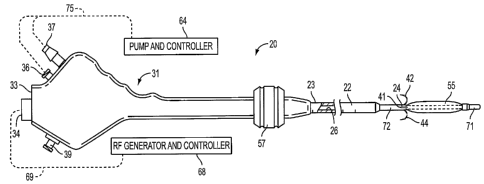
[0031] Referring now to FIGS. 1, a device to treat
urinary incontinence, described in U.S. patent
application Serial No. 09/678,500, which is herein
incorporated by reference in its entirety, is shown.
Apparatus 20 comprises semi-rigid elongated tubular shaft
CA 02493731 2005-O1-24
WO 2004/010843 PCT/US2003/022644
_ g
22 having proximal and distal extremities 23 and 24,
distal region 72, and lumen 26 extending from proximal
extremity 23 to distal extremity 24.
[0032] Handle 31 has proximal and distal ends, and is
configured to be grasped by the human hand. The distal
end of handle 31 is coupled to proximal extremity 23 of
elongated shaft 22. The proximal end of handle 31
preferably comprises rear surface 33 through which
electrical connector 34 extends. Handle 31 further
comprises fluid-in port 36, fluid-out port 37 and,
optionally, auxiliary port 39.
[0033] It will be apparent to those skilled in the art
that handle 31 may'comprise any suitable exterior shape
that is configured to be grasped by a human hand, and is
not intended to be limited by the exterior shapes
depicted herein. An illustrative, alternative handle
shape is depicted in commonly-assigned U.S. patent
application Serial No. 10/207,689, which is herein
incorporated by reference in its entirety.
[0034] Plurality of needle electrodes 41-44, which are
sharpened at their distal extremities, are disposed
within distal region 72 of elongated shaft 22 in a
contracted state. Needle electrodes 41-44 assume a
preformed shape in a deployed state, i.e., when they are
no longer constrained within elongated shaft 22, and
preferably comprise a shape-memory material such as a
nickel-titanium alloy. In the deployed state, needle
electrodes 41-44 curve outwardly and downwardly to
provide a fishhook-like configuration, as shown in FIG.
lA. Needle electrodes 41-44 are disposed in suitable
angular positions, as for example, spaced
circumferentially in a single plane 90° apart. This is
accomplished by slidably mounting needle electrodes 41-44
CA 02493731 2005-O1-24
WO 2004/010843 PCT/US2003/022644
- 9 -
in plurality of PEEK hypotubes 46 in four spaced-apart
lumens 47 of Pebax block 48, as shown in FIG, 1D.
L0035] Pebax block 48 is mounted in a fixed position
in distal region 72 of elongated shaft 22. Needle
electrodes 41-44 extend proximally through hypotubes 46
and are mounted in fixed positions in four lumens 49
spaced apart in Pebax block 51, as shown in FIG. 1C.
[0036] Pebax block 51 is slidably mounted within
elongated shaft 22. Rod 53 has a distal extremity
mounted in a fixed position in centrally disposed lumen
54 of block 51 and extends proximally from block 51 and
into recess 56 of handle 31. Slide block 58 has
outwardly extending protrusion 59 coupled to rod 53, as
shown in FIG. 1B. Knob 57 is slidably mounted on the
exterior of handle 31 and is secured to protrusion 59,
e.g., using a screw. Movement of knob 57 longitudinally
with respect to handle 31 causes needle electrodes 41-44
to be moved between extended and retracted positions in
hypotubes 46.
L0037] Cooling liquid preferably is supplied via
elongated shaft 22 so that it is discharged in the
vicinity of needle electrodes 41-44 via tubing 61.
Tubing 61 is in turn connected to fitting 36. Tubing 62
is connected to fitting 37 and extends distally into
lumen 26 of tubular member 22, through block 51, and
terminates at block 48, where it is placed in
communication with return lumen 63 in block 48. Tubing
61 continues through block 48 and opens into shaft 66,
which extends distally from block 48.
L0038] Expandable member 55 is disposed on shaft 66
and illustratively comprises a balloon. As will be
described hereinbelow with respect to FIGS. 8A-8B,
CA 02493731 2005-O1-24
WO 2004/010843 PCT/US2003/022644
- 10
expandable member 55 alternatively may comprise a
mechanically self-deployable basket.
C0039] Shaft 66 has openings 60 disposed within
expandable member 55. Openings 60.are in fluid
communication with tubing 61 and are used to inflate
expandable member 55. Expandable member 55 is provided
with plurality of openings 74 through which the cooling
liquid introduced into expandable member 55 may escape
and be discharged in the vicinity of needle electrodes
41-44 to cool the tissue being treated, as hereinafter
described. The cooling liquid, after it has performed
its function, is aspirated through central return lumen
63 to fitting 37. Alternatively, openings 74 may be
disposed directly in a lateral surface of shaft 22, as
depicted by openings 74' in FIG. 1B.
C0040] Fittings 36 and 37 are connected by tubing 75
to irrigation pump/controller 64, as depicted in FIG. 1A.
Controller 64 supplies a cooling liquid, such as room
temperature water, to fitting 36 and may also aspirate
the liquid through fitting 37 after it has been used.
C0041] Plurality of insulated wires 65 are connected
to electrical connector 34 with slack being provided
within handle 31. Electrical connector 34 is adapted to
be connected to RF generator/controller 68 by cable 69,
as depicted in FIG. 1A.
C0042] Wires 65 extend distally through lumen 26 of
elongated shaft 22, through lumens in blocks 48 and 51,
and are coupled to four thermocouple wires (not shown)
extending through hollow needles 41-44. The thermocouple
wires are connected to thermocouples 67, which are
mounted in sharpened tips of needles 41-44 for measuring
needle-tip temperatures, as shown in FTG. 1B.
CA 02493731 2005-O1-24
WO 2004/010843 PCT/US2003/022644
- 11
[0043] Referring now to FIG. 1E, a preferred method
for treating urinary incontinence using apparatus 20 is
described. Atraumatic tip 71, which is disposed distal
of expandable member 55, is inserted into urethra U of a
patient with expandable member 55 and needles 41-44 being
provided in contracted states. Elongated shaft 22 is
distally advanced within urethra U so that expandable
member 55 is positioned within bladder 8, e.g., using
markings 76 of FIG. 1B. Expandable member 55 then is
deployed, e.g., by inflating a balloon via fluid-in port
36. Expandable member 55 then is retracted proximally so
that expandable member 55 is anchored against bladder
outlet O, as shown in FIG. 1E.
[00447 After expandable member 55 has been seated
against bladder outlet O, a physician distally advances
knob 57 with respect to handle 31 to cause needles 41-44
to b~ advanced from their retracted positions within
distal region '72 of elongated shaft 22. Needles 41-44
move distally and sidewise beyond the outer cylindrical
profile of elongated shaft 22 and into the urethral
tissue in the vicinity of bladder outlet O, as shown in
FIG. 1E.
[0045] After needles 41-44 have been deployed, radio
frequency energy is supplied from RF generator/
controller 68. As is well known to those skilled in the
art, such a generator may be configured to provide
impedance readings that give an indication of whether or
not needle electrodes 41-44 have been properly positioned
within the tissue.
[0046] Liquid is introduced in the vicinity of needle
electrodes 41-44 via irrigation pump/controller 64 and
openings 74, as described hereinabove, to cool the
mucosal layer of the urethral wall. Radio frequency
CA 02493731 2005-O1-24
WO 2004/010843 PCT/US2003/022644
- 12
energy then is supplied to the needle electrodes at a
power level ranging from 1 to 10 watts for a period of
time ranging from 60 to 90 seconds to achieve
approximately an 70°C temperature in the tissue being
treated, while the overlying mucosal tissue is preserved
by the cooling liquid flow. In accordance with one
aspect of the present invention, it is desirable that the
tissue not to reach a temperature of 100°C. Therefore, RF
generator 68, utilizing the information supplied from
thermocouples 67, is programmed to automatically turn off
if the temperature reaches a pre-set temperature, e.g.,
80°C. Otherwise, for the duration of the treatment, the
RF power is adjusted to maintain the tissue being treated
at the desired target temperature.
[0047) Once the first RF treatment has been completed,
the radio frequency energy is turned off and knob 57 is
retracted to withdraw needle electrodes 41-44 into their
retracted positions within distal region 72 of elongated
shaft 22. The device then may be rotated a predetermined
angle, the needles redeployed and another radio frequency
energy treatment may be applied to surrounding tissue.
During this entire procedure, irrigation liquid is
introduced through openings 74 of expandable member 55
and/or openings 74' of shaft 22. As the irrigation fluid
progressively fills the bladder during each RF treatment,
the geometry of the bladder outlet changes as the bladder
is filled such that repeated seating of the expandable
member results in the electrode location moving
progressively towards the bladder outlet.
(0048] In connection with the RF treatments described
hereinabove, tiny sites of collagen in the vicinity of
the treatment site renature over the ensuing weeks. Such
CA 02493731 2005-O1-24
WO 2004/010843 PCT/US2003/022644
- 13
treatment results in changes in tissue compliance of the
bladder outlet and/or urethral walls to cause a
significant improvement in urinary incontinence.
[0049] Referring now to FIGS. 2-3, a first embodiment
of the present invention that utilizes a plurality of
needleless electrodes to treat urinary incontinence is
described. Apparatus 80 comprises elongated shaft 82
having proximal and distal ends and distal region 86
disposed adjacent to the distal end. Handle 84 is
provided in accordance with handle 31 of FIGS. 1, except
as noted below, and is coupled to the proximal end of
elongated shaft 82.
[0050] Elongated shaft 82 further comprises at least
one needleless electrode 90 capable of transmitting radio
frequency energy. Needleless electrode 90 may comprise a
hollow, curved surface, as depicted in FIGS. 2B-2C, and
preferably is manufactured from stainless steel.
Needleless electrode 90 preferably is embedded into a
lateral surface of elongated shaft 82 so that curved.
regions 107 and 108 of electrode 90 extend within an
interior portion of elongated shaft 82, while outer
surface 101 of electrode 90 is disposed outside of and
faces away from elongated shaft 82, as shown in FIGS. 2B-
2C.
[0051] Elongated shaft 82 further comprises irrigation
port 91 and optional aspiration port 92, which are
coupled to irrigation tubing 104 and aspiration tubing
105, respectively. Irrigation tubing 104 and aspiration
tubing 105 extend proximally from their respective ports
91 and 92, through elongated shaft 82 and handle 84, and
are coupled to fluid-in and fluid-out ports 93 and 94,
respectively. Fluid-in and fluid-out ports 93 and 94 in
.turn are coupled to fluid controller 106.
CA 02493731 2005-O1-24
WO 2004/010843 PCT/US2003/022644
- 14 -
[0052] In FIGS. 2A and 2B, irrigation and aspiration
ports 91 and 92 are illustratively disposed in a lateral
surface of elongated shaft 82 adjacent to electrode 90.
In an alternative embodiment shown in FIG. 2C, irrigation
and aspiration ports 91 and 92 may be omitted and
irrigation and aspiration tubing 104 and 105 may be
coupled to first and second curved ends 107 and 108 of
hollow electrode 90, respectively. In this embodiment,
fluid infused into irrigation tubing 104 flows through
hollow electrode 90 to provide a cooling effect upon
outer surface 101, then is aspirated through tubing 105.
[0053] Apparatus 80 of FIGS. 2 further comprises wire
98 and thermocouple wire 99, each having proximal and
distal ends. The distal ends of wire 98 and thermocouple
wire 99 are coupled to needleless electrode 90, as shown
in FIGS. 2B-2C, while the proximal ends extend through
elongated shaft 82 and handle 84. Wire~~98 and
thermocouple wire 99 preferably are coupled to electrical
connector 110 of handle 84, which in turn is connected to
RF generator/controller 109 by cable 111.
[0054] Apparatus 80 further comprises expandable
member 87 that is deployable at a predetermined distance
distal of needleless electrode 90. Expandable member 87
may comprise a balloon that is disposed on distal region
86 or, alternatively, a self-expanding mechanical basket
as described hereinbelow with respect to FIGS. 8A-8B.
(0055] Referring now to FIG. 3, a preferred method for
using apparatus 80 of FIG. 2A is described. In a first
step, atraumatic tip 85 at the distal end of elongated
shaft 82 is inserted into a patient's urethra U with
expandable member 87 in a contracted state. Expandable
member 87 is positioned within bladder B, e.g., using
measurement indicia 96 disposed near the proximal end of
CA 02493731 2005-O1-24
WO 2004/010843 PCT/US2003/022644
- 15
elongated shaft 82 (see FIG. 2A). Expandable member 87
is deployed and handle 84 is retracted proximally so that
expandable member 87 is anchored against bladder outlet
O.
L0056] In accordance with the present invention,
retracting expandable member 87 against bladder outlet O
positions needleless electrodes 90 at a desired treatment
site within urethra U using only tactile feedback. Once
properly positioned, liquid is introduced to irrigation
port 91 via irrigation pump 106 to provide a cooling
effect to mucosal layer M of the urethra. Radio
frequency energy then is supplied to needleless electrode
90 to achieve a temperature of approximately 70°C in the
tissue being treated, i.e., the submucosal tissue S of
the urethral wall. The overlying mucosal tissue M is
preserved by the cooling liquid flow. Preferably, the
submucosal tissue is not heated to a temperature
significantly higher than 70°C. Therefore, RF generator
109, utilizing the information supplied from thermocouple
97, preferably is programmed to automatically turn off if
the temperature reaches a pre-set temperature, as for
example, 80°C. Otherwise, for the duration of the
treatment, the RF power is adjusted to maintain the sub-
mucosal tissue at the desired target temperature.
[0057] After this first RF treatment has been
completed, the radio frequency energy is turned off and
the device can be advanced into the bladder lumen and
rotated a predetermined angle so that needleless
electrode 90 may contact a new interior surface of
urethra U. Once electrode 90 has been rotated to the
desired angle, handle 84 is retracted proximally to seat
expandable member 87 in bladder outlet O, and RF energy
CA 02493731 2005-O1-24
WO 2004/010843 PCT/US2003/022644
- 16
is provided to needleless electrode 90, as described
hereinabove. Upon completion of the procedure,
expandable member 87 is contracted and elongated shaft 82
is removed from the patient's urethra.
(0058] As described hereinabove with respect to the
embodiment of FIGS. lA-lE, the RF treatments produce
collagen denaturation in small, localized areas where the
treatment is delivered, followed by collagen renaturation
and remodeling over the ensuing weeks and months, thereby
resulting in changes in tissue compliance within the
urethra and/or bladder outlet.
[0059] Referring now to FIGS. 4-5, a further
alternative embodiment of the present invention is
described wherein high intensity focused ultrasound
(HIFU) is applied to treat urinary incontinence. HIFU
involves directing high intensity ultrasound waves at the
selected tissue to create heat in a precise area and
cause coagulation and tissue necrosis.
(0060] In FIG. 4, apparatus 120 comprises elongated
shaft 121 having proximal and distal ends and distal
region 122 disposed adjacent to the distal end. Handle
128 is coupled to the proximal end of elongated shaft 121
and may comprise inflation port 137 that is in fluid
communication with expandable member 123, illustratively
a balloon. Alternatively, expandable member 123 may
comprise a self-expanding mechanical basket as described
hereinbelow with respect to FIGS. 8A-8B.
[0061] Elongated shaft 121 further comprises
therapeutic ultrasound transducer 124 disposed on
elongated shaft 121 just proximal of distal region 122.
Ultrasound transducer 124 is capable of transmitting at
therapeutic ultrasound frequencies. Transducer 124
preferably comprises an annular phased array, and is
CA 02493731 2005-O1-24
WO 2004/010843 PCT/US2003/022644
- 17
coupled to a transmission cable (not shown) disposed in a
lumen of elongated shaft 121. The transmission cable
extends proximally and is coupled to electrical connector
129 of handle 128. Electrical connector 129 in turn is
connected to ultrasound generator/controller 131 by cable
130, as depicted in FIG. 4.
[00621 Referring now to FIG. 5, a preferred method of
using apparatus 120 of FIG. 4 is described. Atraumatic
tip 132 at the distal end of elongated shaft 121 is
inserted into a patient's urethra U with expandable
member 123 in a contracted state. Expandable member 123
is positioned within a patient's bladder B, e.g., using
measurement indicia 133 of FIG. 4. Expandable member 123
then is deployed and handle 128 is retracted proximally
so that expandable member 123 is anchored against bladder
outlet O. In accordance with the present invention,
retracting expandable member 123 against bladder outlet O
positions transducer 124 at a desired treatment site
within urethra U using only tactile feedback.
L0063~ Ultrasound generatorjcontroller 1.31 is turned
on and set to the desired frequency to cause transducer
124 to emit ultrasonic beams. Ultrasound beams 126 are
focused to cause a rise in tissue temperature at a
desired distance beneath mucosal layer M of urethra U.
The heating of the desired submucosal tissue causes
localized denaturation of the tissue. The change in
submucosal tissue created by the denaturation and
renaturation of the collagen results in changes in tissue
compliance of the urethral wall andjor bladder outlet,
thereby reducing urinary incontinence.
(0064) Referring now to FIGS. 6-7, a further
alternative embodiment of the present invention is
described whereby cryogenic therapy is used to treat
CA 02493731 2005-O1-24
WO 2004/010843 PCT/US2003/022644
- 18
urinary incontinence by controlled freezing of selected
urethral tissue.
[0065] In FIG. 6A, cryogenic therapy apparatus 140
comprises elongated shaft 142 having proximal and distal
ends and reduced diameter distal region 144 disposed
adjacent to the distal end. Handle 141 is coupled to the
proximal end of elongated shaft 142 and may comprise
inflation port 159 that is in fluid communication with
expandable member 145, illustratively a balloon.
Alternatively, expandable member 145 may, comprise a self-
expanding mechanical basket as described hereinbelow with
respect to FIGS. 8A-8B.
[0066] Apparatus 140 further comprises at least one
hollow needle 143 and cryogenic probe 152. Needle 143
has proximal and distal ends and sharpened tip 147
disposed at the distal end. The proximal end of each
hollow needle 143 is coupled to knob 146. Although four
hollow needles are illustrated in FIG. 6C, it will be
apparent to those skilled in the art that greater or
fewer needles may be used.
[0067] Handle 141 further comprises proximal port 150
having at least one probe insertion hypotube 151, as
depicted in FIG. 6B. Each probe insertion hypotube 151
corresponds to a respective needle 143. Each probe
insertion hypotube 151 comprises an outer diameter that
preferably is slightly smaller than an inner diameter of
hollow needle 143. A proximal end of each probe
insertion hypotube 151 is affixed to proximal port 150
while a distal end of each hypotube 151 extends into the
proximal end of its respective hollow needle 143 to
create an overlap between the distal end of the hypotube
and the proximal end of the needle, as shown in FIG. 6C.
CA 02493731 2005-O1-24
WO 2004/010843 PCT/US2003/022644
- 19 _
This overlap allows needles 143 to move with respect to
probe insertion hypotubes 157. when knob 146 is actuated.
[0068] Cryogenic probe 152 has proximal and distal
ends and tip 153 disposed at the distal end. Handle 154
is coupled to the proximal end of cryogenic probe 152 and
is configured to be grasped by a physician. Cryogenic
probe 152 is powered and controlled by cryogenic
generator 156 via wire 155, which is coupled to handle
154. Cryogenic probe 152 comprises an outer diameter
7.0 configured to be inserted into probe insertion hypotube
151 and through hollow needle 143.
[0069] Referring now to FIG. 7, a preferred method of
using apparatus 140 of FIG. 6A to treat urinary
incontinence is described. Atraumatic tip 157 at the
distal end of elongated shaft 142 is inserted into a
patient's urethra U with needle 143 in a contracted
state, i.e., retracted within the confines of elongated
shaft 142, and also with expandable member 145 provided
in a contracted state. Expandable member 145 is
positioned within a patient's bladder B, e.g., using
measurement indicia 158. Expandable member 157 then is
deployed within bladder B and handle 141 is retracted
proximally so that expandable member 157 is anchored
against bladder outlet O. In accordance with one aspect
of the present invention, retracting expandable member
157 against bladder outlet O positions needle 143, when
deployed, at a desired treatment site within urethra U
using only tactile feedback.
[0070] Needle 143 then is actuated by distally
advancing knob 146, which urges needle 143 to extend
beyond elongated shaft 142, pierce mucosal layer M of
urethra U, and extend into submucosal layer S. Needle
143 preferably comprises a shape-memory material that
CA 02493731 2005-O1-24
WO 2004/010843 PCT/US2003/022644
- 20
causes the distal end to curve to a predetermined shape
when needle 143 is no longer confined within elongated
shaft 142.
(0071] Cryogenic probe 152 then is inserted into probe
insertion hypotube 151 at proximal port 150 and is
advanced distally via probe insertion hypotube 151 into
hollow needle 143. Cryogenic probe 152 is advanced
distally until it extends distal of needle 143 and into
submucosal layer S of the urethra, as shown in FIG. 7.
Needle 143, having a larger diameter relative to probe
152, serves to dilate the submucosal tissue prior to
insertion of the probe so that the probe encounters
reduced resistance from the tissue.
[0072 Cryogenic generator 156 is turned on and set to
the desired temperature, which preferably is between .
about -80°F and -110°F, to cause local regions of the
submucosal tissue to freeze. The local regions of tissue
and tip 153 may freeze together for about three minutes,
after which time probe 152 is defrosted and removed from
within elongated shaft 142 and handle 141. If desired, a
physician then may re-insert probe 152 into a different
insertion hypotube 151 and the procedure may be repeated
through a different needle 143 to treat another region
within submucosal layer S. In accordance with principles
of the present invention, the application of cryogenic
probe 152 to the submucosal tissue causes small localized
regions of submucosal tissue to undergo necrosis, after
which tissue healing ensues. This results in altered
tissue elasticity, tensile strength, and tissue
compliance in the urethra and/or bladder outlet, and
causes a significant improvement in urinary incontinence.
[0073] Referring now to FIGS. 8A-8B, an alternative
expandable member is described for use with any of the
CA 02493731 2005-O1-24
WO 2004/010843 PCT/US2003/022644
- 21
treatment techniques described hereinabove. Apparatus
170 comprises self-expandable basket 172, shown in a
deployed state in FIG. 8B, instead of a balloon. Basket
172 preferably comprises a plurality of flexible struts
171 joined to rod 176 via hinges 180. Struts 171, which
may be covered by a biocompatible elastomeric membrane
(not shown), are constrained in a contracted position
within central lumen 177 of elongated shaft 181, as
illustrated in FIG. 8A. Struts 171, which preferably
comprise a shape-memory material such as Nitinol, assume
a predetermined curvature extending radially outward from
elongated shaft 181 in the deployed state, i.e., when
struts 171 are no longer effectively constrained within
central lumen 177, as shown in FIG. 8B.
[0074] Rod 176 has proximal and distal ends and is
disposed through central lumen 177. Preferably, rod 176
includes atraumatic distal tip 178 disposed at the distal
end. The proximal end of rod 176 is configured to be
manipulated by a physician.
[0075] In operation, atraumatic tip 178 and elongated
shaft 181 are inserted into the patient's urethra in a
manner described hereinabove. Once distal end 184 of
elongated shaft 181 is positioned within a patient's
bladder, the proximal end of rod 176 is advanced distally
by a physician to self-deploy mechanically expandable
basket 172, as depicted in FIG. 8B. Once basket 172 is
deployed within the bladder, elongated shaft 181 and rod
176 are retracted proximally to cause basket 172 to
become anchored against the bladder outlet.
(0076] At this time, needles 183 may be deployed from
elongated shaft 181 to penetrate the urethral wall to
perform a radio frequency treatment of the tissue.
Alternatively, needles 183 may be omitted and needleless
CA 02493731 2005-O1-24
WO 2004/010843 PCT/US2003/022644
- 22 -
radio frequency waves or ultrasound beams may be used to
treat incontinence, as described hereinabove.
(0077] After the preferred treatment is completed,
basket 172 is returned to the contracted configuration by
retracting rod 176 proximally with respect to elongated
shaft 181 to cause struts 171 to be contained within
central lumen 177.
[0078] Referring now to FIGS. 9-10, apparatus and
methods for longitudinally advancing the spacing between
the tissue treating elements and the expandable member
are described. In FIG. 9, apparatus 200 is provided in
accordance with apparatus 20 of FIGS. 1, except as noted
below. Apparatus 200 Comprises handle 202 and knob 204,
which are similar in structure to handle 31 and knob 57
of FIGS. 1, respectively.
(0079] Apparatus 200 further comprises elongated shaft
206 having proximal and distal ends and actuator 214
disposed about the proximal end. Measurement indicia 212
preferably are provided on a lateral surface of elongated
shaft 206. Illustratively, needle electrodes 210 are
shown for providing energy to the submucosal layer of the
urethral wall, although it will be apparent that
needleless electrodes, an ultrasound transducer or
cryogenic probe, as described hereinabove, may be
substituted for needle electrodes 210.
(0080] Apparatus 200 further comprises shaft 208
having proximal and distal ends and expandable member 209
disposed on the distal end. Shaft 208 preferably is
affixed to an interior surface of handle 202 and may
include inflation lumen 220 extending between the
proximal and distal ends that communicates with
expandable member 209.
CA 02493731 2005-O1-24
WO 2004/010843 PCT/US2003/022644
- 23
[0081 Referring now to FIG. 10A, a side sectional
view of the distal end of apparatus 200 is provided.
Elongated shaft 206 preferably comprises central lumen
217 having an inner diameter slightly larger than an
outer diameter of expandable member shaft 208. Inflation
lumen 220 is in fluid communication with expandable
member 209, while lumens 218 house needle electrodes 210
in the contracted state (see FIG. 10A).
[0082 Region 222 of shaft 208 may be threaded to
provide threaded interface 219 between shaft 208 and
central lumen 217 of elongated shaft 206, as shown in
FIG. IOB. Threaded interface 219 provides for controlled
longitudinal movement of elongated shaft 206 with respect
to shaft 208 when actuator 214 of FIG. 9 is rotated
circumferentially.
[0083 Referring to FIG. lOC, the threaded interface
between elongated shaft 206 and shaft 208 is omitted and
small gap 227 is provided between central lumen 217 and
shaft 208. Gap 227 allows for straight translation of
elongated shaft 206 with respect to shaft 208 when
actuator 214 is longitudinally advanced and handle 202 is
held stationary.
[0084) Using the technique of FIGS. 9-10, a physician
may perform a first treatment at a distance of about xl
from the bladder outlet, as shown in FTG. 10A, assuming
that expandable member 209 is disposed within the bladder
and then retracted against the bladder outlet. The
physician then may perform a second treatment at a
distance of about x2 from the bladder outlet by actuator
214 as described hereinabove. Measurement indicia 212
may be used as a distance guide when a physician
manipulates the distance between x1 and xa u,sing actuation
handle 214.
CA 02493731 2005-O1-24
WO 2004/010843 PCT/US2003/022644
- 24
[00857 Referring now to FIG. 11, an alternative
embodiment of apparatus configured to longitudinally
advance the spacing between the tissue treating elements
and the expandable member is described. In FIG. 11,
apparatus 200' is provided substantially in accordance
with apparatus 200 of FIGS. 9-10, except as noted below.
[00861 In the embodiment of FIG. 11, shaft 208'
extends through elongated shaft 206' and preferably is
coupled to actuator 250 instead of being affixed to an
interior surface of handle 202', as described in the
embodiment of FIGS. 9-10. Actuator 250 may be disposed
about handle 202' in a manner similar to the manner in
which knob 57 of FIG. 1 is disposed about handle 31, as
described hereinabove. Alternatively, actuator 250 may
be disposed proximal of handle 202'. For example, shaft
208' may extend through handle 202', through an aperture
or port (not shown) disposed at the proximal end of
handle 202', and then may be coupled to actuator 250
proximal of the handle.
[00877 Apparatus 200' may comprise a threaded
interface between shaft 208' and a central lumen of
elongated shaft 206', as described in FIG. 10B
hereinabove. The threaded interface provides for
controlled longitudinal movement of shaft 208' with
respect to elongated shaft 206' when actuator 250 is
rotated circumferentially and handle 202' is held
stationary.
[00881 Alternatively, a small gap, such as described
hereinabove with respect to FIG. lOC, may be provided
between the central lumen of elongated shaft 206' and
shaft 208'. This permits straight translation of shaft
208' with respect to elongated shaft 206' when actuator
250 is advanced or retracted and handle 202' is held
CA 02493731 2005-O1-24
WO 2004/010843 PCT/US2003/022644
- 25
stationary. Measurement indicia (not shown) may be
disposed on handle 202' or shaft 208' to determine the
spacing between tissue treating elements 210' and
expandable member 209'.
L0089] Handle 202' also may comprise a central lumen,
e.g., as described hereinabove with respect to FIGS. lOB-
lOC, that guides shaft 208' through handle 202'. The
central lumen of handle 202' may be used alone or in
conjunction with the central lumen of elongated shaft
206' to serve as a guide for shaft 208' along the length
of the device.
[0090] While preferred illustrative embodiments of the
invention are described above, it will be apparent to one
skilled in the art that various changes and modifications
may be made therein without departing from the invention.
The appended claims are intended to cover all such
changes and modifications that fall within the true
spirit and scope of the invention.