Note: Descriptions are shown in the official language in which they were submitted.
CA 02493849 2005-O1-11
WO 2004/007749 PCT/US2003/021968
METHODS AND SYSTEMS FOR EXTENDED
IN VITRO CULTURE OF NEURONAL CELLS
CROSS-REFERENCES TO RELATED APPLICATIONS
This application claims the benefit of U.S. Provisional Patent
Application No. 60/395,973 filed July 12, 2002, which is incorporated herein
by
reference in its entirety.
BACKGROUND OF THE INVENTION
Field of the Invention
The present invention relates generally to a cell culture system that
provides extended in vitro culture of neuronal cells. The invention is
particularly
related to the extended culture of retinal neuronal cells. The cell culture
system is
useful for identifying bioactive agents that can be used for treating
neurodegenerative
diseases, particularly retinal diseases and disorders. The invention also
relates to using
the cell culture method for identifying cells that may be useful for treating
a retinal
degenerative disease or disorder.
Description of the Related Art
In vitro culture of neuronal cells in general, and of retinal neuronal cells
in particular, has been problematic. For many years, it has been believed that
fully
mature neurons lack plasticity and the ability to repair and regenerate after
injury. If
mature central nervous system (CNS) neurons could be cultured and stimulated
to
regenerate, transplantation and functional restoration of damaged or diseased
CNS
tissue might become feasible.
As a first step, groups of investigators have been studying in, vitro
growth of CNS-derived neurons. Some of these studies involve transformed or
immortalized neuronal cells; some cells have been derived from tumorigenic
tissues.
With respect to retinal cultures, in vitro retinal organ cultures, retinal
explant cultures
and retinal explantl membrane culture techniques have been reported. In
addition,
CA 02493849 2005-O1-11
WO 2004/007749 PCT/US2003/021968
investigators have reported analysis of retinal neural cell cultures that are
derived from
embryonic tissue or embryonic stem cells or from neonatal retinas. However,
the
inability to accomplish long-term culture of post-mitotic neuronal cells has
been a
major roadblock within the field of neurobiology. If primary cells obtained
from
mature, fully-differentiated neuronal tissue in general, and mature retinal
neurons in
particular, could be cultured in vitro over an extended period of time, this
would
constitute a valuable tool for neurobiological studies including examination
of cell-to-
cell interactions; selection and analysis of neuroactive compounds and
materials;
provision of a controlled surrogate system for in vivo CNS and ophthalmic
tests; and
potential analysis single cells from a consistent population.
BRIEF SUMMARY OF THE INVENTION
Briefly stated, the present invention provides compositions and methods
for extended cell culture of neuronal cells that may be used for identifying
bioactive
agents and cells useful for treatment of neurodegenerative diseases, including
neurodegenerative retinal diseases and disorders. One aspect of the invention
provides
a cell culture system comprising a mixture of mature neuronal cells and cells
isolated
from a ciliary body. In certain embodiments of the invention the mature
neuronal cells
comprise mature retinal neuronal cells, wherein the mature retinal neuronal
cells are
bipolar cells, horizontal cells, amacrine cells, ganglion cells, and/or
photoreceptor cells.
In another embodiment the invention provides a retinal cell culture
system comprising a mixture of (i) mature retinal neuronal cells; (ii) cells
isolated from
a ciliary body; and (iii) embryonic retinal cells, wherein the mature retinal
neuronal
cells are bipolar cells, horizontal cells, amacrine cells, ganglion cells,
and/or
photoreceptor cells. In certain embodiments, the embryonic retinal cells
comprise
retinal stem cells and in certain other embodiments, the embryonic retinal
cells
comprise embryonic retinal progenitor cells.
The invention also provides a method for producing a retinal cell culture
system comprising co-culturing a mature retinal neuronal cell and a cell
isolated from a
ciliary body. In another embodiment, a method is provided for enhancing
survival of a
mature retinal neuronal cell in vitro comprising co-culturing a mature retinal
neuronal
2
CA 02493849 2005-O1-11
WO 2004/007749 PCT/US2003/021968
cell and a cell isolated from a ciliary body. In certain embodiments, these
methods
comprise co-culturing (i) a mature retinal neuronal cell; (ii) a cell isolated
from a ciliary
body; and (iii) an embryonic retinal cell. In certain specific embodiments,
the
embryonic retinal cell is selected from the group consisting of a retinal stem
cell and an
embryonic retinal progenitor cell.
The present invention also provides a method for identifying a bioactive
agent that is capable of enhancing survival of a neuronal cell, comprising (i)
contacting
a candidate agent with the subject invention cell culture system as described
herein
under conditions and for a time sufficient to permit interaction between a
neuronal cell
of the cell culture system and the candidate agent; and (ii) comparing
survival of a
neuronal cell of the cell culture system in the presence of the candidate
agent with
survival of a neuronal cell of the cell culture system in the absence of the
candidate
agent, and therefrom identifying a bioactive agent that is capable of
enhancing survival
of the neuronal cell. In certain embodiments, the neuronal cell is a retinal
neuronal cell.
° In another embodiment, the invention provides a method for
identifying
a bioactive agent that is capable of inhibiting neurodegeneration of a
neuronal cell
comprising (i) contacting a bioactive agent with a cell culture system as
described
herein, under conditions and for a time sufficient to permit interaction
between a
neuronal cell of the cell culture system and the candidate agent; and (ii)
comparing
structure of a neuronal cell of the cell culture system in the presence of the
bioactive
agent with structure of a neuronal cell of the cell culture system in the
absence of the
bioactive agent, and therefrom identifying a bioactive agent that is capable
of inhibiting
neurodegeneration of the neuronal cell. In certain embodiments, the neuronal
cell is a
retinal neuronal cell.
The invention also provides a method for identifying a bioactive agent
that is capable of treating a retinal disease comprising contacting a
bioactive agent with
the subject invention cell culture system as described herein, under
conditions and for a
time sufficient to permit interaction between a neuronal cell of the cell
culture system
and the candidate agent; and (ii) comparing neurodegeneration of a neuronal
cell of the
cell culture system in the presence of the bioactive agent with
neurodegeneration of a
neuronal cell of the cell culture system in the absence of the bioactive
agent, and
3
CA 02493849 2005-O1-11
WO 2004/007749 PCT/US2003/021968
therefrom identifying a bioactive agent that is capable of treating a retinal
disease. In
certain embodiments, the neuronal cell is a retinal neuronal cell. In certain
specific
embodiments the retinal disease that is treated is macular degeneration,
glaucoma,
diabetic retinopathy, retinal detachment, retinal blood vessel occlusion,
retinitis
pigmentosa, or a retinal disorder associated with Alzheimer's disease.
In another embodiment, the invention provides a method for treating a
retinal disease comprising introducing isolated retinal stem cells into
retinal tissue of a
subject in need thereof, wherein the retinal disease that is treated is
macular
degeneration, glaucoma, diabetic retinopathy, retinal detachment, retinal
blood vessel
occlusion, retinitis pigmentosa, or a retinal disorder associated with
Alzheimer's
disease.
Within one embodiment, the present invention provides methods for
extended culture of mature neuronal cells that feature incubating mature
neuronal cells
with ciliary body cells. Within another embodiment, the invention provides an
in vitro
cell culture system that features a mixture of mature neuronal cells and a
source of
mature retinal stem cells. A ciliary body is a preferred source of retinal
stem cells. In
yet another embodiment, the invention provides a method for screening
bioactive
molecules, using an in vitro cell culture system containing a mixture of
mature neuronal
cells and ciliary body cells. While ciliary body cells are preferred for in
vitro co-culture
with mature neuronal cells, a source of stem cells, including other sources of
CNS stem
cells, may also find use within these methods and systems.
Although this invention is particularly amenable to the in vitro culture
and survival of mature retinal neurons, the methods and systems disclosed are
also
useful for extended in vitro culture of other neuronal cell types obtained
from a variety
of species. The ciliary body cells (and/or a source of stem cells) and the
neuronal cells
need not be obtained from the same species. Further, the source of stem cells
useful
within the present invention may be primary cells; tumorigenic, transformed or
immortalized cells; adult, embryonic or neonatal cells; or retinal or non-
retinal cells.
These methods and systems may be used not only to culture retinal
neurons in vitro, but also may find use with other central nervous system
cells. Also,
other mature, differentiated primary cells that are difficult to culture in
vitro may be
4
CA 02493849 2005-O1-11
WO 2004/007749 PCT/US2003/021968
advantageously co-cultured with ciliary body cells, or more generally with a
source of
stem cells, according to the methods and systems of the present invention.
These and other aspects of the invention will become evident upon
reference to the following detailed description and attached drawings. In
addition,
references set forth herein that describe in more detail certain aspects of
this invention
are therefore incorporated by reference in their entireties.
BRIEF DESCRIPTION OF THE SEVERAL VIEWS OF THE DRAWINGS
Figure 1 illustrates immunohistochemical staining of monkey retinal
cells and chicken retinal cells. Monkey retinal cells were cultured for 3
months (Fig.
1 A, 1 B, and 1 C). Chicken retinal cells were cultured for 14 days (Fig. 1 D
and Fig. 1 E).
Cells were subjected to immunological analysis using an anti-(33-tubulin
antibody (Fig.
lA, 1B, and lE; representative cells are indicated by closed arrows) to
identify ganglion
cells, and using an antibody to calretinin to identify amacrine and horizontal
cells (Fig.
1 A and 1 E, representative cells are circled). The cells were stained with
DAPI to
identify the nuclei (muted staining in Fig. 1 A, 1 B, 1 D, and 1 E;
representatively stained
cells are indicated by open arrow in Fig. 1 A, 1 B, and 1 D). Photoreceptor
cells were
identified with an antibody to recoverin (Fig. 1 C; representative cells are
circled); to
visinin (D, representative cells are shown by open arrowhead); or to rhodopsin
that
stains the projections of the photoreceptor cells (Fig. 1C and 1D;
representative cells are
circled). Scale bars: 20 pm.
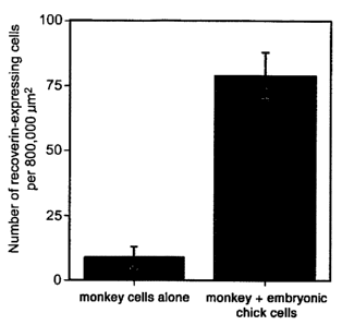
Figure 2 illustrates survival of monkey retinal cells when co-cultured
with monkey ciliary body cells and chicken embryonic cells. Figure 2A presents
a
histogram showing the number of recoverin-expressing cells per 800,000 pmt,
with and
without co-culture with embryonic chick retinal cells. Figure 2B illustrates
the number
of monkey retinal cells that were immunohistochemically stained cells with an
anti-
recoverin antibody when the monkey cells were cultured alone, and Figure 2C
illustrates the number of monkey cells when co-cultured with embryonic chick
retinal
cells.
5
CA 02493849 2005-O1-11
WO 2004/007749 PCT/US2003/021968
DETAILED DESCRIPTION OF THE INVENTION
Prior to setting forth the invention in detail, it may be helpful to the
understanding thereof to define the following terms.
The term "neuron" is used herein to denote a cell that arises from
neuroepithelial cell precursors. Mature neurons (i. e., fully differentiated
cells from an
adult) display several specific antigenic markers.
The term "ciliary body" is used herein to denote a tissue that resides
between the peripheral regions of the retina and the iris, all of which arise
from the
same neuroepithelium during development.
The term "neuroepithelium" is used herein to denote cells and tissues
that arise from the neural epithelium during development; such cells include
retinal
cells, diencephalon cells and midbrain cells. Neuroepithelium is also defined
as
neuroectoderm, and more specifically as ectoderm on the dorsal surface of the
early
vertebrate embryo that gives rise to the cells (neurons and glia) of the
nervous system
1 S (On-line Medical Dictionary, Dept. of Medical Oncology, University of
Newcastle
upon Tyne; March 4, 1998; Retrieved from the Internet:
<URL:http://cancerweb.ncl.ac.uk/cgi-bin/omd?query =neuroepithelium&action=
Seaxch+ OMD.
Neurodegenerative eye diseases, such as glaucoma and macular
degeneration, affect nearly seventeen million patients in the United States
alone.
Considering the loss of quality of life associated with blindness, drug
research and
development in this area is of great importance.
Glaucoma is a broad term used to describe a group of diseases that
causes visual field loss, often without any other prevailing symptoms. The
lack of
symptoms often leads to a delayed diagnosis of glaucoma until the terminal
stages of
the disease. Prevalence of glaucoma is estimated to be three million in the
United
States, with about 120,000 cases of blindness attributable to the condition.
The disease
is also prevalent in Japan with four million cases. In other parts of the
world, access to
treatment is even less, thus glaucoma ranks as a leading cause of blindness
worldwide.
Even if subjects afflicted with glaucoma do not become blind, their vision is
often
severely impaired. The loss of peripheral vision is caused by the death of
ganglion cells
6
CA 02493849 2005-O1-11
WO 2004/007749 PCT/US2003/021968
in the retina. Ganglion cells are a specific type of projection neuron that
connects the
eye to the brain. Glaucoma is often accompanied by an increase in intraocular
pressure.
Current treatment includes use of drugs that lower the intraocular pressure.
However,
lowering the intraocular pressure is often insufficient to completely stop
disease
progression. It is believed that ganglion cells are quite susceptible to
pressure and have
already suffered permanent degeneration prior to the lowering of intraocular
pressure.
In addition, an increasing number of cases of normal tension glaucoma has been
observed, in which ganglion cells degenerate without an observed increase in
the
intraocular pressure. Because current glaucoma drugs only treat intraocular
pressure, a
need exists to identify new therapeutic agents that will prevent or reverse
the
degeneration of ganglion cells. Recent reports suggest that glaucoma is a
neurodegenerative disease, similar to Alzheimer's disease and Parkinson's
disease in
the brain, except that it specifically affects retinal neurons. The retinal
neurons of the
eye originate from diencephalon neurons of the brain. Though it is often not
thought of
as part of the nervous system, retinal neurons . are a key component of
vision,
interpreting the signals from the light sensing cells.
Macular degeneration is a disease that affects central vision, as opposed
to glaucoma that affects peripheral vision. Prevalence of macular degeneration
is
estimated at thirteen million patients in the United States, and it is the
leading cause of
blindness worldwide. Macular degeneration is a disease that causes the loss of
photoreceptor cells in the central part of retina, called the macula. Macular
degeneration can be classified into two types: dry type and wet type. The dry
form is
more common than the wet, with about 90% of age-related macular degeneration
(ARMD) patients diagnosed with the dry form. The wet form of the disease
usually
leads to more serious vision loss. The exact causes of age-related macular
degeneration
are still unknown. The dry form of ARMD may result from the aging and thirming
of
macular tissues, and deposition of pigment in the macula. With wet ARMD,
new,blood
vessels grow beneath the retina and leak blood and fluid. This leakage causes
the
retinal cells to die, creating blind spots in central vision. The only FDA-
approved
protocol available to treat ARMD is a photodynamic therapy that uses a special
drug
combined with laser photocoagulation. This treatment, however, can only be
applied to
7
CA 02493849 2005-O1-11
WO 2004/007749 PCT/US2003/021968
half of the new wet form cases of ARMD. For the vast majority of patients who
have
the dry form of macular degeneration, no treatment is available. Macula exists
in
primates (including humans), but not in rodents; therefore currently no good
animal
models of macula are available. This lack of a good animal model has proved to
be a
major obstacle for developing new drugs to treat this disorder.
Alzheimer's disease (AD) is the most common form of dementia among
the elderly. Dementia is a brain disorder that seriously affects a person's
ability to carry
out daily activities. Alzheimer's is a disease that affects four million
people in the
United States alone. It is characterized by a loss of nerve cells in areas of
the brain that
are vital to memory and other mental functions. Some drugs can prevent AD
symptoms
for a finite period of time, but are no drugs are available that treat the
disease or
completely stop the progressive decline in mental function. Recent research
suggests
that glial cells that support the neurons or nerve cells may have defects in
AD sufferers,
but the cause of AD remains unknown. Individuals with AD seem to have a higher
incidence of glaucoma and macular degeneration, indicating that similar
pathogenesis
may underlie these neurodegenerative diseases of the eye and brain. (See
Giasson et al.,
Free Radic. Biol. Med 32:1264-75 (2002); Johnson et al., Proc. Natl. Acad.
Sci. USA
99:11830-35 (2002); Dentchev et al., Mol. Vis. 9:184-90 (2003)).
The present invention provides an in vitro neuronal cell culture system
that will find use in the identification and biological testing of new
neuroactive
compounds or materials that may be suitable for treatment of neurological
diseases or
disorders in general, and for treatment of degenerative diseases of the eye
and brain in
particular. The cultured mature neurons provided herein are particularly
useful for
compound screening to identify candidate drugs that may enable regeneration of
CNS
tissue that has been damaged by disease. Neurodegenerative diseases or
disorders for
which the present invention may be useful for identifying agents that may
treat, cure,
prevent, ameliorate the symptoms of, or slow or stop the progression of,
include but are
not limited to glaucoma, macular degeneration, diabetic retinopathy, retinal
detachment,
retinal blood vessel (artery or vein) occlusion, retinitis pigmentosa, and
retinal disorders
associated with other neurodegenerative diseases such as Alzheimer's disease
or
Parkinson's Disease.
8
CA 02493849 2005-O1-11
WO 2004/007749 PCT/US2003/021968
In addition, with the advent of novel technologies such as genomics and
proteomics, thousands of new, relatively uncharacterized genes and proteins
have been
identified. One of the bottlenecks of drug discovery and development is
determining
how to prioritize thousands or millions of small molecule and proteinaceous
therapeutic
agent candidates that are available for high-throughput screening. Most of
these high-
throughput assay systems are based on test molecule stimulation or inhibition
of target
cell enzymatic activity, or on test molecule binding to a target molecule or
target cell.
Because in vivo systems feature complex interactions between target molecules
or
target cells and surrounding molecules within the target molecule's cellular
environment, or the target cell's surrounding tissue environment, it is hard
to predict
how a candidate molecule, identified by an isolated biochemical assay, will
exert its
influence in an in vivo setting. For example, certain target proteins, such as
transcription factors and cell-surface receptors, often form mufti-subunit
complexes in
order to exhibit biological function. Furthermore, the response of a target
protein to
potential therapeutic agents is likely to be dependent on its cellular
context. An assay
using the cultured neurological cells provided by the present invention will
better mimic
the in vivo target molecule environment.
Another research and development bottleneck involves correlating
genetic analysis or sequence information to functional biology in order to
validate a
target. Bioinformatics and genomic technologies have identified new genes that
map to
regions of the chromosome associated with genetic mutations or defects that
have been
associated with biological diseases or disorders. However, identifying and
analyzing
the precise biological function of the thousands and millions of interesting
genes (and
their corresponding gene products) is proving to be extremely challenging.
Without
good cellular models, it is difficult to elucidate the true biological
function of each
protein. Thus, although bioinformatics and genomics techniques can now quickly
identify potential disease-causing proteins and candidate therapeutic agents,
characterizing the biological significance and function of each such molecule
continues
to be difficult and time consuming. Consistent and reproducible cell-based
assay
systems, such as provided herein, will accelerate this functional analysis.
Further, use
of the cultured neuronal cells of the present invention may permit
identification of
9
CA 02493849 2005-O1-11
WO 2004/007749 PCT/US2003/021968
bioactive agents that target intracellular functional units or other types of
non-protein
molecules, such as ribosomes, lipids, or carbohydrates.
The next generation of drug discovery platform technology may
incorporate "cellomics." Cellomics will utilize comprehensive analyses of in
vitro
cultured cells. Cell-based screening systems will allow candidate
biopharmaceutical
agents to interact with corresponding target molecules in a more physiological
state
than simple protein-target analysis.
In one embodiment, the present invention provides methods for culturing
retinal neurons in vitro for extended periods of time, preferably longer than
2 weeks or
4 weeks, more preferably longer than 2 months, and still more preferably
longer than 3
months. Retinal neurons have been obtained from post-natal non-human primates
and
post-natal chickens, but any adult or post-natal retinal tissue may be
suitable for use
within the present invention. The source of the retinal cells or tissue may be
mammalian (e.g., human, non-human primate, rodent, canine, porcine, bovine, or
other
mammalian source), avian, or from other genera.
The types of retinal neuronal cells that may be cultured in vitro by this
method include ganglion cells, photoreceptors, bipolar cells, horizontal
cells, and
amacrine cells. A feature of this invention is co-culture of retinal neurons
with ciliary
body cells, and/or with a source of stem cells. The ciliary body is a tissue
in the eye
that includes the group of muscles that act on the eye lens to produce
accommodation
and the arterial circle of the iris. The inner ciliary epithelium is
continuous with the
pigmented retinal epithelium, and the outer ciliary epithelium secretes the
aqueous
humour. The pigmented epithelium from the ciliary body is reported to include
retinal
stem cells (Tropepe et al., Science 287:2032-36 (2000); Fischer et al.,
Develop. Biol.
220:197-200 (2000)). Although ciliary body-derived cells are preferred,
sources of
CNS-derived stem cells may also be used within the invention. Adult or post-
natal
ciliary body cells are preferred. Embryonic cells, particularly, retinal
embryonic cells
may be used in the cell culture system and include embryonic stem cells and
embryonic
neuronal progenitor cells. Sources of adult, embryonic or pre-natal stem cells
(retinal
stem cells or non-retinal stem cells; CNS-derived stem cells or stem cells
derived from
other tissue types; mammalian-, avian- or other genera- and species-derived
stem cells)
CA 02493849 2005-O1-11
WO 2004/007749 PCT/US2003/021968
may also be used. The source of the stem cells, including the source of the
retinal stem
cells, may be primary cells, or may be immortalized, transformed, tumorigenic,
or
genetically manipulated cells that can be cultured in vitro indefinitely.
The present invention provides an effective method for identifying and
analyzing bioactive agents in general, and neuroactive agents in particular.
Through
use of such method, agents useful for treating diseases and disorders of the
central
nervous system and retina, including but not limited to neurodegenerative
diseases,
epilepsy, macular degeneration, and glaucoma, may be selected and tested.
According
to the present invention, a bioactive agent may include, for example, a
peptide, a
polypeptide (for example, a ligand that binds to a neuronal cell receptor, a
growth
factor, trophic factor, or the like), an oligonucleotide or polynucleotide,
antibody or
binding fragment thereof, or small molecule. Candidate agents for use in a
method of
screening for a bioactive agent that is capable of altering (increasing or
decreasing in a
statistically significant manner) neurodegeneration of neuronal cells, such as
retinal
neuronal cells, may be provided as "libraries" or collections of compounds,
compositions, or molecules. Such molecules typically include compounds known
in the
art as "small molecules" and having molecular weights less than 105 daltons,
preferably
less than 104 daltons and still more preferably less than 103 daltons.
Preferably, a
bioactive agent inhibits, impairs, or prevents neurodegeneration of neuronal
cells.
Candidate agents further may be provided as members of a combinatorial
library, which
preferably includes synthetic agents prepared according to a plurality of
predetermined
chemical reactions performed in a plurality of reaction vessels. The resulting
products
comprise a library that can be screened and then followed by iterative
selection and
synthesis procedures, to provide, for example, a synthetic combinatorial
library of
peptides (see, e.g., PCT/L1S91/08694, PCT/LTS91/04666) or other compositions
that
may include small molecules as provided herein (see, e.g., PCT/US94/08542,
U.S.
5,798,035, U.S. 5,789,172, U.S. 5,751,629). Those having ordinary skill in the
art will
appreciate that a diverse assortment of such libraries may be prepared
according to
established procedures.
The present invention provides methods for identifying bioactive agents
that may be useful for treating neurodegenerative diseases, including but not
limited to
11
CA 02493849 2005-O1-11
WO 2004/007749 PCT/US2003/021968
retinal diseases such as glaucoma, macular degeneration, diabetic retinopathy,
retinal
detachment, retinal blood vessel (artery or vein) occlusion, retinitis
pigmentosa, and
retinal disorders associated with other degenerative diseases such as
Alzheimer's
disease. Bioactive agents as described herein may be incorporated into
screening
assays comprising the subject invention cell culture system to determine
whether a
bioactive agent is capable of altering neurodegeneration of neuronal cells
(impairing,
inhibiting, preventing, or accelerating in a statistically significant
manner). A preferred
bioactive agent is one that inhibits or impairs neurodegeneration of a
neuronal cell or
that is capable of regenerating a neuronal cell. A bioactive agent that
inhibits
neurodegeneration of a neuronal cell may be identified by contacting (mixing,
combining, or otherwise permitting interaction between the agent and cells of
the cell
culture system), for example, a candidate agent from a library of agents as
described
herein; with the cell culture system under conditions and for a time
sufficient to permit
interaction between a candidate agent and the cells, particularly the mature
neuronal or
retinal neuronal cells of the cell culture system. A bioactive agent may act
directly
upon a neuronal or retinal neuronal cell to affect survival or
neurodegeneration of the
cell. Alternatively, a bioactive agent may act indirectly by interacting with
one cell that
consequently responds to the agent by affecting survival or neurodegeneration
of
another neuronal cell.
A bioactive agent that effectively alters, preferably inhibits,
neurodegeneration of a neuronal cell may be identified by techniques known in
the art
and described herein for determining the effects of the agent on neuronal cell
structure
or morphology; expression of neuronal cell markers (e.g., (33-tubulin,
rhodopsin,
recoverin, visinin, calretinin, Thy-1, tau, microtubule-associated protein 2,
and the like
(see, e.g., Espanel et al., Int. .7. Dev. Biol. 41:469-76 (1997); Ehrlich et
al., Exp. Neurol.
167:215-26 (2001 ); Kosik et al., J. Neurosci. 7:3142-53 (1987))); and/or cell
survival
(i.e., cell viability or length of time until cell death). Preferably, a
bioactive agent
enhances survival of neuronal cells such as retinal neuronal cells, that is,
the agent
promotes survival or prolongs survival such that the time period in which
neuronal cells
are viable is extended. The ability of a candidate agent to enhance cell
survival or
impair, inhibit, or impede neurodegeneration may be determined by any one of
several
12
CA 02493849 2005-O1-11
WO 2004/007749 PCT/US2003/021968
methods known to those skilled in the art. For example, changes in cell
morphology in
the absence and presence of a candidate agent may be determined by visual
inspection
such as by light microscopy, confocal microscopy, or other microscopy methods
known
in the art. Survival of cells can be determined by counting viable and/or
nonviable
cells, for instance. Immunochemical or immunohistological techniques (such as
fixed
cells staining or flow cytometry) may be used to identify and evaluate
cytoskeletal
structure (e.g., by using antibodies specific for cytoskeletal proteins such
as synapsin,
an intermediate filament protein such as glial fibrillary acidic protein,
fibronectin, actin,
vimentin, tubulin, or the like) or to evaluate expression of cell markers as
described
herein. The effect of a candidate agent on cell integrity, morphology, and/or
survival
may also be determined by measuring the phosphorylation state of neuronal cell
polypeptides, for example, cytoskeletal polypeptides (see, e.g., Sharma et
al., J. Biol.
Chem. 274:9600-06 (1999); Li et al., J. Neurosci. 20:6055-62 (2000)).
Regeneration of
neuronal cells or proliferation of neuronal cells may be determined by any of
several
methods known in the art, for example, by measuring incorporation of labeled
deoxyribonucleotides or ribonucleotides or derivatives thereof, such as
tritiated
thymidine, or such as by measuring incorporation of bromodeoxyuridine (BrdU),
which
can be detected by using antibodies that specifically bind to BrdU.
In some situations, such methods may enable identification of candidate
therapeutic agents that not only improve the symptoms of neurodegeneration,
but also
act to reverse the state of neurodegeneration. The disclosed methods and cell
culture
systems permit very precise measurements of specific interactions occurring
between
neurons, as well as enabling detailed analysis of subtleties in neuron
structure. For
instance, the methods and cultured cells of the present invention are
compatible with
neurochips, cell-based biosensors, and other multielectrode or
electrophysiologic
devices for stimulating and recording data from cultured neurons (see, for
instance,
M.P. Maher et al., J. Neurosci. Meth. 87:45-56, 1999; K.H. Gilchrist et al.,
Biosensors
& Bioelectronics 16:557-64, 2001).
13
CA 02493849 2005-O1-11
WO 2004/007749 PCT/US2003/021968
Screening neurological targets for drug discovery
Neurodegenerative diseases are a major source of morbidity, and an in
vitro neural cell culture model may benefit drug discovery in this area.
Because
culturing of post-mitotic neuronal cells has been so difficult, it is critical
to have a good
paradigm when screening drugs relevant to neurologic and ophthalmic diseases.
As
noted previously, the response of target molecules to potential drug
candidates is likely
to be dependent on the cellular environment of the target molecule. Thus, it
is
important to use in screening assays cultured cells that are closely related
to the cell
types that are to be ultimately treated with the drug.
To properly validate drug / therapeutic agent candidates, tissue-specific
cultured cells for use within a cell-based screening system must be identified
and
evaluated. In the field of neurobiology, cell lines such as PC 12 cells
(derived from a rat
pheochromocytoma), NT2 cells (derived from a human teratocarcinoma), or human
neuroblastoma cell lines have been used to screen drug candidates. While these
cells
have some characteristics of prototypic neurons, these cells are tumor-
derived, and have
an immature neuronal phenotype considered to be different from physiological
neural
cells.
In vitro cell culture system
Others groups have reported in vitro culture of embryonic retinal
neurons, but these cells fail to express all of the retina-specific proteins
that are
expressed by mature retinal cells, or these cells could only be cultured for
short times.
X. Luo et al. (IOVS 42:1096-1106, 2001) have reported culturing of retinal
cells in
vitro, but their system differed from the one disclosed herein in that the in
vitro cell
culture system of the present invention includes co-culture with ciliary body
cells (or
with a source of stem cells).
Only one group has reported that adult and aged human, porcine, and
rodent retinal neurons survived in monolayer culture conditions (Gaudin et
al., Investig.
Ophthalmol. & Visual Sci. 37:2258-68, 1996). This group reported that
photoreceptor
cells regenerated neuritic processes in association with underlying glia,
which were
stated to be essential for their long term survival and neuritogenesis. Gaudin
et al.
14
CA 02493849 2005-O1-11
WO 2004/007749 PCT/US2003/021968
estimated that porcine retinal cells exhibited survival of 5-10% for
originally seeded
neurons after 10 days in vitro; rat retinal cells exhibited survival of ~1%
for originally
seeded neurons after 2 weeks in vitro; and human retinal cells exhibited
survival of
~1% for originally seeded neurons after 2 months in vitro.
The neuronal cell culture system disclosed herein differs from previous
systems in that cells isolated from a ciliary body (and/or a source of stem
cells) are
included with mature neuronal cells as part of an in vitro culture
environment. A
preferred in vitro cell culture system of the present invention includes all
of the major
retinal neuronal cell types in the culture (photoreceptor cells, bipolar
cells, horizontal
cells, amacrine cells, and ganglion cells), and also includes mature retinal
neurons. By
incorporating these different types of cells into the disclosed in vitro
culture system for
maintenance of mature retinal neurons, the system essentially resembles an
"artificial
organ" that is more akin to the natural in vivo state. In one embodiment of
the
invention, the cell culture system of the present invention is a mixture of
mature retinal
and cells isolated from a ciliary body. Cells may be isolated by mechanical
means,
such as dissection and teasing (triturating) or by more rigorous mechanical
methods,
such as sonication. Tissues of the eye may also be treated with enzymes such
as
hyaluronidase, collagenase, and a deoxyribonuclease, to dissociate the cells.
Preferably, the subject invention cell culture system is prepared by a
combination of
mechanical methods and enzymatic digestion. The cell culture system also
comprises
media, nutrients, and conditions such as temperature and an appropriate mix of
gases,
that are required for in vitro culture of cells and that are well known in the
art.
In certain embodiments of the invention, the neuronal cell culture system
comprises a mixture of mature neuronal cells such as mature retinal neuronal
cells, cells
isolated from a ciliary body, and embryonic retinal cells. Embryonic retinal
cells
include, for example, retinal stem cells and embryonic retinal progenitor
cells. A
neuronal progenitor cell has the ability to differentiate into a cell that has
a defined
morphology and histological type. Embryonic progenitor cells include
undifferentiated
cells that display a high proliferative potential and may generate a wide
variety of
differentiated progeny including the major cell phenotypes of a tissue. (See
Gage et al.,
Annu. Rev. Neurosci. 18:159-92 (1995)). Retinal progenitor cells include cells
that
CA 02493849 2005-O1-11
WO 2004/007749 PCT/US2003/021968
differentiate into any one of the five types of mature retinal cells
(photoreceptors,
bipolar cells, horizontal cells, amacrine cells, and ganglion cells). Stem
cells are
capable of dividing into a progenitor cell and another stem cell. Progenitor
cells may
also be derived from adult retina tissue. The mixture of co-cultured cells may
also
include stem cells isolated from the central nervous system, preferably
retinal stem
cells, which may be embryonic or adult-derived retinal stem cells. Embryonic
progenitor cells and CNS stem cells including retinal stem cells may be
obtained from a
mammalian source (e.g., a human, non-human primate, rodent, pig, or other
mammal),
an avian source (e.g., chicken), or other animal.
This in vitro culture system may serve as a physiological retinal model
that can be used to characterize the physiology of the retina in vitro in
large numbers.
This physiological retinal model may also be used as a broader model of
general
neurobiology, and disease models may be built upon this technology by adding
various
stressors, such as glucose oxygen deprivation, pressure, light exposure,
various toxins
or combinations of these. A chronic disease model is of particularly
importance
because most neurodegenerative diseases are chronic. Through use of this in
vitro cell
culture system, the earliest events in long-term disease development processes
may be
identified because an extended period of time is available for cellular
analysis.
The disclosed mixed culture system may lend itself to the identification
of both direct and indirect pharmacologic agent effects. For example, some
drug
candidates may stimulate one cell type in a manner that enhances or decreases
the
survival of other cell types. Cell/cell interactions and cell/extracellular
component
interactions may be important in understanding mechanisms of disease and drug
function. For example, one neuronal cell type may secrete trophic factors that
affect
growth or survival of another neuronal cell type (see, e.g., WO 99/29279).
This in vitro cell culture system may also be used as a model system to
permit identification of bioactive molecules that allow neurons to survive. In
addition,
this in vitro cell culture system may be useful for investigating long term
effects of
bioactive molecules that may not exhibit their effects during short time
frames. Further,
this system may find use in detecting and/or identifying various toxins or
neurotoxins.
The availability of a long-term cell culture system may be particularly
beneficial in the
16
CA 02493849 2005-O1-11
WO 2004/007749 PCT/US2003/021968
field of neurotoxicology because some chemicals and active agents have toxic
effects in
low doses, but only over extended periods of time.
The subject invention cell culture system described herein may be used
as an in vitro model to identify and to evaluate cell types that may be useful
for
treatment of neurodegenerative diseases and disorders. In one embodiment of
the
present invention, various neuronal cell types are added to a mixture of
retinal neuronal
cells (or other neuronal cells) and cells isolated from a ciliary body. As
disclosed
herein, in this cellular model, adding embryonic retinal cells or retinal stem
cells to the
mixture of cells can increase the number of surviving retinal neuronal cells
by
preventing cell death, such as photoreceptor cells, thus indicating that
retinal stem cells
may be useful for treating degenerative retinal diseases. For treatment of
retinal
disorders and dysplasias, transplantation of neural progenitor cells has been
investigated
(see, e.g., WO 00/47238; Seiler et al., Invest. Ophthalmol. Yis. Sci. 39:2121-
31 (1998));
Aramant et al., Restor. Neurol. Neurosci. 2:9-22 (1990)). Whereas the
descendents of
progenitor cells can differentiate along a particular pathway to a fully
differentiated
phenotype, stem cells are capable of dividing into a progenitor cell and
another stem
cell, thus providing a potential continuing source of cells to replace damaged
or dying
cells.
This invention therefore relates to the discovery that retinal stem cells
may be useful for treating neurodegenerative diseases and disorders,
particularly
neurodegenerative retinal diseases as described herein. A subject in need of
such
treatment may be a human or non-human primate or other animal and who has
developed symptoms of a neurodegenerative retinal disease or who is at risk
for
developing a neurodegenerative disease. Treating such a subject is understood
to
encompass preventing further cell death, or replacing, augmenting, repairing,
or
repopulating damaged tissue and cells by administering retinal stem cells.
Preferably,
the retinal stem cells are administered to a subject in need thereof prior to
the end-stage
of a neurodegenerative disease, and preferably at a time point prior to
initiation of
neurodegeneration or at a time point that will prevent, slow, or impair
further
neurodegeneration (that is, for example, soon after an initial diagnosis has
been made).
By way of example, a diagnosis of macular degeneration can be made at early
stages of
17
CA 02493849 2005-O1-11
WO 2004/007749 PCT/US2003/021968
the disease. According to the present invention, introduction of retinal stem
cells at the
time of diagnosis may delay, prevent, impair, or inhibit further
neurodegeneration of
retinal neuronal cells by preventing photoreceptor cell death. While retinal
stem cells
are preferred, another source of stem cell, particularly other central nervous
system
S stem cells may also be used.
The stem cells may be introduced into a subject in need thereof
according to standard transplantation procedures known in the medical arts,
including
grafting, near or at the site of dystrophic tissue, preferably into retinal
tissue, and may
also include injection of retinal stem cells into a site, for example, into
the vitreous of
the eye. The transplantation may be an autograft (stem cells from the subject
to be
treated); syngeneic graft (of the same strain, that is, having the same
histocompatibility
genes); allogeneic graft (same species, but different strains, that is, the
donor and
recipient have different histocompatibility genes); or xenogenic graft (donor
and
recipient belong to different species or genus). For transplantation in
humans, non-
human primates may be used as a source of stem cells. Alternatively,
transgenic
animals, such as a transgenic pig, may be an acceptable source of retinal stem
cells.
Procedures and methods for increasing the likelihood that a tissue graft will
not be
rejected (i.e., decreasing or abrogating the immune response of the recipient
to the
transplanted tissue) by the subject are well known in the art.
The methods and systems of the present invention may also be used to
provide a source of neuronal cell RNA and DNA. For instance, the neuronal
cells
cultured according to the described methods and systems may provide sufficient
and
appropriate material for construction of neuronal cell cDNA libraries. In
addition, such
neuronal cell cultures may be useful in proteomics analyses.
The methods and systems of the claimed invention may find use as a
biosensor to detect molecules used for bioterrorism, and particularly as a
biosensor to
detect neurologically active molecules of bioterrorism. The disclosed methods
and
systems may also be used to identify and develop therapeutic agents that are
capable of
counteracting the effects of such molecules of bioterrorism.
The beneficial effect provided by combining ciliary body cells (and/or
stem cells) with mature neuronal cells may also be used to support extended
culture of
18
CA 02493849 2005-O1-11
WO 2004/007749 PCT/US2003/021968
non-retinal neurons (for instance, other central nervous system and peripheral
nervous
system neurons). In addition, cells other than ciliary body cells (for
instance, non
retinal stem cells or other CNS-derived stem cells) may be advantageously
combined in
co-culture with various types of neuronal cells within the in vitro cell
culture system of
the present invention.
Embryonic retinal stem cells also enhanced the survival of primate
photoreceptors under co-culture conditions. Combinations of factors, such as
ciliary
neurotrophic factor (CNTF), brain-derived neurotrophic factor (BDNF),
fibroblast
growth factor-2 (FGF2), and glial cell line-derived neurotrophic factor (GDNF)
have
been reported to improve the survival of photoreceptors in organ cell culture
systems
(JM Ogilvie et al., Exp. Neurol. 161:676-85, 2000), but none of these factors
sustain
survival of neuronal cells for the period of time achievable with the in vitro
cell culture
system of the present invention. The in vitro cell culture system provided
herein may
be useful in detecting and identifying additional trophic factors that enhance
survival of
mature retinal neurons.
Platform
The in vitro cell culture system described herein permits the survival in
culture of mature primate retinal neurons for over two months. Until now, the
ability to
screen drug candidates using mature retinal neurons has been limited to the
life span of
the neurons in primary culture. Delays in enucleation and delays in tissue
dissociation
have a severe deleterious effect on recovery and survival of neurons (see, for
example,
Gaudin et al, supra). Neurons begin to deteriorate immediately after being
dissociated
from neural tissue. The resulting deterioration of the neurons prevents
adequate
compound screening by the pharmaceutical industry. Also, at present, it is
difficult to
analyze projection neuron or photoreceptor cells. Photoreceptors are the
pririyary cell
type affected in macular degeneration, a leading cause of blindness. Ganglion
cells are
projection neurons in the retina; these cells are affected in glaucoma
patients, also a
leading cause of blindness.
Through use of the methods of the present invention, retinal neurons
may be cultured in vitro for extended periods of time, enabling fully mature
neurons to
19
CA 02493849 2005-O1-11
WO 2004/007749 PCT/US2003/021968
survive for a period of over two months. The ability to culture the
photoreceptors and
associated ganglion projection neurons for extended periods of time enables
screening
of compounds that influence retinal disease. The disclosed methods and cell
culture
systems may also be applicable to brain and spinal cord diseases.
The methods and systems described herein may also be applied to
mature neuronal cells obtained from genetically mutated animal models. For
instance,
mature neurons may be obtained from an animal that expresses the retinal
dystrophic
(rdlrc~ allele. A comparison of wild-type and mutant neuronal cells in
extended cell
culture conditions may aid in identification of bioactive molecules, or in
identification
of up- or down-regulated moieties within these cells upon exposure to stresses
or added/
subtracted compounds or nutrients. Other animal models that carry
characterized
alleles relating to brain, eye, or other CNS disorders or diseases may be
amenable for
use as a source of mature differentiated cells (including mature neuronal
cells) within
the claimed methods and systems.
The cell culture systems and methods of the present invention may be
used in conjunction with any glass surface (including, for instance,
coverslips) that has
been coated with an attachment-enhancing substance, such as poly-lysine,
Matrigel,
laminin, polyornithine, gelatin and/or fibronectin. Feeder cell layers, such
as glial
feeder layers or embryonic fibroblast feeder layers, may also find use within
the
methods and systems provided herein.
Thus, the present invention provides methods for extended culture of
mature neuronal cells that features incubating mature neuronal cells with
ciliary body
cells (andlor with a source of stem cells). The present invention further
provides an in
vitro cell culture system that features a mixture of mature neuronal cells and
ciliary
body cells (and/or a source of stem cells). Also, a method for screening
bioactive
molecules, using an in vitro cell culture system containing a mixture of
mature neuronal
cells and ciliary body cells (and/or a source of stem cells), is provided.
The invention is further illustrated by the following non-limiting
examples.
20
CA 02493849 2005-O1-11
WO 2004/007749 PCT/US2003/021968
EXAMPLES
EXAMPLE 1
PREPARATION OF RETINAL NEURONAL CELL CULTURE SYSTEM
All compounds and reagents were obtained from Sigma Chemical
Corporation (St. Louis, MO), except as noted.
Source of Tissue
Eyes of Macaca nemestrina and Macaca fascicularis were obtained
from the Regional Primate Research Center at the University of Washington
(Seattle,
WA) through the Tissue Distribution Program. The use of animals in these
experiments
was in accordance with the guidelines established by the National Institute of
Health
and the University of Washington Animal Care Committee. Monkeys aged 4 to 17
years were used (monkeys are fully mature at 4 years old) as a source of
retinal tissue.
Chickens were housed in clear Nalgene cages at approximately 25°C. The
chickens
received water and Purina chick starter ad libitum, and were maintained on a
cycle of
16 hours light, 8 hours dark (lights on at 6:00 AM). Chickens were sacrificed
through
use of chloroform overanaesthesia, and the eyes were enucleated.
Tissue Preparation and Cell Culture
Enucleated eyes were cut in half along their equator, and the neural
retina (including or excluding the ciliary body) was dissected from the
anterior part of
the eye in Hank's buffered saline solution (HBSS; Gibco BRL) with 1 mM Hepes
buffer (pH 7.4) and 2% sucrose. Each retina was dissociated with 15 minutes of
incubation at 37° C in S ml of Ca2+-, Mg2+-free HBSS containing 0.125%
trypsin
(Gibco BRL, Invitrogen Life Technologies, Carlsbad, CA), 100 U/ml
hyaluronidase, 10
U/ml collagenase, and 0.1 mg/ml DnaseI, followed by inactivation with 5% fetal
bovine
serum (FBS; Gibco BRL). The enzymatically dissociated cells were triturated 10
times
with a 5-ml plastic pipette, and then triturated 20 times with a fire-polished
glass
pipette. Dissociated cells were collected by centrifugation for 10 min at 1500
Xg,
resuspended in Dulbecco's modified Eagle's medium (DMEM)/F12 medium (Gibco
21
CA 02493849 2005-O1-11
WO 2004/007749 PCT/US2003/021968
BRL) containing 2S pg/ml of insulin, 100 pg/ml of transferrin, 60 ~M
putrescine, 30
nM selenium, 20 nM progesterone, 100 U/ml of penicillin, 100 ~g/ml of
streptomycin,
O.OS M Hepes, and 1% FBS. Cells were plated onto glass coverslips coated with
poly-
D-lysine and Matrigel (Becton Dickinson Biosciences, Franklin Lakes, NJ) at
200,000
S cells per well in 24-well plates. One half of the media in each well was
changed every
48 hours. Cells were incubated at 37° C and S% C02, and maintained from
14 days to 3
months. In some cases, E6 chicken embryonic retinal cells (300,000 cells per
well of a
24-well plate) were plated onto the coated coverslips 1 day before the mature
monkey
ciliary body cells and retinal cells were plated. Briefly, to obtain E6
chicken embryonic
retinal cells, eggs were obtained from H&N International WA and were kept in a
humidified incubator for 6 days (corresponding to embryonic day 6 or "E6").
Day 6
embryos were removed from the eggs, and the embryonic retinal cells were
obtained
and plated, as described above.
EXAMPLE 2
I S IMMUNOCYTOCHEMICAL ANALYSES OF CULTURED CELLS
Immunocytochemical analysis of cultured retinal cells was performed
according to methods well known in the art. Rod photoreceptors were identified
by
labeling with the rhodopsin-specific antibody 4D2 (diluted 1:1000; provided by
Dr. R.
Moday, University of British Columbia, Vancouver, British Columbia, Canada).
The
Tuj 1 antibody, which recognizes (33-tubulin (and is specific to ganglion
cells), was used
to identify ganglion cells. A primate-specific (i. e., does not bind to
chicken cells or
tissue) antibody to recoverin (diluted 1:1000; provided by Dr. J. Hurley,
University of
Washington, Seattle, WA) was used to identify primate photoreceptor cells.
Antibodies
to visinin were used to identify chicken photoreceptor cells. An antibody to
2S bromodeoxyuridine (BrdU, diluted 1:80; Developmental Studies Hybridoma
Bank,
University of Iowa, Iowa City, Iowa) was used to identify BrdU-containing
cells.
Nuclei were stained with 4',6-diamidino-2-phenylindole (DAPI). To detect the
immunoreactivity of the primary antibodies, Alexa 488- or Alexa S68-conjugated
goat
antibodies (Molecular Probes, Eugene, OR) were used. Images were acquired with
a
22
CA 02493849 2005-O1-11
WO 2004/007749 PCT/US2003/021968
Spot slider-RT camera (Diagnostic Instruments, Inc., MI) attached to an
Axioplan2
microscope (Carl Zeiss, NY).
EXAMPLE 3
ANALYSES OF ENHANCED SURVIVAL OF NEURONAL CELLS
When mature retinal neurons were cultured in the absence of the
epithelium of the ciliary body, all of the retinal neurons perished in 1 or 2
days. When
ciliary body cells were co-cultured with mature retinal neurons (under
identical
conditions as those used for culture of mature neurons in the absence of the
epithelium
of the ciliary body), various neurons were identified by immunocytochemical
techniques as described in Example 2. As these cultures were maintained for
extended
time period, the continued survival of retinal neurons was observed. Figure lA-
C
illustrates survival of primate mature retinal neurons after 3 months of in
vitro culture.
The presence of viable cells was confirmed by staining of the nuclei with DAPI
(see
Figures 1 A and 1 B; muted staining with representative cells indicated by an
open
arrow). Immunostaining with a labeled anti-(33-tubulin antibody shows the
presence of
ganglion cells in the long term culture (Figure 1 A and 1 B; representative
cells are
indicated by a closed arrowhead). Amacrine and horizontal cells were
identified by
immunostaining with an antibody that is specific for calretinin (Figure lA;
representative cells are circled). Photoreceptor cells present in the culture
were
identified by immunohistochemistry using an anti-recoverin antibody and an
anti-
rhodopsin antibody (Figure 1 C; representative cells are circled).
To analyze whether this enhancement of survival applied to other species
of retinal neurons, chicken retinas were cultured in a similar manner. When
mature
chicken retinal cells were co-cultured with cells from the chicken ciliary
body, chicken
retinal neurons survived for at least 12 days. Immunocytochemical analyses
were
performed as described in Example 2. Immunostaining with an anti-(33-tubulin
indicated the presence of ganglion cells (Figure lE). Photoreceptor cells were
identified by immunostaining with an anti-visinin antibody and an anti-
rhodopsin
antibody (Figure 1 D). Amacrine and horizontal cells that were present in the
culture
were identified by immunostaining with an antibody that is specific for
calretinin
23
CA 02493849 2005-O1-11
WO 2004/007749 PCT/US2003/021968
(Figure lE; representative cells are circled). Nuclei of cells were indicated
by staining
with DAPI (see Figure 1 D and 1 E). In contrast, when mature chicken retinal
cells were
cultured in the absence of chicken ciliary body cells, no chicken retinal
neurons
survived until the next day. Thus, ciliary body cells promote the survival of
retinal
neurons obtained from a variety of species. Ciliary body cells contain retinal
stem cells
in rodents and birds (Tropepe et al., Science 287:2032-36, 2000; Fischer et
al., Develop. 4
Biol. 220:197-200, 2000).
To determine whether the surviving mature neurons were newly
generated cells, 1 ~M BrdU was added to the cultures after plating. No BrdU-
labeled
neurons were detected, indicating that the surviving neurons were derived from
the
mature retinal neurons that were dissociated and cultured under the described
experimental conditions.
To determine whether embryonic retinal stem cells were capable of
promoting the survival of mature retinal neurons, E6 chicken embryonic retinal
cells
(300,000 cells per 24-well plate well) were added to the culture 1 day before
plating
mature monkey ciliary body cells and retinal cells. An increase in the number
of
recoverin-immunoreactive retinal neurons was observed. The antibodies to
recoverin
that were used bind to primate rod and cone photoreceptors, but do not bind to
any
chicken cell types. More specifically, recoverin-immunoreactive cells
increased from
8.6 to 78.9 cells per 800,000 pmt when embryonic chicken retinal cells were
added as a
source of stem cells (Figure 2A). An almost 10-fold increase in the number of
surviving photoreceptors was observed (Figure 2B and 2C).
From the foregoing, it will be appreciated that, although specific
embodiments of the invention have been described herein for purposes of
illustration,
various modifications may be made without deviating from the spirit and scope
of the
invention.
24