Note: Descriptions are shown in the official language in which they were submitted.
CA 02494198 2005-O1-28
WO 2004/010971 PCT/US2003/023612
IMPLANTABLE MEMS MEDICINE DELIVERY SYSTEM
Field of the Invention
The present invention generally relates to medicine delivery systems suitable
for fabrication
using micro-electro-mechanical system (MEMS) technology. More particularly,
the present invention
relates to a medicine delivery system, adapted to be implanted into a human or
an animal, for
controlling the delivery of a medicine to the human or the animal at specific
times and rates by
rupturing a membrane, without permitting the ruptured membrane to separate
from the medicine
delivery system and be released in the animal or human.
Background of the Invention
Medicine delivery is an important aspect of medical treatment. The efficacy of
many
medicines is directly related to the way in which they are administered. Some
therapies require that
the medicine be repeatedly administered to the patient over a long period of
time. This makes the
selection of a proper medicine delivery method problematic. Patients often
forget, are unwilling, or
are unable to take their medication. Medicine delivery also becomes
problematic when the medicines
are too potent for systemic delivery. Therefore, attempts have been made to
design and fabricate a
delivery device that is capable of the controlled, periodic or continuous
release of a wide variety of
molecules including, but not limited to, drugs and other therapeutics.
Micro-electro-mechanical system (MEMS) technology integrates electrical
components and
mechanical components on a common silicon substrate using microfabrication
technology. Integrated
circuit (IC) fabrication processes, such as photolithography processes and
other microelectronic
processes, form the electrical components. The IC fabrication processes
typically use materials such
as silicon, glass, and polymers. Micromachining processes, compatible with the
IC processes,
selectively etch away areas of the IC or add new structural layers to the IC
to form the mechanical
components. The integration of silicon-based microelectronics with
micromachining technology
permits complete electro-mechanical systems to be fabricated on a single chip.
Such single chip
systems integrate the computational ability of microelectronics with the
mechanical sensing and
control capabilities of micromachining to provide smart devices small enough
to be implanted inside
of a human or animal.
Examples of implantable medicine delivery systems suitable for fabrication
using micro-
electro-mechanical system (MEMS) technology are described in U.S. Patents
5,366,454 (Carne, et
1
CA 02494198 2005-O1-28
WO 2004/010971 PCT/US2003/023612
al.), and 6,123,861 (Santini, Jr., et al.). These patents are described as
improvements over non-
MEMS type of electromechanical devices that are larger and less reliable and
controlled release
polymeric devices, designed to provide medicine release over a period of time
via diffusion of the ,~
medicine through the polymer and/or degradation of the polymer over the
desired time period
following administration to the patient.
U.S. Patent 5,366,454 (Currie, et al.) discloses a medication dispensing
device for
implantation into an animal or human body, and including a substrate having a
plurality of
compartments, a closure member, a rupturable membrane and a membrane rupturing
system. Each
compartment has a charging opening for charging the compartment with a dose of
medicine and a
delivery opening permitting delivery of the medicine. The closure member, made
of silicon, is
anodically bonded to the substrate, also made of silicon, for sealing the
charging openings of the
compartments. The membrane, made of silicon, may be integrally formed with the
substrate or
anodically bonded to the substrate, also made of silicon, for sealing the
delivery openings of the
compartments. The membrane has a predetermined elastic deformation limit and a
predetermined
rupture point. A "V-shaped" groove is formed in the membrane to define a line
of weakness to assist
the rupture of the membrane. The membrane rupturing system associated with
each compartment
ruptures the membrane thereof in response to an electrical signal. The
membrane rupturing system
includes a stress-inducing member maintaining the membrane stressed to
substantially the elastic
deformation limit thereof, and a piezoelectric transducer responsive to the
electrical signal for
applying to the membrane additional stress sufficient to exceed the rupture
point of the membrane,
thereby causing the membrane to rupture. Upon rupture of the membrane, body
fluids are permitted
to enter into the compartment for mixing with the medicine contained therein
so that the medicine is
released in admixture with the body fluids through the delivery opening into
the animal or human
body. The device further includes a control circuit connected to a power
source for supplying the
electrical signal to a respective piezoelectric transducer of each membrane
rupturing system to
activate the respective piezoelectric transducer. A biologically compatible
polymeric film covers the
membrane to bind any broken membrane fragments to the device and to prevent
the fragments from
being released into the human or animal.
U.S. Patent 6,123,861 (Santini, Jr., et al.) discloses a microchip drug
delivery device for
controlling the rate and time of delivery of molecules, such as medicines, in
either a periodic or
continuous manner. This device typically includes hundreds to thousands of
reservoirs, or wells,
formed in a silicon substrate containing the molecules and a release element
that controls the rate of
release of the molecules. The reservoirs can contain multiple medicines or
other molecules in
variable dosages. The filled reservoirs can be capped with materials that
passively disintegrate,
2
CA 02494198 2005-O1-28
WO 2004/010971 PCT/US2003/023612
materials that allow the molecules to diffuse passively out of the reservoir
over time, or materials that
disintegrate upon application of an electric potential. Release from an active
device can be controlled
by a preprogrammed microprocessor, remote control, or by biosensors.
Several methods are used to bond silicon wafers together or to other
substrates, such as glass
substrates, to form larger or more complex micromachined systems, such as
medicine delivery
systems, including: adhesion bonding, anodic bonding, eutectic bonding, glass-
frit bonding, fusion
bonding, low temperature fusion bonding, and microwave bonding. Among these
various bonding
methods engineering tradeoffs exist for the applied temperature, applied
voltage, applied pressure,
applied energy, bonding time, bond strength, material cost, etc.
Adhesion bonding uses an adhesive to bond the substrates together. This is
typically done by
spin coating a thin film of adhesive on one or both substrates before they are
brought into contact.
The substrates are typically baked at a prescribed temperature to cure the
adhesive.
Anodic bonding, otherwise known as electrostatic bonding, typically
hermetically and
permanently joins glass to silicon substrates without using adhesives. The
glass substrate contains
typically has a high percentage of alkali metals, such as sodium oxide. The
silicon and glass
substrates are brought into contact with each other. The silicon and glass
substrates are heated to a
temperature (typically in the range 300-500°C depending on the glass
type) above the softening point
of the glass substrate that results in the sodium oxide splitting up into
sodium and oxygen ions. A
high DC voltage (e.g., up to 1kV) is applied across the substrates creating an
electrical field that
penetrates the substrates. The electric field causes the sodium ions to
migrate from the interface
between the substrates towards the cathode where they are neutralized
providing a depletion layer
with high electric field strength. The resulting electrostatic attraction at
the depletion layer brings the
silicon and glass into intimate contact. The electric field also causes the
oxygen ions to flow from the
glass substrate to the silicon substrate resulting in an anodic reaction at
the interface with the silicon
ions in the silicon substrate to form irreversible silicon-oxygen-silicon
bonds. The result is that the
glass substrate is bonded to the silicon substrate with a permanent chemical
bond. The disadvantages
of anodic bonding include the relatively high temperature required,
temperature non-uniformity
during vacuum sealing, and relatively long bond times (e.g., 10 minutes).
Eutectic bonding and glass-frit bonding use a film of metal and glass ceramic
adhesive,
respectively, to hermetically seal the substrates together under high
temperature.
Fusion bonding uses two silicon substrates having hydrophobic or hydrophilic,
mirror-
polished, flat and clean surfaces. The two surfaces of the substrates contact
each other under high
pressure creating atomic attraction forces that bond the two substrates
together. The atomic attraction
forces are strong enough to allow the bonded substrates to be moved to a
furnace. The bonded
3
CA 02494198 2005-O1-28
WO 2004/010971 PCT/US2003/023612
substrates are annealed at high temperature (e.g., 900°C -
1100°C) in the furnace to form a solid
hermetic seal between the two substrates.
Low temperature fusion bonding advances the glass-frit bonding process. In
contrast to the
glass-frit bonding process, low temperature fusion bonding does not use a
glass ceramic adhesive to
bond the substrates together. The low temperature fusion bonding process uses
low heat to soften the
substrates, and pressure to squeeze and hold the substrates together until
they bond over a prescribed
period of time.
Microwave bonding uses electromagnetic energy to bond two metallized
dielectric or silicon
substrates to each other. The electromagnetic energy in the form of a pulse
heats the metallic
interface between the two substrates to melt the interface together while
permitting the substrates to
remain cool.
It would be desirable to have a medicine delivery system, adapted to be
implanted in a human
or animal, that actively releases a drug or other molecule into the animal or
human by rupturing a
membrane, without permitting the ruptured membrane to separate from the
medicine delivery system
and to be released in the animal or human. Such a system would not permit
disintegrated membrane
material to separate from the drug delivery device to be released in the
animal or human, as disclosed
in U.S. Patent 6,123,861 (Santini, Jr., et al.). Further, such a system would
not require the
biologically compatible polymeric film shown as necessary by U.S. Patent
5,366,454 (Currie, et al.)
to bind any broken membrane fragments to the device and to prevent the
fragments from being
released into the human or animal.
It would also be desirable to have a bonding process to hermetically seal two
substrates
together at a temperature lower than the 300-500°C range used for
anodic bonding. Such a bonding
process would not damage thermally degraded materials, such as the medicine in
the medication
dispensing device as disclosed in U.S. Patent 5,366,454 (Currie, et al.). Such
a bonding process
would also be fast to provide high manufacturing throughput. Further, such a
process would also
apply a relatively low pressure to the substrates.
Summary of the Invention
According to one aspect of the present invention, a medicine delivery system
delivers drugs y
and other molecules reliably for weeks or years at a time.
According to another aspect of the present invention, the medicine delivery
system permits
delivery of medicines in either a periodic or continuous manner.
According to another aspect of the present invention, the medicine delivery
system holds
many different medicines of varying dosages.
4
CA 02494198 2005-O1-28
WO 2004/010971 PCT/US2003/023612
According to another aspect of the present invention, the medicine delivery
system is small
enough to be implanted, injected or swallowed, if desired.
According to another aspect of the present invention, the medicine delivery
system delivers
the medicine by rupturing a membrane, without permitting the ruptured membrane
to separate from
the medicine delivery system.
According to another aspect of the present invention, the medicine delivery
system includes a
control unit and a medicine delivery unit. The medicine delivery unit includes
a plurality of
compartments, a membrane and a plurality of release elements. The control unit
is adapted to
generate a control signal. Each compartment is adapted to contain a
predetermined amount of a
medicine and has a delivery opening permitting delivery of the medicine. The
membrane is adapted
to seal the delivery opening of each compartment. Each release element is
associated with a
corresponding compartment. The release element is adapted to rupture the
membrane along a
predetermined rupture pattern responsive to the control signal. A first
membrane portion partially
separates from a second membrane portion along the predetermined rupture
pattern, while remaining
attached to the second membrane portion at a connection area. The rupture of
the membrane permits
body fluids of the human or animal to mix with the medicine so that the
medicine is released in
admixture with the body fluids through the delivery opening into the human or
animal body.
These and other aspects of the present invention are further described with
reference to the
following detailed description and the accompanying figures, wherein the same
reference numbers are
assigned to the same features or elements illustrated in different figures.
Note that the figures may
not be drawn to scale. Further, there may be other embodiments of the present
invention explicitly or
implicitly described in the specification that are not specifically
illustrated in the figures and vice
versa.
Brief Description of the Drawings
FIG. 1 illustrates a perspective view of a medicine delivery system, including
a control unit
and a plurality of medicine delivery units, in accordance with a preferred
embodiment of the present
invention.
FIG. 2 illustrates a magnified partial top plan view of the medicine delivery
system of FIG. 1.
FIG. 3 illustrates a magnified top plan view of a medicine delivery unit, as
shown in FIGS. 1
and 2, having a release element disposed on a membrane.
FIG. 4 illustrates a magnified lateral cross-sectional view of the medicine
delivery unit taken
along line 4-4 in FIG. 3.
CA 02494198 2005-O1-28
WO 2004/010971 PCT/US2003/023612
FIG. 5 illustrates a longitudinal cross-sectional view of the medicine
delivery unit taken along
line 5-5 in FIG. 3, before the membrane is ruptured.
FIG. 6 is a longitudinal cross-sectional view similar to FIG. 5 but shows the
medicine
delivery unit after the membrane is ruptured.
FIGS. 7A-7K illustrate, in a sequence of steps, a MEMS fabrication process for
making the
medicine delivery unit, as shown in FIGS. 1-6, in accordance with the
preferred embodiment of the
present invention. Cross-hatching has been omitted for the sake of clarity.
FIG. 8 illustrates a flowchart describing a method for sealing the medicine
delivery unit, as
shown in FIGS. 1-6, in accordance with the preferred embodiment of the present
invention.
FIG. 9 illustrates a block diagram of the control unit and the medicine
delivery units, as
shown in FIGS. 1 and 2, in accordance with the preferred embodiment of the
present invention.
Detailed Description of the Preferred Embodiments
FIG. 1 illustrates a perspective view of a medicine delivery system 10,
including a control
unit 12 and a plurality of spaced-apart medicine delivery units 14, in
accordance with a preferred
embodiment of the present invention. The medicine delivery system 10 is
fabricated using the
MEMS technology, as described above, using methods commonly applied to the
manufacture of
integrated circuits such as ultraviolet (UV) photolithography, reactive ion
etching, and electron beam
evaporation, as are well known in the art. The MEMS technology fabrication
procedure permits the
manufacture of medicine delivery systems 10 with primary dimensions (length of
a side if square or
rectangular, or diameter if circular) ranging from less than a millimeter to
several centimeters. The
thickness of a typical medicine delivery system 10 is 300 micrometers, but can
vary from
approximately 10 micrometers to several millimeters, depending on the system's
application.
Changing the system thickness affects the maximum number of medicine delivery
units 14 that may
be incorporated into the system and the volume of each medicine delivery unit
14. "In body"
applications of the device would typically require systems having a primary
dimension of 2 cm or
smaller. Systems for in body applications are small enough to be swallowed or
implanted using
minimally invasive procedures. Smaller in body systems (on the order of a
millimeter) can be
implanted using a catheter or other injection means.
Preferably, the medicine delivery system 10 has a small wafer-like substrate
16 providing the
plurality of spaced-apart medicine delivery units 14. The substrate 16 serves
as a support for the
medicine delivery device 10. The substrate 16 may be any material that is
suitable for etching or
6
CA 02494198 2005-O1-28
WO 2004/010971 PCT/US2003/023612
machining, for providing a support, and is impermeable to medicines and to
surrounding body fluids,
such as, water, blood, electrolytes or other solutions. Examples of materials,
suitable for the substrate
16, include, without limitation, ceramics, semiconductors, glass, and
degradable and non-degradable
polymers.
Biocompatibility of the substrate material is preferred, but not required. For
in body
applications, non-biocompatible materials may be encapsulated in a
biocompatible material, such as
polyethylene glycol) or polytetrafluoroethylene-like materials, before use.
Silicon is an example of a
material that forms a strong, non-degradable, easily etched substrate that is
impermeable to the
enclosed medicines and the surrounding body fluids. Poly(anhydride-co-imide)
is an example of a
material that forms a strong substrate that degrades or dissolves over a
period of time into
biocompatible components. This material is preferred for in body applications
where the system is
implanted and physical removal of the device at a later time is not feasible
or recommended.
Each medicine delivery unit 14 has a compartment 18, adapted to contain or
enclose a
medicine 34 (shown in FIGS. 4-7), which is defined by a cavity, a recess, or a
reservoir formed in the
substrate 16 by etching, machining, or other known process. The compartments
18 are each provided
with a charging opening 20 permitting receipt of medicine 34 in the
compartment 18, and with a
delivery opening 22 permitting delivery of the medicine contained therein. A
cap 24 seals the
charging openings 20, preferably using a bonding method described in FIG. 8,
or a waterproof epoxy
or other appropriate material impervious to the surrounding fluids. A membrane
26 seals the delivery
openings 22.
As best seen in FIG. 4, the medicine 34 is inserted into the charging opening
20 of the
compartment 18 by any method including, without limitation, injection, inkjet
printing, spin coating,
capillary action, pulling or pushing the medicine using a vacuum or other
pressure mechanism,
melting the material into the compartment 18, centrifugation and related
processes, packing solids
into the compartment 18, or any combination of these or other similar filling
techniques.
The medicine 34 may be a solid, liquid or gel in the compartments 18.
Preferably, the
medicine 34 is formed as a solid because the solid medicine has a high
concentration per unit volume,
such as for example in the pico-gram range. The medicine 34 may be any
natural, synthetic, or semi-
synthetic compound or mixture thereof that can be delivered. In one
embodiment, the medicine
delivery system 10 is used to deliver medicines systemically to a patient in
need thereof. In another
embodiment, the construction and placement of the medicine delivery system 10
in a patient enables
the localized release of medicines 34 that may be too potent for systemic
delivery. As used herein,
medicines are compounds or salts, prodrugs, solvates, salts and/or solvates of
prodrugs thereof,
including, without limitation, proteins, nucleic acids, polysaccharides and
synthetic organic
7
CA 02494198 2005-O1-28
WO 2004/010971 PCT/US2003/023612
molecules, having a bioactive effect, for example, anesthetics, vaccines,
chemotherapeutic agents,
hormones, metabolites, sugars, immunomodulators, antioxidants, ion channel
regulators, and
antibiotics. The medicines 34 can be in the form of a single medicine or
medicine mixtures and can
include pharmaceutically acceptable,carriers. In another embodiment, molecules
are released in body
in any system where the controlled release of a small (milligram to nanogram)
amount of one or more
molecules is required, for example, in the fields of analytic chemistry or
medical diagnostics.
Molecules can be effective as pH buffering agents, diagnostic agents, and
reagents in complex
reactions such as the polymerase chain reaction or other nucleic acid
amplification procedures.
Each compartment 18 may contain different medicines 34 depending on the
medical needs of
the patient or other requirements of the medicine delivery system 10. For
applications in medicine
delivery, for example, the medicines 34 in each of the rows can differ from
each other. Further, the
rate of the release of the medicine 34 may differ within each row to release a
medicine at a fast rate
from one compartment 18 and a slow rate from another compartment 18. Each
compartment 18 may
also contain different dosages of the medicines 34. The dosages may also vary
within each row of
medicine delivery units 14.
For in body applications, the entire medicine delivery system 10, except for
the side of the
medicine delivery system 10 providing the delivery openings 22 on the medicine
delivery units 14, is
encased in a material appropriate for the system 10. For in body applications,
the medicine delivery
system 10 is preferably encapsulated in a biocompatible material such as
polyethylene glycol) or
polytetrafluoroethylene.
Use of MEMS technology fabrication techniques permit the incorporation of
hundreds to
thousands of compartments 18 in a single medicine delivery system 10. The
spacing between each
compartment 18 depends on its particular application and whether or not the
release of the medicine
is active or passive. With an active release, the distance between the
reservoirs may be slightly larger
(between approximately 1 and 10 micrometer) than with a passive release due to
the space occupied
by a release element (not shown in FIG. 1) on or near each compartment 18. The
compartments 18
may be made in nearly any shape and depth, and need not pass completely
through the substrate 16.
In a preferred embodiment, the compartments 18 are etched into a silicon
substrate by potassium
hydroxide in the shape of a square pyramid, having side walls sloped at
approximately fifty-four
degrees, which pass completely through the substrate (approximately 300
micrometers) to the
membrane 26 on the other side of the substrate 16, as shown in FIG. 7. The
pyramidal shape permits
easy filling of the compartments 18 through the charging opening 20
(approximately 500 micrometers
by 500 micrometers) on a patterned side of the substrate 16, release through
the delivery opening 22
8
CA 02494198 2005-O1-28
WO 2004/010971 PCT/US2003/023612
(approximately 50 micrometers by 50 micrometers) on the other side of the
substrate 16, and provides
a large cavity inside the medicine delivery unit 14 for storing the medicine.
Referring next to FIGS. 2 - 6, FIG. 2 illustrates a magnified partial top plan
view of the
medicine delivery system 10, of FIG. 1. FIG. 3 illustrates a magnified top
plan view of a medicine
delivery unit 14, as shown in FIGS. 1 and 2, having a release element 28
disposed on the membrane
26. FIG. 4 illustrates a magnified lateral cross-sectional view of the
medicine delivery unit 14, as
shown in FIG. 3, having the release element 28 disposed on the membrane 26.
FIG. 5 illustrates a
longitudinal elevation view of the medicine delivery unit 14, as shown in FIG.
3, before the
membrane 26 is ruptured, in accordance with the preferred embodiment of the
present invention.
FIG. 6 illustrates the longitudinal elevation view of the medicine delivery
unit 14, as shown in FIG. 3,
after the membrane 26 is ruptured, in accordance with the preferred embodiment
of the present
invention.
The release element 28 is associated with each medicine delivery unit 14 for
rupturing the
membrane 26 in response to a control signal 78 (shown in FIG. 9) from the
control unit 12. The size,
shape and placement of the release element 28 may vary, depending on various
engineering
considerations for the particular application. The release element 28 is
preferably disposed on the
membrane 26, either inside and/or outside the compartment 18, using deposition
techniques such as
chemical vapor deposition, electron or ion beam evaporation, sputtering, spin
coating, and other
techniques known in the art. Various release elements may be used to rupture
the membrane 26
including, without limitation, electrostatic, magnetic, piezoelectric,
bimorph, shape memory alloys,
temperature, chemical, and other mechanisms that cause stress or strain on the
membrane 26.
When a temperature element such as a polysilicon piezoresistor is used as the
release element
28 a thermal insulator, such as silicon dioxide, may be used as the membrane
26 to isolate the
temperature element from the medicine 34, if desired. The substrate 16 is
preferably formed of
silicon and acts as a heat sink. The thermal conductivity for silicon is 1.57
W/cm-degrees C, for
silicon dioxide is 0.014 W/cm-degrees C, and for polysilicon is 0.17 W/cm-
degrees C. When the
temperature element 28 is heated, the membrane 26 cracks due to the high
thermal gradient induced
stresses on the membrane 26 causing the medicine delivery unit 14 to open. A
thin film of tensile
silicon nitride may be applied to the membrane 26 to assist in opening the
medicine delivery unit 14
when the temperature element is heated. After the membrane 26 is ruptured, the
tensile silicon nitride
pulls the membrane 26 back to assist in forming the delivery opening 22.
The release element 28 is electrically coupled to the control unit 12 via
electrodes 30 and 32.
Exemplary conductive materials for the electrodes include metals such as
copper, gold, silver, and
zinc and some polymers. Typical film thickness of the electrodes 30 and 32 may
range from 0.05 to
9
CA 02494198 2005-O1-28
WO 2004/010971 PCT/US2003/023612
several microns. When an electric potential is applied to the electrodes 30
and 32, the membrane 26
ruptures along a predetermined pattern to expose the compartment 18 containing
the medicine 34 to
the surrounding fluids.
The predetermined rupture pattern preferably approximates the size and shape
of the release
element 28. Preferably, the predetermined rupture pattern has a width in the
range of 2 to 20
micrometers, a length of a side of the delivery opening 22 in the range of 40
to 500 micrometers, and
spacing between the predetermined rupture pattern and the edge of the delivery
opening 22 in the
range of 2 to 20 micrometers.
An insulating or dielectric material 40 such as silicon oxide (Si02) or
silicon nitride (SiN2) is
deposited over the entire surface of the medicine delivery system 10 by
methods such as chemical
vapor deposition, electron or ion beam evaporation, sputtering, or spin
coating and other techniques
known in the art. Photoresist (not shown) is patterned on top of the
dielectric material 40 to protect it
from etching except on the release element 28 directly over each compartment
18. The dielectric
material 40 can be etched by plasma, ion beam, or chemical etching techniques.
The purpose of this
dielectric material 40 and photoresist film is to protect the electrodes 30
and 32 from corrosion,
degradation, or dissolution in all areas where electrode film removal is not
necessary for release of
the medicine 34.
The membrane 26 has a predetermined elastic deformation limit and a
predetermined rupture
point. The membrane 26 may be formed of a variety of materials including,
without limitation,
dielectric, polysilicon or silicon. The membrane 26 may have a line of
weakness formed therein
along the predetermined rupture pattern to assist with rupturing the membrane
26. Preferably, the
membrane 26 is thinner at the line of weakness than at other areas of the
membrane 26. Such ,
thinning may be formed by a V-shaped indentation in the membrane 26.
Preferably, the membrane 26
is integrally formed with the substrate 16. Alternatively, the membrane 26,
can be formed separately
from the substrate 16 and bonded thereto, such as with a membrane, formed of
silicon, anodically
bonded to a substrate 16, also formed of silicon.
Preferably, the membrane 26 is hermetically sealed over the delivery openings
22 to form a
vacuum in the compartments 18. Various mechanisms for forming the vacuum seal
include, without
limitation, wide area heating mechanisms such as electrostatic bonding, and
local area heating
sources such as laser, microwave, and infrared energy. The local area heating
mechanisms are
preferred over the wide area heating mechanisms because the local area heating
mechanisms operate
at a lower temperature (e.g., 100-150 degrees C) rather than at a higher
temperature (e.g., 300-400
degrees C). Using the lower temperature over the local area prevents damage to
the medicine
delivery unit 10 and to the medicine 34, and creates more strain on the
membrane 26 due to the high
CA 02494198 2005-O1-28
WO 2004/010971 PCT/US2003/023612
temperature gradient along the membrane 26 from the local area to the center
of the membrane 26. In
this case, each compartment 18 is drawn under a vacuum causing the membrane 26
to be drawn
inward into the compartment 18 forming a concave shape. Under the vacuum, the
membrane 26 is
strained to a point near to but less than the predetermined elastic
deformation limit and the
predetermined rupture point of the membrane 26. Since the compartment 18 is
under vacuum, the
membrane 26 is in a pre-stressed condition. The release element 28 causes the
membrane 26 to bend
past its yield point resulting the membrane 26 rupturing along the
predetermined pattern. Because the
membrane 26 is already in a pre-stressed state, the release element 28 does
not require as much
energy to rupture the membrane 26, as compared to a membrane 26 that is not in
a pre-stressed state.
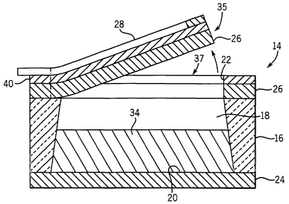
The membrane 26 has a first portion 35 located inside the predetermined
pattern and a second
portion 37 located outside the predetermined pattern. The first portion 35 of
the membrane 26 is
attached to the second portion 37 of the membrane 26 at a connection area 39.
In the preferred
embodiment of the present invention, the first portion 35 of the membrane 26
forms a lid and the
connection area 39 forms a hinge 36. When the membrane 26 ruptures, the lid
separates from the
second portion 37 of the membrane 26, except at the hinge 36, to permit the
medicine 34 to be
delivered through the delivery opening 22, as shown in FIG. 6. The hinge 36
permits the lid to
remain attached to the medicine delivery system 10 so that it is not released
in the animal or human.
The first portion 35 of the membrane 26 and the connection area 39 may have
various sizes, shapes
and positions, depending on various engineering considerations for a
particular application.
FIGS. 7A-7K illustrate, in a sequence of steps, a MEMS fabrication process for
making the
medicine delivery unit 14, as shown in FIGS. 1-6, in accordance with the
preferred embodiment of the
present invention. FIG. 7A illustrates the step of providing the substrate 16.
FIG. 7B illustrates the
substrate 16 having the membrane 26 applied to each opposite side of the
substrate 16. In FIG. 7C,
material 38 for the release element 28 is applied to the membrane 26 on one
side of the substrate 16.
In FIG. 7D, the material 38 for the release element 28 is selectively removed
to form the release
element 28. In FIG. 7E, the insulator 40 is selectively applied to the
membrane 26 and the membrane
material on the bottom side of the substrate 16 is selectively removed. In
FIG. 7F, the medicine
delivery unit 14 is turned over 180 degrees, either physically or for the sake
of illustration. In FIG.
7G, the substrate 16 is etched or machined between the remaining portions of
the membrane material
to form the compartment 18 and the charging opening 20. In FIG. 7H, the
remaining portions of the
membrane material are removed. Alternatively, the remaining portions of the
membrane material stay
depending on the type of material. In FIG 7I, the compartment 18 is filled
with the medicine 34. In
FIG. 7J, the cap 24 is disposed over the compartment 18 to seal the charging
opening 20 under
11
CA 02494198 2005-O1-28
WO 2004/010971 PCT/US2003/023612
vacuum, according to the method described in FIG. 8. In FIG. 7K, the medicine
delivery unit 14 is
again turned over 180 degrees, either physically or for the sake of
illustration.
FIG. 8 illustrates a flowchart describing a method for sealing the medicine
delivery unit 10,
as shown in FIGS. 1-7K. The method starts at step 61. At step 62, the method
provides the substrate
16, having the compartments 18, and the cap 24 in an appropriate manner for
high volume
manufacturing. At step 63, the method charges the compartments 18 with the
medicine 34, as
describe above. At step 64, the method covers the compartments 18 with the cap
24, as described
above. At step 65, the method applies heat 58 to the medicine delivery system
10. In the preferred
embodiment of the present invention the heat is less than 100 degrees C, which
is much less than the
300 - 500 degrees C temperature range used for traditional anodic bonding. At
step 66, the method
applies . a voltage bias 56 across the substrate 16 and the cap 24.
Preferably, a positive voltage is
applied to the cap 24 and a negative voltage is applied to the substrate 16.
Alternatively, the positive
and negative voltages may be reversed, depending on the materials of the cap
24 and the substrate 16.
In the preferred embodiment of the present invention, the voltage bias 56 is
greater than 100 V and
less than the 1 kV used for traditional anodic bonding. At step 67, the method
applies focused energy
54 to the cap 24 to seal the cap 24 to the substrate 16 and to create a vacuum
in the compartments 18.
The focused energy 54 includes, without limitation, microwave, laser,
infrared, lamps, and the like.
The focused energy 54 couples into the cap 24 (e.g., at a wavelength less than
600 nm) to raise the
temperature in a local area over one or more compartments 18 for the duration
of an energy pulse
having a microsecond to millisecond time duration. Such fast heat coupling
assists in bonding the
interface between the cap 24 and the substrate 16, without damaging the cap
24, the substrate 16, or
the medicine 34. Silicon material conducts heat quickly and glass material and
a vacuum conducts
heat slowly. Therefore, when the cap 24 is made of silicon and the substrate
16 is made of glass, the
focused energy 54 conducts slowly to the medicine 34. Note that the focused
energy 54 does not
necessarily need to be aligned with particular features of the medicine
delivery system 10, depending
on the size of the features, the power level and time duration of the focused
energy. At step 68, the
method ends. Although, the method describes a bonding process for assembly of
the medicine
delivery system 10, the method may be used for any kind of micromachined
system or device.
The benefits of the bonding process described in the method include: a fast
manufacturing
throughput, uniform seals, no damage to the medicine 34, a low bonding
temperature permitting more
design flexibility and stable mechanical dimensions with temperature, a flat
assembly process, no
measurable flow of the glass material permitting sealing around previously
machined grooves,
cavities etc. without any loss of dimensional tolerances, parasitic
capacitances are kept extremely
small because the glass material is an insulator, the bonding process may be
performed in vacuum
12
CA 02494198 2005-O1-28
WO 2004/010971 PCT/US2003/023612
permitting hermetically sealed reference cavities to be formed, transparency
of the glass at optical
wavelengths permits simple, but highly accurate, alignment of pre-patterned
glass and silicon wafers
as well as to observe the inside of micro-fluidic devices, a high yield
process that is tolerant to
particle contamination and wafer warp because the electrostatic field
generates a high clamping force
which overcomes surface irregularities, a low cost wafer scale process for
first order packaging can
be done at a chip level if required, mufti-layer stacks permit easy routing to
complex 3-D
microstructures, and a high strength bond that is higher than the fracture
strength of the glass
material.
FIG. 9 illustrates a block diagram of the control unit 12 and the medicine
delivery units 14, as
shown in FIGS. 1 and 2, in accordance with the preferred embodiment of the
present invention. The
medicine delivery system 10 accurately delivers medicine 34 at defined rates
and times according to
the needs of a human or animal patient or other experimental system. The
control unit 12 includes a
controller 70, a memory 72, a sensor 15, a power supply 74, and a
demultiplexer 76. Preferably, the
control unit 12 is constructed as an integrated circuit, but may be
constructed as discrete circuits. The
control unit 12 may have internal or external memory, such as RAM and/or ROM.
The power supply 74 provides power to the appropriate functions in the control
unit 12, such
as the controller 70. Preferably, the power supply 74 is a battery to permit
portable or in body
applications, and is preferably a thin film electrochemical cell deposited on
the substrate 16. The
criteria for selection of the power supply are small size, sufficient power
capacity, ability to be
integrated into the control unit 12, and, in some applications, the ability to
be recharged and the
length of time before recharging is necessary. Alternative batteries of this
type include lithium-based,
rechargeable micro-batteries that are typically only ten microns thick and
occupy 1 cm2 of area. One
or more of these batteries can be incorporated directly into the control unit
12.
The controller 70 generates the control signal 78 to control the medicine
delivery units 14.
The control signal 78 may be carried on a single line carrying multiple
signals, wherein each of the
multiple signals is associated with a corresponding medicine delivery unit 14.
Alternatively, the
control signal may be carried on a plurality of lines, wherein each of the
plurality of lines is
associated with each medicine delivery unit 14. Hence, the controller 70 in
combination with the
control signal 78 actively controls the rupturing of the membrane 26 for each
medicine delivery unit
14.
The control unit 12 is designed based on the period over which the medicine
delivery is
desired, generally in the range of at least three to twelve months for in body
applications. In contrast,
release times as short as a few seconds may be desirable for some
applications. In some cases,
continuous (constant) release from the compartment 18 may be most useful. In
other cases, a pulse
13
CA 02494198 2005-O1-28
WO 2004/010971 PCT/US2003/023612
(bulk) release from the compartment 18 may provide more effective results.
Note that a single pulse
medicine delivery from one compartment 18 can be transformed into a multiple
pulse medicine
delivery by using multiple compartments 18. In addition, delivering several
pulses of medicines in
quick succession can simulate continuous medicine delivery.
The controller 70 controls the time and rate of delivery of the medicine 34
from each
compartment 18 responsive to a software program or circuit, remote control, a
signal from a sensor,
or by any combination of these methods. Preferably, the controller 70 is used
in conjunction with the
sensor 15, the memory 72, the power supply 74, and the demultiplexer 76. The
software program
stored in the memory 72 determines the time and rate of medicine delivery. The
memory 72 sends
instructions to the controller 70. When the time for release has been reached
as indicated by the
software program, the controller 70 sends the control signal 78 corresponding
to the address
(location) of a particular compartment 18 to the demultiplexer 76. The
demultiplexer 76 generates an
electrical signal to the particular compartment 18 addressed by the controller
70.
The sensor 15 advantageously provides a closed loop feedback system to permit
the medicine
delivery system 10 to vary the time, rate and/or dosages of the medicine
responsive to monitored
conditions in the environment, such as the human or animal body.
The medicine delivery system 10 has numerous applications. The medicine
delivery system
can be used to deliver small, controlled amounts of chemical reagents or other
molecules to
solutions or reaction mixtures at precisely controlled times and rates.
Analytical chemistry and
medical diagnostics are examples of fields where the medicine delivery system
10 can be used. The
medicine delivery systems 10 can be implanted into a patient, either by
surgical techniques or by
injection, or can be swallowed. The medicine delivery systems 10 provide
delivery of medicines to
animals or persons who are unable to remember or be ambulatory enough to take
medication. The
medicine delivery systems 10 further provide delivery of many different
medicines at varying rates
and at varying times of delivery.
Hence, while the present invention has been described with reference to
various illustrative
embodiments thereof, the present invention is not intended that the invention
be limited to these
specific embodiments. Those skilled in the art will recognize that variations,
modifications and
combinations of the disclosed subject matter can be made without departing
from the spirit and scope
of the invention as set forth in the appended claims.
14