Note: Descriptions are shown in the official language in which they were submitted.
CA 02494377 2008-07-09
BIOPSY DEVICES AND METHODS
Technical Field
The present invention relates to devices and methods for performing
biopsies, and more particularly to breast implantation clips for use as
markers
in mammography.
Background
Percutaneous biopsy of the breast is a well-accepted alternative to
open surgical biopsy with needle localization for those lesions seen by
mammography or ultrasound but not able to be felt by the surgeon. When
percutaneous biopsy is performed, it is frequently necessary to place a metal
clip at the site of biopsy. This is done for several reasons. For example, the
lesion biopsied might be partially or entirely removed. If the lesion is
proven
to be malignant, it is necessary to subsequently do a wide excisional biopsy
after needle localization to remove any residual malignancy. The clip makes
the site of biopsy apparent, assuring accurate localization. In addition, if
something is seen on both mammography and ultrasound, it is not always
certain that the lesions are one and the same. A biopsy under ultrasound
guidance with placement of a clip allows confirmation by mammography that
the lesion is the same or different than the one seen on the mammogram.
Further, the presence of a clip seen on a mammogram alerts the radiologist
that a biopsy has been performed, prompting the radiologist to more closely
evaluate the site of biopsy.
The vast majority of percutaneous breast biopsies are performed under
art-disclosed stereotactic guidance techniques, and generally use a device
known as the Mammotome (by Johnson & Johnson). The clip that is
1
CA 02494377 2005-01-28
WO 2004/012600 PCT/US2003/023995
employed is generally prone to pinching a minute amount of breast tissue.
Sometimes, the clip may fail to hold onto the tissue or the clip may migrate
to
a different undesired location.
U. S. Surgical has produced a clip from a wire with a memory that is
delivered into the breast and forms a ring. It is larger in diameter than the
Mammotome device clip and can grab significantly more tissue. The clip is
an alloy containing nickel. Recent indications are that U.S. Surgical may no
longer manufacture this clip, thus creating a potential supply issue for
existing
users.
SenoRx, Inc. produces metal markers embedded in Gelfoam pellets (a
product that promotes clotting of blood), called Gel MarkTM. The product is
packaged to include a plurality of pellets and one radiographic marker. The
pellets, however, potentially result in undesired migration of particles.
Although there is current production of a hand-held Mammotome
device, for the purpose of ultrasound guided biopsy, for some users this
device may be awkward and cumbersome to use. The majority of ultrasound-
guided biopsies are done with use of Tru-cut needles. This can be done
through a coaxial needle. A Bard 12 gauge biopsy needle could be used
through an 11 gauge coaxial needle. Through this 11 gauge coaxial needle, a
U. S. Surgical clip might also be delivered, although U.S. Surgical is not
believed to have marketed their clip for use during ultrasound-guided
biopsies.
Another product is manufactured by Inrad. This clip is used for
placement during ultrasound-guided biopsy because the delivery device is
steel and does not provide the flexibility necessary for delivery through the
Mammotome needle. This delivery device has a beveled tip, allowing
advancement through breast tissue without a coaxial needle.
Breast biopsies using an 11 gauge coaxial needle have been
performed. However, most biopsies are typically done using smaller needles,
e.g. a 14 gauge biopsy needle with 13.5 gauge coaxial needle. Such small
sizes, in many environments, however, are believed to be too small to
efficiently allow advancement of the delivery device of current commercially
available clips.
2
CA 02494377 2008-07-09
Turning to another consideration, when a cyst in the breast is aspirated, a
spectrum of different types of fluid can be recovered. These might range in
color
from white to yellow to green or brown. They may be mucousy or bloody and
thus can be thick or thin. Some physicians send all samples for cytology
analysis, while other physicians may send only grossly suspicious samples
(e.g.
mucousy or bloody). Regardless of which cyst fluids are sent for cytology,
once a
cyst is evacuated or in the event that a cyst cannot be fully evacuated
because it
contains a solid component, a radiologist would like to place a clip into the
lesion.
It is often important to mark the cyst so that should the cytology prove
malignant,
or otherwise require further attention, the exact site of the lesion would be
known
and a needle localization could be subsequently performed.
There is a need for improved devices for breast biopsy, cyst aspiration or
both, to overcome the above-discussed disadvantages of current commercial
products.
The following United States patents are also useful to more fully
understand the context and application of the present invention: 6,161,034;
5,526,822; and 5,649,547. Devices disclosed in the above patents may be
modified as desired to incorporate the inventive features disclosed herein.
Summary of the Invention
In one aspect, the present invention meets the above needs by providing
an improved clip for mammography analysis, comprising a first portion that is
straight, arcuate or a combination thereof; and at least a second portion that
is
straight, arcuate or a combination thereof, and which is connected to the
first
arcuate portion at an apex, wherein the first and second portions are adapted
to
be compressed to fit within a tube of a delivery device and to elastically
deform
relative to each other upon exiting the tube for engaging tissue.
In another aspect the present invention contemplates an improved device,
preferably of compact design, for performing a breast biopsy, marking an
aspirated cyst, or both, comprising a gripping portion including finger rests
3
CA 02494377 2005-01-28
WO 2004/012600 PCT/US2003/023995
attached to a hub portion; a tube having defined at one end portion a hole;
and a driver having an actuator member in driving relation therewith; wherein
upon translation of the actuator member the driver advances in the tube to
advance any clip located in the tube for expulsion through hole, and further
wherein the actuator requires only one hand to deploy the clip from the clip
delivery portion and is substantially free of a lock that requires unlocking
to
permit the actuator to operate.
In yet another aspect of the invention, there is contemplated a device
and a method for marking an evacuated cyst, such as a breast cyst,
comprising the steps of inserting a needle into a fluid filled cyst (e.g. a
breast
cyst); removing fluid from the cyst for collapsing the walls of the cyst; and
inserting a clip as a marker into the cyst using the needle.
Description of the Drawings
Fig. 1 illustrates a side view of an illustrative system which is suitable
for use to deploy clips in accordance with the present invention including a
clip delivery device in combination with a biopsy instrument.
Figs. 2A-2C illustrate prior alternative configurations for clips.
Fig. 3 illustrates an enlarged section of Fig. 1 showing deployment of a
clip of the present invention through a deployment tube of a clip delivery
device within a biopsy instrument.
Figs. 4A-4D, illustrate alternative configurations for clips of the present
invention in their deployed state.
Fig. 5A illustrates an alternative configuration for a clip of the present
invention in its deployed state.
Fig. 5B is an end view of the clip of Fig. 5A.
4
CA 02494377 2005-01-28
WO 2004/012600 PCT/US2003/023995
Fig. 5C is an example of an alternative end view of a clip of Fig. 5, in
which the ends are rotated relative to each other for achieving a three
dimensional configuration in a deployed state.
Figs. 6A-6D illustrate alternative configurations for clips of the present
invention in their deployed state.
Figs. 7-8 illustrate a rigid shafted clip delivery device respectively in
pre- and post-deployment states.
Fig. 9 is an end view of the device of Figs. 7 and 8.
Figs. 10, 11A and 11B illustrate a plan view of a biopsy instrument
useful in combination with a clip delivery device of the present invention.
Fig. 11 C illustrates a side section view of a biopsy instrument of Figs.
10, 11 A and 11 B.
Fig. 12A is a side section view of another alternative device to illustrate
a preferred breast cyst aspiration needle device.
Fig. 12B is a top plan view of an end portion of the embodiment of Fig.
12.
Fig. 12C is a sectional view taken through lines 12C-12C of Fig. 12A.
Figs. 13A and 13B illustrate a side section view of another alternative
device to illustrate a preferred cyst aspiration needle device with a side
hole in
combination with a coaxially inserted clip delivery device.
Fig. 14 is an example of an alternative cyst aspiration needle with an
end hole.
5
CA 02494377 2005-01-28
WO 2004/012600 PCT/US2003/023995
Fig. 15 is an example of clip delivery device employed in combination
with an aspiration needle as in Fig. 14.
Figs. 16A-O illustrate yet further examples of alternative clips in
accordance with the present invention.
Detailed Description of the Preferred Embodiment
The present invention is directed to improved devices and methods for
medical diagnosis and treatment, and particularly devices that are employed
for mammographic analysis, such as for the detection and treatment of
cancerous or other abnormal growths. The present invention contemplates an
improved clip, such as for use as a marker, a delivery device for deploying
the
clip, a cyst aspiration device, combinations thereof and methods of using the
same.
As will be seen from the description that follows, the various inventive
features are not confined to a single application, but rather are capable of
numerous variations. Accordingly, though described in a certain context, as
will be apparent, features may be interchangeable among embodiments. For
sake of brevity, while still providing ample instruction to the skilled
artisan, the
features herein are described without limitation in embodiments featuring the
employment of a clip delivery device 110 (110') by itself, with a biopsy
instrument 112, such as a vacuum assisted instrument or a cyst aspiration
device 114 (114') or combinations thereof. Accordingly, it is contemplated
that
the clip delivery device may deploy a clip through an end hole or a side hole
of a rigid, semi-rigid or flexible tube, and possibly thereafter through an
end
hole or a side hole of an outer needle (which itself may be a part of an
integrated or separately connectable device and may be rigid, semi-rigid or
flexible).
In general, one common feature of a number of the different
embodiments herein is the use to precisely deploy clips directly at a biopsy
site, the site of an aspirated cyst, or any other desired site, and thus be
able
to accurately and reliably mark the site with the clip, as will be
demonstrated
6
CA 02494377 2005-01-28
WO 2004/012600 PCT/US2003/023995
by the various embodiments herein, and particularly taking into account the
alternative clip designs of Figs. 4A-4D, 5A-5C, 6A-6D and 16A-160.
As seen from those drawings the clips typically are a relatively fine
structure, and are contemplated as commonly being made of a wire, such as
a surgical stainless steel wire, a titanium wire, nickel containing metals, a
bio-
compatible polymer or the like. Of course, other biocompatible materials may
likewise be employed, such as, other non-corrosive materials or otherwise.
In one aspect of the present invention, the clip may be delivered
through a system or according to a method that uses a biopsy instrument
such as a Mammotome vacuum-assisted biopsy instrument, available
through Johnson & Johnson, pursuant to which a tissue sample may be
obtained with a needle by applying a slight vacuum for drawing, cutting and/or
removing tissue.
In general, any delivery device may be used, whether employed in
combination with a suitable biopsy instrument or not. For example, a device
might be employed in which a tissue sample is obtained with a needle in
combination with a spring loaded mechanism to cut and remove tissue. The
clip might also be delivered during open surgery. Preferably any device may
be employed for performing mammographic analysis provided it is suitable for
stereotactic techniques, ultrasound techniques or a combination thereof.
It is contemplated that the delivery device includes an actuator portion
that may be removably associated with the delivery device, the biopsy
instrument or both. Thus, for example, one preferred apparatus may include a
biopsy instrument that includes a needle portion that is insertable into a
patient. Coupled with or within the needle portion, or integrally defined as
part
of the needle portion may be a cutter (e.g., a movable needle that can be
manually driven, driven by a motor, or both), a vacuum device or a
combination thereof.
In some embodiments, accordingly, the delivery device of the present
invention is flexible over at least a portion of its length to provide better
maneuverability through a tissue mass or otherwise. Therefore, it is
foreseeable that at least a portion of the delivery device is made of plastic
or
another flexible material. However, the present invention also contemplates
the use of a rigid delivery device comprising a harder material such as a
rigid
7
CA 02494377 2005-01-28
WO 2004/012600 PCT/US2003/023995
plastic, metal or otherwise. Combinations of different materials may, of
course, be employed as desired.
Referring to Fig. 1, one example of a system is illustrated as
comprising a clip delivery device 110 that includes a delivery tube 116 (which
optionally may be open, such as by a longitudinal groove, notch, slot or
aperture at an end or over at least a portion of its length, and is
illustrated as
having a blunt tip and a side hole) and a driver 118. A portion of the driver
118
is configured to slide within the delivery tube 116 and push an object, such
as
a clip 120 (shown in an undeployed state) or otherwise out of a tube exit
opening 122. As shown in Fig. 1, the system includes a portion of a biopsy
instrument, particularly one including a needle 124. Referring also to Figs.
10,
11A and 11B, one such biopsy instrument 112 may include a window portion
126 into which the delivery tube 116 of the clip delivery device 110 can be
axially inserted, such as to a predetermined location as suitably defined by a
visual indicator 128.
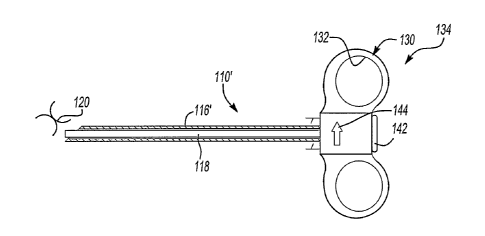
Referring again to Figs. 10, 11A and 11 B, preferably, the clip delivery
device 110 further comprises a gripping portion 130 having a finger rest 132
and a hub portion 134 affixed, integrated with or otherwise attached to the
tube, driver or both. The gripping portion may be enclosed as shown, or it
may be open. It may be adapted for receiving a single finger or a plurality of
fingers.
As seen, advantageously, the clip delivery device 110 (110') may
further comprise or be integrated or used with an aspiration needle device,
which may be open at an end or over at least a portion of its length. The
aspiration needle device may comprise a separable unit configured to
temporarily receive the tube 116 (116') of the device 110 (110'), or
alternatively, the aspiration needle device, the tube 116 (116'), or both may
be
formed with, permanently or temporarily attached to a portion of the delivery
device 110 (110').
For example, one such suitable attachment feature might include a luer
lock or other suitable attachment mechanism, which would permit a user to
readily assemble or disassemble components. For example, without limitation,
as depicted more particularly in Figs. 12A-12C and 13A and 13B, it is
contemplated that a connecting portion 152 connects the needle or device to
8
CA 02494377 2005-01-28
WO 2004/012600 PCT/US2003/023995
a luer lock connector 136 associated with the clip delivery device 110'. See
also Fig. 15. The needle, the tube or both may be connected with such an
attachment mechanism.
Alternatively, the aspiration needle device 114 (114'), tube 116 or both
may be affixed to the delivery device 110' using an adhesive, through welding
(e.g., friction welding), integrally formed or otherwise. Preferably, when
combined together (as seen alternatively, for instance, in the various
embodiments of Figs. 1, 3 11A-11C, 13A, 13B, 14 and 15), the needle 124
(124') and the tube 116 will be coaxially aligned about a center line such as
center line "C". The coaxially alignment may be created using the attachment
device for the needle (e.g., luer lock or otherwise) or it may be created
other
structural alignment features such as a mating configuration between the end
portions of the needle and the tube. Such a needle would typically be
configured with a needle opening 138, an open end or otherwise so that any
needle opening would be aligned with the tube opening 122, as in Fig. 1. With
the addition of a needle 124 to the delivery device 110, it is possible to
perform additional functions such as excise, aspirate or otherwise manipulate
a portion of a tissue sample.
In any of the examples contained herein, it is foreseeable that a clip
120 is compressed upon itself and inserted into the tube portion 116 (116') of
the delivery device 110 (110') at some point prior to the use of the same. As
such, the clip may be inserted into the tube at any time from the manufacture
of the tube or up until just prior to the use of the delivery device. It is
further
contemplated that the clip may comprise a separately manufactured
component, as described in greater detail herein, or it may be a removable
portion of the delivery tube or any other portion of the delivery device.
Also, it is contemplated that more than one clip may be loaded within a
single delivery device. For example, the delivery device may be configured
with a plurality of clips compressed upon themselves and located within the
delivery tube, needle, or otherwise. As such, it would be possible to insert
more than one clip into a breast, either at similar or different locations,
using a
single delivery device.
As can be appreciated, the device 110 is inserted into a tissue sample,
such as a breast or otherwise, and the end portion including the tube 116, the
9
CA 02494377 2005-01-28
WO 2004/012600 PCT/US2003/023995
needle 124 or both, is positioned to the portion of the tissue that is of
interest.
Once positioned, manipulation of the tissue may be performed by, for
example, providing a vacuum through a portion of the needle 124', tube 116'
or otherwise, to pull the tissue into the needle opening 138. Advantageously,
the vacuum force may be used to assist in excising a portion of the tissue for
pathological analysis or otherwise. It should be appreciated that in addition
to
the above described structure any suitable additional blade configuration may
also be employed.
Either before or after the excising or aspiration of the tissue, a clip 120
may be deployed through the tube opening 122 and needle opening 138, if
present. The device and/or needle may then be rotated (e.g., from about 90 to
about 2700, and more preferably about 180 ) so when it is withdrawn, the
needle opening 138 does not hook or otherwise catch onto the clip 120 and
accidentally dislodge it.
In certain embodiments, such as shown in Figs. 1 and 11-13, the
delivery tube, the needle or both has a guide surface such as a ramp 140
(140') on the inside that directs the clip 120 out of the tube opening.
However,
alternatively the delivery device may comprise a rigid or semi-rigid tube
having a tube opening directly defining the axial end of the tube, for
delivery of
a clip. With this alternate configuration, it is foreseeable that the tube is
shaped or comprises needle-like characteristics for allowing the insertion
directly into tissue.
As best seen in Figs. 1, 3 and 11 (with reference to clip deployment
with a biopsy instrument having a needle 124, such as a vacuum assisted
biopsy instrument), Figs. 7 and 8 (with reference to clip deployment
independent of an additional biopsy instrument), and Figs. 13A,13B and 15
(with reference to clip deployment in combination with a cyst aspiration
needle
device), the deployment of the clip comprises advancing (e.g., by sliding) the
clip within the tube using a suitable driver 118. To fully deploy the clip
within
the tissue of interest, the driver extends to the end of the tube and, if
desired,
then through tube opening and any needle opening, if present. This ensures
that the clip 120 is free from both the delivery device and the needle 124
(124') prior to withdrawal or rotation. Though the driver 118 is applying a
force to advance the clip within the tube, in a highly preferred embodiment,
it
CA 02494377 2008-07-09
should be appreciated that it does not impart any significant force to the
clip to
cause the clip to elastically deform about itself upon exiting the delivery
device.
The driver 118 may be solid along its length, or at least partially hollow. As
such, it is foreseeable that the channel within the drives may be used to
provide a
path for fluid, instruments or otherwise. It may be coated or uncoated. For
example, it may have a low friction material over an outer surface.
In one embodiment (e.g., as shown in Figs. 7 and 8), for the clip 120 to be
delivered under ultrasound-guidance (e.g. employing a suitable ultrasound
instrument), it is advantageous for the delivery device 110' to include a
rigid tube
116 (e.g., made of a steel) and the tip to be beveled.
One benefit of the present invention is that clip deployment can be
accomplished with or without a vacuum assist. Precise location of a clip
relative
to the biopsied region is possible, and it is also possible that the clip can
be
deployed to the exact location of the biopsy.
It will be appreciated that, particularly for mammographic analysis, the
area biopsied can sometimes be very small. Also, after biopsying a lesion, the
lesion potentially might become obscured by bleeding. As such, it may be
desirable for the coaxial needle 124 (124') to be at least temporarily left in
place
so that it serves as a valuable landmark for the accurate placement of the
clip
120. Thus the present invention contemplates a step of temporarily placing a
needle (e.g., a 13 gauge needle or another needle, preferably open at both
ends)
in the biopsy region prior to deployment of a clip. Thereafter, the tube of
the
delivery device is inserted into the needle for deployment of the clip.
Though preferred delivery devices are disclosed herein, such disclosure is
not intended to foreclose the adaptation and use of other devices. Additional
examples of delivery devices for use herein are described without limitation
in
U.S. Patent Nos. 5,526,822; and 5,649,547.
The design of the clips 120 helps to avoid the possibility of migration. That
is upon deployment, the ends of the one or plurality of wires are such that
they
catch and attach to the tissue, such as by unfolding upon itself (e.g.,
11
CA 02494377 2005-01-28
WO 2004/012600 PCT/US2003/023995
by rotating at least 45 relative to an opposing portion, such as about its
apex,
more preferably at least 60 , still more preferably greater than 90 , and
possibly greater than 180 ), and employing its intrinsic elasticity to force
an
end of the clip into the tissue. Preferably the stored energy of a clip as it
resides (e.g., in a compressed state relative to its relaxed state) within the
clip
delivery device prior to deployment is sufficient that it can unfold upon
itself
and penetrate tissue in the absence of an externally applied force, as is
common with prior clip devices, such as Fig. 2A (which generally employs a
detachable tensioning wire). Nonetheless, the shape of the clip is such that
uncontrollable spiraling, which might lead to undesired migration can be
avoided.
Accordingly, as the wire of the clip progresses to its relaxed state, upon
exiting the delivery device 110, it is capable of pulling the clip 120 by
itself
(i.e., under its own stored energy and preferably in the absence of additional
user-applied energy) into position, assuring secure deployment and
substantially preventing migration.
Various examples of clips 120 useful in accordance with the present
invention are shown, without limitation, in Figs. 4A-6D and Figs. 16A-160.
In one set of examples of a preferred clip 120, arcuate portions (e.g.
Figs. 4A-D, 6A, 16C, 16D, 16G, 161, 16J and 160) may be joined at an apex,
or a plurality of apexes, to form a clip of the present invention.
Alternatively, in
another set of examples of a preferred clip, the clip 120 of the present
invention may include straight portions (e.g. Figs. 5A, 6B-D, 16A, 16B, 16E,
16F, 16H and 16K-N) joined at an apex or a plurality of apexes. It is further
contemplated that any of the above clip examples may be combined with any
other clips, or the same clip, to form yet more examples of a clip 120 of the
present invention. Furthermore, all of the above clips 120 may further
comprise additional features, which may be resistant to migration through a
breast, such as a barb as in Figs. 16A-E and 16H, 16K and 16L.
In another aspect of the present invention, a preferred delivery device
110 of the present invention comprises an actuator 142 as seen in Figs. 7, 8,
11A, 11 B, 13A and 13B, which is compact in design and is useful by itself or
in combination with another biopsy instrument such as has been described for
performing either or both of the percutaneous or ultrasound guidance
12
CA 02494377 2005-01-28
WO 2004/012600 PCT/US2003/023995
techniques. A preferred driver 118 comprises a pushrod or piston like
configuration, wherein the actuator 142 applies a force to one end thereby
driving the piston or pushrod through the tube along with any clip contained
therein. However other driver and actuator configurations are available and
contemplated, as well known in the art of tissue aspiration and excising.
Advantageously, the design of a preferred gripping portion 130 (e.g.,
handle, or otherwise) and deployment actuator 142 of the present invention
requires only one hand, either left or right (e.g. the devices are designed
for
ambidextrous use), to deploy the clip 120. Further, though a lock may be
employed, a preferred delivery device 110 (110') has no lock that requires
unlocking. Instead, there may be incorporated some slackness or other
approach for providing initial "play" when actuating the actuator 142 (such as
by pressing a button, squeezing a trigger or the like) before which the clip
is
deployed. This can be accomplished with a suitable driver, for example, with
a suitable cable, or more preferably by a push rod (e.g., having a length that
is
slightly shorter than the shaft in which it is disposed). In this manner it is
possible to gain further control to help avoid accidental deployment of the
clip.
As illustrated in the embodiments of Figs. 8-12, a preferred gripping
portion 130 will have a suitable grip portion, such as one that has finger
rests
132 (e.g., at least one and preferably two opposing open or generally
semicircular finger grips or substantially entirely enclosed circular finger
grips)
that help secure control, for either left or right-handed operators. As seen,
for
example in Figs. 7, 8, 11A, 11B, 13A, 13B and 15, in one preferred approach
for the device, the gripping portion 130 optionally may be configured with a
surface marking 144 (e.g., an arrow, text, or otherwise) pointing toward the
direction of the tube opening or needle opening, indicating the direction in
which a clip will be deployed from the device.
Additionally locator features may also be employed. For example, as
shown in Figs. 12A-13B, a notch 146 might be employed to help align the
gripping portion with a needle or other component that is separably attached,
such as by way of a luer lock.
Figs. 7 and 8, and Figs. 13A and 13B illustrate side views of an
example of a preferred gripping portion 130 and deployment actuator 142 of
13
CA 02494377 2005-01-28
WO 2004/012600 PCT/US2003/023995
the present invention, shown in illustrative pre-deployed (Figs. 7 and 13A)
and
deployed (Figs. 8 and 13B) conditions.
Once the button of the actuator 142 is depressed completely and the
clip 120 has been fully deployed, the button of the actuator may include a
feature for automatically locking it into a depressed position, providing
feedback to the physician that the clip has been fully deployed. For example,
a detent, an over center lock, a snap, or the like locking mechanism, might be
employed in the gripping portion which is engaged only upon deployment.
Upon locking of the locking mechanism, there is an audible sound and/or just
prior to locking there is slight increased resistance, which must be overcome,
providing palpable feedback that the clip has been fully deployed.
The actuator 142 may further comprise a return device (not shown) for
retracting the end portion of the driver back within the tube. As such, a
return
device (e.g., a spring or otherwise) may bias the movement of the actuator
142 so that upon release of the same the actuator will retract to
predetermined position (e.g., a stop position, a lock portion, an indentation
or
projection, the original position or otherwise). By retracting the driver into
the
tube, the clip will be substantially free from the device and will not catch
or
otherwise be dislodged from the insert position during rotation or withdrawal
of
the needle from the object into which it is inserted.
The tube and/or needle associated with any delivery device herein may
employ a flexible shaft or a rigid shaft or a combination thereof. It may be
made of a suitable metal (e.g., surgical steel, titanium or the like), plastic
or
other material. It may be coated or uncoated, transparent, opaque or
combinations thereof.
The actuator of the present invention may optionally include a hub
portion that is adapted for temporary or permanent connection with a shaft,
tube or the like. For example, as seen in Fig. 15, a fitting 136 (e.g., a Luer
lock fitting) is provided for attachment of a needle or other projection with
the
hub portion of the actuator.
With reference now to Figs. 12A-15, there are shown alternative
embodiments contemplated within the present invention, in which a cyst
aspiration device 114 (114') is employed, and preferably one through which a
clip 120 could be deployed. Examples of needles 124' (124") are shown in
14
CA 02494377 2005-01-28
WO 2004/012600 PCT/US2003/023995
Figs. 12A-12C, 13A, 13B, 14 and 15. For instance, the needle preferably
includes a shaft 148 (e.g., metal such as steel or titanium, plastic or the
like),
with a cutting portion 150 (e.g., having a tapered tip) and a connecting
portion
152. Though an end hole 138' may be employed (as seen in Figs. 14 and 15),
the cutting portion 150 in Figs. 12A-13B is preferably configured with a side
hole 138 that will align with a fixed or displaceable cutting edge (e.g., a
bevel
on a stylet). Thus, a stylet or other device may also be employed for cutting
tissue, preventing tissue from filling the needle before aspirating a cyst, or
both.
As seen, the use of a typical Luer lock 136 or other suitable end fitting
at the connecting portion preferably allows ease of use with readily available
syringes or with an actuator 142 such as described in the above (e.g., with or
without finger holes) for delivering a clip 120. It also allows for removal of
the
actuator 142 while retaining a needle in place. Thus it is possible that a
clip is
loaded into the device after the needle is inserted into the patient.
The gauge of any aspiration needle 124' (124") of the present invention
may be substantially the same as or larger than the gauge of conventional
needles available for cyst aspiration, it being recognized that frequently the
fluid is thick and will not be able to be practically withdrawn through a
typical
19-gauge needle, in the absence of a thinning protocol (which might be
employed, such as by chemistry, thermally, or otherwise). A larger gauge,
e.g., about 15 to about 18 gauge is preferred in one particular embodiment for
evacuating cysts.
Also, the needle lengths of the present invention may vary as well. For
example, the needle may be configured having a length from about 1 cm to
about 10 cm and, in one embodiment, more preferably about 2 to about 5 cm;
and more preferably about 5 to 10 cm in another embodiment.
At times, having a needle with a long length can prove to be an
advantage. For example, cysts are sometimes deeper than can be reached
with a 2-cm blood-drawing needle. As such, the needle of the present
invention would be produced in one or more lengths and gauges that would
precisely match the steel shafted breast marking clip device. The length or
gauge of the needle could be unusual, but matched to the length of the steel
deployment device so that other commercially available needles controllably
CA 02494377 2005-01-28
WO 2004/012600 PCT/US2003/023995
may be used with it, such as with an adapter, the providing of such an adapter
also being contemplated as within the scope of the present invention.
In another aspect of the present invention, if a cyst warrants marking,
such as for future examination, a clip could be immediately delivered into the
inside of the cyst, while the aspiration needle remains in place. The
aspiration
needle thus also functions as the shaft of a delivery device.
Furthermore, the device is not limited to use only for evacuated cysts
but also could be used for marking solid masses.
As also . discussed further herein, preferred clips should be small
enough to fit through any typical coaxial needle that would be used for breast
biopsy. This will require that the delivery tube preferably be of a thin-wall
construction over a portion of or all of its length (and optionally coated
over at
least a portion of its exterior or interior surface) so the resulting
thickness of
the delivery tube for the clip and therefore strength of the shape memory of
the wire can be maximized. Of course, this device could then be used either
with or without a coaxial needle (which also may be coated over at least a
portion of its exterior or interior surface).
The skilled artisan will appreciate that among the advantages of the
present inventive clip design is that is grabs a relatively large amount of
tissue. Another advantage of the present inventive clip is that is does not
form
a spiral configuration. The proposed clip design is thus highly resistant to
any
accidental migration.
In use, the present inventive clip design also affords the advantage
that, such as using ultrasound guidance, it is possible to place the clip
either
into the central portion of or next to the mass under consideration for
biopsy.
It is generally only necessary to see the tip of the needle well and for there
to
be a positive feel to know that the clip has been deployed.
Biopsies or mass (e.g., breast cyst) aspirations performed in
accordance with the present invention can be performed using any suitable
size needle (e.g., 10 to 20 gauge, and more preferably 11 to 15 gauge). Clips
of the present invention are preferably of a thickness, diameter or other
dimension so that they are capable of passing through the needle. For a wire-
based clip, the wire chosen is thus preferably of a smaller gauge than the
needle, and more preferably a smaller gauge by a factor of at least one half,
16
CA 02494377 2005-01-28
WO 2004/012600 PCT/US2003/023995
so that the wire can be folded upon itself, such as about an apex or flattened
for placement into the needle or other tube for delivery. The clips can be
hollow cored structures, solid structures (e.g., wire) or filled core
structures.
They may be coated or uncoated. For example, they may have a
pharmaceutical agent over some or all of its outer or inner surfaces.
For any of the embodiments of the present invention, a line or other
marking may be inscribed onto the surface of the needle and/or needle hub as
well as onto the surface of the gripping portion and/or hub of the clip
device.
The lines will allow precise alignment of the needle and device to indicate
proper assembly. This would therefore provide confirmation that the opening
of the clip device is aligned with the opening of the needle.
Kits may be provided and used in accordance with the present
invention. Examples of components suitable for inclusion in such a kit
include,
without limitation, one or more of needles, sutures, syringe, anesthetic,
sterile
wipes, a sharps disposal container, gloves or the like.
The devices of the present invention preferably will be packaged in a
sealed sterile container. The container may include a transparent wall, an
opaque wall or a combination thereof. The devices are preferably used only
once and are disposable. In one embodiment, the devices are fabricated with
plastic or metal components that can be recycled.
The present invention also contemplates methods of using the devices
disclosed herein. For example, in one embodiment, a method is
contemplated for performing a biopsy using a clip of the present invention. In
another embodiment, the delivery device herein is used for delivering a clip,
such as during a biopsy. The methods discussed in the Background section
herein are particularly suitable for use of the devices of the present
invention.
Thus, the devices of the present invention may be used for percutaneous
biopsies, ultrasound guided biopsies or a combination thereof. Kits may be
provided and used in performing such procedures.
The present invention is particularly suitable for mammographic
analysis of humans, and particularly female humans, but it is not limited
thereto. Without limitation, it can be used for analysis of other human body
parts, or for analysis of mammals or other animals other than humans.
17
CA 02494377 2005-01-28
WO 2004/012600 PCT/US2003/023995
References to the use of a Mammotome device herein are not
intended to foreclose the use of other like devices for performing one or more
of the tissue removal, marking or other functions performed by the
Mammotome device. Accordingly the present invention also contemplates
substituting for the Mammotome device that is described other such
devices, which preferably will have an elongated delivery tube or like
structure
having chamber through which a clip according to the present invention is
advanced, such as by a push rod or the like.
Though a preferred ejection direction is shown in the accompanying
drawings for the deployment of the clips, it is possible to deploy the clips
so
the apex is the leading portion of the clip.
Further, in addition to the discussion of the clip contained herein, there
may be greater than two straight or arcuate portions for the clips. The
straight
and arcuate portions can be of the same or different size or shape relative to
each other. They may be formed of a single component (e.g., a single wire) or
plural components (e.g., plural wires (2, 3 or more wires) such as might
result
in a structure as in Figs. 4C, 4D, 5A and others illustrated). The portions
need
not be arcuate alone or straight alone, but may be straight, or a combination
of straight and arcuate. In another embodiment, as seen in Figs. 5A, 5B and
6B-6D, the clip may be "X" shaped or may have orthogonally disposed arms.
The clips alternatively may be "N" shaped, arrows, arcs, tetragonal, or any of
a number of different shapes. Combinations of any of the shapes identified
may be employed also. Clips may also include one or a plurality of barbed
ends (such as is illustrated by various of the examples provided in Fig. 16).
When two or more wires are employed they may be configured relative to
each other for deployment in a single common plane or over plural different
planes. Though larger clips are also possible, when deployed, preferred clips
are smaller than about 1 cm in its largest dimension (e.g., length, diameter,
etc.), and more preferably, they are on the order of about 5 mm in its largest
dimension.
As illustrated in Figs. 5A-5C, clips herein may be configured to lie in a
single plane (Fig. 5B) or include one or more portions that lie in a plurality
of
different planes, as in Fig. 5C.
Unless stated otherwise, dimensions and geometries of the various
18
CA 02494377 2005-01-28
WO 2004/012600 PCT/US2003/023995
structures depicted herein are not intended to be restrictive of the
invention,
and other dimensions or geometries are possible. Plural structural
components can be provided by a single integrated structure. Alternatively, a
single integrated structure might be divided into separate plural components.
In addition, while a feature of the present invention may have been described
in the context of only one of the illustrated embodiments, such feature may be
combined with one or more other features of other embodiments, for any
given application. For example, the employment of a Iuer lock may be used in
the various embodiments shown to connect components, omitted or
substituted with an alternative connector, a guide ramp employed or omitted,
side holes might be substituted for end holes, or end holes substituted for
side
holes, even though such feature might not be shown in the accompanying
drawings. Bevel shapes can vary from those depicted. The use of different
material combinations than those shown might also be appropriate, such as
the substitution of metal for plastic, or plastic for metal. It will also be
appreciated from the above that the fabrication of the unique structures
herein
and the operation thereof also constitute methods in accordance with the
present invention.
The preferred embodiment of the present invention has been
disclosed. A person of ordinary skill in the art would realize however, that
certain modifications would come within the teachings of this invention.
Therefore, the following claims should be studied to determine the true scope
and content of the invention.
19