Note: Descriptions are shown in the official language in which they were submitted.
CA 02494538 2005-O1-25
WO 2004/011668 PCT/US2003/023420
METHODS AND APPARATUS FOR SCREENING AND DETECTING
MULTIPLE GENETIC MUTATIONS
This application claims the benefit of U.S. Application Serial No. ,
entitled "Methods and Apparatus for Screening and Detecting Multiple Genetic
.Mutations," filed July 24, 2003, U.S. Provisional Application Serial No.
60/443,989,
filed January 30, 2003, and U.S. Provisional Application Serial No.
60/398,992, filed
July 26, 2002, which are expressly incorporated herein by reference in their
entirety.
FIELD OF THE INVENTION
The methods, apparatus, systems and reagents of these inventions relate to
screening patient samples for genetic polymorphisms and for identifying
specific
polymorphisms within a known set. Particularly, the invention enables both
high
throughput screening and identification of known polymorphisms or mutations
that
relate to disease.
BACKGROUND
Many diseases are caused by known genetic alterations or mutations that occur
in specific, identified regions in the DNA of a patient. Detection of a
disease by direct
analysis of DNA offers significant advantages in certainty and accuracy over
other
diagnostic techniques but requires an assay device and methodology that
accurately
detects and identifies the genetic alterations in a patient sample. When
specific
disease-related alterations or mutations can be detected and identified as
markers of a
disease, the DNA-based assays can be used both to diagnose an individual
patient for
disease and to screen patient groups for the presence of known genetic markers
that
may be correlated with disease. In some cases, populations of patients are
uniquely
susceptible to certain diseases or groups of diseases and DNA-based assays can
be
used to screen patients for any number of a group of diseases based on the
detection of
mutations in a patient's DNA.
CA 02494538 2005-O1-25
WO 2004/011668 PCT/US2003/023420
2 Patent
Attorney Docket: 612,404-427
In recent years, scientists have developed numerous techniques to analyze
genetic material. Together with research that uncovered the specific genetic
markers
underlying certain diseases, researchers have developed techniques to detect
the
presence of specific alterations occurring in identified regions in a
patient's DNA.
Several analytical techniques are available to detect genetic alterations when
present,
however, the detection of small differences within the entirety of a patient's
DNA
requires a sophisticated test regimen and requires highly specialized
biochemical
reagents. For this and other reasons, much of the instrumentation and testing
methods
can only be performed in research environments and requires highly skilled
technicians to conduct the analyses and interpret the results. Also, the
testing of
subtle genetic alterations is often time consuming and expensive to perform.
The
situation is complicated by the fact that many diseases have dozens of
potential
underlying genetic factors that play a role in the onset or progression of the
disease,
and as the number increases, the cost and complexity of an assay to test a
patient's
genetic material substantially increases.
In a clinical environment, a single patient can be tested for the presence of
a
large number of mutations or polymorphisms potentially underlying a suspected
disease. When a screening approach is desired, the design of the assay
technique
becomes even more critical. If available, a screening assay would be highly
desirable
in several circumstances, including, to analyze the genetic basis for disease
by
detecting polymorphisms in patients and correlating the results to the
presence,
absence, or onset of one or more diseases, to screen susceptible groups of
patients for
genetic markers that exist for any of a group of diseases that are known to be
passed
from parent to child within ethnic groups, and to locate asymptomatic Garners
of
diseases who can pass the underlying mutation to offspring. An assay that is
useful
for a screening must be sufficiently reliable and cost-effective so that
multiple tests
can be efficiently performed on a large patient group.
For screening, an ideal testing system would be automated and capable of
screening a large number of patient samples simultaneously and determining
whether
any one of a large number of mutations is present. Such a "high throughput"
system
would also require specially designed data processing so that assay results
could be
mao4ssss.z
CA 02494538 2005-O1-25
WO 2004/011668 PCT/US2003/023420
Patent
Attorney Docket: 612,404-427
efficiently processed, correlated to patient data, and presented in a useful
format for
interpretation by clinical laboratory personnel. For analyzing patient
samples, it is
often desirable to test a large number of samples to first determine whether
any one of
a set of genetic markers is present, followed by analyzing individual samples
to
determine which member of the set of markers is present. In this fashion,
multiple
samples are screened to identify patients who are "positive" for a member of a
set of
markers, followed by identifying the specific mutation or polymorphism in the
patient
sample that yielded the positive signal. Furthermore, many diseases feature
one or
more of a small number of predominant mutations that occur with very high
frequency. The existence of one or more predominant mutations may dictate that
a
testing assay should separately analyze selected mutations individually in a
patient
sample. Thus, the ideal screening and assay system would rapidly indicate, for
an
individual patient, whether or not any one or more of a set of known markers
are
present and would then offer the capability to identify, when a positive
signal was
generated in this screening process, the specific mutation or polymorphism
that
yielded the positive signal from among the larger set tested in the screening
process.
Currently, a number of different techniques exist for direct analysis of a
patient
DNA sample. In one technique, synthetic strands of DNA are produced that have
sequences that may or may not contain a mutation that are complementary. to a
select
group of mutations and that can be used as probes to detect the mutation in a
patient
sample. These synthetic sequences are exposed to a patient sample, and when
the
mutation is present, the synthetic DNA becomes attached to the patient's DNA
by
hybridization. Once hybridized, the probe can be detected by several known
techniques. Also, specific segments of patient DNA that may or may not contain
a
mutation can be amplified and the amplified DNA can be localized on an
electronic
microchip for further testing.
Cystic ~lbrosis (CF) is an example of a genetic disease that is caused,
individually or collectively, by any of a number of different mutations.
Cystic fibrosis
afflicts approximately 30,000 children and adults in the United States;
afflicted
patients typically die in their thirties. One in 31 Americans (one in 28
Caucasians) -
more than 10 million people - is an unlrnowing, symptom-free carrier of a
mutation
that leads to the disease. An afflicted patient must have inherited two
defective copies
IR1:1045858.2
CA 02494538 2005-O1-25
WO 2004/011668 PCT/US2003/023420
4 Patent
Attorney Docket: 612,404-427
of a specific gene - one from each parent - to have CF. Each time two CF
carriers
conceive a child, there is a 25 percent chance that the child will have CF, a
50 percent
change that the child will be an asymptomatic carrier; and a 25 percent chance
that the
child will be a non-carrier.
CF has a variety of symptoms that are manifested clinically. CF causes the
body to produce an abnormally thick sticky mucus, due to the faulty transport
of
sodium chloride (salt) within cells lining organs such as the lungs and
pancreas, to
their outer surfaces. The thick CF mucus also obstructs the pancreas,
preventing
enzymes from reaching the intestines to help break down and digest food. CF
patients
also suffer from persistent coughing, wheezing or pneumonia; excessive
appetite but
poor weight gain and bulky stools. The sweat test is a common diagnostic test
for CF.
This test measures the amount of salt in the skin and a high salt level
indicates that a
person has CF.
The treatment of CF depends upon the stage of the disease and which organs
are involved. One measure of treatment, chest physical therapy, requires vigor
percussion (by using cupped hands) on the back chest to dislodge the thick
mucus
from the lungs. Antibiotics are also used to treat lung infections
administered
intravenously, via pills, and/or medical vapors that are inhaled to open up
clogged
airways. When CF affects the digestive system, the body does not absorb enough
nutrients. Therefore, people with CF may need to eat an enriched diet and take
both
replacement vitamins and enzymes.
CF is known to be caused by a large number of mutations, at least 25 have
been identified as major contributors to the disease. In August 2001, the
American
College of Gynecologists (ACOG) recommended testing the general group of
potential parents for the 25 separate genetic markers to identify asymptomatic
Garners
who risk passing the disease to children. Because of the need to screen a
large group
of patients, a test for CF should rapidly and accurately screen multiple
patient samples
for the presence of any one or a set of known markers followed by the
identification of
one or more specific markers in those patients who test positive for at least
one
member of the set. Detection of whether or not any single one or more of the
mutations exists provides a rapid screening method, and the detection and
identification of the single mutation or number of mutations in a patient
allows
IR1:1045858.2
CA 02494538 2005-O1-25
WO 2004/011668 PCT/US2003/023420
Patent
Attorney Docket: 612,404-427
diagnosis of the disease or identification of a patient as a potential
carrier.
In such situations, the design of the assay and methodology that efficiently
achieves the goals described above is critical. Specifically, the assay must
be rapid,
accurate, and cost effective such that the assay can be performed as a routine
part of
patient care thereby expanding the utility of the assay from diagnosing
individual
patients to screening entire groups. The assay should be able to rapidly test
multiple
patient samples and be flexible enough to selectively recognize predominant
mutations or markers for a disease. Through the ability to screen and identify
a large
number of genetic polymorphisms, the assay could both diagnose disease as well
as
yield epidemiological data about the prevalence of specific polymorphisms and
the
relation to the existence or severity of a condition that may a correlated to
a specific
disease or that exists in a number of pathologies. Because many diseases have
underlying genetic markers that have been identified and localized to
identified
regions of a patient's DNA that can be analyzed, once the specific genetic
markers are
identified, any number of diseases can be analyzed using the same assay format
by
simply altering the gene specific reagents in the assay that hybridize with a
patient's
DNA to detect the known marker and correlating the presence of the marker with
one
or more diseases. Accordingly, once the assay design and methodology are
realized,
one additional disease, a group of diseases, or a group of polyrnorphisms that
are
directly or indirectly correlated to several diseases, can be detected with
the assay
format. As the genetic bases of other diseases are discovered, the gene
specific assay
reagents are readily modified to take advantage of the existing format to
detect and
analyze new diseases. For example, while cystic fibrosis is susceptible of
detection by
screening an identification of a discrete set of markers or mutations that are
known to
contribute to the disease, in other circumstances, the screening process may
identify
other polymorphisrns that are not directly related to a single disease, but
that are
related to multiple diseases or that accompany different conditions such as a
panel of
diseases that may affect a certain population group.
IR1:1045858.2
CA 02494538 2005-O1-25
WO 2004/011668 PCT/US2003/023420
6 Patent
Attorney Docket: 612,404-427
SUMMARY OF THE INVENTION
The present invention provides methods, apparatus and compositions
comprising reagents to screen patient DNA samples for the presence of one or
more of
a predetermined group or set of known genetic markers occurring at identified
loci,
together with the identification of specific markers in the sample. A number
of
patient samples may be individually screened to locate patients that test
positive for
any one or more of the markers in the set, followed by identifying the
specific patients
with one of a set of markers for further analyses to identify the specific
polymorphism
present. In preferred embodiments, a plurality of patient samples are
simultaneously
assayed for a group or set of markers by amplification of the identified
regions of a
patient's DNA that are known to include the mutation or polymorphism of
interest
and the localization or immobilization of the amplification products
("amplicons") on
discrete test sites of an electronically addressable microchip. The microchip
is
comprised of an array of the test sites at which concentrations of the
amplicons are
localized for further reaction with specialized reagents. Once the amplicons
are
localized at the test site, several strategies are employed to interrogate the
amplification products for the presence of any member of the known set of
markers,
followed by identification of the specific member of the set that was
detected.
Specifically preferred is the use of wild-type and/or mutant discriminator
probes that
engage in hybridization reactions to selectively detect any one of a set of
mutations,
followed by reaction with one or more universal reporters that provide
universal
detection capability such that a detectable signal is generated if any one of
the
members of a set of markers is present.
When an appropriate signal is detected in the amplified sample from an
individual patient during the screening phase of the assay, the amplification
products
may then be analyzed in a genotyping assay to identify the specific mutation
that
generated the positive signal. Depending on the prevalence of specific,
predominant
mutations that are known for a particular disease, the assay may isolate a
subset of
predominant mutations for individual analysis in a patient sample before
proceeding
to the analysis of less prevalent mutations. The analysis of a prevalent
mutation may
occur as a discreet step or in parallel with other tests that comprise the
entire screening
or genotyping process. The predominant mutations may be directly correlated
with a
IIt1:1045858.2
CA 02494538 2005-O1-25
WO 2004/011668 PCT/US2003/023420
Patent
Attorney Docket: 612,404-427
specific disease, or may be correlated with any of a number of conditions that
exist in
a certain group of diseases or other conditions where known genetic markers
are
identified.
Because of the large number of mutations that can be analyzed by the system
of the invention, the amplification products of a patient's DNA sample may be
electronically separated and localized onto specific microlocations or test
sites of a
microchip. In a preferred embodiment, immobilized amplicons are exposed to
blocker sequences under hybridizing conditions such that the blocker sequences
bind
to the identified loci of the amplifications and prevent future hybridization
reactions at
the loci. Mutant or wild-type discriminator probes or both are introduced and
hybridize with mutant or wild-type sequences that are not blocked by the
blocker
sequences. A universal reporter construct having a label indicates the
presence or
absence of the known set of markers tested in the assay. Mutant and wild-type
discriminator probes may be used to screen for the presence or absence of any
member
of a set of known mutations, as well as to identify the individual members of
the set
that are present in patient sample. As described in further detail below, a
universal
reporter system provides an efficient strategy for screening the set of known
mutations
and identifying individual mutations within the set. Thus, in a preferred
embodiment,
amplification products of patient samples are exposed to any or all of Mocker
sequences, wild-type discriminator probes, mutant discriminator probes,
universal
reporters, and may be tested in parallel with control standards.
In a preferred embodiment, a first set of hybridization reactions, that may be
referred to as a "screening phase" or "screening run" uses blocker sequences,
mutant
and wild-type discriminator probes, and a universal reporter to detect the
signal
generated by a label that indicates the binding of a mutant discriminator
probe with
the amplification products of any of a plurality of markers comprising a set
of
polymorphisms or mutations desired to be identified in a patient. The first
hybridization occurs at discrete test sites that form an array on a microchip
wherein
selected members of the array of test sites are dedicated to the amplicons of
a single
patient sample. The first set of hybridization reactions yields a positive
signal that is
correlated to a specific patient by identification of the test sites. Where a
positive
signal occurs, a second set of hybridization reactions is performed at the
specific test
maoassss.a
CA 02494538 2005-O1-25
WO 2004/011668 PCT/US2003/023420
Patent
Attorney Docket: 612,404-427
sites dedicated to the patient. The second set of reactions may be termed a
"genotyping phase" or "genotyping run" and uses different groups of blockers
together
with the universal reporters and discriminator probes to distinguish the
individual
mutations or polymorphisms within the set. The microchip component of the
system
is preferred to be electronically addressable so that individual patient
samples can be
localized at predetermined test sites within the array identified by patient.
As is
described in more detail below, the sequential use of different groups of
blocker
sequences at the microlocations of the array is useful in both the screening
aspect of
the invention as well as in the genotyping process. Moreover, the advantageous
use of
universal reporters enables the assay to detect any member of a set of
mutations using
a minimal number of different labels, typically a number that is far fewer
than the total
number of markers tested by the assay. In a preferred embodiment, the assay
both
screens and genotypes for patient samples using universal reporters carrying a
minimal number of separate label species including at least and typically less
than 6
and all integral values therein.
In another embodiment, no "screening run" is performed and only "genotyping
runs" are performed. Preferably, the genotyping runs are performed on an
electronic
array on a microchip. In a preferred embodiment, the array has 100
individually-
addressable sites. More preferably, the array has 400 individually
individually-
addressable sites.
The preferred methodologies of the invention feature the advantageous use of
the blocker sequences to separate and distinguish selected subsets of markers,
wild
type and mutant discriminator probes selectively detect the presence andlor
identity of
members of the known set of mutations, and universal reporters have labels
that
generate a signal upon hybridization with a common sequence of either the
mutant or
wild-type discriminator probes. In one embodiment of the invention,
amplification
products of a single patient are electronically addressed to a number of
predetermined
specific microlocations or on a microchip. As part of a screening step,
different
mixtures or groups of blockers specifically hybridize with identified loci of
the
amplicons. Loci that are not blocked hybridize with mutant or wild-type
discriminator
probes. By selecting mixtures of blocker sequences that are complementary to
the
identified loci of different subsets of the set of known markers, the
detection of
maoa.sasa.a
CA 02494538 2005-O1-25
WO 2004/011668 PCT/US2003/023420
9 Patent
Attorney Docket: 612,404-427
specific subsets can be localized at specific test sites for a specific
patient. The
reaction each discriminator probe generates a discrete signal that is detected
by a
signal detection and processing apparatus. Detection and signal processing
steps
distinguish the labels attached to mutant verses wild-type discriminator
probes,
subtract background signal, and generate a signal or report that identifies
the assay
results for a particular patient.
In the preferred embodiments described below, the specific mixtures of
blocker sequences are selected so that every one of the set of known mutations
is
analyzed within the plurality of test sites dedicated to a single patient.
Once the
selected groups of blockers are applied, mutant and wild-type discriminator
probes are
added to each test site followed by the universal reporters to indicate the
presence of
one or more of a selected subgroup of markers tested at each microlocation and
as
defined by the blocker sequences. The universal reporters may be added at the
same
time as the discrimination probes or after the discrimination probes. By
adding the
universal reporters after the discrimination probes, the amount of universal
reporter
used in the assay can be reduced and the amount of non-specific binding of the
universal reporter to the permeation layer can be minimized. The identity of
the
marker subset screened at each test site is a function of the specific group
of blockers
used at the test site, the subsequent reaction of the wild-type and mutant
discriminator
probes, the second application of a different group of blocker sequences, i.e.
that
block different loci than the first group. Because the universal reporter may
generate
the same signal when more than one mutation sequence is present, the
identification
of the specific mutations is derived from comparing the signal generated by
the
reporter following both applications of blocker groups and the application of
the
discriminator probes.
In CF for example, in the first set of hybridization reactions comprising the
screening run, the assay may test for a total of 25 markers by testing, for
example, a
subset of between one and five mutations at each test site. In this example,
the set of
markers is comprised of 25 mutations or polymorphisms with a single
predominant
mutations and the amplicons from a single patient sample may be addressed to
each of
six test sites. One test site may be used for the predominant mutation, such
that a
group of 24 species of blocker sequences is introduced to the site to
interrogate only
IR1:1045858.2
CA 02494538 2005-O1-25
WO 2004/011668 PCT/US2003/023420
Patent
Attorney Docket: 612,404-427
the one remaining marker. At one other test site, blockers may block 20 of the
identified loci and the remaining five markers are interrogated. Four other
test sites
are used analogously with different groups of blockers such that each marker
is
interrogated at one of the test sites. Because the predominant marker is
interrogated
5 individually at a dedicated test site, if the test site dedicated to the
predominant marker
tests positive, then the final result for that marker is achieved. If one of
the other test
sites generates a positive signal, the assay indicates that a member of a
first subset, i.e.
one or more of the markers interrogated at the site is present. Because more
than one
marker was interrogated at the test site, a subsequent set of reactions is
required to
10 distinguish which one or more of the five possible mutations is present. By
removing
the blockers and discriminators that were applied in the first hybridization
reaction, a
second set of blocker sequences can be applied to discriminate between the
members
of the subset of known mutations identified in the first reaction. The second
set of
hybridization reactions separates the members of the group of five identified
in the
first set by applying a second group of blockers that separate and distinguish
the
individual members of the subset. In this example, the second group of
blockers is
introduced to the test sites such that one test site interrogates one of the 5
members of
the first subset identified in the screening run. Thus, the subsequent
application of
selected blocker groups identifies the individual within the subset identified
in the
screening step. In an alternative embodiment, the screening run may be skipped
and
only the genotyping rends may be performed. Preferably, the genotyping runs
are
performed on an array containing 400 individually-addressable sites or
microlocations. The reaction of a universal reporter generates the signal, as
above,
and the identity of the mutation is indicated by the specific test site at
which signal is
generated. The example of cystic fibrosis is an embodiment of the invention
where a
defined group of markers is directly correlated to a particular disease.
Because the
invention provides the ability to detect a very large number of mutations,
substantially
larger than the 25 mutations detected for CF, the invention can be used to
screen
patient DNA samples for dozens of mutations that may directly or indirectly
correlate
to a number of diseases or which may be identified as accompanying other
mutations
that are associated with a disease or are of other clinical or research
interest. As will
be appreciated from the description of the invention, the assay is capable of
generating
maoasass.z
CA 02494538 2005-O1-25
WO 2004/011668 PCT/US2003/023420
11 Patent
Attorney Docket: 612,404-427
a signal for the presence of a heterozygous mutation as well as a homozygous
mutation. As described above in the context of cystic fibrosis, the presence
of a
heterozygous mutation may indicate the carrier of a disease while the presence
of a
homozygous mutation may indicate the symptomatic presence of the disease.
Because
the detection of a heterozygous mutation will inherently generate a different
signal
than the presence of the homozygous mutation, the assay methodology and
apparatus
distinguishes between a heterozygous and homozygous mutation. For example,
when
the first universal reporter hybridizes with a mutant discriminator probe, the
signal
generated by the label of the first universal reporter is different than the
signal
generated by a second universal reporter that hybridizes with a wild-type
discriminator
probe. Where no mutation is present, mutant discriminator will not be bound
and the
signal will be generated by the second universal reporter binding to wild-type
discriminator probes. For a heterozygous mutation, a signal will be generated
by a
universal reporter binding to both a wild-type discriminator probe and a
mutant
discriminator probe. For a homozygous mutation, wild-type discriminator will
not be
bound and the signal generated will be from a universal reporter binding to
both
mutant discriminator probes. The detection and data processing components of
the
invention process these results by establishing parameters that separate
signal from
noise for each of the three possibilities outlined above, as well as
establishing a
heterozygous ratio reference to utilize the signal generated by two different
species of
label that result from the binding of two different universal reporters. To
facilitate
both qualitative and quantitative analysis of the various reactions described
herein, the
apparatus also employs reference and control reactions to ensure that the
mutation
detection functions are valid.
In another embodiment, a system for detecting members of a set of known
polymorphisrns that occur at identified loci in samples of patient DNA
comprises
loading amplified DNA from the identified loci at an addressable site, mutant
discriminator probes comprising oligonucleotides selective for a member of the
set of
known polymorphisms and a first common nucleotide sequence, and a universal
reporter comprising a label and a nucleotide sequence complementary to the
first
common nucleotide sequence of the mutant discriminator probe.
IR1:1045858.2
CA 02494538 2005-O1-25
WO 2004/011668 PCT/US2003/023420
12 Patent
Attorney Docket: 612,404-427
In another embodiment, a method is provided for detecting members of a set
of known polymorphisms that occur at identi~led loci in samples of patient
DNA.
Initially, the patient sample containing multiple loci is loaded at a site.
Blockers,
which are selected for particular loci, hybridize with the patient sample,
leaving at
least two loci unblocked. Discriminators, which are capable of binding with
the at
least two loci, can then be hybridized with the patient sample. Hybridization
events
between the discriminators and unblocked loci can then be detected, thereby
identifying the unblocked loci. Where a hybridization event is detected, the
blocker
mix can be changed in such a way that enables identification of the loci
involved in
the hybridization event. Preferably, the identity of the loci involved in the
hybridization event is determined by selectively blocking the previously
unblocked
loci. This may be accomplished by changing the blocker mixes sequentially at a
single site or changing the blocker mix simultaneously at the multiple sites.
In another embodiment, a method for detecting members of a set of known
polymorphisms that occur at identified loci in samples of patient DNA
comprises
loading a patient sample containing multiple loci at multiple sites.
Preferably, there is
at least a first and second site. A first set of blockers is selectively
provided for a
subset of the loci to the first site and a second set of blockers, which are
different from
the first set of blockers, is selectively provided for a different subset of
the loci at the
second site. Discriminators are then provided for detecting the unblocked
loci. The
use of an actively addressable electronic microarray facilitates the selective
provision
of the blocker set to a desired site.
In another embodiment, a method is provided for detecting members of a set
r
of known polymorphisms that occur at identified loci in samples of patient
DNA. The
sample of patient DNA to be analyzed is attached to a test site, the patient
sample
having multiple identified loci. A blocker set is provided to the patient
sample so as
to block some, but not all, of the loci. Discriminators are then provided for
detecting
unblocked loci.
Variations on the techniques described herein include where: the patient
samples are amplified by in vitro methods, such as polymerase chain reaction
(PCR),
the ligase chain reaction (LCR), strand displacement amplification (SDA), the
transcription-based amplification system (TAS), the self sustained sequence
IR1:1045858.2
CA 02494538 2005-O1-25
WO 2004/011668 PCT/US2003/023420
13 Patent
Attorney Docket: 612,404-427
replication system (3SR) and the Q[3 replicase amplification system (Q(3). In
one
embodiment, multiple amplifications are accomplished in multiplex polymerase-
based
reactions with specially selected primers for identified loci of genomic DNA
containing the known polymorphisms.
Considering yet further optional variations, the discriminators are capable of
binding to both wild-type and mutant loci. The discriminator probes preferably
include a common tail capable of hybridizing with or complementary to a
universal
reporter. Preferably, the common tails for the mutant discriminators and wild-
type
discriminators have different sequences and therefore, bind to different
universal
reporters. The method may also include the addition of stabilizers capable of
binding
to the patient sample. The stabilizers are chosen for their base-stacking
ability.
Optionally, the stabilizers may also serve a blocking function.
To facilitate rapid and automated performance of the assays of the invention,
the apparatus has a computer controller, dedicated reagent supplies, a
detector, and a
software program to execute the several chemical reactions to detect generated
signals, and to process the data generated by the assay methodology.
DESCRIPTION OF THE FIGURES
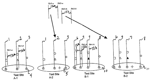
Figure 1 is a schematic example of localization of amplified DNA from 2
individual patients indicated as to A,B two predetermined groups of test sites
labeled
A-1, A-2 for patient A and B-1, B-2 for patient B.
Figure 2 is a schematic of the introduction of a first group of blockers to
the
amplicons at the test sites.
Figure 3 shows the complete specific hybridization of both groups of blockers
and the application of PCR control probes mutant discriminators and the first
and
second universal reporter.
Figure 4 shows the results of an example of a screening run wherein a
confirmation of the amplification is provided by the second universal reporter
and the
presence of a mutation is indicated by the first universal reporter.
Figure 5 is the strategic application of a first group of blockers in a
genotyping
run.
Figure 6 shows the specific hybridization of two blocker groups in a
IR1:1045858.2
CA 02494538 2005-O1-25
WO 2004/011668 PCT/US2003/023420
14 Patent
Attorney Docket: 612,404-427
genotyping run together with the application of mutant and wild-type
discriminators
and the first and second universal reporters.
Figure 7 shows the results of a genotyping run wherein first and second groups
of blockers are specifically hybridized to the amplicons and the hybridization
of the
discriminators and the first and second universal reporters indicate a
heterozygous
mutation in the sample from an individual patient.
Figure ~ is a block diagram of a computer system of the invention.
Figure 9 is a block diagram of a work station useful in the system of the
invention.
DETAILED DESCRIPTION OF THE INVENTION
The present invention provides an assay system, method, and integrated
components and reagents for screening and detection of multiple markers in a
high
throughput format. In this assay, specific regions of chromosomal DNA, which
will
also be referred to herein as identified loci, in patient samples are
amplified and
analyzed in an assay device comprised of a microchip that facilitates reacting
the
amplified DNA with reagents that facilitate identification of specific
markers. The
amplified sample is analyzed in the assay to simultaneously determine the
presence or
absence of one or more polymorphisms and to determine whether the polymorphism
is
present in one or both of a patient's chromosomes. The assay may be conducted
in
two steps wherein, in a first step, one or more patient samples are screened
for any one
or more of a set of markers. When a number of the set of markers is
identified, a
separate reaction identifies the individual members of the set that may be
present. In
preferred embodiments, the apparatus and methodologies have the capability to
analyze several patient samples using a set of reagents that is specifically
selected for
the assay. The following definitions are used herein to describe the several
embodiments of the invention.
An "amplicon" is an amplified polynucleotide sequence derived from a primer
in an amplification reaction wherein a selected sequence is reproduced under
reaction
conditions that extend a primer sequence by sequential addition of nucleotides
to
encompass a target sequence.
IR1:1045858.2
CA 02494538 2005-O1-25
WO 2004/011668 PCT/US2003/023420
15 Patent
Attorney Docket: 612,404-427
"Amplification" refers to the process by which a region of a polynucleotide
sequence is copied and expanded into a large number of amplicons. The
polynucleotides contained in the patient samples are amplified by in vitro
methods,
such as the polymerase chain reaction (PCR), the ligase chain reaction (LCR),
rolling
circle, strand displacement amplification (SDA), nucleic acid sequence based
amplification (NASBA) the transcription-based amplification system (TAS), the
self sustained sequence replication system (3SR) and the Q.beta replicase
amplification system (Q(3).
"Blockers" are polynucleotides that hybridize specifically to polynucleotide
sequences, usually amplicons, and that are designed to prevent binding by wild-
type
and mutant discriminator probes.
"Complementary" refers to the topological compatibility or matching together
of interacting surfaces of two polynucleotides. Thus, the two molecules can be
described as complementary, and furthermore, the contact surface
characteristics are
complementary to each other. A first polynucleotide is complementary to a
second
polynucleotide if the nucleotide sequence of the first polynucleotide is
identical to the
nucleotide sequence of the polynucleotide binding partner of the second .
Thus, the
polynucleotide whose sequence 5'-TATAC-3' is complementary to a polynucleotide
whose sequence is 5'-GTATA-3'.
"Detectable moiety" or "label" refers to a composition detectable by
spectroscopic, photochemical, biochemical, immunochemical, or chemical means.
For example, useful labels include 32P, 355, fluorescent dyes, electron-dense
reagents,
enzymes (e.g., as commonly used in an ELISA), biotin-streptavadin, dioxigenin,
haptens and proteins for which antisera or monoclonal antibodies are
available, or
nucleic acid molecules with a sequence complementary to a target. The label
often
generates a measurable signal, such as a radioactive, chromogenic, or
fluorescent
signal, that can be used to quantitate the amount of bound detectable moiety
in a
sample. The label can be' incorporated in or attached to a primer or probe
either
covalently, or through ionic, van der Waals or hydrogen bonds, e.g.,
incorporation of
radioactive nucleotides, or biotinylated nucleotides that are recognized by
streptavadin. The label may be directly or indirectly detectable. Indirect
detection can
involve the binding of a second directly or indirectly detectable moiety to
the
IR1:1045858.2
CA 02494538 2005-O1-25
WO 2004/011668 PCT/US2003/023420
16 Patent
Attorney Docket: 612,404-427
detectable moiety. For example, the label can be the ligand of a binding
partner, such
as biotin, which is a binding partner for streptavadin, or a nucleotide
sequence, which
is the binding partner for a complementary sequence, to which it can
specifically
hybridize. The binding partner may itself be directly detectable, for example,
an
antibody may be itself labeled with a fluorescent molecule. The binding
partner also
may be indirectly detectable, for example, a nucleic acid having a
complementary
nucleotide sequence can be a part of a branched DNA molecule that is in turn
detectable through hybridization with other labeled nucleic acid molecules.
(See, e.g.,
P. D. Fahrlander and A. I~lausner, Bio/Technology (1988) 6:1165.) Quantitation
of
the signal generated by a label is achieved by known detection and measurement
techniques, e.g., scintillation counting, densitometry, or flow cytometTy.
A "discriminator" or "discriminator probe" is a polynucleotide that
selectively
binds to a polymorphic region of an amplicon, wherein the region may or may
not
contain a mutation. Specific discriminator binding to a known, predetermined
mutation is sometimes referred to herein as "querying." Each different
polymorphic
region may be referred to as a "variant." "Wild-type discriminators" bind to
wild-type
sequence, while "mutant" discriminators bind to variants including recognized
mutations, or simple variants of the wild-type sequence that may be described
as
markers or polymorphisms. A "pair of discriminators" typically consists of the
wild-
type discriminator and the corresponding mutant discriminator for a specific
polymorphism. Discriminators may consist of one that specifically binds to the
sequence of a specific variant or of two polynucleotides that specifically
bind, in
direct apposition, to a contiguous sequence of a specific variant and are
designed so
that a first stabilizes the binding of a second by base stacking. The use of
two
polynucleotides and base stacking to obtain highly stable hybridization
complexes
capable of precise discrimination is described in Radtkey R. et al., Nucleic
Acids
Research, 28(7): i-vi (2000) and Yershov, G. et al., Proc. Nat'1 Acad. Sci.
USA, 93:
4913-18 (May 1996).
"E-stripping" is electronic denaturation of double stranded polynucleotides or
removal of hybridized polynucleotides.
"Heterozygous" means that one chromosome from a patient sample contains a
mutant variant and the other contains the corresponding wild-type variant.
nziaoassss.2
CA 02494538 2005-O1-25
WO 2004/011668 PCT/US2003/023420
17 Patent
Attorney Docket: 612,404-427
"Heterozygous ratio references" are polynucleotides that each bind
specifically
to one discriminator pair. Each heterozygous ratio reference contains one or
more
polynucleotides that bind to one pair of discriminators.
"Homozygous" means that both chromosomes from a patient sample contain a
mutant variant or that both contain a wild-type variant.
"Hybridizing specifically to" or "specific hybridization" or "hybridize to,"
refers to the binding, duplexing, or hybridizing of a nucleic acid molecule
preferentially to a particular nucleotide sequence under stringent conditions
when that
sequence is present in a complex mixture.
"Localized" means that a composition is concentrated at a test site to a level
greater than in ordinary solution and which is greater than would occur
through
passive hybridization. In the context of an amplicon localized at a test site,
the
amplicon has a greater concentration achieved through chemical, electrical, or
biochemical reaction, as opposed to mere selective placement at the test site.
The terms "mutation" or "polymorphism" describe nucleotide sequences that
vary from a wild-type sequence by a known parameter such that the distinction
can be
interrogated with a discriminator probe. In typical usage, mutation usually
refers to
variant the wild-type sequence that is correlated to disease. A polymorphism
may also
be a mutation, but may also refer to a difference from the wild-type sequence
that has
no known correlation to disease. Both mutations and polyrnorphisms may broadly
be
described as "markers" for a disease or a marker may simply represent an
identifiable
sequence in comparison to wild-type. Markers, mutations, or polymorphisms may
be
deletions, substitutions, repeats, transpositions, etc.
An "oligonucleotide" is a polymer of nucleotides.
A "primer" refers to a polynucleotide that is capable of specifically
hybridizing
to a designated polynucleotide template and providing a point of initiation
for
synthesis of a complementary polynucleotide under synthesis inducing
conditions i.e.,
in the presence of nucleotides, a complementary polynucleotide template, and
an agent
for polymerization such as DNA polymerase. A primer is typically single-
stranded
deoxyribonucleic acid, but a wide variety of synthetic and naturally occurring
primers
are useful. A primer is complementary to the template to which it hybridizes
to serve
as a site for the initiation of synthesis.
IR1:1045858.2
CA 02494538 2005-O1-25
WO 2004/011668 PCT/US2003/023420
18 Patent
Attorney Docket: 612,404-427
A "reflex test" occurs when a positive genotyping result requires an
additional
test.
A "reporter" refers to a polynucleotide that is capable of specifically
hybridizing to a designated sequence of another polynucleotide and that
contains a
label. A probe specifically hybridizes to a target complementary
polynucleotide, but
need not reflect the exact complementary sequence of the template. In such a
case,
specific hybridization of the probe to the target depends on the stringency of
the
hybridization conditions. Probes can be labeled with e.g., chromogenic,
radioactive,
chemiluminescent, enzymatic, colorimetric, or fluorescent moieties and used as
detectable moieties.
"Screening" is a step in the assay during which it is determined whether or
not
a sample contains any one of a group of polymorphism that are usually
mutations that
are known to be related to disease. If a sample tests positive, the presence
of a
mutation within the subset is indicated and the sample can then be further
analyzed or
"genotyped.", "Genotyping" refers to determining whether a patient sample is
homozygous for wild-type, homozygous for a particular mutant variant, or
heterozygous for a particular variant.
The terms "selective for" or "selectively hybridize to" describe differential
reactivity between wild type and a mutant variant of a probe in a
hybridization
reaction with a complementary sequence of an amplicon. A mutant discriminator
probe is selective for a specific known polymorphism or mutation such that
hybridization does not occur to a wild-type sequence. Similarly, a wild-type
discriminator probe is selective for the wild-type sequence such that
hybridization
only occurs to the wild-type sequence and not to a polymorphism or mutation.
The terms "stringent conditions" refer to conditions under which a probe will
hybridize preferentially to its target subsequence, and to a lesser extent to,
or not at all
to, other sequences. "Stringent hybridization" and "stringent hybridization
wash
conditions" in the context of nucleic acid hybridization experiments such as
southern
and northern hybridizations are sequence dependent, and are different under
different
environmental parameters. An extensive guide to the hybridization of nucleic
acids is
found in Tijssen (1993) Laboratory Techniques in Biochemistry and Molecular
Biology-Hybridization with Nucleic Acid Probes part I chapter 2 "Overview of
IR1;1045858.2
CA 02494538 2005-O1-25
WO 2004/011668 PCT/US2003/023420
19 Patent
Attorney Docket: 612,404-427
principles of hybridization and the strategy of nucleic acid probe assays,"
Elsevier,
N.Y. Generally, highly stringent hybridization and wash conditions are
selected to be
about 5° C. lower than the thermal melting point (Tm) for the specific
sequence at a
defined ionic strength and pH. The Tm is the temperature (under defined ionic
strength and pH) at which 50% of the target sequence hybridizes to a perfectly
matched probe. Very stringent conditions are selected to be equal to the Trn
for a
particular probe.
A "universal reporter" refers to a polynucleotide that 1) possesses a moiety
(e.g. a nucleotide sequence) that interacts with a series of nucleotide
sequences, each
in the series having the same moiety complementary to the universal reporter
and a
region specific for differing genetic loci, and 2) possesses a detectable
moiety.
Device for simultaneous detection of multiple markers
The apparatus of this invention includes an electronically addressable
microchip device, many of the components of which are described in the
following
applications and patents, which are specifically incorporated herein by
reference:
Application Serial No. 091671,594, filed September 27, 2000, entitled
"ELECTRONIC SYSTEMS, COMPONENT DEVICES, MECHANISMS,
METHODS, AND PROCEDURES FOR MACROSCOPIC AND MICROSCOPIC
MOLECULAR BIOLOGICAL REACTIONS, ANALYSES AND DIAGNOSTICS",
which is a continuation-in-part of application Serial No. 08/986,065, filed
December 5, 1997, entitled "METHODS AND PROCEDURES FOR MOLECULAR
BIOLOGICAL ANALYSIS AND DIAGNOSTICS", now issued as US Patent No.
6,051,380,
which is a continuation-in-part of application Serial No. 08/855,458, filed
May
14, 1997, entitled "METHODS FOR ELECTRONIC FLUORESCENT
PERTURBATION FOR ANALYSIS AND ELECTRONIC PERTURBATION
CATALYSIS FOR SYNTHESIS", now issued as US Patent No. 6,048,690,
which is a continuation-in-part of application Serial No. 08/534,454, filed
September 27, 1995, entitled "APPARATUS AND METHODS FOR ACTIVE
PROGRAMMABLE MATRIX DEVICES", now issued as US Patent No. 5,849,486,
which is a continuation-in-part of application Serial No. 08/304,657, filed
mao4sass.a
CA 02494538 2005-O1-25
WO 2004/011668 PCT/US2003/023420
20 Patent
Attorney Docket: 612,404-427
September 9, 1994, entitled "AUTOMATED MOLECULAR BIOLOGICAL
DIAGNOSTIC SYSTEM," now issued as US Patent No. 5,632,957,
which is a continuation-in-part of application Serial No. 08/271,882, filed
July
7, 1994, entitled "METHODS FOR ELECTRONIC STRINGENCY CONTROL FOR
MOLECULAR BIOLOGICAL ANALYSIS AND DIAGNOSTICS," now issued as
US Patent No. 6,017,696, and
which is a continuation-in-part of Serial No. 08/146,504, filed November 1,
1993, entitled "ACTIVE PROGRAMMABLE ELECTRONIC DEVICES FOR
MOLECULAR BIOLOGICAL ANALYSIS AND DIAGNOSTICS", now issued as
US Patent No. 5,605,662. The microchip device described in the applications
and
patents includes an array of microlocations or test sites each associated with
an
electrode. The electrode is overlaid with a permeation layer that separates
the
polynucleotides from the surface of the electrode. The device also includes an
attachment layer to which molecules such as nucleic acids are bound. Specific
binding entities such as affinity binding pairs are immobilized on the
attachment layer.
For example, streptavidin can be incorporated into the permeation layer,
providing an
affinity binding site for nucleic acids that have been derivatized with
biotin. The
amplification primers may be biotinylated such that the amplicons are
comprised of
amplified loci of a patient sample and a first member of a binding pair
wherein the
second member of the binding pair is integral with the microchip. Charged
molecules
are electronically addressed to a specific test site by biasing the electrode
underlying
the test site with a charge opposite that of the target molecule. This process
also
results in localization of the molecule at the test site. In addition, the
surrounding test
sites may be biased with the same charge as the charged molecule such that the
charged molecule is repelled.
In a preferred embodiment, the microchip device is coupled to a reader that
detects signal generated by the labels attached to universal reporters that
are
hybridized to discriminator probes at the various test sites. In a
particularly preferred
embodiment, the reader detects at least two discrete wavelengths of
fluorescent light
generated by fluorescent labels incorporated into the universal reporters. The
reader
may be comprised of a discrete light source and a detector designed to detect
a signal
from the interaction between light from the source and a label used in the
assay.
maoasasa.a
CA 02494538 2005-O1-25
WO 2004/011668 PCT/US2003/023420
21 Patent
Attorney Docket: 612,404-427
Alternatively, the reader may obtain an image of the device during the assay,
followed
by image analysis to determine the results of the assay. In either embodiment,
the
reader detects the signals) generated by the reporter labels) and determines
intensity
as well as a comparative value compared to like signals generated by the same
label,
or distinct signals generated by a different label or combination of labels.
Where
labeling by different reporters yields different signals, such as different
fluorescent
wavelengths, the detector measures the relative strengths of the signals at
one or more
locations, such as the test sites of the microchip, and may translate any of
these
detections into a signal for further processing by electronic means or through
computer software that manipulates the signal to generate a data report of the
results
of the assay. In a particularly preferred embodiment, the microchip is also
coupled to
a loader capable of transferring sample from one container, such as a
microtiter plate,
to the microchip device and is capable of transferring members of the reagent
set of
the invention to the microchip.
The invention also includes a specific reagent set used in the system. In a
preferred embodiment, the system encompasses integral containers for reagents
such
as sets of primers, discrete groups of Mockers, including the arrangement of
blockers
in predetermined subsets to interrogate subsets of known mutations, wild-type
and
mutant discriminator probes, universal reporters comprising labels and
heterozygous
ratio references. In a particularly preferred embodiment, the apparatus also
includes a
background control and a reagent set of amplification controls.
The blocker compositions each contain groups of sequences that are
specifically hybridized to identified loci on an amplicon that contains one or
more of
the variants being assayed. The individual blocker groups are substantially
homogenous mixtures of discrete nucleotide sequences that specifically
hybridize to
identified loci at the amplicon and prevent the selective binding of mutant or
wild-
type discriminator probes. One blocker group rnay bind a set of identified
loci or may
block only a single locus. In the latter case, blocking more than one
identified locus
requires more than one blocker group. In a preferred embodiment, the blockers
are of
sufficient length to remain hybridized through sequential rounds of
discriminator
hybridization and subsequent denaturation of the discriminators. This length
is
determined empirically. The blocker mixtures are electronically addressed to
selected
mao4s8ss.a
CA 02494538 2005-O1-25
WO 2004/011668 PCT/US2003/023420
22 Patent
Attorney Docket: 612,404-427
microlocations and hybridize to amplicons which prevents the reaction of the
amplicons with selected mutant and/or wild-type discriminators binding of the
mutant
and/or wild-type discriminator probes to the amplified polynucleotides does
not occur
above a threshold level that is capable of being removed as a background
signal.
In preferred embodiments of the assay, the blockers are organized into
screening mixes and genotyping mixes. Each screening mix is unique, consists
of one
or more blockers, and is designed to block a particular subset of the known
markers
being assayed, with each mixture blocking some, but not all, loci. The loci
not
blocked by a particular blocking group will be referred to as a "screening
loci group."
Each genotyping mix is unique, consists of one or more blockers, and is
designed to
block some variants but to leave unblocked one loci from each screening loci
group.
Thus, if blocker mix A blocks all loci except 1-3, Mocker mix B blocks all
loci except
4-6, and blocker mix C blocks all loci except 7-9, genotyping mix A will block
all loci
except 1, 4, and 7, genotyping mix B will block all loci except 2, 5, and ~,
and
genotyping mix C will block all loci except 3, 6, and 9.
In a preferred embodiment, the apparatus also includes amplification controls.
' The amplification process, one example of which is described in more detail
below, is
comprised of any process that accurately and reproducibly copies a defined and
identified region of a gene wherein an identified locus exists that contains a
polymorphism. Because the sequence of at least a portion of the identified
locus is
known, a group of primers can be selected to amplification of discrete genetic
loci in a
patient sample where the mutations are known to occur. The identification of
the
sequence of a primer useful for amplifying a specific locus in a patient
sample is a
routine matter for one of ordinary skill in the art and a preferred set of
reaction
parameters and amplification reagents are described in the examples herein.
The
amplification controls are polynucleotidess that bind specifically to portions
of each
arnplicon in order to verify the presence of a specific amplicon and to verify
that the
amplification reaction has successfully amplified the desired regions of the
pertinent
sample when certain mutations occur. Each amplification control binds to a
different
amplicon, such that a full set of amplification controls contains one
amplification
control per amplicon. In addition, the amplification controls are designed so
that they
can be denatured from the amplicons under less stringent conditions than are
the
IR1:1045858.2
CA 02494538 2005-O1-25
WO 2004/011668 PCT/US2003/023420
23 Patent
Attorney Docket: 612,404-427
blockers. In a preferred embodiment these amplification controls are wild-type
discriminators. Also, in a preferred embodiment, amplification controls are
designed
to have a melting temperature within the operation temperature of the testing
platform
and a temperature empirically determined to be low enough to allow removal by
chemical, thermal or e-stripping without denaturing blockers.
In a preferred embodiment the apparatus also includes a set of heterozygous
ratio references. Each heterozygous ratio reference contains sequences that
are
complementary to one discriminator pair. The heterozygous ratio references are
capable of attachment to the attachment layer of the microchip device. In a
preferred
embodiment, the polynucleotides comprising each heterozygous ratio sequence
are
biotinylated.
The system of the invention also includes a selected group of reagents
including a set of discriminators that are specific for all of the wild-type
and mutant
variants associated with the particular disease being assayed. Each individual
discriminator species contains a nucleotide sequence that is complementary to
a
sequence in an amplicon. Wild-type and mutant discriminators differentially
and
selectively react with either wild-type or discriminator sequences,
respectively,
present in the amplicons, when those regions of the amplicons are not blocked
by the
blocker sequences. The discriminators are preferably designed so that an
entire set of
discriminator pairs can be denatured from the amplicons under less stringent
conditions than are the blockers. In one embodiment, wild-type and mutant
discriminator probes are about 30-40 nucleotides in length and have a melting
temperature between 35° C. and 45° C. In a preferred embodiment,
the wild-type and
mutant discriminator probes have a melting temperature that is about
20° C. less than
the melting temperature of the blocker(s) that bind to the same variant as
does the
discriminator.
The apparatus also includes labels for detecting the binding of at least the
mutant discriminator, and preferably the wild-type discrimination and the
amplification controls to the amplicons. Amplification controls and
discriminators are
associated with distinguishable labels so that more than one amplification
control or
discriminator can be detected at one microlocation. In one embodiment, the
label is
attached directly to the amplification controls and discriminators.
IR1:1045858.2
CA 02494538 2005-O1-25
WO 2004/011668 PCT/US2003/023420
24 Patent
Attorney Docket: 612,404-427
In the preferred embodiment, the label is coupled to a construct that acts as
a
universal reporter to specifically hybridize to a common region in the
amplification
controls and either of the discriminators sometimes referred to herein as a
"tail." In
this embodiment, each amplification control and discriminator is designed to
contain
at least one tail. To distinguish the wild-type discriminators from the mutant
discriminators, the tail sequence of a mutant discriminator may be
complementary to
the nucleotide sequence of a first universal reporter that has a first label.
Accordingly,
the tail sequence of the wild-type discriminator probe may be common and
complementary to the nucleotide sequence of a second universal reporter having
a
second label. Detection of the different signals generated by the first and
second
labels distinguishes the reaction by the wild-type and mutant discriminators
with the
amplified patient DNA. Thus, in the preferred embodiment, the universal
reporter
contains a polynucleotide sequence that is complementary to the above-
described tail
such that the universal reporter binds to the tail of the discriminators under
the
hybridization conditions of the assay. The universal reporters that
specifically bind to
one tail are coupled to a label that is distinguishable from the label coupled
to any
other universal reporter that specifically binds to another tail. Thus, a
first tail is
associated with a first label, a second tail with a second label, and a third
tail with a
third label, and so on, wherein the first, second, third and additional labels
are
distinguishable. In a preferred embodiment more than one amplification control
contains the same tail or tails. In a particularly preferred embodiment a
first group of
the amplification controls contain a first tail while a second group contains
a second
tail.
In the preferred embodiment, wild-type and mutant discriminators contain
different tails, but all of the wild-type discriminators contain a common
first tail and
all of the mutant discriminators contain a common second tail that are
different from
the first tail. In another embodiment, at least one discriminator has a first
tail, at least
one discriminator has a second tail, at least one discriminator has a third
tail, and at
least one discriminator has a fourth tail, wherein the first, second, third;
and fourth
tails are different. To facilitate generating stronger signals for one type or
species of
discriminator probe, the discriminator probes may contain more than one tail
so that a
plurality of individual universal reporter molecules bind to the same
discriminator
IR1:1045858.2
CA 02494538 2005-O1-25
WO 2004/011668 PCT/US2003/023420
25 Patent
Attorney Docket: 612,404-427
probe. Thus, to increase the signal generated by the binding of a mutant
discriminator
probe to an amplicon, the mutant discriminator probe could be designed to have
two
tail regions such that two universal reporters, and accordingly two labels,
are bound to
the same mutant discriminator probe. In a preferred embodiment, the wild-type
and
mutant discriminator probes have the same sequence. In this embodiment, the
wild-
type sequence is of a sufficient length that it is still able to bind to the
amplicon
containing the mutation.
Compounds commonly used to label nucleic acid probes are enzymatic
compounds, fluorescent compounds, phosphorescent compounds, cherniluminescent
compounds, and/or compounds providing a colorimetric, enzymatic, radioactive,
or
other detectable signal. These compounds can be coupled to polynucleotides by
methods well known to those of skill in the art. In a preferred embodiment,
the labels
will be a minimum number of fluorescent labels that generate a signal read by
the
reader that is integral to the assay device. The synthesis of polynucleotides
with
defined sequences, such as those described herein, is well known to one of
ordinary
skill in the art. For instance, the various polynucleotides described above
may be
ordered from several commercial sources, such as Integrated DNA Technologies
(Coralville, IA) or Oligos, Etc. Inc. (Wilsonville, OR), or synthesized using
a
commercially available polynucleotide synthesizer such as the ABI 3900 High
Throughout DNA Synthesizer (Applied Biosystems, Foster City, CA).
Simultaneous assay for multiple mutations
The present invention also relates to a method for simultaneously assaying
multiple markers related to the same disease or to a panel of diseases, or to
a set of
known polymorphisms in multiple patient samples to determine whether a patient
is
heterozygous, homozygous wild-type, or homozygous mutant for each marker,
using
the above-described device. Several diseases, groups of diseases, or
polyrnorphisms
of clinical or research interest are associated with the presence of one or
more known
mutations in the human genome and can be detected using this assay. Examples
of
disease-related mutations that can be detected with this assay are the
mutations
associated with any one or more of Cystic Fibrosis, Beta-Thalassemia,
hereditary
hemochromatosis, Gaucher, Tay-Sachs, Nieman-Pick, HIV, epilepsy, and others.
In
IR1:1045858.2
CA 02494538 2005-O1-25
WO 2004/011668 PCT/US2003/023420
26 Patent
Attorney Docket: 612,404-427
addition to identifying diseases in patient samples, the simultaneous assay
for multiple
markers may also be used for identification in DNA fingerprinting.
The assay is performed on any sample that contains DNA, such as, for
example, blood, urine, sputum, amniotic fluid, or buccal. The loci of the DNA
in the
sample that are identified as containing the known polymorphism(s) are
amplified.
The amplification methods include polymerase chain reaction (PCR), the ligase
chain
reaction (LCR), strand displacement amplification (SDA), the transcription-
based
amplification system (TAS), the self sustained sequence replication system
(3SR) and
the Q(3 replicase amplification system (Q(3). In one embodiment, multiple
amplifications are accomplished in multiplex polymerase-based reactions with
specially selected primers for identified loci of genornic DNA containing the
known
polymorphisms. Polymerase chain reaction and, more specifically, multiplex
i
polymerase chain reaction are described in Innis, M.A. et al., PCR
Applications:
Protocols for Functional Genomics, (San Diego: Academic Pres. 1999).
The resulting amplified polynucleotides for an individual patient are
localized,
preferably by electronic addressing, to a discrete set of test sites on the
microchip
device. Preferably, the group of amplicons for an individual patient are
addressed to a
number of target sites that is less than the total number of polyrnorphisms to
be
detected in the assay. Once the amplicons are electronically addressed, the
detection
of polymorphisms is achieved through hybridization reactions occurring at the
microlocations. A first run of hybridizations is primarily a screening run,
however,
genotyping of single mutations, typically one or more predominant mutations
that is
most commonly associated with a disease, may also occur during the screening
run. A
second run of hybridizations is a "genotyping run" to determine which
particular
mutation or mutations is present in the sample and whether the sample is
homozygous
or heterozygous for that mutation. Additional hybridization runs may be added
to
identify additional mutations or further characterize specific mutations
related to those
identified through the first two runs.
Screening Run
During the screening run, amplicons and blockers are electronically hybridized
to predetermined test sites on the microchip device that are preferably
assigned or
dedicated to an individual patient. Data from a particular test site so
identified can be
IR1:1045858.2
CA 02494538 2005-O1-25
WO 2004/011668 PCT/US2003/023420
27 Patent
Attorney Docket: 612,404-427
correlated with 1) a particular patient and presence or absence of a
particular amplicon
and/or 2) a particular patient and presence or absence of a particular marker
or
markers. In addition, the full set of mutant discriminators, and in some
embodiments
certain wild-type discriminators, are loaded onto the device. Amplification
controls
and discriminators, which may also be associated with labels, are provided
along with
the universal reporter constructs.
Hybridizing ~plicons, blockers, and controls
A complete set of amplicons from one patient, when electronically addressed
to each of a specific number of predetermined microlocations will be
collectively
referred to as a "suite." Electronic addressing of arnplicons to a permeation
layer of
the microchip is described in, for example, U.S. Patent No. 6,051,380, which
is
expressly incorporated herein by reference. The attachment of the amplicon to
the
microlocation can be achieved by covalent chemical binding, or through the use
of a
binding pair of any type. The binding pair may be comprised of any two species
that
react to maintain the location of the amplicon at a specific test site.
Specifically
preferred is the use of an affinity binding pair such as streptavidin, which
is
incorporated into the permeation layer of the microchip device, and biotin-
labeled
'amplicons such that the streptavidin immobilizes the amplicons by the biotin
streptavidin affinity bindings, thereby fixing the amplicons to the permeation
layer.
As noted, the amplicons may also be chemically modified with a linking moiety
to
provide covalent binding to the substrate.
In addition, in a particularly preferred embodiment, each set of amplicons is
addressed to non-adjacent test sites, such that amplicon sets derived from the
same
patient are not addressed to adjacent microlocations. In a preferred
embodiment one
predetermined test site is used to provide a background reading. In one
embodiment
this background test site is empty. In another embodiment, this background
test site is
addressed with a synthetic polynucleotide.
If one of the mutations in the known set is predominant, such as the delta
F508
deletion with cystic fibrosis, that mutation may be both screened and
genotyped
simultaneously in the first set of hybridization reactions. As described in
detail below,
a heterozygous ratio reference that specifically binds the discriminator pair
for delta
508 is then used to query the predominant mutation.
IR1:1045858.2
CA 02494538 2005-O1-25
WO 2004/011668 PCT/US2003/023420
28 Patent
Attorney Docket: 612,404-427
Once electronic addressing of the amplicons is complete, the amplicons are
denatured leaving single stranded polynucleotide. As an option to electronic
addressing of the amplicons, the amplicons can be generated on the microchip
by the
electronic addressing of sense and antisense primer pairs to rnicrolocations
followed
by electronic addressing of patient genomic DNA as template and addition of
amplification reaction components and incubation of the microchip under
conditions
for amplification. This process results in generation of both the sense and
antisense
strands attached to the microlocation. Denaturation of the amplicons occurs as
described with the exception that both single strands remained attached to the
test site.
Those of skill in the art will recognize that there are several ways to
denature double-
stranded polynucleotides. In one embodiment, the amplicons are denatured using
sodium hydroxide. In a particularly preferred embodiment the amplicons are e-
stripped in a similar way to electronic addressing. The electrodes underlying
the test
site to which the sets of amplicons are addressed are positively biased to
denature the
amplicons, repelling the non-biotinylated strand as described in U.S. Patent
No.
6,051,380.
After denaturation of the amplicons, different screening blocker groups are
electronically addressed to a predetermined test sites within each suite and
bind to the
complementary regions of the amplicon as described in more detail herein, the
sequences of the blocker groups are specifically designed to hybridize to the
regions
of the amplicons that contain the mutant or wild-type sequences and hybridize
to the
amplicons to prevent fiu-ther reaction with the mutant or wild-type
discriminator
probes. The blocker groups each include a unique mix of blocker sequences such
that,
at each of the test sites in a suite, a different known group of variants,
i.e., the
screening variant group, remains unhybridized.
In one embodiment, where a mutation is commonly associated with a disease,
which will be referred to as a "predominant mutation," is genotyped in the
first run,
one blocker group blocks all of the variants except the predominant mutation.
This
predominant mutation blocker group is electronically addressed to one test
site per
suite. Test sites at which screening will be performed are referred to as
"screening
sites," while test sites at which genotyping will be performed are referred to
as
"genotyping sites."
IR1:1045858.2
CA 02494538 2005-O1-25
WO 2004/011668 PCT/US2003/023420
29 Patent
Attorney Docket: 612,404-427
In a preferred embodiment, after addressing of the blocker groups,
amplification controls are introduced to each amplicon-bearing test site. This
can be
optional if other methods of amplification confirmation are used. Each
amplification
control is introduced to the particular test site in each of the suites on the
microchip
device that should contain an un-hybridized (i.e. unblocked) region for which
the
particular amplification control is specific. The amplification controls bind
to the
unblocked amplicon region to which at least part of the amplification control
is
complementary. The amplification controls are hybridized in such a way that,
at each
test site per suite, the presence or absence of at least one amplicon is
detected.
In a preferred embodiment, after different Mocker sets are electronically
addressed to each amplicon-bearing test site, the amplification controls are
hybridized
to the particular test site in each of the suites on the microchip device that
should
contain an un-hybridized (i.e. unblocked) region for which the particular
amplification
control is specific.
As described above, these amplification controls are associated with a
construct containing a label. In a preferred embodiment, the construct is a
universal
reporter. After the amplification controls are hybridized the universal
reporter is
loaded onto the microchip device and specifically binds to the tail portion of
the
amplification control. In a particularly preferred embodiment, before
hybridizing the
amplification controls, the universal reporters are mixed with the
amplification
controls and specifically bind to the tail portion of the amplification
control.
After hybridizing of the amplification controls-and, in preferred embodiments,
hybridization to universal reporters, a reader scans the microchip device,
detecting the
signal associated with the amplification controls and with the background
control.
These scans will be referred to as "amplification scans." In embodiments in
which
multiple labels are detected, the reader performs one scan of the microchip
device per
label. In a preferred embodiment in which fluorescent labels are used, each
scan will
detect a different wavelength of fluorescent light.
In the embodiment where amplification controls are used, the controls are
denatured after scanning along with their labels to leave the groups of
blockers
hybridized to the amplicons. Denaturation can occur in a variety of ways,
including
IR1:1045858.2
CA 02494538 2005-O1-25
WO 2004/011668 PCT/US2003/023420
30 Patent
Attorney Docket: 612,404-427
thermal denaturation, chemical denaturation, and e-stripping. In a preferred
embodiment the amplification controls are removed by thermal denaturation.
Mutant discriminators that are selected for each polymorphism are then loaded
onto the microchip device under stringent conditions conducive to selective
hybridization of discriminator probes to the amplicons. In a preferred
embodiment,
discriminator probes are hybridized by a touch down thermal method in which
the
microchip device is heated before and for a short period after the
discriminator probes
are added and then the temperature is slowly decreased. These temperature
changes
are followed by or performed in conjunction with several high salt washes to
further
increase specificity of the discriminator probe binding. In another preferred
embodiment, discriminators are hybridized using non-stringent conditions such
that
both wild-type and mutant signals are approximately equal. Following
hybridization,
discrimination occurs using thermal chemical or e-stripping leaving only
matched
signals.
If the presence of all amplicons could not be verified with the hybridizing of
amplification controls described above, e.g., if the number of amplicons in a
set
exceeds the number of screening test sites multiplied by the number of
different labels
that can be detected at each test site, the presence of the remaining
amplicons can be
verified during the discriminator probe hybridization step, preferably using
wild-type
discriminators. When so used, the wild-type discriminator probes must be
associated
with a different label from the label associated with the mutant discriminator
probes.
In a preferred embodiment in which at least one predominant mutation is
screened and genotyped in the first hybridization run, the wild-type
discriminator that
specifically binds to the identified loci on the amplicons containing the
predominant
mutation is also loaded onto the microchip device such that either the mutant
or wild-
type discriminator will bind to the amplicons at the test site to which all
blocker
groups were addressed. In addition, this set of discriminators will bind to
the
heterozygous ratio reference for the predominant mutation.
In one embodiment in which the universal reporter construct contains a label,
after the discriminators and amplification controls have hybridized to the
amplicons,
the universal reporter is loaded onto the microchip device and specifically
binds to the
common sequence of the tail of the discriminator probes. In a preferred
embodiment,
IR1:1045858.2
CA 02494538 2005-O1-25
WO 2004/011668 PCT/US2003/023420
31 Patent
Attorney Docket: 612,404-427
before the discriminator probes and amplification controls are loaded onto the
microchip device, the universal reporters are mixed with the amplification
controls
and/or discriminator probes and hybridize to the common tail portion of the
amplification controls and/or discriminators.
After hybridizing of the amplification controls and, in some embodiments,
hybridization to universal reporters, a reader scans the microchip device,
detecting the
signal associated with the discriminator probes, with the amplification
controls, if any,
and with the background control. The scans will be referred to as
"discriminator
scans." In embodiments in which multiple labels are detected, the reader
performs
one scan of the microchip device per label. In a preferred embodiment in which
fluorescent labels are used, each scan will detect a different wavelength of
fluorescent
light.
In a preferred embodiment at least some of the steps described above can be
accomplished using a computer system coupled to the device in the manner
described
below. In a preferred embodiment, at least some of the steps described above
are
automated.
Analysis of si ngnalal
Once the hybridization reactions are completed and the labels detected to
yield
a signal, the signal gathered from each test site in the amplification scans
andlor the
discriminator scans is then analyzed. The analysis outlined below may be
performed
by a computer system coupled to the reader. Signal from each test site except
the
background control test site will be referred to as "raw test site signal" or
"RTSS."
Signal used as background signal in the calculations described below will be
referred
to as "background signal." The signal collected from the background control
test site
in a particular scan can be used as the background signal in the calculations
described
below for all test sites. In a particularly preferred embodiment, the
background signal
used in the calculation of signal from the label corresponding to mutant
discriminator
probes at screening test sites is the lowest signal detected at those test
sites per suite.
To determine presence of signal from the amplification scans and from the
screening test sites in the discriminator scans, the signal from each
amplification
control andlor each discriminator probe gathered from each scan is compared to
the
background signal gathered,from that particular scan. The background signal
from a
maoassss.a
CA 02494538 2005-O1-25
WO 2004/011668 PCT/US2003/023420
32 Patent
Attorney Docket: 612,404-427
particular scan will be subtracted from the RTSS gathered from each test site
during a
particular scan to calculate the "adjusted signal." For each scan of each
patient test
site, the background subtracted signal must be greater than a threshold value,
which
will be referred to as a "minimum signal criteria," in order to indicate the
presence of
a useful signal. In one embodiment, the minimum signal criteria is determined
empirically.
In addition to the above calculation, the ratio of RTSS to background signal
is
calculated for each microlocation from each scan to give a "signal to noise
ratio
(SNR)." The SNR must be greater than a threshold value to indicate a readable
signal.
In one embodiment the SNR is determined empirically. In another embodiment,
the
threshold signal value for the SNR is set to a value of 5: l, which has been
shown to be
a meaningful setting to permit further processing.
Because it is known which blocker group, amplification controls, and
discriminator probes were hybridized to each test site, once a useable signal
is
indicated by the above calculations the signal can be correlated to a
particular patient,
a particular amplicon, a particular screening variant group, a particular
variant, or a
particular heterozygous ratio reference based on from which test site and from
which
scan the signal was derived. Such correlation is described in more detail
below.
Yer~ing presence of amplicons
In the amplification scans presence of signal at -a particular test site
indicates
presence of a certain amplicon for a certain patient. For example, if two
amplification
controls (AC), AC-1 with a first label and AC-2 with a second label
distinguishable
from the ~lrst, were addressed to a first test site in each suite, detection
of the first
label at a first test site in a particular suite would indicate presence of
the amplicon to
which AC-1 hybridized for a particular patient. Similarly, presence of the
second
label at the first test site in a particular suite would indicate presence of
the amplicon
to which AC-2 specifically hybridizes. In this way, presence of each amplicon
in a set
for each patient can be analyzed. If absence of an amplicon for a certain
patient is
indicated by lack of signal, the discriminator scans for that patient are not
analyzed
and the assay must be performed again for that patient.
IR1:1045858.2
CA 02494538 2005-O1-25
WO 2004/011668 PCT/US2003/023420
33 Patent
Attorney Docket: 612,404-427
Presence of certain amplicons is also indicated by presence of signal from
label corresponding to amplification controls at screening test sites in the
discrimination scans.
ScYeenirag for groups of variants
Also in the discriminator scans, presence of signal corresponding to the
labels) associated with the mutant discriminator probes at a particular
screening test
site indicates presence of one of the variants in the screening variant group
left
unblocked at that test site. For example, if screening blocker mix D which
left
unblocked screening variant group D, was addressed to a first test site in
each suite,
then the presence of signal at the first test site in a particular suite would
indicate
presence of a mutation in one of the variants in screening variant group D in
a
particular patient sample.
Genotyping the predominant mutation.
Usable signal from the genotyping test site and the heterozygous ratio
reference test site must be manipulated further before presence of signal can
be
correlated with heterozygosity or homozygosity of a particular variant. In a
preferred
embodiment in which label one corresponds to the wild-type variant and label
two to
the mutant variant, a label one/label two scale factor, which will be referred
to as a
"multiplier," is calculated from the heterozygous ratio references, and will
be applied
to the signal gathered from the genotyping test site. This multiplier adjusts
the lower
of the label one or label two signals that correspond to the ratio references
in such a
way that after adjustment the signal from the wild-type ratio reference equals
the
signal from the mutant ratio reference.
In the discussion that follows, raw signal from which the background control
reading from the appropriate scan has been subtracted will be referred to as
adjusted
signal. Raw signal from the ratio reference associated with label one will be
referred
to as "RR-1," while raw signal from the ratio references associated with label
two will
be referred to as "RR-2." Adjusted signal for RR-1 will be referred to as "ARR-
1,"
while adjusted signal for RR-2 will be referred to as "ARR-2."
The multiplier will be calculated as follows. If ARR-1 is greater than or
equal
to ARR-2, then the multiplier for all label 1 adjusted signal will be one,
while the
multiplier for all label 2 adjusted signal will be ARR-1/ARR-2. If ARR-2 is
greater
maoassss.a
CA 02494538 2005-O1-25
WO 2004/011668 PCT/US2003/023420
34 Patent
Attorney Docket: 612,404-427
than or equal to ARR-1, then the multiplier for all label 2 adjusted signal
will be one,
while the multiplier for all label 1 adjusted signal will be ARR-2/ARR-1.
The appropriate multiplier will then be multiplied to the adjusted signal from
each genotyping test site from each scan (i.e., the scan to detect label one
and the scan
to detect label two}. The resulting numbers will be referred to as
"indicators." The
indicator corresponding to label one at each genotyping test site will be
referred to as
Il, while the indicator corresponding to label two will be referred to as I2.
Finally, to correlate I1 and I2 from the genotyping test site to homozygosity
for
the predominant wild-type variant, heterozygosity, or homozygosity for the
predominant mutation, Il and I2 from each test site must be compared. If Il is
greater
than I2 * upper threshold factor (UTF), the patient is homozygous for the wild-
type
variant. If I2 is greater than Il * UTF, the patient is homozygous for the
mutation. If
Il is less than I2 * lower threshold factor (LTF) or I2 is less than Il * LTF,
then the
patient is heterozygous for this particular variant. If Il is between the UTF
and the
LTF, then no determination as to heterozygosity or homozygosity can be made.
In one
embodiment the threshold factors are empirically determined. In another
embodiment
the UTF is 5 and the LTF is 2. In a preferred embodiment in which the
microchip
device is coupled to a computer system, these results (i.e., whether a
particular patient
is homozygous mutant, homozygous wild-type or heterozygous for a particular
variant) are reported to the user by the computer.
In other embodiments in which more than two labels are used, such that more
than one variant can be identified per genotyping test site, multiple
heterozygous ratio
references corresponding to the discriminator probes used to query the
variants would
be required. In addition, the reader would have to scan the chip multiple
times in
order to obtain readings from each label. Finally, the calculations and
analysis
outlined above would have to be done for each variant using the corresponding
heterozygous ratio reference.
If no polymorphisms are detected at the screening test sites, the assay is
complete as to the variants assayed at the screening test sites. In a
preferred
embodiment, if the assay is complete at this point, the computer system
generates a
report of the results of the assay. Referring to Figure 1, in one embodiment,
the
reportable data are stored as a separate file, such as a database file or a
text file. For
IR1:1045858.2
CA 02494538 2005-O1-25
WO 2004/011668 PCT/US2003/023420
35 Patent
Attorney Docket: 612,404-427
example, the report can be downloaded to a report directory on storage device
107 and
formatted for print or display by a database management system. In another
embodiment, the report is created or formatted in a file suitable for viewing
by display
device 121, such as an HTML file within a browser. In still another
embodiment, the
report is stored in a format suitable for printing to a printer or archiving
to a
permanent archive, such as a tape storage device, compact disc, or microfiche.
If a polymorphism is detected at one or more screening test sites, a second
run
of hybridizations is needed to identify which of the polymorphisms in the
known set is
present in the sample. In addition, a second or third run of hybridization may
be made
to further identify the predominant mutation if its presence in the genome has
been
correlated to the presence of some other polymorphism of interest that also
needs to
be genotyped. In a preferred embodiment, the computer system creates a
genotyping
run protocol for each screening variant group identified as containing a
polymorphism, and after a protocol is implemented, prompts the user as to
which
discriminator pairs should be added.
Genotyping Run
During the genotyping run, blocker mixes and heterozygous ratio references
are electronically addressed to assigned, predetermined test sites. Thus, data
from a
particular test site can be correlated with a particular patient and the
presence or
absence of a particular member of a set of known polymorphisms determined.
In addition, discriminator pairs corresponding to one or more screening
variant
groups identified in the screening run as containing a mutation are loaded
onto the
microchip device to query the screening variant group. If the presence of a
polymorphism in more than one screening variant group was identified at one or
more
test site suites in the screening run, more than one query will be needed. In
that case,
discriminator pairs for the polymorphisms in one screening variant group will
be
loaded and detected. Then, these discriminator probes will be removed, and
another
query performed using a set of discriminator pairs corresponding to the other
screening variant group. As many queries as different screening variant groups
identified as containing a polymorphism will be performed. If the same
screening
variant group was identified in multiple test site suites, only one query will
be needed.
maoassss.a
CA 02494538 2005-O1-25
WO 2004/011668 PCT/US2003/023420
36 Patent
Attorney Docket: 612,404-427
Discriminators are labeled such that signal from labels at the various test
sites is
detected and analyzed to determine whether a sample is heterozygous,
homozygous
wild-type, or homozygous mutant.
Addressing the array with ~enotyuin~ blocker groups and heterozy~ous ratio
references
After a positive reading in the screening run, the determination is made that
a
genotyping run is needed and that the mutation belongs to one or more
screening
variant groups. Accordingly, all of the probes are removed, leaving the single-
stranded set of amplicons from each patient, the background control, and the
heterozygous ratio reference for the predominant variant attached to their
predetermined assigned test sites on the microchip device. The polynucleotide
probes
can be removed in a variety of ways, including thermal denaturation, chemical
denaturation, or e-stripping. In a preferred embodiment, all probes are
removed by
washing the array with sodium hydroxide. Then, heterozygous ratio references
and
genotyping blockers are addressed to predetermined test sites.
Groups of heterozygous ratio references are then electronically addressed to
predetermined test sites, with each group assigned to a different test site.
The
heterozygous ratio references can be attached to the permeation layer in a
variety of
ways, as is described in patent no. 6,051,380, which is incorporated herein by
reference. In a particularly preferred embodiment, streptavidin is attached to
the
permeation layer of the microchip device, such that the streptavidin
immobilizes the
heterozygous ratio references, that have been biotinylated, thereby fixing
them to the
permeation layer.
The heterozygous ratio references are grouped such that each group contains
the sequence of one of the variants in each screening variant group. Thus, if
screening
variant group A contains variants 1-3, screening variant group B contains
variants 4-6,
and screening variant group C contains variants 7-9, then heterozygous ratio
reference
group D contains the polynucleotide sequence of variants 1, 4, and 7,
heterozygous
ratio reference group E contains the sequence of variants 2, 6, and 8, and
heterozygous
ratio reference group F contains the sequence of variants 3, 6, and 9.
Each genotyping blocker mix is also electronically addressed to a
predetermined test site in each suite. The blockers in each mix bind to the
amplicon
IR1:1045858.2
CA 02494538 2005-O1-25
WO 2004/011668 PCT/US2003/023420
37 Patent
Attorney Docket: 612,404-427
regions to which they are complementary so that, at each test site in a suite,
one of the
polymorphisms from each of the screening variant groups remain unhybridized.
Thus,
at each test site one variant from the screening variant group identified in
the
screening run can be queried. The relationship between the polymorphisms left
unhybridized or unblocked at each test site during the screening run and
during the
genotyping run is set forth in Table 1 below, where A-Y represent different
variants.
Table 1
GenotypingGenotypingGenotypingGenotypingGenotyping
Test
Site
Pad 1 Pad 2 Pad 3 Pad 4 Pad 5
ScreeningA B C D E
Pad
1
ScreeningF G H I J
Pad
2
ScreeningX L M N O
Pad
3
Screeningp Q R S T
Pad
4
ScreeningU V W X Y
Pad
5
After the blockers have been addressed, the discriminator pairs that
specifically bind
to the variants in one screening variant group identified in the screening run
as containing a
polymorphism are loaded onto the microchip device. If, for example, signal was
detected at
test site 1 in one suite, revealing that the mutation corresponds to
polymorphism 7, 8, or 9,
then only the discriminator sets corresponding to these polymorphisms will be
loaded onto
the microchip device in this query. These discriminators are loaded onto the
microchip
device under stringent conditions conducive to specific binding of the
discriminator probes
to the regions of the amplicons containing the polymorphism to which the
discriminator is
complementary and to the heterozygous ratio references. In a preferred
embodiment, the
discriminator probes are hybridized by a method in which the microchip device
is heated
before and for a short period after the discriminator probes are added and
then the
temperature is slowly decreased. These temperature changes are followed by or
performed
in conjunction with several high salt washes to further increase specificity
of discriminator
probe binding.
IR1:1045858.2
CA 02494538 2005-O1-25
WO 2004/011668 PCT/US2003/023420
38 Patent
Attorney Docket: 612,404-427
As described above, these discriminators are associated with a label. In a
preferred embodiment in which the universal reporter construct contains a
label, after
the discriminator probes and amplification controls have hybridized to the
amplicons,
the universal reporter is loaded onto the microchip device and specifically
binds to the
common sequence or tail of the discriminators. In a particularly preferred
embodiment, before the discriminator probes are loaded onto the microchip
device the
universal reporters are mixed with the discriminator probes and specifically
bind to
the common sequence to which they are complementary.
After addition of the discriminator probes and, in some embodiments,
universal reporters, a reader scans the microchip device, detecting the signal
associated with the discriminators, with the background control, and with the
heterozygous ratio references. The reader will perform one scan of the
microchip
device per number of different labels used in the assay. In a preferred
embodiment in
which fluorescent labels are used, each scan will detect a difFerent
wavelength of
fluorescent light.
If polyrnorphisms were detected in more than one screening variant group,
after scanning, the discriminator probes used in the first query will be
denatured along
with their labels to leave the blocker sequences hybridized. Denaturation can
occur in
a variety of ways, including thermal denaturation, chemical denaturation, and
e-
stripping. In a preferred embodiment the amplification controls are removed by
thermal denaturation. Then, discriminator pairs specific for the polymorphisms
in
another variant group identified in the screening run are loaded onto the
microchip
device. The labeling and detection steps described above are repeated. Then,
these
discriminator probes may be removed and another set used to query another
variant
group, if necessary. As many queries as different screening variant groups
identified
in the screening run will be performed.
In a preferred embodiment at least some of the steps described above can be
accomplished using a computer system coupled to the device in the manner
described
below. In a preferred embodiment, at least some of the steps described above
are
automated.
IR1:1045858.2
CA 02494538 2005-O1-25
WO 2004/011668 PCT/US2003/023420
39 Patent
Attorney Docket: 612,404-427
Analysis of siQrlal
The signal gathered from each scan of each query is then analyzed. The
analysis outlined below may be performed by a computer integral to the
microchip
device. The following description describes analysis of data from one query in
the
genotyping run. Thus, the analysis will be repeated for each query performed
in the
genotyping run. To determine presence of signal, raw test site signal (RTSS)
from
each test site being genotyped, and from each heterozygous ratio reference
test site, is
compared to the background signal gathered from a particular scan. The
background
signal from a particular scan will be subtracted from the RTSS gathered from
each test
site during a particular scan to calculate the "adjusted signal." For each
scan of each
patient test site, the adjusted signal must be greater than a threshold value,
which will
be referred to as a "minimum signal criteria," in order to indicate the
presence of a
useful signal. This minimum signal criteria may be determined in the same ways
as
described above in the section regarding analysis of signal from the screening
run. In
addition to the above calculation, the ratio of RTSS to background signal is
calculated
for each test site from each scan to give a "signal to noise ratio." The SNR
must be
greater than a threshold value, which will be referred to as a "minimum ratio
criteria"
to indicate a readable signal. This minimum ratio criteria may be determined
in the
same ways as described above in the section regarding analysis of signal from
the
screening run. If absence of useful signal is reported in a suite, that
patient sample
must be retested. If absence of useful signal is reported for a heterozygous
ratio
reference, all of the samples on the chip must be retested.
For all of the test sites for which useful signal is generated, the adjusted
signal
from each test site is calibrated using a multiplier calculated from the
signal from the
test site with the corresponding heterozygous ratio reference (i.e., the
heterozygous
ration reference that contains the same sequence as the variant being queried
at a
particular test site a suite). In a preferred embodiment, in which a first
label
corresponds to the wild-type and second label to the polymorphism, a label
one/label
two scale factor, which will be referred to as a "multiplier," is calculated
from each
group of heterozygous ratio references. This multiplier adjusts the lower of
the label
one or label two signals that correspond to the ratio references in such a way
that after
IR1:1045858.2
CA 02494538 2005-O1-25
WO 2004/011668 PCT/US2003/023420
40 Patent
Attorney Docket: 612,404-427
adjustment the signal from the wild-type ratio reference equals the signal
from the
mutant ratio reference.
In the discussion that follows, raw signal from the ratio reference associated
with label one will be referred to as "RR.-1," while raw signal from the ratio
references
associated with label two will be referred to as "RR-2." Adjusted signal for
RR-1
will be referred to as "ARR-1," while adjusted signal for RR-2 will be
referred to as
"~-2.»
The multiplier will be calculated as follows. If ARR-1 is greater than or
equal
to ARR-2, then the multiplier for all label 1 adjusted signal will be one,
while the
multiplier for all label 2 adjusted signal will be ARR-1/ARR-2. If ARR-2 is
greater
than or equal to ARR-l, then the multiplier for all label 2 adjusted signal
will be one,
while the multiplier for all label 1 adjusted signal will be ARR-2/ARR-1.
The appropriate multiplier will then be multiplied to the adjusted signal from
each test site for each scan (i.e., the scan to detect label one and the scan
to detect
label two). The resulting numbers will be referred to as "indicators." The
indicator
corresponding to label one at each test site will be referred to as Il, while
the indicator
corresponding to label two at each test site will be referred to as I2.
Finally, to correlate Il and I2 from the genotyping test site to homozygosity
for
the predominant wild-type variant, heterozygosity, or homozygosity for the
predominant mutant variant, Il and I2 from each test site must be compared. If
Il is
greater than I2 * upper threshold factor (UTF), the patient is homozygous for
the wild-
type variant. If I2 is greater than Il * UTF, the patient is homozygous for
the mutant
variant. If Il is less than I2 * lower threshold factor (LTF) or I2 is less
than Il * LTV,
then the patient is heterozygous for this particular variant. If Il is between
the UTF
and the LTF, then no determination as to heterozygosity or hornozygosity can
be
made. In one embodiment the threshold factors are empirically determined. In
another embodiment the UTF is 5 and the LTF is 2.
In other embodiments in which more than two labels are used, such that more
than one polymorphism can be identified per test site, multiple heterozygous
ratio
references corresponding to the discriminator probes used to query the
polymorphisms
would be required to provide sufficient multipliers for each test site. In
addition, the
reader would have to scan the chip multiple times in order to obtain readings
from
IIt1:1045858.2
CA 02494538 2005-O1-25
WO 2004/011668 PCT/US2003/023420
41 Patent
Attorney Docket: 612,404-427
each label. Finally, the calculations and analysis outlined above would have
to be
done for each variant detected at each test site using the corresponding
heterozygous
ratio reference.
In one embodiment, after the genotyping run the computer system generates a
report of the results of the assay. In one embodiment, the reportable data is
stored as a
separate file, such as a database file or a text file. For example, the report
can be
downloaded to a report directory on storage device 107 and formatted for print
or
display by a database management system. In another embodiment, the report is
created or formatted in a file suitable for viewing by display device 121,
such as an
HTML file within a browser. In still another embodiment, the report is stored
in a
format suitable for printing to a printer or archiving to a permanent archive,
such as a
tape storage device, compact disc, or microfiche.
Reflex Run
If a mutation is identified in either the screening or genotyping runs that is
associated with another mutation or mutations, that associated mutation can be
screened in a reflex run. For example, in cystic fibrosis, intron ~ possesses
a
regulatory element with a variable length thymidine tract (T-tract). A run of
5 T's on
the same chromosome as a Rl 17H mutation results in an altered phenotype.
Thus,
this T-tract mutation is analyzed if an Rl 17H mutant is identified in a
sample.
After it is determined that a reflex run is needed, all of the probes and
blockers
are removed, leaving the single-stranded set of amplicons from each patient,
the
background control, and the heterozygous ratio references at their
predetermined
assigned test sites. Polynucleotides can be removed in a variety of ways,
including
thermal denaturation, chemical denaturation, or e-stripping. In a preferred
embodiment, all oligos are removed by washing the array with sodium hydroxide.
Heterozygous ratio references for the reflex variant are addressed to one test
site in the same manner as described above for the other heterozygous ratio
references.
These heterozygous ratio references are complementary to the discriminator
that
specifically binds to the region of the amplicon containing the reflex
variant. In a
preferred embodiment the heterozygous ratio reference can be addressed to the
same
test site as the previously addressed heterozygous ratio references. In a
particularly
preferred embodiment, in which the predominant mutation is genotyped in the
mao4ssss.a
CA 02494538 2005-O1-25
WO 2004/011668 PCT/US2003/023420
42 Patent
Attorney Docket: 612,404-427
screening run, the heterozygous ratio references for the reflex run are
addressed to the
same test site at the same time as is the heterozygous ratio reference for the
predominant mutation.
A pair of discriminator probes that binds to the reflex variant are loaded
onto
the microchip device under stringent conditions conducive to specific binding
of the
discriminator probes to sequence on the amplicon including reflex variants and
to the
heterozygous ratio references. In a preferred embodiment, the discriminators
are
hybridized by a touch down thermal method in which the microchip device is
heated
before and for a short period after the discriminator probes are added and
then the
temperature is slowly decreased. These temperature changes are followed by or
performed in conjunction with several high salt washes to further increase
specificity
of discriminator binding.
As described above, these discriminator probes are associated with a label. In
a preferred embodiment in which this label is a universal reporter, the
universal
reporter is loaded onto the microchip device after the discriminator probes
are
hybridized. In a particularly preferred embodiment, the universal reporters
are mixed
with the discriminator probes before the probes are loaded onto the microchip
device.
The discriminator probes are then detected and analyzed as described above in
the
genotyping run.
This reflex run can be modified to detect various types of mutations. For
instance, with the T-tract reflex run described in the examples below, the T-
tract
variant has either the T5, T7, or T9 sequence. To determine which of the T-
tracts a
patient sample which is positive for the Rl l7fi mutation has and whether the
patient
is heterozygous, TS/T7 or T7/T9 or TS/T9 or homozygous, TS/T5, T7/T7, or
T9/T9,
the TS and T7 variants are assayed and then they are removed and the TS and T9
variants assayed. This specific embodiment will be described in detail in the
examples below.
IR1:1045858.2
CA 02494538 2005-O1-25
WO 2004/011668 PCT/US2003/023420
43 Patent
Attorney Docket: 612,404-427
EXAMPLE 1
The following is a simplified example of the invention showing an assay of a
genetic sample for 2 patients, and demonstrating the principle of the
invention with 3
amplicons, 2 test sites, and blockers, discriminators, and universal reporters
for only 4
polymorphisms.
Referring to Figure 1, the example shows amplicons from a single patient 1, 2,
3, localized at test sites A-1 4 and A-2 5. The polymorphism interrogated is
represented by (wild-type) and closed (mutant) circles, triangles, squares,
and
diamonds. In Figure 1, the amplicons 1, 2, 3 from a first patient are already
localized
at test sites A-1 4 and A-2 5 while the amplicons of a second patient 6, 7, 8,
9, are in
the process of being addressed to test sites B-1 10 and B-211. In this
example, the
patient whose amplicons are addressed at test sites at B-1 10 and B-211 is
heterozygous for the mutation represented by the triangle.
Referring to Figure 2, groups of blockers are sequentially electronically
addressed to each test site in test site specific groups. In the example of
Figure 2, the
blocker group 12a,12b specific for test sites A-1 4 and B-1 10, designed Sb2-a
and
Sb2-[3 respectively have been introduced and are specifically hybridized to
the
amplicons l, 2, 3, at the identified loci. In this particular example, the
blockers 12a,
12b, have hybridized to the locus comprising the square polymorphism and the
locus
comprising the circle and triangle polymorphisms. Refernng to the test site B-
1 10,
the blockers 12a,12b have specifically hybridized to both the wild-type and
the
mutant in the amplified patient sample. A second group of blockers 13a,13b are
specifically addressed to test sites A-2 5 and B-211. This group is designated
Sb2-
alpha and Sb2-beta to indicate a screening blocker mix specific for test sites
A-2 5 and
B-211 in this example and having individual blocker sequences corresponding to
2
identified loci (alpha, beta).
Referring to Figure 3, the hybridization of both groups of blockers 12a,12b
13a, 13b is shown. Referring to test sites A-2 5 and B-211, the second group
of
blockers is specifically hybridized at the locus comprising the circle only
and the locus
comprising the diamond. Figure 3 is a representation of the screening run
showing the
introduction of amplification control probes 20-1 to 20-9 complementary to
specific
IR1:1045858.2
CA 02494538 2005-O1-25
WO 2004/011668 PCT/US2003/023420
44 Patent
Attorney Docket: 612,404-427
amplicons of patient sample 1-9, mutant discriminator probes selective for the
triangle, circle, square, and diamond 21, 22, 23, 24, and first and second
universal
reporters 25, 26. As described above, the discriminators are selective for the
wild-
type and mutant sequences in the amplicons but are prevented from hybridizing
by the
blockers specifically hybridized at the selected identified loci. The first
universal
reporter 25 and second universal reporter 26 have different labels
(represented by the
star-shaped and circle respectively) that yield different signals as described
above.
Although not shown in Figure 3, wild-type discriminators would also be added
to the
mixture and would be selective for the wild-type sequences present in the
amplicons.
The amplification control run is omitted from this example.
Referring to Figure 4, the invention yields information from the selective
binding of the mutant discriminators 21 at the identified loci not blocked by
the
specific hybridization of the blockers as indicated. Specifically, the signal
from the
first universal reporter 25 and second universal reporter 26 is measured to
determine
that the amplification has occurred and that a mutation is present in the
patient
sample. In this specific example, the signals generated by the universal
reporters 25,
26 indicate that all three amplicons from both patients are present. The
absence of any
signal from the first universal reporter 25 on test sites A-1 4 or A-2 5
dedicated to the
first patient indicates that no mutations are reported for this patient and
the software
program of the invention would recognize that the genotyping run need not be
performed at test sites A-1 4 and A-2 5. With respect to test sites B-1 10 and
B-211,
the signal from the first universal reporter 25 indicates the presence of at
least one
mutation from amongst the first subset comprised of the square, triangle,
circle, and
diamond mutations. The software program would indicate that the genotyping run
must be conducted on test sites B-1 10 and B-211 to determine whether the
mutation
is the triangle or square. The diamond and circle mutations are foreclosed by
knowledge of the content of the blocker group 13a,13b, applied to test site B-
211 as
opposed to test site B-1 10. To perform the genotyping run, the blockers and
discriminators are stripped from the amplicons and a specific genotyping
blocker
group is introduced to test site B-1 10 and test site B-211.
Referring to Figure 5, a blocker group 15a,15b,15c designated as Gla-[3 is
introduced to the test site B-1 10 and a second group of genotype blockers,
designated
mao4ssss.a
CA 02494538 2005-O1-25
WO 2004/011668 PCT/US2003/023420
45 Patent
Attorney Docket: 612,404-427
Gb-2a, (3, 7~ are introduced to test site B-211. In this example, the blockers
at test site
B-1 10, specifically hybridize to the identified loci containing the circle
and triangle
and the diamond polymorphisms. The second group of blockers 14a,14b,14c
addressed to site B-2, specifically hybridize to the circle polymorphism only
and the
diamond polymorphism as shown in Figure 6.
Refernng specifically to Figure 6, test site B-1 10 shows that the blocker
groups 14a,14b,14c have specifically hybridized to each mutation except for
the
square. Analogously, at test site B-211, the blockers have specifically
hybridized to
each mutation except for the triangle. This strategy interrogates the two
polymorphisms that could possibly have resulted in the signal generated at
test site B-
2 in the screening run. The application of mutant discriminators 21, 23 and
wild-type
discriminators 27, 28 for the triangle and square polymorphisms together with
the first
universal reporter 25 and second universal reporter 26 specifically identify
the
mutation in the sample of the second patient.
Referring to Figure 7, the labels of the first universal reporter 25 and
second
universal reporter 26 are indicative that the second patient's sample contains
the wild-
type sequence of the square mutation and a mutant of the triangle. Because
both the
first universal reporter 25 and second universal reporters-26 generate a
signal at test
site B-211, the assay indicates that the patient is heterozygous for the
triangle
mutation.
E~~AMPLE 2
Using the system, apparatus, reagents and methods described above,
Applicants detected the cystic fibrosis-related mutations set forth in Table 2
below.
These mutations are those that the American College of Gynecologists (AGOG)
has
recommended for testing in couples planning to have children.
IR1:1045858.2
CA 02494538 2005-O1-25
WO 2004/011668 PCT/US2003/023420
46 Patent
Attorney Docket: 612,404-427
Table 2: CF Mutation Panel
Mutation Relevant References
G 85 E Tsui 6,001,588
R 117 H (1990) Dean, M, Cell 61:863-870
I 148 T (1994) Bozon D, Hum. Mut.
3:330-332
R 334 W (1991) Gasparini P, Genomics,
10:193-200
R 347 P (1990) Dean, M, Cell 61:863-870
A 455 E Tsui 6,001,588
~F 508 Tsui 5, 776,677
0I 507 Tsui 6,001,588
G 542 X Tsui 6,001,588
G 551 D Cuttin 5,407,796
R 553 X Cuttin 5,407,796
R 560 T Tsui 6,001,588
R 1162 X (1991) Gasparini P, Genomics,
10:193-200
W 1282 X (1990) Vidaud M, Hum Genet.,
85:446-449
N 1303 K (1991) Osborne L, Am J. Hum
Genet.48: 60!
621 +1G~T fisui 6,001,588
711 +1G-~T Tsui 6,001,588
1078 dell (1992) Claustres M Genomics,
13:907-908
1717 -1G~A Tsui 6,001,588
1898 +1G-~A (1990) Guillermit H Hum Genet.
85(4):450-
453
2184 delA 2183 AA -~ G (1994) Bozon
D, Hum. Mut.
2789 +SG-~A (I~~~)'x'erec C Hum Mol. Genet.2(10):1557~
3120 +1 G-~A (1996) Bienvenu Hum Heed 46(3):168-71
3659 delC Tsui 6,001,588
3849 +lOkbC-~T (1994) Highsmith WE, New Eragl.
JMed.
Reflex test
ST/7T/9T (1997) Friedman KJ Hum Mut
10:108-115
* ST in cis can modify R117H phenotype or alone can contribute to congenital
bilateral absence of vas deferens (CBAVD); in the standard assay ST analysis
is
performed only as a reflex test for R117H positives, but the user may choose
to enable
the reflex test upon positive results for any or all other mutations.
181:1045858.2
CA 02494538 2005-O1-25
WO 2004/011668 PCT/US2003/023420
47 Patent
Attorney Docket: 612,404-427
Amplification of the Chromosome Regions Related to Cystic Fibrosis
Two multiplex polymerase chain reactions were performed to amplify portions
of the human genome that include one or more cystic fibrosis mutations using
standard techniques well known in the art. The exons and introns ampli~led and
the
polymorphisms corresponding to each mutation are set forth in Table 3 below.
Exons
3, 4, 9, 11, 12, and 21 were amplified in polymerase chain reaction one. Exons
5,7,
10, 13, 14b, 16, 19, 20 and Intron 19 were amplified in polymerise chain
reaction two.
The contents of each reaction are set forth in Table 4 below, and the primers,
biotinylated and non-biotinylated, are set forth in Tables 5 and 6. Primers
were
ordered from Integrated DNA Technologies, Inc. (Coralville, IA) The
temperature
cycling parameters for the reactions were 96 °C for 10 minutes,
followed by 35 cycles
of 95°C for 30 seconds, 55 °C for 1 minute, 72 °C for 2
minutes, followed by 72 °C
for 7 minutes, followed by a 4 °C hold. After amplification the two
reactions were
mixed and then desalted using Millipore plates. Samples were recovered in 50
mM
histidine.
IR1:1045858.2
CA 02494538 2005-O1-25
WO 2004/011668 PCT/US2003/023420
48 Patent
Attorney Docket: 612,404-427
Table 3 - Exons/Intron and Related Mutations
Exon/IntronRelated Mutations)
Exon 3 G85E
Exon 4 621+1 (G>T), R117H, I148T
Exon 5 711+1 (G>T)
Exon 7 R334W, 1078de1T, R347P
Exon 9 A455E
Exon 10 ~I507, ~F508
Exon 11 G542X, 1717-1(G>A), GSS1D, R560T, R553X
Exon 12 1898+1 (G>A)
Exon 13 2184de1A
Exon 14b 2789+5(G>T)
Exon 16 3120+1(G>A)
Exon 19 3659de1C, R1162X
Intron 3849+lOkb(C>T)
19
Exon 20 W1282X
Exon N1303K
Table 4: Content of Multiplex PCRs
9 plea 6 plea
Volume (uL) per Rxn Volume (uL) per
Rxn
lOX Taq buffer 5 5
MgCl2 (25mM) w/Taq9 9
QIVTPs (lOmM ea) 3 3
AmpliTaq Gold 1 1
Enz
H20 28.5 28.5
Template (100 0.5 0.5
ng/uL)
Primer volume 3 3
total
Total 50 50
IR1:1045858.2
CA 02494538 2005-O1-25
WO 2004/011668 PCT/US2003/023420
49 Patent
Attorney Docket: 612,404-427
Primer concentrations E10, 20, I19, E19 =100nM each E3,12, 21 = 200 nM
each E5, 7, 13, 14 = 200 nM each E9,1 l, 4 = 300 nM
each E16 = 300 nM each
IR1:1045858.2
CA 02494538 2005-O1-25
WO 2004/011668 PCT/US2003/023420
50 Patent
Attorney Docket: 612,404-427
Table 5 -. Biotinylated Primers
1. Exonll. 5'biotin-TCAACTGTGGTTAAAGCAATAGTGTGATA-3'.
2. Exon 4 5'biotin-TTTATCCCTTACTTGTACCAGCTCACTACCTAA-3'
3. Exon 21 5'biotin-TTCACAAGGGACTCCAAATATTGCTGTAG-3'
4. Exon 7 5'biotin-ATTATGGTACATTACCTGTATTTTGTTTATTG3'
5. Exon 10 5'biotin-GATGGGTTTTATTTCCAGACTTCACTTCTAATG3'
6. Exon 19 5'biotin-AATTGTGAAATTGTCTGCCATTCTT3'
7. Exon 16 5'biotin-GATATAGCAATTTTGGATGACCTTCTG3.
8. Exon 20 5'biotin-AATATAATTTAGTTGCCTTTTTTCTGGCTAAGTCC3'
.
9. Exon 12. 5'biotin-TCAAGAGGTAAAATGCAATCTATGATG3'.
10. Exon 13. 5'biotin-TGTCTGTAAACTGATGGCTAACAAAACTA3'.
11. Exon 14b 5'biotin-CACTACCATAATGCTTGGGAGAAAT3'
12. Exon 3 5'biotin-ATGCAACTTATTGGTCCCACTTTTT3'.
13. Exon 5 5'biotin-TGTCAAGCCGTGTTCTAGATAAA.ATAAG3'.
14. Intron 5'biotin-GTTAAACAGTGTTGAATTTGGTGCTA3'
19..
15. Exon 9 5'biotin-AAGAACTACCTTGCCTGCTCCAG3'
Table 6 - Non-Biotinylated Primers
1. Exon 11. 5'CAGAAACAGAATATAAAGCAATAGAGAAATG3'
2. Exon 4. 5'TCACCAAAGCAGTACAGCCTCTCTTA3'.
3. Exon 21. 5'CCATATTTCTTGATCACTCCACTGTT3'
4. Exon 7 5'CAGAACTGAAACTGACTCGGAAGG3'
5. Exon 10 5'ATATAATTTGGGTAGTGTGAAGGGTT3'
6. Exon 19 5'CCCTGAGGGCCAGATGTCA3'.
7. Exon 20 5'CCTATATGTCACAGAAGTGATCCCATC3'
8. Exon 12 5'GAACTGTTTAAGGCAAATCATCTACAC3'
9. Exon 13 5'TTCCCCAAACTCTCCAGTCT3'
10. Exon 14b 5'AGGTGAAGATGTTAG~,AAAAAAATCAACT3'
11. Exon 3. 5'CACAAA.AATGCATATAGTTATGTGATACA3'
12. Exon 5 5'AACTCCGCCTTTCCAGTTGTATAAT3'.
13. Intron 5'GACTTGTCATCTTGATTTCTGGAGAC3'
19.
14. Exon 9 5'AGATCATGTCCTCTAGAAACCGTATGCTATA3'
15. Exon 16 5'TCACATTTGCTTTTGTTATTGTTTTTTTA3'.
The Apparatus
All of the following steps were performed on a Molecular Biology workstation
(MBW) comprising the NanoChip~ from Nanogen Corporation. The MBW
contained both a reader and a loader which were coupled together directly. The
QNet
protocol was used to permit communication between the reader and the loader.
IIt1:1045858.2
CA 02494538 2005-O1-25
WO 2004/011668 PCT/US2003/023420
51 Patent
Attorney Docket: 612,404-427
The reader was coupled to a personal computer (PC) for data processing. The
reader uploads and downloads MBW data directly to the computer. However, the
system is equipped to handle data uploads and downloads over a network
interface,
such as a packet-switched local area network. File transfer between the reader
and the
PC uses the file transfer protocol (FTP), over a network that supports the
transmission control protocol/Internet protocol (TCP/IP) suite. In this case,
the reader
functioned as the FTP host, and the PC functioned as the client, initiating
file
transfers.
The reader received its protocol and the loader's protocol from the PC and
stored the protocols on the reader's storage device. The reader is responsible
for
sending and receiving data to and from the loader. Reader and loader data
gathered
during processing was collected by the reader and uploaded to the PC in
preparation
for further data analysis, processing, and reporting by the PC. The PC ran an
automated software module (ASM) that performed the data analysis, processing,
and
reporting derived from the data uploaded from the reader. Data analysis
included the
signal analysis steps described in the screening and genotyping run headings
of this
example. The automated software module also generated the system reports that
the
system operator can use to analyze assay results. The automated software
module was
also responsible for downloading the reader and loader protocols to the MBW.
The reader used for the assaying methods described herein housed a main
board with 128 megabytes of system memory and accommodated at least 10
gigabytes
of permanent storage. The on-board processor controlling the operation of the
reader
was a 400 MHz Intel Pentium II. The loader used for the assaying methods
described
herein housed a main board with 32 megabytes of system memory and 16 megabytes
of flash-type permanent storage. The on-board processor controlling the
operation of
the loader was a 50 MHz Intel i386. The operating system running on both the
reader
and the loader was the QNX operating system. These components represent the
minimum reader and loader hardware and software specification suggested for
optimal
operation of the assay management application.
Assay Overview - Set-Up
Up to 15 patient samples were assayed on one microchip for 25 markers for
CF set forth in the chart above. The assay included amplification
verification, a
IR1:1045858.2
CA 02494538 2005-O1-25
WO 2004/011668 PCT/US2003/023420
52 Patent
Attorney Docket: 612,404-427
screening run, a genotyping run, and a reflex run. The sequence of the
discriminator
probes used to query each variant and referred to throughout this example are
set forth
at the end of the example. The sequences of the blocker groups are set forth
at the end
of this example.
In this example, amplification veri~lcation was chosen and a reflex run was
performed upon detection of mutations besides Rl 17 H. First, the full set of
amplicons from up to 15 patients was electronically addressed to six test
sites on the
chip. The user added to particular places on the loader 1) a 96 well plate
containing
the de-salted amplicons from up to 15 patients and 2) the 10-well screening
reagent
pack, which contained the reagents set forth in Table 7. The user then
implemented
the loader protocol, the steps of which are set forth below.
Table 7 - Screening Reagent Pack
Well Reagent Description of Reagent
1 Blocker Group All blocked except 621+1 (G>T), G542X,
A1 1898+1 (G>A
2184de1A, 3849+lOkb(C>T)
2 Blocker Group All blocked except R334W, DI507,
A2 1717-1(G>A),
3 Blocker Group A~lrb1-oc~C~d e~ccep~ R117H, 1078de1T,
A3 G551D,
4 Blocker Group A~lib~oc~ed except G85E, I148T, 711+1
A4 (G>T) ,
A455E, R560T
5 Blocker Group All blocked except R347P, R553X,
AS 2789+5 (G>A) ,
3120+1 (G>A) , W1282X
6 Blocker Group All blocked exce t ~1F'S08
A6
7 OF508 Ratio
Reference &
T-tract
8 eferences
9 1/4 low salt
buffer
10 1/4 low salt
buffer
The loader 1) loaded the amplicons, the dF508 Ratio Reference ~ T-tract
Ratio References, and the histidine background control onto predetermined test
sites
on a particular chip and 2) electronically addressed each to the assigned test
site.
Specifically, amplicons from each patient were addressed to six non-adjacent
test
IR1:1045858.2
CA 02494538 2005-O1-25
WO 2004/011668 PCT/US2003/023420
53 Patent
Attorney Docket: 612,404-427
sites. The ratio references were addressed to one of the seven test sites at
the bottom
of each chip that are reserved for the various ratio references and controls
and a
histidine buffer was applied to another of test site at the bottom of the
chip. Finally,
the system directed denaturation of the amplicons by adding 1/4 low salt
buffer to the
chip and by e-stripping the amplicons to render them single stranded.
Specifically, the
loader introduced 1/4 low salt buffer to the chip and e-stripped 45 of the 90
test sites
used for sample addressing in a checkerboard pattern using an amplitude of-
1.4V for
60 seconds. Then, the loader e-stripped the remaining 45 microlocations in the
same
manner.
Screening Run - PCR Amplification Verification, Screening, dF508
Genotyping
Next the loader directed the electronic addressing of the blocker mixes set
forth in table 7 above. Each blocker group Al-A6 was addressed to the
corresponding
predetermined microlocation, 1-6, in each rnicrolocation suite.
Because the user had chosen to include amplification verification in the
assay,
the user then manually added to each chip the amplification controls rnix,
which
contained a high salt mix of 1) the amplification controls that specifically
bind to
exons 5, 7, 9, 12, 13, 14b, 19, and 20, 21 and 2) universal reporters. Two
universal
reporters, coupled to red and green fluorescent labels, Alexa Fluor~ 532 (red)
and
Alexa Fluor~ 647 (green), were used throughout this assay. The Alexa labels
were
purchased from Molecular Probes, Inc. (Eugene, OR). See Table 8 below for the
sequences of the universal reporters and the labels with which each is
associated.
Table 8 - Universal Reporters
Universal Reporter, Red 5' ctcaatgttcggactcag-Alexa Fluor 532-3'
Universal Reporter, Green 5'tgtcaagcgatatactgc-Alexa Fluor 647-3'
The amplification controls for exons 12, 21, 7, 9, and 14b were designed to
bind to the
red universal reporter, while the amplification controls for exons 13, 19, 5,
and 20
were designed to bind to the green universal reporter. These amplification
controls
were wild-type discriminators with tails complementary to one of the universal
reporters, as shown in the table below. Although generally in this assay red
universal
reporters bind to mutant variants and green universal reporters bind to wild-
type
IR1:1045858.2
CA 02494538 2005-O1-25
WO 2004/011668 PCT/US2003/023420
54 Patent
Attorney Docket: 612,404-427
variants, in this amplification control run some of these discriminators bound
to the
red universal reporter and some to the green universal reporter so that two
amplicons
could be detected per test site, as shown in Table 9 below.
IR1:1045858.2
CA 02494538 2005-O1-25
WO 2004/011668 PCT/US2003/023420
55 Patent
Attorney Docket: 612,404-427
Table 9 - Amplification Controls Used in First Amplicon Verification Scan
Exon Detected:Micro- Wild-Type. DiscriminatorUniversal Reporter.
locationFor: to which Tail .
Is Complementary
12 1 1898+1 (G>A) Red
13 1 2184delA Green
21 2 N1303 Red
19. 2 3659de1C Green
7. 3 1078de1T Red
9 4. A455E Red
4 711+1 (G>T) Green
14B 5. 2789+5(G>A) Red
20 5 W1282X Green
After adding the amplification control mix, the user placed the microchips)
5 onto the reader and implemented the following reader protocol. To promote
hybridization and to remove unhybridized amplification controls, the reader
automatically increased the temperature of the chip to 56 °C for 60
seconds, decreased
the temperature of the chip to 40 °C for 30 seconds, and then performed
eight high
salt washes of the chip. Next, the reader performed scans of the test sites to
which
samples had been addressed and to which the background control was addressed -
one
to detect red label and one to detect green label - and offloaded the data
collected to
the PC.
The amplification controls were then removed. The reader automatically
increased the temperature of the chip to 56 °C, performed 3 high salt
washes, and then
lowered the temperature of the chip to 24 °C. This temperature increase
caused
denaturation of the amplification controls but allowed the blocker sequences
to remain
hybridized.
Next, the user added to each microchip a screening reporter mix, which was a
reagent mix in high salt containing 1) mutant discriminator probes for all of
the
variants queried 2) wild-type discriminator probes for ~F508, 3) amplification
controls that specifically bind to exons 3, 4, 10, 11, 16, and intron 19,
(i.e., wild-type
discriminator probes that bind specifically to the above exons) and 4) red and
green
mao4ssss.a
CA 02494538 2005-O1-25
WO 2004/011668 PCT/US2003/023420
56 Patent
Attorney Docket: 612,404-427
universal reporters. In this portion of the assay, red universal reporters
were bound
specifically to mutant variants and green universal reporters were bound
specifically
to wild-type variants. The user then implemented the following reader
protocol.
The reader increased the temperature of the chip to 56 °C for 60
seconds,
decreased the temperature to 42 °C for 30 seconds, performed 8 high
salt washes, and
lowered the temperature to 24 °C. The reader then performed two
discriminator scans
(to detect red and green label) of the test sites to which the samples, the
background
control, and the dF508 heterozygous ratio reference had been addressed and
offloaded
this data to the PC.
The ASM then analyzed the data collected from the test sites. The ASM
calculated an adjusted signal and a signal to noise ratio for data gathered
from each
test site for each scan. The reading from the test site to which the histidine
control
was addressed was used as background signal for calculating adjusted signal
for the
amplification control readings from the first set of scans and for all of the
dF508
readings (from test site 6 of each suite and from the test site with the dF508
heterozygous ratio reference). In contrast, to analyze the readings from test
sites 1-5
in each suite, the lowest reading from all of the test sites in a suite was
used as the
background signal. For signal from each test site to be considered usable, the
adjusted
signal (raw test site signal (RTSS) minus background signal) had to be greater
than
50, and the signal to noise ratio had to be greater than five for all signal
except that
from test sites 1-5, where the signal to noise ratio had to be greater than
two.
The signal from each test site from the amplification scan was analyzed as
follows. If any signal failed to meet the above criteria, the system reported,
in a final
report that was generated after all necessary runs had been completed, which
PCR
product failed in which suite and that the corresponding sample required a
retest.
Neither screening nor genotyping results were reported for any suite that had
any
failed PCR. Usable signal at each test site was analyzed as follows:
~ Exonl2 presence indicated by RED on 1. If no RED signal, then there must
be a RED signal on 1 in the subsequent screen run and the genotype for
1898+1 (G>A) must be homozygous mutant.
~ Exonl3 presence indicated by GREEN on 1. If no GREEN signal, then there
must be a RED signal on 1 in the subsequent screen run and the genotype for
IR1:1045858.2
CA 02494538 2005-O1-25
WO 2004/011668 PCT/US2003/023420
57 Patent
Attorney Docket: 612,404-427
218 4 de lA must be homozygous mutant.
~ Exon21 presence indicated by RED on 2. If no RED signal, then there must
be a RED signal on 2 in the subsequent screen run and the genotype for
N13 0 3 K must be homozygous mutant.
~ Exon19 presence indicated by GREEN on 2. If no GREEN signal, then there
must be a RED signal on 2 in the subsequent screen run and the genotype for
3 6 5 9 de 1 C must be homozygous mutant.
Exon7 presence indicated by RED on 3. If no RED signal, then there must be
a RED signal on 3 in the subsequent screen run and the genotype for
10 7 8 de 1 T must be homozygous mutant.
~ Exon9/ Intron9 presence indicated by RED on 4. If no RED signal, then
there must be a RED signal on 4 in the subsequent screen run and the genotype
for A4 55 E must be homozygous mutant.
~ Exon5 / Int ron5 presence indicated by GREEN on 4. If no GREEN signal,
then there must be a RED signal on 4 in the subsequent screen run and the
genotype for 711+1 (G>T) must be homozygous mutant.
Exonl4B presence indicated by RED on 5. If no RED signal, then there must
be a RED signal on 5 in the subsequent screen run and the genotype for
2 7 8 9+5 ( G>A) must be homozygous mutant.
~ Exon2 0 presence indicated by GREEN on 5. If no GREEN signal, then there
must be a RED signal on 5 in the subsequent screen run and the genotype for
W1282X must be homozygous mutant.
IR1:1045858.2
CA 02494538 2005-O1-25
WO 2004/011668 PCT/US2003/023420
58 Patent
Attorney Docket: 612,404-427
The various signals from the discriminator scan were also analyzed. First,
presence of amplicons was analyzed by analysis of the signal from the red and
green scans. If any signal failed to meet the above criteria, the ASM
reported, in
the final report, which PCR product in which suite failed and that the
corresponding sample required a retest. Neither screening nor genotyping
results
were reported for any suite that had any failed PCR. Usable signal was
analyzed
as follows:
~ Intronl9 presence indicated by GREEN or RED on 1. If using RED to
indicate presence, the genotype for 3 849+lOkb (C>T) must be homozygous
mutant.
~ Exonll presence indicated by GREEN or RED on 2. If using RED to indicate
presence, the genotype for 1717-1 (G>A} must be homozygous mutant.
~ Exon4 presence indicated by GREEN or RED on 3. If using RED to indicate
presence, the genotype for R117H must be homozygous mutant.
~ Exon3 presence indicated by GREEN or RED on 4. If using RED to indicate
presence, the genotype for G85E must be homozygous mutant.
~ Exonl6/ Intronl6 presence indicated by GREEN or RED on 5. If using
RED to indicate presence, the genotype for 3120+1 (G>A) must be
homozygous mutant.
~ ExonlO presence indicated by GREEN or RED on 6.
IR1:1045858.2
CA 02494538 2005-O1-25
WO 2004/011668 PCT/US2003/023420
59 Patent
Attorney Docket: 612,404-427
The ASM reported these results in the final report.
To determine presence of the various screening variant groups the data from
the red scan was analyzed as follows. If the signal from a particular
microlocation did
not meet the minimum signal criteria or signal to noise criteria, the ASM
discontinued
analysis of the suite to which the test site belonged and reported, in the
final report,
"no designation" for that patient sample and that the sample required a
retest. Usable
signal was analyzed as follows.
~ 621+1 (G>T) , G542X, 1898+1 (G>A) , 2184de1A, 3849+l0kb (C>T)
screening variant group indicated by RED on 1
~ R334W, dI507, 1717-1 (G>A) , 3659de1C, N1303K screening variant
group indicated by RED on 2
~ R 117 H, 10 7 8 de 1 T, G5 51 D, R 116 2 X screening variant group indicated
by
RED on 3
~ G85E, I148T, 711+1 {G>T) , A455E, R560T screening va~~iant group
indicated by RED on 4
R347P, R553X, 2789+5 (G>A) , 3120+1 {G>A) , W1282X variant
screening group indicated by RED on 5
The ASM reported presence of a particular screening variant group at a
particular test
site in a final report. If three or more mutations were detected for a single
sample in
the screening run, the system reported the following warning: "Alert: Three or
more
mutations indicated for sample X. This sample should be repeated for
confirmation."
To genotype the dF508 variant, signal from the red and green scans of test
site
six in each suite and from the dF508 heterozygous ratio reference test site
was
analyzed. If the signal from a particular suite did not meet the minimum
signal
criteria or signal to noise criteria, the ASM discontinued analysis of that
suite and, in
the final report, reported "no designation" for that patient sample for dF508
and that
the sample required a retest. And if the signal from the heterozygous ratio
reference
test site did not meet the minimum signal criteria or signal to noise
criteria, the ASM
discontinued analysis of all dF508 test sites on that chip and reported the
failure and
that a retest was required in the final report.
IIt1:1045858.2
CA 02494538 2005-O1-25
WO 2004/011668 PCT/US2003/023420
60 Patent
Attorney Docket: 612,404-427
To convert the signal from the dF508 test sites in each suite to genotyping
data, a multiplier was calculated as described in the detailed description
above, using
the red scan data and the green scan data from the test site to which the
heterozygous
ratio reference corresponding to the dF508 variant was addressed. If the
adjusted red
signal was greater than the adjusted green signal at the heterozygous ratio
reference
test site, the multiplier for red signal from the dF508 test site was 1, and
the multiplier
for all green signal was adjusted red signal/adjusted green signal; if the
green signal
was greater than the red signal, the multiplier for green signal was l, and
the
multiplier for red signal was adjusted green signal/adjusted red signal. All
signals
from each dF508 test site in each suite were multiplied by the appropriate
multiplier
(i.e., signal from the red scan is multiplied by the red multiplier and signal
from the
green scan is multiplied by the green multiplier) to obtain an indicator. By
comparing
the indicators as follows, the following determinations could be made for each
dF508
test site:
~ Red indicator to green indicator ratio is greater than 5:1 indicates a
mutant
(homozygous) genotype.
~ Green indicator to red indicator ratio is greater than 5:1, indicates a wild-
type
(homozygous) genotype.
~ Red indicator to green indicator ratio is less than 2:1 OR the green
indicator to red
indicator ratio is less than 2:1 indicates a heterozygous mutant.
~ If red indicator to green indicator ratio is between 2:1 and 5:1 OR the
green
indicator to red indicator ratio is between 2:1 and 5: l, no designation can
be made.
The ASM reported "mutant," "wild-type," "heterozygote," or "no designation"
for
each dF508 test site in the final report.
If no mutations were indicated at any of the screening test sites, the
analysis
was complete and the ASM generated the final report. If one or more mutations
were
detected, the ASM created the necessary protocols) for genotyping. The
protocols,
which could be run in any order, were displayed on the PC monitor. When a user
selected a protocol to run, the ASM prompted the user as to which microchip
and
discriminator mix to use. When the user frnished running one protocol, the
cartridge
IR1:1045858.2
CA 02494538 2005-O1-25
WO 2004/011668 PCT/US2003/023420
61 Patent
Attorney Docket: 612,404-427
was ejected and a "complete" icon displayed next to that protocol. The user
then
selected one of the remaining protocols. The variants genotyped in each
protocol
depend on which microlocation contained a mutant in the screening run. This
relationship is set forth below.
IR1:1045858.2
CA 02494538 2005-O1-25
WO 2004/011668 PCT/US2003/023420
62 Patent
Attorney Docket: 612,404-427
Test Site 1 positive in screen
3849+1 Okb(C>T~ genotyped at test site 1
G542X genotyped at test site 2
1898+1 (G>A~ genotyped at test site 3
2184delA genotyped at test site 4
621+1 (G>T~ genotyped at test site 5
Test Site 2 positive in screen
D I 5 0 7 genotyped at test site 1
1717 -1 ( G>A) genotyped at test site 2.
R3 3 4 W genotyped at test site 3
3 659de1C genotyped at test site 4
N13 0 3 K genotyped at test site 5
Test Site 3 positive in screen
Test site 1 NOT SCANNED
G551D genotyped at test site 2
1078de1T genotypedattestsite3
R1162X genotyped at test site 4
R117H genotyped at test site 5
Test Site 4 positive in screen
G8 5 E genotyped at test site 1
R5 6 0 T genotyped at test site 2
A4 5 5 E. genotyped at test site 3
711+1 (G>T) genotyped at test site 4.
I 14 8T. genotyped at test site 5
Test Site 5 positive in screen
3120+1 (G>A) . genotyped at test site 1
R553X genotyped at test site 2
R3 4 7 P genotyped at test site 3
2789+5 (G>A) genotyped at test site 4.
W12 82X genotyped at test site 5
IR1:1045858.2
CA 02494538 2005-O1-25
WO 2004/011668 PCT/US2003/023420
63 Patent
Attorney Docket: 612,404-427
Genotyping Run
The user selected one of the protocols displayed on the monitor, washed the
appropriate microchip with sodium hydroxide, and placed in the loader the chip
and a
genotyping reagent pack with the reagents set forth in the following table.
The user
then implemented the loader protocol, the steps of which are set forth below.
Table 10 - Genotyping Reagent Pack
Well Reagent Description of Reagent
1 Blocker Group All blocked except G85E, ~I507, 3120+1(G>A),
B1 3 849+1 Okb(C>T)
2 Blocker Group All blocked except 1711-1(G>A), G542X,
B2 GSS1D, R553X,
R560T
3 Blocker Group All blocked except 1078de1T, R347P,
B3 R334W, A455E,
1898+1 (G>A)
4 Blocker Group All blocked except 711+1 (G>T), 2184de1A,
B4 2789+5(G>A),
R1162X, 3659de1C
5 Blocker Group All blocked except 621+1(G>T), R117H,
BS I148T, W1282X,
N1303K
6 HRR Group 1 G85E, DI507, 3120+1(G>A), 3849+lOkb(C>T)
7 HRR Group 2 1711-1(G>A), G542X, GSS1D, R553X, R560T
8 HRR Group 3 1078de1T, R347P, R334W, A455E, 1898+1
(G>A)
9 HRR Group 4 711+1(G>T), 2184de1A, 2789+5(G>A), R1162X,
3659de1C
HRR Group 5 621+1(G>T), R117H, I148T, W1282X, N1303K
The loader 1) loaded the heterozygous ratio reference (HRR) groups onto the
10 chip and 2) electronically addressed them to a predetermined test site.
Then, it 1)
loaded the blocker mixes onto the chip and 2) electronically addressed them to
a
predetermined test site.
The user then removed the chip from the loader and added the appropriate
genotyping reporter mixes for that protocol, as prompted by the ASM. The
genotyping reporter mix was a reagent mix in high salt containing 1 ) wild-
type and
mutant discriminators for each of the variants in the screening variant group
identified
maoassss.a
CA 02494538 2005-O1-25
WO 2004/011668 PCT/US2003/023420
64 Patent
Attorney Docket: 612,404-427
as containing a mutant in the screening run and 2) red and green universal
reporters.
The user then placed the chip in the reader and implemented the following
reader
protocol.
The reader increased the temperature of the chip to 56 °C for 60
second,
decreased the temperature to 42 °C for 30 seconds, performed 8 high
salt washes, and
lowered the temperature to 24 °C. Then, the reader performed
discriminator scans (to
detect red and green label) of the test sites with the samples, the test site
with the
histidine background, and the microlocations with the heterozygous ratio
references,
offloading this data to the computer system. The reader then increased the
temperature of the chip to 56 °C for 60 seconds, performed four high
salt washes of
the chip to denature the discriminators and lowered the temperature of the
chip to 24
°C. Then, it ejected the microchip that was analyzed, displaying a
"complete" icon
next to that protocol. If more than one screening variant group was identified
as
containing a mutation in the screening run, the user selected another protocol
to run,
and the ASM again prompted the user as to which microchip and genotyping
reporter
mix to use. This process was repeated until all of the necessary protocols had
been
run.
After all protocols had been run, the ASM analyzed the data collected.
Background calculations for the sample and heterozygous ratio reference test
sites
were calculated and analyzed. If the signal from a particular sample test site
did not
meet the minimum signal criteria or signal to noise criteria, the ASM
discontinued
analysis of the suite to which the test site belonged and reported "no
designation" for
that patient sample and that the sample required a retest in the final report.
And if the
signal from the heterozygous ratio reference test site did not meet the
minimum signal
criteria or signal to noise criteria, the ASM discontinued analysis of all
test sites on
that chip and reported the failure and that a retest was required in the final
report.
If the signal was usable, adjusted signal from each genotyping test site was
converted into a genotyping indication by performing the same calculations as
were
described above in analyzing the dF508 variant during the screening run.
Multipliers
for each test site were calculated using the heterozygous ratio reference
corresponding
to the variant being genotyped at a particular test site. For example, the
multiplier
calculated from the test site containing HRR 1 was used to adjust the signal
from test
IR1:1045858.2
CA 02494538 2005-O1-25
WO 2004/011668 PCT/US2003/023420
65 Patent
Attorney Docket: 612,404-427
site 1 in each suite in which screening variant group 1 was identified as
containing a
mutation; the multiplier calculated from the test site containing HRR-2 was
used to
adjust the signal from test site 2, and so on.
In the final report, the ASM reported "mutant," "wild-type," and
"heterozygote," or "no designation" for each test site that was genotyped. If
all of the
test sites in a particular suite produced wild-type designations, the ASM
displayed a
warning on the PC monitor indicating that the user most likely used an
incorrect
reporter. The user may then return to the reader and re-run the query with the
correct
reporter mix. If three or more mutations were detected for a single sample in
a
genotyping run, the following alert would be reported for that sample: "Alert:
Three
or more mutations are indicated for sample X. This sample should be retested
for
confirmation."
Unless the T-tract analysis was required (i.e., when a positive result is
indicated for the Rl 17H mutation or for any other mutation selected by the
user in the
user configurable setup to reflex to T-tract), the assay was complete and the
ASM
generated a final report. If a mutation was detected that required a reflex
run, the
ASM created the necessary protocol(s).
Reflex Run
If a reflex run was required the user washed the appropriate microchip with
sodium hydroxide and added a first set of T-tract reporters to the chip. This
set of
reporters contained a high salt mix of 1) discriminators complementary to the
amplicon sequence containing the ST variant 2) discriminators complementary to
amplicon sequence containing the 7T variant, and 3) universal reporters. In
this
portion of the assay, the green universal reporter binds to the ST variant
while the red
universal reporter binds to the 7T variant. The user then implemented a reader
protocol, the steps of which are set forth below.
The reader increased the temperature to 56 °C for 60 second,
decreased
temperature to
38 °C for 30 seconds, performed eight low salt washes, and decreased
temperature to
24 °C. Then, the reader performed a red/green scan of test site 6, the
background
control test site, and the T-tract heterozygous ratio reference test site,
which was
addressed with the dF508 heterozygous ratio reference during the screening
run. The
IR1:1045858.2
CA 02494538 2005-O1-25
WO 2004/011668 PCT/US2003/023420
66 Patent
Attorney Docket: 612,404-427
data collected was offloaded to the PC. After the scan the reader increased
the
temperature of the chip to 56 °C, performed four low salt washes, and
then set the
temperature at 24 °C.
After the washes, the user added a second mix of T-tract reporters to the
microchip. This set of reporters contained a high salt mix of 1) discriminator
probes
complementary to the amplicon sequence containing the ST variant 2)
discriminator
probes complementary to amplicon sequence containing the 9T variant, and 3)
universal reporters. In this portion of the assay, the green universal
reporter binds to
the ST variant while the red universal reporter binds to the 9T variant. The
user then
implemented a reader protocol, using the same heating, washing, and scanning
steps
set forth above in the first T-tract analysis.
The ASM then analyzed the collected data. If the signal from a particular
sample test site did not meet the minimum signal criteria or signal to noise
criteria, the
ASM discontinued analysis of the suite to which the test site belonged and
reported
"no designation" for that patient sample and that the sample required a retest
in the
final report. If the signal from the T-tract heterozygous ratio reference test
site did not
meet the minimum signal criteria or signal to noise criteria, the ASM
discontinued
analysis of the entire chip and reported the failure and that a retest was
required in the
final report.
The usable signal from each scan was analyzed in the same manner as was the
data from the genotyping run, wherein an adjusted signal was scaled using a
multiplier
and the signal from both the red and the green scan of one microlocation
compared to
make a genotyping determination. In this run, the mutant, wild-type, or
heterozygote
indications collected from the 5/7 and 5/9 scans must then be further
correlated to
arrive at a the final genotype, as follows:
IR1:1045858.2
CA 02494538 2005-O1-25
WO 2004/011668 PCT/US2003/023420
67 Patent
Attorney Docket: 612,404-427
If the signal from the 5/7 scans indicated WT and the signal from the 5/9
scans
indicated WT, this indicated presence of the ST variant.
If the signal from the 5/7 scans indicated Mut and there was no signal from
the 5/9
scans, this indicated presence of the 7T variant. '
If there was no signal from the 5/7 scans and the signal from the 5/9 scans
indicated
Mut, this indicated presence of the 9T variant.
If the signal from the 5/7 scans indicated Het and the signal from the 5/9
scans
indicated WT, this indicated presence of the ST/7T heterozygote.
If the signal from the 5/7 scans indicated WT and the signal from the 5l9
scans
indicated Het, this indicated presence of the ST/9T heterozygote.
If the signal from the 5/7 scans indicated Mut and the signal from the 5/9
scans
indicated Mut, this indicated the presence of the 7T/9T heterozygote.
No designation could be made for all other combinations.
At this point, the assay was complete and the ASM reported these results as
well as all of the other results gathered in each run in the final report.
mao4ssss.a
CA 02494538 2005-O1-25
WO 2004/011668 PCT/US2003/023420
6S Patent
Attorney Docket: 612,404-427
Table 11- Discriminator Sequences
Discriminator Sequence
for:
dI507, Wild-type5'aagatgatattttctttaactgagtccgaacattgag3'
Screening, 5'aaagatarittctttaatttgcagtatatcgcttgaca3'
dI507
Mutant
dI507, Mutant 5'aaagatattttctttaatggcagtatatcgcttgaca3'
I507V, Mutant 5'aagacgatarittctttaactgagtccgaacattgag3'
I506V, Mutant 5'aagatgacattttctttaactgagtccgaacattgag3'
F508C, Mutant 5'ctgagtccgaacattgagggaaacaccacaga3'
1717-1, Wild 5'ctgagtccgaacattgaggatgtcctattacc3'
type
1717-1, Mutant5'gcagtatatcgcttgacagatgtcttattacc3'
Screening, 5'gcagtatatcgcttgacaagatgtcttattacc3'
1717-1,
mutant
3659de1C, Wild-type5'ctgagtccgaacattgagttgacttggtaggt3'
Screening 3659de1C,5'gcagtatatcgcttgacattgacttgtaggttt3'
Mutant
3659de1C, Mutant5'gcagtatatcgcttgacattgacttgtaggtt3'
G542X, Wild-type5'ctgagtccgaacattgagcttctccaagaact-3'
G542X, Mutant 5'gcagtatatcgcttgacaccttctcaaagaac-3'
R553X, Wild-type5'ctgagtccgaacattgagtgctcgttgacc3'
R553X, Mutant 5'gcagtatatcgcttgacattgctcattgacct3'
Screening R553X,5'gcagtatatcgcttgacataattcttgctca'
Mutant
G85E, Wild-type5'ctgagtccgaacattgagagattccatagaac3'
G85E, Mutant 5'gcagtatatcgcttgacaaagatttcatagaac3'
I148T, Wild-type5'ctgagtccgaacattgagcatcacattggaatg'
I148T, Mutant 5'gcagtatatcgcttgacaatcacactggaatg3'
R117H, Wild-type5'ctgagtccgaacattgagggaacgctctatc3'
R117H, Mutant 5'gcagtatatcgcttgacaggaacactctatcg3'
711+1, Wild-type5'ctgagtccgaacattgagggtacatacttcatc3'
711+1, Mutant 5'gcagtatatcgcttgacaaggtacataattcat3'
R334W, Wild 5'ctgagtccgaacattgagcatcctccggaaaa3'
type
R334W, Mutant 5'gcagtatatcgcttgacacatcctctggaaaa3'
1078de1T, Wild-5'ctgagtccgaacattgaggttctttgtggtgt3'
type
1078de1T, Mutant5'gcagtatatcgcttgacagttcttgtggtgtt3'
mao4ssss.a
CA 02494538 2005-O1-25
WO 2004/011668 PCT/US2003/023420
69 Patent
Attorney Docket: 612,404-427
Screening, 5'gcagtatatcgcttgacagttcttgtggtgt3
1078de1T, Mutant.
A455E, Wild 5'ctgagtccgaacattgagtggcggttgc3'
type
A455E, Mutant 5'gcagtatatcgcttgacattggaggttgct3'
4508, Wild 5'ctgagtccgaacattgaggaaacaccaaaga3'
type
X508, Mutant 5'gcagtatatcgcttgacaataggaaacaccgat3'
GSS1D, Wild 5'ctgagtccgaacattgagcgttgacctccac3'
type
GSS1D, Mutant 5'gcagtatatcgcttgacacgttgatctccact3'
Screening GSS1D,5'tctccactcagttgcagtatatcgcttgaca3'
Mutant
R560T, Wild 5'ctgagtccgaacattgagtattcaccttgcta3'
type
R560T , Mutant5'gcagtatatcgcttgacaattcacgttgcta3'
2184de1A, Wild-5'ctgagtccgaacattgagattgttttlttgtttc3'
type
2184delA , 5'gcagtatatcgcttgacaattgttttttgtttct3'
Mutant
2789+5, Wildtype5'ctgagtccgaacattgagaagtgagtattcc3'
2789+5 , Mutant5'gcagtatatcgcttgacaaaagtgaatattcca3'
3120+1, Wild 5'ctgagtccgaacattgagacatacctggatg3'
type
3120+1 Mutant 5' - gcagtatatcgcttgacaacatatctggatg
- 3'
81162, Wild 5'ctgagtccgaacattgagctcggctcaca3'
type
81162, Mutant 5'gcagtatatcgcttgacagactcagctcaca3'
N1303K, Wild 5'ctgagtccgaacattgaggatccaagtttttt3'
type
N1303K, Mutant5'gcagtatatcgcttgacaatccaacttttttc3'
R347P, Wild 5'ctgagtccgaacattgagcattgttctgcg3'
type
R347P , Mutant5'gcagtatatcgcttgacaattgttctgcc
Stabilizer, 5'catggcggtcactcggcaatttccctg3'
R347P
1898+1, Wild 5' - ctgagtccgaacattgagtgaaaggtatgttc
type -3'
1898+1 , Mutant5'gcagtatatcgcttgacattgaaagatatgttct
621+1 Wild 5' ctgagtccgaacattgagataagaaggtaatac
type 3'
621+1 , Mutant5' gcagtatatcgcttgacatataagaagttaatact
3'
W1282X, Wild 5'ctgagtccgaacattgagacagtggaggaaa3'
type
W1282X , Mutant5'gcagtatatcgcttgacaacagtgaaggaaag3'
3849+lOkb, 5'ctgagtccgaacattgagaaatggcgagta3'
Wild
IR1:1045858.2
CA 02494538 2005-O1-25
WO 2004/011668 PCT/US2003/023420
70 Patent
Attorney Docket: 612,404-427
type
3849+lOkb, 5'gcagtatatcgcttgacaaaaatggtgagtaa3'
Mutant
T-tract, ST 5'ctgagtccgaacattgagtgtgtttttaacaggg3'
T-tract, 7T 5'gcagtatatcgcttgacatgtgtLtttttaacaggg3'
T-tract, 9T 5'gcagtatatcgcttgacatgt~aacagg3'
Amplicon 5' gcagtatatcgcttgacatgaaaggtatgttc3'
Confirmation,
Exon
12
Amplicon 5'gcagtatatcgcttgacagatCCaagtttttt3'
Confirmation,
Exon
21
Amplicon 5'gcagtatatcgcttgacaGTTCTTTGTGGTGT
Confirmation,
Exon
7
Amplicon 5'gcagtatatcgcttgacaTGGCGGTTGC3'
Confirmation,
Exon
Amplicon 5'geagtatatcgcttgacaAaagtgagtattcc3'
Confirmation,
Exon
14b
IR1:1045858.2
CA 02494538 2005-O1-25
WO 2004/011668 PCT/US2003/023420
71 Patent
Attorney Docket: 612,404-427
Table 12 - Blocker Sequences
Blocker Se uence
3849+lOkb Blocker5' ca attaaaat a as acaccct aaa aaat
ctattcat 3'
X507 Blocker 5'gatattttctttaatggtgccaggcataatccaggaaaactgagaacagaatg3'
screenin run
b508 Blocker 5'tgcittgatgacgcttctgtatctatattcatcataggaaacacc3'
screenin run
~507/~508 wild-5'tattcatcataggaaacaccaaagatgatattttcittaatggtg3'
~3~~7 ~Toc'~Cer,5'ctatattcatcataggaaacaccaaagatattttctttaatggtg3'
Q~'b~'~locker,5'ctatattcatcataggaaacaccgatgatattttctttaatggtg3'
,
+ Blocker 5'tat a atttataagaagktaatacttcctt cacag
ccccat cacata3'
2184De1A Blocker5'gtctgtttaaaagattgtttttttgtttctgtccaggag3'
4D' Blocker 5'gtctgtttaaaagattgitttttgtttctgtccaggag3'
T8~'8'~+I Blocker5' at aaca aaaaa aaatattt aaa at cttt
aataccttact3'
N1303K Blocker5'cact cata atccaasttttttctaaat cca
3'
W1282X Blocker5'caataactttgcaaca a aaa ccttt a ataccac3'
711+1 Blocker 5' cctaaaa attaaatcaata acatamttcatcaaattt
c3'
R117H Blocker 5'cggataacaagga aacrctctatc cgatttatctag
c3'
I148T Blocker 5' ccattttt ccttcatcacaytg aatgcagatgagaata
c3'
G85E Blocker 5'ccttacccctaaatataaaaagattycatagaacataaatctcc3'
1162 Blocker 5'caatgaacttaaagactcr ctcaca atc catct
aaataaaaa3'
3659De1C Blocker5'gtatggtttggttgacttggtaggtttaccttctg3'
Blocker 5'gtatggtttggttgacttgtaggtttaccttctg3'
3I2~+lBlocker 5'c acttatttttacataYct at as caaatat
as a3'
2789+5 Blocker5'- ct ctcctt aaa artattccat cctatt
a att -3'
A455E Blocker 5' a aca ct atccact a ca caa 3'
R334W Blocker 5'ctaatcaaa aatcatcctcy aaaatattcaccaccatctca3'
1078DeIT Blocker5'gctcagccttcttcttctcagggttctttgtggtgtttttatctg3'
Blocker 5'gctcagccttcttcttctcagggttcttgtggtgtttttatctg3'
1t~47P Blocker5'ttct catt ct cscat c cactc caatttccct
ct a3'
1717-1 Blocker5' a at c attaccaaaaata aaaatta a a
cac3'
G542X Genotyping5'actttctcmaagaactatattgtctttctctgcaaacttg3'
G~SIl~ Screening5'ttgacctccactcagtgtgattccaccttctccaac3'
G~SITS%R553X/R565'-tat tca cct tgc taa aga aat tct
tgc tcg ttg acc tcc act-3'
G342X/G551D/R555'-ttg ctc gtt gac ctc cac tca gtg
tga ttc cac ctt ctc caa gaa cta ta-3'
1~537XBlocker 5'caataatta attcacctt ctaaa aaattctt
ctc a3'
R560T Blocker 5'ctt cta accaataatta attcac cta3'
IR1:1045858.2
CA 02494538 2005-O1-25
WO 2004/011668 PCT/US2003/023420
72 Patent
Attorney Docket: 612,404-427
In a preferred embodiment, at least some of the steps described above can be
accomplished using a computer system coupled to the device in the manner
described
below. In a preferred embodiment, at least some of the steps described above
are
automated and the computer and software components are readily adapted for any
disease, group of diseases, or set of known polymorphisms.
In a preferred embodiment the reader and the loader are coupled to a computer
system capable of directing many of the steps described in the method for
using the
device set forth above and/or capable of analyzing the data gathered by the
reader.
Figure 8 is a block diagram of a system 100 designed in accordance with the
methods
and systems disclosed herein. System 100 comprises Molecular Biology
Workstation
(MBW), which is comprised of a reader 102 having a dedicated CPU 103 and
computer interface 104, and a loader 105 with a dedicated CPU 106.
Loader 105 is responsible for initiating and executing the steps leading to
electronic addressing of the amplicons, heterozygous ratio references,
background
control, if any, and blockers to predetermined microlocations. In one
embodiment,
loader 105 comprises a processor for executing the instructions which make up
the
protocol responsible for accomplishing the above steps. In another embodiment,
the
system has a combined loader 105 and reader 102 which operate under the
control of a
central processor.
Reader 102 is responsible for detecting the hybridizing of amplification
controls detecting signal, and offloading data to computer system 107 over
communication link 108 for further processing. In a preferred embodiment, the
reader .
102 is also responsible for directing the loading of the discriminators onto
the
microchip device. In one embodiment, reader 102 comprises a processor for
executing the instructions which make up the protocol responsible for the
above-
described hybridization of amplification controls, detecting signal, and
offloading data
for further processing. In another embodiment, reader 102 operates under the
control
of a central processor 103 included with MBW. In yet another embodiment,
reader
102 operates under the control of a central processor included in computer
system
107.
Computer interface 104 establishes electronic data communication between
MBW and computer system 107. Computer interface 104 can comprise a physical
mao4sssa.a
CA 02494538 2005-O1-25
WO 2004/011668 PCT/US2003/023420
73 Patent
Attorney Docket: 612,404-427
link, such as a full-duplex USB connection, or other high-speed physical layer
cable
supporting an industry standard communication protocols. In another
embodiment,
communication link 108 can comprise a wireless link, supporting wireless data
communications over an air interface. In still other embodiments, data
communication over communication link 108 can occur as part of a private or
public
local area network (LAIC, wide area network (WAIF, or personal area network
(PAID. In one embodiment, loading, reading and data analysis and processing
is performed within a single integrated computer system 109 comprising
multiple
hardware and software subsystems programmed to execute the methods described
herein and comprising a dedicated CPU 110. Thus, the assay methods can be a
complete run-time environment including the loading, electronic addressing,
reading,
and data analysis steps that are implemented as loadable software modules.
Such a
modular system would permit a user to plug in those modules required for a
particular
customer environment. A further advantage to system modularity is the
immediate
feedback that the system can offer. For example, a system operator can know
immediately if one step or microlocation in the assay has failed without
having to wait
until the entire assay run is complete. A modular system permits minimal user
intervention by eliminating frequent tray changes and manual pipetting steps
normally
associated with assay management systems. The integrated system 109 may also
have
a communication link 108 to facilitate data communication to a separate
computer
system 107.
Computer system 107 can be a personal computer (PC) running a personal
computer operating system, such as [Microsoft Windows 2000]. Figure 9 is a
block
diagram of a computer system 111 upon which the methods for assaying can be
implemented. Computer system 111 includes a bus 112 or other communication
mechanism for communicating information, and a processor 113 coupled with bus
112 for processing information. Computer system 111 further comprises a random
access memory (RAM) or other dynamic storage device 114 (referred to as main
memory), coupled to bus 112 for storing information and instructions to be
executed
by processor 113. For example, RAM 114 can store the instructions comprising
one
or more software modules, such as an automation software module (ASlVn for
data
analysis or a reader or loader software module for performing the steps
involved in
IR1:1045858.2
CA 02494538 2005-O1-25
WO 2004/011668 PCT/US2003/023420
~4 Patent
Attorney Docket: 612,404-427
reading and loading respectively. Main memory 114 can also be used for storing
temporary variables or other intermediate information during execution of
instructions
by processor 113. Computer system 111 also comprises a read only memory (ROM)
and/or other static storage device 115 coupled to bus 112 for storing static
information
and instructions for processor 113. Data storage device 116, for storing
information,
(such as data collected from a reader), and instructions, (such as the
instructions ,
derived from an assay protocol), is connected to bus 112.
A data storage device 116 such as a magnetic disk or optical disk and its
corresponding disk drive can be coupled to computer system 111. Computer
system
111 can also be coupled via bus 112 to a display device 117, such as a cathode
ray
tube (CRT), far displaying information to a computer user. Computer system 111
can
further include a keyboard 118 and a pointer control 119, such as a mouse.
The assaying methods described herein can also be deployed on computer
system 111 in a stand-alone environment or in a clientlserver network having
multiple
computer systems 111 connected over a local area network (LAN) ar a wide area
network (WAN). Network interface 120 can be used to format data for
transmission
over such a network. In one embodiment, data is gathered from an MBW and
passed
via a protocol suite, such as TCP/IP, over the LAN or WAN infrastructure to a
workstation or server attached to the network for further data processing. In
another
24 embodiment, the LAN or WAN is part of the Internet. In this embodiment,
computer
generated reports and forms can be formatted using a mark-up language, such as
HTML or XML for static or dynamic data processing, messaging, and user input.
IR1:104585~.2