Note: Descriptions are shown in the official language in which they were submitted.
CA 02494928 2005-02-07
WO 2004/015394 PCT/US2003/025195
CANCER DIAGNOSTICS AND PROGNOSTICS
FIELD OF THE INVENTION
This invention relates to methods of predicting and diagnosing cancer.
BACKGROUND OF THE INVENTION
Cancer is a category of related diseases in which normal, healthy cells become
cancerous cells. Normally, cells grow and divide in a relatively orderly
manner to
produce more cells only when required by the body. In cancer, however, cells
continue to divide and proliferate even when new cells are not required. This
can lead
~ o to the formation of a mass of tissue, such as a growth or tumor. Cancer is
one of the
leading causes of death worldwide. Prostate, breast, and cervical cancer are
among
the most prevalent forms of cancer, and cause many deaths.
Centrosomes play critical roles in processes that affect the genetic stability
of
human cells. They are involved in mitotic spindle organization, cytokinesis
and cell
15 cycle progression, processes essential for ensuring the fidelity of
chromosome
segregation. Centrosomes are the primary microtubule-organizing centers in
animal
cells and they contribute to the organization of microtubule spindles in
mitosis and
control progression through cytokinesis and entry into S phase.
SUMMARY OF THE INVENTION
20 The invention is based, in part, on the discovery that occurrence of
centrosomal abnormalities in cells correlates with the future occurrence of
cancer.
Thus, the invention provides new methods for predicting and diagnosing cancer,
as
well as providing a prognosis for the severity of a given tumor.
The invention features methods of predicting the evolution of an in situ
lesion
25 in a patient by examining a microtubule organizing center of a cell in a
tissue sample
(e.g., prostate, breast, uterine cervix, brain, lung, colon, or any other
tissue in which
carcinomas can occur) from the in situ lesion of the patient, detecting a
centrosome
abnormality in the cell, and determining the degree of severity of any
centrosome
CA 02494928 2005-02-07
WO 2004/015394 PCT/US2003/025195
abnormality detected, in which the degree of severity of any centrosome
abnormality
correlates with the probability that the iyz situ lesion will evolve into a
high grade
invasive cancer. These methods, and any other methods of the invention, can be
entirely or partially automated.
The invention also features methods of predicting cancer in a patient by
examining a microtubule organizing center of a cell in a tissue sample (e.g.,
prostate,
breast, uterine cervix, brain, lung, colon, or any other tissue in which
carcinomas can
occur) from the patient, and detecting a centrosome abnormality (e.g., a
diameter of a
centrosome greater than twice the diameter of centrosomes present in normal
~o epithelium in the same tissue sample, a centrosome in which the ratio of
the
centrosame's greatest and smallest diameter exceeds about 2, abnormal shape,
absence of a centrosome, or centrosomes that are organized as multiple small
dots,
increased level of pericentrin) in the cell, in which the presence of a
centrosome
abnormality indicates an increased probability that the patient will develop
cancer.
15 This method can be repeated for multiple cells, in which case, the
centrosome
abnormality detected is the presence of more than two centrosomes in more than
about 5% of the cells whose microtubule organizing centers are examined or in
which
or the ratio of centrosomes to nuclei is greater than about 2.5.
In another aspect, the invention encompasses methods of predicting the degree
20 of aggressiveness of a cancer in a patient by examining a microtubule
organizing
center of a cell in a tissue sample (uterine cervix, breast, prostate, or any
other tissue
in which carcinomas can develop) from a precancerous lesion of the patient,
detecting
a centrosome abnormality in the cell, and determining the degree of severity
of any
centrosome abnormality detected, in which the degree of severity of any
centrosome
25 abnormality correlates with the probability that the patient has or will
develop
aggressive cancer (e.g., an approximately 2- to 4-fold increase in the
incidence of
centrosomal abnormality compared to normal cells correlates with
histologic/cytologic grade of cancer).
The invention also encompasses methods of predicting cancer in a patient by
3o examining a mitotic spindle of a cell in a tissue sample (e.g., uterine
cervix, breast,
prostate, or any other type of tissue in which carcinoma can develop) from the
patient,
and detecting any mitotic spindle abnormality in the cell, wherein detection
of a
CA 02494928 2005-02-07
WO 2004/015394 PCT/US2003/025195
mitotic spindle abnormality indicates an increased probability that the
patient has or
will develop cancer.
In addition, the invention includes methods of predicting cancer in a subj
ect,
in which the method includes measuring the level of pericentrin in a cell
culture or
tissue sample of interest, comparing the level of pericentrin in the cell
culture or tissue
sample of interest to the concentration of pericentrin in a normal, healthy
control cell
culture or tissue sample, and predicting an enhanced probability of developing
cancer
if the level of pericentrin in a cell culture or tissue sample of interest is
greater (e.g., at
least about twice) than that in the normal, healthy control cell culture or
tissue sample.
Also, the invention features systems for detecting centrosome abnormalities
automatically, in which the system includes a cell culture or tissue sample to
be
examined, a means for automatically preparing the cell culture or tissue
sample (e.g.,
immunohistochemistry, immunofluoresence, paraffin-embedding of multiple
samples)
for examination, a high magnification microscope, an XY stage adapted for
holding a
~ 5 plate containing a cell culture or tissue sample and having a means for
moving the
plate for proper alignment and focusing on the cell culture or tissue sample
arrays, a
digital camera, a light source having optical means for directing excitation
light to cell
culture or tissue sample arrays and a means for directing fluorescent light
emitted
from the cells to the digital camera, a computer means for receiving and
processing
2o digital data from the digital camera, wherein the computer means includes a
digital
frame grabber for receiving the images from the camera, a display for user
interaction
and display of assay results, digital storage media for data storage and
archiving, and
a means for control, acquisition, processing, and display of results, and a
computer
means for detecting centrosome abnormalities in the cell culture or tissue
sample.
25 As used herein, "evolution" of cells refers to Darwinian selection for
cells that
have increased proliferation, increased survivability, and increased
resistance to
chemotherapy.
As used herein, "development" of cells or tissues or tumors refers to their
progression through the stages of healthy to preinvasive to low, medium, and
high (or
3o aggressive) grades of cancer (e.g., as measured by the Gleason scale, the
changes used
to describe the aggressive of cells in a Pap smear or in indications of breast
cancer, or
CA 02494928 2005-02-07
WO 2004/015394 PCT/US2003/025195
the various scales or measuring units employed to measure severity,
development, or
progression of any carcinomas).
Unless otherwise defined, all teclnucal and scientific terms used herein have
the same meaning as commonly understood by one of ordinary skill in the art to
which this invention belongs. Although methods arid materials similar or
equivalent
to those described herein can be used in the practice or testing of the
present
invention, suitable methods and materials are described below. All
publications,
patent applications, patents, and other references mentioned herein are
incorporated
by reference in their entirety. In case of conflict, the present
specification, including
1 o definitions, will control. In addition, the materials, methods, and
examples are
illustrative only and not intended to be limiting.
The invention provides a number of advantages. It allows the early prediction
and diagnosis of cancer from tissue samples. This can enhance patient
survivorship
by allowing treatment for cancer to commence earlier than it would otherwise.
This is
particularly true with respect to three of the most common cancers: prostate,
breast,
and cervical. The invention also provides specific diagnostic features of
centrosome
abnormalities, thus enhancing the efficiency and accuracy of cancer prediction
and
diagnosis. Furthermore, it allows the determination of a prognosis about the
severity
of a particular cancer (e.g., prostate cancer), thus allowing treatment
decisions (e.g.,
2o decision to elect surgery if prognosis is for aggressive cancer) to be made
earlier than
would otherwise be possible.
The details of one or more embodiments of the invention are set forth in the
accompanying drawings and the description below. Other features, objects, and
advantages of the invention will be apparent from the description and
drawings, and
from the claims.
BRIEF DESCRIPTION OF THE DRAWINGS
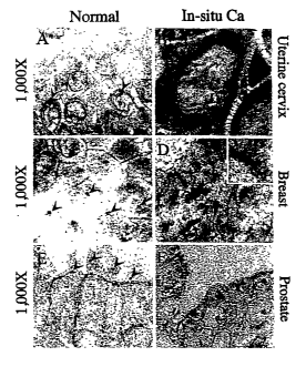
FIGS. lA-F are a series of micrographs that illustrate centrosome defects in
carcinoma in situ. Photomicrographs (1000X) of normal epithelium (lA, 1C, and
IE)
and iya situ carcinoma (1B, 1D and 1F) immunostained with antibodies to
pericentrin
3o to visualize centrosomes. In normal epithelia, centrosomes are round and
uniform in
size (arrowheads, lA, IC and lE) while in carcinoma iu situ they are larger
4
CA 02494928 2005-02-07
WO 2004/015394 PCT/US2003/025195
(arrowheads in 1B, 1D, 1F), multiple (1B), or structurally abnormal
(arrowheads in
1D and 1F). Nuclei are stained light blue with hematoxylin. The inset in 1D
shows
higher magnification of an elongated centrosome.
FIGS. 2A-C are a series of graphs that illustrate that centrosome defects are
prevalent in carcinoma ih situ. Centrosome defects are present in 62, 75 and
28
percent of CIC (2A), DCIS (2B) and PIN (2.C) lesions, respectively (N, normal
epithelia). First column (2A-C), cumulative defects; second column (A'-C'),
breakdown of centrosome defects by category (#, number, Sz, size, Sh, shape).
FIGS. 3A-L are a series of graphs that illustrate that the incidence of
1 o centrosome defects increases with increasing histologic grade. The
cumulative
incidence of centrosome defects in each pre-invasive lesion (left column)
includes
grades I-3 for CIC (3A, I-3) and low (3L) and high (3H) grades for DCIS (3E)
and
PIN (31). N identifies normal epithelium. Each subcategory of centrosome
defects
increases with grade including increased centrosome number (3B, 3F, 3J), shape
abnormalities (3C, 3C~ 3K), and size (3D, 3H, 3L).
FIGs. 4A-I are a series of photomicrographs (at left) and graphs (at right)
that
illustrate that mitotic spindle defects are common in CIC and DCIS. Examples
of
bipolar mitotic spindles imrnunostained with g-tubulin in CIC and DCIS (4A and
4C,
respectively). Examples of multipolar spindles (4B, CIC; 4D, 4F, DCIS) and
multiple spindles (4E, DCIS). Quantitative analysis of the number of bipolar
spindles
(x axis) and mulitipolar spindles (y axis) in each CIC lesion (4G) and DCIS
lesion
(4H). Each circle represents a single lesion. Filled circles represent lesions
with ten
or more mitoses and were included in the estimation of the extent of mitotic
spindle
defects in CIC and DCIS. On average 10% and 17% of the spindles, in CIC and
DCIS lesions with more than 10 immunostained spindles (red circles in 4G and
4H),
axe abnormal.
FIGS. SA-I are a series of photomicrographs (at left) and graphs (at right)
that
illustrate that centrosome abnormalities correlate with chromosome instability
in
carcinoma iya situ. Examples of ih situ hybridization reactions performed on
samples
of CIC (SB), DCIS (SD) and PIN (SF). Many cells have more than two signals for
chromosome #8 (arrowheads in SB, SD, SF) and thus exhibit chromosome
instability
(CIN+). Cells in adjacent normal epithelium (SA, SC, SE) rarely have more than
two
CA 02494928 2005-02-07
WO 2004/015394 PCT/US2003/025195
signals. Quantitative analysis of chromosomal instability (CIN+) in CIC (SG),
DCIS
(SH) and PIN (SI) lesions with normal centrosomes (SN) or abnormal centrosomes
(SA). CIN is present in most lesions with abnormal centrosomes and a small
fraction
of lesions lacking centrosome abnormalities.
FIGS. 6A-J are a series of photomicrographs (at top) and graphs (at bottom)
that illustrate centrosome and spindle defects and chromosome instability in
cell lines
derived from in situ lesions. Immunofluorescence images showing centrosomes
and
spindles in cell lines derived from normal epithelium (1560NPTX, 6A, 6B,
mitosis,
6E, interphase) and high grade PIN lesion from the same prostate gland
(1560PINTX,
6C, 6D, mitosis, 6F, 6C~ interphase). Quantification of this data shows that
1560PINTX has a 2-4-fold higher incidence of centrosome defects (6H), spindle
defects (61) and chromosome instability (6J) than1560NPTX.
FIG 7 is a schematic diagram depicting a centrosome-mediated model for
tumor progression.
15 FIG 8 is a diagram of the components of a cell-based scanning system. An
inverted fluorescence microscope is used 1, such as a Zeiss Axiovert inverted
fluorescence microscope that uses standard objectives with magnification of 1-
100x to
the camera, and a white light source (e.g., 100W mercury-arc lamp or 75W xenon
lamp) with power supply 2. There is an XY stage 3 to move the plate 4 in the
XY
2o direction over the microscope objective. A Z-axis focus drive 5 moves the
objective
in the Z direction for focusing. A joystick 6 provides for manual movement (if
desired) of the stage in the XYZ direction. A high resolution digital camera 7
acquires
images from each well or location on the plate. There is a camera power supply
8, an
automation controller 9, and a central processing unit 10. The PC 11 provides
a
25 display 12, and has associated software. The printer 13 provides for
printing of a hard
copy record.
Like reference symbols in the various drawings indicate like elements.
Detailed Description of the Invention
The invention includes methods of predicting the evolution of ih situ lesions
in
3o a patient by examining a microtubule organizing center of a cell in a
tissue sample. It
can also involve methods of predicting the development of cancer in a patient
by
CA 02494928 2005-02-07
WO 2004/015394 PCT/US2003/025195
examining a tissue sample for centrosome abnormalities. In addition, the
invention
includes methods of predicting the degree of aggressiveness of a cancer in a
patient by
examining a tissue sample for the degree of severity of centrosome
abnormalities.
These methods can be employed to predict cancer in any tissue that contains
centrosomes (e.g., prostate, breast, or uterine cervix, epithelial, lung,
colon, brain, and
all other carcinomas). A particular advantage of the invention is that its
methods can
be carried by human inspection or can be automated. Automation of tissue
preparation, examination for centrosome abnormalities, and analysis can
enhance the
speed, efficiency, and accuracy of the resulting predictions about cancer.
Methods of Analyzing Cells
There are numerous methods that can be used to analyze cells for centrosome
defects. Some examples of these methods are provided below.
First, a tissue sample is taken from a patient using standard biopsy
techniques.
Once taken, the sample can be prepared in a variety of ways. For example, it
can be
formalin-fixed and paraffin-embedded. Visualizing centrosomes can be enhanced
by
staining of the tissue (e.g., immunostaining with pericentrin antibodies).
Standard
histopathologic criteria can be applied to newly prepared hematoxylin- and
eosin-
stained sections to confirm the presence of carcinoma ih situ in the tissue
sample
(Rosai, J., Ake~~caya's SuYgical Pathology, (Mosby, New York), 1996). Once
stained,
or otherwise prepared for inspection, a microscope (e.g., high-resolution
light or
electron microscope) or other appropriate device for detecting subcellular
structures
can be used to detect and view centrosomes.
Reference tissue samples can be used to judge centrosome abnormality. For
example, samples of normal, healthy tissue of the same tissue type or origin
that
contain normal centrosomes can be compared to any tissue samples being assayed
for
the presence of centrosomal abnormalities.
One method of obtaining a reference tissue sample involves deriving both the
tissue sample to be assayed and the reference tissue sample from the same
tissue of
3o the same patient. For example, tissue samples can be taken from the same
prostate
gland of a patient, one sample from a location known to be normal and healthy,
and
the other from a location to be assayed.
CA 02494928 2005-02-07
WO 2004/015394 PCT/US2003/025195
Alternatively, the reference tissue sample can be taken from the same patient
at
an earlier point in time (analogous to dental records) to be used in the
future as a
reference. Or, it can be taken from a different patient whose tissue is known
to be
normal and healthy. Exemplary normal and healthy tissue samples can be
preserved
and used as references. A reference tissue known to be normal and healthy
could be
preserved for future comparison. Or, the appearance of a reference tissue
known to be
normal and healthy could be recorded onto another medium (e.g., an image on
paper,
a computer image) for visual, or other (e.g., automated or computer),
comparison to
the tissue to be assayed. Many other methods are possible.
1 o Alternatively, cell lines can be employed. For example, cell lines to be
compared (e.g., a normal, healthy cell line and a cell line to assayed) can be
grown on
glass coverslips in Defined Keratinocyte-SFM media containing 5% fetal bovine
serum and antibiotics. After permeabilization of cells in microtubule
stabilization
buffer containing 0.1% triton-X 100 cells were fixed in cold (-20°C)
methanol and
centrosomes immunostained as described in Pihan, G. A., et al. (Cancer Res,
58:3974-85, 1998). Immunofluorescence and FISH can also be employed.
Some examples of centrosomal abnormalities include:
(1) centrosomes with diameters greater than twice the diameter of centrosomes
present in normal, healthy samples of the same tissue type or origin,
20 (2) centrosomes in which the ratio of a centrosome's greatest and smallest
diameter exceeds about 1.5-2,
(3) tissues in which there are more than two centrosomes per cell in more than
about 5% of the cells examined or yielding a ratio of centrosomes to nuclei of
greater than about 2.5,
25 (4) abnormally shaped centrosomes,
(5) absence of centrosomes,
(6) centrosomes that are organized as multiple small dots in comparison to the
organization of normal, healthy centrosomes, and
(7) increased levels (or concentrations) of pericentrin within a cell.
3o In general, centrosomal abnormalities can include any difference from the
centrosomes in samples of normal, healthy tissue of the same tissue type or
origin.
Differences can be in shape, size, color, orientation, proximity to other
cellular or
CA 02494928 2005-02-07
WO 2004/015394 PCT/US2003/025195
subcellular structures, timing of appearance, movement over time, or any other
aspect
of appearance or behavior, either at one sampling time or over multiple
sampling
times.
The frequencies of centrosomal abnormalities in different tissue samples can
be compared. For example, the frequency of centrosomal abnormalities in a
normal,
healthy reference sample can be compared to the corresponding frequency in the
tissue being assayed. The increased probability of developing cancer or of
developing
a more aggressive cancer correlates with the difference in frequency of
centrosomal
abnormalities between the reference tissue sample and the tissue sample being
assaying.
Mitotic spindles can also be examined using similar methods as those used to
visualize or detect centrosomal abnormalities. For example, g-tubulin can be
used to
stain mitotic spindles in archival formalin-fixed paraffin-embedded tissues
because it
decorates spindle poles while a large fraction of a and b tubulins are
cytoplasmic and
obscure the spindle microtubule signal.
Automated Centrosome Anal
The invention includes automation of any of the above aspects of sampling,
examining, or analyzing centrosomal abnormalities. For example, a computer can
be
2o programmed to compare images of normal, healthy centrosomes (e.g., shape,
color,
size, number, orientation, appearance, behavior, etc.) to images of
centrosomes from a
patient's tissue sample or cell culture. These images can be generated by
preparing a
the cell tissues or cell cultures in a variety of ways to highlight the
centrosomal aspect
or aspects of interest so that they can be visualized by a microscope, or
other device
for visualizing or detecting particular characteristics of centrosomes.
Preparation of
cell tissues or cell cultures can involve such techniques as staining using a
two-color
immunofluoresence, two-color immunohistochemistry, or both simultaneously. In
addition, automation can allow one to greatly increase'the volume of analyses
that can
be made. For example, one could use punch-embedded paraffin slides to analyze
100
so or more tumors per slide fox centrosomal abnormalities. FIG. 8 depicts an
example of
an automated system than can be used to examine and analyze tissue or cell
samples
for centrosomal abnormalities.
CA 02494928 2005-02-07
WO 2004/015394 PCT/US2003/025195
For example, cells from a patient to be examined for centrosomal abnormalities
can be cultured using standard cell culture techniques. Then, these cells can
be loaded
onto an automated system. The system can automatically prepare the cell
samples by
staining, or some other means of enhancing visualization. Then, the system can
examine the samples using a microscope. The microscope can visualize
characteristics of interest in the samples, and then transmit information
regarding
those characteristics to a computer. The computer can then compaxe
characteristics of
interest in the cells (e.g., shape, size, color, or number of centrosomes) to
reference
characteristics (e.g., of normal, healthy cells, or of previously analyzed
samples taken
~ o from the same patient). The computer can be programmed to decide whether
or not
the centrosomes in one sample are sufficiently similar to or different from
those in a
different sample to allow a prediction regarding a cancer, and, if so, to
identify a
particular prediction.
The invention includes the use of a high magnification, high resolution, three-
~5 dimensional acquisition microscope. The microscope can be a microscope
capable of
taking pictures in a Z-series that can visualize centrosomes in all planes of
a cell. The
light source can be white light, fluorescence, or multiple wavelength
fluorescence.
The invention can use conventional immunohistochemical methods or
immunofluorescence methods, using conventional methods for preparing samples
for
2o immunohistochemistry or immunofluorescence.
As a practical example, a patient could provide a tissue sample at age 20,
which
could be examined and analyzed using an automated system, and the resulting
centrosomal information stored in her medical records. Then, the patient could
provide a second tissue sample at age 25 (and at subsequent intervals), which
could be
25 examined and analyzed again, and then compared to results for the original
sample. A
change in centrosomal characteristics (e.g., a statistically significantly
greater ratio of
centrosomes to nuclei in the latter sampled tissue compared to the earlier
sampled
tissue) could result in a prediction that the patient is undergoing early
development of
cancer in that tissue. The patient could then begin cancer therapy earlier
than if she
3o had waited unfit symptoms of cancer appeared. Her chances for survival
might thus
be increased.
to
CA 02494928 2005-02-07
WO 2004/015394 PCT/US2003/025195
There are many ways in which the methods of this invention can be
automated. These include any of the methods disclosed in WO 00/26408 and in
United States Patent Nos. 6,SS3,135, 6,418,236, 6,372,183, 6,330,349,
6,328,567,
6,317,617, 6,215,892, 6,200,781, 6,190, I70, 6,127, I33, 6,088,473, 6,048,314,
s 6,011,862, 5,984,870, 5,812,419, 5,790,690, 5,717,602, S,6S6,499, S,6S0,122,
5,631,165, 5,620,898, S,S26,258, and S,S09,042, all of which are hereby
incorporated
by reference in their entirety. Examples of commercially available systems
that can
be used to automate examination and analysis of centrosomal abnormalities in
tissue
or cell samples are the Discovery-1TM or Discovery-TMATM systems(along with
o MetaMorph~, MetaFluor , or MetaVueTM systems) from Molecular Devices
Corporation.
Centrosome Abnormalities
Chromosomal instability (CIN) is believed to be caused by continuous
~ 5 chromosome missegregation during mitosis and is the most common form of
genetic
instability in human cancer (Lengauer C., et al., Nature, 396:643-9, 1998).
Together
with structural chromosome changes, CIN is thought to be important to promote
Darwinian genornic evolution characteristics of cancer (Cahill, D. P., et al.,
Trerads
Cell Biol, 9:M57-60, 1999). The combined effect of C1N and chromosome breakage
2o and misrepair can explain the progressive loss of tumor suppressor genes
and
accumulation of extra copies of tumor promoting genes (oncogenes, cell
survival
genes) characteristic of cancer. In fact, loss of heterozygocity in cancer
primarily
affects whole chromosomes or large chromosomal domains suggesting that it
results
from non-disjunction of whole normal or structurally abnormal chromosomes
25 (Thiagalingam, S., et al., Pros Natl Acad Sci USA, 98:2698-702, 2001). CIN
is
thought to facilitate the inexorable evolution of cancers toward cellular
states that
support tumor cell growth, dissemination, and resistance to therapy (Lengauer
C., et
al., Nature, 396:643-9, 1998; Cahill, D. P., et al., Trefads Cell Biol, 9:M57-
60, 1999;
Lengauer, C., et al., Nature, 386:623-7, 1997; Pihan, G A., et al., Cancer
Res,
30 58:3974-8S, 1998; Pihan, G A., et al., Semira CafZCer-Biol, 9:289-302,
1999). A
common element in the chain of events associated with loss of fidelity in
chromosome
segregation is centrosome dysfunction (for review, see Pihan, G A., et al.,
Senain
11
CA 02494928 2005-02-07
WO 2004/015394 PCT/US2003/025195
Cancer Biol, 9:289-302, 1999; Brinkley, B. R., Trends Cell Biol, 11:18-21,
2001;
Doxsey, S., Nat Rev Mol Cell Biol, 2:688-98, 2001; Lingle, W L., et al., Curr
Top
Dev Biol, 49:313-29, 2000; Marx, J., Science, 292:426-9, 2001; Winey, M., Curr
Biol,
9:8449-S2, 1999).
Centrosomes are the primary microtubule-organizing centers in animal cells,
and they contribute to the organization of microtubule spindles in mitosis and
control
progression through cytokinesis and entry into S phase (Doxsey, S., Nat Rev
Mol Cell
Biol, 2:688-98, 2001; Hinchcliffe, E. H., et al., Genes Dev, 15:1167-81, 2001;
Khodjakov, A., et al., J Cell Biol, 153:237-42, 2001; Piel, M., et al.,
Science,
291:1SS0-3, 2001). Centrosome defects have been detected in aggressive
carcinomas
of multiple origins (Pihan, G A., et al., Cancer Res, 58:3974-8S, 1998;
Lingle, W. L.,
et al., Proc Natl Acad Sci USA, 9S:29S0-S, 1998). The invention is based, at
least in
part, on the discovery that centrosome defects in a tissue are strongly
correlated to
whether or not that the tissue will develop cancer, the evolution of such a
cancer, and
~5 the resulting severity of that cancer.
The established role of centrosomes in organizing mitotic spindles suggested a
model in which tumor cells with multiple centrosomes organize multipolar
spindles
that in turn missegregate chromosomes and contribute to genetic instability.
This
phenomenon can occur in diploid cells or in cells that previously failed in
cell division
2o to create polyploid cells with excess centrosomes (Meraldi, P., et al.,
Embo J, ?1:483-
92, 2002). Despite the occurrence of centrosome defects in human cancers, and
their
important role in the assembly of mitotic spindles and chromosome segregation,
a role
for centrosomes in the earliest steps of human tumor development has not
elsewhere
been established. The invention is based, at least in part, on the discovery
that
25 centrosome defects and genetic instability occur in some low grade prostate
tumors
and are present prior to development of aggressive tumors. However, it appears
that
centrosome defects have not previously been linked to the earliest stages of
human
cancer where they would have the highest potential to contribute to the early
stages of
the disease, and possibly serve as prognostic markers for tumor development
and
3o therapeutic targets for treatment.
Pre-invasive cancer lesions in humans known as carcinoma in situ provide a
unique opportunity to directly examine this issue in some detail. This
invention is
12
CA 02494928 2005-02-07
WO 2004/015394 PCT/US2003/025195
based, at least in part, on the recognition that centrosome defects occur in
carcinomas
in situ from multiple tissue sources and co-segregate with other tumor-like
features
associated with centrosome dysfunction, such as spindle abnormalities,
cytologic
changes, and chromosomal instability
Centrosome Defects and Precancerous Lesions
Experimental results upon which this invention is based, at least in part,
demonstrate that centrosome defects play a critical role in carcinogenesis.
Centrosome defects occur frequently in advanced forms of some of the most
common
~ o human cancers, and contribute to genetic instability by impairing the
fidelity of
chromosome segregation during mitosis (Lengauer C., et al., Nature, 396:643-9,
1998; Cahill, D. P., et al., T~ehds Cell Biol, 9:M57-60, 1999; Brinkley, B.
R., Trends
Cell Biol, 11:18-21, 2001; Doxsey, S., Nat Rev Mol Cell Biol, 2:688-98, 2001;
Marx,
J., Scier2ce, 292:426-9, 2001; Lingle, W. L., et al., Proc Natl Acad Sci USA,
95:2950-
~5 5, 1998). Carcinoma in situ is the immediate precursor of invasive
epithelial cancers
and it shares some, but not all, genotypic and phenotypic characteristic of
invasive
cancer (Bostwik, D. G, Semira Urol Oncol, 17:187-98, 1999; Shultz, L. B., et
al., CuYr-
Opin Oncol, 11:429-34, 1999; Wolf, J. K., et al., Cancer Invest, 19:621-9,
2001). The
experimental results disclosed herein show that centrosome defects are present
at the
2o earliest morphologically recognizable stages of tumor development in some
of the
most common human cancers. They provide a mechanistic explanation for the
commonly observed CIN and aneuploidy observed in most lesions found in
experimental models of carcinogenesis and human carcinoma in situ (Bulten, J.,
et al.,
Am JPathol, 152:495-503, 1998; Levine, D. S., et al., Proc Natl Acad Sci USA,
25 88:6427-31, 1991; Li, R., et al., Py~oc Natl Acad Sci USA, 94:14506-11,
1997; Wang,
X. W., et al., Proc Natl Acad Sci USA, 96:3706-11, 1999; Weinberg, D. S., et
al., Af°ch
Pathol Lab Med, 117:1132-7, 1993). These data demonstrate the presence of
centrosome defects in the generation of genetic instability during the early
stages of
the tumorigenic process.
3o Furthermore, centrosome defects correlate with the histologic/cytologic
grade
of the in situ lesion, and the centrosome has a role in the induction of the
morphologic
phenotype characteristic of carcinoma ira situ. Centrosomes have been shown to
play
13
CA 02494928 2005-02-07
WO 2004/015394 PCT/US2003/025195
a role in cell polarity, shape, and motility, all of which are perturbed in
iya situ cancers.
Moreover, the presence of mitotic spindle defects in many carcinoma ira situ
of the
uterine cervix (CIC, or carcinoma iya situ of the cervix) and carcinoma in
situ of the
female breast (DCIS, or ductal carcinoma in situ) lesions, and the co-
segregation of
centrosome abnormalities with CIN in these lesions, show that centrosome
defects
have an important functional impact in in situ carcinoma.
The experimental results herein demonstrate a role for centrosome defects in
the development of aggressive tumors, rather than those that remain benign.
For
example, there is a high prevalence of centrosome abnormalities in lesions
with a high
1 o rate of progression to high-grade cancer (DCIS (ductal carcinoma in situ)
and
CIC(carcinoma ifi situ of the cervix)), and a low prevalence of centrosome
defects in
lesions associated with progression to low grade invasive cancers, such as
prostate
intraepithelial neoplasia (PLN). It has been shown that most invasive cancers
of the
breast and uterine cervix are aggressive high-grade cancers. Because DCIS and
CIC
are usually indistinguishable cytologically from aggressive cancers it is
believed that
they give rise to these aggressive cancers. In contrast, cancers of the
prostate are
usually low-grade cancers consistent with the low-grade appearance of most PIN
lesions. These results support the centrosome-mediated model of tumorigenesis
where centrosome defects induce dramatic and persistent changes in chromosome
2o number, thereby shuffling the genome and allowing selection of the most
aggressive
phenotypes such as those seen in invasive cancers.
The invention is based, at least in part, on the discovery that the presence
of
centrosome abnormalities in cells at the earliest stages of disease allows
prediction of
the evolution of i~c situ lesions into high-grade invasive cancers. This
discovery is of
particular interest for the management of prostate cancer since the majority
of these
tumors are biologically low grade. Currently, these cancers are often treated
by
prostatectomy because there is no effective prognostic indicator of aggressive
disease.
Since centrosome abnormalities predict the development of high grade cancer,
such
prediction can provide a sorely needed surrogate marker for high grade cancer.
3o Centrosome defects correlate with aggressive disease, as can be shown by
examining
PIN lesions from patients who subsequently progressed to invasive cancer.
14
CA 02494928 2005-02-07
WO 2004/015394 PCT/US2003/025195
Centrosome defects in early (precancerous) lesions are worse in lesions that
subsequently progress to worse, or more aggressive, tumors.
.An interesting observation was the presence of low, yet measurable, levels of
centrosome defects in morphologically normal epithelium adjacent to CIC
lesions
(FIG 2A). This may be due to the presence of human papillomavirus infection.
It is
well established that papillomavirus is the cause of nearly all carcinomas of
the
cervix, and is present in all precursor lesions (Monger, K., Front Biosci,
7:d641-9,
2002). Moreover, it has recently been demonstrated that papillomavirus can
rapidly
induce centrosorne abnormalities in squamous epithelial cells (Duensing, S.,
et al.,
7o Biochim BiophysActa, 2:M81-8, 2001).
Another important discovery is the functional impact of abnormal centrosomes
in in situ carcinomas. It has been demonstrated in experimental systems and
cell lines
(Brinkley, B. R., Trends Cell Biol, 11:18-21, 2001; Ring, D., et al., J Cell
Biol,
94:549-56, 1982) that multipolar spindles formed by supernumerary centrosomes
may
~ 5 coalesce to form bipolar spindles, mitigating the functional consequences
of
centrosome defects on chromosome segregation. Whether coalescence occurs in
ira
situ cancers is not know. However, even if it does, it is not sufficient to
completely
suppress the effect of supernumerary centrosomes on spindle multipolarity.
Whether centrosome defects are cause or consequence of the in situ carcinoma
2o phenotype, centrosomal abnormalities can be predictive of the development
of cancer.
Thus, identification of centrosomal abnormalities can be important for
predictive
testing and effective cancer-specific therapeutic interventions. There are
many ways
in which centrosome defects can arise. These include changes in proteins
involved in
cell cycle control, in centrosome structure or function, and in DNA repair.
For
25 instance, mutation or elimination of p53 (Fukasawa, K., et al., Science,
271:1744-7,
1996; Tarapore, P., et al., OrZCOgene, 20:3173-84, 2001; Wang, X. J., et al.,
Oncogene,
17:35-45, 1998), or pS3 downstream effectors or regulators, such as Mdm2
(Carroll,
P. E., et al., Oracogene, 18:1935-44, 1999), p21 Waf/Cipl (Fukasawa, K., et
al.,
Science, 271:1744-7, 1996; Carroll, P. E., et ccl., Oncogene, 18:1935-44,
1999; Mantel,
so C., et al., Blood, 93:1390-8, 1999), or GADD45 (Wang, X. W , et al., Proc
Natl Acad
Sci USA, 96:3706-11, 1999; Hollander, M. C., et al., Nat Genet, 23:176-84,
1999),
induce centrosome abnormalities. Abrogation of postmitotic pS3-dependent
CA 02494928 2005-02-07
WO 2004/015394 PCT/US2003/025195
checkpoints may be critical in allowing tetraploid cells with supernumerary
centrosomes to continue to cycle (Andreassen, P., R., et al., Mol Biol Cell,
12:1315-28,
2001; Khan, S. H., et al., CahcerRes, 58:396-401, 1998; Lanni, J. S., et al.,
Mol Cell
Biol, lB:lOSS-64, 1998). Similarly, alteration in the levels of centrosome-
associated
proteins such as pericentrin (Pihan et al., Cancer Res, 61:2212-9, 2001;
Purohit, A., et
al., J Cell Biol, 147:481-92, 1999), g-tubulin (Shu, H. B., et al., J Cell
Biol, 130:1137-
47, 1995), aurora (Meraldi, P., et al., Embo J, 21:483-92, 2002; Bischoff, J.
R., et al.,
Embo J, 17:3052-6S, 1998; Zhou, H., et al., Nat Genet, 20:189-93, 1998), polo
(Corm
et al., Cancer Res., 60:6826-31), TACC (gaff, J. W., et al., Cell, 57:611-9,
1989), and
RanBP (Wiese, C., et al., SciefZCe, 291:653-6, 2001) lead to abnormal
centrosomes.
Moreover, mutation or functional abrogation of proteins involved in DNA repair
such
as Xrcc3 (Griffin, C. S., et al., Nat Cell Biol, 2:757-61, 2000), Xrcc2
(Griffin, C. S., et
al., Nat Cell Biol, 2:757-61, 2000), BRCA1 (Bertwistle, D., et al., Beast
Cancer Res,
1:41-7, 1999; Xu, X., et al., Mol Cell, 3:389-9S, 1999), BRCA2 (I~raakman-van
der
Zwet, M., et al., Mol Cell Biol, 22:669-79, 2002; Tutt, A., et al., Curr Biol,
9:1107-10,
1999), Mrell (Yamaguchi-Iwai, Y, et al., Embo J, 18:6619-29, 1999), or DNA
polymerase beta (Bergoglio, V , et al., Cancer Res, 62:3511-4, 2002), or
genome
damage signaling proteins such as ATR (Smith, L., et al., Nat Geyaet, 19:39-
46, 1998)
can also lead to centrosome abnormalities. Lastly, centrosome abnormalities
can also
2o arise by mutation of the adenomatous polyposis coli gene (APC) whose
product
interacts with microtubules (Foddle, R., et al., Nat Cell Biol, 3:433-8,
2001), by
cytokinesis failure (Doxsey, S., Nat Genet, 20:104-6, 1998), and by ectopic
assembly
of centrosome components into acentriolar microtubule organizing centers
(Doxsey,
S., Nat Rev Mol Cell Biol, 2:688-98, 2001; Pihan, G . A., et al., Cancer Res,
61:2212-
9, 2001; Purohit, A., et al., J Gell Biol, 147:481-92, 1999).
EXAMPLES
The invention is further described in the following examples, which do not
limit the scope of the invention described in the claims. The general
experimental
3o procedures are described first.
16
CA 02494928 2005-02-07
WO 2004/015394 PCT/US2003/025195
Experimental Pxocedures
lmmunohistochemical Staining and Analysis
Fornialin-fixed paraffin-embedded tissue from carcinoma in situ of the uterine
cervix, female breast, and male prostate was selected from the files of the
Pathology
Department at UMass Memorial Health Care. Samples were immunostained with
pericentrin antibodies as described (Pihan, G. A., et al., Cancers Res,
58:3974-85,
1998; Pihan et al., Cancer Res, 61:2212-9, 2001; Purohit, A., J Cell Biol,
147:481-92,
1999). Standard histopathologic criteria was applied to newly prepared
hematoxylin
and eosin stained sections to confirm the presence of carcinoma in situ in the
~o specimen (Rosai, J., Akerman's Surgical Pathology, (Mosby, New York),
1996).
Centrosomes were considered abnormal if they had a diameter greater than twice
the
diameter of centrosomes present in normal epithelium within the same section,
if the
ratio of a centrosome's greatest and smallest diameter exceeded 2, or if there
were
more than two centrosomes in more than 5% of the cells examined (Pihan et al.,
Cancer Res., 61:2212-2219, 2001). g-tubulin was chosen to stain mitotic
spindles in
archival formalin fixed paraffin embedded tissues because it decorates spindle
poles
while a large fraction of a and b tubulins are cytoplasmic and obscure the
spindle
microtubule signal. Multipolar mitoses, an obvious consequence of
supernumerary
centrosomes, are common in carcinoma cell lines with abnormal centrosomes as
we
2o and others have previously shown (Pihan et al., Cancers Res., 58:3974-85,
1998; Sato
et al:, Clin. Cancer Res., 5:963-70, 1999; Lingel et al., Am. J. Pathol.,
155:1941-51,
1999; Saunders et al., PNAS, 97:303-8, 2000).
Chromosomal Instabilit~Analysis
Tissue sections parallel to those used for pericentrin immunohistochemistry
were used to stain for the centromeres of chromosome 1 and 8 (Pihan et al.,
Cancer
Res., 58:3974-85, 1998). Briefly, after de-paraffinization, sections were co-
denatured
with biotinylated centromeric probes specific for chromosomes I or 8 and
hybridized
overnight at 37°C in a Hybrite oven (Vysis, Chicago, IL) in the
hybridization buffer
3o recommended by the probe manufacturer. After appropriate stringency washes
sections were placed on the automatic immunostainer and an ABC/DAB protocol
similar to the one used above for immunohistochemistry was used to reveal the
17
CA 02494928 2005-02-07
WO 2004/015394 PCT/US2003/025195
hybridized probe. Nuclei were lightly counterstained with hematoxylin. For
quantitative analysis, the number of hybridization signals in 100 to 200
nuclei from in
situ carcinoma and morphologically normal adjacent epithelium was recorded
(Pihan
et al., Cancer Res., 58:3974-85, 1998). Using these probes it has been shown
that
normal diploid tissue has 10-15% cells with more than 3 signals per nucleus
(Pihan et
al., Caracer~ Res., 58:3974-85, 1998; Bulten et al., Am. J. Pathol., 152:495-
503, 1998).
In tissue sections some nuclei are truncated leading to artificially increased
numbers
of diploid cells with apparently less than two signals per nuclei. For this
reason,
computed signal gains (greater than two) were computed, and not apparent
losses.
Due to this limitation, no attempt was made to obtain an absolute measure of
chromosome instability in sections, as it can be done on cell lines (Lengauer
et al.,
Nature, 386:623-7, 1997; Pihan et al., Cahce~ Res., 58:3974-85, 1998). Rather,
tumors with likely aneuploidy/CIN were defined as those in which the fraction
of
nuclei with more than two signals exceeded 20% (Bulten et al., Am. J. Pathol.,
152:495-503, 1998), and used this measurement as an index of chromosome
instability/aneuploidy.
Analysis of Cell Lines Derived from PIN or Normal Prostate Epithelium
During attempts to establish isogenic pairs of neoplastic and normal
epithelial
2o cell lines from patients with prostate cancer at NCI, one pair of normal
and high grade
PIN cell lines was derived from the same patient (Bright et al., Cancer Res.,
57:995-
1002, 1997). Pathologic examination of the donor prostate showed only normal
glands and extensive high grade PIN, but no invasive carcinoma. To study
centrosomes, cell lines were grown on glass coverslips in Defined Keratinocyte-
SFM
media containing 5% fetal bovine serum and antibiotics. After permeabilization
of
cells in rnicrotubule stabilization buffer containing 0.1 % triton-X 100 cells
were fixed
in cold (-20°C) methanol and centrosomes immunostained as described
(Pihan et al.,
Cancer Res., 58:3974-85, 1998). Ira situ hybridization with probes to
chromosomes 1
and 8 were carried out as previously described (Pihan et al., Caracef-Res.,
58:3974-85,
1998). Tm_m__unofluorescence and FISH were carried out in four different
experiments
and results averaged.
1s
CA 02494928 2005-02-07
WO 2004/015394 PCT/US2003/025195
Example 1. Centrosome Defects Occur in a Significant Number of Pre-Invasive
Cancerous Lesions
Carcinoma in situ of the uterine cervix (CIC), the female breast (DCIS), and
the male prostate (PIN) was studied. These lesions are precursors of the most
common human cancers. Moreover, breast and prostate cancers are the second
leading cause of cancer death in women and men, respectively.
Using antibodies to the centrosome protein pericentrin (Doxsey et al., Cell,
76:639-50, 1994), we examined microtubule organizing centers in sections of
tumor
and nontumor tissues as described (Pihan et al., Cancer Res., 58:3974-85,
1998;
1 o Pihan et al., Cancer Res., 61:2212-2219, 2001). Several distinct
centrosome
abnormalities were detected in these lesions, including supernumerary
centrosomes
(FIG. 1B arrowheads), abnormally-shaped centrosomes, such as elongated or cork-
screw forms (FIG. ID and F) and centrosomes of larger diameter than those in
normal
epithelium within the same tissue section (FIG. 1B and D). Also observed were
cells
that apparently lacked centrosomes, or whose centrosomes were organized as
multiple
small dots. Because this phenotype could partly be a consequence of cell
truncation
during tissue sectioning, these were not scored as defects even though a
similar
phenotype was observed in tumor cell lines. Quantification of centrosome
defects in
all precancerous lesions demonstrated that 36-72% had abnormal centrosomes
(FIG. 2A-C), while nontumor cells had undetectable or low levels of defects
(FIG. 2A-C). Centrosome defects were more prevalent in DCIS and CIC lesions
than
in PIN lesions. Differences in centrosome abnormalities between DCIS and CIC,
on
one hand, and PIN, on the other, are consistent with differences in
histological,
cytological, and genetic features of these lesions. DCIS and CIC show a high
degree
of nuclear atypia, cytologic disarray, loss of cell polarity, and genetic
instability. In
fact, on cytologic features alone, they are often indistinguishable from
invasive breast
and cervical cancers (Cram et al., J. Cell. Biochem. Suppl., 23:71-9, 1995;
O'Connell
et al., Breast Cancer Res. Treat., 32:5-12, 1994). This is in contrast to PIN
lesions
that show preservation of Bell polarity, and glandular architecture, and can
only be
3o distinguished from normal glands by rather subtle changes in nuclear and
nucleolar
features.
19
CA 02494928 2005-02-07
WO 2004/015394 PCT/US2003/025195
In summary, it was demonstrated that centrosome abnormalities occur in pre-
invasive lesions, and that they are more common in CIC and DCIS than in PIN
lesions. Similar results were obtained using g-tubulin antibodies in
interphase cells,
although fewer defects were observed than with pericentrin antibodies.
Examble 2. The Incidence of Centrosome Defects Tncreases with Higher
Histolo~ic
Grade of In Situ Carcinomas
In situ carcinomas of different histologic/cytologic grade differ in their
associated risk of progression to invasive carcinoma. A 2-4-fold increase in
the
o incidence of centrosome defects with increasing histologic/cytologic grade
in all three
precancerous lesions was observed (FIG. 3). Most DCIS lesions exhibited
centrosorne defects (FIG. 3E), while only 36% of high-grade PIN lesions had
this
phenotype (FIG. 31). The surprisingly high incidence of centrosome defects in
DCIS
is consistent with the cytologic similarity between DCIS and invasive breast
cancer
(Pihan et al., Cancer Res., 58:3974-85, 1998). CIC lesions of histologic grade
2 and 3
(collectively "high grade" lesions) showed a high incidence of centrosome
defects,
nearly as high as that seen in DCIS lesions (FIG. 3A). Centrosome
abnormalities in
all three types of lesions was greater in those lesions associated with a
higher
propensity to evolve into invasive carcinoma. This trend demonstrates an
important
2o role for centrosomes in generating the cytologic and genetic changes that
occur during
tumor progression.
Example 3. Mitotic Spindle Abnormalities are Frequent in Carcinoma Ira Situ
One expected consequence of supernumerary centrosomes in mitotic cells is
the development of multipolar mitotic spindles (Pihan et al., Cancer' Res., 5
8:3974-85,
1998; Purohit et al., J. Cell. Biol., 147:481-92, 1999). To identify abnormal
spindles,
sections were stained with g-tubulin, which provided the best marker for
spindle poles
in this immunohistochemical procedure (see Experimental procedures). Although
the
total number of mitotic figures was generally low, mitotic spindles were found
in 74%
(29/39) of CIC lesions, 35% (12/34) of DCIS lesions, and in none of the PIN
lesions
(0/42) and nontumor cells. The low incidence of spindles in PIN lesions is
likely the
result of delayed fixation and the relatively slow growth of prostate tumor
cells
CA 02494928 2005-02-07
WO 2004/015394 PCT/US2003/025195
compared with the other ire situ lesions (DCIS, CIC). Of the tumors with
spindles,
75% (9/12) of DCIS and 34% (10/29) of CIC had at least one abnormal spindle
(FIG. 4H and G). Defective spindles included multipolar spindles (3 or more
poles,
FIG. 4B, D, and F), multiple bipolar spindles in single cells (FIG. 4E), and
asymmetric bipolar and multipolar spindles (FIG. 4D and F).
To get a measure of the extent of this phenotype in in situ lesions, and to
avoid
the inherent bias introduced in the data by low spindle counts, abnormal
spindles in
cells with 10 or more spindles were counted. The average number of multipolar
spindles in cases so selected was 10.1 +/- 7.8 and 16.6 +/- 4.1, respectively
(FIG. 4I).
1 o Monopolar spindles were also detected, but they could not be authenticated
due to the
compounding effect of truncation artifacts induced by tissue sectioning.
Mitotic
figures were infrequently observed in normal epithelium adjacent to lesions.
This is
most likely due to the low mitotic rate of these tissues, but in all cases
they appeared
structurally normal (symmetric, bipolar, n=4). Because of the low incidence of
spindles in nontumor tissues, and to control for the nonspecific effects of
the
immunohistochemical procedure on mitotic cells, results from in situ
carcinomas were
compared with those of a highly proliferative epithelium. In biopsies from
patients
with celiac spree, a form of malabsortion, the small intestinal epithelium has
increased mitotic activity due to increased rates of mucosal regeneration. In
these
2o biopsies, abnormal mitoses (n= 45) were never observed, indicating that the
observations in in situ carcinomas are not an artifact of staining in archival
tissue
biopsies.
Example 4. Centrosome Defects Correlate with C1N in Precancerous Lesions
Both chromosome instability (Lengauer C., et al., Nature, 396:643-9, 1998;
Pihan et al., Cancer Res., 58:3974-85, 1998; Lingle et al., PNAS, 95:2950-5,
1998)
and centrosome defects are common features of epithelial cancers (Marx,
Science,
292:426-9, 2001; Lingle et al., PNAS , 95:2950-5, 1998; Pihan et al., Cancer
Res.,
61:2212-9, 2001; Lingle et al., Ana. J. Pathol., 155:1941-51, 1999). To
determine
3o whether a correlation exists between centrosome defects and CIN in
carcinoma in
situ, consecutive serial tissue sections were examined for these anomalies
(for
methods, see Pihan et al., CanceY Res., 58:3974-85, 1998; Ghadami et al.,
Genes
21
CA 02494928 2005-02-07
WO 2004/015394 PCT/US2003/025195
Chr ofnosonaes Cancer, 27:183-90, 2000; Pihan et al., Cancer Res., 61:2212-9,
2001;
Bright et al., Cancer Res., 57:995-1002, 1997).
While CIN was observed in many in situ lesions, it was never seen in normal
epithelium in the same tissue section (FIG. SA, C, and E). Moreover, in all
three in
situ carcinomas there was a statistically significant non-random association
(Fisher
exact test p < 0.005) between centrosome defects and CIN (FIG. SG-I). In fact,
most
lesions with centrosome defects showed CIN (63-71 %, FIG. 5). Conversely, the
fraction of cases that lacked centrosome defects, lacked CIN (81-95%, FIG. 5).
This
correlation between centrosome defects and CIN was significant despite the
vastly
~o different degrees of centrosome defects between DCIS, CIC, and PIN (FIG.
2).
Interestingly, there were more lesions that had centrosome defects and no CIN
(~30%) than lesions with CTN and no centrosome defects (~10-20%), showing that
centrosome defects precede CIN in the progression of the tumor-like phenotype
in
precancerous lesions (Pihan et al., Cancer Res., 58:3974-85, 1998; Doxsey,
Nat. Rev.
~5 Mol. Cell. Biol., 2:688-98).
Thus, centrosome abnormalities can be used to predict C1N and the
development and progression of a cancer.
Example 5. Centrosome Abnormalities and CIN in Cell Lines Derived from PIN and
Normal Tissues
One of the only known in situ carcinoma cell lines available (Bright et al.,
Cancer Res., 57:995-1002, 1997) was investigated for centrosome defects and
CIN.
Cell lines provide a better quantitative measure of these features and can
ultimately be
used to examine the molecular mechanism responsible for centrosome
abnormalities.
A line derived from a high-grade PIN lesion (1560PINTX) and a control line
derived
from normal prostate epithelium (1560NPTX) both originated from the same
surgically-excised prostate gland (Bright et al., Cancer Res., 57:995-1002,
1997).
3o hnmunofluorescence analysis using pericentrin antibodies to detect
centrosome
defects revealed a significantly higher incidence of centrosome abnormalities
in PIN
cells than in normal cells (~4-fold higher, FIG. 6H). As in tumors, the
incidence of
22
CA 02494928 2005-02-07
WO 2004/015394 PCT/US2003/025195
multipolar spindles paralleled the incidence of centrosome defects, being
higher in
PIN cells than in normal cells (FIG. 6I). The level of CIN was also
consistently
higher in PIN-derived cells compared with controls (FIG. 6J).
Thus, centrosome abnormalities can be used to predict CIN and the
development and progression of a cancer (e.g., PIN cells).
Example 6. Diagnosis of Prostate Cancer
The etiology of prostate carcinoma is unknown. Understanding the
fundamental cellular mechanisms involved in disease onset and progression is
- essential for designing methods for the detection and treatment of this
major form of
human cancer. This invention allows the development of early and effective
prognostic methods for aggressive disease and production of novel therapies
based on
the identification of new targets for prostate cancer.
Prostate tumor virulence correlates with aberrant cytoarchitecture (Gleason
~ 5 grades 4, 5) and high grade tumors exhibit genetic instability. However,
little is
known about the molecular and biologic basis of these aberrant cellular
features.
Centrosomes and associated microtubules play a critical role in mitosis by
coordinating spindle assembly and cytokinesis with chromosome segregation and
in
interphase by regulating cell polarity and shape. All these processes are
disrupted in
2o prostate carcinoma. Several significant observations demonstrate that
centrosomes
confiribute to all known cellular and genetic changes in prostate cancer.
Centrosome
defects are present in pre-invasive lesions and become more severe during
tumor
progression, paralleling changes in Gleason grade and genetic instability.
Overexpression of the centrosome protein pericentrin produces features
25 indistinguishable from prostate tumor cells and induces or exacerbates
prostate cell
transformation in vitro. The novel discovery of centrosome defects and
elevated
pericentrin levels in prostate carcinoma and pre-invasive lesions shows a
previously
unexplored mechanism for generating the cellular and genetic changes that
occur
during prostate cancer progression. The observation that pericentrin interacts
with
3o several kinases (PKA, PKC, and others) that are themselves implicated in
cancer led
to the discovery that the oncogenic potential of pericentrin results from loss
of
pericentrin's interaction with these kinases.
23
CA 02494928 2005-02-07
WO 2004/015394 PCT/US2003/025195
The majority of patients diagnosed with prostate cancer have clinically
indolent tumors, while a minority develops more aggressive, often fatal
cancer. An
effective prognostic test could eliminate the unnecessary treatment of
patients with
indolent disease, target patients with aggressive disease for early
intervention and
potentially increased survival, and facilitate better targeting and refinement
of
therapies. The development of such a test has become ever more critical due to
the
dramatic increase in the population at risk for this age-related cancer (aging
Baby
Boom generation), and the increased number of individuals diagnosed with
prostate
cancer through more sensitive measures of prostate specific antigen (PSA). We
have
~ o determined that centrosomes were abnormal in nearly all aggressive tumors,
but only
in a fraction of precancerous (PIN) lesions. Centrosome defects in PIN lesions
can
predict progression to clinically aggressive tumors examined after
prostatectomy or
death. This approach can be used to develop clinical assays to test for
defects in
needle biopsies as well as for changes in molecular components of centrosomes
in
patient sera.
Prostate carcinoma is the most common gender-specific cancer in the United
States, accounting for nearly one third of all cancers affecting American men.
The
lifetime risk of developing invasive prostate carcinoma in the United States
stands at
~20% (37-40), while that of octogenarians, based on histopathologic
examination of
2o the prostate at autopsy, approaches 80%. Despite such an alarmingly high
incidence,
the lifetime risk of dying from prostate carcinoma is much lower, currently
estimated
to be around 3.6% (1128, Surveillance Epidemiology, & End Results Website at
NCI,
2,001). The trend toward higher incidence and lower mortality will increase in
the
next few decades due to the combination of two factors: 1) the aging of the
Baby
Boom generation, which will result in an increase in the population at risk
for this
age-dependent cancer, and 2) the clinical implementation of ever more
sensitive
assays for prostate specific antigen (PSA), which are able to detect
increasingly
smaller cancer burdens long before the development of clinical symptoms.
However,
it is currently impossible to predict tumor behavior by non-invasive means, so
radical
so treatment is suggested for essentially all patients with disease,
highlighting the critical
need to develop a non-invasive test to distinguish clinically indolent (low
grade)
carcinoma from potentially fatal disease (high grade). Such a test could spare
the
24
CA 02494928 2005-02-07
WO 2004/015394 PCT/US2003/025195
majority of patients with indolent prostate cancer from unneeded
prostatectomy, thus
accruing significant cost savings in health care and avoiding much therapy-
related
morbidity. This test would also enable caretakers to focus therapy on the more
homogeneous group of patients with aggressive disease, where the efficacy of
newer
s therapies could be assessed more quickly.
Currently, one of the best predictors of prostate cancer progression is the
Gleason score. Because the Gleason score is well known to one of ordinary
skill in
the art, its details are not provided here. This score is a measure of
progressively
aberrant cytoarchitectural features (cytologic anaplasia) and glandular de-
1 o differentiation, recorded as Gleason grades. Recent results indicate that
the
proportion of the tumor with the highest Gleason grades (4, 5) appears to have
greater
predictive power than the Gleason score itself. The intimate relationship
between
features of high Gleason grades (progressive glandular de-differentiation,
cytologic
anaplasia) and genetic instability (aneuploidy) suggests that these tumor-
associated
15 features may be mechanistically linked. Thus, defects in molecular
components and
subcellular structures that control cell and tissue architecture and genetic
fidelity are
likely to contribute to tumor progression and dictate the clinical behavior of
tumors,
and, thus, to predict aggressive cancer. We have searched for the biological
factors
that contribute to the constellation of features found in high Gleason grade
prostate
2o carcinoma in order to exploit these unexplored factors for disease
diagnosis and
therapy.
All features of high grade prostate carcinoma result from a previously
overlooked phenomenon, namely, defects in centrosome structure and function.
Loss
of glandular differentiation, cell shape and polarity, and the development of
genetic
25 instability could all be caused by centrosome dysfunction. Centrosomes are
tiny
cellular organelles that nucleate microtubule growth in interphase and mitosis
and
organize the mitotic spindle to mediate chromosome segregation into daughter
cells.
As organizers of microtubules, centrosomes also play an important role in many
microtubule-mediated processes, such as establishing cell shape and cell
polarity,
3o processes essential for epithelial gland organization. Centrosomes also
coordinate
numerous intracellular activities, in part by providing docking sites for
regulatory
molecules such as those that control cell cycle progression, centrosome and
spindle
CA 02494928 2005-02-07
WO 2004/015394 PCT/US2003/025195
function, and cell cycle checkpoints. The invention is based, at least in
part, on the
elucidation of a centrosome-mediated model for prostate tumor progression
(FIG. 7).
Centrosomes are defective in the majority of aggressive prostate carcinomas
and centrosome defects increase with increasing Gleason grade. Centrosome
defects
in prostate tumors correlate with genetic instability, loss of normal cellular
architecture, and glandular dedifferentiation, demonstrating a strict
relationship
between defective centrosomes and these tumor-associated features. We
discovered
that a fraction (~20%) of precursor lesions to prostate carcinoma (prostate
intraepithelial neoplasia, PII~ have abnormal centrosomes. This exciting
observation
1 o has important implications for prostate cancer etiology and prognosis. The
presence
of dysfunctional centrosomes early in the tumorigenic process demonstrated
that they
contribute to genetic instability and cytologic anaplasia that occur later in
the disease,
and that they can predict development of high grade carcinomas. Data also
shows
that a similar fraction of PIN lesions exhibit aneuploidy, an indicator of
aggressive
disease.
The most compelling experimental evidence for our centrosome-based model
for prostate cancer progression is the remarkable observation that genetic
instability
and cellular changes characteristic of advanced Gleason grades can be induced
in
normal cells and exacerbated in tumor cells by overexpressing the centrosome
protein
2o pericentrin. Pericentrin is essential for centrosome and spindle
organization and
function. Artificial elevation of pericentrin levels induces genetic
instability,
cytologic anaplasia, centrosome defects, microtubule disorganization, and
spindle
dysfunction in human, mouse, and monkey cells and normal prostate cells, and
exacerbates these features in prostate tumor cells. These cells exhibit other
tumor-like
features, such as accelerated growth in uitro and aberrant mitotic checkpoint
control.
Moreover, pericentrin levels are elevated in tumors and in the subset of PIN
lesions
with centrosome defects. Thus, pericentrin is strongly involved in tumor
progression.
Pericentrin interacts with PKA, PKC, and others. The central role of
pericentrin in tumor-related functions is mediated through interactions with
several
so essential cellular components. Among these are proteins involved in the
nucleation of
centrosomal microtubules (e.g., g tubulin) and assembly of pericentrin onto
centrosomes cytoplasmic dynein. Pericentrin also interacts with protein
kinases that
26
CA 02494928 2005-02-07
WO 2004/015394 PCT/US2003/025195
are themselves involved in cancer, namely PKA, PKC, and others. The tumor-like
features of pericentrin lie in domains that bind PKA, PKC, and others. All
three
kinases bind pericentrin (PKA, PKC, and others). Expression of the PKC binding
domain of pericentrin uncouples the pericentrin-PKC interaction in the cell
and
induces aneuploidy (binucleate cells) through cytokinesis failure. In a
converse
experiment, expression of the pericentrin-binding domain of PKC induces
cytokinesis
failure and aneuploid cells. The phenotype is specific for PKC bII as 7 other
isoforms
have little effect on aneuploidy. Disruption of the pericentrin-PKA
interaction by
similar methods produces spindle defects and binucleate cells. Importantly,
expression of a pericentrin mutant lacking the PKA binding domain produces a
less
severe phenotype than the full-length protein, showing that PKA binding to
pericentrin contributes to pericentrin-induced aneuploidy. The pericentrin-
bound
fraction of all three kinases act independently or cooperatively to control
genetic
fidelity, and disruption of any of these interactions (e.g., by pericentrin
overexpression) induces aneuploidy.
Through its interaction with molecules that are individually essential for
spindle function, cytokinesis and chromosome segregation, pericentrin can be
viewed
as a hub of activities involved in maintaining genetic stability. It is easy
to imagine
how elevated pericentrin levels disrupt these activities and induce features
of
2o aggressive prostate cancer. For example, spindle defects or cytokinesis
failure lead to
genetic instability, while breakdown in microtubule arrays could cause changes
in cell
polarity and shape leading to glandular disorganization. Our pericentrin- and
centrosome-mediated model of prostate tumor progression explains all forms of
genetic instability both ih vivo and i~ vitro, including chromosomal
instability,
multiple-DNA-content stemlines, near diploid cancer, as well as hypo- and
hypertetraploid tumors.
A novel centrosome protein called centriolin is homologous to two different
oncogenes. A domain at the amino terminal region is homologous to oncoprotein
18
or stathmin, while domains in the central region and C-terminus are homologous
to
3o transforming acid coiled coil, or TALC, proteins. In studies designed to
elucidate
centriolin function, we discovered that alteration of protein levels is
sufficient to drive
cells out of the cell cycle. This was accomplished by reducing cellular levels
of
27
CA 02494928 2005-02-07
WO 2004/015394 PCT/US2003/025195
centriolin using small interfering RNAs (siRNA/RNAi) or by overexpression of a
domain at the N-terminus of the protein. The ability to drive cells out of
cycle
provides a more powerful method for blocking cell proliferation than arresting
cells
within the cycle. Moreover, driving cells out of cycle suggests that they may
enter a
unique senescent state that may ultimately lead induce differentiation.
Expression of
the amino terminal domain of centriolin can eliminate prostate tumor cells in
men
with prostate cancer (including late stage cancers) by forcing cell cycle
exit, inducing
differentiation, and returning cells to normal function. Therapy can be based
on
imposing a Go-like state on prostate or any other tumor cells.
o Prostate carcinoma is unique among solid tumors including breast, lung, and
colon in that there is a relatively wide spectrum of cytologic, biologic, and
genetic
features ranging from the relatively normal in indolent, low grade, carcinomas
to the
extensively abnormal in high grade, biologically aggressive, carcinomas.
Centrosome
dysfunction drives the transition from low grade tumors to high grade forms
~ 5 associated with cancer dissemination and death. Briefly stated, centrosome
defects
are found in a fraction of PIN lesions and low grade tumors, and increase
during
tumor progression to become ubiquitous in malignant prostate carcinoma.
Pericentrin
levels axe elevated in tumors with centrosome defects, and artificial
elevation of
pericentrin in cultured cells induces or exacerbates prostate tumor-like
features. The
20 oncogenic properties of pericentrin lie within domains that interact with
kinases that
are themselves implicated in tumorigenesis (PKA, PKC, and others). We recently
discovered a novel centrosome gene that induces cell cycle exit when
functionally
abrogated, suggesting a unique approach to block tumor cell proliferation.
This
method can be used to induce cell cycle exit of prostate tumor proliferation.
Inhibit
25 prostate tumor cell proliferation through prostate-specific targeting and
expression of
a retrovirus containing a centriolin construct that drives cell cycle exit.
For example,
one can construct a "double targeting" self activation replication-defective
retroviral
vector that has receptors for PSMA and expresses a dominant negative Go-
inducing
centriolin construct under transcriptional control of the prostate-specif c
probasin
3o promotor. The Go virus can be specifically targeted with the expression of
the Go
virus to prostate cancer cell lines. The Go-inducing retrovirus can be
specifically
targeted to, and arrest, prostate tumor cells in xenographs and in the TRAMP
prostate
28
CA 02494928 2005-02-07
WO 2004/015394 PCT/US2003/025195
cancer mouse model. One can inhibit prostate tumor cell proliferation through
prostate-specific targeting and expression of a retrovirus containing a
centriolin
constnzct that drives cell cycle exit. To do this, ones can construct a
"double
targeting" self activation replication-defective retroviral vector that has
receptors for
PSMA and expresses a dominant negative Go-inducing centriolin construct under
transcriptional control of the prostate-specific probasin promotor. Next, one
tests the
specific targeting and expression of the Go virus to prostate cancer cell
lines. The Gp-
inducing retrovirus can be specifically targeted to, and arrest, prostate
tumor cells in
xenographs and in the TRAMP prostate cancer mouse model
o We have observed centrosome defects in a set of PIN biopsies from patients
who proved to have aggressive carcinoma after prostatectomy. The presence of
centrosome defects in pre-invasive lesions, and the ability to induce
centrosome
defects and tumor-like features in prostate cells by overexpressing
pericentrin,
demonstrates that centrosome defects drive prostate tumorigenesis and
accelerate
~ 5 tumor progression. Examination of the PIN biopsies and prostatectomy
tissues
revealed a correlation between the presence of defective centrosomes in P1N
lesions
and the subsequent development of aggressive carcinoma.
We have obtained 200 cases of PIN lesions (detected in needle biopsies) that
progressed to invasive cancer (detected after prostatectomy) through a
collaboration
2o with several institutions, including Walter-Reed Medical Hospital. Biopsies
with PIN
lesions in which prostatectomy showed only indolent disease have been provided
(n=S7). Immunohistochemical and immunofluorescence can be used to identify
centrosome defects in the PIN lesions and aggressive tumors; we have
identified
centrosome features that can be analyzed for predictive value (see above). In
25 addition, the level of pericentrin in PIN lesions has predictive power, as
we have
shown that pericentrin levels are increased in all tumors and that they
increase from
low to high grade. Centrosome defects can be seen in all PIN lesions from
patients
who subsequently develop high grade tumors. Centrosomes contribute to changes
associated with high grade tumors. This observation has important prognostic
value.
3o The current clinical management of patients with "PIN-only" sextant
biopsies is
controversial because tumor progression from this stage has not been
established by
other researchers. Centrosome defects in PIN can define patients at high risk
of
29
CA 02494928 2005-02-07
WO 2004/015394 PCT/US2003/025195
developing high grade prostate carcinoma and assist clinicians in their
therapeutic
decision. Examination of the above centrosome features and pericentrin levels
enables one to identify even subtle changes.
Studies on the histopathology, DNA content, and molecular composition of
human material have demonstrated that PIN lesions in proximity to invasive
carcinoma are structurally and genetically related to the carcinoma,
demonstrating
that the invasive component arose from neighboring PIN lesions. Centrosome
defects
contribute to tumor progression, and such defects are present (or more severe)
in PIN
lesions adj acent to invasive carcinomas, whereas PIN lesions distant from the
tumor,
1 o and those adj acent to low grade tumors, may have no centrosome defects.
Tissue
derived from radical prostatectomies by immunoperoxidase labeling to determine
whether centrosome defects are present exclusively (or are more severe) in PIN
lesions adjacent to invasive carcinoma can be compared with those more distant
from
tumor tissue.
OTHER EMBODIMENTS
A number of embodiments of the invention have been described.
Nevertheless, it will be understood that various modifications may be made
without
departing from the spirit and scope of the invention. Accordingly, other
embodiments
2o are within the scope of the following claims.