Note: Descriptions are shown in the official language in which they were submitted.
CA 02495075 2005-02-08
WO 2004/021003 PCT/US2003/018058
SELF-CALIBRATION SYSTEM FOR A MAGNETIC BINDING ASSAY
Background of the Invention
Various analytical procedures and devices are commonly employed in
assays to determine the presence and/or absence of analytes in a test sample.
For instance, immunoassays utilize mechanisms of the immune systems, wherein
antibodies are produced in response to the presence of antigens that are
pathogenic or foreign to the organisms. These antibodies and antigens, i.e.,
immunoreactants, are capable of binding with one another, thereby causing a
highly specific reaction mechanism that can be used to determine the presence
or
concentration of that particular antigen in a biological sample.
There are several well-known immunoassay methods that use
immunoreactants labeled with a detectable component so that the analyte can be
detected analytically. For example, "sandwich-type" assays typically involve
mixing the test sample with antibodies to the analyte. These antibodies are
mobile
and linked to a label or probe, such as dyed latex, a colloidal metal sol, or
a
radioisotope. This mixture is then contacted with a chromatographic medium
containing a band or zone of immobilized antibodies to the analyte. The
chromatographic medium is often in the form of a strip resembling a dipstick.
When the complex of the analyte and the labeled antibody reaches the zone of
the
immobilized antibodies on the chromatographic medium, binding occurs and the
bound labeled antibodies are localized at the zone. This indicates the
presence of
the analyte. This technique can be used to obtain quantitative or semi-
quantitative
results. Some examples of such sandwich-type assays are described by U.S.
Patent Nos. 4,168,146 to Grubb, et al. and 4,366,241 to Tom, et al.
An alternative technique is the "competitive-type" assay. In a "competitive-
type" assay, the label is typically a labeled analyte or analyte-analogue that
competes for binding of an antibody with any unlabeled analyte present in the
sample. Competitive assays are typically used for detection of analytes such
as
haptens, each hapten being monovalent and capable of binding only one antibody
molecule. Examples of competitive immunoassay devices are described in U.S.
Patent Nos. 4,235,601 to Deutsch, et al., 4,442,204 to Liotta, and 5,208,535
to
Buechler. et al.
CA 02495075 2005-02-08
WO 2004/021003 PCT/US2003/018058
Magnetic binding assays have been widely used for separation of biological
species (e.g., proteins, cells, and micro-organisms) from complex samples
because they can be easily manipulated by magnetic fields and require no
special
and expensive instruments. In this manner, magnetic immunoassays can provide
a fast and simple technique to determine the presence or absence of the
species.
In such assays, various signal-generating mechanisms have been used, including
color (absorption and reflectance), fluorescence, chemilluminescence,
radioactivity
and enzymes.
However, conventional magnetic immunoassays generally require control
samples to generate a calibration curve each time they are used to obtain
quantitative information for analytes. Specifically, when analyzing the
presence or
absence of a biological species within a test sample, multiple control samples
are
simultaneously tested for known amounts of the species in an attempt to
calibrate
the test assay at approximately the same conditions. Unfortunately, this
calibration
technique is often inconvenient, costly, and cumbersome on the tester.
As such, a need currently exists for an accurate calibration system for
assays that is readily controllable and relatively inexpensive.
Summary of the Invention
In accordance with one embodiment of the present invention, a self
calibrated, magnetic binding assay (e.g., sandwich, competitive, etc.) is
disclosed
for detecting the presence or quantity of an analyte residing in a test
sample. The
magnetic binding assay comprises detection probes capable of generating a
detection signal and magnetic calibration probes capable of generating a
calibration signal, wherein the amount of the analyte within the test sample
is
proportional (e.g., directly or inversely) to the intensity of the detection
signal
calibrated by the intensity of the calibration signal. In some embodiments,
the
detection probes, calibration probes, or combinations thereof are conjugated
with a
specific binding member. The specific binding member can, for example, be
selected from the group consisting of antigens, haptens, antibodies, and
complexes thereof.
Generally speaking, the detection probes and calibration probes can be
formed from any material that is capable of generating a detectable signal.
For
example, in some embodiments, such probes are selected from the group
2
CA 02495075 2005-02-08
WO 2004/021003 PCT/US2003/018058
consisting of chromogens, catalysts, fluorescent compounds, chemiluminescent
compounds, phosphorescent compounds, radioactive compounds, direct visual
labels, liposomes, and combinations thereof. For instance, the detection
probes
and calibration probes can be fluorescent compounds, such as fluorescent
particles. In one particular embodiment, the detection probes are fluorescent
non-
magnetic compounds and the calibration probes are fluorescent magnetic
particles. If desired, the fluorescent magnetic particles can be conjugated
with a
specific binding member or blocked.
In accordance with another embodiment of the present invention, a method
is disclosed for detecting the presence or quantity of an analyte residing in
a test
sample. The method comprises:
i) providing a magnetic binding assay comprising detection probes capable
of generating a detection signal and magnetic calibration probes capable of
generating a calibration signal;
ii) contacting a test sample containing the analyte with the detection probes
and the calibration probes;
iii) separating the detection probes and the calibration probes from the test
sample using a magnetic device;
iv) exciting the separated detection probes (complexed and/or
uncomplexed) and the separated calibration probes (complexed and/or
uncomplexed), wherein the excitation causes the separated detection probes to
emit the detection signal and the separated calibration probes to emit the
calibration signal;
v) measuring the intensity of the detection signal and the intensity of the
calibration signal; and
vi) comparing the intensity of the detection signal to the calibration signal,
wherein the amount of the analyte within the test sample is proportional to
the
intensity of the detection signal calibrated by the intensity of the
calibration signal.
The separated detection and calibration probes (complexed and/or
uncomplexed) are thus capable of indicating the presence or quantity of
analyte in
the test sample. Specifically, the amount of the analyte within the test
sample is
proportional to the intensity of the detection signal generated by the
separated
detection probes (complexed and/or uncomplexed) at the detection zone
calibrated
3
CA 02495075 2005-02-08
WO 2004/021003 PCT/US2003/018058
by the intensity of the calibration signal generated by the separated
calibration
probes (complexed and/or uncomplexed) at the detection zone. For example, in
one embodiment, the amount of the analyte within the test sample is
proportional
to the intensity of the detection signal divided by the intensity of the
calibration
signal.
The separated detection probes and calibration probes may be excited
simultaneously or separately. Likewise, the intensity of the detection signal
and
the calibration signal may be measured simultaneously or separately. Further,
in
one embodiment, the method further comprises generating a calibration curve by
plotting the intensity of the detection signal calibrated by the intensity of
the
calibration signal for a plurality of predetermined analyte concentrations.
Other features and aspects of the present invention are discussed in greater
detail below.
Brief Description of the Drawings
A full and enabling disclosure of the present invention, including the best
mode thereof, directed to one of ordinary skill in the art, is set forth more
particularly in the remainder of the specification, which makes reference to
the
appended figures in which:
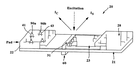
Fig. 1 is a perspective view of one embodiment of an assay of the present
invention;
Fig. 2 is a graphical illustration of the mechanism used for one embodiment
of a sandwich assay of the present invention;
Fig. 3 is a graphical illustration of the mechanism used for another
embodiment of a sandwich assay of the present invention;
Fig. 4 is a graphical illustration of the mechanism used for one embodiment
of a competitive assay of the present invention;
Fig. 5 is a graphical illustration of the mechanism used for another
embodiment of a competitive assay of the present invention;
Fig. 6 is a graphical illustration of one embodiment for covalently
conjugating an antibody to carboxylate nanoparticles;
Fig. 7 shows the excitation (EX) and emission (EM) spectra of a calibration
probe (C) and a detection probe (FP) in accordance with one embodiment of the
present invention;
4
CA 02495075 2005-02-08
WO 2004/021003 PCT/US2003/018058
Fig. 8 shows the normalized fluorescent intensity versus the amount of
leutinizing harmone (LH) as discussed in Example 1;
Fig. 9 shows the normalized fluorescent intensity versus the amount of
leutinizing harmone (LH) as discussed in Example 2; and
Fig. 10 shows the normalized fluorescent intensity versus the amount of C-
reactive protein (CRP) as discussed in Example 4.
Repeat use of reference characters in the present specification and
drawings is intended to represent same or analogous features or elements of
the
invention.
Detailed Description of Representative Embodiments
Definitions
As used herein, the term "analyte" generally refers to a substance to be
detected. For instance, analytes can include antigenic substances, haptens,
antibodies, and combinations thereof. Analytes include, but are not limited
to,
toxins, organic compounds, proteins, peptides, microorganisms, amino acids,
nucleic acids, hormones, steroids, vitamins, drugs (including those
administered
for therapeutic purposes as well as those administered for illicit purposes),
bacteria, virus particles and metabolites of or antibodies to any of the above
substances. Specific examples of some analytes include ferritin; creatinine
kinase
MIB (CK-MB); digoxin; phenytoin; phenobarbitol; carbamazepine; vancomycin;
gentamycin; theophylline; valproic acid; quinidine; leutinizing hormone (LH);
follicle
stimulating hormone (FSH); estradiol, progesterone; IgE antibodies; vitamin B2
micro-globulin; glycated hemoglobin (Gly. Hb); cortisol; digitoxin; N-
acetylprocainamide (NAPA); procainamide; antibodies to rubella, such as
rubella-
IgG and rubella IgM; antibodies to toxoplasmosis, such as toxoplasmosis IgG
(Toxo-IgG) and toxoplasmosis IgM (Toxo-IgM); testosterone; salicylates;
acetaminophen; hepatitis B virus surface antigen (HBsAg); antibodies to
hepatitis
B core antigen, such as anti-hepatitis B core antigen IgG and IgM (Anti-HBC);
human immune deficiency virus 1 and 2 (HIV 1 and 2); human T-cell leukemia
virus 1 and 2 (HTLV); hepatitis B a antigen (HBeAg); antibodies to hepatitis B
a
antigen (Anti-HBe); thyroid stimulating hormone (TSH); thyroxine (T4); total
triiodothyronine (Total T3); free triiodothyronine (Free T3); carcinoembryoic
antigen
(CEA); and alpha fetal protein (AFP). Drugs of abuse and controlled substances
5
CA 02495075 2005-02-08
WO 2004/021003 PCT/US2003/018058
include, but are not intended to be limited to, amphetamine; methamphetamine;
barbiturates, such as amobarbital, secobarbital, pentobarbital, phenobarbital,
and
barbital; benzodiazepines, such as librium and valium; cannabinoids, such as
hashish and marijuana; cocaine; fentanyl; LSD; methaqualone; opiates, such as
heroin, morphine, codeine, hydromorphone, hydrocodone, methadone, oxycodone,
oxymorphone and opium; phencyclidine; and propoxyhene. Other potential
analytes may be described in U.S. Patent No. 4,366,241 to Tom et al.
As used herein, the term "test sample" generally refers to a material
suspected of containing the analyte. The test sample can be used directly as
obtained from the source or following a pretreatment to modify the character
of the
sample. The test sample can be derived from any biological source, such as a
physiological fluid, including, blood, saliva, ocular lens fluid, cerebral
spinal fluid,
sweat, urine, milk, ascites fluid, raucous, synovial fluid, peritoneal fluid,
amniotic
fluid or the like. The test sample can be pretreated prior to use, such as
preparing
plasma from blood, diluting viscous fluids, and the like. Methods of treatment
can
involve filtration, distillation, concentration, inactivation of interfering
components,
and the addition of reagents. Besides physiological fluids, other liquid
samples
can 'be used such as water, food products and the like for the performance of
environmental or food production assays. In addition, a solid material
suspected of
containing the analyte can be used as the test sample. In some instances it
may
be beneficial to modify a solid test sample to form a liquid medium or to
release the
analyte.
Detailed Description
Reference now will be made in detail to various embodiments of the
invention, one or more examples of which are set forth below. 'Each example is
provided by way of explanation of the invention, not limitation of the
invention. In
fact, it will be apparent to those skilled in the art that various
modifications and
variations can be made in the present invention without departing from the
scope
or spirit of the invention. For instance, features illustrated or described as
part of
one embodiment, can be used on another embodiment to yield a still further
embodiment. Thus, it is intended that the present invention covers such
modifications and variations as come within the scope of the appended claims
and
their equivalents.
6
CA 02495075 2005-02-08
WO 2004/021003 PCT/US2003/018058
In general, the present invention is directed to a self-calibrated magnetic
binding assay (e.g., sandwich, competitive, etc.) for detecting the presence
or
quantity of an analyte residing in a test sample. The magnetic binding assay
includes detection probes capable of generating a detection signal (e.g.,
fluorescent non-magnetic particles) and calibration probes capable of
generating a
calibration signal (e.g., fluorescent magnetic particles). The amount of the
analyte
within the test sample is proportional (e.g., directly or inversely) to the
intensity of
the detection signal calibrated by the intensity of the calibration signal. It
has been
discovered that the self-calibration system provides an accurate, inexpensive,
and
readily controllable method of determining the presence of an analyte in a
test
sample.
Referring to Figs. 1-2, for instance, one embodiment of a flow-through
sandwich assay 20 that can be formed according to the present invention will
now
be described in more detail. As shown, the assay 20 contains a porous membrane
23 optionally supported by a rigid material 21. In general, the porous
membrane
23 can be made from any of a variety of materials through which the test
sample is
capable of passing. For example, the materials used to form the porous
membrane 23 can include, but are not limited to, natural, synthetic, or
naturally
occurring materials that are synthetically modified, such as polysaccharides
(e.g.,
cellulose materials such as paper and cellulose derivatives, such as cellulose
acetate and nitrocellulose); silica; inorganic materials, such as deactivated
alumina, diatomaceous earth, MgS04, or other inorganic finely divided material
uniformly dispersed in a porous polymer matrix, with polymers such as vinyl
chloride, vinyl chloride-propylene copolymer, and vinyl chloride-vinyl acetate
copolymer; cloth, both naturally occurring (e.g., cotton) and synthetic (e.g.,
nylon or
rayon); porous gels, such as silica gel, agarose, dextran, and gelatin;
polymeric
films, such as polyacrylamide; and the like. In one particular embodiment, the
porous membrane 23 is formed from nitrocellulose and/or polyester sulfone
materials. It should be understood that the term "nitrocellulose" refers to
nitric acid
esters of cellulose, which may be nitrocellulose alone, or a mixed ester of
nitric
acid and other acids, such as aliphatic carboxylic acids having from 1 to 7
carbon
atoms.
The assay 20 may also contain a wicking pad 28. The wicking pad 28
7
CA 02495075 2005-02-08
WO 2004/021003 PCT/US2003/018058
generally receives fluid that has migrated through the entire porous membrane
23.
As is well known in the art, the wicking pad 28 can assist in promoting
capillary
action and fluid flow through the membrane 23.
To initiate the detection of an analyte within the test sample, a user may
directly apply the test sample to a portion of the porous membrane 23 through
which it can then travel to reach one or more detection and calibration zones
(described below). Alternatively, the test sample may first be applied to a
sampling
pad (not shown) that is in fluid communication with the porous membrane 23.
Some suitable materials that can be used to form the sampling pad include, but
are not limited to, nitrocellulose, cellulose, porous polyethylene pads, and
glass
fiber filter paper. If desired, the sampling pad may also contain one or more
assay
pretreatment reagents, either diffusively or non-diffusively attached thereto.
In the illustrated embodiment, the test sample travels from the sampling pad
(not shown) to a conjugate pad 22 that is placed in communication with one end
of
the sampling pad. The conjugate pad 22 is formed from a material through which
the test sample is capable of passing. For example, in one embodiment, the
conjugate pad 22 is formed from glass fibers. Although only one conjugate pad
22
is shown, it should be understood that other conjugate pads may also be used
in
the present invention.
To facilitate detection of the presence or absence of an analyte within the
test sample, various detection probes 41 may be applied to the conjugate pad
22.
While contained on the conjugate pad 22, these probes 41 remain available for
binding with the analyte as it passes from the sampling pad through the
conjugate
pad 22. Upon binding with the analyte, the probes 41 can later serve to
identify
the presence or absence of the analyte. The detection probes 41 may be used
for
both detection and calibration of the device 20. In alternative embodiments,
however, separate calibration probes 43 can also be applied to the conjugate
pad
22 for use in conjunction with the detection probes 41 to facilitate
simultaneous
calibration and detection, thereby eliminating inaccuracies often created by
conventional assay calibration systems. It should be understood, however, that
the detection probes 41 and/or the calibration probes 43 may be applied
together
or separately at any location of the device 20, and need not be applied to the
conjugate pad 22. Further, it should also be understood that the detection
probes
8
CA 02495075 2005-02-08
WO 2004/021003 PCT/US2003/018058
41 and/or the calibration probes 43 may be applied to the same or different
conjugate pads.
Any substance generally capable of generating a signal that is detectable
visually or by an instrumental device may be used as the detection probes 41
and/or the calibration probes 43. Various suitable substances can include
chromogens; catalysts; fluorescent compounds; chemiluminescent compounds;
phosphorescent compounds; radioactive compounds; direct visual labels,
including
colloidal metallic (e.g., gold) and non-metallic particles, dye particles,
enzymes or
substrates, or organic polymer latex particles; liposomes or other vesicles
containing signal producing substances; and the like. For instance, some
enzymes suitable for use as probes are disclosed in U.S. Patent No. 4,275,149
to
Litman, et al., which is incorporated herein in its entirety by reference
thereto for all
purposes. One example of an enzyme/substrate system is the enzyme alkaline
phosphatase and the substrate nitro blue tetrazolium-5-bromo-4-chloro-3-
indolyl
phosphate, or derivative or analog thereof, or the substrate 4-
methylumbelliferyl-
phosphate. Other suitable probes may be described in U.S. Patent Nos.
5,670,381 to Jou, et al. and 5,252,459 to Tarcha, et al., which are
incorporated
herein in their entirety by reference thereto for all purposes.
In some embodiments, the detection probes 41 and/or the calibration
probes 43 can contain a fluorescent compound that produces a detectable
signal.
The fluorescent compounds can be fluorescent molecules, polymers, dendrimers,
particles, and the like. Some examples of suitable fluorescent molecules, for
instance, include, but are not limited to, fluorescein, europium chelates,
phycobiliprotein, rhodamine and their derivatives and analogs. Moreover, some
commercially available examples of suitable fluorescent particles include
fluorescent carboxylated microspheres sold by Molecular Probes, Inc. under the
trade names "FIuoSphere" (Red 580/605) and "TransfluoSphere" (543/620), as
well as "Texas Red" and 5- and 6-carboxytetramethylrhodamine, which are also
sold by Molecular Probes, Inc.
Regardless of the technique used to impart the probe with a signal
generating capability, it is typically desired that the detection probes 41
and/or the
calibration probes 43 be magnetically responsive probes. Generally, a material
is
considered "magnetically responsive" or "magnetic" if it is influenced by the
9
CA 02495075 2005-02-08
WO 2004/021003 PCT/US2003/018058
application of a magnetic field, such as, for example, if it is attracted or
repulsed or
has a detectable magnetic susceptibility or induction. For instance, some
examples of suitable magnetically responsive materials that can be used to
impart
magnetic properties to a probe include, but are not limited to, paramagnetic
materials, superparamagnetic materials, ferromagnetic materials, ferrimagnetic
materials, and metamagnetic materials. Specific examples are metals such as
iron, nickel, cobalt, chromium, manganese, and the like; lanthanide elements
such
as neodymium, erbium, and the like; alloys such as magnetic alloys of
aluminum,
nickel, cobalt, copper and the like; oxides such as ferric oxide (Fe304),
ferrous
oxide (Fe203), chromium oxide (Cr02), cobalt oxide (Co0), nickel oxide (Ni02),
manganese oxide (Mn203) and the like; composite materials such as ferrites and
the like; and solid solutions such as magnetite with ferric oxide and the
like.
In some embodiments, the detection probes 41 and/or the calibration
probes 43 are fluorescent and magnetic. Fluorescent magnetic probes are
generally well known in the art and often include a magnetically responsive
component and a fluorescent component. In some embodiments, for example,
one or more fluorescent dyes can be applied to magnetic particles to form the
probes, while in other embodiments, fluorescent dyes) can be applied to non-
magnetic particles that are coupled with magnetic particles. Some examples of
suitable fluorescent dyes include, but are not limited to, monomethine dyes,
trimethine dyes, pentamethine dyes, quinoline dyes, squaric acid-based dyes,
and
the like. The monomethine dyes that are pyridines typically have a blue or
blue-
green fluorescence emission, while quinolines typically have a green or yellow-
green fluorescence emission. The trimethine dyes are substantially shifted
toward
red wavelengths, while the pentamethine dyes are shifted even further, often
exhibiting infrared fluorescence emission. Specific examples of such
fluorescent
dyes include, but are not limited to, phthalocyanines, 2,3-naphthalocyanines,
squaraines and croconic acid derivatives. Other examples of suitable
fluorescent
magnetic particles are believed to be described in U.S. Patent Nos. 4,731,337
to
Luotola, et al. and 6,268,222 to Chandler, et al., which are incorporated
herein in
their entirety by reference thereto for all purposes.
When the detection probes 41 and/or the calibration probes 43 are particles,
such as described above, the mean diameter of the particulate probes may
CA 02495075 2005-02-08
WO 2004/021003 PCT/US2003/018058
generally vary as desired depending on factors such as the type of particle
chosen,
the pore size of the membrane, and the membrane composition. For example, in
some embodiments, the mean diameter of the particulate probes can range from
about 0.01 microns to about 1,000 microns, in some embodiments from about 0.01
microns to about 100 microns, and in some embodiments, from about 0.01
microns to about 10 microns. In one particular embodiment, the particulate
probes
have a mean diameter of from about 1 to about 2 microns. Generally, the
particles
are substantially spherical in shape, although other shapes including, but not
limited to, plates, rods, bars, irregular shapes, etc., are suitable for use
in the
present invention. As will be appreciated by those skilled in the art, the
composition, shape, size, and/or density of the particles may widely vary.
The detection probes 41 and/or the calibration probes 43 may be capable of
bonding (covalently or non-covalently) or physically adsorbing the analyte.
However, it is often desired to modify the probes in some manner so that they
are
more readily able to bond to the analyte. In such instances, the detection
probes
41 and/or the calibration probes 43 can be modified with certain specific
binding
members 90a and/or 90b that are adhered thereto to form probe conjugates.
Specific binding members generally refer to a member of a specific binding
pair, i.e., two different molecules where one of the molecules chemically
and/or
physically binds to the second molecule. For instance, immunoreactive specific
binding members can include antigens, haptens, aptamers, antibodies, and
complexes thereof, including those formed by recombinant DNA methods or
peptide synthesis. An antibody can be a monoclonal or polyclonal antibody, a
recombinant protein or a mixtures) or fragments) thereof, as well as a mixture
of
an antibody and other specific binding members. The details of the preparation
of
such antibodies and their suitability for use as specific binding members are
well
known to those skilled in the art.
Other common specific binding pairs include but are not limited to, biotin
and avidin, carbohydrates and lectins, complementary nucleotide sequences
(including probe and capture nucleic acid sequences used in DNA hybridization
assays to detect a target nucleic acid sequence), complementary peptide
sequences including those formed by recombinant methods, effector and receptor
molecules, hormone and hormone binding protein, enzyme cofactors and
11
CA 02495075 2005-02-08
WO 2004/021003 PCT/US2003/018058
enzymes, enzyme inhibitors and enzymes,' and the like. Furthermore, specific
binding pairs cari include members that are analogs of the original specific
binding
member. For example, a derivative or fragment of the analyte, i.e., an analyte-
analog, can be used so long as it has at least one epitope in common with the
analyte.
The specific binding members 90a and/or 90b can generally be attached to
the probes 41 and/or 43 using any of a variety of well-known techniques. For
instance, covalent attachment of the specific binding members 90a and/or 90b
to
the probes 41 and/or 43 (e.g., microparticles) can be accomplished using
carboxylic, amino, aldehyde, bromoacetyl, iodoacetyl, thiol, epoxy and other
reactive or linking functional groups, as well as residual free radicals and
radical
cations, through which a protein coupling reaction can be accomplished. A
surface
functional group can also be incorporated as a functionalized co-monomer
because the surface of the microparticle can contain a relatively high surface
concentration of polar groups. In addition, although microparticle probes are
often
functionalized after synthesis, in certain cases, such as poly(thiophenol),
the
microparticles are capable of direct covalent linking with a protein without
the need
for further modification. For example, referring to Fig. 6, one embodiment of
the
present invention for covalently conjugating a probe is illustrated. As shown,
the
first step of conjugation is activation of carboxylic groups on the probe
surface
using carbodiimide. In the second step, the activated carboxylic acid groups
are
reacted with an amino group of an antibody to form an amide bond. The
activation
and/or antibody coupling can occur in a buffer, such as phosphate-buffered
saline
(PBS) (e.g., pH of 7.2) or 2-(N-morpholino) ethane sulfonic acid (MES) (e.g.,
pH of
5.3). As shown, the resulting probes can then be blocked with ethanolamine,
for
instance, to form the probe conjugate. Besides covalent bonding, other
attachment techniques, such as adsorption, may also be utilized in the present
invention.
Referring again to Figs. 1-2, a test sample containing an analyte can
initially
be applied to the sampling pad. From the sampling pad, the test sample can
then
travel to the conjugate pad 22, where the analyte mixes with the detection
probes
41 and/or the calibration probes 43. Depending on the type of probes selected,
the analyte may bind with the detection probes 41 and/or the calibration
probes 43
12
CA 02495075 2005-02-08
WO 2004/021003 PCT/US2003/018058
to form complexes 49 (See Fig. 2). For instance, in one embodiment, a test
sample containing an analyte is mixed with (1 ) fluorescent non-magnetic
particles
41 conjugated with a first binding member 90a and (2) fluorescent magnetic
particles 43 conjugated with a second binding member 90b. In such an instance,
the analyte forms sandwich complexes 49 with the fluorescent non-magnetic
particles 41 and the fluorescent magnetic particles 43. Moreover, because the
conjugate pad 22 is in fluid communication with the porous membrane 23, the
complexes 49 can migrate from the conjugate pad 22 to a detection zone 31
present on the porous membrane 23.
At the detection zone 31, the complexes 49 and any unbound conjugated,
fluorescent magnetic particles 43 are then captured by a magnetic device 60
and
separated from the rest of the sample using conventional techniques. A
magnetic
field generator, for instance, can be used to generate a magnetic field that
elicits a
response from the magnetically responsive probes. Suitable magnetic field
generators include, but are not limited to, permanent magnets and
electromagnets.
The magnetic separation process typically involves mixing the sample with the
magnetic particles in a liquid medium to bind the analyte by affinity
reaction, and
then separating the unbound magnetic particles and analyte complexes from the
sample medium by applying a magnetic field. Most, if not all of the magnetic
particles, except those particles that are colloidal, settle in time. The
liquid
medium, therefore, can be agitated to keep the particles suspended for a
sufficient
period of time to allow the bioaffinity binding reaction to occur. Examples of
known
agitation methods include shaking, swirling, rocking, rotation, or similar
manipulations of a partially filled container. Some commercially available
examples of suitable magnetic separation devices include the Dynal MPC series
of
separators manufactured by Dynal, Inc., Lake Success, New York, which employ a
permanent magnet located externally to a container holding a test medium and
provide only for separation. Mixing of the magnetic particles in the test
medium for
affinity binding reaction is done separately. In addition, other methods for
capturing magnetic particles may be described in U.S. Patent Nos. 5,200,084 to
Liberti, et al.; 5,647,994 to Tuunanen, et al.; 5,795,470 to Wangi, et al.;
and
6,033,574 to Siddigi, which are incorporated herein in their entirety by
reference
thereto for all purposes.
13
CA 02495075 2005-02-08
WO 2004/021003 PCT/US2003/018058
Once captured, the fluorescence signal of the fluorescent magnetic particles
43, complexed and uncomplexed, and the complexes 49 can be measured using
conventional techniques. For example, in one embodiment, the particles 43 and
complexes 49 can be excited with the same external source. In this embodiment,
the source supplies radiation at an excitation wavelength, thereby causing the
particles 43 to emit light at a wavelength that is different than the
wavelength
emitted by the complexes 49. This enables the presence of the complexes 49 and
particles 41 to be separately measured. Alternatively, the particles 43 and
complexes 49 can also be measured separately using separate external sources.
Generally speaking, fluorescence is the result of a three-stage process that
occurs in certain fluorescent compounds. In the first stage, energy is
supplied by
an external source, such as an incandescent lamp or a laser and absorbed by
the
fluorescent compound, creating an excited electronic singlet state. In the
second
stage, the excited state exists for a finite time during which the fluorescent
compound undergoes conformational changes and is also subject to a multitude
of
possible interactions with its molecular environment. During this time, the
energy
of the excited state is partially dissipated, yielding a relaxed state from
which
fluorescence emission originates. The third stage is the fluorescence emission
stage wherein energy is emitted, returning the fluorescent compound to its
ground
state. The emitted energy is lower than its excitation energy (light or laser)
and
thus of a longer wavelength. This shift or difference in energy or wavelength
allows the emission energy to be detected and isolated from the excitation
energy.
Fluorescence detection generally utilizes wavelength filtering to isolate the
emission photons from the excitation photons, and a detector that registers
emission photons and produces a recordable output, usually as an electrical
signal
or a photographic image. There are generally four recognized types of
detectors:
spectrofluorometers and microplate readers; fluorescence microscopes;
fluorescence scanners; and flow cytometers. One suitable fluorescence detector
for use with the present invention is a FluoroLog III Spectrofluorometer,
which is
sold by SPEX Industries, Inc. of Edison, New Jersey.
Although not required, the selection criteria of particularly desired
detection
and calibration probe pairs include: (1 ) little or no spectral overlap for
either the
absorption spectra or the fluorescence spectra so that emission intensities
can be
14
CA 02495075 2005-02-08
WO 2004/021003 PCT/US2003/018058
measured separately; (2) no significant fluorescent energy transfer between
the
detection and calibration probes when brought into a close proximity so that
they
emit independently; and (3) relatively long emission wavelength (e.g., greater
than
about 600 nm) so that the autofluorescence of biological fluids has a minimal
effect
on the fluorescence measurement. Fig. 7, for example, illustrates an exemplary
calibration probe and detection probe having excitation spectra with little
overlap
so that they can be independently excited.
Further, if desired, a technique known as "time-resolved fluorescence
detection" may also be utilized in the present invention. Time-resolved
fluorescence detection is designed to reduce background signals from the
emission source or from scattering processes (resulting from scattering of the
excitation radiation) by taking advantage of the fluorescence characteristics
of
certain fluorescent materials, such as lanthanide chelates of europium (Eu
(III))
and terbium (Tb (III)). Such chelates can exhibit strongly red-shifted, narrow-
band,
long-lived emission after excitation of the chelate at substantially shorter
wavelengths. Typically, the chelate possesses a strong ultraviolet absorption
band
due to a chromophore located close to the lanthanide in the molecule.
Subsequent to light absorption by the chromophore, the excitation energy can
be
transferred from the excited chromophore to the lanthanide. This is followed
by a
fluorescence emission characteristic of the lanthanide. The use of pulsed
excitation and time-gated detection, combined with narrow-band emission
filters,
allows for specific detection of the fluorescence from the lanthanide chelate
only,
rejecting emission from other species present in the sample that are typically
shorter-lived or have shorter wavelength emission. Other time-resolved
techniques for measuring fluorescence are described in U.S. Patent No.
5,585,279
to Davidson and 5,637,509 to Hemmila, et al., which are incorporated herein in
their entirety by reference thereto for all purposes.
Regardless of the technique used to measure fluorescence, the absolute
amount of the analyte can be, ascertained by comparing the fluorescence signal
of
the captured, fluorescent non-magnetic particles 41 with the captured,
fluorescent
magnetic particles 43. The fluorescence intensity of the captured, fluorescent
non-
magnetic particles 41, IS, can be compared to the fluorescence intensity of
the
captured, fluorescent magnetic particles 43, I~. The total amount of the.
captured
CA 02495075 2005-02-08
WO 2004/021003 PCT/US2003/018058
fluorescent magnetic particles 43 is predetermined and known and thus can be
used for calibration purposes. For example, in one embodiment, the amount of
analyte is directly proportional to the ratio of IS to I~. Based upon the
intensity
range in which the detection zone 31 falls, the general concentration range
for the
analyte may be determined. As a result, calibration and sample testing may be
conducted under approximately the same conditions at the same time, thus
providing reliable quantitative or semi-quantitative results, with increased
sensitivity.
If desired, the ratio of IS to I~ may be plotted versus the analyte
concentration for a range of known analyte concentrations to generate a
calibration
curve. To determine the quantity of analyte in an unknown test sample, the
signal
ratio may then be converted to analyte concentration according to the
calibration
curve. It should be noted that the capturing efficiency of the complexed and
uncomplexed fluorescent magnetic particles is generally the same for any given
sample. Accordingly, the variation in capturing efficiency is not believed to
significantly interfere with the results from sample-to-sample because the
ratio of
fluorescence intensities (i.e., IS/I~) is used instead of absolute
fluorescence. It
should also be noted that alternative mathematical relationships between IS
and I
may be plotted versus the analyte concentration to generate the calibration
curve.
For example, in one embodiment, the value of IS /(IS + I~) may be plotted
versus
analyte concentration to generate the calibration curve.
Various other embodiments are also contemplated by the present invention.
For instance, referring to Fig. 3, the assay 20 described above and
illustrated in
Fig. 1 can be modified to form another format of a sandwich assay. In one
embodiment, for instance, a test sample containing an analyte can initially be
mixed with (1) fluorescent non-magnetic particles 141a conjugated with a first
binding member 190a, (2) fluorescent magnetic particles 143, and (3) non-
fluorescent magnetic particles 141 b conjugated with a second binding member
190b. In this particular embodiment, the fluorescent magnetic particles 143
can be
blocked with a blocking agent, such as (3-casein, to prevent nonspecific
binding to
the analyte, thereby allowing such particles 143 to act only as a calibration
probe.
Further, the first specific binding member 190a and the second specific
binding
member 190b may be analogs of the analyte.
16
CA 02495075 2005-02-08
WO 2004/021003 PCT/US2003/018058
The term "blocking agent" means a reagent that adheres to the probe
surface so that it "blocks" or prevents non-analyte materials from binding to
the
surface. Blockers can include, but are not limited to, ~i-casein, albumins
such as
bovine serum albumin, pluronic or other surtactants, polyethylene glycol,
polyvinyl
alcohol, or sulfur derivatives of the above compounds, and any other blocking
material known to those of ordinary skill in the art.
Referring again to Fig. 3, the analyte forms sandwich complexes 149 with
the conjugated, fluorescent non-magnetic particles 141 a and the conjugated,
non-
fluorescent, magnetic particles 141 b. Because the conjugate pad 22 is in
fluid
communication with the porous membrane 23, the complexes 149 can migrate
from the conjugate pad 22 to the detection zone 31 present on the porous
membrane 23. At the detection zone 31, the complexes 149 and any unbound
particles 143 and/or 141 b are then captured by the magnetic device 60 and
separated from the rest of the sample. As described above, the absolute amount
of the analyte can be ascertained by comparing the fluorescence intensity of
the
captured, fluorescent non-magnetic particles 141a, IS, to the fluorescence
intensity
of the captured, fluorescent magnetic particles 143, I~. In particular, the
total
amount of the captured fluorescent magnetic particles 143 is predetermined and
known and thus can be used for calibration purposes. Accordingly, the amount
of
analyte in this embodiment is directly proportional to the ratio of IS to I~.
Moreover, referring to Fig. 4, the assay 20 described above and illustrated
in Fig. 1 can also be modified to form a competitive assay. In one embodiment,
for
instance, a test sample containing an analyte can initially be mixed with (1 )
fluorescent non-magnetic particles 241 conjugated with a first binding member
290a and (2) fluorescent magnetic particles 243 conjugated with a second
binding
member 290b. In this particular embodiment, the first binding member 290a can
be identical to the analyte, while the second binding member 290b can be an
analog of the analyte.
Upon mixing, the analyte competes with the conjugated, fluorescent non-
magnetic particles 241 for the conjugated, fluorescent magnetic particles 243
such
that complexes 249a of the analyte and the fluorescent magnetic particles 243
and
complexes 249b of the fluorescent magnetic particles 243 and the fluorescent,
non-magnetic particles 241 are formed. Because the conjugate pad 22 is in
fluid
17
CA 02495075 2005-02-08
WO 2004/021003 PCT/US2003/018058
communication with the porous membrane 23, the complexes 249a and 249b can
migrate from the conjugate pad 22 to the detection zone 31 present on the
porous
membrane 23. At the detection zone 31, the complexes 249a and 249b and any
unbound particles 243 are then captured by the magnetic device 60 and
separated
from the rest of the sample. As described above, the absolute amount of the
analyte can be ascertained by comparing the fluorescence intensity of the
captured, fluorescent non-magnetic particles 241, IS, to the fluorescence
intensity
of the captured, complexed or uncomplexed, fluorescent magnetic particles 243,
I~.
In particular, the total amount of the captured fluorescent magnetic particles
243 is
predetermined and known and thus can be used for calibration purposes.
Accordingly, the amount of analyte in this embodiment is inversely
proportional to
the ratio of IS to I~.
Referring to Fig. 5, the assay 20 described above and illustrated in Fig. 1
can also be modified to form another format of a competitive assay. In one
embodiment, for instance, a test sample containing an analyte can initially be
mixed with (1 ) fluorescent non-magnetic particles 341 a conjugated with a
first
binding member 390a (2) fluorescent magnetic particles 343, and (3) non-
fluorescent magnetic particles 341 b conjugated with a second binding member
390b. In this particular embodiment, the first binding member 390a can be
identical to the analyte, while the second binding member 390b can be an
analog
of the analyte. Further, the fluorescent magnetic particles 343 can be blocked
with
a blocking agent, such as ~3-casein, to prevent nonspecific binding to the
analyte,
thereby allowing such particles to act only as a calibration probe.
Upon mixing, the analyte competes with the conjugated, fluorescent non-
magnetic particles 341 a for the conjugated, non-fluorescent magnetic
particles
341 b such that complexes 349a of the analyte and the non-fluorescent magnetic
particles 341 b and complexes 349b of the non-fluorescent magnetic particles
341 b
and the fluorescent non-magnetic particles 341 a are formed. Because the
conjugate pad 22 is in fluid communication with the porous membrane 23, the
complexes 349a and 349b can migrate from the conjugate pad 22 to the detection
zone 31 present on the porous membrane 23. At the detection zone 31, the
complexes 349a and 349b and any unbound particles 343 and/or 341 b are then
captured by the magnetic device 60 and separated from the rest of the sample.
As
18
CA 02495075 2005-02-08
WO 2004/021003 PCT/US2003/018058
described above, the absolute amount of the analyte can be ascertained by
comparing the fluorescence intensity of the captured, fluorescent non-magnetic
particles 341 a, IS, to the fluorescence intensity of the captured,
fluorescent
magnetic particles 343, I~. In particular, the total amount of the captured
fluorescent magnetic particles 343 is predetermined and known and thus can be
used for calibration purposes. Accordingly, the amount of analyte in this
embodiment is inversely proportional to the ratio of IS to I~.
Although various embodiments of assay configurations have been
described above, it should be understood, that an assay of the present
invention
may generally have any configuration desired. For example, a competitive assay
may be formed such as shown in Fig. 4 and described above, except that the
particles 241 are fluorescent, magnetic particles and the particles 243 are
fluorescent, non-magnetic particles. Likewise, a competitive assay may be
formed
such as shown in Fig. 5 and described above, except that the particles 341 a
are
non-fluorescent, magnetic particles and the particles 341 b are fluorescent,
non-
magnetic particles. In addition, besides flow-through, membrane-based assay
devices, other types of assay devices may also be used in the present
invention,
such as capillary, fluid-based, and solution-based assays. Various other assay
configurations are also described in U.S. Patent Nos. 5,395,754 to Lambotte,
et
al.; 5,670,381 to Jou, et al.; and 6,194,220 to Malick, et al., which are
incorporated
herein in their entirety by reference thereto for all purposes.
Moreover, although various embodiments have been described above that
relate specifically to the use of fluorescence as the mechanism for
calibration and
detection, other well known detection mechanisms are equally applicable in the
present invention. For example, in some embodiments, the detection and/or
calibration probes may be chemiluminescent or phosphorescent compounds.
Chemiluminescent probes, for instance, may be excited through the use of a
suitable reactant as is well known in the art. Still other embodiments and
configurations are also contemplated by the present invention.
Thus, the present inventors have discovered that the manipulation of
magnetic particles can be utilized to establish a separation and detection of
an
analyte. Specifically, magnetic separation and detection techniques (e.g.,
fluorescence) are built into an integrated system. Further, the system is self-
19
CA 02495075 2005-02-08
WO 2004/021003 PCT/US2003/018058
calibrated to eliminate the requirement of control calibration samples when
using
conventional external calibration techniques. In one embodiment, self-
calibration
is accomplished through the use of fluorescent magnetic probes. The
fluorescence emitted from the fluorescent magnetic probes and fluorescent non-
magnetic probes can be separately measured on the same sample. Because the
number of magnetic particles is predetermined, the system is self-calibrated
when
determining the amount of the captured fluorescent non-magnetic probes, and
subsequently, the amount of analyte. Furthermore, because the fluorescence of
the calibration and detection probes are simultaneously measured under
identical
conditions, potential interference from many variations, such as temperature
and
instrument instability, can be avoided to improve detection reliability and
consistency.
The present invention may be better understood with reference to the
following examples.
EXAMPLE 1
The ability of the present invention to detect the presence of an analyte
using a sandwich assay, such as shown in Fig. 3, was demonstrated. Initially,
the
following components were added to six Eppendorf vials:
(1 ) 25 microliters of covalently conjugated, non-fluorescent magnetic
particles
(3 milligrams per milliliter in PBS buffer);
(2) 15 microliters of covalently conjugated, fluorescent non-magnetic
particles
(2 milligrams per milliliter in PBS buffer);
(3) 10 microliters of fluorescent magnetic particles blocked by a-casein (3
milligrams per milliliter in PBS buffer); and
(4) Leutinizing hormone (LH) analyte ranging from 0, 10 microliters (1
microgram per milliliter), 20 microliters (1 microgram per milliliter), 40
microliters (1 microgram per milliliter), 40 microliters (2 micrograms per
milliliter) and 80 microliters (2 micrograms per milliliter).
To each of the Eppendorf vials, an appropriate amount of PBS buffer was
added to a final volume of 150 microliters. The samples were incubated at room
temperature for 10 minutes with gentle shaking. The magnetic particles were
then
separated by a magnetic separator obtained from Dynal, Inc. The supernatant
from each vial was discarded and the magnetic particles were re-suspended in
1.5
CA 02495075 2005-02-08
WO 2004/021003 PCT/US2003/018058
milliliters of PBS. 300 microliters of the fluorescent magnetic particle
suspension
was used for each fluorescence measurement. A "Flourolog III
Spectrofluorometer", which was obtained from SPEX Industries, Inc. of Edison,
N.J., was used to measure the fluorescence of the sample using a right angle
mode. An excitation wavelength of 470 manometers and an emission wavelength
of 560 manometers were used for the fluorescent magnetic particles, while an
excitation wavelength of 570 manometers and an emission wavelength of 605
manometers were used for the fluorescent, non-magnetic particles. The
integration
time was 0.2 seconds.
The normalized and calibrated fluorescence intensity as a function of the
dose of LH in each sample is shown in Fig. 8. Normalized intensity was
obtained
by dividing the measured fluorescence intensity of the sample by the
fluorescence
intensity of a control sample. The control sample was the sample without the
analyte.
The particles used in Example 1 were formed as follows:
Non-Fluorescent Magnetic Particles
125 microliters of 10% carboxylate-modified paramagnetic particles (0.35
microns, Estapor~ Superparamagnetic microspheres, obtained from Bang's
Laboratories, Inc.) were washed once with 1.5 milliliters of carbonate buffer
and
twice with PBS using a magnetic separator. The washed particles were re-
suspended in 0.6 milliliters PBS and 15 milligrams carbodiimide (from
Polysciences, Inc.). The mixture was allowed to react at room temperature (RT)
for 30 minutes on a shaker. The activated particles were then washed twice
with a
borate buffer. The activated particles were again re-suspended in 1.2
milliliters of
a borate buffer. Thereafter, 30 microliters of LH ~i-monoclonal antibody (9.8
mg/ml, obtained from Fitzgerald Industries International, Inc.) was added to
the
activated particles. The reaction mixture was allowed to react at room
temperature
on a shaker overnight. The activated particles were then collected and
incubated
in 1 milliliter of 0.1 molar ethanolamine under gentle shaking for 15 minutes.
The
particles were then washed twice with PBS and stored at 4°C in a buffer
that
contained 0.1 molar PBS, 0.15 molar NaCI, 1 % ~3-casein, 5% glycerol and 0.1
NaN3.
21
CA 02495075 2005-02-08
WO 2004/021003 PCT/US2003/018058
Fluorescent Non-Magnetic Particles
The "fluorescent non-magnetic" particles were covalently conjugated
according to the procedure described above, except that the binding member was
LH a-monoclonal antibody (9.8 milligrams per milliliter, obtained from
Fitzgerald
Industries International, Inc.) instead of LH ~3-monoclonal antibody. The
particles
utilized were FIuoSpheres~ carboxylate-modified microspheres, which were
obtained from Molecular Probes, Inc. The particles had a particle size of 0.5
microns, and were red fluorescent with an excitation wavelength of 580
nanometers and an emission wavelength of 605 nanometers.
Fluorescent Magnetic Particles
100 microliters of a 2.76% solids suspension of fluorescent
superparamagnetic particles (obtained from Polysciences, Inc. of Warrington,
Pennsylvania) were combined with 1 milliliter of a borate buffer (0.1 molar,
pH =
8.5) in an Eppendorf tube. Such particles have a mean diameter of between 1 to
2
microns and are believed to be iron-containing microspheres that have a
polystyrene surface that allows for passive adsorption and functional group
reactions with proteins. The particles were separated by a magnetic separator
obtained from Dynal, Inc. and re-suspended in 200 microliters of a 10
milligram per
milliliter solution of ~i-casein in a 0.1 molar borate buffer. The suspension
was
incubated for 30 minutes with gentle mixing. The above step was repeated
twice.
The separated particles were re-suspended in 200 microliters of PBS and stored
at
4°C.
Leutinizinq Hormone (LH)
The "leutinizing hormone (LH)" was obtained from Fitzgerald Industries
International, Inc.
EXAMPLE 2
The ability of the present invention to detect the presence of an analyte
using a sandwich assay, such as shown in Fig. 1, was demonstrated. Initially,
the
following components were added to six Eppendorf vials:
(1 ) 5 microliters of covalently conjugated, fluorescent non-magnetic
particles (2
milligrams per milliliter in PBS buffer);
(2) 15 microliters of physical absorption conjugated, fluorescent magnetic
particles (3 milligrams per milliliter in PBS buffer); and
22
CA 02495075 2005-02-08
WO 2004/021003 PCT/US2003/018058
(3) Leutinizing hormone (LH) analyte ranging from 0, 5, 10 microliters, 20,
40,
and 100 microliters (2 micrograms per milliliter).
To each of the Eppendorf vials, an appropriate amount of PBS buffer was
added to a final volume of 150 microliters. The samples were incubated at room
temperature for 25 minutes with gentle shaking. The magnetic particles were
then
separated by a magnetic separator obtained from Dynal, Inc. The supernatant
from each vial was discarded and the magnetic particles were re-suspended in
1.5
milliliters of PBS. 300 microliters of the fluorescent magnetic particle
suspension
was used for each fluorescence measurement. A "Flourolog III
Spectrofluorometer', which was obtained from SPEX Industries, Inc. of Edison,
N.J., was used to measure the fluorescence of the sample using a right angle
mode. An excitation wavelength of 470 nanometers and an emission wavelength
of 560 nanometers were used for the fluorescent magnetic particles, while an
excitation wavelength of 570 nanometers and an emission wavelength of 605
nanometers were used for the fluorescent, non-magnetic particles. The
integration
time ranged from 0.2 to 1 second.
The normalized and calibrated fluorescence intensity as a function of the
dose of LH in each sample is shown in Fig. 9.
The particles used in Example 2 were formed as follows:
Fluorescent Non-Magnetic Particles
The "fluorescent non-magnetic" particles were formed as described above
in Example 1.
Fluorescent Magnetic Particles
2.76 milligrams of fluorescent superparamagnetic particles (2.5% solids in
an aqueous suspension) were obtained from Polysciences, Inc. of Warrington,
Pennsylvania. The particles were washed three times with borate buffers and
separated by a magnetic separator obtained from Dynal, Inc. The washed
particles were re-suspended in a 200-microliter borate buffer, and 82
micrograms
of (3-leutinizing hormone ((3-LH) monoclonal antibody (1 milligram per
milliliter,
obtained from Fitzgerald Industries International, Inc.) were added. The
mixture
was gently mixed overnight at room temperature. The particles were then
collected by a magnetic separator and incubated with 200 microliters of (3-
casein
(10 milligrams per milliliter in borate buffer) for 30 minutes with gentle
mixing to
23
CA 02495075 2005-02-08
WO 2004/021003 PCT/US2003/018058
block the nonspecific binding sites. The blocked particles were washed twice
with
PBS and stored in 0.1 molar PBS.
Leutinizing Hormone (LH)
The "leutinizing hormone (LH)" was obtained from Fitzgerald Industries
International, Inc.
EXAMPLE 3
A self-calibrated magnetic binding assay was compared to a non-calibrated
magnetic binding assay.
Without Self-Calibration
Initially, the following components were added to 5 Eppendorf vials (Vial
Nos. 2-6 in Table I):
(1 ) 15 microliters of covalently conjugated, non-fluorescent magnetic
particles (3 milligrams per milliliter in 0.1 molar PBS buffer);
(2) 15 microliters of covalently conjugated, fluorescent non-magnetic
particles
(2 milligrams per milliliter in PBS buffer);
(3) 20 microliters leutinizing hormone (LH) analyte (1 microgram per
milliliter);
and
(4) 20 microliters of PBS.
A control Eppendorf vial was also formed with only 20 microliters of PBS
(Vial No. 1 in Table I).
The samples were incubated at room temperature for 20 minutes with
gentle shaking. The magnetic particles were then separated by a magnetic
separator obtained from Dynal, Inc. The supernatant from each vial was
discarded
and the magnetic particles were re-suspended in 1.5 milliliters of PBS. 300
microliters of the fluorescent magnetic particle suspension was used for each
fluorescence measurement. A "Flourolog III Spectrofluorometer", which was
obtained from SPEX Industries, Inc. of Edison, N.J., was used to measure the
fluorescence of the sample using a right angle mode. An excitation wavelength
of
570 nanometers and an emission wavelength of 605 nanometers were used for to
take fluorescence measurements on different days.
24
CA 02495075 2005-02-08
WO 2004/021003 PCT/US2003/018058
Table I lists the relative fluorescence data for each day.
Table I: Fluorescent Measurements
Std.
Vial No. No:2~ ~No.3No.4 No.-5 TNo.6
1 Dev%
Day1 13 254 215 263 285 291 11
Day 12 235 207 300 263 299 15
2
Day 12 183 176 213 270 266 20
3
Day 18 265 226 275 282 293 10
4
Day 9 207 193 246 236 244 10
Day 14 227 202 252 262 274 12
6
Std.
23 13 8 11 6 7
Dev%
With Self-Calibration
5 Initially, the following components were added to 5 Eppendorf vials (Vial
Nos. 9-13 in Table II):
(1 ) 15 microliters of covalently conjugated, non-fluorescent magnetic
particles
(3 milligrams per milliliter in 0.1 molar PBS buffer);
(2) 15 microliters of covalently conjugated, fluorescent non-magnetic
particles
(2 milligrams per milliliter in PBS buffer);
(3) 20 microliters of fluorescent magnetic particles blocked by ~-casein (3
milligrams per milliliter in PBS buffer); and
(4) 20 microliters leutinizing hormone (LH) analyte (1 microgram per
milliliter);
and
(5) 20 microliters of PBS.
A control Eppendorf vial was also formed with only 20 microliters of PBS
(Vial No. 8 in Table II).
The samples were incubated at room temperature for 20 minutes with
gentle shaking. The magnetic particles were then separated by a magnetic
separator obtained from Dynal, Inc. The supernatant from each vial was
discarded
and the magnetic particles were re-suspended in 1.5 milliliters of PBS. 300
microliters of the fluorescent magnetic particle suspension was used for each
fluorescence measurement. The "Flourolog III Spectrofluorometer" was used to
CA 02495075 2005-02-08
WO 2004/021003 PCT/US2003/018058
measure the fluorescence of the sample using a right angle mode. An excitation
wavelength of 470 manometers and an emission wavelength of 560 manometers
were used for the fluorescent magnetic particles, while an excitation
wavelength of
570 manometers and an emission wavelength of 605 manometers were used for
the fluorescent, non-magnetic particles. Table II lists the relative
fluorescence data
for each day.
Table II: Fluorescent Measurements
a : . v. ~ ~ =Std
Vial No No.9 No. No. No 12 -_.:
8 . 10 11 -- . No 13 Dev%::
_. ~ : . _~ _ .
_ W
Day 31 352/47344/43 300/41318/44 369/39 12
1 /32
Day 31 324/42329/41 323/46338/47 418/43 14
2 /42
Day 28/39 307/40333/42 282/42288/40 425/46 12
3
Day 30/41 267/36292/36 271 281 356/43 8.8
4 /41 /38
Day 21 252/33292/34 258/38275/36 328/37 10
5 /29
Day 21 237/33307/38 265/40288/35 358/39 12
6 /25
Std.
13 3 3 4 5 6
Dev%
As can be seen from the comparisons of each set of samples for the two
systems, the standard deviations (Std. Dev%) for the self-calibrated system
were
significantly smaller than the standard deviations without self calibration,
even
under carefully controlled conditions. Because the self calibrated system is
less
dependent on the measurement conditions, it is anticipated that the standard
deviations for the self-calibrated system would be even smaller than the
standard
deviations without self-calibration when the conditions are not carefully
controlled.
The particles used in Example 3 were formed as follows:
Non-Fluorescent Magnetic Particles
The "non-fluorescent magnetic" particles were formed as described above
in Example 1.
Fluorescent Non-Magnetic Particles
The "fluorescent non-magnetic" particles were formed as described above
in Example 1.
26
CA 02495075 2005-02-08
WO 2004/021003 PCT/US2003/018058
Fluorescent Magnetic Particles
The "fluorescent magnetic particles" were formed as described in Example
2.
Leutinizing Hormone (LH)
The "leutinizing hormone (LH)" was obtained from Fitzgerald Industries
International, Inc.
FXOMPI F d
The ability of the present invention to detect the presence of an analyte
using a sandwich assay, such as shown in Fig. 3, was demonstrated. Initially,
the
following components were added to six Eppendorf vials:
(1 ) 30 microliters of covalently conjugated, non-fluorescent magnetic
particles
(2 milligrams per milliliter in PBS buffer);
(2) 20 microliters of covalently conjugated, fluorescent non-magnetic
particles
(2 milligrams per milliliter in PBS buffer);
(3) 15 microliters of fluorescent magnetic particles blocked by ~3-casein (1
milligram per milliliter in PBS buffer); and
(4) C-reactive protein (CRP) analyte ranging from 0, 5, 10, 20, 50, and 100
microliters (0.2 micrograms per milliliter in PBS).
The samples were incubated at room temperature for 20 minutes with
gentle shaking. The magnetic particles were then separated by a magnetic
separator obtained from Dynal, Inc. The supernatant from each vial was
discarded
and the magnetic particles were re-suspended in 1.5 milliliter of PBS. 300
microliters of the fluorescent magnetic particle suspension was used for each
fluorescence measurement. A "Flourolog III Spectrofluorometer", which was
obtained from SPEX Industries, Inc. of Edison, N.J., was used to measure the
fluorescence of the sample using a right angle mode. An excitation wavelength
of
470 nanometers and an emission wavelength of 560 nanometers were used for
the fluorescent magnetic particles, while an excitation wavelength of 570
nanometers and an emission wavelength of 605 nanometers were used for the
fluorescent, non-magnetic particles. The integration time ranged from 0.2 to 1
second. The normalized fluorescence intensity as a function of the dose of CRP
in
each sample is shown in Fig. 10.
27
CA 02495075 2005-02-08
WO 2004/021003 PCT/US2003/018058
The particles used in Example 4 were formed as follows:
Non-Fluorescent Magnetic Particles
125 microliters of 10% carboxylate-modified paramagnetic particles (0.35
microns, Estapor~ Superparamagnetic microspheres, available from Bang's
Laboratories, Inc.) were washed once by 1.5 ml carbonate buffer and twice by
phosphate buffer saline (PBS) using a magnetic separator. The washed particles
were re-suspended in 0.6 milliliters PBS and 15 milligrams carbodiimide (from
Polysciences, Inc.). The mixture was allowed to react at room temperature (RT)
for 30 minutes on a shaker. The activated particles were then washed twice
with a
borate buffer. The activated particles were again re-suspended in 1.2 ml
borate
buffer. Thereafter, 30 microliters of anti-C-reactive protein (anti-CRP1 )
monoclonal
antibody (Mab A5804, 2 mg/ml, obtained from BiosPacific, Inc.) were added to
the
activated particles. The reaction mixture was allowed to react at room
temperature
on a shaker overnight. The activated particles were then collected and
incubated
in 1 milliliter of 0.1 molar ethanolamine under gentle shaking for 15 minutes.
The
particles were then washed twice with PBS and stored at 4°C in a buffer
that
contained 0.1 molar PBS, 0.15 molar NaCI, 1 % ~3-casein, 5% glycerol and 0.1
NaN3.
Fluorescent Non-Magnetic Particles
The "fluorescent non-magnetic" particles were covalently conjugated
according to the procedure described above, except that the binding member was
anti-C-reactive protein (anti-CRP2) monoclonal antibody (2 mg/ml, obtained
from
BiosPacific, Inc.) instead of anti-CRP1. The particles utilized were
FIuoSpheres~
carboxylate-modified microspheres, which were obtained from Molecular Probes,
Inc. The particles had a particle size of 0.5 Nm, and were red fluorescent
with an
excitation wavelength of 580 nanometers and an emission wavelength of 605
nanometers.
Fluorescent Magnetic Particles
100 microliters of a 2.76% solids suspension of fluorescent
superparamagnetic particles (obtained from Polysciences, Inc. of Warrington,
Pennsylvania). Such particles have a mean diameter between 1 to 2 microns, and
are believed to be iron-containing microspheres that have a polystyrene
surface
that allows for passive adsorption and functional group reactions with
proteins. 1
28
CA 02495075 2005-02-08
WO 2004/021003 PCT/US2003/018058
milliliter of a borate buffer (0.1 molar, pH = 8.5) was then added to the
particles in
an Eppendorf tube. The particles were separated by a magnetic separator
obtained from Dynal, Inc. and the particles were re-suspended in 200
microliters of
a 10 mg/ml solution of ~i-casein in a 0.1 M borate buffer. The suspension was
incubated for 30 minutes with gentle mixing. The above step was repeated
twice.
The separated particles were re-suspended in 200 microliters of PBS and stored
at
4°C.
C-Reactive Protein (CRP)
The "C-reactive protein (CRP)" was obtained BiosPacific, Inc.
While the invention has been described in detail with respect to the specific
embodiments thereof, it will be appreciated that those skilled in the art,
upon
attaining an understanding of the foregoing, may readily conceive of
alterations to,
variations of, and equivalents to these embodiments. Accordingly, the scope of
the present invention should be assessed as that of the appended claims and
any
equivalents thereto.
29