Note: Descriptions are shown in the official language in which they were submitted.
CA 02495314 2010-09-23
1
MEDICAL IMPLANT
Technical Field
[0002] The invention relates generally to implanting a device such as a
surgical sling at
an anatomical site in the body of a mammal. More particularly, the invention
relates to
delivering and placing the surgical sling (entirely or partially within an
envelope or a
sheath) at an anatomical site in the body of a female patient or a male
patient.
Background Information
[0003] It is known to use a surgical sling to repair and restore living
tissue. For example,
surgical slings may be used to support and/or reinforce a damaged or weakened
portion in
the body of a patient. A sling used for such a purpose generally is made
sufficiently
porous to allow for growth of tissue through the mesh after implantation. The
healing
tissue grows through openings in, for example, an implanted synthetic mesh,
thereby
assimilating the tissue with the mesh and adding structural integrity to the
tissue.
[0004] Surgical slings may be produced with yarns including monofilament and
multifilament yarns. Multifilament yarn have small void areas or interstitial
spaces
between the yarn
CA 02495314 2010-09-23
2
filaments. The yarns in the surgical sling may be made of materials such as
polypropylene and polyesters.
Summary of the Invention
[0005] The crevices and voids of a surgical sling may harbor bacteria or other
pathogens
that contaminate the surgical sling during implantation. Following
implantation of the
surgical sling in the patient, the bacteria or other pathogens harbored in the
sling are
introduced to the anatomical site where the sling is implanted. Typically, the
anatomical
site being repaired is poorly accessible to antimicrobial drugs applied
intraoperatively to
combat bacteria or other pathogens that may be picked up and introduced to the
anatomical site during the surgery to implant the mesh.
[0006] The invention relates to an implant, such as a surgical sling or mesh,
for
implantation at an anatomical site in the body of a patient, such as at the
mid-urethra. The
implant can be disposed within an envelope or a sheath for delivery to and
placement at
the anatomical site, and such a combination (of an implant and an envelope) is
referred to
herein as an implant assembly. Implants and implant assemblies according to
the
invention are relatively inexpensive, provide effective therapy, and require
minimal
training before use. Implants and implant assemblies according to the
invention can be
used to treat female urinary incontinence, including stress incontinence, for
example. The
invention also relates generally to methods of making and using implants and
implant
assemblies.
[0007] The invention, in one embodiment, addresses deficiencies in the prior
art by
providing an implant assembly that reduces or prevents contamination of the
implant and
contamination of the patient's tissue during delivery of the implant assembly
to the
anatomical site. An operator can adjust and position the implant assembly at
the
anatomical site in the patient's body and maintain the correct position of the
implant at the
anatomical site during and after withdrawal of an envelope, which at least
partially
encloses the implant. Also, the implant or implant assembly can be easily
associated with
a delivery device. The delivery device can be used to position the implant
assembly (such
as a sling in an envelope) at the patient's urethra. Transvaginal,
transabdominal (e.g.,
percutaneous), supra-pubic, pre-pubic, or transobturator approaches can be
used to install
and position the implants and the implant assemblies. Without limitation,
exemplary
delivery devices and methodologies that may be employed in combination with
the
CA 02495314 2010-09-23
3
implant assemblies of the invention can be found in U. S. patent nos.
7,025,772,
6,991,597, 7,235,043 and 6,936,052 and in U.S. patent application publication
nos.
20020151909 and 20020156489, filed in the United States Patent Office on March
7,
2002.
[0008] The US patent application entitled "Medical Slings" by Rao et al (US
publication
No. 20050038451), the US patent applications entitled "Systems, Methods and
Devices
Relating to Delivery of Medical Implants" by Chu et al. (US publication Nos.
20050131391 and 20050131392 and 20050131393) and the US patent entitled
"Systems,
Methods and Devices Relating to Delivery of Medical Implants" by Chu et al.
(US patent
No. 7,364,541), the US patent application entitled "Medical Slings" by Chu (US
publication No. 20050038452) and the US patent entitled "Spacer for Sling
Delivery
System" by Chu et al (US patent No. 7,402,133) are all filed on even date
herewith.
[0009] In one aspect, the invention provides an assembly for delivering an
implant to an
anatomical location in a body. According to this aspect, the assembly includes
an
envelope having two sleeves. The two sleeves of the envelope enclose the
implant, such
as a sling for the treatment of female urinary incontinence. In one
embodiment, the
assembly further includes a scaffold. The scaffold is sized and shaped to be
enclosed
within a lumen of the envelope. The scaffold is configured to couple the first
sleeve and
the second sleeve together. In one embodiment, each of the sleeves of the
envelope is
about the same length. In another embodiment, the scaffold includes a fold to
form a
hinge between the first and second sleeves. The scaffold may be manufactured,
for
example, from rigid or flexible materials.
[0010] In another aspect of the invention, at least one sleeve of the envelope
includes a
tongue, which overlaps at least a portion of the implant enclosed within the
envelope. In
one embodiment of this aspect of the invention, the tongue of the first sleeve
is positioned
within the lumen of the second sleeve. In some configurations, the tongue is
an integral
part of at least one sleeve.
[0011] In another aspect, the assembly includes a tab joined to the envelope.
The tab may
be a positioning member for positioning the implant in the body of the
patient. In one
embodiment of this aspect of the invention, the envelope includes a first
sleeve section
and a second sleeve
CA 02495314 2005-02-14
WO 2004/016180 PCT/US2003/025377
-4-
section which interoperate to enclose at least a portion of the implant. A tab
on the end of the
first sleeve section is passed through the first sleeve section to a first end
of the envelope. A tab
access for accessing the tab is located at the first end of the envelope. The
tab is accessible
through the tab access to position the implant in the patient's body.
Similarly, a tab on the end of
the second sleeve section is passed through the second sleeve section to a
second end of the
envelope and the tab is accessible through a tab access located at the second
end of the envelope.
In one embodiment, the tab access is a linear cut through at least one sleeve
from the outside of
the sleeve, into the lumen of the sleeve. Alternatively, the tab access is a
perforation or a series
of perforations through the sleeve.
[0012] In another aspect, the assembly includes an envelope with one or more
sleeves having a
first side and a second side. The envelope encloses at least a portion of an
implant, for example,
a sling having a length and a width. The first side of the envelope includes
at least one
discontinuity that exposes the width along a first portion of the sling. In
one embodiment, the
discontinuity is a gap disposed between the first and second portions of the
first side of the
envelope. The mid-length portion of the sling is devoid of covering (e.g., not
enclosed) by the
envelope. This mid-length portion of the sling may be de-tanged. In another
embodiment, the
first side of the envelope has a first slit-shaped aperture and a second slit-
shaped aperture. The
first and second slit-shaped apertures may be intermediately located along the
length of the sling.
The sling that is at least partially enclosed by the envelope threads out of
the envelope through
the first slit-shaped aperture and threads back into the envelope through the
second slit-shaped
aperture, which creates a mid-length envelopeless sling loop that is external
to the envelope. The
mid-length envelopeless sling loop may be de-tanged and is devoid of covering,
e.g., external to
the envelope.
[0013] Additional embodiments of the assembly according to the invention
include at least one
loading member included on an end of an envelope. The loading member
facilitates
interoperability of the envelope with a delivery device. The loading member
may be bonded to
the implant, the envelope, the scaffold, or some combination. The loading
member may be, for
example, a guide member, a guide tube, or some other type of male or female
structure (such as a
hook, loop, etc.) disposed at an end of the envelope for mating with a
complementary structure
on the delivery device. At least one of the sleeves of the envelope may be
manufactured from a
composite of two or more materials. In one embodiment, the two sleeves of the
invention are
CA 02495314 2005-02-14
WO 2004/016180 PCT/US2003/025377
-5-
joined by a hinge. The envelope of the delivery device may include a visual
indication mark, for
example, a spacer, a clamp, a tinted area or other indication mark that
provides a visual
indication of the placement of the sling delivery device, i. e., the envelope
and/or implant. The
visual indication may be employed to inform the operator about the orientation
of, for example,
the sling.
[00141 In another aspect, the invention relates to a method for delivering an
implant assembly in
a patient's body. In one embodiment, an assembly is provided that includes an
envelope having
at least two sleeves that enclose at least a portion of the implant. In a
further embodiment, the
assembly includes a scaffold that is sized and shaped to fit within the lumen
of the envelope. In
another embodiment of this aspect of the invention, at least one of the
sleeves has a tongue
portion that overlaps the implant. In yet another embodiment, the assembly
includes an envelope
having at least one tab passed through a sleeve section to an end of the
envelope and at least one
tab access for accessing the tab that is located at an end of the envelope.
[00151 In one embodiment, the implant assembly is positioned at the anatomical
site in the
patient's body. When the operator is satisfied with the position of the
implant assembly, the
envelope is withdrawn from the patient's body. The envelope may be withdrawn
from the
patient's body by pulling the tabs and pulling the hinge and the remaining
portions of the
envelope from the body of the patient. Preferably, the implant assembly is
positioned at the mid
urethra and a tab on a first sleeve section of an envelope is pulled through
the tab access on an
end of the envelope. The pulled tab tears a portion of the envelope away from
the implant and a
hinge section is pulled intravaginally to withdraw the envelope from a
patient's body. The
envelope may also be withdrawn by cutting the tab accesses, separating the
envelope into two
sides along the cut tab access, accessing the internal tabs, pulling the
internal tabs, and pulling
the hinge and remaining portions of the envelope intravaginally from the body
of the patient via
the vagina. In another embodiment, the envelope may be withdrawn by cutting
the hinge section
3o and separating the first sleeve from the second sleeve. Alternatively, the
envelope may be
withdrawn by cutting the scaffold and separating the first sleeve from the
second sleeve. In
another embodiment, the envelope may be withdrawn by cutting and separating
the external tabs
or the loading members, separating the first sleeve from the second sleeve and
scaffold,
separating the second sleeve from the scaffold, then pulling the scaffold from
the body of the
patient. After withdrawal of the envelope, the implant remains where it was
positioned at the
CA 02495314 2005-02-14
WO 2004/016180 PCT/US2003/025377
-6-
anatomical site, for example, at the mid-urethra of the patient to treat
female urinary
incontinence.
[0016] The foregoing and other objects, aspects, features, and advantages of
the invention will
become more apparent from the following description and from the claims.
Brief Description of the Drawings
[0017] In the drawings, like reference characters generally refer to the same
parts throughout the
different views. Also, the drawings are not necessarily to scale, emphasis
instead generally
being placed upon illustrating the principles of the invention.
[0018] FIG. IA illustrates one embodiment of an assembly for delivering an
implant to a body.
[0019] FIG. 1B illustrates an embodiment of one of the sleeves illustrated in
FIG. 1A.
[0020] FIG. 1 C illustrates a cross section at 1 C-1 C of the embodiment of
the sleeve illustrated in
FIG. 1B.
[0021] FIG. 1D illustrates an embodiment of one of the sleeves illustrated in
FIG. 1A.
[0022] FIG. 1E illustrates a cross section at lE-lE of the embodiment of the
sleeve illustrated in
FIG. 1D.
[0023] FIG. 1 F illustrates another embodiment of one of the sleeves
illustrated in FIG. 1 A.
[0024] FIG. 1 G illustrates a cross section at 1 G- 1 G of the embodiment of
the sleeve illustrated in
FIG. 1 F.
[0025] FIG. 1H illustrates one embodiment of a method for making the assembly
for delivering
an implant to a body illustrated in FIG. 1A.
[0026] FIG. 11 illustrates a top view of one embodiment of an assembly for
delivering an
implant to a body illustrated in FIG. 1A.
[0027] FIG. 1J illustrates one embodiment of a placement fork according to the
invention.
[0028] FIG. 1K illustrates a cross section at lK-1K of an embodiment of the
assembly for
delivering an implant to a body illustrated in FIG. 11.
[0029] FIG. 1L illustrates an embodiment of a portion of the assembly for
delivering an implant
to a body illustrated in FIG. IA.
CA 02495314 2005-02-14
WO 2004/016180 PCT/US2003/025377
-7-
[0030] FIG. 1M illustrates another embodiment of the assembly for delivering
an implant to a
body illustrated in FIG. 1A.
[0031] FIG. 2A illustrates another embodiment of an assembly for delivering an
implant to a
body.
[0032] FIG. 2B illustrates another embodiment of a portion of the assembly for
delivering an
implant to a body illustrated in FIG. 2A.
[0033] FIG. 2C illustrates another embodiment of an assembly for delivering an
implant to a
body illustrated in FIG. 2A.
[0034] FIG. 3A illustrates one embodiment of an assembly for delivering an
implant to a body.
[0035] FIGS. 3B-3G illustrate one embodiment of a method of making an assembly
for
delivering an implant to a body illustrated in FIG. 3A.
[0036] FIG. 3H illustrates a top view of one embodiment of the assembly for
delivering an
implant to a body illustrated in FIG. 3A.
[0037] FIG. 31 illustrates a cross-section at 31-31 of one embodiment of the
assembly for
delivering an implant to a body illustrated in FIG. 3H.
[0038] FIG. 4 illustrates another embodiment of an assembly for delivering an
implant to a body.
[0039] FIG. 5A illustrates one embodiment of an assembly for delivering an
implant to a body.
[0040] FIG. 5B illustrates a top view of one embodiment of a scaffold for use
in the assembly
illustrated in FIG. 5A for delivering an implant to a body.
[0041] FIG. 5C illustrates a side view of the scaffold illustrated in FIG. 5B.
[0042] FIG. 5D illustrates another embodiment of the scaffold for use in the
assembly illustrated
in FIG. 5A.
[0043] FIG. 5E illustrates another embodiment of one of the sleeves
illustrated in FIG. 5A.
[0044] FIG. 5F illustrates another one of the sleeves for use in the assembly
illustrated in FIG.
5A for delivering an implant to a body.
[0045] FIG. 5G illustrates one embodiment of a method of making the assembly
for delivering
an implant to a body illustrated in FIG. 5A.
CA 02495314 2010-09-23
8
[0046] FIG. 5H illustrates a top view of one embodiment of the assembly for
delivering
an implant to a body illustrated in FIG. 5A.
[0047] FIG. 51 illustrates a cross-section at 51-51 of one embodiment of the
assembly for
delivering an implant to a body illustrated in FIG. 5H.
[0048] FIG. 6 illustrates a fragmented plan view of another embodiment of an
assembly
for delivering an implant to a body.
[0049] FIG. 7 illustrates a fragmented plan view of another embodiment of an
assembly
for delivering an implant to a body.
Description
[0050] In general, the invention described herein is an assembly for
implanting an
implant into the body of a patient. Referring to FIGS. IA, 2A, 3A, 4, 5A, 6,
and 7 in one
aspect, the assembly 120 includes an envelope 20 enclosing an implant for
example a
sling 10. In accordance with the assembly 120 of the invention, an implant
(for example,
a surgical sling 10 formed from a mesh or other suitable material) can be
entirely
surrounded by or enclosed within the envelope 20, or only partially covered by
the
envelope 20.
[0051] In one embodiment according to the invention, the implant is a surgical
mesh. The
surgical sling 10 may be fabricated from one or more yarns and the yarns may
be made
from one or more materials. Materials that may be employed include
polypropylene,
polyesters, polyolefins, polytetrafluoroethylene, polyethylene, polyurethanes,
nylons, and
co-polymers, without limitation such as those described in U. S. Patent No.
6,042, 592.
The sling 10 may also be a hybrid of synthetic materials and tissues. The
sling 10 may
also be made from absorbable materials, such as, polyglycolic acid, polylactic
acid and
other suitable absorbable materials. Exemplary slings 10 that may be employed
with the
invention are described in, for example, United States Patent Application No.
6,755,781
filed July 27, 2001, United States Patent No. 7,070,558 filed June 12, 2003
and United
States Patent No. 6,953,428 filed March 7, 2002. Other exemplary slings 10
that may be
employed with the invention are described in the U.S. patent application
publication No.
20050038451 by Rao et al. and U. S. patent application publication No.
20050131393 by
Chu, filed on even date herewith.
CA 02495314 2010-09-23
9
[0052] The assembly 120 of the invention, which includes the envelope 20 and
the sling
10, may further include one ore more spacers (not shown). Exemplary spacers
that may
be employed with the invention are described, for example, in the U. S. patent
No.
7,402,133 entitled "Spacer for Sling Delivery System" by Chu et al. filed on
even date
herewith.
[0053] Each of the one or more yarns used to make the sling 10 may include a
plurality of
filaments. Alternatively, a monofilament yarn may be employed. In one
embodiment, the
sling 10 is formed from a mesh and the mesh may be a polypropylene
monofilament
tricot mesh for use in surgical applications. Within the mesh, each yarn may
have void
areas between yarn filaments. The process used to fabricate the mesh may
create crevices
in the mesh. Multifilament yarns have multiple voids or interstitial spaces
between the
yarn filaments. Mesh, according to the invention, may be produced according to
a variety
of fabrication processes known to the skilled artisan including, but not
limited to,
knitting, weaving, or braiding. Meshes fabricated using multifilament yarns
may have
both crevices and interstitial voids. According to the illustrative embodiment
of the
invention, the surgical mesh is enclosed within the envelope 20 that entirely
or partially
surrounds the surgical mesh. The envelope 20 surrounding the mesh reduces the
likelihood that the mesh will become contaminated with foreign matter, such as
bacteria,
during mesh placement at an anatomical site in the body of the patient.
[0054] The illustrative envelopes 20 may be of varying construction, for
example, the
envelope 20 may be used to assist in handling the sling 10 and/or to assist in
adjusting the
sling 10 during surgical placement. The envelope 20 also aids in preventing
the sling 10
from stretching or becoming misshapen due to handling prior to placement at
the
anatomical site. The envelope 20 may be likened to a pouch or a sleeve that
partially or
entirely surrounds the sling 10. The thickness of the material used to make
the envelope
20 may range from about 0.0001 inch to about 0.01 inch, preferably being about
0.0003
inch thick. In the illustrative embodiment, the envelope 20 defines a lumen
185 through
which at least a portion of the sling 10 can pass.
CA 02495314 2005-02-14
WO 2004/016180 PCT/US2003/025377
-10-
[0055] The material used to make the envelope 20 may be selected from the
group including
polypropylene, polyethylene, polyester, polytetrafluoroethylene (e.g.,
TEFLON), TYVEK ,
MYLAR , and co-polymers, thereof. In one embodiment according to the
invention, the
material used to make the envelope 20 is an absorbent material, such as, for
example, a sponge-
like material. The envelope 20 may be pre-soaked in a solution containing a
drug such as an
antibiotic prior to surgical implantation of the implant 10 in a patient's
body.
[0056] In some configurations sling 10 is made of a non-wettable material such
as a
polypropylene, polyethylene, polyester, polytetrafluoroethylene, TYVEK
available from
DuPont, PA, MYLAR available from DuPont, PA, or co-polymers thereof.
Polytetrafluoroethylene, which is suitable for use in accordance with the
present invention, is
available from DuPont (Wilmington, Delaware, under the trade designation
TEFLON). Such
non-wettable materials do not take up any liquids, for example, therapeutic
agents in solution.
[0057] To permit therapeutic agents to bond or absorb to these non-wettable
material sides, the
sling 10 may be treated with a substance that is wettable such as, for
example, a wettable coating
composition. The wettable coating composition may be a synthetic coating
(e.g.,
polyvinylpyrrlidone or PVP), a natural coating (e.g., collagen) or a
physically absorbent material
(e.g., sponge comprising cellulose or open celled polyurethane). The wettable
coating
composition may be hydrophilic. Suitable hydrophilic coatings may be water
soluble and
include, for example, such coatings available under the trade designations
Hydroplus and
Hydropass. Similarly, a hydrophobic coating may be employed on one or more
surfaces of the
sling 10. Suitable hydrophobic coatings that may be employed in accordance
with the invention
include but are not limited to polytetrafluoroethylene, silicon, and Pyrelene.
[0058] Therapeutic agents may also be employed with sling 10. For example, the
hydrophilic
coating and the therapeutic agent are mixed to form a single coating.
Alternatively, the
therapeutic agents may be compressed into the material of the sling 10, rather
than being applied
as a coating.
[0059] The therapeutic agents can be, for example, hydrophilic or hydrophobic.
Hydrophilic
drugs that may be employed in accordance with the invention include oxybutynin
chloride,
lidocaine, ketorolac, ketorolac tromethamine, which is available under the
trade designation
Toradol from Roche Pharmaceuticals (Nutley, NJ) and hyoscyamine sulfate which
is available
under the trade designation CYTOSPAZ from Polymedica (Woburn, MA), for
example.
CA 02495314 2005-02-14
WO 2004/016180 PCT/US2003/025377
-11-
Suitable hydrophobic drugs include but are not limited to ibuprofen,
ketoprofen, and diclofenac.
The drug can be mixed with the coating and applied with the coating. Where the
bonding
between the coatings and drugs is weak, the drug that is absorbed will readily
release to be
delivered to the sides it contacts. Alternatively, a stronger bonding affinity
may provide a slower
timed release of the drug.
[0060] Where the coating applied to the surface of the sling 10 has an ionic
charge, drugs
comprising a complementary charge will bond to the coating when the coating
and the drug are
exposed to one another. The strength of any bonding will impact how readily
the drug is
released from the sling 10. Where the ionic bonding between the coating and
the drug is weak,
the drug will release more readily. In embodiments where rapid drug release is
desirable,
covalent bonding between the side coating and the drug should be avoided.
[0061] In general, the therapeutic agent for use in connection with the
present invention can be
any pharmaceutically acceptable therapeutic agent. Preferred therapeutic
agents include anti-
inflammatory agents, analgesic agents, local anesthetic agents, antispasmodic
agents, and
combinations thereof.
[0062] Anti-inflammatory agents include steroidal and non-steroidal anti-
inflammatory agents.
Examples of non-steroidal anti-inflammatory drugs, include aminoarylcarboxylic
acid
derivatives such as enfenamic acid, etofenamate, flufenamic acid, isonixin,
meclofenamic acid,
mefanamic acid, niflumic acid, talniflumate, terofenamate and tolfenamic acid;
arylacetic acid
derivatives such as acemetacin, alclofenac, amfenac, bufexamac, cinmetacin,
clopirac, diclofenac
sodium, etodolac, felbinac, fenclofenac, fenclorac, fenclozic acid, fentiazac,
glucametacin,
ibufenac, indomethacin, isofezolac, isoxepac, lonazolac, metiazinic acid,
oxametacine,
proglumetacin, sulindac, tiaramide, tolmetin and zomepirac; arylbutyric acid
derivatives such as
bumadizon, butibufen, fenbufen and xenbucin; arylcarboxylic acids such as
clidanac, ketorolac
and tinoridine; arylpropionic acid derivatives such as alminoprofen,
benoxaprofen, bucloxic acid,
carprofen, fenoprofen, flunoxaprofen, flurbiprofen, ibuprofen, ibuproxam,
indoprofen,
ketoprofen, loxoprofen, miroprofen, naproxen, oxaprozin, piketoprofen,
pirprofen, pranoprofen,
protizinic acid, suprofen and tiaprofenic acid; pyrazoles such as difenamizole
and epirizole;
pyrazolones such as apazone, benzpiperylon, feprazone, mofebutazone, morazone,
oxyphenbutazone, phenybutazone, pipebuzone, propyphenazone, ramifenazone,
suxibuzone and
thiazolinobutazone; salicylic acid derivatives such as acetaminosalol,
aspirin, benorylate,
CA 02495314 2005-02-14
WO 2004/016180 PCT/US2003/025377
-12-
bromosaligenin, calcium acetylsalicylate, diflunisal, etersalate, fendosal,
gentisic acid, glycol
salicylate, imidazole salicylate, lysine acetylsalicylate, mesalamine,
morpholine salicylate, 1-
naphthyl salicylate, olsalazine, parsalmide, phenyl acetylsalicylate, phenyl
salicylate,
salacetamide, salicylamine o-acetic acid, salicylsulfuric acid, salsalate and
sulfasalazine;
thiazinecarboxamides such as droxicam, isoxicam, piroxicam and tenoxicam;
others such as e-
acetamidocaproic acid, s-adenosylmethionine, 3-amino-4-hydroxybutyric acid,
amixetrine,
bendazac, benzydamine, bucolome, difenpiramide, ditazol, emorfazone,
guaiazulene,
nabumetone, nimesulide, orgotein, oxaceprol, paranyline, perisoxal, pifoxime,
proquazone,
proxazole and tenidap; and pharmaceutically acceptable salts thereof.
[00631 Examples of steroidal anti-inflammatory agents (glucocorticoids)
include 21-
acetoxyprefnenolone, aalclometasone, algestone, arnicinonide, beclomethasone,
betamethasone,
budesonide, chloroprednisone, clobetasol, clobetasone, clocortolone,
cloprednol, corticosterone,
cortisone, cortivazol, deflazacort, desonide, desoximetasone, dexamethasone,
diflorasone,
diflucortolone, difluprednate, enoxolone, fluazacort, flucloronide,
flumehtasone, flunisolide,
fluocinolone acetonide, fluocinonide, fluocortin butyl, fluocortolone,
fluorometholone,
fluperolone acetate, fluprednidene acetate, fluprednisolone, flurandrenolide,
fluticasone
propionate, formocortal, halcinonide, halobetasol priopionate, halometasone,
halopredone
acetate, hydrocortamate, hydrocortisone, loteprednol etabonate, mazipredone,
medrysone,
meprednisone, methyolprednisolone, mometasone furoate, paramethasone,
prednicarbate,
prednisolone, prednisolone 25-diethylaminoacetate, prednisone sodium
phosphate, prednisone,
prednival, prednylidene, rimexolone, tixocortal, triamcinolone, triamcinolone
acetonide,
triamcinolone benetonide, triamcinolone hexacetonide, and pharmaceutically
acceptable salts
thereof.
[00641 Analgesic agents include narcotic and non-narcotic analgesics. Narcotic
analgesic agents
include alfentanil, allylprodine, alphaprodine, anileridine, benzylmorphine,
bezitramide,
buprenorphine, butorphanol, clonitazene, codeine, codeine methyl bromide,
codeine phosphate,
codeine sulfate, desomorphine, dextromoramide, dezocine, diampromide,
dihydrocodeine,
dihydrocodeinone enol acetate, dihydromorphine, dimenoxadol, dimepheptanol,
dimethylthiambutene, dioxaphetyl butyrate, dipipanone, eptazocine,
ethoheptazine,
ethylmethlythiambutene, ethylmorphine, etonitazene, fentanyl, hydrocodone,
hydromorphone,
hydroxypethidine, isomethadone, ketobemidone, levorphanol, lofentanil,
meperidine,
CA 02495314 2005-02-14
WO 2004/016180 PCT/US2003/025377
-13-
meptazinol, metazocine, methadone hydrochloride, metopon, morphine, myrophine,
nalbuphine,
narceine, nicomorphine, norlevorphanol, normethadone, normorphine,
norpipanone, opium,
oxycodone, oxymorphone, papaveretum, pentazocine, phenadoxone, phenazocine,
pheoperidine,
piminodine, piritramide, proheptazine, promedol, properidine, propiram,
propoxyphene,
rumifentanil, sufentanil, tilidine, and pharmaceutically acceptable salts
thereof.
[00651 Non-narcotic analgesics include aceclofenac, acetaminophen,
acetaminosalol, acetanilide,
acetylsalicylsalicylic acid, alclofenac, alminoprofen, aloxiprin, aluminum
bis(acetylsalicylate),
aminochlorthenoxazin, 2-amino-4-picoline, aminopropylon, aminopyrine, ammonium
salicylate,
amtolmetin guacil, antipyrine, antipyrine salicylate, antrafenine, apazone,
aspirin, benorylate,
benoxaprofen, benzpiperylon, benzydamine, bermoprofen, brofenac, p-
bromoacetanilide, 5-
bromosalicylic acid acetate, bucetin, bufexamac, bumadizon, butacetin, calcium
acetylsalicylate,
carbamazepine, carbiphene, carsalam, chloralantipyrine, chlorthenoxazin(e),
choline salicylate,
cinchophen, ciramadol, clometacin, cropropamide, crotethamide, dexoxadrol,
difenamizole,
diflunisal, dihydroxyaluminum acetylsalicylate, dipyrocetyl, dipyrone,
emorfazone, enfenamic
acid, epirizole, etersalate, ethenzamide, ethoxazene, etodolac, felbinac,
fenoprofen, floctafenine,
flufenamic acid, fluoresone, flupirtine, fluproquazone, flurbiprofen,
fosfosal, gentisic acid,
glafenine, ibufenac, imidazole salicylate, indomethacin, indoprofen,
isofezolac, isoladol,
isonixin, ketoprofen, ketorolac, p-lactophenetide, lefetainine, loxoprofen,
lysine acetylsalicylate,
magnesium acetylsalicylate, methotrimeprazine, metofoline, miroprofen,
morazone, morpholine
salicylate, naproxen, nefopam, nifenazone, 5' nitro-2' propoxyacetanilide,
parsalmide, perisoxal,
phenacetin, phenazopyridine hydrochloride, phenocoll, phenopyrazone, phenyl
acetylsalicylate,
phenyl salicylate, phenyramidol, pipebuzone, piperylone, prodilidine,
propacetamol,
propyphenazone, proxazole, quinine salicylate, ramifenazone, rimazolium
metilsulfate,
salacetamide, salicin, salicylamide, salicylamide o-acetic acid,
salicylsulfuric acid, salsalte,
salverine, simetride, sodium salicylate, sulfamipyrine, suprofen,
talniflumate, tenoxicam,
terofenamate, tetradrine, tinoridine, tolfenamic acid, tolpronine, tramadol,
viminol, xenbucin,
zomepirac, and pharmaceutically acceptable salts thereof.
[00661 Local anesthetic agents include amucaine, amolanone, amylocaine
hydrochloride,
benoxinate, benzocaine, betoxycaine, biphenamine, bupivacaine, butacaine,
butaben,
butanilicaine, butethamine, butoxycaine, carticaine, chloroprocaine
hydrochloride, cocaethylene,
cocaine, cyclomethycaine, dibucaine hydrochloride, dimethisoquin,
dimethocaine, diperadon
CA 02495314 2005-02-14
WO 2004/016180 PCT/US2003/025377
-14-
hydrochloride, dyclonine, ecgonidine, ecgonine, ethyl chloride, beta-eucaine,
euprocin,
fenalcomine, fomocaine, hexylcaine hydrochloride, hydroxytetracaine, isobutyl
p-
aminobenzoate, leucinocaine mesylate, levoxadrol, lidocaine, mepivacaine,
meprylcaine,
metabutoxycaine, methyl chloride, myrtecaine, naepaine, octacaine, orthocaine,
oxethazaine,
parethoxycaine, phenacaine hydrochloride, phenol, piperocaine, piridocaine,
polidocanol,
pramoxine, prilocaine, procaine, propanocaine, proparacaine, propipocaine,
propoxycaine
hydrochloride, pseudococaine, pyrrocaine, ropavacaine, salicyl alcohol,
tetracaine hydrochloride,
tolycaine, trimecaine, zolamine, and pharmaceutically acceptable salts
thereof.
[0067] Antispasmodic agents include alibendol, ambucetamide, aminopromazine,
apoatropine,
bevonium methyl sulfate, bietamiverine, butaverine, butropium bromide, n-
butylscopolammonium bromide, caroverine, cimetropium bromide, cinnamedrine,
clebopride,
confine hydrobromide, confine hydrochloride, cyclonium iodide, difemerine,
diisopromine,
dioxaphetyl butyrate, diponium bromide, drofenine, emepronium bromide,
ethaverine,
feclemine, fenalamide, fenoverine, fenpiprane, fenpiverinium bromide,
fentonium bromide,
flavoxate, flopropione, gluconic acid, guaiactamine, hydramitrazine,
hymecromone, leiopyrrole,
mebeverine, moxaverine, nafiverine, octamylamine, octaverine, oxybutynin
chloride,
pentapiperide, phenamacide hydrochloride, phloroglucinol, pinaverium bromide,
piperilate,
pipoxolan hydrochloride, pramiverin, prifinium bromide, properidine,
propivane,
propyromazine, prozapine, racefemine, rociverine, spasmolytol, stilonium
iodide, sultroponium,
tiemonium iodide, tiquizium bromide, tiropramide, trepibutone, tricromyl,
trifolium, trimebutine,
n,n-ltrimethyl-3,3-diphenyl-propylamine, tropenzile, trospium chloride,
xenytropium bromide,
and pharmaceutically acceptable salts thereof.
[0068] Two particularly preferred therapeutic agents for the practice of the
present invention are
(a) ketorolac and pharmaceutically acceptable salts thereof (e.g., the
tromethamine salt thereof,
sold under the commercial name Toradol(M) and (b) 4-diethylamino-2-
3o butynylphenylcyclohexylglycolate and pharmaceutically acceptable salts
thereof (e.g., 4-
diethylamino-2-butynylphenylcyclohexylglycolate hydrochloride, also known as
oxybutynin
chloride, sold under the commercial name Ditropan ).
[0069] The amount of the therapeutic agent present in the polymeric matrix is
an amount
effective to reduce the pain or discomfort associated with the medical device.
Typically, the
therapeutic agent is present in a polymeric matrix in a range from about 0.1%
to about 30% by
CA 02495314 2005-02-14
WO 2004/016180 PCT/US2003/025377
-15-
weight of the polymeric matrix (including 0.1%,
0.2%,0.5%,l%,2%,3%,4%,5%,6%,7%,
8%, 9%, 10%, 11%, 12%, 13%, 14%, 15%, 16%, 17%, 18%, 19%, 20%, 21%, 22%, 23%,
24%,
25%,26%,27%, 28%,29%,30% and ranges between any two of these points, for
instance, 0.1-
10%, 10-20% and 20-30%, etc.). Where the oxybutynin chloride and ketorolac
tromethamine
are used a range of 2-20% is typical, more typically 5-15%.
[0070] Alternatively, other therapeutic agents as known to those in the field
as useful to enhance
the efficacy of the sling 10 or reduce adverse reactions to the sling 10, for
example, are
contemplated with respect to the invention.
[0071] After placing the envelope 20 and sling 10 combination at the
anatomical site, the
operator withdraws the envelope 20 from the patient's body. The method of
envelope 20
withdrawal may vary according to the various envelope 20 constructions. In
some embodiments,
the envelope 20 may be withdrawn from the body without being torn. In other
embodiments, the
operator tears or cuts the envelope 20 prior to withdrawal.
[0072] In one illustrative embodiment, the envelope 20 of FIGS. 1A, 1H and 1K
includes a first
sleeve 20A, a second sleeve 20B, tabs 188, 198, tab accesses 153A, 153B, 163A
and 163B and
the envelope hinge 200. The tabs 188 and 198 are internal tabs enclosed within
the envelope 20.
The tab 188 may be accessed from the inside of the envelope 20 and pulled
outside the envelope
20 via the tab access 153A. The envelope 20 encloses the sling 10. The tab
accesses 153A and
153B simplify accessing the tabs 188 and 198 to enable withdrawal of envelope
20.
[0073] Referring to FIGS. 1B and 1C, sleeve 20A, includes an inner surface 30,
an outer surface
40, a proximal end 154, a distal end 150, a first lumen 185A, a tab 188
coupled to a top section
150A, and a bottom section 150B. The sleeve 20A further includes the tab
access 153A.
[0074] As shown in FIGS. 1B-1E, the tab access 153A and 153B may be one or
more cuts 220A
and 230A, respectively, disposed through a surface or a tearable region 222A
of a sleeve 20A of
envelope 20. The tearable region 222A includes a material that is easily torn
open. Such easily
torn materials include, but are not limited to, a material with a molecular
orientation such as a
linear low density polyethylene or linear polytetrafluoroethylene. The
envelope 20 may be
manufactured partially or entirely from these materials. For example, the
envelope 20 may
include sections including linear low density polyethylene along with sections
including a series
of perforations, cuts or apertures. Referring to FIGS. 1A, 1D and 1F, the
envelope 20 includes a
tearable region 222A and 222B with tabs 188 and 198, respectively, that may be
torn away.
CA 02495314 2005-02-14
WO 2004/016180 PCT/US2003/025377
-16-
[0075] In one illustrative embodiment, referring again to FIGS. 1B-1D, the tab
access 153A is
formed from one or more perforations, apertures or cuts 220A that are disposed
lengthwise along
the length of the top surface 250A of the first sleeve 20A of envelope 20. In
another exemplary
embodiment (not shown) the tab access 153A may be placed transverse or at
another angle
relative to the length of a sleeve 20A of envelope 20.
[0076] In one illustrative embodiment, illustrated in FIGS. 1B-1D the tab
access 153A includes
the series of cuts 220A disposed on the top surface 250A along the length of
sleeve 20A between
one aperture, a first hole 151A, and another aperture, a second hole 152A. In
another illustrative
embodiment, the tab access 153B includes the series of cuts 230A disposed on
the bottom
surface 250B between one aperture, the first hole 151B, and another aperture,
the second hole
152B. The diameter of the holes 151A, 152A, 151B and 152B may range between
about 1/64
inches and about 1/4 inches, preferably being about 3/32 inches. In the
illustrative embodiment,
the sleeve 20A top surface 250A and bottom surface 250B have tab accesses 153A
and 153B
respectively. In another embodiment, either the top surface 250A or the bottom
surface 250B of
sleeve 20A has a tab access 153A or 153B.
[0077] FIGS. IA, 1D and 1E illustrate another illustrative embodiment of the
sleeve 20A of
assembly 120. The tab 188 is coupled to the top section 150A and pulled into
the first lumen
185A of sleeve 20A. In one illustrative embodiment, the tab 188 is positioned
in the first lumen
185A between the first hole 151A and the second hole 152A of tab access 153A.
In one
embodiment, sleeve 20A has two first holes 151A and 151B, two second holes
152A and 152B,
and the tab 188. Referring now to FIG. 1E, the first holes 151A and 151B maybe
positioned
between about 0.5 inch to about 2 inches, preferably about 1.5 inches from the
proximal end 154
of sleeve 20A. Each of the first holes 151A and 151B maybe positioned at the
same distance,
or, alternatively, at different distances from the proximal end 154. The
second holes 152A and
152B may be positioned between about 0.05 inch to about 1 inch, preferably
about 0.5 inch from
the proximal end 154 of sleeve 20A. Each of the second holes 152A and 152B may
be
positioned at the same distance, or, alternatively, at different distances
from the proximal end
154. The tab 188 that is coupled to top section 150A and pulled into the first
lumen 185A of
sleeve 20A may be positioned between about 0.25 inch to about 1.75 inch,
preferably about 1
inch from the proximal end 154 of sleeve 20A.
CA 02495314 2005-02-14
WO 2004/016180 PCT/US2003/025377
-17-
[0078] FIG. 1F illustrates a second sleeve 20B of the envelope 20 shown in
FIG. 1A. For
example, FIGS. 1F and 1G illustrate the sleeve 20B including an inner surface
30, an outer
surface 40, a proximal end 160, a distal end 164, a second lumen 185B, a tab
198 coupled to a
top section 160A, and a bottom section 160B. The sleeve 20B further includes
the tab accesses
163A and 163B. The tab access 163A is formed from one or more perforations,
apertures or cuts
220B disposed lengthwise along the length of the top surface 260A of sleeve
20B. Similarly, the
tab access 163B is formed from one or more perforations, apertures or cuts
230B that are
disposed lengthwise along the length of the bottom surface 260B of sleeve 20B.
The tab
accesses 163A and 163B may be placed transverse or at another angle relative
to the length of
sleeve 20B of envelope 20.
[0079] In one illustrative embodiment, the tab access 163A includes a series
of cuts 220B
disposed on the top surface 260A lengthwise along the length of sleeve 20B
between one
aperture, a first hole 161A, and another aperture, a second hole 162A. The tab
access 163B
includes a series of cuts 230B disposed on the bottom surface 260B lengthwise
along the length
of sleeve 20B between one aperture, a first hole 161B, and another aperture, a
second hole 162B.
The diameter of the holes 161A, 162A, 161B and 162B may range between about
1/64 inches
and about 1/4 inches, preferably about 3/32 inches. In one embodiment, either
the top surface
260A or the bottom surface 260B of sleeve 20B has a tab access 163A or 163B.
In another
embodiment, the tab 198 is coupled to the top section 160A and is pulled into
the second lumen
185B of sleeve 20B such that the tab 198 is positioned in the second lumen
185B between the
first holes 161A and 161B and the second holes 162A and 162B.
[0080] FIGS. 1F and 1G illustrate the sleeve 20B having two first holes 161A
and 161B, two
second holes 162A and 162B and the tab 198. Referring now to FIG. 1 G, the
first holes 161 A
and 161B may be positioned between about 0.5 inch to about 2 inches,
preferably about 1.5
inches from the distal end 164 of sleeve 20B. Each of the first holes 161A and
161B may be
positioned at the same distance, or, alternatively, at different distances
from the distal end 164 of
sleeve 20B. The second holes 162A and 162B may be positioned between about
0.05 inch to
about 1 inch, preferably about 0.5 inch from the distal end 164 of sleeve 20B.
Each of the
second holes 162A and 162B may be positioned at the same distance, or,
alternatively, at
different distances from the distal end 164. The tab 198 that is coupled to
top section 160A and
pulled into the second lumen 185B of sleeve 20B may be positioned between the
first holes
CA 02495314 2005-02-14
WO 2004/016180 PCT/US2003/025377
-18-
161A and 161B and the second holes 162A and 162B. In one embodiment, the tab
198 is
positioned between about 0.25 inch to about 1.75 inch, preferably 1 inch from
the distal end 164
of sleeve 20B. Referring now to FIGS. 1A, 1H, and 1K, the tab accesses 153A
and 153B are
positioned at the first end 201 of the envelope 20 and the tab accesses 163A
and 163B are
positioned at the second end 202 of envelope 20.
[0081] Referring now to FIG. 1H, the bottom section 150B of the first sleeve
20A is placed at
about a 90 angle relative to the length of the first sleeve 20A. Similarly,
the bottom section
160B of the second sleeve 20B is placed at about a 90 angle relative to the
length of the second
sleeve 20B. The sleeves 20A and 20B are aligned in proximity to one another
such that the
bottom sections 150B and 160B of sleeves 20A and 20B, respectively, face one
another.
Referring now to FIGS. 1A and 1K, the bottom sections 150B and 160B act as
hinge sections
when the bottom sections 150B and 160B are joined to one another by adhesive,
staples, heat
bonding or other means known to the skilled person to form hinge 200. In one
embodiment, a
clip (not shown) maybe employed to join the bottom sections 150B and 160B,
forming hinge
200.
[0082] As shown in FIG. 11, the sleeves 20A and 20B form envelope 20 and the
proximal end
154 of first sleeve 20A is positioned on the opposite end from the distal end
164 of the second
sleeve 20B. The tab access 153A is located at a first end 201 of the envelope
20 and the tab
access 163A is located at the second end 202 of the envelope 20. The tabs 188
and 198 are
located at the first end 201 and the second end 202, respectively, of the
envelope 20. The tab
188 is passed through the lumen 185A of sleeve 20A and is placed at the first
end 201 of
envelope 20 about the region of the tab access 153A and the tab 198 is passed
through the lumen
185B of sleeve 20B and is placed at the second end 202 of envelope 20 about
the region of the
tab access 163A. Referring again to FIG. 1K, the bottom sections 150B and 160B
of sleeve 20A
and sleeve 20B form the hinge 200 of the envelope 20. The envelope 20 has a
proximal end 154
and a distal end 164, formed by the proximal end 154 and distal end 164 of
sleeves 20A and
20B, respectively. Referring again to FIG. 11, the length of envelope 20, from
the proximal end
154 to the distal end 164 ranges between about 4.0 inches to about 28.0
inches, or between about
12.0 inches and about 24.0 inches, most preferably 20.0 inches.
[0083] Referring now to FIGS. 1D, 1F and 1K, in a particular embodiment, the
top section 150A
is not fully pulled into the sleeve 20A, such that an overlap region 150C of
the top section 150A
CA 02495314 2005-02-14
WO 2004/016180 PCT/US2003/025377
-19-
remains on the outer surface 40. Similarly, the top section 160A of sleeve 20B
is not fully pulled
into the sleeve 20B, such that an overlap region 160C of top section 160A
remains on the outer
surface 40. The overlap regions 150C and 160C range between about 0.04 inches
to about 1.2
inches, preferably about 0.3 inches. The overlap region 160C of sleeve 20B of
envelope 20 may
lay on top of the overlap region 150C of sleeve 20A of envelope 20. The
overlap regions 150C
and 160C protect the sling 10 enclosed within envelope 20. For example, in
embodiments of the
invention where the envelope 20 is employed to implant a mid-urethral sling
10, the overlap
regions 150C and 160C of the envelope 20 prevents the sling 10 from stretching
caused by, for
example, a hemostat inadvertently applied to the sling 10 during surgery.
[0084] In one embodiment, referring now to FIGS. 1D, IF and 1K, the tab
accesses, 153A,
153B, 163A and 163B or a portion of a tab access, for example one or more hole
151A, 152A,
151B and 152B and/or one or more cut 220A, 220B, 230A, and 230B may be
employed to
sterilize envelope 20 and/or the sling 10 enclosed therein. In one embodiment,
ethylene oxide
(ETO) gas is supplied to the lumen 185 of envelope 20 through a portion of or
the entire tab
access 153A, 153B, 163A or 163B. In one illustrative embodiment, ETO is be
supplied into the
first hole 151B of envelope 20 prior to operator placement inside the
patient's body.
[0085] In one illustrative embodiment, referring now to FIG. 1J, a placement
fork 170 is
employed to position an implant, such as a sling 10, within envelope 20. The
placement fork 170
has a fork handle 173 and fork prongs 171. In one embodiment, the placement
fork 170 has one
or more fork prongs 171. In another embodiment, the placement fork 170 has two
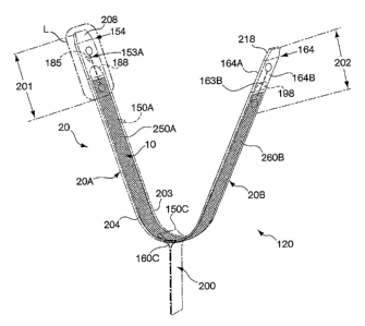
or more fork
prongs 171. The fork prongs 171 may be configured to have a pointed end 175.
Alternatively,
the fork prongs 171 may be configured with a flat end 175. In one embodiment,
the fork prongs
171 are configured with a rounded end 175. In other embodiments, a fork prong
171 has a sharp
end 175 or a dull end 175. Placement forks 170 may have multiple fork prongs
171 having
different configurations.
[0086] In one embodiment, referring to FIGS. 11, 1J and 1K, the placement fork
171 is employed
to place a sling 10 for treatment of female urinary incontinence in sleeves
20A and 20B of
envelope 20. Each of sleeves 20A and 20B measure 9.75 inches and the sling 10
measures 18
inches. The length of each fork prong 171 may range from about 0.300 inch to
about 0.400 inch,
preferably about 0.350 inch long. The diameter of each fork prong 171 may
measure between
about 0.028 inch to about 0.040 inch, preferably about 0.030 inch. In one
embodiment, there are
CA 02495314 2005-02-14
WO 2004/016180 PCT/US2003/025377
-20-
two fork prongs 171 on the placement fork 170. In another embodiment, each
fork prong 171 is
spaced from about 0.090 inch to about 0.110 inch, preferably about 0.100 inch
from the adjacent
fork prong 171 on the placement fork 170. In one embodiment, the fork prongs
171 have
rounded ends 175. The fork handle 173 length may range from about 10 inches to
about 12
inches, preferably about 11 inches long.
[0087] The placement fork 170, including the handle 173 and fork prongs 171,
may be
configured to fit within the lumen 185 of envelope 20. The fork handle 173 and
the fork prongs
171 of the placement fork 170 may be fabricated from various materials,
including medical grade
stainless steel, for example 304 stainless tool steel, or medical grade
plastic, for example nylon
or TEFLON . In one embodiment, a material, such as a soft plastic, may be
employed to cover
the placement fork 170 handle 173 to provide ease of gripping and/or comfort
to the operator
using the placement fork 170.
[0088] Optionally, referring still to FIGS. 1J and 1K, when placing a sling
10, the overlap region
160C is pulled in the direction perpendicular to the envelope 20 and the
overlap region 150C is
pulled out of the lumen 185B of second sleeve 20B in the direction
perpendicular to the envelope
20. The placement fork 170 may be held by the fork handle 173. The fork prongs
171 may be
used to position the sling 10 inside envelope 20. The placement fork 170 may
have one or more
fork prongs 171. In one embodiment, the positioning fork 170 has two fork
prongs 171.
[0089] In one illustrative embodiment, the implant, a sling 10, is pierced by
the fork prongs 171
and positioned in the lumen 185B of second sleeve 20B of envelope 20.
Thereafter, the sling 10
is positioned in the lumen 185A of first sleeve 20A of envelope 20. In another
embodiment, the
prongs 171 of the placement fork 170 are employed to position the sling 10
within the lumen
185B of the second sleeve 20B and within the lumen 185A of the first sleeve
20A, without
piercing the sling 10. To ensure proper sling 10 placement within the lumen(s)
185A and 185B
of the sleeves 20A and 20B, respectively, the length of the handle 173 of
placement fork 170
may be selected according to the combined lengths of the sleeves 20A and 20B.
[0090] Referring again to FIG. 1K, the overlap regions 150C and 160C, cover
the sling 10
enclosed within envelope 20. In another illustrative embodiment according to
the invention,
referring to FIG. 1K, the sling 10 is placed within the envelope 20 after the
bottom sections 150B
and 160B of sleeves 20A and 20B are joined to form hinge 200. Referring now to
FIG. 1 A, the
tabs 208 and 218 are joined to the envelope 20 after the hinge 200 is formed.
The tabs 208 and
CA 02495314 2005-02-14
WO 2004/016180 PCT/US2003/025377
-21-
218 are external to the envelope 20 and they are positioning members for
positioning the
envelope 20 inside the body of the patient. The tabs 208 and 218 are
positioned at the proximal
end 154 and the distal end 164 of envelope 20, respectively. The implant, for
example the sling
10, may be manually inserted into lumen 185 of envelope 20. Alternatively, the
sling 10 may be
inserted into lumen 185 with the aid of a placement fork 170 described above
with reference to
1 o FIG. 1 J.
[0091] In another illustrative embodiment, referring to FIGS. 1K and 1J, the
sling 10 is manually
placed within the first lumen 185A of the first sleeve 20A and/or placed with
the aid of the
placement fork 170 or a grasping device, such as, for example, forceps.
Thereafter, sleeve 20B
is joined with the sleeve 20A to form the envelope 20, including the hinge
200. The remaining
portion of the sling 10 may be placed within the second lumen 185B of sleeve
20B prior to or
after forming the hinge 200.
[0092] In yet another embodiment, referring to FIGS. 1D, IF and 1K the sling
10 is placed, via
the tab accesses, 153A, 153B, 163A, and 163B, within the envelope 20 after the
sleeves 20A and
20B are joined at hinge 200. According to this embodiment, referring also to
FIGS. 1A and 1J,
the sling 10 is inserted into, for example, cut 230B of tab access 163B
manually and/or with the
aid of a grasping device or the placement fork 170 and the sling 10 is
positioned within lumen
185. In another embodiment, the sling 10 is placed within the envelope 20 via
the tab accesses,
153A, 153B, 163A, and/or 163B after the sleeves 20A and 20B are joined at
hinge 200, and after
tabs 208 and 218 are joined to the envelope 20.
[0093] With reference to FIGS. 1L-1M, in another aspect the invention includes
a method for
positioning the sling 10 at an anatomical site in the body of a patient.
According to one
embodiment of this method of the invention, the operator positions the
assembly 120 of the
envelope 20 enclosing sling 10 at the anatomical site, for example, the
urethra 999 or
bladderneck of a female patient. Referring to FIG. 1L, the tab 188 and the tab
access 153A are
located at the first end 201 of the envelope. The operator accesses the tab
188 by pulling tab 188
through cut 220A of tab access 153A. Referring to FIG. 1M, the operator
intravaginally grasps
hinge 200 and simultaneously pulls the tab 188 in the direction indicated by
arrow 189. The
force exerted on tab 188 coupled to the top section 150A tears the envelope 20
along the top
surface 250A of the first sleeve 20A. Thereafter, the top section 150A of
sleeve 20A is torn
away from the envelope 20 in a single piece to expose the implant sling 10.
Tab 188, the top
CA 02495314 2005-02-14
WO 2004/016180 PCT/US2003/025377
-22-
section 150A, the top of sleeve 20A, and a portion of the proximal top section
154A are torn
away from the envelope 20 and the implant sling 10 is exposed. The exposed
portion of the sling
is adjacent the urethra 999.
[0094] Referring again to FIGS. 1A, 1F, 11, 1K, and 1M, the operator similarly
accesses tab 198
by pulling tab 198 through cut 220B of tab access 163A. The tab 198 and the
tab access 163A
10 are located at the second end 202 of the envelope 20. The operator
intravaginally grasps hinge
200 and simultaneously pulls tab 198. The force exerted on tab 198 coupled to
the top section
160A tears the envelope 20 along the top surface 260A of the sleeve 20B away
from the
envelope 20. The force exerted on tab 198 coupled to the top section 160A
tears these portions
of sleeve 20B away from the envelope 20. Thus, tab 198, the top section 160A,
the top of sleeve
20B, and a portion of the distal top section 164A are withdrawn from envelope
20. The portion
of the sling 10 previously enclosed by the envelope 20 is thereby exposed.
Finally, the hinge
200 and the remainder of envelope 20 including the tabs 208 and 218 are
withdrawn via the
patient's vagina. The sling 10 remains inside the body of the patient at the
anatomical site where
the operator positioned the sling 10, for example, at the anatomical site of
the urethra 999.
[0095] In another illustrative embodiment, FIGS. 2A-2C illustrate the envelope
20, described in
accordance with FIGS 1A-1M, further including a first guide tube 350A and a
second guide tube
350B. As illustrated in FIGS. 1K and 2A the assembly 120 includes the envelope
20 including
first sleeve 20A, second sleeve 20B, tabs 188, 198, tab accesses 153A, 153B,
163A, and 163B,
the envelope hinge 200, the envelope 20 enclosing all or part of sling 10. A
first guide tube
350A is coupled to the proximal end 154 of first sleeve 20A and the second
guide tube 350B is
coupled to the distal end 164 of the second sleeve 20B of envelope 20. The
first guide tube
350A, the proximal end 154, and the tab access 153A and the second guide tube
350B, the distal
end 164, and the tab access 163A are positioned on opposite ends of the
envelope 20 formed by
sleeve 20A and sleeve 20B. The tab access 153A and 153B are positioned at the
first end 201 of
the envelope 20 and the tab accesses 163A and 163B are positioned at the
second end 202 of the
envelope 20.
[0096] The guide tubes 350A and 350B facilitate interoperability of the
assembly 120 with a
delivery device. Other structures that facilitate interoperability of the
assembly 120 with a
delivery device to introduce the assembly 120 to the body of the patient may
be employed in
accordance with the assembly 120 of the invention. Suitable configurations and
structures
CA 02495314 2010-09-23
23
include, for example, loops, apertures, male and/or female connectors, guide
tubes and the
like. Such other structures that may be employed are described in more detail
in United
States patent Nos. 6,991,597, 6,936,052 and 7,025,772 and United States patent
application
publication Nos. 20020151909, 20020156489 and 20030009181, filed in the United
States
Patent Office on March 7, 2002 and United States patent application
publication No.
20040122474 filed December 19, 2002.
[0097] Referring to FIGS. 2A-2C, the operator may employ the assembly 120 of
the
invention to position the implant at an anatomical site in the body of a
patient. The operator
will employ the first and second guide tube 350A and 350B to position the
envelope 20
enclosing sling 10 at an anatomical site in the body of a patient. Other
structures as
described and incorporated herein above may be used to position the envelope
20 enclosing
sling 10 at an anatomical site in the body of a patient. The envelope 20 may
be positioned
with the aid of a delivery device that may be employed to access the patient's
urethra 999
according to supra-pubic, pre-pubic, transvaginal, or transobturator
approaches. The
operator positions the envelope 20 enclosing sling 10, at the anatomical site,
for example,
the urethra 999 or bladderneck. Referring to FIG. 2B, the operator accesses
the tab 188 by
pulling tab 188 through cut 220A of tab access 153A. Referring to FIG. 2C, the
operator
intravaginally grasps hinge 200 and simultaneously pulls the tab 188 in the
direction
indicated by arrow 189. The force on tab 188 coupled to the top section 150A
tears the
envelope 20 along the top portion of the first sleeve 20A. Thereafter, the top
section 150A
of sleeve 20A is torn away from the envelope 20 in a single piece to expose
the implant
sling 10. Tab 188, the top section 150A, the top surface 250A of sleeve 20A,
and a
CA 02495314 2005-02-14
WO 2004/016180 PCT/US2003/025377
-24-
portion of the proximal top section 154A are torn away from the envelope 20
and the implant
sling 10 is exposed. The exposed portion of the sling 10 is adjacent the
urethra 999.
[0098] Referring now to FIGS. lK, lF and 2C, the operator similarly accesses
tab 198 by pulling
tab 198 through cut 220B of tab access 163A. The operator intravaginally
grasps hinge 200 and
simultaneously pulls tab 198. The force exerted on tab 198 coupled to the top
section 160A tears
these portions of sleeve 20B away from the envelope 20. Thus, tab 198, the top
section 160A,
the top surface 260A of sleeve 20B, and a portion of the distal top section
164A are withdrawn
from envelope 20. The portion of the sling 10 previously enclosed is thereby
exposed. Finally,
the hinge 200 and the remainder of envelope 20 including the first guide tube
350A and the
guide tube 350B are withdrawn via the patient's vagina. The sling 10 remains
inside the body of
the patient at the anatomical site where the operator positioned the sling 10,
for example, at the
anatomical site of the urethra 999.
[0099] Referring now to FIGS. 1D, 2B and 2C, in another illustrative
embodiment, the tab
accesses 153A and 153B include perforations (not shown) about the perimeter of
the
perpendicular axis 24A of sleeve 20A of envelope 20. The operator can access
the tab 188 by
grasping and withdrawing, for example, the guide tube 350A and the portion of
the envelope 20
lying between the perforation and the envelope 20 proximal end 154. The tab
188 is thereby
exposed and is accessible to the operator. In one embodiment, the envelope 20
tab access 153A
has perforations about the perimeter of the perpendicular axis 24A of envelope
20 and also has a
nick (not shown) at one or more portions of the perforated perimeter that
enables the tab access
153A to be pulled or torn to expose the tab 188 enclosed within the envelope
20.
[0100] In yet another embodiment, referring now to FIGS. 11, 1K, and 2A the
tab access 153A
and 153B includes shapes (not shown) cut out of one or more of the top
surfaces 250A, 260A
and bottom surfaces 250B, 260B of sleeves 20A and 20B, respectively. Suitable
cut out shapes
include, oval and triangle, and the triangular cut out shape may have, for
example, rounded
edges.
[0101] Referring now to FIGS. 1D, 1F, and 2A, in one illustrative embodiment
for positioning
the assembly 120 in the body of a patient, the envelope 20 enclosing an sling
10 is positioned
inside the body of a patient. The tab access 153A, including a cut 220A
disposed between the
first hole 151A and the second hole 152A, and the tab access 153B, including a
cut 230A
disposed between the first hole 151B and the second hole 152B, are cut at
position 437A. The
CA 02495314 2005-02-14
WO 2004/016180 PCT/US2003/025377
-25-
cut at position 437A cuts through the second holes 152A and 152B. The guide
tube 350A and a
portion of the envelope material are withdrawn. Thereafter, the sides of
envelope 20 sleeve 20A
are pulled apart by opening and separating each of the cuts 220A and 230A. In
one embodiment,
the sides of envelope 20 separate until the point where the first holes 15 1A
and 151B are located.
The first holes 151A and 151B may provide, for example, relief to the cuts
220A and 230A.
[0102] In another illustrative embodiment, tab accesses 163A, 163B on sleeve
20B are cut at
position 437B and tab 198 is similarly accessed by separating the sides of the
envelope 20. The
guide tube 350B and a portion of the envelope are withdrawn.
[0103] According to this embodiment, after the sling 10 is positioned, the
operator cuts the
envelope 20 at position 437A, withdraws the first guide tube 350A, and
separates the envelope
20 to provide accesses to the tab 188. The operator intravaginally grasps
hinge 200 and
simultaneously pulls the tab 188 and the force on tab 188 coupled to the top
section 150A tears
the envelope 20 along the top portion of the first sleeve 20A. The top section
150A of sleeve
20A is torn away from the envelope 20 in a single piece to expose the implant
sling 10. Tab 188,
the top section 150A, the top surface 250A of sleeve 20A, and a portion of the
proximal top
section 154A are torn away from the envelope 20 and the implant sling 10 is
exposed. The
exposed portion of the sling 10 is adjacent the anatomical site
[0104] The operator cuts the envelope 20 at position 437B, withdraws the
second guide tube
350B, and separates the envelope 20 to provide accesses to the tab 198. The
operator grasps
hinge 200 and simultaneously pulls tab 198. The force exerted on tab 198
coupled to the top
section 160A tears these portions of sleeve 20B away from the envelope 20.
Thus, tab 198, the
top section 160A, the top surface 260A of sleeve 20B, and a portion of the
distal top section
164A are withdrawn from envelope 20. The portion of the sling 10 previously
enclosed is
thereby exposed. Finally, the hinge 200 and the remainder of envelope 20 are
withdrawn via the
vagina. This embodiment avoids guide tubes 350A and 350B being pulled back
through the
patient's body when the operator withdraws the remainder of the envelope. The
sling 10 remains
inside the body of the patient at the anatomical site where the operator
positioned the sling 10.
[0105] Referring again to FIG. 2A, in another embodiment, the tab access 153A
and 153B is a
cut out shape, for example, an oval (not shown). In one embodiment, the cut
out oval may be cut
over positions 437A and 437B. When the operator cuts positions 437A, 437B
withdrawing a
portion of the envelope 20 and first guide tube 3 50A and a portion of the
envelope 20 and a
CA 02495314 2005-02-14
WO 2004/016180 PCT/US2003/025377
-26-
second guide tube 350B. The portion of the envelope 20 below positions 437A
and 437B may
be separated until the point at the bottom of the oval, providing access to
the tabs 188, 198
enclosed within the envelope 20.
[0106] In another aspect, the invention includes an assembly 120 for
delivering the envelope 20
and an implant, for example, a sling 10 to an anatomical site in the body of a
patient. In one
embodiment, shown in FIG. 3A, the assembly 120 includes the envelope 20
including two or
more sleeves 20A, 20B, one or more tabs 208, 218 and a tongue 155. The tongue
155 simplifies
placement and/or withdrawal of the envelope 20 from inside the body of the
patient and prevents
damage to the sling 10 thereby maintaining the integrity of the sling 10
enclosed by the envelope
20.
[0107] Referring to FIG. 3D, generally, the tongue 155 is an extension of the
top section 150A
of the sleeve 20A. The tongue 155 extends from one end of the sleeve 20A. In
one
embodiment, the tongue 155 is separate, i.e., non-contiguous with the bottom
section 150B of the
sleeve 20A. In another embodiment, tongue 155 extends from the top section
150A beyond the
lumen 185A of the sleeve 20A. The tongue maybe flexible and it may be any flat
shape, for
example, rectangular or oval. The length of the tongue measures between about
0.2 inches and
about 5 inches, preferably about 1 inch in length. The width of the tongue
measures between
about 0.1 inches and about 2.0 inches. The thickness of the tongue 155 is at
least as thick as the
sleeve from which the tongue is made. In another embodiment, the tongue 155 is
bonded at an
end of sleeve 20A, in such embodiments the tongue 155 thickness may be
greater, less, or equal
to the thickness of the sleeve 20A to which the tongue 155 is bonded. The
tongue 155 may be
bonded to sleeve 20A by adhesives, sutures, or heat bonding as well as by
other bonding means
known to the skilled person.
[0108] Referring now to FIG. 3B, in one embodiment of the invention, the
tongue is made by
cutting two angles into the first sleeve 20A, as indicated by the arrow 176
and arrow 178. The
3o angles 176 and 178 are cut in the range of from about 40 to about 170 , or
about 80 to about
130 , preferably about 110 , from the longitudinal axis 22A of the sleeve 20A
illustrated in FIG.
3B. Preferably, the angles 176 and 178 are obtuse, ranging from greater than
90 to about 130
as measured from the longitudinal axis 22A on the side of the distal end 150
of the sleeve 20A.
In one embodiment, the two angles 176 and 178 are cut into the first sleeve
20A for a distance
between about 0.01 inch and about 0.05 inch, preferably about 0.04 inch into
the sleeve 20A as
CA 02495314 2005-02-14
WO 2004/016180 PCT/US2003/025377
-27-
measured along the perpendicular axis 24A. Referring now to FIGS. 3A and 3D,
the angles 176
and 178 and the depth of the cut into the sleeve 20A along the perpendicular
axis 24A at which
the angles 176 and 178 are placed, determines the placement of tongue 155 of
envelope 20, by,
for example, sizing or placing the tongue 155 to cover the sling 10 enclosed
by envelope 20.
[0109] Referring to FIG. 3C, the distal end portion 150 of sleeve 20A is
trimmed along the
longitudinal axis 22A from the distal end 150 to angle 176 and from the distal
end 150 to the
angle 178. In general, the amount of the first sleeve 20A that is trimmed
along the longitudinal
axis 22A from the distal end 150 to each respective angle 176 and 178 is
substantially the same.
The first sleeve 20A has a first lumen 185A, and its structure is tube-like,
and after the first
sleeve 20A is trimmed, the distal end 150 portion has a top section 150A and a
bottom section
150B. After the first sleeve 20A is trimmed, the longitudinal axis 22A of
first sleeve 20A from
the angle 178 to the end 150 of top section 150A, may measure between about
2.0 inches to
about 14.0 inches, preferably about 10.0 inches. As illustrated in FIG. 3D,
the top section 150A
is trimmed along the perpendicular axis 24A forming a tongue 155 which
measures between
about 0.2 inches and about 5 inches, preferably about 1 inch in length as
measured along the
longitudinal axis 22A of envelope 20. The envelope 20 may be trimmed by
employing, for
example, a razor. The width of the tongue 155 measured along the perpendicular
axis 24A of
envelope 20, is sized so that it is equal to or smaller than the inner
diameter of a second lumen
185B of the second sleeve 20B of envelope 20, illustrated in FIG. 3F. For
example, the width of
the tongue 155 may measure between about 0.1 inches and about 2.0 inches.
[0110] Referring now to FIGS. 3E and 3F, the second sleeve 20B of the envelope
20 shown in
FIG. 3A is illustrated. Referring to FIG. 3E, the second sleeve 20B has the
inner surface 30, the
outer surface 40, the proximal end 160, and the distal end 164. The second
sleeve 20B includes
the second lumen 185B. The length of the second sleeve 20B between the
proximal end 160 and
the distal end 164, along the longitudinal axis 22B, is between about 4.0
inches and about 28.0
inches, preferably about 20.0 inches. The perpendicular axis 24B of second
sleeve 20B
measures between about 0.2 inches to about 2.0 inches, preferably between
about 0.4 inches and
about 0.8 inches, preferably about 0.6 inches.
[0111] Still referring to FIG. 3E, the two angles 176,178, are cut into the
second sleeve 20B in
the range of about 40 to about 170 , or about 800 to about 130 , preferably
about 90 from the
longitudinal axis 22B of second sleeve 20B proximal end 160. In one
embodiment, the two
CA 02495314 2005-02-14
WO 2004/016180 PCT/US2003/025377
-28-
angles 176 and 178 are cut into the second sleeve 20B for a distance between
about 0.01 inch
and about 0.05 inch, preferably about 0.04 inch into the sleeve 20A as
measured along the
perpendicular axis 24B. The second sleeve 20B is trimmed along the
longitudinal axis 22B from
the proximal end 160 to the angle 176 and from the proximal end 160 to the
angle 178. In
general, the amount of the second sleeve 20B that is trimmed along the
longitudinal axis 22B
from the proximal end 160 to each respective angle, 176 and 178, is
substantially the same. The
second sleeve 20B second lumen 185B includes a tube-like structure. Referring
now to FIGS.
3E and 3F, after the tube-like second sleeve 20B is trimmed, the proximal end
160 includes a top
section and a bottom section 160B. Referring now to FIG. 3F, the longitudinal
axis 22B of
second sleeve 20B from the angle 176 and the angle 178 to the end of bottom
section 160B may
measure between about 2.0 inches to about 14.0 inches, preferably about 10.0
inches. The top
section is trimmed along the perpendicular axis 24B of sleeve 20B, from the
legs of angles 176
and 178, which are at an angle to the longitudinal axis 22B of sleeve 20B.
[0112] As shown in FIGS. 3D, 3F, and 3G, the bottom section 150B of the first
sleeve 20A is
placed at about a 90 angle relative to the longitudinal axis 22A of the first
sleeve 20A.
Similarly, the bottom section 160B of the second sleeve 20B is placed at about
a 90 angle
relative to longitudinal axis 22B of the second sleeve 20B. The sleeves 20A
and 20B are aligned
in proximity to one another such that the bottom sections 150B and 160B of
sleeves 20A and
20B, respectively, face one another.
[0113] Referring now to FIG. 31, the bottom sections 150B and 160B are joined
to one another
by adhesive, staples, heat bonding or other means known to the skilled person
to form hinge 200.
In one embodiment, a clip (not shown) may be employed to join bottom sections
150B and
160B, which act as hinge sections when they form hinge 200. As shown in FIG.
3H, the
proximal end 154 of first sleeve 20A and the distal end 164 of the second
sleeve 20B are
positioned on opposite ends of the envelope 20 formed by sleeve 20A and sleeve
20B. The
length of envelope 20, measured along the longitudinal axis 22 from proximal
end 154 to the
distal end 164, ranges between about 4.0 inches to about 28.0 inches, or
between about 12.0
inches and about 24.0 inches, most preferably 20.0 inches.
[0114] Referring again to FIG. 3I, the tongue 155 of the first sleeve 20A is
tucked inside the
lumen 185B of the second sleeve 20B. The tongue 155 is sized and shaped so
that it fits within
the lumen 185B of the second sleeve 20B. When the tongue 155 is tucked inside
lumen 185B,
CA 02495314 2005-02-14
WO 2004/016180 PCT/US2003/025377
-29-
the sling 10 enclosed within envelope 20 is shielded by the tongue 155 in the
region 255 of the
envelope 20 corresponding to the tongue 155. For example, in embodiments where
the envelope
20 is employed to implant a mid-urethral sling 10, the tongue 155 of the
envelope 20 prevents
damage to the sling 10 caused by, for example, patient tissue rubbing against
the sling 10 in the
region of the envelope 20 corresponding to the tongue 155 during sling 10
placement. Also, the
tongue 155 prevents abrasion to patient tissue caused by the sling 10 during
envelope 20
placement.
[01151 Referring to FIGS. 3A and 3D, in another embodiment, two sleeves 20A,
each having a
tongue 155, are provided. The two sleeves 20A are positioned so that their
tongues 155 face one
another and are positioned on the same plane. Each sleeve 20A tongue 155 is
configured to fit
inside the lumen 185A of sleeve 20A that it faces. According to this
embodiment, the two sleeve
20A tongues 155 combine to provide the desired coverage and protection of the
sling 10
enclosed within the envelope 20. In one illustrative mbodiment, referring,
still to FIGS. 3A and
3D, two sleeves 20A having a perpendicular axis 24A measuring about 1 inch may
be identically
cut to provide a tongue 155 width that measures about 0.5 inch, as measured
from angle 178
along the perpendicular axis 24A. The two sleeves 20A are positioned so that
their tongues 155
face one another and are positioned on the same plane. Each of the two sleeves
20A tongue 155
is inserted into the lumen 185A of the sleeve 20A that it faces and the two
tongues 155 protect a
sling 10 enclosed by the two sleeves 20A when they form an envelope 20.
[01161 As shown in FIGS. 3A and 3G-3I, and as described above, sleeves 20A and
20B are
joined to form envelope 20. The first lumen 185A and the second lumen 185B
form the single
lumen 185 of the envelope 20. The joined bottom sections 150B and 160B of
sleeves 20A and
20B act as hinge sections to form hinge 200 of envelope 20. Various methods
may be employed
to join bottom sections 150B and 160B, for example, heat bonding, adhesives,
or staples. In one
embodiment, a clip (not shown) maybe employed to join bottom sections 150B and
160B,
forming hinge 200. The length of hinge 200 may range between about 2.0 inches
to about 14.0
inches in length. The hinge 200 may be trimmed to a shorter length that may be
grasped by the
operator during the method of delivering the implant assembly, described
below. One or both of
sleeves 20A and 20B of envelope 20 may be made from a composite of two or more
materials.
[01171 In another aspect, the invention is a method for delivering an implant
assembly 120 to a
patient's body. Referring to FIGS. 3H-3I, the sling 10 is placed within the
envelope 20 as is
CA 02495314 2005-02-14
WO 2004/016180 PCT/US2003/025377
-30-
described above with reference to FIGS. 1J and 1K. At least a portion of the
sling 10 is enclosed
within the envelope 20 and in one embodiment, the envelope 20 encloses
substantially all of the
sling 10. With reference to FIG. 3A, in another aspect the invention is a
method for positioning
an implant, for example, a sling 10 at an anatomical site in the body of a
patient. According to
one embodiment of this method of the invention, the operator positions the
envelope 20
enclosing sling 10, at the anatomical site, for example, the urethra 999 or
bladderneck.
[0118] With reference to FIGS. 3A and 3I, the operator grasps hinge 200 and
cuts hinge 200
near position 200A. Position 200A is the area of the hinge 200 adjacent
implant sling 10, where
bottom sections 150B and 160B of sleeves 20A and 20B are not sealed. In one
embodiment, the
operator cuts one bottom section, 150B or 160B, at position 200A to free it
from the other
bottom section 150B or 160B. In another embodiment, the operator cuts hinge
200 at position
200A and thereafter withdraws hinge 200 via the vagina. In one embodiment, the
joined bottom
sections 150B and 160B of hinge 200 are pulled apart. In another embodiment,
the operator
unfastens and withdraws a clip (not shown) surrounding the bottom sections
150B and 160B,
which act as hinge sections, that form hinge 200.
[0119] The operator next grasps tab 208 and pulls tab 208 and the attached
sleeve 20A in the
direction indicated by arrow 289, withdrawing the tab 208 and attached sleeve
20A. Thereafter,
the operator grasps tab 218 and pulls tab 218 and the attached sleeve 20B in
the direction
indicated by arrow 389, withdrawing the tab 218 and attached sleeve 20B. The
sling 10
previously enclosed by the envelope 20 is thereby exposed and the sling 10
remains inside the
body of the patient at the anatomical site where the sling 10 was positioned
by the operator, for
example, at the anatomical site of the urethra 999.
[0120] FIG. 4 illustrates the envelope 20 having the tongue 155, as described
above with
reference to FIGS. 3A-3I, further including the first guide tube 350A and the
second guide tube
350B, as described above with reference to FIGS. 2A-2C. The guide tubes 350A
and 350B may
provide the operator a mechanical advantage and simplify grasping and
withdrawing the sleeves
20A and 20B during the surgical procedure to position the sling 10 at an
anatomical site in the
body of a patient, for example, at the urethra 999.
[0121] In another aspect, the invention includes an assembly 120 for
delivering an envelope 20
and an implant sling 10 to an anatomical site in the body of a patient. In one
embodiment,
shown in FIG. 5A the assembly 120 includes the envelope 20 including two or
more sleeves
CA 02495314 2005-02-14
WO 2004/016180 PCT/US2003/025377
-31-
20A, 20B, one or more guide tubes 350A, 350B, and a scaffold 305. Referring
also to FIG. 51,
the scaffold 305 joins sleeve 20A and 20B to form envelope 20. In one
embodiment, the
scaffold 305 has a fold 310 that is positioned, for example, between sleeves
20A and 20B. In
one embodiment the fold 310 positioned between sleeves 20A and 20B forms a
hinge 200. The
scaffold 305 simplifies placement and/or withdrawal of the envelope 20 from
inside the body of
the patient and maintains the integrity of the envelope 20.
[0122] Referring now to FIGS. 5A-5G, the scaffold 305 generally is a flat
piece of material that
is dimensioned to fit within the lumens 185A and 185B of the sleeves 20A and
20B to join the
sleeves 20A and 20B that form the envelope 20. The scaffold 305 may be
fabricated from one or
more materials such as, for example, polypropylene, polyethylene, polyester,
polytetrafluoroethylene, TYVEK , MYLAR , and co-polymers thereof as well as
one or more
dissolvable materials or one or more absorbent materials such as, for example,
a sponge-like
material such as polyglycolic acid, polylactic acid and other suitable
absorbable materials. The
scaffold 305 may be firm or rigid, but preferably, it is flexible. Referring
to FIG. 5C, the
scaffold thickness may range from about 1 mil to about 6 mil, preferably about
3.5 mils.
Referring to FIG. 5B, the scaffold 305 longitudinal axis 32 is measured from
the scaffold 305
first side 305A to the scaffold second side 305B. The longitudinal axis 32
ranges between about
4 inches and about 60 inches, preferably being about 23 inches. The scaffold
perpendicular axis
34 ranges between about 0.2 inches to about 2 inches, preferably between about
0.4 inches to
about 0.8 inches, preferably being about 0.6 inches.
[0123] FIGS. 5A and 5D illustrate the scaffold 305 having a fold 310. The fold
310 may be
employed to position an implant enclosed by envelope 20. The fold 310 may aid
in withdrawal
of the scaffold 305 or withdrawal of the envelope 20 after assembly 120
placement (described
below). In one embodiment, a portion of the scaffold 305 longitudinal axis 32
is pinched to form
a fold 310. Adhesives, staples, heat bonding or other bonding means known to
the skilled person
maybe employed to maintain the fold 310 in the scaffold 305. The fold 310, as
measured
perpendicular to the longitudinal axis 32 of scaffold 305, ranges from about
0.5 inches to about 6
inches, preferably 2 inches long. In another embodiment, two or more pieces of
material may be
bonded together by adhesives, staples, heat bonding or other bonding means
known to the skilled
person to make scaffold 305 and the two pieces of material may form a bond
joint to provide the
fold 310.
CA 02495314 2005-02-14
WO 2004/016180 PCT/US2003/025377
-32-
[0124] Referring to FIG. 5A, the scaffold 305 thickness and the material used
to manufacture the
scaffold 305 may be selected to provide the envelope 20 with integrity
appropriate for the
surgical procedure, e.g., to withstand the forces imposed on the envelope 20
when placing the
sling 10 in the patients body. In another embodiment, the scaffold 305
material may be selected
because it forms a strong bond with the sleeve 20A, 20B or the guide tube
350A, 350B. The
to scaffold 305 material may be the same or different than the material used
to manufacture the
sleeves 20A, 20B. The scaffold 305 may be bonded inside the lumens 185A and
185B of the
sleeves 20A and 20B by adhesives, suturing, heat bonding or any other means
known to the
skilled person. In one embodiment, the scaffold 305 is bonded to a portion of
the sleeve 20A
inside the lumen 185A.
[0125] Referring to FIG. 5A, in one embodiment according to the invention, the
assembly 120
includes an envelope 20 including a first sleeve 20A, a second sleeve 20B, the
scaffold 305 and a
sling 10. Guide tubes 350A and 350B are disposed on the proximal end 154 and
the distal end
164 of the envelope 20. Referring now to FIG. SE, the sleeve 20A may have a
tongue 155
prepared in the same manner as was described with respect to the FIGS. 3B-3D.
Referring still
to FIG. SE, the bottom section 150B, described in relation to FIG. 3D, is
removed by trimming
the bottom section 150B along the perpendicular axis 24A of sleeve 20A. In one
embodiment,
the bottom section 150B of sleeve 20A is trimmed along the perpendicular axis
24A from about
the angle 176 vertex to about the angle 178 vertex.
[0126] Referring to FIG. SF, the second sleeve 20B of the envelope 20 shown in
FIG. SA is
illustrated. The second sleeve 20B has an inner surface 30, an outer surface
40, a proximal end
160, and a distal end 164. The second sleeve 20B is a tube-like structure
including a second
lumen 185B. The length of the second sleeve 20B between the proximal end 160
and the distal
end 164, along the longitudinal axis 22B, measures between about 2.0 inches
and about 14.0
inches, preferably being about 10.0 inches. The perpendicular axis 24B of
second sleeve 20B
measures between about 0.2 inches to about 2.0 inches, preferably between
about 0.4 inches and
about 0.8 inches, preferably about 0.6 inches.
[0127] As illustrated in FIGS. SG and 51, the first side 305A of scaffold 305
is inserted into
lumen 185A and the second side 305B of scaffold 305 is inserted into lumen
185B. The scaffold
305 may be bonded to a portion or along the entire length of the lumens 185A
and 185B of
sleeves 20A and 20B, respectively, by employing adhesive, staples, heat
bonding or other means
CA 02495314 2005-02-14
WO 2004/016180 PCT/US2003/025377
-33-
known to the skilled person. In another embodiment, the scaffold 305 is simply
positioned
inside the lumens 185A and 185B of the sleeves 20A and 20B without being
bonded to the
sleeves 20A or 20B of envelope 20. One or both of sleeves 20A and 20B may be
made from a
composite of two or more materials.
[0128] In one embodiment, referring now to FIGS. 5E and 5F, sleeve 20A,
measured from the
proximal end 154 to the end of the tongue 155 along the longitudinal axis 22A
measures about
10.75 inches, with the length of tongue 155 measuring about 1 inch, and the
longitudinal axis
22B of sleeve 20B measures about 9.75 inches. Referring again to FIG. 51, in
one embodiment,
the scaffold 305 joins sleeves 20A and 20B to form envelope 20 and the sling
10 is positioned
inside the lumen 185 of envelope 20. When the sleeve 20A tongue 155 is tucked
inside the
lumen 185B of sleeve 20B, the envelope 20 measures about 19.5 inches. In an
embodiment
where sling 10 measuring about 14 inches long is enclosed by envelope 20,
about 5.5 inches of
space of lumen 185 is free from the sling 10. The proximal end 154 and the
distal end 164 of
envelope 20 may each extend up to about 2 inches beyond the sling 10.
[0129] Referring now to FIGS. 5F and 51, the scaffold 305 may be used to join
two sleeves 20B
and 20B that do not have a tongue 155. In such embodiments, a portion of the
sling 10 enclosed
by the envelope 20 may be exposed.
[0130] One or both of sleeves 20A and 20B of envelope 20 may be made from a
composite of
two or more materials. The scaffold 305 may or may not have a fold 310.
Referring now to
FIG. 5A, the scaffold 305 may be used to join sleeves 20A and 20B with or
without guide tubes
350A and 350B. For example, in some embodiments, the scaffold 305 joins
sleeves 20A and
20B having tabs (not shown).
[0131] In another illustrative embodiment according to the invention,
referring again to FIG. 5A,
the sling 10 is placed within the lumen 185 of envelope 20 after sleeves 20A
and 20B are joined
when scaffold 305 is positioned between the sleeves 20A and 20B, but prior to
guide tubes 350A
and 350B being joined to the envelope 20. A first guide tube 350A and a second
guide tube
350B are described above with reference to FIGS. 2A-2C. Tabs 208 and 218
described with
reference to FIGS. 1A and 3A may alternatively be joined to the envelope 20
proximal end 154
and distal end 164, respectively. The implant, for example a sling 10, may be
inserted into the
lumen 185 of envelope 20 manually and/or with the aid of a grasping device or
with the aid of a
placement fork 170 described above with reference to FIG. 1J. The envelope 20
may enclose at
CA 02495314 2005-02-14
WO 2004/016180 PCT/US2003/025377
-34-
least a portion of the sling 10. In one embodiment, referring to FIG. 51, the
envelope 20 includes
a tongue 155, which may be employed to overlay or protect the sling 10
enclosed within
envelope 20.
[0132] Referring to FIGS. 1J and 51, in one embodiment, the sling 10 inside
lumen 185 lies
between scaffold 305 and the top surfaces, 250A and 260A of the sleeves 20A
and 20B of
envelope 20. In another illustrative embodiment, at least the first end 305A
of scaffold 305 is
placed inside of the first lumen 185A of sleeve 20A and the sling 10 is placed
within the first
lumen 185A of the first sleeve 20A manually and/or with the aid of a grasping
device or with the
aid of a placement fork 170. Thereafter, sleeve 20B is joined with sleeve 20A
by inserting at
least the second end 305B of scaffold 305 inside the lumen 185B of sleeve 20B
to form envelope
20. In one embodiment the first end 305A and the second end 305B of scaffold
305 are bonded
inside the lumens 185A and 185B of the sleeves 20A and 20B, respectively. The
remaining
portion of sling 10 may be placed within the second lumen 185B of sleeve 20B.
Optionally, the
remaining portion of sling 10 may be placed within the second lumen 185B of
sleeve 20B prior
to or after forming a fold 310 within scaffold 305.
[0133] The operator may employ the assembly 120 of the invention illustrated
in FIG. 5A to
position the sling 10 at an anatomical site in the body of a patient. The
operator will employ the
first and second guide tubes 350A and 350B to position the envelope 20
enclosing sling 10 at an
anatomical site in the body of a patient with the aid of a delivery device.
Suitable delivery
devices may be used to position the envelope 20 of the assembly 120 enclosing
at least a portion
of an implant according to a plurality of surgical approaches, for example,
according to
transvaginal, transabdominal (e.g., percutaneous), supra-pubic, pre-pubic, or
transobturator
approachs. The operator positions the envelope 20 enclosing sling 10 at the
anatomical site, for
example, the urethra 999 or bladdemeck.
[0134] With continued reference to FIG. 5A, the operator grasps the scaffold
305 that is between
the sleeves 20A and 20B and then cuts the scaffold 305. In one illustrative
embodiment, the
operator cuts and withdraws the fold 310 within the scaffold 305. Next, the
operator grasps the
first guide tube 35OA and pulls guide tube 350A, the attached sleeve 20A, and
a portion of
scaffold 305 in the direction indicated by arrow 289, withdrawing the guide
tube 350A, attached
sleeve 20A and a portion of scaffold 305 from the patient's body. Thereafter,
the operator grasps
the second guide tube 3 50B and pulls guide tube 350B, the attached sleeve 20B
and a portion of
CA 02495314 2005-02-14
WO 2004/016180 PCT/US2003/025377
-35-
scaffold 305 in the direction indicated by arrow 389, withdrawing the guide
tube 350B, the
attached sleeve 20B and a portion of scaffold 305 from the patient's body. In
one embodiment,
the guide tubes 35 OA and 350B provide the operator with a mechanical
advantage simplifying
grasping the sleeves 20A and 20B during the surgical procedure. The sling 10
previously
enclosed by the envelope 20 is thereby exposed and remains inside the body of
the patient at the
1o anatomical site where the sling 10 was positioned by the operator, for
example, at the anatomical
site of the urethra 999.
[0135] Referring still to FIG. 5A, in another illustrative embodiment, the
operator cuts envelope
20 at position 437A and withdraws the first guide tube 350A and a portion of
envelope 20. The
operator cuts envelope 20 at position 437B and withdraws the second guide tube
350B and a
portion of the envelope 20. According to this embodiment, the scaffold 305 may
be bonded to
the lumens 185A and 185B of envelope 20. The operator cuts the scaffold 305
and grasps and
withdraws the first envelope 20A and a portion of scaffold 305 from the
patient. Thereafter, the
operator grasps and withdraws the second envelope 20B and a portion of
scaffold 305 from the
patient. The sling 10 previously enclosed by the envelope 20 is thereby
exposed and remains
inside the body of the patient at the anatomical site where the sling 10 was
positioned by the
operator, for example, at the anatomical site of the urethra 999.
[0136] In another embodiment, referring still to FIG. 5A, the guide tube 350A,
the proximal end
154 of sleeve 20A and the scaffold 305 first end 305A are bonded together.
Similarly, the guide
tube 350B, the distal end 164 of sleeve 20B and the scaffold 305 second end
305B are bonded
together. The operator cuts and withdraws guide tubes 350A and 350B at
positions 437A and
437B on envelope 20. Thereafter, the operator grasps scaffold 305 between the
sleeves 20A and
20B and intravaginally withdraws the scaffold 305 from the envelope 20.
Optionally, the
scaffold 305 fold 310 is grasped. Finally, the operator grasps and withdraws
the first sleeve 20A
and thereafter grasps and withdraws the second sleeve 20B. The sling 10
previously enclosed by
the envelope 20 is thereby exposed and the sling 10 remains inside the body of
the patient at the
anatomical site where the sling 10 was positioned by the operator, for
example, at the anatomical
site of the urethra 999. Alternatively, after cutting the envelope 20 at
positions 437A and 437B
and withdrawing guide tubes 350A and 350B, the operator first grasps and
withdraws the first
sleeve 20A, then grasps and withdraws the second sleeve 20B, and finally
grasps and withdraws
the scaffold 305. In one embodiment, when the sleeves 20A and 20B are
withdrawn from the
CA 02495314 2005-02-14
WO 2004/016180 PCT/US2003/025377
-36-
patient prior to the scaffold 305, the scaffold 305 acts as a slide that
enables the sleeves 20A and
20B to disengage from the envelope 20.
[0137] Referring to FIGS. 6-7, the implant assembly 120, and more
specifically, the envelope 20
of the assembly 120 has a proximal end 154 and a distal end 164 and the
envelope 20 encloses at
least a portion of the implant, for example a sling 10 for treatment of female
urinary
incontinence. The implant sling 10 may be made from a mesh. Referring now to
FIG. 6, in one
embodiment, the envelope 20 includes a single sleeve 20C.
[0138] In one illustrative embodiment, the sling 10 includes a de-tanged mid-
length portion 244,
which has end points 249A and 249B, and two tanged end-length portions 248A
and 248B.
Suitable implant slings 10 include the de-tanged slings such as the slings
described in, for
example, U. S. S.N. 10/092,872 filed March 7, 2002, the entirety of which is
incorporated by
reference herein. The two tanged end-length portions 248A and 248B extend from
the end
points 249A, 249B of the de-tanged mid-length portion 244 to sling ends 252A
and 252B,
respectively. The two tanged end-length portions 248A and 248B are, in a
preferred
embodiment, of substantially equal length, such that the de-tanged mid-length
portion 244 is
substantially centered along the long axis of the sling 10.
[0139] The envelope 20 includes a first sleeve surface 256 extending along the
long axis of the
envelope 20 and a second sleeve surface 258 disposed on the opposite side of
the envelope 20
from the first sleeve surface 256. In one embodiment, the first sleeve surface
256 has at least
one discontinuity 264. Referring to FIG. 6, for example, in one embodiment,
the discontinuity
264 in the first sleeve surface 256 is, for example, a gap 268, indicated by
double-ended arrows
269. The gap 268 is defined by a first sleeve end point 276A and a second
sleeve end point
276B. Referring now to FIG. 7, in an alternative embodiment, the envelope 20
includes two
discontinuities 264 in the first sleeve surface 256, for example, slit-shaped
apertures 272A,
272B. In still other embodiments, the first sleeve surface 256, or the second
sleeve surface 258,
may include any number of discontinuities 264.
[0140] Referring to the embodiment illustrated in FIG. 6, a first region 273A
in the first sleeve
surface 256 extends from the proximal end 154 to the end point 276A and a
second region 273B
in the first sleeve surface 256 extends from the distal end 164 to the end
point 276B. The
separation of the first region 273A from the second region 273B by the gap 268
allows the
envelope 20 to be removed when the physician cuts the second sleeve surface
258. The gap also
aids in removal of the envelope 20 by preventing friction between the first
region 273A and the
CA 02495314 2005-02-14
WO 2004/016180 PCT/US2003/025377
-37-
second region 273B that might otherwise occur if the regions 273A and 273B
overlapped.
Friction between the regions 273A and 273B would make the process of removing
the envelope
20 from about the sling 10 more difficult. The gap 268 eliminates the
possibility that the first
region 273A and the second region 273B may stick to one another.
[0141] The gap 268 in the first sleeve surface 256 may be made in a variety of
ways. As an
example, the envelope 20 is flattened and a rectangular window, having width
substantially equal
to the width of the first sleeve surface 256, is cut out of a mid-length
section of the first sleeve
surface 256.
[0142] Referring still to the embodiment illustrated in FIG. 6, the de-tanged
mid-length portion
244 of the sling 10 is preferably in alignment with, and therefore exposed at,
the gap 268. In
other words, end points 249A, 249B of the de-tanged mid-length portion 244
preferably coincide
with the first sleeve end point 276A and the second sleeve end point 276B,
respectively.
[0143] Referring to the embodiment illustrated in FIG. 7, the sling 10 is
shown to be at least
partially enclosed by the envelope 20. The sling 10 exits the envelope 20 at
one slit-shaped
aperture 272A and 272B, and re-enters the envelope 20 at the other slit-shaped
aperture 272B
and 272A. Preferably, the end points 249A and 249B of the de-tanged mid-length
portion 244
coincide with the slit-shaped apertures 272A and 272B of the first sleeve
surface 256, such that
the de-tanged mid-length portion 244 of the sling 10 is located outside the
envelope 20, between
the slit-shaped apertures 272A and 272B, and the two tanged end-length
portions 248A and
248B are enclosed within the envelope 20.
[0144] Variations, modifications, and other implementations of what is
described herein will
occur to those of ordinary skill in the art without departing from the spirit
and the scope of the
invention. Accordingly, the invention is not to be defined only by the
preceding illustrative
description of certain embodiments.
What is claimed is: