Note: Descriptions are shown in the official language in which they were submitted.
CA 02495392 2008-07-18
rBRAC + Y SEED DEPLOYMENT SYSTEM
Background of the Invention
This invention relates to the field of brachytherapy and the manufacture and
handling of small radioactive seeds. Brachytherapy involves the implantation
of snnall
radioactive seeds, or pellets into tumors to . eradicate cancerous cells, and
is an
alternative to external radiation therapy such as electron beam irradiation.
Brachytherapy has been used in t$e treatment of nunQerous types of cancer,
including cervical, breast, lung, head and neck, and prostate. As an example
of the
ls procedure the treatment of prostate cancer will be used. This is in no way
intended to
limit the scope of this application, as the use of the invention disclosed
herein has
general application in the handling of the radioactive pellets, or seeds, as
will be
obvious to those skilled in the art.
The treatment of prostate cancer using radioactive seed implantation has been
known for some time. Currently either Palladium-103 or Iodine-125 seeds are
used,
with apparent activities ranging from about 0.25 mcuries to 1.2 mcuries,
depending on
the prostate size and aggressiveness of the cancer. Recent advances in
ultrasound
imaging and other technological advancements have enabled this procedure to
become a
very viable alternative to other treatments such as external beam irradiation
and radical
prostatectomy. The procedure involves ultrasound mapping of the prostate gland
and
size of tumor using a transrectal ultrasound probe. A radiation oncologist
will then
decide on the number and positioning of the radioactive seeds needed to
deliver a
sufficient amount of radiation to kill the cancerous cells. The requisite
number of
radioactive seeds are typically loaded into 18 gauge brachytherapy needles.
Needles
may contain anywhere from one to seven seeds, usually separated by bio-
absorbable
spacers of catgut or other suitable suture material. To prevent the seeds
and/or the
CA 02495392 2005-01-26
WO 2004/014215 _PCT/US2003/024397
spacers from falling out of the needle accidentally, the distal end of the
needle, the tip, is
plugged with 'a small amount of bone-wax. Bone-wax is a medical grade beeswax
material. The seeds are prevented from falling out of the proximal or hub end
of the
needle by a blunt obturator, which is ultimately used to force the seeds from
the 18
gauge needle once in position in the prostate. The needles are inserted into
the prostate
transperineally.
In a typical procedure the needles loaded with seeds are inserted into the
prostate
gland under the guidance of the ultrasound rectal probe. A metal grid,
abutting the
peritoneum, having X-Y coordinates is matched to a grid overlaid on the real-
time
ultrasound picture, so that the requisite number of seeds can be placed at
each location
in accordance with the mapping planes used by the radiation oncologist to
optimize dose
delivery. Once the tip of the needle is visualized in the correct location on
the
ultrasound screen, the needle is withdrawn over the obturator whilst
maintaining the
position of the obturator, such that a pattern Qeseeds and spacers is laid
down as
required. Typically about 85 seeds are placed during the procedure, but the
number can
be as high as about 140 or as low as about 40. Thus a typical procedure uses
about 30
needles per patient.
Currently the seeds and the spacers are loaded into the needles by the
radiation
oncologist or radiation physicist by hand. This is a laborious task, and can
take up to an
hour to complete. This can tie-up Operating Room time, and at a=minirrium is
wasting
radiation oncologist or physicist time. Furthermore, during this time the
person doing
the loading is exposed to undesirable levels of radiation, and the loading
task is
extremely fatiguing. Some mechanical assist devices exist, but they are either
unreliable,
and can jam or, even worse, break or crush a seed allowing radioactive
material to
escape. In addition, verification of seed loading per needle is generally not
readily
accomplished. A system marketed by Northwest Radiation Therapy Products
organizes
the seeds, spacers, and needles on a stand. This lessens operator movement,
but the
process is still time consuming.
An alternate approach for delivering the seeds to the patient is typified by
instruments called the Mick Applicator and the Quick Seeder Applicator sold by
Mick
Radio-Nuclear Instruments, Inc. In this system the empty needles are first
inserted into
-2-
CA 02495392 2005-01-26
WO 2004/014215 PCT/US2003/024397
the patient at the predetermined locations. Then using the Mick Applicator one
seed at
a time is delivered from a pre-loaded cartridge, indexing back a pre-
determined distance
after delivering each individual seed. In the case of the Quick Seeder
Applicator; a
cartridge pre-loaded with seeds and spacers is attached to the needle. This
device
transfers a column of seeds and spacers by indexing back a pre-determined
distance to
accomplish the delivery. Again the cartridges are loaded either by hand or by
using a
device that consists of a chamber in which the seeds and spacers are lined up
before
being pushed into the cartridge. This is time consuming because seeds and
spacers still
have to be hand loaded into the transfer chamber, thus offering little benefit
over
straight hand loading. The invention described herein overcomes the
deficiencies in the
prior art and provides an improved mearis for loading needles. No supplier
provides
pre-loaded needles for brachytherapy.
Another approach for delivering the seeds to the patient is.typified by the
RAPID
StrandTm Rigid Absorbable Permanent Implant Device sold by Nycomed Amersham.
This device consists of ten Iodine-125 seeds spaced at a fixed distance within
a
polyglactin 910 absorbable suture. The suture material containing the seeds is
stiffened
and then sterilized. RAPID StrandTM is implanted in the patient using standard
implantation techniques and disposable needles. Disadvantageously, RAPID
StrandTm
frequently jams the implantation needle, requiring the implanting physician to
discard
the needle and seeds. Further, because RAPID StrandTM uses a bioabsorbable
suture,
once the body absorbs the suture, the seeds may migrate and not accomplish
their
intended purpose.
United States Patent Number 5,928,130 by Schmidt describes a tool for
implanting radioactive seeds that includes a needle, spacers and seeds loaded
into a
transparent or translucent sleeve, and an obturator to facilitate the
displacement of
spacers and seeds and deposit them into tissue.
Notwithstanding the various efforts in the prior art, there remains a need for
a
preloaded brachytherapy seed system as described in detail below.
Proper seed placement and seed retention at the implantation site strongly
influence the success or failure of a brachytherapy procedure. As described
above, seed
implantation devices generally contain a plurality of seeds that may be
separated by
-3-
CA 02495392 2005-01-26
WO 2004/014215 PCT/US2003/024397
spacers. Prior implantation devices and methods do not reliably maintain
proper seed
spacing during and after implaritation. Therefore, a device and/or method of
reliably
maintaining proper seed spacing during and after implantation would be of
great benefit
to brachytherapy patients.
Loose seeds, especially those that are extra-capsular (located outside the
capsule
of the prostate), tend to migrate within the patient. Because extra-capsular
tissue is less
dense than tissue within the capsule of the prostate, prior brachytherapy seed
implantation devices and methods can not effectively maintain the location of
seeds in
the extra-capsular material. These seeds readily migrate and fail to provide
radiation
where needed. Migrating radioactive seeds not only fail to provide needed
radiation
therapy at the treatment site, but may cause damage to other radiation-
sensitive areas of
the body. Therefore, a device and/or method of preventing migration of
radioactive
seeds would be of great benefit to brachytherapy patients.
Summary of the Invention
A preferred embodiment of the present brachytherapy seed deployment system
comprises at least two seeds and a filament joining the at least two seeds.
The seeds
may be attached to the filament with, for example, adhesive or cylindrical
collars. The
collars may be heat shrunk around the seeds. More than one filament may be
provided.
To maintain proper spacing between adjacent seeds,'spacers may be positioned
between
the seeds. A distal end of the filament rimay include an anchor, such as a
knot in the
filament, a hook or a T bar. A distal seed may also include an anchor, such as
a barb,
instead of or in addition to the anchor on the filament. The filameint may
comprise a
material that is rigid outside the human body and flexible inside the human
body. For
example, the filament may comprise a material that is rigid at temperatures
below
normal human body temperature and flexible at normal human body temperature.
Alternatively, the filament may comprise a material including a matrix that is
rigid
outside the body and flexible within the body.
Another preferred embodiment of the present brachytherapy seed deployment
system comprises at least two seeds, a filament joining the at least two
seeds, a sleeve
including a central lumen for containing the seeds and filament, and a needle
including a
-4-
CA 02495392 2005-01-26
WO 2004/014215 PCT/US2003/024397
central lumen for containing the sleeve. The system may further comprise an
obturator
insertable through a proximal end of the sleeve for ejecting the seeds and
filament from
the sleeve. The sleeve may fi.lrther comprise a hub, and the needle may also
further
comprise a hub.
Another preferred embodiment of the present brachytherapy seed deployment
system comprises a method of assembling a brachytherapy seed deployment
system.
The method comprises the steps of providing a fixture including a longitudinal
slot,
securing a filament within the slot, pulling the filament taut, - sliding at
least one
cylindrical collar onto the filament, inserting a seed into the at least one
collar, locating
the seed and collar at a desired point along a length of the filament, and
securing the
seed and collar to the filament. The method may further comprise the step of
securing
an anchor to a distal end of the filament. The method may further comprise the
step of
providing a second filament. The first and second filaments may be located on
opposite
sides of the seed. The seed and collar may be secured to the filament by heat
shrinking.
The method may further comprise the step of inserting at least one spacer
between
adjacent seeds. The method may further comprise the step of knotting and
cutting an
end of the filament. The method may further comprise the step of placing the
seed,
collar and filament into a sleeve.
Another preferred embodiment of the present brachytherapy seed deployment
system comprises a method of implanting brachytherapy seeds: The method
comprises
the steps of providing a needle, the needle containing a sleeve, the sleeve
containing at
least two seeds joined to one another by a filament, penetrating a cancerous
region with
the needle, and ejecting the seeds from the sleeve and implanting the seeds in
the
cancerous region. The seeds may be ejected from the sleeve by an obturator
inserted
through a proximal end of the sleeve. A distal end of the filament may include
an
attached anchor, such as a knot in the filament, a hook or a T bar. The
optional anchor
preferably engages tissue in the cancerous region. Preferably, as the needle
and sleeve
are withdrawn from the cancerous region, the anchored distal end of the
filament creates
tension in the filament, maintaining a desired spacing of the seeds. The
distal seed may
include a notch, such that a rigid wire is engageable with the notch to hold
the distal
seed in place in the cancerous region as the sleeve and needle are withdrawn.
-5-
CA 02495392 2005-01-26
WO 2004/014215 PCT/US2003/024397
Another preferred embodiment of the present' brachytherapy seed deployment
system comprises at least two seeds, and a laminate encapsulating the at least
two seeds.
The laminate may comprise at least two sheets of a biocompatible polymeric
material.
The system may further comprise at least one spacer between adjacent seeds.
Another preferred embodiment of the present brachytherapy seed deployment
system comprises a method of securing at least two brachytherapy seeds to a
filament.
The method comprises the steps of providing a filament, securing a first seed
to the
filament, and securing a second seed to the filament such that the two seeds
are tethered
to one another by the filament. The seeds may be secured to the filament via
an
adhesive, or the seeds may be secured to the filament via a heat-shrunk
collar. The
method may further comprise the step of placing a spacer between the seeds.
The
spacer may be secured to the filament.
Further features and advantages of the present invention will become apparent
to
those of skill in the art in view of the detailed description of preferred
embodiments
which follows, when considered together with the attached drativ3ngs and
claims.
Brief Description of the Drawings
The novel features, advantages, and aspects of the present invention will be
more readily understood upon reading the following detailed description taken
in
connection with the accompanying drawings in which:
20' Figure 1 is a partially exploded perspective view of components of the
invention;
Figure 2A is a schematic exploded view of components of the invention;
Figure 2B is a side elevational view of an assembled, loaded deployment device
in accordance with the present invention;
Figure 2C are schematic views of an obturator lock in accordance with the
present invention;
Figure 2D is a side elevational view as in Figure 2B, without brachytherapy
seeds and with the obturator fully distally advanced;
Figure 3 is a front perspective view of a sleeve assembly pre-loaded with
seeds
and spacers;
Figures 4A, 4B, 4C and 4D are views of seed and spacer retaining elements;
-6-
CA 02495392 2005-01-26
WO 2004/014215 PCT/US2003/024397
Figure SA is a cross-sectional view of a sleeve hub;
Figure 5B is a cross-sectional view of an alternate sleeve hub;
Figure 6 is a schematic view of needles, pre-loaded sleeves, and obturators
organized on a needle drape;
Figures 7A and 7B are a needle loading report;
Figure 8 is an exploded perspective view of a folded needle drape, and an
outer
pouch;
Figure 9 is a partially exploded perspective view of a needle drape, pouch,
needle canister lid and needle canister base;
Figure 10 is an exploded perspective view of a shipping assembly;
Figure 11 is an exploded perspective view of a calibration seed pig;
Figure 12 is a front elevational perspective view of a closed needle drape
attached to a needle stand;
Figure 13 is a view of a needle drape attached to a needle stand as in Figure
11,
Is with the needle drape open;
Figure 14 is a perspective view of a preferred embodiment of the present
-brachytherapy seed deployment system, illustrating a method of securing the
seeds to a
filament;
Figure 15 is a perspective view of the brachytherapy seed deployment system of
Figure 14 including spacers;
Figure 16 is a perspective view of another preferred embodiment of the present
brachytherapy seed deployment system, illustrating another method of securing
the
seeds to a filament;
Figure 17 is a perspective view of the brachytherapy seed deployment system of
Figure 16 including spacers;
Figure 18 is a perspective view of another preferred embodiment of the present
brachytherapy seed deployment system, illustrating another method of securing
the
seeds to at least two filaments;
Figure 19 is a perspective view of the brachytherapy seed deployment system of
Figure 18 including spacers;
-7-
CA 02495392 2005-01-26
WO 2004/014215 PCT/US2003/024397
Figure 20 is a perspective view of another preferred embodiment of the present
brachytherapy seed deployment system, illustrating another method of securing
the
seeds to at least two filaments; ,
Figure 21 is a perspective view of the brachytherapy seed deployment system of
Figure 20 including spacers;
Figure 22 is a perspective view of another preferred embodiment of the present
brachytherapy seed deployment system, illustrating a method of securing the
filament;
Figure 23 is a perspective view of another preferred embodiment of the present
brachytherapy seed deployment system, illustrating another method of securing
the at
least two filaments;
Figure 24 is a perspective view, of another preferred embodiment of the
present
brachytherapy seed deployment system, illustrating another method of securing
the
filament;
Figure 25 is a perspective view of another preferred embodiment of the present
-brachytherapy seed deployment system, illustrating another method of securing
the at
least two filaments;
Figure 26 is a perspective view of another preferred embodiment of the present
brachytherapy seed deployment system, illustrating another method of securing
the
filament;
Figure:27 is a perspective view of another preferred embodiment of the present
brachytherapy seed deployment system, illustrating another method of securing
the at
least two filaments;
Figure 28 is a perspective view of a preferred method of assembling the
brachytherapy seed deployment system of Figure 16;
Figure 29 is a perspective view of a preferred method of assembling the
brachytherapy seed deployment system of.Figure 20;
Figure 30 is a perspective view of a preferred method of inserting the present
brachytherapy seed deployment system into a sleeve;
Figure 31 is a side elevation view and cross-sectional end view of the
brachytherapy seed deployment system of Figure 14 loaded into a sleeve,
including a
detail view of a distal end of the sleeve, and a detail view of the end view;
-8-
CA 02495392 2005-01-26
WO 2004/014215 PCT/US2003/024397
Figure 32 is a side elevation view and cross-sectional end view of the
brachytherapy seed deployment system of Figure 15 loaded into a sleeve,
including a
detail view of a distal end of the sleeve, and a detail view of the end view;
Figure 33 is a side elevation view and cross-sectional end view of the
brachytherapy seed deployment system of Figure 16 loaded into a sleeve,
including a
detail view of a distal end of the sleeve, and a detail view of the end view;
Figure 34 is a side elevation view and cross-sectional end view of the
brachytherapy seed deployment system of Figure 17 loaded into a sleeve,
including a
detail view of a distal end of the sleeve, and a detail view of the end view;
Figure 35 is a side elevation view and cross-sectional end view of the
brachytherapy seed deployment system of Figure 18 loaded into a sleeve,
including a
detail view of a distal end of the sleeve, and a detail view of the end view;
Figure 36 is a side elevation view and cross-sectional end view of the
brachytherapy seed deployment system of Figure 19 loaded into a sleeve,
including a
detail view of a distal end of the sleeve, and a detail view of the end view;
Figure 37 is a side elevation view and cross-sectional end view of the
brachytherapy seed deployment system of Figure 20 loaded into a sleeve,
including a
detail view of a distal end of the sleeve, and a detail view of the end view;
Figure 38 is a side elevation view and cross-sectional end view of the
brachytherapy seed deployment system of Figure 21 loaded into a sleeve,
including a
detail view of a distal end of the sleeve, and a detail view of the end view;
Figure 39 is a side elevation view of a preferred method of implanting the
brachytherapy seed deployment system of Figures 17, 21, 26 and 27;
Figure 40 is a detail side elevation view of a distal = end of the present
brachytherapy seed deployment system loaded into a sleeve and needle, and a
cross-
sectional end view of the present brachytherapy seed deployment system loaded
into a
sleeve and needle;
Figure 41 is a schematic partial cross-sectional side view of another
preferred
device and method of implanting the present brachytherapy seed deployment
system;
Figure 42 is a schematic side view of another preferred seed for use in the
present brachytherapy seed deployment system;
-9-
CA 02495392 2005-01-26
WO 2004/014215 PCT/US2003/024397
Figure 43 is a schematic view of extra-capsular seeds;
Figure 44 is a schematic view of another preferred device and method of
implanting the present brachytherapy seed deployment system; and
Figure 45 is a schematic view of another preferred device and method of
implanting the present brachytherapy seed deployment system.
Detailed Description of Preferred Embodiments
Referring to Figures 1 through 3, there is illustrated a brachytherapy seed
deployment device 20 in accordance with one aspect of the present invention.
The
deployment device 20 is adapted to controllably deploy a plurality of
radioactive seeds
and spacers along a linear path in a target tissue site. Radioactive seeds and
spacers may
be individually deployed, with tactile feedback to the operator as each seed
and spacer
leaves the device. The risk of inadvertent deployment or loss of radioactive
seeds or
spacers is minimized or reduced by the distal tip design as will be discussed
below.
The deployment device 20 *comprises an elongate needle 22 for penetrating
tissue to reach the target site as is known in the art. Needle 22 comprises an
elongate
tubular body 24 extending between a proximal end 26 and -a sharpened distal
tip 28 for
puncturing tissue. See Figure 2A. Proximal end 26 is provided with a Hub 30 as
is
understood in the art. In general, needle 22 has an axial length which is
sufficient to
reach the target tissue, from a predetermined access point. Thus, depending
upon the
target and access point, various needle lengths may be utilized. Axial lengths
within the
range from about 6 inches to about 12 inches and, in one embodiment, about 7.9
inches,
are utilized in a system intended to treat the prostate gland.
Needles of various diameters may also be utilized, with an optimum diameter
for
any particular application selected to be sufficient to carry an appropriate
seed while
minimizing the cross-section of the puncture. In a system intended for
treating the
prostate gland, needles within the range from about 26 gauge to about 12
gauge, and, in
one embodiment, 18 gauge, will norm.ally be used. Needles made from any of a
variety
of materials including stainless steel, nitinol, or others may be utilized as
will be
understood in the art.
-10-
CA 02495392 2005-01-26
WO 2004/014215 _ PCT/US2003/024397
Tubular sleeve 32 is dimensioned to be axially slidably positioned within the
needle 22. Sleeve 32 comprises an elongate tubular body 34, extending between
a
proximal end 36 and a distal end 38. Proximal end 36 is provided with a Hpb
40. The
distal end 38 of sleeve 32 is preferably provided with one or more
brachytherapy seed
retention structures, which will be disclosed in greater detail below.
Sleeve 32 is adapted to slidably receive one or more radioactive seeds for
deployment at a tissue site. Generally, from one to about seven seeds, usually
separated
by bioabsorbable spacers of cat gut or other suitable material will be
preloaded into the
sleeve depending upon the particular clinical needs of the patient. The
preloading
io process preferably occurs at the point of manufacture or at a loading
station which is
remote from the clinical site.
Tubular body 34 is preferably manufactured from a material which permits
visual observation of the contents, so that the physician or other clinical
staff may
observe the number and location of seeds and spacers within the sleeve 32.
Materials
having sufficient structural integrity and transparency for this purpose can
be readily
determined through routine experimentation by those of skill in the art in
view of the
objectives recited herein. In one embodiment, intended for use with an 18
gauge needle,
tubular body 34 comprises a polyimide extrusion having an outside diameter of
about
0.039 inches, an inside diameter of about 0.036 inches and a wall thickness of
about
0.0015 inches. The specific dimensions of the sleeve 32 will be determined to
cooperate with the Needle 22 as will be apparent to those of skill in the art
in view of
the disclosure herein.
An obturator 42 is adapted to be axially slidably received within the sleeve
32.
Obturator 42 comprises an elongate body 44 having a proximal end 46 and a
distal end
48. A Hub or control 50 is provided at the proximal end 46. The body 44 may
comprise either a solid rod or a tubular element. Solid rods or tubes of
stainless steel or
other medically acceptable metals may be utilized. Alternatively, extruded rod
or tubing
of a polymeric material may also be utilized. In one embodiment, the body 44
comprises ABS plastic. The outside diameter of the body 44 is adapted to be
slidably
received within the sleeve 32. Thus, in a system adapted for use with an 18
gauge
-11-
CA 02495392 2005-01-26
WO 2004/014215 PCT/US2003/024397
needle, and a sleeve 32 having an inside diameter of about 0.036 inches, the
outside
diameter of body 44 of Obturator 42 is about 0.032 inches.
The role of the obturator 42 is to distally advance the seeds 70 and spacers
72
from the distal end 38 of the sleeve 32. As a consequence, distal end 48 of
obturator 42
is preferably blunt, and provided with as large a cross-sectional area, as
will be slidably
accommodated within the sleeve 32. Slidability may be optimized by providing
the
distal end 48 with a slight chamfer or break to reduce snagging. The axial
length of the
obturator 42 is preferably sufficient that the distal end 48 will reach the
distal end 38 of
the sleeve 32 at or about the time the Hub 50 engages Hub 40. In this manner,
the entire
contents of the sleeve 32 may be deployed into the patient.
Referring to Figure 2B, the foregoing elements are illustrated in a loaded and
locked orientation, such as for shipping and handling. The sleeve 32 carries a
deployable load 60 such as a plurality of radioactive seeds 70 and spacers 72.
Sleeve 32
is positioned within Needle 22, and obturator 42. is advanced distally through
the central
15, lumen 76 of sleeve 32 to about the point of contact with the proximal end
of the
deployable load 60.
The loaded needle and sleeve assembly is coaxially positioned within an outer
needle shield 52. Needle shield 52 preferably extends between a proximal end
54 and a
distal end 56. The axial length of the needle shield 52 is preferably longer
than the axial
length of the Needle 22, to minimize the risk of needle sticks during
handling. The
proximal end 54 of needle shield 52 is preferably frictionally engaged with
the Hub 30,
such as at a distal tapered engagement surface 58. Needle shield 52 may be
removed at
the clinical site, to expose the distal tip 28 of the Needle 22 for insertion
at the treatment
site.
In one embodiment in which needle 22 extends approximately 7.9 inches distally
of the hub 30, needle shield 52 has an axial length of about 8.25 inches. The
inside
diameter of the needle shield 52 is sufficient to slidably receive needle 22
axially
therethrough. In one embodiment intended for use with an 18 gauge needle, the
needle
shield 52 has an inside diameter of about 0.1.6 inches and an outside diameter
of about
.20 inches. Needle shield 52 may be manufactured by any of a variety of
techniques
-12-
CA 02495392 2005-01-26
WO 2004/014215 PCT/US2003/024397
well known in the art, such as extrusion of any of a variety of polymers well
known in
the medical device arts.
The deployment device 20 is further illustrated with an obturator lock 62. See
Figure 2B. In the loaded configuration, the proximal end 46 of the obturator
42 is
positioned proximally of the hub 40 of sleeve 32 by a distance which
corresponds to the
axial length of the deployable load 60. Distal advancement of the obturator
hub 50 will
deploy the deployable load 60 out of the distal end 38 of the sleeve and
distal tip 28 of
the needle 22. Premature advancement of the hub 50, such as during handling or
positioning of the needle 22, may accidentally deploy a portion or all of the
deployable
load 60 prior to the time that the needle 22 is appropriately positioned at
the treatment
site. In many radiation treatments, particularly in the prostate gland, a
large number of
needles 22 will be loaded with unique patterns or numbers of radioactive
seeds. As a
consequence, inadvertent loss of radioactive seeds from the sleeve 32 can
significantly
complicate and delay the procedure while the unique pattern of seeds and
spacers for
that needle is reconstructed. In addition, the possibility of accidental
deployment of
radioactive seeds in the operating room is disadvantageous to the clinical
staff.
. The present invention thus provides a lock for resisting distal advancement
of
the obturator 42 until the desired deployment time. Lock 62 thus axially fixes
the
position of the obturator 42 with respect to the hub 30, until the lock is
released. This
lock may be accomplished using any of a variety of structures, such as Toohey-
Borst
type hubs, clamps, cams or other friction generating structures at about the
hub 30.
Alternatively, as illustrated in Figure 2B, the obturator lock 62.comprises an
elongate
axial support, such as a tubular body 64, which is dimensioned to extend
coaxially
around the obturator 42, but not around the hub 30 or hub 50. In this manner,
hub 50
cannot be advanced distally towards hub 30 until the obturator lock 62 has
been
removed. Obturator lock 62 in the illustrated embodiment comprises a tubular
body 64
having an axially extending longitudinal slit 66 to allow the obturator lock
62 to be
advanced laterally onto and removed from the obturator 42. A pull tab 68 may
be
provided on the tubular body 64, preferably centered approximately 1 SO apart
from the
longitudinal slit 66. The pull tab 68 may be pulled away from the obturator
42, thereby
causing the tubular body 64 to be peeled away from the obturator 42. This low
profile,
-13-
CA 02495392 2005-01-26
WO 2004/014215 PCT/US2003/024397
low cost locking structure enables the positioning of the brachytherapy seed
deployment
device 20 at the treatment site, and then rapid removal of the obturator lock
62 by
pulling pull tab 68 when the time is appropriate to deploy the deployable load
60.
Although the present invention is described primarily herein in the context of
the
radiation delivery device, it will be understood by those of skill in the art
that the
deployable load 60 may comprise any of a variety of devices, structures, or
materials
that may desirably be implanted within the body. For example, any of a wide
variety of
medications may be included within the sleeve 32. Drugs in solid or liquid
form, time
release structures, such as microporous materials or gels, or prosthetic
devices may
alternatively be deployed from the system disclosed herein.
Referring to Figure 2D, the deployment system of Figure 2B is illustrated,
with
the obturator lock 62 removed, and the obturator hub 50 advanced to its distal
limit of
travel in contact with the hub 40.
The obturator lock 62 may be manufactured in any of a variety of ways, which
are known in the art. For example, obturator lock 62 may be extruded in
tubular form,
with the longitudinal slit 66 and tab 68 formed as a post-extrusion step.
Materials such
as various densities of polyethylene, polyethylene terephthalate, nylon,
PEBAX, or
others well known in the catheter and medical device arts may be utilized. In
one
embodiment, the obturator lock 62 comprises an extruded polypropylene tube
having an
inside diameter of about 0.06 inches, an outside diameter of about 0.09
inches, and an
axial length of about 3.3 inches.
Preferably, the needle 22 is provided with markings along its axial length to
allow visual observation of the depth of penetration at the treatment site. In
addition, a
distal zone 29 is preferably provided with a textured surface or radiopaque
coating to
enhance visualization as will be understood in the art.
Referring to Figure 3, the tubular body 34 is illustrated with a load of seeds
70
and spacers 72. The seeds 70 and spacers 72 together define a deployable load
length
74. The length 74 will vary depending upon the clinical needs of the patient.
In
general, load lengths within the range of from about 0.30 inches to about 3.31
inches are
utilized in most applications where the device is utilized to deliver
radioactive seeds to
the prostate gland.
-14-
CA 02495392 2005-01-26
WO 2004/014215 PCT/US2003/024397
Referring to Figure 4A, there is disclosed an enlarged distal end 38 of sleeve
32
including a seed retention structure to prevent inadvertent loss of seeds from
the sleeve
32. Central-lumen 76 within sleeve 32 is in communication with a distal
opening 78,
for deploying radioactive seeds and spacers or other material. At least one
retention
structure 80 is provided, for resisting accidental distal loss of the
radioactive seeds 70 or
spacers 72. In the illustrated embodiment, the retention structure 80
comprises one or
more interference surfaces 82. Interference. surfaces 82 are movably
positioned at least
part way across the path of the load to retain the load within the central
lumen 76. The
interference surface 82 is movable so that it can be advanced from a first
position in
which it obstructs the load to a second position in which the load may be
distally
deployed through the distal opening 78. Preferably, the interference surface
82 is biased
in the direction of the first position.
In this manner, a seed may be forcibly advanced through the distal opening 78
by
pushing the interference surface 82 out of the way.' Once the seed has been
deployed
from the distal opening 78, the. interference surface 82 returns to its first
position,
thereby providing tactile feedback to the clinician that the seed has been
deployed and
resisting accidental deployment of subsequent radioactive seeds or spacers.
The interference surface 82 may be provided on any of a variety of structures,
such as radially inwardly extending tabs, flanges, tapered surfaces, inserts
or other
interference elements, as will be apparent to those of skill in the art in
view of the-
disclosure herein. The interference surface 82 may be integrally formed with
the sleeve
32, or may be manufactured separately and attached to the tubular body 34
during the
manufacture process.
In the illustrated embodiment, the interference surface 82 is provided on the
radially inwardly facing surface of an inclined flange 84. The illustrate
flange 84 is in
the form of an annular frusto-conical tip on the tubular body 34, inclining
radially
inwardly in the distal direction. Preferably, one or more axially extending
slots 86
extend from the distal limit of the inclined flange 84, in a proximal
direction, to
facilitate the enlargement of the distal opening 78 when the clinician puts
sufficient
distal pressure on the obturator hub 50 to deploy a seed or spacer.
-15-
CA 02495392 2005-01-26
WO 2004/014215 PCT/US2003/024397
The interference surface 82, whether carried by inclined flange 84 or other
structure, can extend circumferentially either entirely around or only part
way around
the distal opening 78. - For example, in the embodiment illustrated in Figure
4A, the
inclined flange 84 extends substantially the entire circumference of the
distal opening
. , ,
78. Alternatively, inclined flange 84 may extend no more than about 180 , no
more
than about 90 , or even no more than about 10 or 15 of the circumference of
distal
opening 78. The foregoing circumferential lengths of inclined flange 84 may
represent a
single continuous flange, or the sum of a plurality of distally inclined tabs.
For
example, by removing portions of the flange, a plurality of spaced apart tabs
may be
provided such as two or four or six or more tabs, spaced apart around the
circumference
of distal opening 78. The number and spacing of these tabs can be selected to
achieve a
desired minimum deployment force and tactile feedback as will be apparent to
those of
skill in the art in view of the disclosure therein.
The illustrated inclined flange 84 can be manufactured in any of a variety of
ways, depending in part upon the material of tubular body 34. For example,
molding,
machining, or attachment of a separately formed tip such as with adhesives,
thermal
bonding or other technique may be used. In one embodiment, the flange 84 is
formed in
a polyimide tubular body 34 by advancing the tube into a frusto-conical bore
with a
corresponding mandrel positioned within central lumen 76, under the
application of
heat.
An alternate retention structure 80 is illustrated in Figure 4B. An inclined
tab 88
is created by forming a slot 90 in a generally U-shaped configuration, or by
forming two
parallel slots 90 at the distal end of the tubular body 34. The resulting tab
88 may then
be bent radially inwardly to provide an interference surface 82 in the path of
the
brachytherapy seed. One or two or more inclined tabs 88 may be provided in a
common
plane transverse to the longitudinal axis of the tube 34, depending upon the
desired
performance characteristics of the device.
A similar structure may be provided at the proximal end of the deployable load
60, if desired, to prevent proximal loss or travel of seeds 70 or spacers 72.
The
proximal stop 92 may be formed by a slot 94 in the wall of the tubular body
34, such as
in a U- or V-shape. The resulting proximal stop 92 may be bent radially
inwardly to
-16-
CA 02495392 2005-01-26
WO 2004/014215 PCT/US2003/024397
provide a ramp 98 and a stop surface 96. As will be apparent to those of skill
in the art,
ramp 98 allows distal advancement of seeds through the central lumen 78 but
proximal
travel of seeds will be prevented by stop surface 96. , ,
The provision of a proximal stop 92 is optional, and may be desirable in
embodiments in which shipping of loaded sleeves is accomplished without an
obturator
42 positioned within the tubular body 34 proximally of the deployable load 60.
Referring to Figure 4C, there is illustrated an alternate retention structure
80 and
optional proximal stop 92. Retention structure 80 is formed'by a crimp or dent
100 in
the wall 34 of tubular body 32. The crimp 100 provides an interference surface
82,
which interferes with the distal travel of a brachytherapy seed 70 or spacer
72. Upon
application of sufficient distal force on the brachytherapy seed 70, the
interference
surface 82 is pushed out of the path of travel such that the seed 70 is
deployed through
the distal opening 78. Crimp 100 may be in the form of an annular indentation,
or one
or more discrete indentations or dents around the circumference of the tubular
body 34.
For example, two opposing crimps may be provided or three crimps provided with
120
spacing, or four crimps at 90 spacing around the circumference of the tubular
body 34.
The precise location, depth, and number of crimps 100 may be determined
through
routine experimentation, depending upon the desired performance of the device.
Similarly, the optimal proximal stop 92 is provided by one or more crimps or
dents 102. The resulting structure provides a ramp 98 to permit distal travel
of
radioactive seeds 70 under distal pressure by an obturator or other loading
device. The
stop surface 96 inhibits proximal travel of the seeds or spacers.
The crimp 100 or 102 may be provided in any of a variety of manners dep ending
upon the construction materials and wall thickness of the tubular body 34. For
example,
certain materials may retain a crimp provided by controlled mechanical
compression of
the tubular body 34. The compression may be accomplished with or without the
application of heat, depending upon the material and wall thickness. In one
embodiment in which the tubular body 34 comprises a polyimide extrusion, the
crimp
100 and, optionally, crimp 102 is provided by compressing the wall 34 at an
elevated
temperature within the range of from about 600 F to about 800 F.
-17-
CA 02495392 2005-01-26
WO 2004/014215 PCT/US2003/024397
An alternate retention technique is schematically illustrated in Figure 4D. In
this
embodiment, the distal opening 78 is obstructed by a removable plug such as a
wax or
gel. Suitable materials for the plug include medical grade bone-wax, available
from
medical goods suppliers. Care should be taken to ensure consistent needle to
needle
plug size, so that the seeds may be precisely placed at the treatment site.
Referring to Figure 5A, there is illustrated a cross-sectional view through a
sleeve hub 40, for connecting to the proximal end 36 of sleeve 32. In general,
sleeve
hub 40 comprises a proximal connector 106 such as a standard luer connector or
a
simple annular flange. The distal end 108 of sleeve hub 40 is provided with a
lumen or
bore 104 for receiving the proximal end 36 of sleeve 32. Sleeve 32 is
preferably
advanced into lumen 104 during the manufacturing process and secured in any of
a
variety of ways such as through the use of adhesives, solvent bonding, thermal
bonding,
or other techniques known in the medical device manufacturing arts.
A proximally extending annular recess 110 defines a distal projection or nose
112, which may serve as the male component of a luer connector. For this
purpose, the
wall of annular recess 110 may be provided with radially inwardly directed
threads as
are well understood in the art. In this manner, the hub 40 may be advanced
distally
toward and connected to the hub 30 by a partial rotation of hub 40 with
respect to hub
30. A gripping surface 114 may be provided on the hub 40, including friction
enhancing surface structures such as a plurality -of axially extending ribs as
is
understood in the art. Preferably, the hub 40 is in the form of a male luer
connector
which may be securely engaged with a complementary female luer connector on
hub 30
of the needle 22. In particular, the projection 112 is provided with a tapered
surface
116, which fits within a complimentary tapered surface 118 surrounding a
cavity in the
proximal end of hub 30.
Brachytherapy needles 22 are currently marketed by more than one
manufacturer, and complete uniformity in the design of hub 30 has not been
achieved.
The taper angle on interior surface 118 on the needle hub 30 is not uniform
for all
manufacturers. For example, some needles 22 are available having a taper on
surface
118 of about 6 degrees, while other commonly available commercial needles 22
have a
taper angle on surface 118 of about 2 degrees. If the taper angle on surface
118 does not
-18-
CA 02495392 2005-01-26
WO 2004/014215 PCT/US2003/024397
correspond closely to the taper angle on surface 116, a secure fit between the
needle 22
and sleeve 32 will not be'achieved.
Accordingly, referri.ng to Figure 5B, there is provided in, accordance with
another aspect of the present invention a universal hub 40 for attachment to
the
proximal end 36 of tubular body 34. The projection 112 is provided with a
first taper
zone 120 having a first taper angle, and a second, distal taper zone 122
having a second,
greater taper angle. The projection 112 on hub 40 can thus accommodate needle
hubs
30 of differing internal tapers on surface 118. In one embodiment, the tapered
surface
120 extends at an angle of approximately 2 degrees with respect to the
longitudinal axis
of tubular body 34, and tapered surface 122 resides at an angle of
approximately 6
degrees with respect to the longitudinal axis of tubular body 34. Alternative
tapers may
readily be selected, depending upon the construction of the corresponding
needle hubs
which are desirably accommodated. In addition, three or more distinct taper
surfaces
may be provided on projection 112, if desired to accommodate a larger number
of
corresponding needle hubs.
Referring to Figure 6, there is illustrated a schematic plan view of a drape
124 in
accordance with the present invention, adapted to carry a plurality of
brachytherapy seed
deployment devices 20. The drape 124 comprises a back portion 126, a left flap
128
and a right flap 130 for folding over the brachytherapy seed deployment
devices 20.
Alternatively, a single flap may be utilized to cover the entire front surface
of the drape
124. The right flap 130 in the illustrated embodiment is provided with at
least one
removable attachment structure such as an adhesive patch 131. Adhesive patch
131
may be removably attached to the back surface of left flap 128 to releasably
close the
drape 124.
The illustrated drape 124 is additionally provided with a bottom flap 132 and
a
top flap 134. A support 136 is preferably provided with one or more attachment
structures such as apertures 138, for attaching the drape 124 to a support
structure as
will be described.
The drape 124 is preferably additionally provided with a needle carrier 140.
In
the illustrated embodiment, needle carrier 140 is secured to the back 126,
such that it
will be covered by the closed right and left flaps 130 and 128. Needle carrier
140 is
-19-
CA 02495392 2005-01-26
WO 2004/014215 PCT/US2003/024397
provided with a plurality of pairs of opposing apertures such as 142 and 144
adapted to
receive a brachytherapy seed deployment device 20 therethrough. Although 30
opposing pairs of apertures are illustrated in Figure 6, the capacity of the
needle carrier
140 may be varied as desired. Preferably, each opposing pair of apertures 142
and 144
is provided with an identifying indicium 146 such as a letter or number to
allow
identification of each unique deployment device 20.
The drape 124 may be manufactured in any of a variety of ways, such as by
cutting a desired profile on a polymeric sheet comprising any of a variety of
medical
grade, sterilizable materials. Suitable materials include polypropylene. In
one
embodiment, the back portion 126 has a vertical dimension of about 18 inches
and a
horizontal dimension of about 14.5 inches. The width in the horizontal
direction of
.each of the left flap 128 and right flap 130 from the fold to the outer edge
is
approximately 7.75 inches. The needle carrier, 140 comprises polypropylene,
and is heat
sealed at the top and bottom edges to the back 126. The back portion 126 or
other
portion of the drape 124. may optionally be additionally provided with a
radiation
attenuation layer such as a thin lead sheet to contribute to the radiation
attenuation
function of the needle pig 152 as will be discussed.
Figures 7A and 7B illustrate pages 1 and 2 of a needle loading report, which
will
accompany the loaded drape 124. On page 1 of the needle loading report
illustrated at
Figure 7A, the spatial orientation of each needle at the treatment site is
identified, as
well as the number of radioactive seeds per needle. Page 2 of the needle
loading report
illustrated at Figure 7B discloses the precise seed and spacer arrangement for
each
needle contained in the drape 124. Additional patient information is also
included. In
accordance with the present invention, each of the needles is preloaded at the
point of
manufacture for a unique patient's needs, and delivered to the treatment site.
The
clinical staff receive the loaded drape 124 and corresponding needle report,
which
enables them to both identify the precise desired location of each needle as
well as audit
the contents of each needle compared to the desired needle loading report, due
to the
transparent wall of the tubular sleeve 32.
Figure 8 illustrates a needle drape 124 including four brachytherapy seed
deployment devices 20, in which the left flap 128, right flap 130, and bottom
flap 132
-20-
CA 02495392 2005-01-26
WO 2004/014215 PCT/US2003/024397
are folded closed. For shipping, the entire folded needle drape is positioned
within a
sterile pouch 148.
Referring to Figure 9, the pouch 148 containing the needle drape 12,4 is
rolled
following the loading process and positioned within a chamber 150 in a needle
pig 152.
The needle pig 152 comprises a needle canister base 154 having the chamber 150
therein, together with a corresponding needle canister lid 156. Preferably,
the needle
canister base 154 and lid 156 are made from lead, or other material which
helps
attenuate radiation from the brachytherapy seeds.
Referring to Figure 10, a shipping assembly 158 is illustrated for shipment of
the
needle pig 152 to the clinical site. In the illustrated embodiment, a foam or
other
support160 is provided with a needle pig cavity 162 for removably received the
needle
pig 152. A calibration pig cavity 164 is also provided, for receiving a
calibration seed
pig 166 which will be described below. The support 160, optionally with an
additional
foam base 168 is positioned within a shipping box 170. A foam lid 172 or other
cushioning or closure element is positioned on top of the calibration seed pig
166 and
needle pig 152, and placed within the box 170. The various components of the
shipping
assembly 158 preferably sufficiently attenuate radiation from the
brachytherapy seeds
that the loaded shipping assembly 158 may be transported under ordinary
shipping
conditions such as via Federal Express or other commercial carrier.
Referring to Figure 11, there is illustrated an exploded view of an exemplary
calibration seed pig 166. Due to the known characteristics of radioactive
decay, the
activity of the brachytherapy seeds 70 is constantly declining until the
radiation has
dropped below a therapeutically usef-ul range, and ultimately becomes fully
dissipated.
As a consequence, the activity must be assayed or calibrated at the time of
the clinical
procedure, to enable delivery of the desired radioactive dose. This is
accomplished in
the context of the present brachytherapy system by providing a calibratiorl
seed pig 166
which includes brachytherapy seeds 70 of the same activity as the seeds 70
which have
been preloaded into each of the brachytherapy seed deployment devices 20. The
provision of extra calibration seeds in the separate calibration seed pig 166
enables the
clinical staff to calibrate the activity of the seeds without needing to
disassemble any of
the preloaded deployment devices or break the sterile seal on the needle pig
152.
-21-
CA 02495392 2005-01-26
WO 2004/014215 PCT/US2003/024397
The calibration seed pig 166 includes a pig base 174, constructed from a
suitable
radiation attenuating material such as lead. The pig base 174 is provided with
a cavity
176 for receiving a glass vial 178. Glass vial 178 includes a plurality of
seeds 70 having
the same activity as the corresponding seeds in the associated deployment
devices. A
lid 180 is provided for the glass vial 178. The glass vial 178 is positioned
within the
cavity 176. The cavity 176 may be lined by an annular foam insert 182, to
provide
additional cushioning for the glass vial 178. The pig base 174 is closed by a
corresponding pig lid 184. Preferably, a label 186 is provided on the pig base
174, and
may be held thereto by an outer layer of shrink wrap 188.
Any of a variety of alternate constructions for the calibration seed pig may
be
devised, in view of the disclosure herein, to achieve the advantages of
the'present
invention. In general, the distinct calibration seed pig enables the
calibration of the
brachytherapy seed deployment system without needing to open the sterile drape
which
includes the deployment device.
Referring to Figures 12 and 13, there is illustrated a drape stand 190 in
accordance with another aspect of the brachytherapy seed delivery system of
the present
invention. The drape stand 190 comprises a support surface 192 for supporting
a drape
124. Preferably, the support surface 192 lies in a plane which is inclined
with respect to
the horizontal, such as within the range of from about.45 to about 90 .
Alternatively,
the support surface 192 can be parallel to horizontal, although this
orientation will
require a greater countertop surface area.
The support surface 192 may be supported by or attached to a frame 194, and a
base 196. Preferably, the base 196 is designed to fit on an existing surgical
table, and
has dimensions of approximately 9 inches by about 14 inches. The support
surface 192
is preferably additionally provided with one or two or more attachment
structures 198,
such as post or clips for retaining a drape 124 thereon. In the illustrated
embodiment,
first and second posts 198 are adapted to receive first and second apertures
138 (see
Figure 6) to retain the drape 124 thereon.
As illustrated in Figure 13, the right and ]eft flaps 130 and 128 of the drape
124
may be opened, while the drape 124 is secured to the support surface 192, to
facilitate
-22-
CA 02495392 2005-01-26
WO 2004/014215 PCT/US2003/024397
sequential removal of each brachytherapy seed deployment device 20 as it may
be
needed during the procedure.
The drape stand 190 may be manufactured either as a one-time use disposable
device, or as a reusable device. Preferably, the drape stand 190 is reusable,
and may be
manufactured from any other variety of materials such as stainless steel, or
plastics
which are well known in the medical device arts.
In addition to other advantages discussed previously herein, two types of
customized dosing profiles are facilitated by the present invention. In the
first, seed to
seed activity may be varied within a single sleeve 32, to achieve higher
resolution
dosing patterns compared to the standard uniform seed activity devices
currently in use.
For example, at least a first seed within a sleeve 32 may be provided with a
first activity,
and at least a second seed in the same sleeve may be provided with a second,
different
activity. By "different", the inventors contemplate a measurable, intended
different
activity, and not merely manufacturing tolerance differences. Two or more
seeds may
be provided at the first activity, and two or more seeds may be provided at
the second
activity. Additional combinations may also be provided, based upon patient
needs. In
addition, more than two different activities may be provided in a single
sleeve 32. For
example, at least a first seed may be provided at a first activity, at least a
second seed at
a second activity, and at least a third seed at a third activity within a
single sleeve 32.
In this manner, the activity and resulting delivered dose can be controllably
varied along the axial direction of the needle. One or more needles prepared
in this
manner will have a first zone which exhibits at least a first activity, and a
second zone
which exhibits at least a second, different activity.
A second form of dose customization that can be readily accomplished in
accordance with the present invention results from needle to needle variations
in
activity. A first sleeve 32 may be provided with one or more seeds having a
first
activity, and a second sleeve 32 may be provided with one or more seeds having
a
second, different activity. Combinations of the two forms of dose
customization can
also be used to optimize conformity between the three dimensional delivered
dose
profile and the desired treatment site.
-23-
CA 02495392 2005-01-26
WO 2004/014215 PCT/US2003/024397
Once the three dimensional shape of the desired target tissue has been
established for a particular patient, and tissue to be avoided (e.g., urethra,
rectum) has
been mapped, the sleeves are loaded with seeds and spacers in a pattem to most
closely
conform to the target tissue in both the axial dimension and the transverse
(to the axis of
the needles) dimension. The deployment devices are assembled and loaded into
the
drape and prepared for shipment to the clinical site. At the site, the drape
is preferably
placed on a drape stand and each needle is removed and advanced into the
target tissue
at its unique, predetermined site to produce the predetermined three
dimensional dosing
profile. Preprocedure calibration can be enabled by either providing
calibration seeds at
each activity level, or 'providing calibration seeds at a single level or two
levels from
which calibration values for the other levels can be extrapolated. "
Figures 14-21 illustrate preferred embodiments of the present brachytherapy
seed deployment system. Each embodiment includes a plurality of spaced seeds
200.
that are secured to a filament 202. Alternatively, the filament 202 may
include only one
attached seed 200, such that the filament 202 serves as a tether for the seed
200, as
explained below.
A knot 214 may be tied at a distal end of the filament 202, or at a proximal
end
of the filament 202, or at both ends, or at neither end. The knot 214 or knots
214 help to
maintain the position of the seeds 200 and filament 202 with respect to the
sleeve 32,
once the seeds 200 and filament 202 are placed within the sleeve 32. The
filament 202
preferably comprises a bio-compatible, non-absorbable material such as a monof
lament
7-0 suture. If desired, the filament 202 may comprise an elastic material.
Preferred
monofilament materials include polypropylene, silk, PGA, and polyglactin 910.
Alternatively, the filament 202 may comprise an absorbable material.
Preferably,
however, the body would not absorb the absorbable filament 202 until after the
effective
life of the seeds 200 had expired. The absorbable filament 202 would thus
maintain
proper seed spacing throughout the useful life of the seeds 200. Those of
skill in the art
will appreciate that a variety of other suitable filament materials could be
used instead.
The seeds 200 may be evenly or unevenly spaced along the filament 202 as
needed to treat the cancerous tissue. For example, the system may be assembled
in
several standard configurations such as three seeds 200 at 1 centimeter
intervals, four
-24-
CA 02495392 2005-01-26
WO 2004/014215 PCT/US2003/024397
seeds 200 at 1 centimeter intervals, etc. Alternatively, the system may be
custom
assembled in configurations determined by a patient treatment plan submitted.
by the
treating physician.
i
The filament 202 helps to maintain the proper spacing between adjacent
implanted seeds 200. Because each seed 200 is securely fastened to the
filament 202,
no seed 200 can migrate away from the next adjacent seed 200 farther than the
length of
filament 202 between those two seeds 200. The filament 202 thus reduces the
likelihood that an implanted seed 200 will migrate to an area of the body
remote from
the treatment site. Physicians performing brachytherapy with the present seed
deployment system thus have greater flexibility as to seed deployment
patterns.
In certain treatment situations, extra-capsular seeds 200 are advantageous.
These seeds 200, shown in solid lines in Figure 43, are located external to
the prostate
capsule 204. When such seeds 200 would benefit the patient, a physician may
deploy a
string of seeds 200 such that one or more of the seeds 200 are extra-capsular.
The extra-
capsular seeds 200 are unlikely to migrate to undesired areas of the body,
even though
they are 'located in less dense tissue, because they are tethered to intra-
capsular seeds
200. If the filament 202 is elastic, the extra-capsular seeds 200 are able to
migrate
within a well-defined range depending upon the elasticity of the filament 202.
In the embodiment of the present brachytherapy seed deployment system
illustrated in Figure 14, each seed 200 is independently secured to a single
filament 202
using a biocompatible glue or adhesive 206. Preferably, a small amount of glue
206 is
applied at an outer midpoint of the seed 200 using a micro-applicator 208. The
glue 206
may, however, be applied in any other area of the seed 200. Preferred glues
include
cyanoacrylate and urethane UV cure. Those of skill in the art will appreciate
that other
adhesives could be used instead.
To maintain the desired spacing between adjacent seeds 200 before, during and
after implantation, the assembled seed deployment system may include a benign
spacer
210 between adjacent seeds 200. The spacers 210 are preferably constructed of
a
biocompatible material, and may be -absorbable or non-absorbable. The spacers
210
may also serve as anchors. For example, a physician may implant a string of
seeds 200
including one or more spacers 210 at a distal end. By implanting only the
spacer 210 or
-25-
CA 02495392 2005-01-26
WO 2004/014215 PCT/US2003/024397
spacers 210 within the prostate capsule 204, the seeds 200 would be free to
migrate
within the range of the filament 202 that tethers them to the anchored spacer
210 or
spacers 210. If desired, the filament 202 may include only one attached seed
200.
In the embodiment of Figure 15, each seed 200 is independently secured to a
single filament 202 using a biocompatible glue 206, and a spacer 210 is
provided
between adjacent seeds 200. In the illustrated embodiments, the spacers 210
are not
secured to the filaments 202. However, those of skill in the art will
appreciate that the
spacers 210 could be secured to the filaments 202 using any of the same
attachment
methods disclosed herein, or any other methods known to those skilled in the
art.
In the embodiment illustrated in Figure 16, each seed 200 is independently
secured to a single filament 202 using a biocompatible collar 212, and in the
embodiment illustrated in Figure 17 a spacer 210.is provided between adjacent
seeds
200. The collar 212 fits snugly around the circumference of the seed 200 and
traps the
filament 202 between an outer wall of the seed 200 and an inner wall of the
collar 212.
The collar 212 may be glued, heat shrunk or otherwise attached to the seed 200
to
prevent the seed 200 from detaching from the filament 202. In one embodiment,
the
collar 212 secures the seed 200 to the filament 202 in a loose enough fashi.on
that the
seed 200 and collar 212 rnay be slid along the filament. A physician
implanting the
seeds 200 may thus alter a spacing between seeds 200, as described in detail
below.
The collar 212 is preferably substantially shorter than the seed 200 as
measured
in a longitudinal direction. A preferred collar length is 1-2 mm. The collar
212 is
preferably located at or near a midpoint of the seed 200. The collar 212 is
preferably
constructed of a material that will not attenuate the radioactive properties
of the seed
200. If the collar 212 is to be heat shrunk, then preferably the collar 212 is
constructed
of a material that shrinks upon the application of heat. Preferred collar
materials are
polyimide, polyesters, polyethylenes, polyamides, ptfe's, polypropylene and
ploysulfones. Those of skill in the art will appreciate that other collar
materials could
be used instead. In the event that the material used for the collar 212 does
not diminish
the effectiveness of the seeds 200, the collar 212 may be the same length as
or longer
than the seed 200 and hold the entire seed 200 adjacent the filament 202, as
will be
understood by those of skill in the art.
-26-
CA 02495392 2005-01-26
WO 2004/014215 PCT/US2003/024397
In addition to the adhesive 206 and collar 212, the seeds 200 and/or spacers
210
could be secured to the filaments 202 using other methods known to those
of'skill in the
art. ,
In the embodiment illustrated in Figure 18, each seed 200 is independently
secured to two strands of filament 202 using a biocompatible glue 206, and in
the
embodiment illustrated in Figure 19 a spacer 210 is provided between adjacent
seeds
200. In the embodiment illustrated in Figure 20, each seed 200 is
independently secured
to two strands of filament 202 using a biocompatible collar 212, and in the
embodiment
illustrated in Figure 21 a spacer 210 is provided between adjacent seeds 200.
Preferably, the two filament strands 202 are located at positions on the seeds
200 that
are diametrically opposed to one another. Ends of the filaments 202 may be
tied
together in a knot 214 at distal and/or proximal ends of the assembly, or the
ends may
remain loose. Those of skill in the art will appreciate that more strands
could be
provided.
In the above described embodiments, the seeds 200 are attached to the
filaments
202 in a linear fashion. Those of skill in the art will appreciate that the
seeds 200 could
be attached to the filaments 202 in a variety of other arrangements. For
example,
several seeds 200 may be secured via filaments 202 to a central seed* 200 in a
radial, or
hub and spoke, pattern. During implantation, one or more of the radially
attached seeds
200 may be intra=capsular, leaving the remaining seeds 200 to migrate in the
vicinity of
the cancerous tissue due to their tethering to the intra-capsular seeds 200.
The embodiments depicted in Figures 14-21 are preferably implanted using the
device 20 described above. Before implantation, the seeds 200 (and spacers 210
if
provided) are loaded into a sleeve 32. Figures 31-38 illustrate the
embodiments of
Figures 14-21, respectively, inserted into sleeves 32. The sleeve 32 is then
preferably
inserted into a needle 22, as illustrated in Figure 40. Preferably, there is
sufficient
clearance between the seed/filament assembly and the inside wall of the sleeve
32 so
that the assembly does not get jammed in the sleeve 32 during implantation.
During implantation, the physician expels the seeds 200 from of the sleeve
using
the obturator 42, as shown in Figure 39. Spacing between adjacent seeds 200 is
advantageously maintained in embodiments including spacers 210. In embodiments
-27-
CA 02495392 2005-01-26
WO 2004/014215 PCT/US2003/024397
without spacers 210, preferably the most distal seed 200 is securely anchored
into the
surrounding tissue 204 so that tension in the filament 202 causes the seeds
200 to be
properly spaced as the physician withdraws the sleeve 32 and needle 22. The
embodiments illustrated in Figures 22-27 and 42 provide preferred structures
for
anchoring the most distal seed 200 in the surrounding tissue 204. Any of these
structures may be used with any of the embodiments depicted in Figures 14-21.
In the embodiments of Figures 22 and 23, the anchoring structure comprises a
hook 216. In the embodiments of Figures 24 and 25 a knot 214 in the filament
202 or
filaments 202 comprises the anchoring structure, and in the embodiments of
Figures 26
and 27 a "T" bar 218 comprises the anchoring structure. The anchors 214, 216,
218
may be secured to the filament 202 or filaments 202 using tying, bonding,
swaging or
other mechanical means. Using the embodiments of Figures 26 and 27 as an
example, a
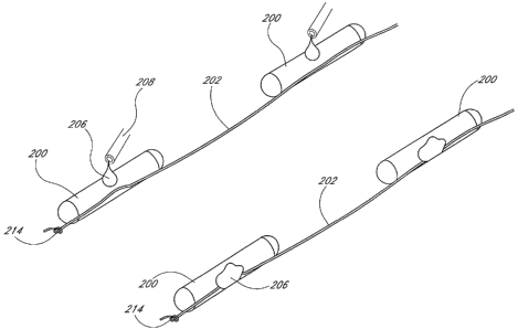
typical implantation procedure is illustrated in Figure 39. Although the
system depicted
in this figure includes spacers 210, the spacers 210 are unnecessary to
maintain the
spacing between adjacent seeds 200.
The physician penetrates the prostate capsule tissue 204 with the needle 22 as
shown in the upper figure. Holding the obturator 42 steady as he or she
withdraws the
sleeve 32, the physician then expels the seeds 200 until the most distal seed
200
protrudes from the distal end 28 of the needle 22. The T bar 218 engages and
becomes
anchored in the tissue 204. 'As the physician withdraws the needle 22 and
sleeve 32, the
anchored T bar 218 creates tension in the filament 202 that properly spaces
the seeds
200.
Figure 42 illustrates another preferred configuration for a most distal seed
200.
The seed 200 includes at least one barb 220 secured to an outer surface. The
seed 200
may include two or more barbs 220, as shown. The barbs 220 may be attached to
a
collar 212 wrapped about the seed 200, as shown, or may be glued directly to
the seed
200. The seed 200 is inserted into the sleeve 32 in the direction of the arrow
A, such
that the inner wall of the sleeve 32 compresses the barbs 220 against the seed
200.
During implantation, when the seed 200 exits the distal end 38 of the sleeve
32, the
barbs 220 extend outward and engage the prostate capsule 204 and anchor the
seed 200
in the surrounding tissue. Similar to the anchors 214, 216, 218 described
above, the
-28-
CA 02495392 2005-01-26
WO 2004/014215 PCT/US2003/024397
barbs 220 create tension in the filament 202 or filaments 202 as the physician
withdraws
the sleeve 32. The tension causes the seeds 200 to maintain the desired
spacing.
Figure 41 illustrates another preferred embodiment of the present
brachytherapy
seed deployment system. In this embodiment, the most distal seed 200 includes
a notch
222 in an outer surface. A rigid wire 224 inserted longitudinally through the
sleeve 32
is removably engageable with the notch 222. As the physician withdraws the
sleeve 32,
he or she applies a pushing force to the wire 224 to maintain the position of
the most
distal seed 200 within the prostate capsule 204. This pushing force creates
tension in
the filament 202 or filaments 202 that causes the seeds 200 to maintain the
desired
spacing as the physician withdraws the sleeve 32. Once the sleeve is removed
and the
seeds 200 are properly spaced, the wire 224 may be disengaged from the distal
seed 200
and withdrawn.
Figure 28 illustrates a preferred method of assembling the embodiment of
Figure
16, and Figure 29 illustrates a preferred method ' of assembling the
embodiment of
Figure 20. A fixture 226 comprising a longitudinal slot 228 preferably
supports the
components during the assembly process. The type of filament 202 to be used is
preferably selected first. If desired, an anchor 214, 216, 218 such as those
described
above is secured to a distal end of the filament 202 or filaments 202. The
distal end is
then secured within the fixture slot 228 and the filament 202 or filaments 202
are pulled
taut.
The desired number of collars 212 are slid down the filament 202 or filaments
202 to their approximate final attachment points. The seeds 200 are then slid
into the
collars 212. If a single filament 202 is used, then the filament 202 may be
located on
any side of the seed 200. If dual filaments 202 are used, then preferably the
filaments
202 are located on opposite sides of the seed 200.
The seeds 200 and collars 212 are slid along the filament 202 or filaments 202
to
their exact final locations and are secured in place using one of the methods
described
above or another equivalent method. If desired, spacers 210 may then be added
between
the seeds 200. Any remaining filament 202 at the ends of the assembly is cut
and may
be knotted if desired. If the ends are knotted, preferably the knots 214 are
located an
-29-
CA 02495392 2005-01-26
WO 2004/014215 PCT/US2003/024397
appropriate distance from the end seeds 200 so as to enable the assembly to
slide easily
into a sleeve 32.
Although not pictured, the embodiments including glue 206 rather than collars
212 are assembled in substantially the same fashion just described. However,
the step
of threading the collars 212 onto the filament 202 or filaments 202 is
eliminated.
Once the seeds 200 and filaments 202 (aind collars 212 and spacers 210, if
provided) are assembled, the assembly is preferably loaded into a sleeve 32
for
implantation. Figure 30 illustrates a preferred method of loading the sleeve
32. A
proximal end of the hub 40 on the sleeve 32 includes an opening 230 having a
diameter
lo significantly larger than a diameter of the seed/filament assembly.
Therefore, to more
easily guide the assembly through the hub 40 and into the tubular portion of
the sleeve
32, a conical adapter 232 may be used. The adapter 232 includes a central
lumen 234
having a diameter slightly larger than that of the seed/filament assembly. A
proximal
face 236 of the adapter 232 abuts an edge of the fixture 226 such that the
lumen 234 is
substantially coaxial with the fixture slot 228. The distal end 238 of the
adapter 232 is
then inserted into the hub 40 such that a distal face of the adapter
substantially abuts a
proximal end of the tubular portion of the sleeve 32. Using the obturator 42,
the
assembler then pushes the seed/filament assembly through the adapter 232,
through the
hub 40 and into the sleeve 32.
Figure 44 illustrates another preferred erribodiment of the present
brachytherapy
seed deployment system. In this embodiment, the filament 202 comprises a
material
that is rigid outside the body, and flexible inside the body. For example, the
material
may be rigid at temperatures below normal human body temperature (98.6 F),
and
flexible at normal human body temperature. Alternatively, the material may
comprise a
matrix that is rigid when exposed to air, but flexible inside the body. Thus,
during
implantation the rigidity of the filament 202 maintains proper spacing between
adjacent
seeds 200. Shortly after implantation, however, the filament 202 becomes
flexible.
Figure 45 illustrates another preferred embodiment of the present
brachytherapy
seed deployment system. In this embodiment, the filament 202 is replaced by a
polymer
laminate 240. The seeds 200 are laminated between two sheets of a
biocompatible
polymeric material. Preferred materials include cyanoacrylates and hotmelts.
Spacers
-30-
CA 02495392 2005-01-26
WO 2004/014215 PCT/US2003/024397
210 may be provided between adjacent seeds 200, and anchoring structures such
as
those described above may be secured to a distal end 242 of the laminate 240.
The
entire laminate structure is inserted into the prostate capsule 204 in a
similar manner as
that described above. In one embodiment, if the laminate 240 is bioabsorbable,
a
filament 202 or filaments 202 may be secured to the seeds 200 as described
above prior
to the seeds 200 being laminated between the sheets. Thus, once the laminate
240
dissolves, the filament 200 maintains the proper seed spacing. If the laminate
240
permits the radiation to properly treat the cancerous tumor without
dissolving, a
filament 202 may not be necessary.
Advantageously, in one embodiment the collars 212 hold the seeds 200 to the
filament 202 or filaments 202 snugly enough that the seeds 200 will not travel
along the
filament 202 or filaments 202 after implantation, but loosely enough that an
end user of
the present brachytherapy seed deployment system may change the spatial
configuration
of the seeds 200 before implantation. For example; if a physician has
available one or
more pre-assembled systems including seeds joined by filaments, he or she may
disassemble the systems and rearrange the spacing between the seeds in order
to tailor
the standard seed arrangement to fit a course of treatment.
To rearrange the seeds, the user removes the sleeve 32 from the needle 22. He
or she then removes the seeds 200 (and spacers 210 if provided) from the
sleeve 32 by
pushing them out with the obturator 42. He or she positions the seeds 200 and
spacers
210 in the slot 228 on the fixture 226, and removes any spacers 210. Using
sterile
tweezers, he or she manipulates the spacing between the seeds 200 by sliding
the
seed/collar assembly along the filament 202 to the desired new location. He or
she may
then add spacers 210 if desired, and reinsert the assembly into the sleeve 32
as described
above.
Although exemplary embodiments of the invention have been shown and
described, many changes, modifications and substitutions may be made by one
having
ordinary skill in the art without necessarily departing from the spirit and
scope of this
invention.
-31-