Note: Descriptions are shown in the official language in which they were submitted.
CA 02495745 2005-02-15
WO 2004/019256 PCT/US2003/025931
METHODS AND DEVICES FOR ANALYSIS OF X-RAY IMAGES
Technical Field
'The present invention is in the field of x-ray imaging and analysis thereof.
In
particular, methods and compositions for the accurate analysis of bone mineral
density
andlor bone structure based on x-rays are described.
Background
X-rays and other x-ray image analysis are important diagnostic tools,
particularly for
bone related conditions. Currently available techniques for the noninvasive
assessment of
the skeleton for the diagnosis of osteoporosis or the evaluation of an
increased risk of
fracture include dual x-ray absorptiometry (DXA) (Eastell et al. (1998) New
Engl J. pled
338:736-746); quantitative computed tomography (QCT) (Cane (1988) Radiology
166:509-
522); peripheral DXA (pDXA) (Patel et al. (1999) J Cli~a Dehsitom 2:397-401);
peripheral
QCT (pQCT) (Gluer et. al. (1997) Senzin Nucl Med 27:229-247); x-ray image
absorptiometry (RA) (Gluer et. al. (1997) Senaih Nucl Med 27:229-247); and
quantitative
ultrasound (QUS) (Njeh et al. "Quantitative Ultrasound: Assessment of
Osteoporosis and
Bone Status" 1999, Martin-Dunitz, London England; U.S. Patent No. 6,077,224,
incorporated herein by reference in its entirety). (See, also, WO 9945845; WO
99/08597;
and U.S. Patent No. 6,246,745).
DXA of the spine and hip has established itself as the most widely used method
of
measuring BMD. Tothill, P. and D.W. Pye, (1992) Br JRadiol 65:807-813. The
fundamental principle behind DXA is the measurement of the transmission
through the
body of x-rays of 2 different photon energy levels. Because of the dependence
of the
attenuation coefficient on the atomic number and photon energy, measurement of
the
transmission factors at 2 energy levels enables the area densities (i.e., the
mass per unit
projected area) of 2 different types of tissue to be inferred. In DXA scans,
these are taken to
be bone mineral (hydroxyapatite) and soft tissue, respectively. However, it is
widely
recognized that the accuracy of DXA scans is limited by the variable
composition of soft
tissue. Because of its higher hydrogen content, the attenuation coefficient of
fat is different
from that of lean tissue. Differences in the soft tissue composition in the
path of the x-ray
beam through bone compared with the adjacent soft tissue reference area cause
errors in the
BMD measurements, according to the results of several studies. Tothill, P. and
D.W. Pye,
(1992) Br JRadiol, 65:807-813; Svendsen, O.L., et al., (1995) JBorae Min Res
10:868-873.
CA 02495745 2005-02-15
WO 2004/019256 PCT/US2003/025931
Moreover, DXA systems ale largetand expensive, ranging in price between
$75,000 and
$150,000.
Quantitative computed tomography (QCT) is usually applied to measure the
trabecular bone in the vertebral bodies. Cann (1988) Radiology 166:509-522.
QCT studies
are generally performed using a single kV setting (single-energy QCT), when
the principal
source of error is the variable composition of the bone marrow. However, a
dual-kV scan
(dual-energy QCT) is also possible. This reduces the accuracy errors but at
the price of
poorer precision and higher radiation dose. Like DXA, however, QCT are very
expensive
and the use of such equipment is currently limited to few research centers.
Quantitative ultrasound (QUS) is a technique for measuring the peripheral
skeleton.
Njeh et al. (1997) Osteoporosis Int 7:7-22; Njeh et al. Quantitative
Ultrasound: Assessment
of Osteoporosis and Bone Status. 1999, London, England: Martin Dunitz. There
is a wide
variety of equipment available, with most devices using the heel as the
measurement site. A
sonographic pulse passing through bone is strongly attenuated as the signal is
scattered and
absorbed by trabeculae. Attenuation increases linearly with frequency, and the
slope of the
relationship is referred to as broadband ultrasonic attenuation (BUA; units:
dB/MHz). BUA
is reduced in patients with osteoporosis because there are fewer trabeculae in
the calcaneus
to attenuate the signal. In addition to BUA, most QUS systems also measure the
speed of
sound (SOS) in the heel by dividing the distance between the sonographic
transducers by
the propagation time (units: m/s). SOS values are reduced in patients with
osteoporosis
because with the loss of mineralized bone, the elastic modulus of the bone is
decreased.
There remain, however, several limitations to QUS measurements. The success of
QUS in
predicting fracture risk in younger patients remains uncertain. Another
difficulty with QUS
measurements is that they are not readily encompassed within the WHO
definitions of
osteoporosis and osteopenia. Moreover, no intervention thresholds have been
developed.
Thus, measurements cannot be used for therapeutic decision-making.
There are also several technical limitations to QUS. Many devices use a foot
support that positions the patient's heel between fixed transducers. Thus, the
measurement
site is not readily adapted to different sizes and shapes of the calcaneus,
and the exact
anatomic site of the measurement varies from patient to patient. It is
generally agreed that
the relatively poor precision of QUS measurements makes most devices
unsuitable for
monitoring patients' response to treatment. Gluey (1997) JBo~e Miya Res
12:1280-1288.
Radiographic absorptiometry (RA) is a technique that was developed many years
ago for assessing bone density in the hand, but the technique has recently
attracted renewed
interest. Gluey et al. (1997) Semira Nucl Med 27:229-247. With this technique,
BMD is
2
CA 02495745 2005-02-15
WO 2004/019256 PCT/US2003/025931
measured in the phalanges '' The priincipal disadvantage of RA of the hand is
the relative
lack of high turnover trabecular bone. For this reason, RA of the hand has
limited sensitivity
in detecting osteoporosis and is not very useful for monitoring therapy-
induced changes.
Peripheral x-ray absorptiometry methods such as those described above are
substantially cheaper than DXA and QCT with system prices ranging between
$15,000 and
$35,000. However, epidemiologic studies have shown that the discriminatory
ability of
peripheral BMD measurements to predict spine and hip fractures is lower than
when spine
and hip BMD measurements are used. Cummings et al. (1993) Lancet 341:72-75;
Maxshall
et al. (1996) Br Med J 312:1254-1259. 'The main reason for this is the lack of
trabecular
bone at the measurement sites used with these techniques. In addition, changes
in forearm
or hand BMD in response to hormone replacement therapy, bisphosphonates, and
selective
estrogen receptor modulators are relatively small, making such measurements
less suitable
than measurements of principally trabecular bone for monitoring response to
treatment.
Faulkner (1998) JClin Densitom 1:279-285; Hoskings et al. (1998) NEngl JMed
338:485-
492. Although attempts to obtain information on bone mineral density from
dental x-rays
have been attempted (See, e.g., Shrout et al. (2000) J. Periodonod. 71:335-
340; Verhoeven
et al. (1998) Cdirz Oral Implants Res 9(5):333-342), these have not provided
accurate and
reliable results.
Furthermore, current methods and devices do not generally take into account
bone
structure analyses. See, e.g., Ruttimann et al. (1992) Oral Surg Oral Med Oral
Pathol
74:98-110; Southard & Southard (1992) Oral Surg Oral llsled Oral Patlaol
73:751-9; White
& Rudolph, (1999) Oral Surg Oral Med Oral Pathol Orad Radiol Endod 88:628-35.
Thus, although a number of devices and methods exist for evaluating hone
density,
there are a number of limitations on such devices and methods. Consequently,
the inventors
have recognized the need, among other things, to pxovide methods and
compositions that
result in the ability to obtain accurate bone mineral density and bone
structure information
from x-xay images and data.
Summary
The present invention meets these and othex needs by providing compositions
and
methods that allow for the analysis of bone mineral density and/or bone
structure from x-ray
images. The x-ray images can be, for example, dental or hip radiographs. Also
provided
are x-ray assemblies comprising accurate calibration phantoms including, in
particular,
calibration phantoms which act as references in order to determine bone
structure from an
x-ray image.
CA 02495745 2005-02-15
WO 2004/019256 PCT/US2003/025931
In one aspect, the i~iventior,~ includes a method to derive quantitative
information on
bone structure and/or bone mineral density from a x-ray image comprising (a)
obtaining an
x-ray image, wherein the x-ray image includes an external standard for
determining bone
structure; and (b) analyzing the image obtained in step (a) to derive
quantitative information
on bone structure. The x-ray image (radiograph) can be, for example, a hip
radiograph or a
dental x-ray obtained on dental x-ray film with or without an external
standard comprising a
calibration phantom that projects free of the mandible ox maxilla. The
calibration phantom
can comprise geometric patterns, for example, made of plastic, metal or metal
powder.
In certain embodiments, the image is obtained digitally, for example using a
selenium detector system or a silicon detector system or a computed
radiography system. In
other embodiments, the image can be digitized for analysis.
In any of the methods described herein, the analysis can comprise using one or
more
computer program (or units). Additionally, the analysis can comprise
identifying one or
more regions of anatomical interest (ROI) in the image, either prior to,
concurrently or after
analyzing the image, e.g. for information on bone mineral density andlor bone
structure.
Bone structural or bone density information at a specified distance from the
ROI and/or
areas of the image having selected bone structural or bone density information
can be
identified manually or, preferably, using a computer unit. The region of
interest can be, for
example, in the mandible, maxilla or one or more teeth. The bone density
information can
be, for example, areas of highest, lowest or median density. Bone structural
information
can be, for example, trabecular thickness; trabecular spacing; two-dimensional
or three-
dimensional spaces between trabeculae; two-dimensional or three-dimensional
architecture
of the trabecular network.
In other aspects, the invention includes a method to derive quantitative
information
on bone structure from an x-ray image comprising: (a) obtaining an x-ray
image; and (b)
analyzing the image obtained in step (a) using one or more indices selected
from the group
consisting of Hough transform, skeleton operator, morphological operators,
mean pixel
intensity, variance of pixel intensity, fourier spectral analysis, fractal
dimension,
morphological parameters and combinations thereof, thereby deriving
quantitative
information on bone structure. The various analyses can be performed
concurrently ox in
series, for example a skeleton operator can be performed before a Hough
transform.
Further, when using two or more indices they can be weighted differently.
Additionally,
any of these methods can also include analyzing the image for bone mineral
density
information using any of the methods described herein.
4
CA 02495745 2005-02-15
WO 2004/019256 PCT/US2003/025931
In another aspect, any of thN methods described herein can further comprise
applying one or more correction factors to the data obtained from the image.
For example,
correction factors can be programmed into a computer unit. The computer unit
can be the
same one that performs the analysis of the image ~r can be a different unit.
In certain
embodiments, the correction factors account for the variation in soft-tissue
thickness in
individual subjects.
In another aspect, any of the methods described herein can further comprise
compressing soft tissue in the image to a selected thickness while obtaining
the x-ray image
In any of the assemblies described herein, the calibration phantom can be
integrated
into the assembly, for example integrated into the hygienic cover, x-ray film
(e.g., between
one or two layers of the filin) and/or holder. Alternatively, the calibration
phantom can be
temporarily attached to the assembly, for example by insertion into a
compartment of the
hygienic cover or by mechanical attachment to the x-ray film. In certain
embodiments, the
calibration phantom comprises a plurality of geometric patterns (e.g.,
circles, stars, squares,
crescents, ovals, multiple-sided objects, irregularly shaped objects and
combinations
thereof) that serve as a reference for bone structure characteristics (e.g.,
trabecular
thickness; trabecular spacing; two-dimensional or three-dimensional spaces
between
trabecular; two-dimensional and/or three-dimensional architecture of the
trabecular
network). The calibration phantom (or geometric patterns therein) can be made,
for
example, of metal, plastic, metal powder or combinations thereof. In any of
the assemblies
described herein, the film can be integral to the hygienic cover.
In a still further aspect, the invention includes a method of diagnosing a
bone
condition (e.g., osteoporosis, risk of fracture) comprising analyzing an x-ray
obtained by
any of the methods described herein.
In a still further aspect, the invention includes a method of treating a bone
condition,
for example by diagnosing the condition as described herein and selecting and
administering one or more therapies to the subject.
In another aspect, the invention includes a method to derive information on
bone
structure from an image comprising: (a) obtaining an image from a subject; (b)
analyzing
the image obtained in step (a) to derive quantitative information on bone
structure. In
certain embodiments, the analysis comprises comparing the information on bone
structure
obtained from the image to a database of bone structure measurements obtained
from
selected subjects. The image can be, for example, an x-ray image or an
electronic image.
In certain embodiments, the image comprises an external standard. The selected
subjects
making the database can be, for example, normal subjects, subjects with
osteoporosis or
CA 02495745 2005-02-15
WO 2004/019256 PCT/US2003/025931
combinations thereof. Further, the~database can comprises demographic data and
data on
bone structure in the subjects, for example, wherein the subjects are age, sex
and race-
matched.
In certain embodiments, the bone stntcture information is selected from the
group
S consisting of trabecular thickness; trabecular spacing; trabecular
connectivity, two-
dimensional or three-dimensional spaces between trabecular; two-dimensional or
three-
dimensional architecture of the trabecular network and/or combinations
thereof. Further,
any of the methods described herein may further include the step of locating
one or more
regions of interest (ROI) in the image (e.g., x-ray image), fox example, an
ROI that is
positioned using a regularized active shape algorithm or an ROI that is
located
automatically.
In certain aspects, step (b) comprises analyzing the image obtained in step
(a) using
one or more indices selected from the group consisting of trabecular density,
trabecular
perimeter, star volume, trabecular bone pattern factor, trabecular thickness,
trabecular
orientation, orientation-specific trabecular assessment, trabecular
connectivity and
combinations thereof. In certain embodiments, the indices include at least one
of the
indices trabecular density and density is a ratio of trabecular area to total
area. In other
embodiments, at least one of the indices is orientation-specific trabecular
assessment as
determined using Fourier analysis. In other embodiments, at least one of the
indices is
trabecular thickness as determined by Euclidean distance transformation. In
other
embodiments, at least one of the indices is trabecular orientation as
determined using 2D
fast Fourier Transform (FFT). In other embodiments, at least one of the
indices is trabecular
connectivity as determined using node count. Further, two or more indices may
be
analyzed.
In another aspect, the invention includes a method of diagnosing a bone
condition in
a subject comprising analyzing information from an image according to any of
the methods
described herein, wherein if the analysis indicates that the bone structure
information
obtained from the subject differs from that of normal control subjects, a bone
condition is
diagnosed. The bone condition may be, for example, osteoporosis.
In yet another aspect, the invention comprises a method of treating a bone
condition
comprising (a) obtaining an image from a subject; (b) analyzing the image
obtained in step
(a) to derive quantitative information on bone structure; (c) diagnosing a
bone condition
based on the analysis of step (b); and (d) selecting and administering a
suitable treahnent to
the subject based on the diagnosis. In certain embodiments, analysis of the
information
obtained in step (b) is conducted by comparing the information with
information in a
6
CA 02495745 2005-02-15
WO 2004/019256 PCT/US2003/025931
database of bone structure ineasur~ments obtained from selected subjects. The
treatment
may comprise, for example, administering one or more antiresorptive agents;
administering
one or more anabolic agents; or combinations thereof.
In another aspect, the invention includes a method of deternlining bone
mineral
density from an x-ray image, the method comprising the steps of (a)
determining density of
one or more internal standards in the image; (b) creating a weighted mean
between the
values obtained in step (a); (c) utilizing the weighted mean to determine bone
mineral
density of bone in the image. The internal reference can be, for example,
selected from the
group consisting of air, fat, water, metal and combinations thereof.
In another aspect, the invention includes a method of determining bone
structure
from an x-ray image comprising the steps of (a) identifying one or more
internal standards
on the x-ray image; (b) determining the density or structure of the standard;
and (c) utilizing
the density, structure or combinations thereof of the standard to determine
bone structure of
the x-ray image. The internal reference can be, for example, a tooth, a
portion of a tooth,
cortical bone, air, subcutaneous fat, and muscle.
In another aspect, the invention includes a method of evaluating bone disease
in a
subject, the method comprising the steps of: (a) obtaining an x-ray image from
the subject,
wherein the image includes one or more bones; (b) assessing bone mineral
density in at least
one anatomic region of the image; (c) assessing bone structure in the region;
and (d)
combining the assessments of bone mineral density and bone structure to
evaluate bone
disease. The bone disease can be, for example, the risk of bone fracture such
as
osteoporotic fracture. The evaluation can include, for example, diagnosing
bone disease,
monitoring the progression of bone disease (e.g., by evaluating bone disease
at various
discrete time points) and the like. In certain embodiments, the methods
described herein
further comprising selecting a therapy based on the evaluation of bone disease
and
administering the therapy to the subject. In further embodiments, the methods
described
herein the evaluation comprises monitoring the progression of bone disease
during or after
administration of the selected therapy. In still further embodiments, any of
the methods
described herein can further comprise the step of assessing one or more macro-
anatomical
parameters in the image and combining the assessment of bone mineral density,
bone
structure and macro-anatomical parameters to diagnose bone disease.
In yet another aspect, the invention includes a method of treating bone
disease in a
subject, the method comprising the steps of: (a) obtaining an image (e.g., x-
ray or electronic
image) from the subject, wherein the image includes one or more bones; (b)
assessing bone
mineral density in at least one anatomic region of the image; (c) assessing
bone structure in
7
CA 02495745 2005-02-15
WO 2004/019256 PCT/US2003/025931
the region; (d) combining the assessments of bone mineral density and bone
structure to
evaluate bone disease; (e) selecting a therapy based on the evaluation of bone
disease; and
(f) administering the therapy to the subject. In certain embodiments, steps
(a) to (d) are
performed two or more times. In other embodiments, steps (a) to (e) are
performed two or
more times.
In another aspect, the invention includes a method for evaluating bone disease
in a
subject, the method comprising the steps of: (a) obtaining an image (e.g., x-
ray or electronic
image) of the subject wherein the image includes one or more bones; (b)
assessing bone
structure of the bone in the image; (c) assessing one or more macro-anatomical
parameters
in the image; and (d) combining the assessments bone structure and macro-
anatomical
parameter assessment to evaluate bone disease. The bone disease can be, for
example, the
risk of bone fracture such as osteoporotic fracture. The evaluation can
comprise, for
example, diagnosing bone disease and/or monitoring the progression of bone
disease over
two or more discrete time points (e.g., by repeating steps (a) to (d) at two
or more time
points). The methods can further comprise selecting a therapy based on the
evaluation of
bone disease and administering the therapy to the subject. Further, the
evaluation may
comprise monitoring the progression of bone disease during or after
administration of the
selected therapy.
These and other embodiments of the subject invention will readily occur to
those of
skill in the art in light of the disclosure herein.
Brief Description of the Figures
Fig. 1 shows an example of a dental x-ray. A calibration phantom 110 is seen.
Regions of interest 120 have been placed for measurement of bone mineral
density or
structure.
Fig. 2 shows another example of a dental x-ray. A calibration phantom 110 is
seen.
Regions of interest 120 have been placed for measurement of bone mineral
density or
structure.
Fig. 3 shows an example of an analysis report resulting from a measurement of
mandibular or maxillary bone mineral density. A subject (~ is more than one
standard
deviation below the mean of age-matched controls (x-axis age, y-axis arbitrary
units BMD).
Fig. 4 shows an example of a V-shaped calibration phantom 114 mounted on a
tooth
120. Gums are also shown 130.
Fig. 5 shows an example of a holder 115 for a calibration phantom 110. The
holder
115 is mounted on a tooth 120. Gums are also shown 130.
CA 02495745 2005-02-15
WO 2004/019256 PCT/US2003/025931
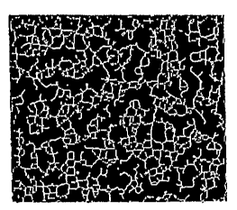
Fig. 6, panels B through E shows gray value profiles along different rows of
pixels
used for locating dental apices. From top to bottom, the characteristic peaks
for the dental
roots (shown in dental x-ray panel A) gradually disappear.
Fig. 7 shows a Hough transform (panel A) of a test image (panel B). All
collinear
points from the same line are transformed into sinusoidal curves that
intersect in a single
point (circles).
Fig. 8 shows a Hough transform (panel A) of a skeletonized trabecular bone x-
ray
image (panel B). The white regions in panel A indicate longer segments and
predominant
angles.
Fig. 9 shows the effect of varying size of structuring element E2; calibration
phantom image with lines of varying width (1, 3, 5, 7, 9, 11, 13 pix) (top
left); skeleton
operation performed using E2 with a diameter of 3 pix (top right), 7 pix
(bottom left), and 11
pix (bottom right), respectively.
Fig. 10 shows the effect of varying size of structuring element E2; gray scale
image
of trabecular bone (top left, panel A); skeleton operation performed using E2
with a diameter
of 3 pix (top right, panel B); 7 pix (bottom left, panel C) and 11 pix (bottom
right, panel D),
respectively.
Fig. 11 shows gray value surface plot of an anatomical region of interest from
a
dental x-ray (inset) used for fractal analysis.
Fig. 12 shows an example of a hygienic cover holder that includes compartments
for
a calibration phantom and a fluid-filled bolus back.
Fig. 13 shows an example of an anatomical region of interest (black dot),
determined
relative to the teeth or to the convexity/concavity of the mandible.
Fig. 14 shows an example of three anatomical region of interests (black dots),
determined relative to the teeth or to the convexitylconcavity of the
mandible.
Fig. 15 is a side view of an exemplary system for minimizing tube angulation
as
described herein. In the Figure, the system is shown as a dental x-ray system.
An extension
tubing (200) is attached to a ring-shaped Rinn holder (102). The outer
diameter of the
extension tubing is slightly smaller than the inner diameter of the tube
located in front of the
dental x-ray systemldental x-ray tube. The extension tubing can then be
inserted into the
metal tube thereby reducing tube angulation and resultant errors in bone
apparent density
and bone structural measurements.
Detailed Description
Before describing the present invention in detail, it is to be understood that
this
invention is not limited to particular formulations or process parameters as
such may, of
9
CA 02495745 2005-02-15
WO 2004/019256 PCT/US2003/025931
course, vary. It is also to be understood that the terminology used herein is
for the purpose
of describing particular embodiments of the invention only, and is not
intended to be
limiting.
The practice of the present invention employs, unless otherwise indicated,
conventional methods of x-ray imaging and processing within the slcill of the
art. Such
techniques are explained fully in the literature. See, e.g., X-Ray Structure
Determination: A
Practical Guide, 2nd Edition, editors Stout and Jensen, 1989, John Wiley &
Sons, publisher;
Body CT: A Practical Approach, editor Slone, 1999, McGraw-Hill publisher; The
Essential
Physics of Medical Imaging, editors Bushberg, Seibert, Leidholdt Jr & Boone,
2002,
Lippincott, Williams & Wilkins; X-ray Diagnosis: A Physician's Approach,
editor Lam,
1998 Springer-Verlag, publisher; and Dental Radiology: Understanding the X-Ray
Image,
editor Laetitia Brocklebank 1997, Oxford University Press publisher.
All publications, patents and patent applications cited herein, whether above
or
below, are hereby incorporated by reference in their entirety.
It must be noted that, as used in this speciEcation and the appended claims,
the
singular forms "a", "an", and "the" include plural referents unless the
content clearly
dictates otherwise. Thus, for example, reference to "a calibration phantom"
includes a one
or more such phantoms.
Definitions
Unless defined otherwise, all technical and scientific terms used herein have
the
same meaning as commonly understood by one of ordinary skill in the art to
which the
invention pertains. Although any methods and materials similar or equivalent
to those
described herein can be used in the practice for testing of the present
invention, the
preferred materials and methods are described herein.
The term "subject" encompasses any warm-blooded animal, particularly including
a
member of the class Mammalia such as, without limitation, humans and nonhuman
primates
such as chimpanzees and other apes and monkey species; farm animals such as
cattle,
sheep, pigs, goats and horses; domestic mammals such as dogs and cats;
laboratory animals
including rodents such as mice, rats and guinea pigs, and the like. The term
does not denote
a particular age or sex and, thus, includes adult and newborn subjects,
whether male or
female.
"Osteoporosis" refers to a condition characterized by low bone mass and
mieroarchitectural deterioration of bone tissue, with a consequent increase of
bone fragility
and susceptibility to fracture. Osteoporosis presents commonly with vertebral
fractures or
CA 02495745 2005-02-15
WO 2004/019256 PCT/US2003/025931
hip fractures due to the decease in,bone mineral density and deterioration of
structural
properties and microarchitecture of bone.
A "subject" preferably refers to an animal, for example a mammal such as a
human.
As used herein the term "patient" refers to a human subject.
"Computational unit" refers to any current or future software, chip or other
device
used far calculations, such as bone structure, naw developed or developed in
the future. The
computational unit may be designed to control the x-ray assembly or detector
(as well as
other parameters related to the x-ray detector). Other applications of the
computational unit
to the methods and devices described herein will be recognized by those
skilled in the art.
The computational unit may be used for any other application related to this
technology that
may be facilitated with use of computer software or hardware.
"Bone structure" refers to two-dimensional or three-dimensional arrangement
(e.g.,
architecture or microarchitecture) of bone tissue. Generally, bone tissue
includes two types
of bone -- an outer layer of cortical bone that is generally mostly solid with
some canals or
pores therein and an inner layer of trabecular (or cancellous) bone that
generally is sponge-
like ar honeycomb-like in structure. Structural features or cortical and
trabecular bone
include, but are not limited to, trabecular thickness; trabecular spacing; two-
dimensional or
three-dimensional spaces between trabeculae; two-dimensional or three-
dimensional
architecture of the trabecular network, solid material (typically greater than
3000 ~.m),
primary and/or secondary trabeculae (typcially 75 to 200 ~.m), primary and
secondary
osteons (typically 100 to 300 Vim, plexiform, interstitial bone, trabecular
packets, lamellae
(typically 1 to 20 ~,m), lacunae, cement Lines, canaliculi, collagen-mineral
composite
(typically 0.06 to 0.4 ~.m), cortical pores, trabecular connectivity, nodes
and branch points,
and the like. One or more of these and other structural features may be
measured in the
practice of the present invention. Preferably, measurements are the sub-
millimeter range,
more typically in the 10 - 500 ~.m range. Non-limiting examples of
microarchitecture
parameters include trabecular structure thresholded binary image parameters
such as
trabecular area; total area; trabecular area /total area; trabecular perimeter
area; trabecular
distance transform; marrow distance transform; trabecular distance transform
regional
maxima values (mean, min., max, std. Dev); marrow distance transform regional
maxima
values (mean, min., max, std. Dev); star volume (see, e.g., Ikuta et. al.
(2000) JBMR
18:217-277; Vesterby (1990) Bone 11:149-155; and Vesterby et al. (1989) Bone
10:7-13);
trabecular Bone Pattern Factor (Hahn et. al., (1992) Bone 13:327-330); TBPf =
(Pl - P2) l
(A1 - A2 ) where Pl and A1 are the perimeter length and trabecular bone area
before
dilation and P2 and A2 corresponding values after a single pixel dilation as
well as
11
CA 02495745 2005-02-15
WO 2004/019256 PCT/US2003/025931
trabecular skeleton parameters such as connected skeleton count or Trees (T);
node count
(N); segment count (S); node-to-node segment count (NIA; node-to-free-end
segment count
(NF); node-to-node segment length (NNL); node-to-free-end segment length
(NFL); free-
end-to-free-end segment length (FFL); node-to-node total struts length
(NN.TSL) (see, e.g.,
Legrand et. al., (2000) JMBR 15:13-19; free-end-to-free-ends total struts
length( FF.TSL);
total struts length (TSL); FF.TSL/ TSL; NN.TSL/ TSL; Loop count (Lo); Loop
area; mean
distance transform values for each connected skeleton; mean distance transform
values for
each segment (Tb.Th ); mean distance transform values for each node-to-node
segment
(Tb.Th.NN); mean distance transform values for each node-to-free-end segment
(Tb.Th.NF); orientation (angle) of each segment; angle between segments;
length-thickness
ratios (NNL/Tb.Th.NN ) and (NFL/ Tb.Th.NF); and interconnectivity index (ICI)
where ICI
_ (N '~ NN)/ ( T * (NF + 1).
"Macro-anatomical parameter" refers to any parameter describing the shape,
size or
thickness of bone and/or surrounding structure, typically parameters that are
greater than
O.Smm in size in at least one dimension. Macro-anatomical parameters include,
for
example, in the hip joint thickness of the femoral shaft cortex, thickness of
the femoral neck
cortex, hip axis length, CCD (caput-collum-diaphysis) angle and width of the
trochanteric
region.
General Overview
Methods and compositions useful in analyzing x-ray images are described. In
particular, the invention includes methods of obtaining and/or deriving
information about
bone mineral density and/or bone structure from an x-ray image. Additionally,
the present
invention relates to the provision of accurate calibration phantoms for use in
determining
bone structure and methods of using these calibration phantoms. In particular,
the present
invention recognizes for the first time that errors arising from misplacement
of interrogation
sites in dental or hip x-rays of bone density and/or bone structure can be
corrected by
positioning the x-ray tube, the detector and/or the calibration reference with
respect to an
anatomical landmark (or anatomical region of interest).
Advantages of the present invention include, but are not limited to, (i)
providing
accessible and reliable means for analyzing x-rays; (ii) providing non-
invasive
measurements of bone structure and architecture; (iii) providing methods of
diagnosing
bone conditions (e.g., osteoporosis, fracture risk); (iv) providing methods of
treating bone
conditions; and (iv) providing these methods in cost-effective manner.
12
CA 02495745 2005-02-15
WO 2004/019256 PCT/US2003/025931
1Ø Obtaining Dada from 3X Rays
An x-ray image can be acquired using well-known techniques from any local
site.
For example, in certain aspects, 2D planar x-ray imaging techniques are used.
2D planar x-
ray imaging is a method that generates an image by transmitting an x-ray beam
through a
body or structure or material and by measuring the x-ray attenuation on the
other side of the
body or the structure or the material. 2D planar x-ray imaging is
distinguishable from
cross-sectional imaging techniques such as computed tomography or magnetic
resonance
imaging. If the x-ray image was captured using conventional x-ray film, the x-
ray can be
digitized using any suitable scanning device. Digitized x-ray images can be
transmitted
over a networked system, e.g. the Internet, into a remote computer or server.
It will be
readily apparent that x-ray images can also be acquired using digital
acquisition techniques,
e.g. using photostimulable phosphor detector systems or selenium or silicon
detector
systems, the x-ray image information is already available in digital format
which can be
easily transmitted over a network.
Any x-rays can be used including but not limited to digital x-rays and
conventional
x-ray film (which can be digitized using commercially available flatbed
scanners). In
certain embodiments, the x-ray is of the hip region, for example performed
using standard
digital x-ray equipment (Kodak DirectView DR 9000, Kodak, Rochester, NY).
Patients are
typically positioned on an x-ray table in supine position, parallel to the
long axis of the
table, with their arms alongside their body. The subject's feet may be placed
in neutral
position with the toes pointing up or in internal rotation or may be placed in
a foot holder
such that the foot in a neutral position (0° rotation) or in any
desired angle of rotation (e.g.,
internal or external) relative to neutral (see, also Example 8 below). Foot
holders suitable
for such purposes may include, for example, a base plate extending from the
foot, for
example, from the mid to distal thigh to the heel. The base plate preferably
sits on the x-ray
table. The patients' foot is positioned so that the posterior aspect of the
heel is located on
top of the base plate. The medial aspect of the foot is placed against a
medial guide
connected rigidly to the base plate at a 90° angle by any suitable
means (e.g., straps, velcro,
plastic, tape, etc.). A second, lateral guide attached to the base plate at a
90° angle with a
sliding mechanism can then be moved toward the lateral aspect of the foot and
be locked in
position, for example when it touches the lateral aspect of the foot. The use
of a foot holder
can hel~improve the reproducibility of measurements of bone structure
parameters or
macro-anatomical parameters.
13
CA 02495745 2005-02-15
WO 2004/019256 PCT/US2003/025931
Generally, the ray is centered onto the hip joint medial and superior to the
greater
trochanter. A calibration phantom, such as an aluminum step wedge may also be
included
in the images to calibrate gray values before further image analysis,
In other embodiments, dental x-rays are preferred because of the relative ease
and
lack of expense in obtaining these images. Further, the mandible and maxilla
are primarily
composed of trabecular bone. Since the metabolic turnover of trabecular bone
is
approximately eight times greater than that of cortical bone, areas of
predominantly
trabecular bone such as the vertebral body are preferred sites for measuring
bone mineral
density. Lang et al. (1991) Radiol Clira North Arra 29:49-76. Thus, the fact
that trabecular
bone is clearly visible on the dental x-ray image, thus lending itself to
quantitative analysis
of bone mineral density and structure. Jeffcoat et al. (2000) Periodontol
23:94-102;
Southard et al. (2000) J Deht Res 79:964-969. Further, the earliest bone loss
in osteoporosis
patients occurs in areas of trabecular bone. Multiple dental x-ray images are
commonly
made in most Americans throughout life. Indeed, there are approximately 750
million U.S.
dental visits annually and 150 million of these patients result in more than 1
billion dental x-
rays taken each year. Thus, the ability to diagnose osteoporosis on dental x-
rays would be
extremely valuable since it would create the opportunity for low-cost mass
screening of the
population.
Preferably, x-ray imaging is performed using standard x-ray equipment, for
instance
standard dental x-ray equipment (e.g. General Electric Medical Systems,
Milwaukee, WI).
X-rays of the incisor region and canine region are acquired using a standard x-
ray imaging
technique with 80 kVp and automatic exposure using a phototimer or using a
manual
technique with l OrnA tube current. X-ray images are acquired, for example, on
Kodak
Ultraspeed film (Kodak, Rochester, NY). X-ray images may be digitized using a
coxnmexcial flatbed scanner with transparency option (Acer ScanPremio ST).
1.1. Calibration Phantoms
It is highly preferred that the x-ray images include accurate reference
markers, for
example calibration phantoms for assessing bone mineral density and/or bone
structure of
any given x-ray image. Calibration references (also known as calibration
phantoms) for use
in imaging technologies have been described. See, e.g., U.S. Patent No.
5,493,601 and U,S.
Patent No. 5,235,628. U.S. Patent No. 5,335,260 discloses a calibration
phantom
representative of human tissue containing variable concentrations of calcium
that serves as
reference for quantifying calcium, bone mass and bone mineral density in x-ray
and CT
imaging systems. However, currently available calibration phantoms are not
always
14
CA 02495745 2005-02-15
WO 2004/019256 PCT/US2003/025931
accurate. Because bone m~,neral density accounts for considerably less than
100% of
fracture risk in osteoporosis (~uyang et al. (1997) Calif Tissue Tnt, 60:139-
147) some of the
methods and devices described herein are designed to assess not only bone
mineral density
but also bone structure. By assessing both these parameters, more accurate
testing and
screening can be provided for conditions such as osteoporosis.
Thus, in certain aspects, the current invention provides for methods and
devices that
allow accurate quantitative assessment of information contained in an x-ray
such as density
of an anatomic structure and/or morphology of an anatomic structure. Any
suitable
calibration phantom can be used, for example, one that comprises aluminum or
other radio-
opaque materials. U.S. Patent No. 5,335,260 describes other calibration
phantoms suitable
for use in assessing bone mineral density in x-ray images. Examples of other
suitable
calibration reference materials can be fluid or fluid-like materials, for
example, one or more
chambers filled with varying concentrations of calcium chloride or the like.
Numerous calibration phantoms (or reference calibrations) can be used in the
practice of the present invention. Typically, the system used to monitor bone
mineral
density and/or bone structure in a target organism comprises an x-ray (e.g., a
dental or hip
radiograph), which provides information on the subject; an assembly including
a calibration
phantom, which acts as a reference for the data in the dental x-ray; and at
least one data
processing system, which evaluates and processes the data from the dental x-
ray image
andlor from the calibration phantom assembly.
It will be readily apparent that a calibration phantom can contain a single,
known
density or structure reference. Furthermore, a gradient in x-ray density can
be achieved by
varying the thickness or the geometry of the calibration phantom along the
path of the x-ray
beam, for example, by using a V-shape of the calibration phantom of varying
thickness (Fig.
4). The calibration phantom can also include angles. For example, the
calibration phantom
can be "T"-shaped or "L"-shaped thereby including one or more 90 degree
angles.
The calibration phantom can contain several different areas of different radio-
opacity. For example, the calibration phantom can have a step-like design,
whereby
changes in local thickness of the wedge result in differences in radio-
opacity. Stepwedges
using material of varying thickness are frequently used in radiology for
quality control
testing of x-ray beam properties. By varying the thickness of the steps, the
intensity and
spectral content of the x-ray beam in the projection image can be varied.
Stepwedges are
commonly made of aluminum, copper and other convenient and homogeneous
materials of
known x-ray attenuation properties. Stepwedge-like phantoms can also contain
calcium
phosphate powder or calcium phosphate powder in molten paraffin.
CA 02495745 2005-02-15
WO 2004/019256 PCT/US2003/025931
Alternatively, continuous Wedges may be used or the calibration reference may
be
designed such that the change in radio-opacity is from periphery to center
(for example in a
round, ellipsoid, rectangular, triangular of other shaped structure). As noted
above, the
calibration reference can also be constructed as plurality of
separate.chambers, for example
fluid filled chambers, each including a specific concentration of a reference
fluid (e.g.,
calcium chloride). In addition to one or more fluids, a calibration phantom
can also contain
metal powder, e.g. aluminum or steel powder, embedded within it (for example,
embedded
in a plastic).
In certain embodiments, the calibration phantom is specifically designed to
serve as
a reference for bone structure (e.g., trabecular spacing, thickness and the
like). For
example, the calibration wedge can contain one or more geometric patterns with
known
dimensions, e.g. a grid whereby the spacing of a grid, thickness of individual
grid elements,
etc. are known. This known geometric pattern of radio-opaque elements in the
calibration
phantom can be used to improve the accuracy of measurements of trabecular bone
structure
in an x-ray. Such measurements of trabecular bone structure can include, but
are not limited
to, trabecular spacing, trabecular length and trabecular thickness. Such
measurements of
trabecular spacing, trabecular length and trabecular thickness can, for
example, be
performed in a dental or hip x-ray. These calibration phantoms can be made up
of a variety
of materials include, plastics, metals and combinations thereof. Further, the
reference
components can be solid, powdered, fluid or combinations thereof. Thus, the
calibration
wedge can also be used to improve measurements of bone structure.
Since the present invention contemplates analysis of dental x-ray images for
information on bone structure, bone mineral density or both structure and
density, it will be
apparent that calibration phantoms will be selected based on whether
structure, density or
both are being measured. Thus, one or more calibration phantoms may be
present.
Whatever the overall shape or composition of the calibration phantom, when
present, the at least one marker be positioned at a known density andlor
structure in the
phantom. Furthermore, it is preferred that at least one geometric shape or
pattern is
included in the calibration phantom. Any shape can be used including, but not
limited to,
squares, circles, ovals, rectangles, stars, crescents, multiple-sided objects
(e.g., octagons),
V- or U-shaped, inverted V- or U-shaped, irregular shapes or the like, so long
as their
position is known to correlate with a particular density of the calibration
phantom. In
preferred embodiments, the calibration phantoms described herein are used in
2D planar x-
ray imaging.
16
CA 02495745 2005-02-15
WO 2004/019256 PCT/US2003/025931
The calibration phantoms can be imaged before or after the x-ray image is
taken,
Alternatively, the calibration phantom can be imaged at the same time as the x-
ray image.
The calibration phantom can be physically connected to an x-ray film and/or
film holder.
Such physical connection can be achieved using any suitable mechanical or
other '
attachment mechanism, including but not limited to adhesive, a chemical bond,
use of
screws or nails, welding, a VelcroTM strap or VelcroTM material and the like.
Similarly, a
calibration phantom can be physically connected to a detector system or a
storage plate for
digital x-ray imaging using one or more attachment mechanisms (e.g., a
mechanical
connection device, a VelcroTM strap or other VelcroTM material, a chemical
bond, use of
screws or nails, welding and an adhesive). The external standard and the film
can be
connected with use of a holding device, for example using press fit for both
film and
external standard.
Additionally, the calibration phantom assembly can be attached to an
anatomical
structure, for example one or more teeth, mucus membranes, the mandible and/or
maxilla.
For instance, the calibration phantom can be attached (e.g., via adhesive
attachment means)
to the epithelium or mucous membrane inside overlying the mandible or the
maxilla.
Alternatively, the calibration phantom can be placed on or adjacent to a
tooth, for example,
a V- or U-shaped (in the case of the maxilla) or an inverted V- or U-shaped
(in the case of
the mandible) calibration phantom can be used. The opening of the V or U will
be in
contact with the free edge of at least one tooth or possibly several teeth
(Fig. 4).
In preferred embodiments, when an x-ray of an anatomic structure or a non-
living
object is acquired a calibration phantom is included in the field of view. Any
suitable
calibration phantom can be used, for example, one that comprises aluminum or
other radio-
opaque materials. U.S. Patent No. 5,335,260 describes other calibration
phantoms suitable
for use in assessing bone mineral density in x-ray images. Examples of other
suitable
calibration reference materials can be fluid or fluid-like materials, for
example, one or more
chambers filled with varying concentrations of calcium chloride or the like.
In a preferred
embodiment, the material of the phantom is stainless steel (e.g., AISI grade
316 comprising
carbon (0.08%); manganese (2%); silicon (1%); phosphorus (0.045%); sulphur
(0.03%);
nickel (10-14%); chromium (16-18%); molybdenum (2-3%); plus iron to make up
100%).
The relative percentages of the components may be with respect to weight or
volume.
It will be apparent that calibration phantoms suitable for attachment to an
anatomical
structure can have different shapes depending on the shape of the anatomical
structure (e.g.,
tooth or teeth) on which or adjacent to which it will be placed including, but
not limited to,
U-shaped, V-shaped, curved, flat or combinations thereof. For example, U-
shaped (or
17
CA 02495745 2005-02-15
WO 2004/019256 PCT/US2003/025931
inverted U-shaped) calibration phantoms can be positioned on top of molars
while V-shaped
(or inverted V-shaped) calibration phantoms can be positioned on top of
incisors. Further, it
will be apparent that in certain instances (e.g., teeth on the mandible), the
calibration
phantom can rest on top of the tooth just based on its gravity or it can be
attached to the
tooth (e.g.; using adhesive). In the case of the teeth on the maxilla, the
calibration phantom
will typically be attached to the tooth, for example with use of an adhesive.
Any of these attachments may be permanent or temporary and the calibration
phantom can be integral (e.g., built-in) to the film, film holder and/or
detector system or can
be attached or positioned permanently or temporarily appropriately after the
film and/or
film holder is produced. Thus, the calibration phantom can be designed for
single-use (e.g.,
disposable) or for multiple uses with different x-ray images. Thus, in certain
embodiments,
the calibration phantom is reusable and, additionally, can be sterilized
between uses.
Integration of a calibration phantom can be achieved by including a material
of known x-ray
density between two of the physical layers of the x-ray film. Integration can
also be
achieved by including a material of known x-ray density within one of the
physical layers of
the x-ray film. Additionally, the calibration phantom can be integrated into
the film cover.
A calibration phantom or an external standard can also be integrated into a
detector system
or a storage plate for digital x-ray imaging. For example, integration can be
achieved by
including a material of known x-ray density between two of the physical layers
of the
detector system or the storage plate. Integration can also be achieved by
including a
material of know x-ray density within one of the physical layers of the
detector system or
the storage plate.
In certain embodiments, for example those embodiments in which the calibration
phantom is temporarily attached to a component of the x-ray assembly system
(e.g., x-ray
film holder, x-ray filrri, detector system or the like), cross-hairs, lines or
other markers may
be placed on the apparatus as indicators for positioning of the calibration
phantom. These
indicators can help to ensure that the calibration phantom is positioned such
that it doesn't
project on materials that will alter the apparent density in the resulting
image.
Any of the calibration phantom-containing assemblies described herein can be
used
in methods of analyzing and/or quantifying bone structure (or bone mineral
density) in an x-
ray image. The methods generally involve simultaneously imaging or scanning
the
calibration phantom and another material (e.g., bone tissue from a subject)
for the purpose
of quantifying the density of the imaged material (e.g., bone mass). In the
case of dental
radiographs, the calibration phantom, the x-ray tube or dental x-ray film is
typically
positioned in a manner to ensure inclusion of the calibration phantom and a
portion of the
1~
CA 02495745 2005-02-15
WO 2004/019256 PCT/US2003/025931
mandible and/or maxilla on the dental x-ray image. Preferably, the calibration
phantom, the
x-ray tube and the dental x-ray film are positioned so that at least a portion
of the section of
the mandible or maxilla included on the image will contain predominantly
trabecular bone
rather than cortical bone.
Thus, under the method of the present invention, the calibration phantom is
preferably imaged or scanned simultaneously with the individual subject,
although the
invention allows for non-simultaneous scanning of the phantom and the subject.
Methods
of scanning and imaging structures by x-ray imaging technique are well known.
By placing
the calibration phantom in the x-ray beam with the subject, reference
calibration samples
allow corrections and calibration of the absorption properties of bone. When
the phantom is
imaged or scanned simultaneously with each subject, the variation in x-ray
beam energy and
beam hardening are corrected since the phantom and the subject both see the
same x-ray
beam spectrum. Each subject, having a different size, thickness, muscle-to-fat
ratio, and
bone content, attenuate the beam differently and thus change the effective x-
ray beam
spectrum. It is necessary that the bone-equivalent calibration phantom be
present in the
same beam spectrum as the subject's bone to allow accurate calibration.
X-ray imaging assemblies that are currently in use do not take into account
the
position of the calibration phantom in relation to the structures being
imaged. Thus, when
included in known assemblies, calibration phantam(s) are often positioned such
that they
project on materials or structures (e.g., bone) that alter apparent density of
the calibration
phantom in the resulting x-ray image. Clearly, this alteration in apparent
density will affect
the accuracy of the calibration phantom as a reference for determining bone
mineral density.
Therefore, it is an object of the invention to provide methods in which the
calibration
phantom projects free of materials or structures that will alter the apparent
density of the
reference. In the context of dental x-rays, for instance, the methods
described herein ensure
that the calibration phantom projects free of bone (e.g., teeth, jaw) tissue.
This can be
accomplished in a variety of ways, for example, positioning the calibration
phantom in the
x-ray film or in the x-ray film holder such that it will appear between the
teeth in the dental
x-ray.
The calibration phantom materials and methods of the present invention are
preferably configured to be small enough and thin enough to be placed inside
the mouth,
and the method of the present invention can be used to quantify bone mass
using standard
dental x-ray systems, for example by including temporary or permanent
calibration
phantoms in dental x-ray film holders. Further, it is highly desirable that
the calibration
phantom be positioned so that at least a portion doesn't project on structures
or materials
19
CA 02495745 2005-02-15
WO 2004/019256 PCT/US2003/025931
that will alter the apparent density car structural characteristics of the
calibration phantoms.
It is also preferable to position calibration phantom at a defined distance
relative to at least
one tooth or the mandible or the maxilla whereby a substantial portion of the
calibration
phantom projects free of the tooth, the mandible or the maxilla on the x-ray
image. Any
suitable distance can be used, for example between about 1 mm and 5 cm or any
value
therebetween.
A cross-calibration phantom can be used to optimize system performance, e.g. x-
ray
tube settings or film processor settings, or to improve the comparability of
different
machines or systems, typically located at different sites. For this purpose, a
separate image
may be obtained which does not include a patient or a body part. The image
includes the
primary calibration phantom used in patients, e.g. a step-wedge of known
density, and the
cross-calibration phantom. The apparent density of the primary calibration
phantom is then
calibrated against the density of the cross-calibration phantom. The resultant
cross-
calibration of the primary phantom can help to improve the accuracy of
measurements of
bone density, bone structure and macro-anatomical parameters. It can also help
improve the
overall reproducibility of the measurements. In one embodiment of the
invention, an x-ray
technologist or a dental hygienist will perform a cross-calibration test once
a day, typically
early in the morning, prior to the first patient scans. The results of the
cross-calibration or
the entire cross-calibration study can be transmitted via a network to a
central computer.
The central computex can then perform adjustments designed to maintain a high
level of
comparability between different systems.
1.2. Inherent Reference Markers
In certain embodiments of the invention, information inherent in the anatomic
structure or the non-living object can be used to estimate the density and/or
structure of
selected bone regions of interest within the anatomic structure or the non-
living object. For
example, since the x-ray density of muscle, fat, water (e.g., soft tissue),
metal (e.g., dental
fillings) and air are typically known, the density of air surrounding an
anatomic structure or
non-living object, the density of subcutaneous fat, and the density of muscle
tissue can be
used to estimate the density of a selected region of bone, fox example within
the distal
radius. For instance, a weighted mean can be determined between one or more of
the
internal standards (e.g., air, water, metal, andlor fat) and used as internal
standards to
determine bone density in the same x-ray image. Similarly, the density of a
tooth or a
portion of a tooth can be used to estimate the density of a selected region of
bone, e.g. an
area in the mandible.
CA 02495745 2005-02-15
WO 2004/019256 PCT/US2003/025931
The information inherent in the anatomic structure can also be combined with
information provided by the calibration phantom and the combination can result
in an
improved accuracy of the calibration phantom.
1.3. Holders and Hygienic Covers
As noted above, in certain embodiments, a holder can be used to position the
calibration phantom. The holder can be U-shaped or V-shaped (Fig. 5) for ease
in
attachment to a tooth. The attachment can be, for example, with an adhesive.
The
calibration phantom, in turn, can be attached to the holder. Similarly, the
calibration
phantom can be attached to holders comprising one or more molds of at least
one or more
teeth. Additionally, the holder can be used to position both the film and the
calibration
phantom relative to the osseous structure that will be included in the x-ray
image. In
another embodiment, a holding device that can hold the x-ray film is
integrated in the
calibration phantom. This holding device can hold the film in place prior to
taking the x-
ray. The holding device can be spring-loaded or use other means such as
mechanical means
of holding and stabilizing the x-ray film.
In certain embodiments, the holder may comprise a disposable or sterilizeable
hygienic cover. See, e.g., WO 99/08598, the disclosure of which is
incorporated by
reference herein in its entirety. Furthermore, the holder may comprise
multiple
components, for example, the calibration phantom and a integrated or
insertable bolus back
that can serve to enhance the accuracy of the calibration phantom by
accounting for the
effect of soft tissue that may project with the calibration phantom and/or
with the bone.
In certain embodiments, the calibration phantom can be configured so that it
stabilizes against the surrounding tissues on its own without the use of an
additional holder.
The calibration phantom can be protected with a hygienic cover.
The holder (e.g., hygienic cover) may be comprised of a rigid material, a
flexible
material or combinations thereof. Furthermore, the holder may include one or
more
pockets/compartments adapted to receive additional components such as the
calibration
phantom, a bolus back or the like. Additionally, one or more portions of the
holder may be
radiolucent.
2Ø Analysis and Manipulation of Data
The data obtained from x-ray images taken as described above is then
preferably
analyzed and manipulated. Thus, the systems and assemblies described herein
can also
include one or more computational units designed, for example, to analyze bone
density or
21
CA 02495745 2005-02-15
WO 2004/019256 PCT/US2003/025931
bone structure data in the image; to identify an anatomical landmark in an
anatomical
region; to correct for soft tissue measurements; and/or to evaluate bone
density and structure
of the image. The computational unit can also further comprise a database
comprising, for
example, reference anatomical maps and the computational unit is further
designed to
compare the anatomical map with the reference anatomical map. The reference
anatomical
map may be historic (from the same or another patient, generated as part of an
interrogation
protocol), or theoretical or any other type of desired reference map.
Any x-ray image can be analyzed in order to obtain and manipulate data. Thus,
data
points, derived data, and data attributes database according to the present
invention may
comprise the following: (1) the collection of data points, the data points
comprising
information obtained from an x-ray image, for example, bone mineral density
information
or information on bone structure (architecture); and (2) the association of
those data points
with relevant data point attributes. The method may further comprise (3)
determining
derived data points from one or more direct data points and (4) associating
those data points
with relevant data point attributes. The method may also comprise (5)
collection of data
points using a remote computer whereby the remote computer operates in a
network
environment.
In certain preferred embodiments, the information is obtained from a dental x-
ray
image. As described herein, dental x-ray images can be acquired at a local
site using known
techniques. If the x-ray image was captured using conventional x-ray film, the
data points
(information) of the x-ray image can be digitized using a scanning device. The
digitized x-
ray image information can then be transmitted over the network, e.g. the
Internet, into a
remote computer or server. If the x-ray image was acquired using digital
acquisition
techniques, e.g. using phosphorus plate systems or selenium or silicon
detector systems, the
x-ray image information is already available in digital format. In this case
the image can be
transmitted directly over the network, e.g. the Internet. The information can
also be
compressed and/or encrypted prior to transmission. Transmission can also be by
other
methods such as fax, mail or the like.
2.1. Data Points
Thus, the methods of and compositions described herein make use of collections
of
data sets of measurement values, for example measurements of bone structure
and/or bone
mineral density from x-ray images. Records may be formulated in spreadsheet-
like format,
for example including data attributes such as date of x-ray, patient age, sex,
weight, current
medications, geographic location, etc. The database formulations may further
comprise the
22
CA 02495745 2005-02-15
WO 2004/019256 PCT/US2003/025931
calculation of derived or calculated'data points from one or more acquired
data points. A
variety of derived data points may be useful in providing information about
individuals or
groups during subsequent database manipulation, and are therefore typically
included
during database formulation. Derived data points include, but are not limited
to the
following: (1) maximum bone mineral density, determined for a selected region
of bone or
in multiple samples from the same or different subjects; (2) minimum bone
mineral density,
determined for a selected region of bone or in multiple samples from the same
or different
subjects; (3) mean bone mineral density, determined for a selected region of
bone or in
multiple samples from the same or different subjects; (4~ the number of
measurements that
are abnormally high or low, determined by comparing a given measurement data
point with
a selected value; and the like. Other derived data points include, but are not
limited to the
following: (1) maximum value of a selected bone structure parameter,
determined for a
selected region of bone ox in multiple samples from the same or different
subjects; (2)
minimum value of a selected bone structure parameter, determined for a
selected region of
bone or in multiple samples from the same or different subjects; (3) mean
value of a
selected bone structure parameter, determined for a selected region of bone or
in multiple
samples from the same or different subjects; (4) the number of bone structure
measurements
that are abnormally high or low, determined by comparing a given measurement
data point
with a selected value; and the like. Other derived data points will be
apparent to persons of
ordinary skill in the art in light of the teachings of the present
specification. The amount of
available data and data derived from (or arrived at through analysis of) the
original data
provide provides an unprecedented amount of information that is very relevant
to
management of bone related diseases such as osteoporosis. For example, by
examining
subjects over time, the efficacy of medications can be assessed.
Measurements and derived data points are collected and calculated,
respectively, and
may be associated with one or mare data attributes to form a database. The
amount of
available data and data derived from (or arrived at through analysis of) the
original data
provide provides an unprecedented amount of information that is very relevant
to
management of bone related diseases such as osteoporosis. For example, by
examining
subjects over time, the efficacy of medications can be assessed.
Data attributes can be automatically input with the x-ray image and can
include, for
example, chronological information (e.g., DATE and TIME). Other such
attributes may
include, but are not limited to, the type of x-ray imager used, scanning
information,
digitizing information and the lilce. Alternatively, data attributes can be
input by the subject
and/or operator, for example subject identifiers, i.e. characteristics
associated with a
23
CA 02495745 2005-02-15
WO 2004/019256 PCT/US2003/025931
particular subject. These identifiersr include but are not limited to the
following: (1) a
subject code (e.g., a numeric or alpha-numeric sequence); (2) demographic
information
such as race, gender and age; (3) physical characteristics such as weight,
height and body
mass index (BMI); (4) selected aspects of the subject's medical history (e.g.,
disease states
or conditions, etc.); and (5) disease-associated characteristics such as the
type of bone
disorder, if any; the type of medication used by the subject. In the practice
of the present
invention, each data point would typically be identified with the particular
subject, as well
as the demographic, etc, characteristic of that subject.
Other data attributes will be apparent to persons of ordinary skill in the art
in light of
the teachings of the present specification.
2.2. Storage of Data Sets and Association of Data Points with Relevant
Data Attributes
A number of formats exist for storing data sets and simultaneously associating
related attributes, including but not limited to (1) tabular, (2) relational,
and (3) dimensional.
In general the databases comprise data points, a numeric value which
correspond to physical
measurement (an "acquired" datum or data point) or to a single numeric result
calculated or
derived from one or more acquired data points that are obtained using the
various methods
disclosed herein. The databases can include raw data or can also include
additional related
information, for example data tags also referred to as "attributes" of a data
point. The
databases can take a number of different forms or be structured in a variety
of ways.
The most familiar format is tabular, commonly referred to as a spreadsheet. A
variety of spreadsheet programs are currently in existence, and are typically
employed in the
practice of the present invention, including but not limited to Microsoft
Excel spreadsheet
software and Corel Quattro spreadsheet software. In this format, association
of data points
with related attributes occurs by entering a data point and attributes related
to that data point
in a unique row at the time the measurement occurs.
Further, rational, relational (Database Design for Mere Mortals, by Michael J.
Hernandez, 1997, Addison-Wesley Pub. Co., publisher; Database Design for
Smarties, by
Robert J. Muller, 1999, Morgan Kaufmann Publishers, publisher; Relational
Database
Design Clearly Explained, by Jan L. Harrington, 1998, Morgan Kaufinann
Publishers,
publisher) and dimensional (Data-Parallel Computing, by V.B. Muchnick, et al.,
1996,
International Thomson Publishing, publisher; Understanding Fourth Dimensions,
by David
Graves, 1993, Computerized Pricing Systems, publisher) database systems and
management
may be employed as well.
24
CA 02495745 2005-02-15
WO 2004/019256 PCT/US2003/025931
Relational databases typically support a set of operations deftned by
relational
algebra. Such databases typically include tables composed of columns and rows
for the data
included in the database. Each table of the database has a primary key, which
can be any
column or set of columns, the values for which uniquely identify the rows in a
table. The
5 tables in the database can also include a foreign key that is a column or
set of columns, the
values of which match the primary key values of another table. Typically,
relational
databases also support a set of operations (e.g., select, join and combine)
that form the basis
of the relational algebra governing relations within the database.
Such relational databases can be implemented in various ways. For instance, in
Sybase~ (Sybase Systems, Emeryville, CA) databases, the tables can be
physically
segregated into different databases. With Oracle0 (Oracle Inc., Redwood
Shores, CA)
databases, in contrast, the various tables are not physically separated,
because there is one
'instance of work space with different ownership specifted for different
tables. In some
configurations, databases are all located in a single database (e.g., a data
warehouse) on a
single computer. In other instances, various databases are split between
different
computers.
It should be understood, of course, that the databases are not limited to the
foregoing
arrangements or structures. A variety of other arrangements will be apparent
to those of
skill in the art.
2.3. Data Manipulation
Data obtained from x-ray images as described herein can be manipulated, for
example, using a variety of statistical analyses, to produce useful
information. The
databases of the present invention may be generated, fox example, from data
collected for an
individual or from a selected group of individuals over a deftned period of
time (e.g., days,
months or years), from derived data, and from data attributes.
For example, data may be aggregated, sorted, selected, sifted, clustered and
segregated by means of the attributes associated with the data points. A
number of data
mining software programs exist which may be used to perform the desired
manipulations.
Relationships in various data can be directly queried and/or the data analyzed
by
statistical methods to evaluate the information obtained from manipulating the
database.
For example, a distribution curve can be established for a selected data set,
and the
mean, median and mode calculated therefor. Further, data spread
characteristics, e.g.
variability, quartiles and standard deviations can be calculated.
CA 02495745 2005-02-15
WO 2004/019256 PCT/US2003/025931
The nature of the relationship between any variables of interest can be
examined by
calculating correlation coefficients. Useful methods for doing so include but
are not limited
to the following: Pearson Product Moment Correlation and Spearman Rank Order
Correlation.
Analysis of variance permits testing of differences among sample groups to
determine whether a selected variable has a discernible effect on the
parameter being
measured.
Non-parametric tests may be used as a means of testing whether variations
between
empirical data and experimental expectancies are attributable merely to chance
or to the
variable or variables being examined. These include the Chi Square test, the
Chi Square
Goodness of Fit, the 2 x 2 Contingency Table, the Sign Test, and the Phi
Correlation
Coefficient.
There are numerous tools and analyses available in standard data mining
software
that can be applied to the analysis of the databases of the present invention.
Such tools and
analyses include, but are not limited to, cluster analysis, factor analysis,
decision trees,
neural networks, rule induction, data driven modeling, and data visualization.
Some of the
more complex methods of data mining techniques are used to discover
relationships that axe
more empirical and data-driven, as opposed to theory-driven, relationships.
Exemplary data mining software that can be used in analysis and/or generation
of
the databases of the present invention includes, but is not limited to: Link
Analysis (e.g.,
Associations analysis, Sequential Patterns, Sequential time patterns and Bayes
Networks);
Classification (e.g., Neural Networks Classification, Bayesian Classification,
k-nearest
neighbors classification, linear discriminant analysis, Memory based
Reasoning, arid
Classification by Associations); Clustering (e.g., k-Means Clustering,
demographic
clustering, relational analysis, and Neural Networks Clustering); Statistical
methods (e.g.,
Means, Std dev, Frequencies, Linear Regression, non-linear regression, t-
tests, F-test, Chit
tests, Principal Component Analysis, and Factor Analysis); Prediction (e.g.,
Neural
Networks Prediction Models, Radial Based Functions predictions, Fuzzy logic
predictions,
Times Series Analysis, and Memory based Reasoning); Operating Systems; and
Others
(e.g., Parallel Scalability, Simple Query Language functions, and C++ objects
generated for
applications). Companies that provide such software include, for example, the
following:
Adaptative Methods Group at UTS (UTS City Campus, Sydney, NSW 2000), CSI~,
Inc.,
(Computer Science Innovations, Inc. Melbourne, Florida), IBM~ (International
Business
Machines Corporation, Armonk, N~, Oracle0 (Oracle Inc., Redwood Shores, CA)
and
SAS~ (SAS Institute Inc., Cary, NC).
26
CA 02495745 2005-02-15
WO 2004/019256 PCT/US2003/025931
These methods and~rocesses may be applied to the data obtained using the
methods
described herein, for example, databases comprising, x-ray image data sets,
derived data,
and data attributes.
In certain embodiments, data (e.g., bone structural information or bone
minderal
density information) is obtained from normal control subjects using the
methods described
herein. These databases are typically referred to as "reference databases" and
can be used to
aid analysis of any given subject's x-ray image, for example, by comparing the
information
obtained from the subject to the reference database. Generally, the
information obtained
from the normal control subjects will be averaged or otherwise statistically
manipulated to
provide a range of "normal" measurements. Suitable statistical manipulations
and/or
evaluations will be apparent to those of skill in the art in view of the
teachings herein. The
comparison of the subject's x-ray information to the reference database can be
used to
determine if the subject's bone information falls outside the normal range
found in the
reference database or is statistically significantly different from a normal
control. Statistical
significance can be readily determined by those of skill in the art. The use
of reference
databases in the analysis of x-ray images facilitates that diagnosis,
treatment and monitoring
of bone conditions such as osteoporosis.
For a general discussion of statistical methods applied to data analysis, see
Applied
Statistics for Science and Industry, by A. Romano, 1977, Allyn and Bacon,
publisher
The data is preferably stored and manipulated using one or more computer
programs
or computer systems. These systems will typically have data storage capability
(e.g., disk
drives, tape storage, CD-ROMs, etc.). Further, the computer systems may be
networked or
may be stand-alone systems. If networked, the computer system would be able to
transfer
data to any device connected to the networked computer system for example a
medical
doctor or medical care facility using standard e-mail software, a central
database using
database query and update software (e.g., a data warehouse of data points,
derived data, and
data attributes obtained from a large number of subjects). Alternatively, a
user could access
from a doctor's office or medical facility, using any computer system with
Internet access,
to review historical data that may be useful for determining treatment.
If the networked computer system includes a World Wide Web application, the
application includes the executable code required to generate database
language statements,
for example, SQL statements. Such executables typically include embedded SQL
statements. The application further includes a configuration file that
contains pointers and
addresses to the various software entities that are located on the database
server in addition
to the different external and internal databases that are accessed in response
to a user
27
CA 02495745 2005-02-15
WO 2004/019256 PCT/US2003/025931
request. The configuration'file also directs requests for database server
resources to the
appropriate hardware, as may be necessary if the database server is
distributed over two or
more different computers.
Usually each networked computer system includes a World Wide Web browser that
provides a user interface to the networked database server. The networked
computer system
is able to construct search requests for retrieving information from a
database via a Web
browser. With access to a Web browser users can typically point and click to
user interface
elements such as buttons, pull down menus, and other graphical user interface
elements to
prepare and submit a query that extracts the relevant information from the
database.
Requests formulated in this manner are subsequently transmitted to the Web
application that
formats the requests to produce a query that can be used to extract the
relevant information
from the database.
When Web-based applications are utilized, the Web application accesses data
from a
database by constructing a query in a database language such as Sybase or
Oracle SQL
which is then transferred to a relational database management system that in
turn processes
the query to obtain the pertinent information from the database.
Accordingly, in one aspect the present invention describes a method of
providing
data obtained from x-ray images on a network, for example the Internet, and
methods of
using this connection to provide real-time and delayed data analysis. The
central network
can also allow access by the physician to a subject's data. Similarly, an
alert could be sent
to the physician if a subject's readings are out of a predetermined range,
etc. The physician
can then send advice back to the patient via e-mail or a message on a web page
interface.
Further, access to the entire database of data from all subjects may be useful
to the for
statistical or research purposes. Appropriate network security features (e.g.,
for data
transfer, inquiries, device updates, etc.) are of course employed.
Further, a remote computer can be used to analyze the x-ray that has been
transmitted over the network automatically. For example, x-ray density
information or
structural information about an object can be generated in this fashion. X-ray
density
information can, for example, be bone mineral density. If used in this
fashion, the test can
be used to diagnose bone-related conditions such as osteoporosis.
2.4. Graphical User Interface
In certain of the computer systems, an interface such as an interface screen
that
includes a suite of functions is included to enable users to easily access the
information they
seek from the methods and databases of the invention. Such interfaces usually
include a
28
CA 02495745 2005-02-15
WO 2004/019256 PCT/US2003/025931
main menu page from which a user oan initiate a variety of different types of
analyses. For
example, the main menu page for the databases generally include buttons for
accessing
certain types of information, including, but not limited to, project
information, inter-project
comparisons, times of day, events, dates, times, ranges of values, etc.
2.5. Computer Program Products
A variety of computer program products can be utilized for conducting the
various
methods and analyses disclosed herein. In general, the computer program
products
comprise a computer-readable medium and the code necessary to perform the
methods set
forth supra. The computer-readable medium on which the program instructions
are encoded
can be any of a variety of known medium types, including, but not limited to,
microprocessors, floppy disks, hard drives, ZIP drives, WORM drives, magnetic
tape and
optical medium such as CD-ROMs.
For example, once an x-ray image or data from that image is transmitted via a
local
or long-distance computer network and the data on the x-ray received by a
remote computer
or a computer connected to the remote network computer, an analysis of the
morphology
and density of the bone can be performed, for example using suitable computer
programs.
This analysis of the object's morphology can occur in two-dimensions, although
it is also
possible in three-dimensions, in particular when x-ray images have been
acquired through
the anatomic object using multiple different x-ray transmission angles or x-
ray planes. For
example, in imaging osseous structures, such analysis of the transmitted x-ray
image can be
used to measure parameters that are indicative or suggestive of bone loss or
metabolic bone
disease. Such parameters include all current and future parameters that can be
used to
evaluate osseous structures. For example, such parameters include, but are not
limited to,
trabecular spacing, trabecular thickness, trabecular connectivity and
intertrabecular space.
Information on the morphology or 2D or 3D structure of an anatomic object can
be ,
derived more accurately, when x-ray image acquisition parameters such as
spatial resolution
are known. Other parameters such as the degree of cone beam distortion can
also be helpful
in this setting.
As noted above, an x-ray image can be transmitted from a local site into a
remote
server and the remote server can perform an automated analysis of the x-ray.
Further, the
remote server or a computer connected to the remote server can then generate a
diagnostic
report. Thus, in certain embodiments, a computer program (e.g., on the remote
server or on
a computer connected to the remote server) can generate charges for the
diagnostic report.
3 5 The remote server can then transmit the diagnostic report to a physician,
typically the
29
CA 02495745 2005-02-15
WO 2004/019256 PCT/US2003/025931
physician who ordered the test or v~hho manages the patient. The diagnostic
report can also
be transmitted to third parties, e.g. health insurance companies. Such
transmission of the
diagnostic report can occur electronically (e.g. via e-mail), via mail, fax or
other means of
communication. All or some of the transmitted information (e.g., patient
identifying
information) can be encrypted to preserve confidentiality of medical records.
Thus, one exemplary system is described herein for analyzing bone morphology
or
structure in a subject system via a dental x-ray that includes at least a
portion of the
mandible and/or maxilla of a subject, followed by evaluation or the x-ray
image. Dental x-
rays are obtained in any conventional method. The x-ray produces an image that
can be
interpreted (for example, employing a selected algorithm and/or computer
program) by an
associated system controller to provide a bone mineral density or bone
structure evaluation
for display.
In a further aspect of the present invention, the monitoring system can
comprise two
or more components, in which a first component comprises an x-ray image and
calibration
phantom that are used to extract and detect bone-related data on the subject,
and a second
component that receives the data from the first component, conducts data
processing on the
data and then displays the processed data. Microprocessor functions can be
found in one or
both components. The second component of the monitoring system can assume many
forms
3Ø0.0 Correction Factors
Although the presence of calibration phantoms greatly aids in increasing the
accuracy of data obtained from dental x-rays, the pxesent inventors also
recognize that, in
certain instances, there may be a need to apply one or more correction factors
to further
enhance accuracy of the data obtained from any given x-ray image. Such
correction factors
will take into account one or more of a wide variety of influences (e.g.,.
soft tissue
thickness, region from which the data is extracted and the like) that can
alter apparent
density or structure information on the x-ray image.
In this regard, one or more reference databases can be used for calibration
and
normalization purposes. For example, image normalization or correction of soft-
tissue
attenuation can be performed using patient characteristic data such as patient
weight, height
and body mass index. In one example, a higher soft-tissue attenuation can be
assumed in
high weight and low height subjects; a lower soft-tissue attenuation will be
assumed in low
weight and high height subjects.
CA 02495745 2005-02-15
WO 2004/019256 PCT/US2003/025931
In another embodiment, a standard calibration curve is applied to x-ray
images,
whereby the calibration curve can be derived from reference x-rays obtained
with use of
calibration phantoms. For example, 100 patients can undergo dental x-rays with
a
calibration phantom and a standard calibration curve can be derived from these
images.
3.1Ø0. Anatomical Landmarks
In one embodiment, identification of anatomic landmarks of the structure to be
analyzed or identification of anatomical landmarks adjacent to the structure
to be analyzed
with subsequent positioning and computer analysis of the x-ray image relative
to these
anatomic landmarks or with subsequent positioning and computer analysis of
anatomical
region of interest (ROI) relative to these anatomic landmarks. The pxesent
invention
includes also positioning dental or other x-ray detectors, positioning the
dental x-ray tube,
and analyzing the resulting images using landmarks based on either 1) textural
information,
2.) structural information, 3.) density information (e.g. density), or 4) 2 or
3 dimensional
contour information 5) a combinations thereof of the tissue or structure to be
measured and
of tissues or structures adjacent to the measurement site. The invention also
includes
methods and devices that are not necessarily based solely on anatomical
landmarks, but in
some applications can be combined with anatomical landmark embodiments.
Preferably,
many of the embodiments described herein are designed for automated use with a
minimum
of operator intervene and preferably remote or computer control of such
devices.
In one embodiment, an alignment device may be used to ensure perpendicular or
near perpendicular alignment of the dental x-ray tube relative to the dental
film, thereby
decreasing geometric distortion resulting from tube angulation. For example, a
dental film
holder is positioned relative to an anatomical landmark, e.g. the posterior
wall of the
mandible in the incisor region. A side-view of an exemplary alignment system
using a
dental x-ray film holder is shown in Figure 15. The system includes bite block
(100),
stainless steel rod (101), film (103), optional calibration phantom (104),
Rinn holder (102)
typically having a ring or donut shape, and extension tubing (200). The
extension tubing is
designed to fit within the Rinn holder and may be temporarily or permanently
attached. The
system can achieve high reproducibility of the film position relative to an
anatomical
landmark such as the alveolar ridge or the posterior wall of the mandible. The
extension
tubing allows for alignment of the x-ray tube so that it is near perpendicular
to the Rinn
instrument and, ultimately, the dental film.
Since manual alignment of the dental x-ray tube, namely the tube (e.g., metal)
located in front of the dental x-ray tube for pointing and alignment purposes,
is often not
31
CA 02495745 2005-02-15
WO 2004/019256 PCT/US2003/025931
very accurate with alignment errors of 3, 5 or even more degrees, a mechanical
or
electromagnetic device is preferably used in order to achieve perpendicular or
near
perpendicular alignment between the metal tube anterior to the x-ray tube and
the Rinn
holder. For example, the metal tube can be physically attached to the Rinn
holder with use
of one or more VelcroTM straps or it can be aligned using optical aids such as
levels, cross-
hairs, light sources (points or areas), etc. Alternatively, such physical
attachment can be
achieved with use of one or more magnets rigidly attached to the dental x-ray
system metal
tube and the Rinn holder. In this embodiment, the magnets on the Rinn holder
and the
dental x-ray system metal tube will be aligned and brought into physical
contact. In another
embodiment, an extension tube is attached, for example with an adhesive, to
the Rinn
holder. The extension tubing can also be an integral part of the Rinn holder.
The extension
tubing can be designed so that its inner diameter is slightly greater than
tlxe outer diameter
of the dental x-ray system metal tube. The dental x-ray system metal tube is
then inserted
into the extension tubing attached to the Rinn holder thereby greatly reducing
alignment
error of the x-ray tube relative to the x-ray film. Alternatively, the
extension tubing can be
designed so that its outer diameter is slightly smaller than the inner
diameter of the dental x-
ray system metal tube. The dental x-ray system metal tube is then advanced
over the
extension tubing attached to the Rinn holder thereby greatly reducing
alignment error of the
x-ray tube relative to the x-ray film. One of skill in the art will easily
recognize in view of
the teachings hexein that many other attachment means can be used for properly
aligning the
dental x-ray tube with the dental x-ray film. Combinations of attachment
mechanisms are
also possible.
The anatomical landmark that is selected is part of an anatomical region. .An
anatomical region refers to a site on bone, tooth or other definable biomass
that can be
identified by an anatomical features) or location. An anatomical region can
include the
biomass underlying the surface. Usually, such a region will be definable
according to
standard medical reference methodology, such as that found in Williams et al.,
Gray's
Anatomy, 1980. The anatomical region can be selected from the group consisting
of an
edge of the mandible, an edge of the maxilla, an edge of a tooth, valleys or
grooves in any
of these structures or combinations thereof. The dental x-ray image can be
readily taken so
as to include the anatomical site. Other anatomical regions include but are
not limited to the
hip, the spine, the forearm, the foot, and the knee.
For example, the region of interest is placed between the dental apices and
the
inferior mandibular cortex. The apices can be found automatically in the
following way: for
each row of pixels, the gray value profile is examined. While a profile that
intersects bone
32
CA 02495745 2005-02-15
WO 2004/019256 PCT/US2003/025931
and dental roots in an alternating fashion has several distinct peaks and
valleys, a profile
that only covers trabecular bone shows irregular changes in the gray values
(Fig. 6). The
dental apices are located in the transitional region between these two
patterns.
The measurement techniques to assess trabecular bone structure are preferably
designed to work without user intervention. In order to fully automate the
process of
analyzing dental x-rays, it is necessary to develop a technique to locate the
regions of
interest (ROIs) that are used for the calculation of the structural parameters
of the trabecular
bone. If the profile for a particular row of pixels contains distinct peaks,
their number,
width and height can be determined. Next, the rows below these lines can be
evaluated until
the peaks have disappeared. This line determines the boundary, 5 mm below
which the ROI
can be placed in the center between the longitudinal axes of the roots, which
can also be
determined from the row profiles (Fig. 6). At a pixel size of 0.042mm x
0.042mrn, which
corresponds to a resolution of 600dpi, the ROI has a size of 5.4mm x 5.4mm
(128x128
pixels). Far other scanning resolutions, the pixel resolution of the ROI can
be adjusted
accordingly.
In the case of an edentulous patient, bone mineral density can be measured in
all
ROIs that are located on a line that is, for example, 8 mm inferior and
parallel to the
alveolar ridge. The ROIs can be moved from left to right on a pixel-by-pixel
basis.
Eventually, the ROI with the f owest BMD can be chosen for further evaluation
of the
structural bone parameters. This helps to avoid inclusion of regions on the x-
ray where bone
mineral density may be overestimated due to projection of the curved parts of
the mandible
near the canine teeth. Alternatively, the ROI with the median BMD can be used.
Other
statistical parameters can be employed for this purpose.
Thus, software or other computational unit can identify the selected anatomic
landmark in an interrogated x-ray image and direct analysis of the image using
various
parameters and analytic functions. Further, such software or other
computational analytical
unit can be used to identify areas of particular density at a certain distance
from the selected
landmark. Similarly, manual or computer analysis can be used to identify areas
of lowest,
highest, median or average density (ox structural characteristics) in relation
to the selected
landmark.
Further, the same landmarlc may be compared at different times (infra-landmark
comparison) or one or more landmarks may be compared (inter-landmark
comparison). For
instance, an infra-landmark comparison can be used during a single
interrogation protocol
that entails multiple interrogations of the same region with reference to a
particular
33
CA 02495745 2005-02-15
WO 2004/019256 PCT/US2003/025931
anatomical landmark. Statistical analysis as described herein and known in the
art can be
performed.
Thus, the invention provides for means of assessing bone structure, i.e. the
two-
dirnensional or three-dimensional architectural organization of the trabecular
bone
including, but not limited to, measurement of trabecular spacing, trabecular
thickness,
trabecular length and trabecular connectivity. Other examples of measurements
of bone
structure are provided in Table 1. These measurements can be used alone or
enhanced with
use of calibration phantoms or external standards that can allow a correction
or
normalization of image intensity and that can in certain embodiments also
allow a
correction of geometric distortions for example resulting from cone beam
geometry of an x-
ray beam.
As described herein, one or more measurements of bone structure can be used to
select a therapy, for example the use of anabolic or antiresorptive agent in
the case of bone
loss or deterioration. In certain embodiments, measurements of bone structure
are
conducted over time to longitudinally monitor a subject's bone health
longitudinally over
time. 'Measurements can be performed at different time points Tl, T2, ..., Tn
and changes
in the bone structure parameters can be registered and used to track a
patient's bone health.
In either single or longitudinally measurements, a physician can be apprised
of the
measurements and can include a pre-determined cut-off value (e.g., when a bone
structure
parameter measured in a patient is more than one or two standard deviations
different from
a normal, healthy reference population) and use this information to select a
therapy.
The data obtained and analyzed as described herein can be used to monitor a
patient's response to therapy. For example, information regarding bone
structural
information in a patient receiving an anabolic or antiresorptive drug and be
evaluated at
different time intervals T1, T2,..., Tn and changes in bone structure
parameters can be used
in order to assess therapeutic efficacy. A physician can use this information
to adjust the
dose of a drug administered (e.g., for treatment of osteoporosis) or to change
the, drug
regimen.
Other techniques using x-ray information such as tomosynthesis can also be
used for
measuring bone structure and for selecting therapy (or therapies) or
monitoring therapy (or
therapies).
Bone structure can be measured using a number of different technical
approaches.
These include but are not limited to the Hough Transform, analysis of density
and size
distribution of trabeculae, multidimensional classification schemes, mean
pixel intensity,
34
CA 02495745 2005-02-15
WO 2004/019256 PCT/US2003/025931
variance of pixel intensity, courier spectral analysis, fractal dimension and
morphological
parameters.
3.1.1Ø Hough Transform
The Hough transform (See, e.g., Hough "Machine analysis of bubble chamber
pictures" in International Conference on High Energy Accelerators and
Instrumentation.
1959. CERN) can be used to detect geometric objects in binary images. As an
entixely new
approach to assessing bone structure, the invention includes the use of such
methods to
analyze direction and length of trabecular structures in bone x-ray images.
For this purpose,
the region of interest (ROI) can be blurred with a Gaussian filter. The pixel
values of the
filtered ROI can then be subtracted from those in the original ROI, and the
value 128 can be
added at each pixel location. This results in an image with a mean gray value
of 128, which
is also used as a threshold to yield a binary image in which the trabeculae
are represented by
the white pixels.
After a skeletonization step, a Hough transform with the line parameterization
p = x cos B + y sin B can be applied to the binary image in order to find
straight line
segments. Here p is the perpendicular distance of the line from the origin and
~ is the angle
between the x-axis and the normal. Each point (x, y) in the original image is
transformed
into a sinusoidal curve p = x cos 8 + y sin B in the ( p, ~) plane of the
transformed image (see
Fig. 7)). Ideally, the curves from collinear points in the original image
intersect in a single
point in the transformed image. However, the (p, ~) plane can be divided into
bins, where
each bin counts the number of transformed curves that pass through it. This
number
corresponds to the number of collinear points on a line segment in the
original image, and
thus the length of this segment. Furthermore, the transformed image provides
information
on the predominant angles of the line segments in the original image (see fig.
8).
The average length and the variance of the line segments, which are calculated
for
all bins with a count above a certain threshold, can be used as structural
parameters for the
shape of the bone trabeculae. Average length as well as the variability of the
length to
decrease in patients with osteoporosis. The threshold has the effect that only
segments of a
certain minimal length are included in the calculation. Choosing the threshold
so that it
provides the best discrimination between healthy and diseased individuals can
be readily
determined by one of skill in the art in view of the teachings herein.
The "center of mass" of the transformed image h, given as
CM = ~ ( p, ~)T * H( p, 8) ~ H( p, B) , in which each bin is interpreted as an
element
~P.B) ~P.B)
CA 02495745 2005-02-15
WO 2004/019256 PCT/US2003/025931
with a mass equivalent to its count, is a way to measure the predominant
angles of the
trabecular segments. The angle at cm is measured with respect to the alveolar
rim to obtain
a standardized value. More importantly, the variance of the segment angles
(again
measured after thresholding the bin counts) provides information on the
anisotropy of the
trabecular structure. Histomorphological studies of osteoporotic vertebrae
have shown that
the variability of trabecular orientations decreases with the disease.
3.1.2Ø Analysis of Density and Size Distribution of Trabeculae
Morphological operations such as variations of dilation and erosion and
combinations thereof can also be used to detect the size of structures in gray
scale or binary
images. For example, a skeleton operator can be used to extract and quantify
trabeculae of
different sizes and directions, which results in a measure of the size
distribution of
trabecular structures. This skeleton operator is based on the work described
in Kumasaka et
al. (1997) Dentomaxillofac Rad 26:161-168 and works as follows:
Let a two-dimensional structuring element a be a function over the window
- rn <_ i, j <_ rn (rn>0) with E(i, j) a ~0,1} . The dilation operator sets a
pixel value, f(x,y) in a
gray scale image f to the maximum of those values within the window of size m,
for which
e(i, j)=1:
~ f' O+ E~(x, y) = max ~ f (x + i, y + j)IE(i, j) =1}
-m5i>j<-m
The erosion operator is defined accordingly, using the minimum instead of the
maximum:
'f' ~ E~(x, y) = min ~f (x + i, y + j)I E(i, j) =1}
-m<_i,j<m
'Opening' is the operation of maximum search after minimum search:
fE =(f ~E)~E
Accordingly, the 'closing' operation is defined as the minimum search after
maximum search:
fE =(f ~E)~E
If a ftxed structuring element El is given as El (i, j)=1 for -1 <_ i, j <_ 1,
the skeleton
operation is then deftned as
'STrrrbeculae (f ) _ (f ~ E2 ) - C(f ~ E2 ) El
36
CA 02495745 2005-02-15
WO 2004/019256 PCT/US2003/025931
E2 is another structuring element that is of circular shape and can be varied
in size,
and therefore renders the skeleton operator sensitive to the size of the
structures in the
image. The erosion of f with EZ erases the structures that are smaller than Ez
and extracts
those trabeculae that are at least equal in size. Those structures that are
exactly equal in size
is reduced to a width of one pixel: The opening step with El causes all
structures that are
one pixel wide to disappear (second term in (1)). After subtraction of this
term from the
first one, only those trabecular structures that exactly match the size of E2
remain. Finally,
the image is thresholded with a level of 1. The effect of this operator is
illustrated in Fig. 9.
Fig. 10 demonstrates the use of the skeleton operator with the same structural
element diameters as in Fig. 9 on a gray scale region of interest from a
dental x-ray
containing trabecular bone. The number of bright pixels in the binary images
resulting from
each skeleton operation corresponds to the portion of trabeculae of the
particular size in the
original image. If the percentage of the bright pixels with respect to the
total number of
pixels in each skeletonized image is plotted against the diameter of E2, the
"center of mass"
of the curve, i.e. the predominant structure size, can be used as an index to
discriminate
between osteoporotic and healthy bone.
Furthermore, the skeleton operator is preferably optimized and extended to
detect
structures that are oriented only in a specific direction. This can be
achieved by adding
erosion operations to the skeleton operator with structural elements in which,
for example,
only the diagonal pixels are set to 1.
This can be used to calculate an anisotropy index, similar to the one derived
from
the Hough transform. Both anisotropy indices are tested with respect to their
potential to
distinguish healthy from osteoporotic bone.
In a similar manner the sizes of the marrow spaces can be examined. The
skeleton
operator is then defined as
'S'Mnrrow W = O ~ E2 ~ - ~O ~ ~' z ) EI
3.1.3Ø Multidimensional Classification Schemes
In certain embodiments, it is preferred to use multiple indices to measure
bone
structure parameter. Thus, novel approaches that integrate one or more
suitable indices can
be employed. The indices can be optimized and incorporated into a mufti-
dimensional
classification scheme, for example using a nearest neighbor classification.
Cover et al.
(1967) IEEE T~a~s Ir foam Theory 13(1):21-7. (See, Example 3).
Table 1 provides examples of different analyses and anatomical / physiological
correlates of the parameters that can be measured.
37
CA 02495745 2005-02-15
WO 2004/019256 PCT/US2003/025931
Table 1
Anal sis Anatomical / Ph siolo ical Correlates
Hou h transformlen th and direction of trabeculae; anisotro
Morphological thickness and direction of trabeculae; anisotropy;
o erators thickness and length of
marrow s aces
Mean ixel intensibone mineral densi
'Variance of complexity of trabecular structure
pixel
intensi
Fourier spectralcomplexity of trabecular structure
anal sis
Fractal dimensioncom lexi of trabecular structure
Morphological length, size of trabeculae; complexity of trabecular
ammeters structure; length, size
of marrow s aces; com lexi of marrow s ace
3.1.3.1 Mean pixelintensity
Mean pixel intensity is a general parameter for the bone mineral density. The
degree
to which x-rays passing through bone tissue are absorbed depends on the bone's
mineral
content. Bone with a higher mineral density absorbs a larger portion of x-
rays, and
therefore appears brighter on the x-ray image.
The mean pixel intensity f (x, y) in the ROI is calibrated against an aluminum
calibration wedge that is included in the image. The log of the average pixel
intensity for
each thickness level of the calibration wedge is plotted against the
thickness, which allows
f (x, y) to be converted into a standardized aluminum thickness equivalent,
which is used as
the value for this parameter. The automatic recognition of the different
thickness levels of
the calibration wedge are made possible by different geometric patterns
scribed into the
wedge which are shown in the x-xay image and can be localized automatically.
3.1.3.2. Variance of pixel intensity
The variance of the pixel gray values in the roi, var f(x,y), describes the
variability
of the pixel intensities and can therefore be a measure of the degree of
trabeculation, A loss
of trabecular bone is predicted to be reflected by a decreased var f(x,y).
Southard &
Southard (1992) Oral Sing Oral Med Oral Pathol 74:111-117.
3.1.3.3. Fourier spectral analysis
The spatial frequency spectrum of a texture provides information about its
coarseness. Fine textural structures and edges in an image correspond to high
frequencies in
the frequency domain, while coarse textures are represented by lower
frequencies. Applied
to x-ray images of trabecular bone, this means that a region with coarse or
little
3~
CA 02495745 2005-02-15
WO 2004/019256 PCT/US2003/025931
trabeculation should exhibit a Fourier spectral energy concentration at low
spatial
frequencies, whereas a region of fine trabecular structure should show a
spectral energy
concentration at high frequencies.
Typically, the 2-dimensional Fourier coefFicients for the selected ROI. These
2-
dimensional coefficients are used to determine a 1-dimensional power spectrum
F(u) by
averaging all coefficients over circles with radii that correspond to the
discrete spatial
frequencies u. The mean transform coefftcient absolute value IF(u)~ and the
mean spatial
N
F(ad)I ' a
first moment M, - u=z N ' 1 of the absolute coefficients are determined after
exclusion of
the first ("DC") coefficient. Mt provides a measure for which frequencies
contribute most to
the energy of the spectrum, similar to the "center of mass" of a geometric
object.
3.1.3.4. Fractal dimension
A different approach to analyze the texture in an image is by fractal
analysis.
Fractals are objects that exhibit certain statistical self similar or self
affme properties, so
that a portion of the object, scaled to its original size, has for example the
same surface area
(3-d) or the same perimeter (2-d) as the original object. In the context of
fractal analysis,
the gray values in a particular texture can be interpreted as an altitude, and
the resulting 3-
dimensional surface is analyzed (Fig. 11).
Fractal dimension (fd) is the rate at which the perimeter or surface area of
an object
increases as the measurement scale is reduced. Russ "The Image Processing
Handbook,"
Third edition ed. 1999, Boca Raton: CRC press. It is a measure for the
complexity of a
boundary or surface and corresponds to the intuitive notion of an object's
roughness.
Without being bound by one theory, it is postulated that osteoporotic
trabecular bone, in
which trabeculae become thinner and lose their continuity, and therefore
complexity is
increased, should have a higher fractal dimension than healthy bone.
The results from the several ways in which FD can be measured are not
comparable.
Thus, various methods can be tested to determine which one (or combination)
provides the
best discrimination between normal and osteoporotic subjects.
The first method is applied in the frequency domain after calculation of the
ROI's 2=
D power spectrum using a fast Fourier transform (FFT). From the 2-D Fourier
coefftcients
the I-D power spectrum is produced as described above for the Fourier
analysis. When this
1-D power spectrum is plotted as the logarithm of the power versus the
logarithm of the
39
CA 02495745 2005-02-15
WO 2004/019256 PCT/US2003/025931
frequency, it must have a negative slope of magnitude b with 1 < b < 3
according to fractal
theory. The FD value is then calculated as FDl = 3.5 - b/2 .
Another approach, the Minkowski method, measures the difference (summed over
the ROI) between an upper and lower envelope fitted to the surface as a
function of the size
of the neighborhood used. Peleg et al. (1984) Anal Mach Intell 6(4):518-523.
If 8
(8=1,2,3,...) is the distance between the envelopes and the surface, then the
upper envelope
us and the lower envelope is are given by
uo(i~j) =lo(i~j) =.f(i~j)
us+1 (i, j) = max~us (i, j) + 1, max f us (rn, ra)}~
u(m,n)-(i.l)Ihl
is+~ (i, j) = mind s (i, j) -1, min ~ls (na, n)}~
'I(m,n)-(i,j)~51
where f(i~) is the gray value of pixel (i,j) in the ROI. The log of the area
A(8),
plotted against log(8), yields a line with a negative slope of magnitude b'.
The fractal
dimension is then given by FDZ = 2 - b' . The area is calculated as A(s) = V s
2 s-' with
vs = ~~us(i~j)-ls(i~J)~~
(i,j)EROI
3.1.3.5. Morphological Parameters
While the previous features and parameters provide rather general information
on
trabecular bone structure, the following examples describe more detailed
aspects.
The gray scale region of interest is first binarized. As described in White et
al.
(1999) Oral Surg Oral Med Oral Patholo Oral Radiol Eradod 88:628-635, this can
be
achieved in the following way: The ROI is blurred by means of a Gaussian
filter. The
blurred ROI is then subtracted from the original ROI, and the value 128 is
added at each
pixel location. This results in an image with a mean gray value of 128, which
is also used
as a threshold, resulting in an image, in which trabeculae are white and
marrow space is
black.
From this binary image, the total number of white pixels represents the
trabecular
area, which is calculated as a percentage of the total ROI area. The number of
pixels on the
outer trabecular border measures the peripheral length of the trabeculae. The
same
parameters can be measured for the marrow space by counting the black pixels.
After skeletonization of the binary image, the total length of the trabeculae
is
determined by the total number white pixels. Furthermore, the counts of the
terminal points
and of the branch points are expressed as a proportion of trabecular length.
An estimate of
CA 02495745 2005-02-15
WO 2004/019256 PCT/US2003/025931
the average length of the trabeculae'is calculated as the ratio of total
trabecular length and
the sum of terminal points and branch points.
3.2Ø0. Soft Tissue
Variations in soft tissue thickness can be significant in analyzing and
evaluating
bone density and bone structure in x-rays. Accordingly, the invention also
includes
methods and devices fox correcting for soft tissue in assessment of bone
structure or dense
tissue, particularly for diagnosing and/or predicting osteoporosis or other
bone conditions.
In certain embodiments, the x-ray image is a dental x-ray image and such
correction
methods involve (a) interrogating at least a portion of a subject's mandible
and/or maxilla
with an x-ray detector; (b) producing an x-ray image of the interrogated
mandible and/or
maxilla; (c) obtaining data from the x-ray image regarding bane density or
bone structure;
(d) interrogating the surrounding soft tissue to determine soft tissue
thickness; and (e)
correcting the data obtained from the x-ray image by correcting for soft
tissue thickness.
Such study groups include: non-osteoporotic premenopausal, non-osteoporotic
postmenopausal, osteoporotic postmenopausal patients. It will be apparent,
although
exemplified with respect to dental x-rays, that many of the methods described
herein can be
applied to other x-ray images, e.g, hip or spine x-ray images.
Soft tissue thickness measured in a subject can also be compared to reference
soft
tissue thickness obtained from a control population (e.g, age-, sex-, race-,
or weight-
matched normal subjects). Reference soft tissue thickness can be generated by
measuring
soft tissue thickness in healthy subjects with normal vascular, cardiac,
hepatic, or renal
function and no other underlying medical condition. Reference soft tissue
thickness can be
expressed as but are not limited to, mean and standard deviation or standard
error.
Reference soft tissue thickness can be obtained independently for patients 15-
20, 20-30, 30-
40, 40-50, 50-60, 60-70, 70-80, and 80 and more years of age and are
preferably obtained
separately fox men and women and for race (e.g. Asian, African, Caucasian, and
Hispanic
subjects). Additionally, reference soft tissue thickness can be obtained for
different subject
weights within each age, sex, and racial subgroup.
Individual patients can be compared to reference soft tissue thickness. If
patient's
soft tissue thickness is elevated, a correction factor can be applied. The
amountlmagnitude
of correction factor is influenced by the magnitude of increase in soft tissue
thickness that
can be influenced by the magnitude of fat, fibrous, and muscle tissue
contribution. Clinical
study groups can be evaluated to generate databases for further study or to
generate more
refined correction factors. Such study groups include: non-edematous non-
osteoporotic
41
CA 02495745 2005-02-15
WO 2004/019256 PCT/US2003/025931
premenopausal, non-edematous nod-osteoporotic postrnenopausal, non-edematous
osteoporotic postmenopausal; edematous non-osteoporotic premenopausal,
edematous non-
osteoporotic postmenopausal, and edematous osteoporotic postmenopausal
patients. In each
study group the following procedures can be performed for comparison: dual x-
ray
absorptiometry ("DXA") of the spine, hip, or calcaneus, along with SOS and BUA
measurements or quantitative computed tomography ("QCT"). Thus, correction for
soft
tissue thickness can also improve the accuracy and discriminatory power in the
analysis of
x-rays and other x-rays. Such methods can also be used to identify population
with an
increased or decreased risk of bone conditions such as osteoporosis.
4Ø Applications
The measurements of bone mineral density or trabecular architecture, for
example in
the mandible or maxilla or in the hip or in the spine, can be used to derive
an assessment of
bone health in any subject. Additionally, the analysis and manipulation of
data from x-rays
allows for the assessment of bone health that in turn can be used to prescribe
a suitable
treatment regime. Efficacy of a treatment regime can also be assessed using
the methods
and devices described herein (for example, using measurements of bone mineral
density or
trabecular architecture in the mandible or the maxilla or the hip or the spine
taken at two
separate time points Tl and T2 to detect any difference in bone mineral
density or
trabecular architecture).
In addition, the methods described herein permit, for example, fully automated
assessment of the structural organization and architectural arrangement of
trabecular bone
on standard hip radiographs as well as improved tools for monitoring
progression of
osteoporosis and therapeutic response. In certain embodiments, the methods
involve
binarizing and skeletonizing trabecular bone using morphological operators
with detection
of branch points and endpoints of the skeleton network and classification into
free-end
segments and node-to-node segments. In other embodiments, the methods involve
measuring trabecular density, trabecular perimeter, trabecular bone pattern
factor, segment
count, segment length, angle of segment orientation and ratio of node-to-node
segments to
free-end segments based on the binarized andlor skeletonized images. In still
further
embodiments, the methods involve (a) measuring trabecular thickness using a
Euclidean
distance transform (see, also Example 3); (b) assessing trabecular orientation
using a 2D
Fast Fourier Transform; and/or (c) creating a bone structure index for
diagnosing
osteoporosis or for predicting fracture risk combining at least two or more of
these
structural parameters.
42
CA 02495745 2005-02-15
WO 2004/019256 PCT/US2003/025931
In certain embodiments, the radiograph is of a subject's hip. Furthermore, to
help
control the influence of radiographic positioning on the accuracy of bone
structure
measurements, the methods may include one or more of the following: evaluating
the
angular dependence of bone structure measurements in the hip, for example by
comparing
anteroposterior radiographs of the hip joint in healthy to osteoporotic
patients (subjects)
with the femur radiographs in neutral position and in various degrees of
internal and
external rotation or by obtaining radiographs of the hip with different
degrees of tube
angulation. Bone structure measurements can be compared between the different
positions
to determine which bone structure parameters show the least dependence on
radiographic
positioning and/or using a foot holder to fix the patients' foot in neutral
position in case pair
wise coefficients of variation between the results for the 0° neutral
position and a 15°
internal or external rotation position exceed 10% for the majority of the
structural
parameters measured.
In other embodiments, methods of monitoring bone structure over time (e.g.,
longitudinally) are also provided, for example to assess progression of
osteoporosis and/or
response to therapy. In certain embodiments, the methods involve automated
placement of
regions of interest (ROI) in the hip joint, for example by creating and using
a general model
of the proximal femur that includes six defined regions of interest (ROI's).
The methods described herein, which allow, in part, for the measurement of
bone
structure are useful in both the diagnosis and treatment of osteoporosis.
Ultimately, these
techniques could help screen large numbers of women at risk for osteoporosis
in a highly
cost-effective and accurate manner using standard, widely available
radiographic equipment
without the need for expensive dedicated capital equipment. It is clear that a
program of
this type would be powerfully enabling for therapeutic intervention with new
anabolic or
anti-resorptive drugs that are needed to prevent the expected pandemic of
osteoporotic
fractures.
4.1. Kits
The invention also provides kits for obtaining information from x-ray images,
for
example for obtaining information regarding bone structure from an x-ray such
as a dental
x-ray. In certain embodiments, the kit comprises one or more computer (e.g.,
software)
programs, for example for receiving, analyzing and generating reports based an
x-ray
images. In further embodiments, the kits can include calibration phantoms, for
example
calibration phantoms integrated or attachable-to a holder, hygienic cover, x-
ray film and/or
x-ray film holders.
43
CA 02495745 2005-02-15
WO 2004/019256 PCT/US2003/025931
The invention also provides for therapeutic kits, for example for treating
osteoporosis or dental disease. In certain embodiments, the kits comprise a
calibration
phantom for use with one or more x-ray films, a computer software product, a
database, a
therapeutic drug and, optionally, instructions for use (e.g., instructions
regarding positioning
the calibration phantom while taking the x-ray, using the software to analyze
the x-ray,
dosages and the like. The therapeutic drug can be, for example, anti-
resorptive or anabolic.
4.2. Diagnosis and Prediction
In yet another aspect, methods of diagnosing or predicting bone-related
disorders
(e.g., osteoporosis, Paget's Disease, osteogenesis imperfecta, bone cancers),
periodontal
disease or oral implant failure in a subject are provided, for example using
any of the kits,
methods and/or devices described herein. It will be apparent that these
methods are
applicable to any bone-related disorder including, for example, osteoporosis,
bone cancer,
and the like, as well as to periodontal disease and implant failure.
Osteoporosis alone is a major public health threat for 25 million
postrnenopausal
women and 7 million men. In 1995, national direct expenditures for
osteoporosis and
related fractures were $13 billion. Changing demographics, with the growth of
the elderly
population, steadily contribute to increasing numbers of osteoporotic
fractures and an
incipient and potentially economically unmanageable epidemic of osteoporosis.
Projections
put the total cost of osteoporosis in the United States alone at mare than 240
billion dollars
per year in 40 years.
Less than 20% of the patients know they have the disease and many fewer
receive
physician directed specific therapy. A major impediment in successfully
dealing with the
impending osteoporosis epidemic is not a lack of treatment modalities but the
inability to
identify persons at risk and who require treatment, The limited access to
osteoporosis
testing is largely the result of the high cost of the currently available
systems resulting in a
small installed base limited to hospitals and specialty clinics.
The devices and methods described herein address these and other issues by
providing inexpensive and reliable bone structural analysis screens and
resulting diagnosis
of bone condition and/or presence of disease. Indeed, while measurements of
bone mineral
density (BMD) are technically relatively easy to perform, low BMD accounts for
considerably less than 100% of fracture risk although it is well established
that progressive
disruption of trabecular structure and architecture contribute in a major way
to fracture risk
in older individuals.
44
CA 02495745 2005-02-15
WO 2004/019256 PCT/US2003/025931
Thus, in certain embodimems, the methods comprise using a computer program to
analyze bone mineral density or bone structure of a x-ray image (e.g., dental
x-ray image)
and comparing the value or measurement obtained from the image with a
reference standard
or curve, thereby determining if the subject has a bone-related condition such
as
osteoporosis or thereby determining a subject's fracture risk. The x-ray image
can also
include a calibration phantom, for example a calibration phantom as described
herein.
In certain embodiments, measurements of bone structure can be combined or
correlated with measurements of macro-anatomical parameters(e.g., cortical
thickness on a
hip x-ray), for example using statistical or mathematical methods, to create
an index for the
severity of the disease. Subsequently, the index can be used for diagnosing
osteoporosis or
for predicting fracture risk combining at least two or more of these bone
structure or
morphological parameters.
4.3. Treatment
The methods and devices described herein can also be used to develop an
appropriate treatment regime for a subject in need thereof. Additionally, the
invention
allows for the ongoing analysis of the efficacy of a subject's treatment
regime.
Although estrogen deficiency after menopause is one of the most well
documented
causes of osteoporosis that can be prevented by hormone replacement therapy
(HRT), HRT
may also cause an increase (approximately 35%) in the risk of breast cancer in
long-term
users. Lancet (1997)350:1047-1059. Consequently, much effort has been devoted
to
developing alternative treatments for osteoporosis. Among those treatments,
bisphosphonates are becoming increasingly recognized as the treatment of
choice. Lin
(1996) Bone 18:75-85; Liberman et al. (1995) NEngl JMed 333:1437-1443;
Mortensen et
al. (I998) JClin Endocrinol Metab 83:396-402. Another new class of therapeutic
agents
recently introduced is the selective estrogen receptor modulators (SERMs).
Delmas et al.
(1997) NEngl JMed 337:1641-1647; Lufkin et al. (1998) JBoneMin Res 13:1747-
1754.
Anabolic therapies such as parathyroid hormone have also been suggested fox
treatment of
osteoporosis. Roe et al. (1999) JBorae Miner Res 14(suppll):5137, Abst#1019;
Lane et al.
(1998) J Clin Invest 102:1627-33.
The combined results of these and other studies suggest that effective
treatments for
osteoporosis can be developed once the condition is diagnosed. For instance,
using any of
the methods, kits, andfor devices described herein, the presence of
osteoporosis in a subject
can be diagnosed and that subject provided with appropriate therapy (e.g., one
or more anti-
resorptive agents and/or one or more anabolic agents). Periodontal disease can
be similarly
CA 02495745 2005-02-15
WO 2004/019256 PCT/US2003/025931
diagnosed and treatments ranging frnm oral hygiene practices to surgery can be
recommended. Over time, the methods described herein can be used to assess the
e~cacy
of the selected treatment and the treatment regime altered as necessary. Thus,
in certain
embodiments, treatment of bone related disorders are provided.
4.4. Decision Trees
Thus, diagnosing, predicting, developing treatment regimes, assessing
treatment
efficacy and the like can be readily accomplished using the methods described
herein. In
certain aspects, these applications will be accomplished using algorithms or
decision trees
(also known as logic trees or flow charts). One exemplary decision tree is
provided in
regard to predicting bona problems. It will be readily apparent that such
decision trees are
equally applicable to other applications (e.g., designing treatment regimes,
assessing
treatment efficacy, etc.).
One exemplary method for predicting bone problems (e.g., osteoporoses, etc.),
I S periodontal disease or oral implant failure employs a decision tree (also
called classification
tree) which utilizes a hierarchical evaluation of thresholds (see, fox
example, J.J. Oliver, et.
aI, in Proceedings of the 5th Australian Joint Conference on Artificial
Intelligence, pages
361-367, A. Adams and L. Sterling, editors, World Scientific, Singapore, 1992;
D.J. Hand,
et al., Pattern Recognition, 31(5):641-650, 1998; J.J. Oliver and D.J. Hand,
Journal of
Classification, 13:281-297, 1996; W. Buntine, Statistics and Computing, 2:63-
73, 1992; L.
Breiman, et al., "Classification and Regression Trees" Wadsworth, Belmont, CA,
1984;
C4.5: Programs for Machine Learning, J. Ross Quinlan, The Morgan Kaufmann
Series in
Machine Learning, Pat Langley, Series Editor, October 1992, ISBN 1-55860-238-
0).
Commercial software for structuring and execution of decision trees is
available (e.g.,
CART (5), Salford Systems, San Diego, CA; C4.5 (6), RuleQuest Research Pty
Ltd., St Ives
NSW Australia) and may be used in the methods of the present invention in view
of the
teachings of the present specification. A simple version of such a decision
tree is to choose
a threshold bone structure or bone mineral density reading at a particular
anatomical
landmark (e.g., edge of mandible or maxilla, the end of a tooth root, etc.).
If a value is
equal to or below the threshold bone data value, then more of the image is
evaluated. If
more of the image is below the threshold value, then a bone problem,
periodontal disease or
implant failure is predicted.
Fox example, a first level decision is made by the algorithm based on the most
recent
x-ray images obtained and analyzed as described herein is compared to initial
thresholds
that may indicate an impending or current bone- or periodontal-related event.
For example,
46
CA 02495745 2005-02-15
WO 2004/019256 PCT/US2003/025931
the algorithm may compare~the current bone structure measurements (time=n) or
a predicted
bone structure measurement (time=n+1) to a threshold value. If the bone
structure
measurement is greater than the threshold value then a decision is made by the
algorithm to
suggest further future x-rays. If the bone structure measurement is less than
ox equal to the
S threshold levels) then the algorithm continues with the next level of the
decision tree.
The next level of the decision tree may be an evaluation of the subject's age
and/or
gender at time (n) that x-ray is taken, which is compared to a threshold bone
measurement
for "normal" subjects of that age and/or gender. For example, if the subject's
bone
measurement is greater than the threshold bone structure level for that
particular age and/ox
gender, then a decision is made by the algorithm to prompt further monitoring
in the future.
If the information on bone structure is less than or equal to the threshold,
then the algorithm
continues with the next level of the decision tree.
The next level of the decision tree may be, for example, an evaluation of the
subject's soft tissue (e.g., gum) thickness (n), which is compared to a
threshold
measurement. For example, if the soft tissue is significantly below or above
the normal
range of thickness, then a decision is made by the algorithm to examine more
of the x-ray
image ox to predict a bone-related problem.
The decision tree could be further elaborated by adding further levels. For
example,
after a determination that a bone and/or periodontal events are possible, the
subject can be
x-rayed again to see if values have changed. Again, age, gender, weight, soft
tissue
thickness and the like can also be tested and considered to confirm the
prediction.
In such decision trees, the most important attribute is typically placed at
the root of
the decision tree. In one embodiment of the present invention the root
attribute is the
current bone structure measurement(s). In another embodiment, a predicted bone
structure
measurement at a future time point may be the root attribute. Alternatively,
bone mineral
density and/or implant structure could be used as the root attribute.
Further, thresholds need' not (but can) be established a priori. The algorithm
can
learn from a database record of an individual subject's readings and
measurements. The
algorithm can train itself to establish threshold values based on the data in
the database
record using, for example, a decision tree algorithm.
Further, a decision tree may be more complicated than the simple scenario
described
above. For example, if soft tissue of a particular subject is very thick, the
algorithm may set
a threshold for the bone measurements that is higher or lower than normal.
By selecting parameters (e.g., current or future bone information, etc.) and
allowing
the algorithm to train itself based on a database record of these parameters
for an individual
47
CA 02495745 2005-02-15
WO 2004/019256 PCT/US2003/025931
subject, the algorithm can avaluate Pach parameter as independent or combined
predictors
of disease andlor implant failure. Thus, the prediction model is being trained
and the
algorithm determines what parameters are the most important indicators. A
decision tree
may be learnt in an automated way from data using an algorithm such as a
recursive
partitioning algorithm. The recursive partitioning algorithm grows a tree by
starting with all
the training examples in the root node. The root node may be "split," for
example, using a
three-step process as follows. (1) The root node may be split on all the
attributes available,
at all the thresholds available (e.g., in a training database). To each
considered split a
criteria is applied (such as, GIlVI index, entropy of the data, or message
length of the data).
(2) An attribute (A) and a threshold (T) are selected which optimize the
criteria. This
results in a decision tree with one split node and two leaves. (3) Each
example in the
training database is associated with one of these two leaves (based on the
measurements of
the training example). Each leaf node is then recursively split using the
three-step process.
Splitting is continued until a stopping criteria is applied. An example of a
stopping criteria
is if a node has less than 50 examples from the training database that are
associated with it.
In a further embodiment, at each level of the decision in the decision tree,
the
algorithm~software can associate a probability with the decision. The
probabilities at each
level of decision can be evaluated (e.g., summed) and the cumulative
probability can be
used to determine whether disease andlor implant failure is predicted.
Receiver
Operating Characteristic (ROC) curve analysis can be applied to decision tree
analysis
described above. ROC analysis is another threshold optimization means. It
provides a way
to determine the optimal true positive fraction, while minimizing the false
positive fraction.
A ROC analysis can be used to compare two classification schemes, and
determine which
scheme is a better overall predictor of the selected event (e.g., evidence of
osteoporosis); for
example, a ROC analysis can be used to compare a simple threshold classifier
with a
decision tree. ROC software packages typically include procedures for the
following:
correlated, continuously distributed as well as inherently categorical rating
scale data;
statistical comparison between two binormal ROC curves; maximum likelihood
estimation
of binormal ROC curves from set of continuous as well as categorical data; and
analysis of
statistical power for comparison of ROC curves. Commercial software for
structuring and
execution of ROC is available (e.g., Analyse-It for Microsoft Excel, Analyse-
It Software,
Ltd., Leeds LS 12 SXA, England, UK; MedCalc~, MedCalc Software, Ma~riakerke,
Belgium; AccuROC, Accumetric Corporation, Montreal, Quebec, GA).
Related techniques that can be applied to the above analyses include, but are
not
limited to, Decision Graphs, Decision Rules (also called Rules Induction),
Discriminant
48
CA 02495745 2005-02-15
WO 2004/019256 PCT/US2003/025931
Analysis (including Stepwise Discriminant Analysis), Logistic Regression,
Nearest
Neighbor Classification, Neural Networks, and Naive Bayes Classifier.
All of these aspects of the invention can be practiced separately or in
combination.
Typically, the use of combinations of the embodiments listed above is more
advantageous.
Further, although preferred embodiments of the subject invention have been
described in
some detail, it is understood that obvious variations can be made without
departing from the
spirit and the scope of the invention.
EXPERIMENTAL
Below are examples of specific embodiments for carrying out the present
invention.
The examples are offered for illustrative purposes only, and are not intended
to limit the
scope of the present invention in any way.
Example 1: In vivo reproducibility and in vivo diagnostic sensitivity
A. Dental X-Rays
In order to test in vivo reproducibility of data obtained from dental x-rays,
the
following experiment was performed. Subjects sat in a dental chair and an x-
ray was taken
of the area of the incisor teeth and of the molar teeth of the mandible. A
calibration
phantom step wedge was attached to the dental x-ray film. The dental x-ray
film was
exposed using standard x-ray imaging techniques for x-rays of the incisor
area. The subjects
walked around for 15 minutes at which point that test was repeated using the
same
procedure.
X-ray films were digitized on a commercial flat-bed scanner with transparency
option (Acex ScanPrennio ST). The regions of interest (ROIs) were placed
manually at the
same position with respect to the dental roots in all digitized x-rays of the
same subject
using the NIH Image software program (http:/lrsb.info.nih.gov/nih-
image/Default.html).
The reproducibility of the measurement of the average gray values inside the
ROIs was
determined as the coefficient of variation (COV=standard deviation of
measurements/mean
of measurements). Overall results are given as root mean square ( RMS = ~i'x2
/ra ) over
both subjects. The data are summarized in Table 2.
Table 2: Reproducibility of measurements of average gray values
in digitized dental x-rays
Re ion COV Sub'ect A COV Sub'ect RMS
B
Incisor 2.9% n=3 ~ 5.9% n=3 4.6%
Molar 3.0% n=3 4.1% n=4 3.6%
All re ions: 4.2%
49
CA 02495745 2005-02-15
WO 2004/019256 PCT/US2003/025931
The data show that reproducibility is achieved that is already comparable with
that
of many ultrasound systems to diagnose osteoporosis.
B. Hip Radiographs
To test whether bone texture analysis in hip x-rays can detect differences
between
normal and osteoporotic bone, sample hip x-ray images were acquired in two
patients with a
Fuji FCR 5000 computed radiography system (Fuji Medical Systems, Stemford,
CT). The
first patient had normal bone mineral density in the hip as measured by DXA.
In the second
patient, femoral neck BMD measured by DXA was one standard deviation below
normal.
For x-ray imaging, patients were positioned on the x-ray table in supine
position
parallel to the long axis of the table. The patient's arnls were placed
alongside their body.
Patient comfort was ensured with a pillow underneath the patient's neck.
However, no
pillows were used underneath the knees. The x-ray technologist checked that
the patient lies
straight on the table by looking from the head down towards the feet (which
were placed in
neutral position with the toes pointing up. The ray was centered onto the hip
joint medial
and superior to the greater trochanter.
Anteroposterior hip radiographs were acquired using the following parameters:
Film-focus distance: 100 cm; tube voltage: 65 kVp; exposure: phototimer for
automatic
exposure or approximately 20 mAs for manual exposure; collimation: limited to
the hip
joint, including proximal femoral diaphysis; centering: over femoral head (see
above); tube
angulation: zero degrees. An aluminum step wedge (BioQuest, Tempe, AZ) was
included in
the images to calibrate gray values before further image analysis. Processing
was
performed using ImageJ, a Java version of T1IH image
(http://rsb.info.nih.gov/ij~.
Six regions of interest were selected manually at the approximate locations as
shown
in Figure 9. Trabeculae were extracted through background subtraction. The
resulting
binarized images are shown in the Figures. In a next step, the trabecular bone
in the selected
regions of interest was skeletonized.
The binarized ROI's in the normal and the osteopenic patient were used to
determine
the trabecular density ratio (trabecular area vs. ROI area). The following
bone structure
measurements were obtained from the skeletonized ROI's; mean segment length,
total
skeleton length (normalized by ROI area), skeleton segment count (normalized
by ROI
area), and skeleton node count (normalized by ROI area). Results are shown in
Tables 3
through 7.
3S
CA 02495745 2005-02-15
WO 2004/019256 PCT/US2003/025931
Table 3: Trabecular Density Ratio (Trabecular-At-ea lROI Area)
ROI ROI ROI ROI D ROI ROI
A B C E F
Normal 0.473 0.482 0.514 0.494 0.476 0.485
Osteo enia 0.382 0.455 0.492 0.426 0.424 0.455
_ 81% 94% 96% ~ 86% 89% 94%
~% Osteopenia vs.
Normal
Table 4: Mean skeleton segrraent length
ROI ROI ROI C ROI ROI ROI
A B D E F
Normal 7.116 8.071 10.765 8.175 8.272 7.313
Osteo enia 7.146 9.877 10.004 6.699 8.607 9.750
~% Osteopenia vs. I00% 122% 93% 82% 104% I33%
Normal
Table S: Total ~S'keleton Length (rrorrnalized by ROI area)
ROI ROI ROI C ROI ROT ROI
A B D E F
Normal 0.0736 0.0758 0.0906 0.0889 0.08060.0785
_Osteopenia 0.0503 0.0589 0.0672 0.0584 0.06810.0543
Osteopenia vs. 68% 78% 74% 66% 84% 69%
Normal
Table 6: Skeleton segment count (>zo~rnalized by ROI area)
_ ROI ROI ROI C ROI ROT ROI
A B D E F
Normal 0.0100 0.0094 0.0084 O.OI09 0.00970.0107
Osteo enia 0.0070 0.0060 0.0067 0.0087 0.00790.0056
_ 68% 63% 80% 80% 81% 52%
Osteopenia vs.
Normal
Table 7: Skeleton node count (normalized by ROI area)
ROI ROI B ROI ROI ROT ROI
A C D E F
Normal 0.0198 0.0210 0.0229 0.0244 0.0156 0.0240
Osteopenia 0.0090 0.0117 0.0132 0.0113 0.0088 0.0081
_ 46% 56% 58% 47% 56% 34%
Osteo~penia vs.
Normal
These results demonstrate that the evaluation of trabecular structure reveals
significant differences between normal and osteopenic bone and that selective
analysis of
trabeculae oriented in certain directions in the different ROI allows fox the
assessment of
structures critical for biomechanical stability of the proximal femur.
Example 2: Image Processing Techniques
Techniques to analyze structure of trabeculae in different regions of the
femoral
head, neck, and proximal shaft are developed in Matlab (The MathWorks, Inc.,
Natick, MA)
on PC's. The following techiques (modules) are developed: algorithms for
software analysis
of density, length, thickness, and orientation of trabeculae in different
regions of interest
(ROI) in the radiograph and a technique for automated placement of these ROI.
Six regions of interest are selected in the proximal femur for bone
microstructure
evaluation. The size and shape of these ROI are designed to capture the Local
changes of
trabecular density and structure (see, e.g., Figure 9), and may reflect the
location of the
different compressive and tensile groups of trabeculae. Singh et al. (1970)
JBone Joint
51
CA 02495745 2005-02-15
WO 2004/019256 PCT/US2003/025931
S'urg Ajn. 1970. 52:457-467. Thus, a classification scheme based on
statistical convergence
of multiple parameters that would provide a high precision index for
predicting hip fractures
is developed.
Example 3: Bone Structure Analysis of Hip Radiographs
The trabeculae in the femur is extracted using the background subtraction
method,
essentially as described in Geraets et al. (1998) Bone 22:165-173. A copy of
the image is
blurred with a 15x15 Gaussian filter, and the result represents the non-
uniform background.
This background image is subtracted from the original image to obtain an image
of
trabecular structure. This image is then transformed into binary image of
trabecular
structure by applying a threshold value of 0. An example of the end result is
shown in
Figure 10.
In a second step, parameters relevant to the geometry and connectivity of
trabecular
structure are measured on the trabecular skeleton or centerline. The
skeletonization is
performed using morphological hit-or-miss thinning for example as described in
Soille,
"Morphological image analysis: principles and application" Springer, 1998: p.
129-154.
The branch points and end points of the skeleton network are detected, and the
skeleton
segments are classified as free-end segments and node-to-node segments.
One or more of the following parameters from the binarized and from the
skeletonized ROI's are used: trabecular density; ratio of trabecular area to
total ROI area;
trabecular perimeter; star volume (Ikuta et al. (2000) J Bozze Miner Res.
18:271-277;
Vesterby (1990) Bohe 11:149-155); trabecular bone pattern factor (Hahn et al.
(1992) Bone
13:327-330); Euclidean distance transform; assessment of trabecular
orientation using
Fourier analysis; and orientation-specific trabecular assessment. Further, one
or more of the
following parameters can be measured in each ROI on the network of
skeletonized
trabeculae as a whole, all skeleton segments, and each type of segment:
segment count;
segment length; angle of segment orientation; and Interconnectivity Index
(Legrand et al.
(2000) J. Bone Mine" Res. 15:13-19): normalized ratio of the number of node-to-
node
segments to free-end segments.
For example, in Euclidean Distance Transform each pixel on the binarized
trabeculae is assigned a value equal to its Euclidean distance from the
structure boundary.
Thus, thicker trabeculae will have larger distance transform values in the
center, thereby
estimating trabecular thickness calculates the mean of the distance transform
values along
the trabecular skeleton (see Figure 11). Further, multiplying this value by 2
provides a
measurement of trabecular thickness.
52
CA 02495745 2005-02-15
WO 2004/019256 PCT/US2003/025931
Similarly, predominant trabeculae orientation may be evaluated using the 2D
Fast
Fourier Transform (FFT). A rectangular region is selected within each ROI and
multiplied
with a 2D Kaiser window before applying the transform (see Figure 12, left).
The log of the
Fourier magnitude is taken to form an image representing the frequency domain
of the ROI.
The result is then filtered with a 5x5 Gaussian filter to reduce local
variation. An example
image is shown in Figure 12, center. The Fourier image is subsequently
thresholded at a
fixed magnitude level. This binary image is resampled to a square image to
normalize the
length of the vertical and horizontal axes, and the direction and length of
its major axis are
determined (Figure 12, right), The angles will be measured with respect to the
axes of tha
femoral neck and shaft. The axes are determined by fztting lines to the two
longest segments
of the centerline of the binarized femur (see also Figure 14). The ROI's are
looted such
that they include the different groups of compressive and tensile trabeculae
in the proximal
femur that each can be characterized by a specific direction. A fully
automated technique to
evaluate the different quantitative structural parameters explained above for
those
trabeculae in each of the ROI that are oriented in the characteristic
direction expected for
the particular ROI is developed.
The orientation of each trabecular skeleton segment is found through the
gradient of
the line fitted to the skeleton points. Based on this orientation information,
only those
trabeculae are considered in the evaluation of the structure parameters that
are
approximately oriented in the characteristic direction for a particular ROI.
Example 4; Multidimensional Classification
Example 3 describes a number of parameters that are measured to assess
trabecular
structure in different regions of the proximal femur. In this Example, the
different structural
parameters are combined in each section, and a single index is determined over
all regions
of interest.
A training set of hip x-ray images of a group of subjects are divided into the
two
categories "osteoporosis" and "no osteoporosis", based on previous DXA
results.
Subsequently, for all x-rays in the training set, the parameters listed in
Example 3 are
calculated fox all regions of interest placed as described in Example 3,
resulting in a set of
m-dimensional prototype feature vectoxs fl = ( f;"..., f m )T for the training
set
I={I~),z=1,...,n.
For each parameter a single scalar index value is calculated. All index values
are
combined into one n-dimensional feature vector. In one step, the system is
trained with the
data from clinical validation studies with premenopausal, postmenopausal
healthy and
53
CA 02495745 2005-02-15
WO 2004/019256 PCT/US2003/025931
postmenopausal osteoporotic subjects. The subject groups are preferably
divided into a
"fracture" and a "no fracture" category. 'The feature vectors calculated from
the x-ray
images are used as prototype patterns.
For each patient, a feature vector is calculated from the x-ray as calculated
for the
prototype patterns and an individual patient classified as category C if the
majority of the k
closest prototype patterns is of the category C. The distance d between the
patient's feature
vector f = ( fi, fz,..., fn)T and a prototype pattern p = (pl, pz,..., pn)T is
defined by the
Euclidean norm L2:
n
d~.l~~P)=Lz~.f~P)= ~,~.f; -Pt)'
=i
The optimum scale for the different parameters is also preferably determined.
However, for some parameters differences in the index values between the
categories is
smaller than for others. Also, the optimum k will be determined. Increasing k
is expected to
improve the accuracy of the classification, but it has to be smaller than the
number of
I S prototypes in each category. The exact percentage value of the majority of
the k closest
prototype patterns that determines the classification provides a measure for
the reliability of
the classification. The higher the percentage of prototype patterns from a
particular
category C, the more significant the information provided by the
classification is likely to
be.
This classification approach is validated with a series of leave-one-out
experiments
using the 0° neutral position images of the femoral position study (see
Example 8) and the
baseline hip x-rays of the short-term in vivo reproducibility study. For these
experiments,
each subject is preferably used as a test case once. The training set for the
system consists
of the patterns calculated for all or most of the remaining subjects. The test
case is correctly
classified using this training set, and the diagnostic sensitivity and
specificity of the
combination of bone structure parameters is determined.
In addition to the measurements described above (which provide index values
for
the parameters "length of trabeculae", "direction of trabeculae and
anisotropy", and
"trabecular thickness"), additional measurements for other parameters in the
classification
system that have been explored in the past to study bone density and structure
from x-ray,
CT, and MR images such as: (1) mean pixel intensity; {2) variance of pixel
intensity; (3)
Fourier spectral analysis; (4) fractal dimension; (5) morphological parameters
such as the
trabecular area, trabecular periphery, total trabecular length, number of
terminal and branch
points, as well as similar parameters for the bone marrow can be used.
54
CA 02495745 2005-02-15
WO 2004/019256 PCT/US2003/025931
Example 5: Automated Placement of Region of Interest (ROI)
Analysis of x-rays (e.g., hip radiographs) may be facilitated by development
of
techniques that locate one or more regions of interest (ROI) used for the
calculation of the
structural parameters of the trabecular bone. For example, the general
position of the femur
can be located using a binary image of the hip radiographs thresholded at the
appropriate
gray value. In a typical hip radiograph, the femur is a bright structure
extending from the
pelvis. (Figure 13). By thresholding the digitized radiograph at the typical
femur intensity
value, a binary image showing the femur is produced. The relatively thin
structuxe of the
femoral shaft can be extracted by applying a morphology operation on the
binary image.
The morphological top-hat filter (opening subtracted from input) With an
upright rectangular
structuring element segments the femoral shaft. The xesult is shown in Figure
13 with
outline of the binarized Femur superimposed on the original radiograph. The
region is
cropped for further processing, preferably leaving enough room to include the
femoral head.
To position the set of predetermined ROI, a regularized active shape algorithm
can
be used (Behiels et al. (1999) Proceedings of the 2nd International Conference
on Medical
Image Computing and Computer-Assisted Intervention - MICCAI'99, Lecture notes
in
Computer Science 1679:128-137; Cootes (1994) Image and Vision Computing 12:355-
366).
A general model of the proximal femur is created by manually outlining the
shape in a
training set of typical hip radiographs to form a mean shape. The six
predefined ROI are
then embedded into this model. This mean model is scaled down 80%,
isometrically along
its centerline. This transformation is applied to the predefined ROT as well.
The outline of
the rescaled model is then used as the initial template and is positioned
within the proximal
femur in the input image. The control points of the contour are subsequently
expanded
outwards away from the nearest centerline point. The energy function to be
optimized in
this iterative process can take into account local features, such as gradient,
intensity,
deviation from the mean model, and curvature of contour segments. Figure 14
illustrates the
propagation of the initial control points towards the femur edge. When the
iteration is
completed, a deformation field for the model area is calculated. This
deformation field is
interpolated for the model ROI inside the boundaries of the femur model. The
result is a
new set of ROI that is adapted to the input image, but similar to the model
ROT with respect
to anatomical landmarks (see Figure 9).
55
CA 02495745 2005-02-15
WO 2004/019256 PCT/US2003/025931
Example 6: Data Analysis
Patients are selected into one of three groups: healthy premenopausal (PRE);
healthy
postmenopausal (POST), and osteoporotic postmenopausal (OSTEO) women. All
groups
are studied by: (1) dental x-ray images of the periapical and canine region;
(2) quantitative
computed tomography of the spine and (3) hip; (4) dual x-ray absorptiometry of
the spine
and (5) hip; (6) single x-ray absorptiometry of the calcaneus, and (7)
ultrasound of the
calcaneus using standard techniques. A diagnosis of osteoporosis is made when
at least one
atraumatic vertebral fracture as determined by a semi-quantitative assessment
of
morphologic changes of the thoracic and lumbar spine on lateral conventional x-
rays is
observed.
The means and standard deviations of the different bone structure measurements
(see above) and bone mineral density measurements (mandibular BMD, QCT spine,
QCT
hip, DXA spine, DXA hip, SXA calcaneus, ultrasound calcaneus) are calculated
for each
patient group. The Student's t-test (t-values and p-values) and percent
decrement are used
for comparing the different measurements for reflecting intergroup
differences. Annual,
age-related changes are expressed as percent changes relative to the predicted
values at age
30 and as fractional standaxd deviation (SD) of PRE. Correlations with age
along with p-
values are also be reported. Odds ratios (for 1 SD change in the measured
parameter) and
95% confidence limits based on the age-adjusted logistic regression are
calculated to
measure the discriminative ability (for discriminating between the
postmenopausal
osteaporotic and the normal postmenopausal group) and the risk of osteoporotic
fracture
associated with the measured parameter. The pairwise comparisons of the
discriminative
abilities are tested using age-adjusted receiver operating characteristic
(ROC) curve
analysis.
Pairwise comparisons of all techniques are obtained by pooling all subjects
(PRE,
POST, OSTEO) and using Pearson's correlation coefficients (r), percent
standard errors of
the estimate (CV), and p-values for testing significance of correlations.
To compare measurements for their diagnostic ability, a kappa score analysis
is
performed on the normal postmenopausal women (POST) and the osteoporotic
postmenopausal women (OSTEO). This is done by classifying every woman from the
postmenopausal groups as osteopenic if her T-score with respect to the
reference group
(PRE) is less (or in case of structural parameters also greater) than 2.5. The
T-score for an
individual woman and a particular measurement is defined as the measurement
minus the
mean measurement of young normals (PRE) divided by the SD of the measurement
in the
56
CA 02495745 2005-02-15
WO 2004/019256 PCT/US2003/025931
PRE group. Note that the T-score is measuring the position of an individual
woman with
respect to the PRE group and is different from the Student's t-value.
Example 7: Longitudinal Monitoring of Bone Structure
Algorithms and software to match follow-up dental x-rays obtained at a time
point
T2 relative to baseline x-rays of the mandible obtained at an earlier time
point Tl are
developed. For purposes of monitoring of therapeutic response, bone structure
parameters
have to be measured at the same location of the mandible at different points
in time. Thus,
in order to compensate for differences in patient positioning and in order to
find
corresponding regions of interest (ROI's) for comparison of the results
between baseline
and follow-up examinations, it is desirable to register two dental x-ray
images.
Due to possible slight differences in the projection angle of the x-ray beam
on the
film in the two images to be registered, an elastic matching step is
preferably included. The
first step, however, is a global affine transformation, for which the mutual
information is
used as a cost function. Wells et al. (1996) Medicallmage Analysis 1:35-51.
'The mutual
information IMN of two images M and N is defined as
Pnsw (m~ h)
INr>N = ~ P~ (m~ n) 1°g pNr (m)PN (~)
(m,n)
Here, the gray values occurnng in the two images are regarded as random
variables,
and the mutual information provides a measure of the strength of the
dependence between
these variables, p,~ and pN are the distributions of M and N respectively, and
p,~ is the joint
distribution of M and N. Maintz et al. (1998) SPIE Medical Imaging - Image
Processing.
These distributions can be approximated from the marginal and joint gray value
histograms,
more accurately with the use of a Parzen window function. Powell's method can
be used as
an optimization scheme to find the best affine transformation for N to match
it with M.
Press et al. ("Numerical Recipes in C." 2nd edition, 1992, Cambridge
University Press.
This global transformation is followed by local elastic adjustments to improve
the
match. To achieve this, the conditional probability densities p(ra~m) are
estimated from the
joint histogram of the globally registered images. The transformation vector
field t(x) is then
determined such that N(x - t(x)) is as similar to M(x) as possible by
maximizing the local
gray value correspondence, which for a fixed value of x is defined as
~x (t) = f 1N(x~ - x)PtNtx~ - t) ~ Mtx~))~
Here, w is a window function whose width determines the size of the region
that is
used to compute t(x). To determine the window function, an approach similar to
the one
described in Warfield et a1. "Brain Warping" 1999, Academic Press, p:67-84 is
used. A
57
CA 02495745 2005-02-15
WO 2004/019256 PCT/US2003/025931
number of successively wider'window functions wt are combined into a single
window
w = ~ W; w; , where the weights WI are given as
r
Wi =~ 1 det(Q; ) 'with Q; = f w; (x' - x)~N(x')~NT (x')dx' .
~t det(Qr )
The exact location of the ROI after automatic placement in the baseline image
for a
particular patient is kept in a database. When the patient returns for a
follow-up exam, the
new image is registered with the baseline image, and thus transformed into the
coordinate
system of the baseline image. The bone structure in the registered follow-up x-
ray can then
be measured at exactly the same position as in the baseline image.
Example 8: Influence of Positioning of the Femur on Bone Structural
Measurements
T'he effects) of the positioning of the femur on each parameter of the bone
structure
assessments is (are) examined. Hip x-rays are obtained in normal
postmenopausal women
and postmenopausal women with osteoporosis in neutral position and in various
degrees of
internal and external rotation.
The diagnosis of osteoporosis is made when at least one atraumatic vertebral
fracture as determined by a semi-quantitative assessment of morphologic
changes of the
thoracic and lumbar spine on lateral conventional radiographs is observed.
See, also,
Genant et al. (1993) J. Bone Miher Res. 8:1137-1148.
Standard anteroposterior hip radiographs are obtained with the extremity at
30°
internal rotation, 15° internal rotation, 0°, 15°
external rotation, and 30° external rotation.
These angles are achieved by placing the foot and ankle against a 30°
or a 15° degree wedge
in either internal or external rotation of the femur. The foot is secured
against the wedge
using Velcro straps.
The effect of positioning is assessed by calculating the pair wise coefficient
of
variation (CV%) between the results for the 0° position and the other
positions for each
individual subjects. The angular dependency will be expressed for each of the
angles 30°
internal rotation, 15° internal rotation, 15° external rotation,
and 30° external rotation as the
root-mean-square of these CV% values over all subjects. In general, parameters
with the
least dependency on angular positioning of the femur are selected.
If the pair wise coefficient of variation between the results for the
0° neutral position
and the 15° internal or external rotation position exceed 10% for the
majority of the
structural parameters measured, a foot holder that fixes the patients' foot in
neutral position
can be used The foot holder is designed with a base plate extending from the
mid to distal
58
CA 02495745 2005-02-15
WO 2004/019256 PCT/US2003/025931
thigh to the heel. The base plate preferably sits on the x-ray table. The
patients' foot is
positioned so that the posterior aspect of the heel is located on top of the
base plate. The
medial aspect of the foot is placed against a medial guide connected rigidly
to the base plate
at a 90° angle. A second, lateral guide attached to the base plate at a
90° angle with a sliding
mechanism will then be moved toward the lateral aspect of the foot and will be
locked in
position as soon as it touches the lateral aspect of the foot. The foot will
be secured to the
medial and lateral guide using Velcro straps. It is expected that the degree
of involuntary
internal or external rotation can be limited to less than 5° using this
approach.
Example 9: Influence of X-Ray Tube Angulation on Bone Structural
Measurements
The effects) of the positioning of the x-ray tube on each parameter of the
bone
structure assessments is (are) examined. Dental x-rays are obtained in normal
postmenopausal women and postmenopausal women with osteoporosis. The diagnosis
of
osteoporosis is made when at least one atraumatic vertebral fracture as
determined by a
semi-quantitative assessment of morphologic changes of the thoracic and lumbar
spine on
lateral conventional radiographs is observed. See, also, Genant et al. (1993)
J. Borne Miner
Res. 8:1137-1148.
Standard anteroposteriox dental radiographs are obtained in the incisor region
of the
mandible. The x-ray tube is aligned with an angle of 0°, 10°,
20°, 30°, and -10°, - 20°, and -
30° relative to the dental x-ray film. These angles are achieved with
use of a goniometer
applied to the metal tube located in front of the dental x-ray tube. The
dental x-ray film is
positioned at the posterior mandibular wall in the incisor region.
'The effect of positioning is assessed by calculating the pair wise
coefficient of
variation (CV%) between the results for the 0° position and the other
tube positions for each
individual subject. The. angular dependency will be expressed for each of the
angles as the
root-mean-square of these CV°I° values over all subjects.
The results indicate that a 10 degree tube angulation can result in a 12%
error in
apparent density.
A mechanical alignment system is then applied to the Rinn holder. For this
purpose,
an extension tubing is attached to the Rinn holder. The extension tubing is
designed so that
its inner diameter is slightly greater (and fits over) than the outer diameter
of the dental x-
ray system metal tube (Fig. 15). The dental x-ray system metal tube is then
inserted into the
extension tubing attached to the Rinn holder which reduces alignment error of
the x-ray
tube relative to the x-ray film. One group of patients then undergo two x-xays
each of the
59
CA 02495745 2005-02-15
WO 2004/019256 PCT/US2003/025931
incisor region. The results indicate that the short-term in-vivo
reproducibility error of
dental bone density and bone structure measurements is reduced with use of the
mechanical
alignment system by reducing x-ray tube angulation relative to the dental film
and the
anatomic landmarks in the mandible.
Example 10: Measurement of Bone Structure and Selecting Therapy
An x-ray image of a mandible or a hip or spine or other bone is analyzed using
a
computer program capable of assessing bone structure, for example as described
above.
The computer program derives a measurement of one or more structural
parameters of the
trabecular bone. The measurement of the structural parameters) is compared
against a
database containing information on one or more structural parameters in
normal, healthy
age-, sex-, and race matched controls. If the patient's measurement of bone
structure differs
by more than 2 standard deviations from the age-, sex-, and race matched mean
of normal,
healthy subjects, a report is sent to the physician who then selects a therapy
based on the
measurement of bone structure.
Example 11: Measurement of Bone Structure and Monitoring Therapy
One or more x-ray images (mandible, hip or spine or other bone) are obtained
from a
patient undergoing therapy for osteoporosis, for example using an anabolic or
an
antiresorptive drug at two different time points T1 and T2. The x-rays are
analyzed using a
computer program capable of assessing bone structure. The computer program
derives a
measurement of one or more structural parameters of the trabecular bone for
both time
points T1 and T2. The measurement of the structural parameters) at Tl and T2
is compared
against a database containing information on one or more structural parameters
in normal,
healthy age-, sex-, and race matched controls for each time point. If the
results indicate that
the patient has lost 5% or more bone between time points Tl and T2 despite
therapy, a
physician selects a different, more aggressive therapy.
60