Note: Descriptions are shown in the official language in which they were submitted.
CA 02495906 2004-10-18
WO 03/099167 PCT/US03/13533
-1-
ENDOLUMINAL DEVICE HAVING BARB ASSEMBLY
AND METHOD OF USING SAME
FIELD OF THE INVENTION
This invention relates generally to endoluminal devices, and more particularly
concerns implants such as stems and grafts for placement in an area of a body
lumen that has been
weakened by damage or disease, such as by aneurysms of the abdominal aorta. In
particular, the
present invention relates to such devices having barbs that engage the body
lumen upon or after
deployment of the device. The invention also relates to methods for using such
barbed endoluminal
devices.
BACKGROUND OF THE INVENTION
A stent is an elongated device used to support an intraluminal wall. In the
case of a
stenosis, a stent provides an unobstructed conduit through a body lumen in the
area of the stenosis.
Such a stent may also have a prosthetic graft layer of fabric or covering
lining the inside and/or
outside thereof. A covered stmt is commonly referred to in the art as an
intraluminal prosthesis, an
endoluminal or endovascular graft (EVG), an endoluminal device, or a stem-
graft. As used herein,
the term "implant" shall mean any covered stmt or uncovered stent or other
medical device suitable
for implantation in a body and for use in connection with the present
invention.
A stent-graft may be used, for example, to treat a vascular aneurysm by
removing the
pressure on a weakened part of an artery so as to reduce the risk of rupture.
Typically, a stmt is
implanted in a blood vessel at the site of a stenosis or aneurysm
endoluminally, i.e. by so-called
"minimally invasive techniques" in which the stmt, restrained in a radially
compressed configuration
by a sheath or catheter, is delivered by a stmt delivery system or
"introducer" to the site where it is
required. The introducer may enter the body from an access location outside
the body, such as
through the patient's skin, or by a "cut down" technique in which the entry
blood vessel is exposed
by minor surgical means. The term "proximal" as used herein refers to portions
of the stmt or
delivery system relatively closer to the end outside of the body, whereas the
term "distal" is used to
refer to portions relatively closer to the end inside the body.
When the introducer has been threaded into the body lumen to the stmt
deployment
location, the introducer is manipulated to cause the stmt to be ejected from
the surrounding sheath or
catheter in which it is restrained (or alternatively the surrounding sheath or
catheter is retracted from
the stmt), whereupon the stmt expands to a predetermined diameter at the
deployment location, and
the introducer is withdrawn. Stent expansion may be effected by spring
elasticity, balloon expansion,
CA 02495906 2004-10-18
WO 03/099167 PCT/US03/13533
-2-
or by the self-expansion of a thermally or stress-induced return of a memory
material to a pre-
conditioned expanded configuration.
Among the many applications for stem-grafts is that of deployment in lumen for
repair of aneurysms, such as abdominal aortic aneurysms (AAA). An AAA is an
area of increased
aortic diameter that generally extends from just below the renal arteries to
the aortic bifurcation.
AAA generally results from deterioration of the arterial wall, causing a
decrease in the structural and
elastic properties of the artery. In addition to a loss of elasticity, this
deterioration also causes a slow
and continuous dilation of the lumen.
The standard surgical repair of AAA is an extensive and invasive procedure
typically
requiring a weeklong hospital stay and an extended recovery period. To avoid
the complications of
the surgical procedure, practitioners commonly resort to a minimally invasive
procedure using
endoluminal stent-grafts to reinforce the weakened vessel wall, as mentioned
above. At the site of
the aneurysm, the practitioner deploys the stmt-graft, anchoring it above and
below the aneurysm to
relatively healthy tissue. The anchored stem-graft diverts blood flow away
from the weakened
arterial wall, minimizing the exposure of the aneurysm to high pressure.
Intraluminal stems for repairing a damaged or diseased artery or to be used in
conjunction with a graft for delivery to an area of a body lumen that has been
weakened by disease or
damaged, such as an aneurysm of the abdominal aorta, are well established in
the art of medical
science. The use and description of such intraluminal stems are set forth in
U.S. Patent Nos.
5,681,346; 5,800,526; and 5,843,164. These references are each incorporated in
their entirety as
part of this specification. One aspect of the use of such intraluminal stems
are the means by which
such devices are secured within the intraluminal body in which they are to be
deployed. This is
important because subsequent movement of the stmt (or "migration") could cause
the aneurysm to
become exposed to blood pressure. In particular, if the device migrates
proximally over time, a leak
at the distal end of the device (i.e., a "type I endoleak") could cause blood
to undesirably flow to the
aneurysm.
Stems with fixed barbs have been used to engage the vessel wall as the
deployment
sheath is pulled back from the stem. However, such stems with fixed integrated
barbs are difficult to
load into the catheter deployment system. Fixed barbs are not flush to the
perimeter of the stmt and
therefore have a tendency to prevent the stmt from being loaded or to cause
the stem to become
lodged inside the catheter during loading. Moreover, catheter deployment
systems used to deploy
stents with barbs are commonly scratched during the deployment of the stmt.
Scratching of the
CA 02495906 2004-10-18
WO 03/099167 PCT/US03/13533
-3-
catheter deployment system can cause plastic particulate from the catheter
deployment system to enter
the bloodstream, potentially forming an embolus.
Accordingly, it can be seen that while the art has advanced the use of barbs
to
minimize migration of a deployed stmt-graft, such barbs bring with them
additional or new problems
such as damaging the wall of the vessel or hindering the placement of the
stent and body graft. While
the art has attempted to address such problems, there still remains a need for
improvement in the art.
Such improvement is critical inasmuch as scratching of the deployment system
can cause plastic or
other particulate from the deployment system to enter the blood stream,
potentially forming an
embolus.
SUMMARY OF THE INVENTION
In view of its purposes and the needs of the prior art, the present invention
provides
an endoluminal device comprising an implant and a barb or barb assembly.
According to a first
embodiment, a device for implantation in a body lumen comprises an implant and
at least one barb
assembly. The implant may be a stent having a radially compressed
configuration and a radially
expanded configuration and comprising at least one filament which pivots as
the stem moves between
the radially compressed configuration and the radially expanded configuration.
The barb assembly
comprises: (i) a first portion attached to the stem, (ii) a bend, and (iii) a
second portion, disposed
opposite the first portion relative to the bend and having a bearing surface.
The second portion is
adapted to protrude radially inward when the stmt is in the radially
compressed configuration. The
filament radially contacts and imparts a radially outward force against the
bearing surface as the stmt
moves from the radially compressed configuration to the radially expanded
configuration to cause the
second portion to protrude radially outward (or "flip" outwardly) when the
stmt is in its radially
expanded configuration. A method for implanting an endoluminal device
according to this first
embodiment in a body lumen comprises the steps of compressing the endoluminal
device into a
radially compressed configuration and retaining the device in an introducer;
introducing the
introducer into the body lumen to a deployment location; and deploying the
endoluminal device from
the introducer and into the body lumen.
According to a second embodiment of the present invention, a device for
implantation
in a body lumen from a proximal access location comprises an implant and at
least one barb. The
implant may be a stmt having a radially compressed configuration for insertion
into a sheath and
comprising at least one filament. The barb comprises (i) a base segment
attached to the filament and
(b) a curved segment extending from the base segment and terminating in a
point. The curved
CA 02495906 2004-10-18
WO 03/099167 PCT/US03/13533
-4-
segment is curved proximally and radially inwardly but not to such an extent
so as to extend radially
within the periphery defined by the stmt. A method for implanting an
endoluminal device according
to this embodiment in a body lumen comprises the steps of compressing the
endoluminal device into a
radially compressed configuration and retaining the device in an introduces;
introducing the
introduces into the body lumen to a deployment location; deploying the
endoluminal device from the
introduces and into the body lumen; and twisting the implant between 1 and 15
degrees to cause the
curved segment to engage the body lumen.
According to a third embodiment of the present invention, a device for
implantation
in a body lumen from a proximal access location comprises an implant and at
least one barb
assembly. The implant may be a stent having a radially compressed
configuration and a radially
expanded configuration and defining a plurality of cells each having a cell
height. The barb assembly
comprises: (i) a wire extending from the top of a cell to the bottom of a cell
and having a length
greater than the cell height and a substantially uniform cross-sectional area;
and (ii) a hook attached
to the wire and extending radially outward. The wire is formed to arc radially
inwardly when the
stent is in its radially compressed configuration and is capable of being
arced radially outward when
the stent is in its radially expanded configuration. A method for implanting
an endoluminal device
according to this embodiment in a body lumen comprises the steps of
compressing the endoluminal
device into a radially compressed configuration and retaining the device in an
introduces; introducing
the introduces into the body lumen to a deployment location; deploying the
endoluminal device from
the introduces and into the body lumen; and imparting a radially outward force
against the barb
assembly to cause the barb assembly to arc radially outwardly and cause the
hook to engage the body
lumen.
The foregoing general description and subsequent detailed description are
representative, not restrictive, of the invention.
BRIEF DESCRIPTION OF THE DRAWINGS
The invention is best understood when the following detailed description is
read with
reference to the attached drawing, in which:
Fig. 1 depicts a view of a portion of an endoluminal device according to a
first
embodiment of the present invention;
Fig. 2 depicts an enlarged portion of the device shown in Fig. 1 and shows a
barb
assembly according to the present invention;
CA 02495906 2004-10-18
WO 03/099167 PCT/US03/13533
-5-
Fig. 3a depicts a perspective view of a portion of the device shown in Fig. 1
in its
radially expanded configuration and shows a barb assembly according to the
first embodiment of the
presentinvention;
Fig. 3b depicts a perspective view of a portion of the device shown in Fig. 1
in its
radially compressed configuration and shows a barb assembly according to the
first embodiment of
the present invention;
Fig. 4a depicts view of a portion of an endoluminal device according to a
second
embodiment of the present invention;
Fig. 4b depicts a top view of the device shown in Fig. 4a in its radially
expanded and
engaged configuration;
Fig. 4c depicts a top view of the device shown in Fig. 4a in its radially
compressed
configuration;
Fig. Sa depicts a view of a portion of an endoluminal device according to a
third
embodiment of the present invention;
Fig. Sb depicts a side view along the lines A-A of a portion of the device
shown in
Fig. Sa in its radially expanded configuration and shows a barb assembly
according to the third
embodiment of the present invention; and
Fig. 5c depicts a side view along the lines A-A of a portion of the device
shown in
Fig. Sa in its radially compressed configuration and shows a barb assembly
according to the third
embodiment of the present invention.
DETAILED DESCRIPTION OF THE INVENTION
The invention will next be illustrated with reference to the figures wherein
the same
numbers indicate similar elements in all figures. Such figures are intended to
be illustrative rather
than limiting and are included herewith to facilitate the explanation of the
apparatus of the present
invention.
The present invention is directed to devices for implantation in a body lumen.
Such
devices include an endoluminal device used to treat an Abdominal Aortic
Aneurysm (AAA). Such an
endoluminal device typically comprises a stmt having a graft extending along a
portion of the stent.
Devices according to the present invention may also include other implants
which have a stem-like
structure and, after implantation of which, migration is sought to be
minimized. The body lumen in
which a device of the present invention may by implanted include any body
lumen in which such
devices are typically implanted to perform a wide range of medical functions.
In the AAA
CA 02495906 2004-10-18
WO 03/099167 PCT/US03/13533
-6-
application, the body lumen is at least one artery, such as the aorta or the
aorta and one or both iliac
arteries.
The device of the present invention uses an implant and a barb or barb
assembly.
The implant used in the present invention can be any number of suitable stents
known in the art. A
number of suitable stmt configurations are described and referenced in co-
pending U.S. patent
application number 09/442,165, entitled MULTI-SECTION FILAMENTARY ENDOLUMINAL
STENT, assigned to the assignee of this application and incorporated herein by
reference. The stmt
may be wound, braided, or made from a laser-cut tube. The stent may be self-
expanding or may be
capable of expansion by an external force, such as a balloon. The material of
the stems may also be
any suitable material typically used for such applications, such as nitinol.
In the embodiments
discussed, the stem has a braided section 102 and a wound section 104, as
shown for example in Fig.
1. In the embodiments described, each stmt has a radially compressed
configuration suitable for
loading into an introducer and a radially expanded configuration which it
assumes or is caused to
assume upon deployment in a body lumen. Also, the stems described herein have
a filament, which
can be a wire, strand, or a remaining portion from a laser-cut tube.
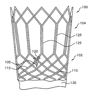
Fig. 1 depicts a device according to a first embodiment of the present
invention. Fig.
1 shows an expanded filamentary stent 100 having a braided section 102 and a
would section 104, as
is described in the '165 application. Stent 100 comprises a first filament 110
and a second filament
115, both of which extend along both braided section 102 and wound section
104. Within the wound
section, a plurality of hexagonal cells 125 (also referred to herein as
"vertical cells") are formed by
the filaments, with each cell having a base defined by two segments of the
hexagonal cell. First
filament 110 and second filament 115 also form a plurality of intersections,
such as intersection 120,
defined by the two filaments crossing one another.
The device shown in Fig. 1 also includes a self deploying barb assembly 105,
which
is attached to stent 100 adjacent intersection 120. Figs. 2, 3a, and 3b show
self-deploying barb
assembly 105 in more detail. As shown therein, self deploying barb assembly
105 comprises: (i) a
first portion 270 attached to the stmt, (ii) a bend 280, and (iii) a second
portion 275, disposed
opposite the first portion from the bend and having a bearing surface 285.
Bearing surface 285 is the
underside of second portion 275, as viewed in Fig. 2. Barb assembly includes a
first wire 235 and a
second wire 245, each of which extending across first portion 270 and second
portion 280 and each
having a bend 275. As shown in these figures, a first end of first wire 235
and a first end of second
wire 245 are disposed within first portion 270 and are attached to stent 100.
The other ends of the
CA 02495906 2004-10-18
WO 03/099167 PCT/US03/13533
two wires are attached to one another to form a point. More specifically,
second wire 245 is attached
to first filament 110 and first wire 235 is attached to second filament 115 in
the area of intersection
120. A wide variety of ways to attach the wires to the filaments may be
employed, e.g. welding,
suturing, gluing, and the like, so long as the means for attachment do not
adversely affect the
biocompatibility of the stent.
Self deploying barb assembly 105 is pre-fabricated and made of a biocompatible
wire,
such as nitinol or a material compatible with the biocompatible material of
stent 100. In this
particular example, self deploying barb assembly 105 is in the area of
intersection 120, which is in a
row of stent 100 between braided section 102 and wound section 104. More
specifically, barb
assembly 105, including bend 120, is disposed adjacent intersection 120. The
present invention is not
limited to this configuration. Self-deploying barb assemblies 105 may also be
fixed to vertical cell
segments 125 or to another row within braided section 102. Stent 100 may
include a plurality of self
deploying barb assemblies 105 attached along the perimeter of stmt 100 and
having variable
dimensions and geometry, as long as both stmt 100 and self deploying barb
assemblies 105 function
within a medically acceptable tolerance.
In some embodiments, the device may also include a graft 130 as shown in Fig.
1.
Such grafts may be used in an endoluminal device for treating AAA. Grafts
serve to prevent blood
from flowing across the device to an aneurysm sac. The material for such
grafts may be any suitable
material used for such purposes, and the graft may be a braided or non-braided
graft, and may
comprise any graft material known in the art. Suitable graft materials
include, but are not limited to,
polyethyleneterepthalate (PET), polyetheretherketone (PEEK), polysulfone,
polytetrafluroethylene
(PTFE), expanded polytetrafluroethylene (ePTFE), fluorinated ethylene
propylene (FEP),
polycarbonate urethane, a polyolefin (such as polypropylene, polyethylene, or
high density
polyethylene (HDPE)), silicone, and polyurethane. Preferably, and as shown in
Fig. 1, graft 130 is
affixed to stem 100 at an area remote from (i.e., axially distant from) barb
assembly 105. Typically,
the portion where the barbs are located are intended to be placed in the body
lumen at a location
where there is healthy tissue; on the other hand, a graft is located at a
position along the device
corresponding to an unhealthy portion of the body lumen, such as an aneurysm
sac.
Fig. 2 shows self deploying barb assembly 105 in more detail including first
flat wire
235, a first wire hinge 240, second flat wire 245, a second wire hinge 250, an
apex weld 255, a first
posterior tab 260, and a second posterior tab 265. Apex weld 255 joins first
flat wire 235 to
overlapping second flat wire 245, as mentioned above. To prepare the device,
self-deploying barb
CA 02495906 2004-10-18
WO 03/099167 PCT/US03/13533
-g_
assembly 105 is typically pre-fabricated from a suitable material, such as
spring steel, nitinol, or
other suitable metals. The assembly is then affixed to first filament 110 and
to second filament 115
using first wire hinge 240 and second wire hinge 250, respectively, in the
area where first filament
110 and second filament 115 form intersection 120. According to an embodiment
of the invention, a
first posterior tab 260 and a second posterior tab 265 limit rotation of the
hinge on self deploying
barb assembly 105, causing the barb to engage as the diameter of stmt 100
changes upon expansion.
Fig. 3a shows a three-dimensional view of a segment of the device of Figs. 1
and 2
including stent 100, comprising first filament 110 and second filament 115,
with the device in its
radially expanded configuration. Also shown is an engaged barb assembly 105.
When the diameter
of stmt 100 is increased, the forces exerted on barb assembly 105 cause it to
flip from a sub-surface
profile in a generally outward direction relative to an axis of stem 100 to
engage the vessel wall, as
discussed in more detail below. As used herein, the term "engage" means when a
portion of the barb
assembly protrudes into and contacts the body lumen in a way which decreases
migration of the
device relative to the body lumen.
Fig. 3b is a three-dimensional view of a segment of the device of Figs. 1 and
2
including a stmt 100 comprising first filament 110 and second filament 115,
and an unengaged self-
deploying barb 105. When stent 100 is compressed in the deployment catheter,
it is formed to be
biased in a radially inward direction relative to an axis of stmt 100, and
thereby preventing the point
of barb assembly 105 from scratching the catheter wall.
As can be seen when comparing Figs. 3a and 3b, second portion 275 of barb
assembly 105 (i.e., that portion below the bend 280) swings radially outward
to engage the lumen
wall as stmt 100 radially expands. Thus, second portion 275 is adapted to
protrude radially inward
when stmt 100 is in its radially compressed configuration. This can be done in
any number of ways,
such as by using a shape memory alloy, such as nitinol which could be
configured to have the desired
shape in the radially compressed configuration. Spring steel or other metals
could also be used.
Barb assembly 105 is caused to take its shape as shown in Fig. 3a due to a
filament or intersection
radially contacting and imparting a radially outward force against bearing
surface 285 of the barb
assembly 105. More specifically, the radially outward force from stent 100, as
it moves from its
radially compressed configuration to its radially expanded configuration, is
preferably directed
somewhere on the bearing surface 285 of second portion 275. To facilitate this
extension of barb
assembly, it is desirably to cause the force be directed to the end of the
second portion furthest from
bend 280.
CA 02495906 2004-10-18
WO 03/099167 PCT/US03/13533
-9-
As is known, the angle of some intersections of certain types of stems changes
as the
stent moves from a radially compressed configuration to a radially expanded
configuration. This is
true for braided stents or braided portions of stents, such as braided portion
102, in which angle a is
shown in Fig. 2. This means that, as stmt 100 expands, first filament 110 and
second filament 115
swing relative to one another as angle a increases. Thus, the swinging of
second filament 115
against bearing surface 285 of second portion 280 can enhance the radial
expansion of barb assembly
105 in concert with the radially outward force caused by the expanding stmt
generally. Preferably, a
protuberance 290 is formed on the radially inner side of second portion 280
for abutting against stem
100 as the stem moves between the radially compressed configuration and the
radially expanded
configuration. Such a protuberance is located at a position such that a
filament crosses and contacts
the protuberance during radial expansion of the stem.
A method for implanting an endoluminal device in a body lumen involves first
compressing the endoluminal device into a radially compressed configuration
and retaining it in an
introduces. Such an introduces may be a delivery catheter as are well known in
the art, such as those
described in U.S. Patent Application No. 09/573,273, entitled STENT DELIVERY
SYSTEM FOR
PREVENTION OF KINHING, AND METHOD OF LOADING AND USING SAME, assigned to
the assignee of this application and incorporated herein by reference. Next,
the introduces is
introduced or threaded into the body lumen via a vascular access site to a
deployment location, such
as by using a well-known percutaneous cut-down technique referred to above.
Examples of the
vascular access site include the femoral artery. The access site may be
surgically exposed and
punctured with, for example, an 18-gauge needle. Then, the device is deployed
from the introduces
and into the body lumen. This is typically done by first aligning the distal
end of the device, then
retracting an outer sheath of the introduces. After or upon deployment, the
endoluminal device
expands to form a radial expanded portion and the at least one filament
radially contacts the second
portion and imparts a radially outward force against the bearing surface as
the implant (e.g., stmt)
moves from its radially compressed configuration to its radially expanded
configuration to cause the
second portion to protrude radially outward and engage the body lumen when the
stmt is in its
radially expanded configuration. In the event that the stem is self expanding,
the radial expansion of
the stent is caused by the removal of the stent from the introduces. On the
other hand, if the stent is
not self expanding, the radial expansion of the stmt is caused by expanding a
balloon (or some other
external source of radially outward force) from within the stem.
CA 02495906 2004-10-18
WO 03/099167 PCT/US03/13533
-10-
According to another embodiment of the present invention, Fig. 4a shows a
device
comprising a filamentary stent 400 and a corkscrew barb 405. The stent is
similar to stmt 105 shown
in Fig. 1 in that it has a braided section 402 and a wound section 404. As
discussed in connection
with the first embodiment, a vertical segment 410, a first filament 415, and a
second filament 420 are
shown. The barb 405 comprises (i) a base segment 407 attached to one or more
filaments (including
an intersection)t and (b) a curved segment 409 extending from the base segment
and terminating in a
point. The curved segment is curved proximally and radially inwardly but not
extending radially
within the periphery defined by said stmt. The downward curvature of barb 405
is shown in Fig. 4a
while the radially inward curvature is shown in Figs. 4b and 4c.
Barb 405 is a biocompatible material, such as nitinol or a material compatible
with
the biocompatible material of stmt 400. Barb 405 is preferably welded at the
base of vertical
segment 410 where first filament 415 and second filament 420 intersect. Barb
405 is corkscrewed to
the longitudinal axis of stmt 400. The degree of skewness can range from a
small degree to a large
degree. The degree of skewness, of course, should be sufficient to allow the
barb to hold the stem in
place, without causing any damage to the introducer. Preferably, the
longitudinal axis of base
segment 407 is at least somewhat parallel, more preferably about parallel, to
a line intersecting the
longitudinal axis at a right angle (90 degrees). When the proximal end of stem
400 is deployed, stmt
400 may be rotated to implant barbs 405 into the vessel wall, thereby securing
the vessel wall to the
stmt graft. Barbs 405 are preferably configured such that only a slight
rotation of the catheter (e.g.,
about 15° or less) is required to twist the barbs into the vessel wall.
As in the first embodiment, the
device may further comprise a graft 430 which is affixed to stem 400 remote
from barb 405.
Fig. 4b shows filamentary stem 400 with a plurality of corkscrewed barbs 405.
Barbs 405 are pointing in an outward direction, i.e., as they would point in a
deployed configuration.
This is after the device has been deployed and twisted in the body lumen to
cause an increase in angle
(3.
Fig. 4c shows the compressed filamentary stmt 400 with a plurality of
corkscrewed
barbs 405. When stmt 400 is compressed for loading into the stmt deployment
catheter, barbs 405
are aligned so that the points of barbs 405 do not scrape the inner surface of
the outer sheath. Barbs
405 are preferably just slightly curved, as shown in Fig. 4c, as further
precaution that the points do
not scratch the sheath.
A method to deploy a stmt according to this embodiment of the invention again
involves compressing the endoluminal device into a radially compressed
configuration and retaining
CA 02495906 2004-10-18
WO 03/099167 PCT/US03/13533
-11-
the device in an introducer; introducing the introducer into the body lumen to
a deployment location;
and deploying the endoluminal device from the introducer and into the body
lumen. This method also
involves twisting the stmt between 1 and 15 degrees to cause the curved
segment to engage the body
lumen. This twisting or rotation involves rotation in an engaging direction.
Similarly, if it is desired
to disengage the implant, then rotation in the opposite direction would
disengage the engagement
means.
According to another embodiment of the present invention, Fig. Sa shows a
device
comprising a filamentary stmt 500 and a barb assembly 505. The stmt is similar
to stent 105 shown
in Fig. 1 in that it has a braided section 502 and a wound section 504. As
discussed in connection
with the first embodiment, a vertical segment 510, a first filament 515, and a
second filament 520 are
shown. The barb assembly comprises: (i) a wire 507 extending from the top of a
cell to the bottom
of a cell and having a length greater than the cell height and a substantially
uniform cross-sectional
area and (ii) a hook 509 affixed to the wire and extending radially outward.
The term substantially
uniform is intended to mean that there is not a change in cross sectional area
of greater than 10% and
there are no step changes in cross sectional area. The wire is formed to arc
radially inwardly, as
shown in Fig. Sc, when the stent is in its radially compressed configuration
and is capable of being
arced radially outwardly, as shown in Fig. Sb, when the stmt is in its
radially expanded
configuration.
The mechanism can involve using stent wires (or ribbon) such that there are
two
support wires of the same length, on either side of a third wire of a longer
length than the supports.
As a result the longer wire is bowed and can be placed on the inner or outer
side of the stent by
pushing on the bowed wire. An illustrative example of such apparatus is
depicted in the Figs. Sa-Sc,
but the embodiment is not limited thereby. Preferably in this embodiment, the
barb assembly is
attached at a point where the cell height remains fairly constant as the
device is radially expanded.
This is generally true for the vertical segments 510 of the wound section 504
of stmt 500. In
addition, a graft 530 may be included in the device but is preferably remote
from barb assembly 505.
The hook(s)/barb(s) can be cut, etched, or attached to the longer wire in any
way
(facing up, down or both). The barbs can be set on the inner side of the stent
for loading and
deployment. Then, to deploy the barbs to the outer side post implantation of
the device a balloon can
be inflated or an inner member dilator/sheath on the delivery system can be
advanced in the barb area
to push or set the barbs to the outer side of the stent.
CA 02495906 2004-10-18
WO 03/099167 PCT/US03/13533
- 12-
A method to deploy a device according to this embodiment of the invention
again
involves compressing the endoluminal device into a radially compressed
configuration and retaining
the device in an introducer; introducing the introducer into the body lumen to
a deployment location;
and deploying the endoluminal device from the introducer and into the body
lumen. This method also
involves imparting a radially outward force against the barb assembly to cause
the barb assembly to
arc radially outwardly and cause the hook to engage the body lumen.
In connection with any of the embodiments discussed herein, radiopaque markers
may
be used in the construction of the attachment means. Such markers assist in
deploying, moving or
removing the stmt since the status of the barb can be determined. Preferably,
radiopaque material
l0 can be used in the construction of the engagement means, thereby permitting
the artisan to further
reduce the risk of damage.
In another embodiment of the present invention, the barbs are supported such
that
during loading into the catheter, in the fully loaded state and during
deployment there is no contact
between the barbs and the catheter wall. Then, either once the barbed area is
exposed or the entire
15 stmt-graft system is deployed, the barbs are deployed into place by means
such as inflating a balloon
or advancing a dilator to push the barbs out into place.
Although illustrated and described herein with reference to certain specific
embodiments, the present invention is nevertheless not intended to be limited
to the details shown.
Rather, various modifications may be made in the details within the scope and
range of equivalents of
20 the claims and without departing from the spirit of the invention.