Note: Descriptions are shown in the official language in which they were submitted.
CA 02496091 2005-02-15
WO 2004/025331 PCT/US2003/029332
1
MICROSCOPE WITH EXTENDED FIELD OF VISION
FIEhD AND BACKGROUND OF THE INVENTION
[0001] The present invention relates generally to the
field of microscopy and in particular to a new and
useful apparatus and method for high-speed image
scanning, image capturing, and mosaicking to enlarge
the field of view with respect to microscopic obj acts .
[0002] A microscope is an indispensable tool for micro-
assembly and micro-manipulation. However,
conventional microscopes suffer from the limitation
that high magnification reduces the size of the field
of view, which is the maximum object size which may be
imaged by a lens. As a result, many micro-assembly or
micro-manipulation tasks that require micron to
sub-micron precision over millimeter work volume are
beyond the capability of fixed optical microscopes.
[0003] For example, in vitro fertilization requires the
manipulation of two microscopic biological cells, a
spermatazoid and an ovule. The two biological cells
CA 02496091 2005-02-15
WO 2004/025331 PCT/US2003/029332
2
may be separately located in two different zones of
interest on a biological plate. If a first zone of
interest is magnified for viewing and manipulation of
one biological cell, a biological cell located at a
distant second zone of interest may fall outside the
field of view of the first zone of interest. This is
especially problematic where the biological cell at
the second zone of interest is moving.
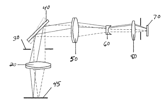
(0004 Another example involves micro-assembly by mobile
robot using relative positioning. Stick-and-slip
microrobots for instance offer a very high relative
accuracy (a few nm) in a large working space. However,
high resolution sensors that can work on a large scale
are usually expensive and volume-consuming. Moreover,
one sensor is required for each degree of freedom.
One way to reduce the number of sensors is to use
mufti-dimensional sensors. Using a pattern-matching
algorithm, it is possible to track the motion of an
object via a CCD-camera looking through a microscope.
[0005] The accuracy of this sensing method depends on
magnification but an accuracy of half a micron to a
quarter of micron can be reasonably achieved. X and
Y movement and rotation can be sensed without
defocusing the microscope and the Z position can be
obtained by focusing-defocusing the picture. However,
the working volume will be limited to the size of the
picture itself which is a problem if" accuracy is
required. If the robot has to perform a task like a
pick-and-place manipulation.this volume may not be
large enough. The whole assembly setup might have to
be moved under the microscope which may be a problem
if delicate assembly is required.
CA 02496091 2005-02-15
WO 2004/025331 PCT/US2003/029332
3
[0006] A common solution to the problem of reduced field
of view is to move the platform supporting the sample
or to move the microscope itself. The bandwidth of
the motion is limited by the inertia of the platform
or microscope, and the vibration resulting from the
motion can blur the image or even modify the scene.
[0007] Mosaicking, or forming a single large image from
smaller images, is used in applications such as NASA
planetary flybys and photo-stitching software in some
consumer digital cameras. However, implementation of
mosaicking for performance of dynamic micro-assembly
and micro-manipulation tasks with real-time vision
guidance requires an optical system with a
sufficiently fast refresh rate. Confocal microscopes
employ high speed scanning to form images but only a
single pixel data is obtained at each scan.
[0008] Similarly, U.S. Patent 6,433,907 to Lippert et
al. teaches a display apparatus that includes a
scanning assembly that scans a plurality of light
beams produced from spatially distinct regions, in a
raster pattern. The scanning assembly includes
mirrors that pivot to sweep the beams . Beam color and
intensity is modulated to form a respective pixel of
an image. By properly controlling the color and
intensity of the beam for each pixel location, the
display can produce a contiguous image from the pixels
from each distinct region. Like confocal microscopes,
the Lippert '907 apparatus involves acquisition of
single pixel.data, or pixel-by-pixel scanning.
[0009] U.S. Patent 6,313,452 to Paragrano et al.
discloses a microscopy system that utilizes a
plurality of images to create a single mosaic image.
CA 02496091 2005-02-15
WO 2004/025331 PCT/US2003/029332
4
The system comprises a stage, at least one magnifying
lens, a lens controller, a video capture device, and
a processing subsystem. However, no high-speed
scanning or capture devices are included.
[00103 U.S. Patents 6,101,265 and 6,226,392 both to
Bacus et al. teach an apparatus and method for
acquiring and storing multiple images from a specimen
via a microscope and digital scanner, and providing a
user a reconstructed image of the entire specimen at
low magnification. The reconstructed image is formed
of a large number of tiled images which are
coordinated and assembled to form the reconstructed
image. High-speed scanning, imaging, and refreshing
are not taught.
[0011] U.S. Patent 6,272,235 also to Baccus et al.
further teaches that acquired images are coherently
seemed together to provide virtual digitized images at
either at low or high resolution. A data structure is
formed with the virtual digitized images along with
their mapping coordinates. The data structure is
formed with compressed data so that it can be
transmitted over low bandwidth channels, such as the
Internet, without loss of resolution.
[0012] An optical system is needed that addresses the
field of vision limitations of conventional
microscopes without movement of the microscope stage'
or sample. The optical system should be capable of
capturing images at fast refresh rates so that a
virtual reconstructed image can be constructed
quickly, where a view of the reconstructed image
cannot be differentiated from the specimen view by the
human eye. Such an optical system thereby overcomes
CA 02496091 2005-02-15
WO 2004/025331 PCT/US2003/029332
the disadvantages of traditional motorized stages
which are significantly slower. The optical system
should be capable of spanning images which are focused
and undistorted. Image processing in stitching the
images together should be performed without any
particular imaging algorithms.
SUN~1ARY OF THE INVENTION
[0013] It is an object of the present invention to
create a mosaic image, or selept portions of a mosaic
image, of a large field of view through a microscope
at fast refresh rates.
[0014] It is another object of the present invention to
provide a mosaic image with a high resolution that is
free of blurring or aberrations.
[0015] It is yet another object of the present invention
that the mosaic view be formed by stitching smaller
images together on the fly.
[0016] It is yet another object of the present invention
to provide for a virtual enhancement in resolution by
combining several overlapping images of the same
scene. '
[0017] Accordingly, an optical system is provided for
enlarging the field of view with respect to an object
by high-speed scanning, image capture, and image
mosaicking along an optical path. The optical system
includes one or more lenses forming an objective lens
assembly positioned downstream from the object along
the optical path so that the object is positioned at
a focal plane of the objective lens assembly. The
purpose of the objective lens assembly is to collect
the light from the specimen and to bend to light rays
CA 02496091 2005-02-15
WO 2004/025331 PCT/US2003/029332
6
to form collimated or nearly collimated light beams.
By collimated or nearly collimated, it is meant that
the back focal distance of the objective lens assembly
be in the range of 50mm to infinity. A back focal
distance of infinity means that the light is perfectly
collimated.
[0018] The optical system further includes an iris
downstream from the objective lens assembly, one or
more galvanometric scanning mirrors placed downstream
from the iris for high-speed scanning, one or more
imaging lenses, such as a converging lens and a
diverging lens combined with an iris, downstream of
the galvanometric scanning mirrors, and a high-speed
digital imaging device downstream of the imaging
lenses. The optical system also includes a means for
processing and constructing scanned and captured
images into a mosaic image on the fly without any
particular imaging algorithms.
[0019] The various features of novelty which
characterize the invention are pointed out with
particularity in the claims annexed to and forming a
part of this disclosure. For a better understanding
of the invention, its operating advantages and
specific objects attained by its uses, reference is
made to the accompanying drawings and descriptive
matter in which a preferred embodiment of the
invention is illustrated.
BRIEF DESCRIPTION OF THE DRAWINGS
(0020] Tn the drawings:
CA 02496091 2005-02-15
WO 2004/025331 PCT/US2003/029332
7
[0021] Fig. 1 is a schematic diagram of a first
embodiment of the optical path of the
invention;
[0022] Fig. 2 is a schematic diagram of a second
embodiment of the optical path of the
invention;
[0023] Fig. 3 is a schematic diagram of the optical
path of the invention, including a
computer; and
[0024] Fig. 4 is a schematic diagram of the
dimensions of a mosaic image achieved
by the present invention.
[0025] Fig. 5 is a diagram that shows a field of
view that has been enlarged via
mosaicking.
DESCRIPTION OF THE PREFERRED EMBODIMENTS
[0026] Referring now to the drawings, in which like
reference numerals are used to refer to the same or
similar elements, Fig. 1 shows an optical system.
[0027] The optical system can be divided into six
elements. The first element is an illumination system
1. A specimen can be illuminated from the bottom due
to a dedicated illumination system. It can also be
illuminated from the top due to a light beam injected
into the optical path. Another possibility is side
illumination using fiber bundles for instance.
[0028] The second element is an objective lens assembly
3 that consists of one or more lenses. An object is
placed at the focal plane of the lens assembly such
that each ray emitting off the same point on the
object exit parallel to each other from the lens
CA 02496091 2005-02-15
WO 2004/025331 PCT/US2003/029332
8
assembly. One or more lenses in the assembly can be
moving to achieve a specific function like focusing or
aberration compensation.
[0029] An iris 30 is placed just after the lens assembly
to enhance image contrast. Other features like
polarizers, quarter-wave plates or filters may also be
placed just after the lens assembly to modify some
characteristics of the light passing through it such
as polarization.
[0030] The third element is a scanning system 5. It
consists of one or several mirrors assembled such that
any ray reflecting on the mirrors with a given angle,
can be reflected off at another angle in a
controllable manner.
[0031] The fourth element is an imaging lens assembly 7.
The imaging lens assembly 7 consists of one or more
lenses such that any beam of parallel rays is focused
on a single point in the imaging plane. The fourth
element has the opposite function of the second
element.
[0032] The fifth element is an imaging device 9 that can
collect photon intensity andlor photon frequencies
and/or the difference of phase with a reference signal
and/or the polarization. Examples of device 9 are
Charge Coupled Devices (CCD) or CMOS sensors, a second
iris, a filter, a polarizer or any optical device that
modify wave characteristics such that the
polarization, the phase or the wavelength could be
used to achieve a particular image quality.
[0033] Finally, the sixth element is a processing unit
11. This unit can be one or more computers or any
stand-alone system or set of hardware capable of
CA 02496091 2005-02-15
WO 2004/025331 PCT/US2003/029332
9
controlling the mirrors position in real-time,
grabbing pictures or any set of collected data from
the imaging device 9, and eventually modulating the
illumination on the specimen in a synchronized manner
with the mirror motion and tuning the aperture of the
iris and/or the orientation of the polarizers and/or
the type of filters in real-time. The processing unit
11 further processes the acquired data and/or
reconstructs an enlarged field of view, and provides
a user interface through either a screen or a data
port.
[0034] Fig. 2 shows another exemplary embodiment of the
optical system which can be divided into three
sections. The first section has the main purpose of
scanning, and in its simplest form, includes an
obj ective lens 20, an iris 30, and high-speed scanning
mirrors 40. An object 45 is placed at the focal plane
of lens 20 such that each ray reflecting off the
object is collimated at the scanning mirrors 40 which
is downstream from the lens 20 and an iris 30. The
iris 30 is placed just after the lens 20 to enhance
image contrast.
[0035] A second section designed for the purpose of
image capture, conditions the image according to the
required performance such as the desired magnification
and digital imaging size. A Galilean-like optical
system (5 times ratio) is used, which consists of a
converging lens 50 and a diverging lens 60 downstream
from the scanning mirrors 40. Further downstream,
rays exit parallel to the Galilean expander and form
an image on the high-speed digital imaging device 70
by means of a fourth lens 80.
CA 02496091 2005-02-15
WO 2004/025331 PCT/US2003/029332
(0036] A third section is designed for processing and
constructing the captured images of the digital
imaging device into a mosaic image . The third section
includes a computing device 90 with software as shown
in Fig. 3. Construction of the mosaic image involves
use of a frame grabber for acquiring the images from
the high-speed digital imaging device 70, a dedicated
algorithm for organizing, processing, and constructing
the mosaic image, and pattern recognition for
synchronization of the images. The scanning pattern
as well as integrated manipulation is also preferably
shared through a network such as the Internet allowing
collaborative observation and manipulation.
[0037] In one preferred embodiment of the present
invention, lens 20 is an achromat lens with a focal
length ranging from about 25 mm to 75 mm, and most
preferably about 50mm, and a diameter ranging from
about l0mm to 40mm, and most preferably about 25.4mm.
Achromat lenses are preferred to minimize or eliminate
chromatic aberrations from the resulting images.
(0038] The scanning mirrors 40 are a moving mirror ~XBy
galvanometer system which is placed in the optical
path for the purpose of optical scanning. A
galvanometer is an electromagnetic actuator, similar
in principle to a DC motor, but with no commutation, .
so the amount of shaft rotation is limited to about 20
degrees. A mirror is mounted on the output shaft of
the galvanometer to reflect and direct the light beam
coming from the objective lens assembly to the imaging
lens assembly. Such galvanometers can achieve
millisecond settling time over small motion ranges.
It is further noted that the scanning mirrors 40 is
CA 02496091 2005-02-15
WO 2004/025331 PCT/US2003/029332
11
self-contained and portable so that it can be inserted
into the optical path over any type of sample.
[0039] The illumination system could also be
synchronized with the scanning system such that only
a selected region of the specimen is locally
illuminated. The intensity could also be modulated to
provide for uniform lighting regardless of mirrors
positions and associated optical imperfection. This
could be achieved with an appropriate feedback system
or through an appropriate calibration.
[0040] The scanning mirrors 40 operate at a speed to
allow ample time to settle to prevent image blur, the
optical system has only minimal complexity, and image
acquisition is based on readily available software.
A program, which can be written in any programming
language, including C++, is used to coordinate the
motion control with the image acquisition and image
processing. After initial startup and during the
process of image acquisition, the scanning mirrors
operate at a speed within the range of 0=-300 tiles per
second, and most preferably as fast as 0-250 tiles per
second. That is, to construct a 5x5 mosaic of tiles,
the entire 5x5 mosaic can be refreshed at a rate of 10
complete mosaics per second. As a result, the bulk of
the operation time at the present is spent on image
acquisition and processing. The scanning motion is
preferably programmed for rapid tracking of multiple,
and possibly disconnected, events. The motion of the
scanning mirrors 40 is coordinated to create subpixel
accuracy.
[0041] The capability of positioning the center of the
field of view with subpixel accuracy allows multiple
CA 02496091 2005-02-15
WO 2004/025331 PCT/US2003/029332
12
images to be taken of the same scene from slightly
different angles. The data from the multiple images
can be combined to form an image with a virtual
resolution higher than the optical and imaging
resolution by means of super resolution image
processing algorithms. As an example of this
technique, super resolution image processing
algorithms are used by law enforcement agencies to
create high resolution images of a license plate from
multiple frames of low resolution video footage of the
license plate taken from slightly different angles as
the car drives away.
[0042] But it is noted that since the optical layout
allows scanning without disturbing the specimen, there
are applications where the user would want to scan
slower than 250 image tiles per second to reduce the
amount of raw image data that must be stored (large
population study of a slow biological process would be
an example).
[0043] Converging lens 50 has a focal length between
about 25mm and 75mm, and most preferably about 50 mm,
and a diameter between about 10mm and 40mm, and most
preferably about 25.4mm. Diverging lens 60, which is
downstream of the converging lens along the optical
path, has a focal length between about 3mm and 15mm,
and most preferably about 9mm, and a diameter between
about 3mm and l5mm, and most preferably about 9mm.
The fourth lens 80, which is downstream of the
diverging lens, is preferably a biconvex lens and has
a focal length between about 25mm and 500mm, and most
preferably about 100mm. Lens 80 also has a diameter
between about 5mm and 200mm, and most preferably about
CA 02496091 2005-02-15
WO 2004/025331 PCT/US2003/029332
13
l5mm.
[0044] Images scanned by the scanning mirrors 40 are
captured from the scanner via the high-speed digital
imaging device 70 which is preferably a charge-coupled
device ("CCD") camera. The CCD then relays the images
to a processing unit 11 such as a computer 90 for
processing the images and constructing a mosaic image
representing the scene viewed under the microscope.
[0045] The motion of the scanning mirrors 40 is
coordinated with image capture to enable precise image
alignment during the mosaic forming process.
Therefore, the processing unit can accomplish
stitching on the fly without any particular imaging
algorithms. As compared to traditional mosaicking
methods that use features in the image for alignment,
and require time consuming stitching algorithms, the
subpixel positioning accuracy of the current invention
allows for a very quick direct memory copy. It is
noted that traditional mosaicking algorithms can be
applied, at a cost to image acquisition speed, but
with a potential increase in image alignment quality.
[0046] As shown in Fig. 4, the preferred embodiment of
the invention produces a mosaic image that is 4 x 4,
wherein an image area of each dimension, defined by a
x b, is 2mm x l.5mm and an overall field of view is
8mm x 6mm, as defined by c x d, at a resolution of
6.25 x 6.25 ~.~.m2/pixel, or 320 x 240, as defined by a
x f. It is noted that the dimensions and image sizes
specified above will depend on the selection of
digital imaging device and the lens combination chosen
for the specific imaging task at hand. As is common
in microscopy, the camera' s pixel density and the lens
CA 02496091 2005-02-15
WO 2004/025331 PCT/US2003/029332
14
combination are selected to provide the appropriate
magnification. Similarly, the size of the mosaic
image will depend on the task at hand and can be
generalized to be m tiles wide by n tiles high
(m x n) .
[0047] As shown in Fig. 5, a field of view 100 is
enlarged wherein the pieces Q, R, S, T, U, V are
mosaicked to produce a mosaic image 110.
[0048] Although a preferred embodiment has been shown,
the invention is not limited to the lens and image
parameters and measurements which have been provided.
Both optical and mechanical design parameters of the
system may be optimized to improve image acquisition,
processing, image resolution and field of view.
[0049] To improve the overall image quality or to
achieve imaging performance specifications, different
lens assemblies could potentially be used for the
objective lens assembly as well as for the imaging
lens assembly. Configurations known as
Petzval,.Telephoto, Zeiss Tessar, Cooke Triplet or F-
. Theta are among the candidates. The book Optical
System Design from R. Fischer and Biljana Tadic-Galeb
published by SPIE-Press/McGraw Hill (ISBN 0-07-134916-
2) gives an overview of these systems (see in
particular pages 130 to 139).
[0050] With regard to optical design parameters, the
size, shape and distance from the object to lens 20 or
lens assembly 3, will directly affect the size of the
field of vision as well as the image quality (such as
optical aberrations, image distortions, etc.) as the
system is working off the optical axis.
[0051] With regard to mechanical design parameters, the
CA 02496091 2005-02-15
WO 2004/025331 PCT/US2003/029332
settling time of the scanner will define the refresh
rate. Many of these parameters are closely related.
For example, a larger mirror will allow, among others,
a larger field of vision but at the cost of a longer
settling time and therefore a lower refreshing rate.
A larger CCD array provides higher image resolution
but requires more data transfer and image processing
time.
[0052] A target refresh rate is 25 Hz or at least 25 Hz.
The refresh rate is fast for a number of reasons,
including high-speed scanning and low inertia of the
scanning mirrors 40 and digital imaging with a high-
speed CCD or CMOS camera. Also, any motion errors
that are produced, occur after the objective lens 20,
and therefore, such errors are not magnified, as
compared to moving stages, in which error or
oscillation in the stage motion is magnified together
with the sample. Because error is not magnified and
the scanning mirror position is accurate, scanned and
captured images do not need to be stitched together
with software, but can be directly placed next to each
other with subpixel accuracy, permitting faster
refresh rates and more time for image processing. The
scanning pattern is also adapted to the regions of
interest in order to obtain the highest refresh rates .
[0053] The timing budget for a complete image is 40 ms.
Factors which contribute to the time consumption are
scanner motion (including motion profile generation
and physical movement from one scan area to the next ) ,
and image acquisition and processing. These two
operations can be executed in parallel since they
involve different processors.
CA 02496091 2005-02-15
WO 2004/025331 PCT/US2003/029332
16
[0054] Although specific embodiments have been shown for
microscopic applications, the invention is not limited
to those embodiments and may be used in macroscopic
applications such as law enforcement or other large
scale image capturing applications.
[0055] In one alternative embodiment of the invention,
other optical systems such as a laser beam can be
injected between the scanning and image capture
optical sections to perform different functions like
machining, manipulation, heating, cutting, welding or
fluorescent stimulation simultaneously with the
imaging.
[0056] While a specific embodiment of the invention has
been shown and described in detail to illustrate the
application of the principles of the invention, it
will be understood that the invention may be embodied
otherwise without departing from such principles.