Note: Descriptions are shown in the official language in which they were submitted.
CA 02496136 2005-02-18
WO 2004/017867 PCT/US2003/026154
-1-
COMPOSITE PROSTHESIS
Description
Technical Field
This invention relates to prosthesis for implantation within the human or
animal body for the repair of damaged lumens such as blood vessels.
Background of the Invention
Although this invention will be discussed with respect to its application to
repair of abdominal aortic aneurysms the invention is not so limited and may
apply
to prosthesis for repair of other lumens within the human or animal body.
Throughout this specification when discussing the application of this
invention to the aorta the term distal with respect to a prosthesis is
intended to refer
to the end of the prosthesis furthest away in the direction of blood flow from
the
heart and the term proximal is intended to mean the end of the prosthesis
which
when implanted would be nearest to the heart.
In our earlier patent specification published number WO98/53761 an
endoluminal prosthesis was disclosed which in particular was useful for repair
of
aortic aneurysms. A problem with such a prosthesis is that for different
persons or
animals different size prostheses must be constructed because the specific
dimensions of an aorta are quite variable in each of length, diameter and
angulation
between the renal artery region and the region of the aortic bifurcation.
Summary of the Invention
It is the object of this invention to provide a composite prosthesis which
can be assembled to fit a range of lengths of aorta thereby saving inventory
costs
and enabling off the shelf supply of a prosthesis assembly.
In one form therefore although this may not necessarily be the only or
broadest form the invention is said to reside in a prostheses assembly adapted
for
deployment in an aorta to span an aortic aneurysm, comprising at least first
and
second members with an end portion of one member to be joined to an end
portion
of the other member portion when in and when expanded within a lumen of a
patient, wherein each member comprises a stent arrangement associated with a
graft
CA 02496136 2005-02-18
WO 2004/017867 PCT/US2003/026154
-2-
arrangement, wherein the end portion of one member has at least part of its
stent
arrangement on the inner surface of its graft, and wherein the end portion of
the said
other member has at least part of its stent arrangement on the inner surface
of its
graft.
In an alternative form the invention is said to reside in a two part stent
graft prostheses assembly comprising at least first and second members to be
located within and joined together within a lumen of a patient, wherein one
member
is to be initially located and expanded within the lumen, said one member
having one
end portion with one or more stents on the inner surface of the graft, wherein
the
other member is to be sequentially located within and expanded within the said
lumen and has a second end portion to be located within the said one end
portion,
and wherein the said second end portion has a graft portion with a stent or
stents
on the inside surface thereof, so that when the said one and said other end
portions
are in engagement with one another there is no stent material between the
engaging
portions.
Preferably the said one member has a stent or stents on the outer surface
of a further part or the remainder of the graft of the said one member and the
said
other member has a stent or stents on the outer surface of a further part or
the
remainder of the graft of the said other member.
Preferably the stent graft prosthesis member for use with the above
assembly of claim comprises a stent or stents on one graft surface at one end
portion
thereof, and further comprises a stent or stents on at least a part of the
other graft
surface which part is spaced longitudinally from the said one end portion.
In an alternative form the invention is said to reside in a composite
prosthesis adapted for deployment in a lumen, the prosthesis comprising a
first
substantially tubular prosthesis portion and a second substantially tubular
prosthesis
portion, characterized by each prosthesis portion having a plurality of self
expanding
stents on an outer surface thereof along the length of each portion and at
least one
self expanding stent on an inside surface thereof at each end of each portion,
each
prosthesis portion having a connecting end adapted to engage with the
connecting
end of the other prosthesis portion and a remote end at the opposite end to
the
CA 02496136 2005-02-18
WO 2004/017867 PCT/US2003/026154
-3-
connecting end, each connecting end having the same outside diameter as the
other
connecting end, whereby in use the connecting end of the first prosthesis
portion can
be deployed either inside or outside the connecting end of the second
prosthesis
portion with at least two stents overlapping.
In further form the invention is said to reside in a composite prosthesis
adapted for deployment in an aorta to span an aortic aneurysm adjacent to or
including an aortic bifurcation, the prosthesis comprising a substantially
tubular
proximal prosthesis portion and a substantially tubular distal prosthesis
portion,
characterized by each prosthesis portion having a plurality of self expanding
stents
on an outer surface thereof along the length of each portion and at least one
self
expanding stent on an inside surface thereof at each end of each portion, each
prosthesis portion having a connecting end adapted to engage with the
connecting
end of the other prosthesis portion and a remote end at the opposite end to
the
connecting end, each connecting end having the same outside diameter as the
other
connecting end, whereby in use the connecting end of the proximal prosthesis
portion can be deployed either inside or outside the connecting end of the
distal
prosthesis portion with at least two stents overlapping such that the either
the distal
or proximal prosthesis portion can be deployed first and the other prosthesis
portion
deployed so that its connecting end is within the connecting end of the first
deployed
prosthesis portion.
It will be seen that by these general forms of the invention the amount of
overlap of the first and second or proximal and distal prosthesis can be
varied thereby
enabling a variety of lengths of aorta or other body lumen or the region being
spanned in the aorta to be allowed for. The ability to deploy with the
connecting end
either inside or outside means that the either the first or second prosthesis
portion
can be deployed first and then the other one deployed inside it. This gives a
physician considerable flexibility and means that a hospital can have a stock
of
prostheses which can be readily assembled depending upon the observed
vasculature.
CA 02496136 2005-02-18
WO 2004/017867 PCT/US2003/026154
-4-
Having the same diameter for each connecting end means that an
interference fit is obtained whether one connecting end goes inside or outside
the
other connecting end.
In one form of the invention the second or distal prosthesis portion may
be a bifurcated graft having a body portion and two leg portions.
Alternatively the
second or distal prosthesis portion may be an aorto-uni-iliac prosthesis.
The bifurcated second or distal prosthesis portion may have a shorter leg
and a longer leg and there may be self expanding stents on the outside of the
shorter
leg and the inside of the distal end of the longer leg.
There may be further included at least one leg prosthesis portion. The leg
prosthesis portion may be adapted to be deployed in to either the longer or
shorter
legs of the bifurcated second or distal prosthesis portion or into the end of
the aorto-
uni-iliac prosthesis.
The first or proximal prosthesis portion may be provided with a proximally
extending self expanding stent. Such a proximally extending self expanding
stent
may include barbs to engage against the wall of a lumen to hold the graft in
place.
This proximally extending self expanding stent may be adapted to span across
the
renal arteries to provide good mounting of the composite prosthesis within the
aorta.
Each of the stents may be zig zag or z-stents made from nitinol or stainless
steel.
Where it is desirable for the prosthesis portions to be flexible to allow for
angulation of or curves in the aorta the stents along the length of the
prosthesis
portion may be spaced apart along the graft material. Spacing of stents may be
from
0 mm to 8 mm. More flexibility may be provided on the proximal portion than
the
distal portion.
In an alternative form the stents may be balloon expandable stents.
Brief Description of the Drawing
This then generally describes the invention but to assist with
understanding reference will now be made to the accompanying drawings which
show preferred embodiments of the invention.
I
CA 02496136 2009-10-19
-5-
FIG. 1 shows a first embodiment of composite prosthesis
according to the invention in an exploded view;
FIG. 2 shows an assembled view of the embodiment shown in
FIG. 1;
FIG. 3 shows an alternate assembled view of the embodiment
shown in FIG. 1;
FIG. 4 shows an alternative embodiment of a distal prosthesis
portion according to the invention;
FIG. 5 shows an assembled view of part of the embodiment
shown in FIG. 1 and the embodiment shown in FIG. 4;
FIG. 6 shows an alternate assembled view of part of the
embodiment shown in FIG. 1 and the embodiment shown in FIG. 4;
FIG. 7 shows a detailed cut away view of the connecting region
of a prosthesis assembly of one embodiment of the invention showing the
bottom up approach; and
FIG. 8 shows a detailed cut away view of the connecting region
of a prosthesis assembly of one embodiment of the invention showing the top
down approach.
Detailed Description
U.S. Patent No. 5,387,235 entitled "Expandable Transluminal
Graft Prosthesis For Repair Of Aneurysm" discloses apparatus and methods
of retaining grafts onto deployment devices. These features and other
features disclosed in U.S. Patent No. 5,387,235 could be used with the
present invention.
U.S. Patent No. 5,720,776 entitled "Barb and Expandable
Transluminal Graft Prosthesis For Repair of Aneurysm" discloses improved
barbs with various forms of mechanical attachment to a stent. These features
and other features disclosed in U.S. Patent No. 5,720,776 could be used with
the present invention.
CA 02496136 2009-10-19
-6-
U.S. Patent No. 6,206,931 entitled "Graft Prosthesis Materials"
discloses graft prosthesis materials and a method for implanting,
transplanting
replacing and repairing a part of a patient and particularly the manufacture
and use of a purified, collagen based matrix structure removed from a
submucosa tissue source. These features and other features disclosed in
U.S. Patent No. 6,206,931 could be used with the present invention.
PCT Patent Publication No. WO 98/53761 entitled "A Prosthesis
And A Method And Means Of Deploying A Prosthesis" discloses an introducer
for a prosthesis which retains the prosthesis so that each end can be moved
independently. These features and other features disclosed in PCT Patent
Publication No. WO 98/53761 could be used with the present invention.
PCT Patent Publication No. WO 99/29262 entitled "Endoluminal
Aortic Stents" discloses a fenestrated prosthesis for placement where there
are intersecting arteries. This feature and other features disclosed in PCT
Patent Publication No. WO 99/29262 could be used with the present
invention.
PCT Patent Publication No. WO 03/034948 entitled "Prosthesis For
Curved Lumens" discloses prostheses with arrangements for bending the
prosthesis for placement into curved lumens. This feature and other features
disclosed in PCT Patent Publication No. WO 03/034948 could be used with
the present invention.
U.S. Patent Publication No. 2003/0233140 discloses release
wire systems for the release of stent grafts retained on introducer devices.
This feature and other features disclosed in U.S. Patent Publication
No. 2003/0233140 could be used with the present invention.
. . I I I I . CA 02496136 2009-10-19
-7-
U.S. Patent No. 6,939,370 discloses introducer devices adapted
for deployment of stent grafts particularly in the thoracic arch. This feature
and other features disclosed in U.S. Patent No. 6,939,370 could be used with
the present invention.
U.S. Patent No. 7,232,459 discloses stent grafts that are useful
in treating aortic aneurysms particularly in the thoracic arch. This feature
and
other features disclosed in U.S. Patent No. 7,232,459 could be used with the
present invention.
U.S. Patent No. 7,238,198 discloses arrangements for fastening
stents onto grafts particularly for exposed stents. This feature and other
features disclosed in U.S. Patent No. 7,238,198 could be used with the
present invention.
U.S. Patent No. 7,294,147 discloses retention arrangements for
retaining onto and releasing prostheses from introducer devices.
i i.i . ~ i . ..
CA 02496136 2009-10-19
- $ -
This feature and other features disclosed in U.S. Patent No. 7,294,147 could
be used with the present invention.
U.S. Patent Publication No. 2003/0120332 discloses
arrangements on stent grafts for enhancing the adhesion of such stent grafts
into walls of vessels in which they are deployed. This feature and other
features disclosed in U.S. Patent Publication No. 2003/0120332 could be
used with the present invention.
Now looking more closely to the drawings and in particular the
embodiment shown in FIGS. 1, 2 and 3 it will be seen that the composite
prosthesis includes a first or proximal prosthesis portion 2, a second or
distal
prosthesis portion 4 and leg prosthesis portion 6. The first or proximal
prosthesis portion 2 comprises a fabric material graft body 34 of
substantially
tubular form with self expanding zig zag stents 10 on the outside along most
of its length and self expanding zig zag stents 14 within the tubular body 8
at
the proximal end 16 and distal end 12. Extending from the proximal end 16 is
a supra-renal zig zag stent 18 with barbs 20 extending distally to provide
fixation into the wall of the aorta.
The zig-zag stents are also well known as Gianturco Z-stents
commercially available from William A Cook Australia Pty Ltd, Brisbane,
Australia or Cook Inc, Bloomington, Indiana, USA. The graft material is
typically DACRON material available from a number of medical graft
manufacturers.
The zig zag stent within the proximal end 16 of the first or
proximal prosthesis portion 2 assists with sealing of the graft against the
walls
of the aorta and the external zig zag stents provide a smooth inner surface
for
the flow of blood through the prosthesis. The internal zig zag stent 14 at the
distal end 12 provides an outer surface of the tubular body 8 which is smooth
and can seal within the proximal end of the second or distal prosthesis
portion 4 when it is deployed within the second or distal prosthesis portion
4.
CA 02496136 2005-02-18
WO 2004/017867 PCT/US2003/026154
-9-
The second or distal prosthesis portion 4 comprises a fabric material graft
body 26 and has an internal zig zag stent 22 at its proximal end 24 so that
the outer
surface of its tubular body 26 is smooth and can seal within the distal end of
the first
or proximal prosthesis portion 2 when it is deployed within the first or
proximal
prosthesis portion 2. The external zig zag stents 25 provide a smooth inner
surface
for the flow of blood through the prosthesis.
Towards the distal end of the second or distal prosthesis portion 4 the
tubular body 26 bifurcates into a longer leg 28 and a shorter leg 30 each of
which
has zig zag stents 29 on its outside surface except the terminal zig zag stent
32 on
the longer leg.
The leg prosthesis portion 6 which is adapted to extend into the
contralateral-iliac artery is comprised from a tubular fabric material body 34
with
outside zig zag stents 36 along its length except for internal zig zag stents
38 at its
proximal and distal ends.
FIG. 2 shows the assembled prosthesis in a first arrangement or
configuration in which the connecting end 24 of the second or distal
prosthesis
portion 4 is deployed within the distal connecting end 12 of the first or
proximal
prosthesis portion 2. It will be realized that the amount of overlap between
the first
or proximal prosthesis portion 2 and the second or distal prosthesis portion 4
can be
varied for different lengths of an aorta from the renal arteries to the aortic
bifurcation. It is preferable, however, that there is at least a longitudinal
or axial
overlap of two stents. This means that there will be a smooth inner surface of
one
portion engaged against a smooth outer surface of the other portion to provide
a
good seal. The leg prosthesis portion 6 is deployed with its proximal end 37
within
the shorter leg 30 of the second or distal prosthesis portion 4.
The prosthesis in FIG. 2 is assembled in what is also known as a top down
approach or assembly. The physician will deploy the proximal prosthesis
portion 2
first in the aorta of a patient followed by deploying or placing the distal
prosthesis
portion 4 in the aorta with the proximal end 24 of the distal prosthesis
portion 4
inside the distal end 12 of the proximal prosthesis portion 2.
CA 02496136 2005-02-18
WO 2004/017867 PCT/US2003/026154
-10-
FIG. 3 shows the assembled prosthesis in a second arrangement in which
the connecting end 12 of the first or proximal prosthesis portion 2 is
deployed within
the connecting end 24 of the second or distal prosthesis portion 4. It will be
realized
that the amount of overlap between the first or proximal prosthesis portion 2
and the
second or distal prosthesis portion 4 can be varied for different lengths of
an aorta
from the renal arteries to the aortic bifurcation. It is preferable, however,
that there
is at least an overlap of two stents. This means that there will be a smooth
inner
surface of one portion engaged against a smooth outer surface of the other
portion
to provide a good seal. The leg prosthesis portion 6 is deployed with its
proximal
end 37 within the shorter leg 30 of the second or distal prosthesis portion 4.
The prosthesis in FIG. 3 is assembled in a second arrangement or
configuration in what is also known as a bottom up approach or assembly. Here,
the
physician deploys the distal prosthesis portion 4 first in the aorta of a
patient
followed by deploying or placing the proximal prosthesis portion 2 through the
distal
prosthesis portion 4 and into the aorta with the distal connecting end 12 of
the
proximal prosthesis portion 2 inside the proximal connecting end 24 of the
distal
prosthesis portion 4.
FIG. 4 shows an aorto-uni-iliac prosthesis portion 38 which is suitable as
an alternative second or distal prosthesis portion. This portion 38 is adapted
to be
deployed inside or outside a first or proximal prosthesis portion as depicted
in FIGS.
or 6 and to extend from the aorta into either of the iliac arteries. In such a
situation a plug would normally be deployed into the other iliac artery.
The aorto-uni-iliac prosthesis portion 38 comprises a tubular slightly
tapered body 40 with a proximal end 42 and a distal end 52. The taper is used
because the iliac arteries are normally of lesser diameter than the aorta. The
aorto-
uni-iliac prosthesis portion 38 has an internal zig zag stent 46 at its
proximal end 42
so that the outer surface of its tubular body 40 is smooth and can seal within
the
distal end of the first or proximal prosthesis portion 2 when it is deployed
within the
first or proximal prosthesis portion 2. The external zig zag stents 48 provide
a
smooth inner surface for the flow of blood through the prosthesis. The
internal zig
zag stent 50 at the distal end 52 provides an outer surface of the tubular
body 40
CA 02496136 2005-02-18
WO 2004/017867 PCT/US2003/026154
-11-
which is smooth and can seal within and against the wall of the iliac artery
when it
is deployed.
FIG. 5 shows an assembled aorto-uni-iliac prosthesis in a first arrangement
or configuration in which the connecting end 42 of the aorto-uni-iliac
prosthesis
portion 38 is deployed within the proximal connecting end 12 of the first or
proximal
prosthesis portion 2. It will be realized that the amount of overlap between
the first
or proximal prosthesis portion 2 and the aorto-uni-iliac prosthesis portion 38
can be
varied for different lengths of an aorta from the renal arteries to the aortic
bifurcation
and to a suitable landing spot in one of the iliac arteries. It is preferable,
however,
that there is at least an overlap of two stents. This means that there will be
a
smooth inner surface of one portion engaged against a smooth outer surface of
the
other portion to provide a good seal.
FIG. 6 shows an assembled aorto-uni-iliac prosthesis in a second
arrangement or configuration in which the distal connecting end 12 of the
first or
proximal prosthesis portion 2 is deployed within the connecting end 42 of the
aorto-
uni-iliac prosthesis portion 38. It will be realized that the amount of
overlap between
the first or proximal prosthesis portion 2 and the aorto-uni-iliac prosthesis
portion 38
can be varied for different lengths of an aorta from the renal arteries to the
aortic
bifurcation and to a suitable landing spot in one of the iliac arteries. It is
preferable,
however, that there is at least an overlap of two stents. This means that
there will
be a smooth inner surface of one portion engaged against a smooth outer
surface of
the other portion to provide a good seal.
The use of the aorto-uni-iliac prosthesis in either the first or second
configuration again depends upon the whether the physician prefers to utilize
a top
down or bottom up approach or assembly as previously described.
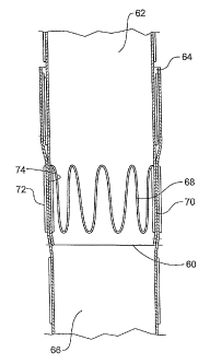
FIG. 7 shows an detailed cut away view of the connecting region of a
prosthesis assembly of one embodiment of the invention showing the bottom up
approach.
In this embodiment the distal end 60 of the proximal portion 62 is
deployed within the proximal end 64 of the distal prosthesis portion 66. The
end
stent 68 of the proximal portion 62 is inside the graft material of that
portion and
CA 02496136 2005-02-18
WO 2004/017867 PCT/US2003/026154
-12-
hence the outer surface 70 in that region is smooth. The end stent but one 72
of the
distal prosthesis portion 66 is on the outside of the graft material so that
the inner
surface 74 in that region is smooth. These smooth surfaces 70 and 74 engage
with
each other when the prosthesis is assembled and provide a seal between the
proximal and distal portions.
FIG. 8 shows an detailed cut away view of the connecting region of a
prosthesis assembly of one embodiment of the invention showing the top down
approach.
In this embodiment the proximal end 64 of the distal portion 66 is
deployed within the distal end 64 of the proximal prosthesis portion 62. The
end
stent 76 of the distal portion 66 is inside the graft material of that portion
and hence
the outer surface 78 in that region is smooth. The end stent but one 82 of the
proximal prosthesis portion 62 is on the outside of the graft material so that
the inner
surface 80 in that region is smooth. These smooth surfaces 78 and 80 engage
with
each other when the prosthesis is assembled and provide a seal between the
proximal and distal portions.
INTENDED USE
In one embodiment the composite prosthesis of the present invention is
intended to treat aneurysms of the abdominal aortic or aortoiliac by excluding
the
aneurysmal portion of that vessel from arterial flow and pressure. The
composite is
a multi-piece device and is to be used in instances where the implanting
physician
desires the ability to vary the overall length of the device by 'tromboning'
or for
applications where an increase in the angulation of the neck is required. The
device
is inserted via surgical cutdown into a femoral artery, the device is advanced
into the
desired position over a stiff wire guide using endovascular interventional
techniques.
A range of endovascular graft lengths and diameters are offered to the
implanting
physician to cater for individual patient anatomies.
The composite prosthesis of the present invention in one embodiment is
a self-expanding, fully supported and modular bifurcated system developed for
endovascular repair of infrarenal abdominal aortic aneurysms (AAA). The main
body
of the graft consists of two parts, a distal bifurcated graft and a proximal
tubular
CA 02496136 2005-02-18
WO 2004/017867 PCT/US2003/026154
-13-
extension graft. The other components of the graft are the iliac legs which
when
coupled with the main bifurcated body provide a variety of overall device
lengths.
Ancillary devices such as body extenders, aorto-uni-iliac converters, and
iliac plugs
may also be required. Each individual device has it's own separate delivery
system.
The bifurcated graft has one long limb with an iliac cuff and one short limb
on the contra-lateral side.
There is a radiopaque marker at the graft bifurcation and a 'tick' marker
at the distal end of the contra-lateral limb.
This bifurcated graft is pre-mounted into a deployment device with a
tethered top stent introduction system which provides a controlled release for
the
graft. This graft is attached to the delivery system at both ends and released
by
three independent trigger wires. The first wire releases the compressed short
leg,
the second wire releases the proximal end of the graft and the third wire
releases the
distal end of the graft.
The proximal extension graft is a tubular structure with an exposed
proximal attachment stent to allow for suprarenal fixation. Small radiopaque
markers
indicate the proximal edge of the graft.
This proximal extension graft is pre-mounted into a deployment device
with a top cap introduction system which provides a controlled release for the
graft.
The exposed attachment stent is constrained within a top cap and held there by
a
trigger wire. The distal end of the graft is also attached to the delivery
system and
held by an independent wire.
The iliac legs are tubular grafts which are used to extend the composite
graft into the iliac arteries. An iliac leg must be placed into the short limb
from the
contra-lateral side while a separate iliac leg can also be placed if needed
into the long
limb via the ipsilateral side.
Each component comes in a range of lengths and diameters which allows
the physician to tailor the device to individual patient anatomies and to
select the
best iliac landing site. The diameter at the connecting end of both the
proximal
tubular extension graft and the distal bifurcated graft may be 22 or 24 mm.
The
diameter of the proximal end of the proximal tubular extension graft may be
from 22
CA 02496136 2005-02-18
WO 2004/017867 PCT/US2003/026154
-14-
to 34 mm. The length of the proximal tubular extension graft may be from 73 to
131 mm. The diameter of the distal end of the a distal bifurcated graft may be
from
12 to 24 mm. The length of the distal bifurcated graft to the bifurcation may
be
from 50 to 95 mm and the overall length may be from 100 to 180 mm. Spacing of
the stents on the proximal extension graft may be from 1 to 8 mm. Spacing of
the
stents on the distal bifurcated graft may be from 0 to 1 mm on the body
portion and
from 1 to 3 mm on the longer leg portion.
Throughout this specification various indications have been given as to the
scope of the invention but the invention is not limited to any one of these
but may
reside at two more of these combined together. The examples are given for
illustration only and not for limitation.
Throughout this specification unless the context requires otherwise the
words comprise and include and variations such as comprising and including
will be
understood to imply the inclusion of stated integers or group of integers but
not the
exclusion of any other integer or group of integers.