Note: Descriptions are shown in the official language in which they were submitted.
CA 02496400 2005-02-21
WO 2004/022580 PCT/US2003/028482
BH3 PEPTIDES AND METHOD OF USE THEREOF
STATEMENT AS TO FEDERALLY SPONSORED RESEARCH
This invention was made with U.S. government support under NIH grant CA92625.
The government has certain rights in the invention.
FIELD OF THE INVENTION
This invention relates generally to methods and compositions for the
regulation of
apoptosis.
BACKGROUND OF THE INVENTION
Programmed cell death, referred to as apoptosis, plays an indispensable role
in the
development and maintenance of tissue homeostasis within all multicellular
organisms (Raff,
Nature 356: 397-400, 1992). Genetic and molecular analysis from nematodes to
humans has
indicated that the apoptotic pathway of cellular suicide is highly conserved
(Hengartner and
Horvitz, Cell 76: 1107-1114, 1994). In addition to being essential for normal
development
and maintenance, apoptosis is important in the defense against viral infection
and in
preventing the emergence of cancer.
Diverse intrinsic death signals emanating from multiple subcellular locales
all induce
the release of cytochrome c from mitochondria to activate Apaf 1 and result in
effector
caspase activation. Proteins in the BCL-2 family are major regulators of the
commitment to
programmed cell death as well as executioners of death signals at the
mitochondrion.
Members of this family include both pro- and anti-apoptotic proteins and share
homology in
up to four conserved regions termed BCL-2 homology (BH) 1-4 domains (Adams and
Cory,
CA 02496400 2005-02-21
WO 2004/022580 PCT/US2003/028482
1998). The family can be divided into three main sub-classes. The anti-
apoptotic
proteins, which include BCL-2 and BCL-XL, are all "multidomain," sharing
homology
throughout all four BH domains. However, the pro-apoptotic proteins can be
further
subdivided and include multidomain proteins, such as BAX and BAK, which
possess
sequence homology in BHl-3 domains. The more distantly related "BH3-only"
proteins are to
date all pro-apoptotic and share sequence homology within the amphipathic a-
helical BH3
region, which is required for their apoptotic function (Chittenden et al.,
1995; O'Connor et al.,
1998; Wang et al., 1996; Zha et al., 1997).
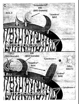
Multidomain pro-apoptotic proteins such as BAX and BAK upon receipt of death
signals participate in executing mitochondria) dysfunction. In viable cells,
these proteins exist
as monomers. In response to a variety of death stimuli, however, inactive BAX,
which is
located in the cytosol or loosely attached to membranes, inserts deeply into
the outer
mitochondria) membrane as a homo-oligomerized multimer (Eskes et al., 2000;
Gross et al.,
1998; Wolter et al., 1997 ). Inactive BAK resides at the mitochondrion where
it also
undergoes an allosteric conformational change in response to death signals,
which includes
homo-oligomerization (Griffiths et al., 1999; Wei et al., 2000). Cells
deficient in both BAX
and BAK are resistant to a wide variety of death stimuli that emanate from
multiple locations
within the cell (Wei et al., 2001).
The BH3-only molecules constitute the third subset of this family and include
BID,
NOXA, PUMA, BIK, BIM and BAD (Kelekar and Thompson, 1998). These proteins
share
sequence homology only in the amphipathic a-helical BH3 region which mutation
analysis
indicated is required in pro-apoptotic members for their death activity.
Moreover, the BH3-
only proteins require this domain to demonstrate binding to "multidomain" BCL-
2 family
members. Multiple binding assays, including yeast two-hybrid, co-
immunoprecipitation from
detergent solubilized cell lysates and in-vitro pull down experiments indicate
that individual
BH3-only molecules display some selectivity for multidomain BCL-2 members
(Boyd et al.,
1995; O'Connor et al., 1998; Oda et al., 2000; Wang et al., 1996; Yang et al.,
1995). The BID
protein binds pro-apoptotic BAX and BAK as well as anti-apoptotic BCL-2 and
BCL-XL
(Wang et al., 1996; Wei et al., 2000). In contrast, BAD, NOXA and BIM as
intact molecules
display preferential binding to anti-apoptotic members (Boyd et al., 1995;
O'Connor et al.,
1998; Oda et al., 2000; Yang et al., 1995).
2
CA 02496400 2005-02-21
WO 2004/022580 PCT/US2003/028482
SUMMARY OF THE INVENTION
The present invention is based on the discovery that the BH3 domain from the
BCL-2
family of proteins alone can function as a specific death ligand. The peptides
are refered to
herein as BH3 peptides.
In one aspect the invention provides an isolated peptide having a sequence of
SEQ ID
NO: 1, 2 or 10. The peptide induces BAIL oligomerization and cytochrome c
release from
mitochondria. In another aspect the invention provides an isolated peptide
having a sequence
of SEQ ID NOs: 3-9 or 11. The peptide binds BCL-2 or MCL-1. For example, SEQ
ID
NO:1-5 binds BCL-2. Alternatively, SEQ ID NO: 6 and 7 bind MCL-1.
Also include in the invention is a chimeric peptide having a first domain and
a second
domain. The first domain having and amino acid sequence of SEQ ID NOs: 1-11.
The second
domain having a translocation sequence which facilitates transport across a
biological
membrane. Examples, of translocation sequence includes polyarginine.
In another aspect the invention includes a nucleic acid encoding any one of
the
peptides of the invention.
Also included in the invention is a vector containing one or more of the
nucleic acids
described herein, and a cell containing the vectors or nucleic acids described
herein.
The invention is also directed to host cells transformed with a vector
comprising any of
the nucleic acid molecules described above.
In another aspect, the invention includes a composition that includes the
peptides of the
invention and a carrier or diluent.
In yet a further aspect the invention provides methods of treating a cell
proliferative
disorder, e.g., cancer in a subject by administering to a subject a BH3
peptide.
In another aspect the invention includes a method of inducing apoptosis in a
cell by
contacting said cell with SEQ ID NOs l, 2 or 10 such that apoptosis is
induced. Alternatively,
the invention provides a method of sensitizing a cell to apoptosis by
contacting said cell with a
composition comprising any of SEQ ID NOs: 3-7 or 11 such that as to sensitize
the cell to
apoptosis.
A further aspect the invention includes a method of screening for an apoptotic
sensitizer compound by contacting mitochondria overexpressing BCL-2 with a BID-
like BH3
peptide to form a BCL-2 - peptide complex and contacting the complex with a
test compound.
3
CA 02496400 2005-02-21
WO 2004/022580 PCT/US2003/028482
Cytochrome c release from the mitochondria is determined and compared to
Cytochrome c
release from the mitochondria not exposed to the compound. An increase of
cytochrome c
release in the presence of the test compound compared to the absence of the
compound
indicates the compound is an apoptotic sensitizer compound.
In another aspect, the invention relates to a transgenic animal containing a
heterologous gene construct encoding a protein comprising BCL-2 protein, or a
cell isolated
from this animal. The gene construct is ubiquitously expressed. Alternatively,
the gene
construct is constitutively expressed. The gene constructs contain one or more
regulatory
sequences, such as a promoter. For example, the gene construct is under the
control of an
inducible promoter. The transgenic animal is useful for in vitro testing to
determine the effect
of a BCL-2 antagonist.
In another aspect, the invention relates_to a method of using cell lines
isolated from a
transgenic animal in an i~ vitro assay to determine the inhibition of a BCL-2
protein,
inhibition of an anti-apoptotic BCL-2 protein family member or determine the
effects or
antagonist thereof.
In another aspect, the invention relates to a transgenic non-human animal
containing a
recombinant nucleic acid molecule stably integrated in its genome, where the
recombinant
nucleic acid molecule encodes a BCL-2 protein.
In a further aspect, the invention relates to a method for the production of a
transgenic
non-human animal, which includes introduction of a recombinant nucleic acid
molecule into a
germ cell, an embryonic cell, an egg cell or a cell derived therefrom, where
the recombinant
nucleic acid molecule encodes a BCL-2 protein.
Unless otherwise defined, all technical and scientific terms used herein have
the same
meaning as commonly understood by one of ordinary skill in the art to which
this invention
belongs. Although methods and materials similar or equivalent to those
described herein can
be used in the practice or testing of the present invention, suitable methods
and materials are
described below. All publications, patent applications, patents, and other
references
mentioned herein are incorporated by reference in their entirety. In the case
of conflict, the
present specification, including definitions, will control. In addition, the
materials, methods,
and examples are illustrative only and are not intended to be limiting.
Other features and advantages of the invention will be apparent from the
following
detailed description and claims.
4
CA 02496400 2005-02-21
WO 2004/022580 PCT/US2003/028482
BRIEF DESCRIPTION OF THE DRAWINGS
Figure 1. is a bar chart showing BIDBH3, myrBID, and BIMBH3 proteins and
peptides
induce cytochrome c release from mitochondria.
Figure 2. is a photograph of an immunoblot showing BAX and BAK expression in
mitochondria isolated from mouse liver and FL5.12 cells.
Figure 3. is a bar chart showing Cytochrome c release induced by BIMBH3 and
BIDBH3 is
dependent on the presence of the multi-domain pro-apoptotic BAK.
Figure 4A. is an immunoblot showing BAK oligomerization in Wt liver
mitochondria treated
with 100 ~M BIDBH3.
Figure 4B. is a line graph showing peptide induced cytochrome C release. Wt
liver
mitochondria were treated as in (A) and cytochrome c release measured by
ELISA.
Figure 4C. is a blot showing BAK oligomerization induced by treatment if in
mitochondria
from FL5.12 cells with 100 ~M of the indicated peptides. Markers 1, 2, 3, 4
correspond to size
of monomer, dimer, trimer and tetramer.
Figure 4D. is a blot showing BAX oligomerization in mitochondria from FL5.12
cells.
Figure SA. is a bar chart showing BCL-2 inhibits the release of cytochrome c
in mitochondria
isolated from parental and BCL-2 over-expressing FL5.12 cells.
Figure SB. is a blot showing the oligomerization of BAK in mitochondria from
parental and
FL5.12-BCL-2 cells treated with 10 ~M BIDBH3, incubated with cross-linking
agent BMH,
and SDS-PAGE and immunoblot for BAK.
Figure 6 A. is a bar chart showing BADBH3 enables cytochrome c release by
BIDBH3,
BIMBH3 and myrBID .
5
CA 02496400 2005-02-21
WO 2004/022580 PCT/US2003/028482
Figure 6B. is a graph showing BADBH3 enables cytochrome c release by BIDBH3 in
a dose
dependent fashion.
Figure 6C. is a graph showing BADBH3 enables cytochrome c release by BIMDBH3
in a
dose dependent fashion.
Figure 6D. is a graph showing BADBH3 enables cytochrome c release by myrBID in
a dose
dependent fashion.
Figure 6E. is bar chart showing the dose response of BADBH3 and BII~BH3
enabling
myrBID-induced release of cytochrome c from mitochondria of FL5.12-BCL-2
cells.
Figure 7A. is a graph showing binding of BIDBH3 and BADBH3 binding to GST-BCL-
2
Figure 7B. is a graph showing displacement of BIDBH3 binding to GST-BCL-2 by
BADBH3
Figure 7C. is a schematic model of the BID-Like domain.
Figure 7D. is a is a schematic model of the BAD-Like domain.
30
Figure 8. is a bar chart showing rBBADBH3 sensitizes Jurkat cells to r8BIDBH3
killing.
Figure 9. is a schematic representation of a BH3-mimetic screening strategy.
Figure 10. is a schematic showing the germ-line transmission of tet-Bcl-2
allele.
Figure 11. is a bar chart showing that the loss of BCL-2 expression induced by
doxycycline
treatment induces a dramatic, 1-2 log decrease in WBC and a remission of the
leukemia
Figure 12. are photographs of a Western Blot depicting the expression of hBcl-
2 in the spleen.
Figure 13. is bar chart depicting the requirement of BCL-2 expression for
leukemia cell
survival.
Figure 14. is bar chart depicting the requirement of IL-7 for leukemia cell
growth in culture.
6
CA 02496400 2005-02-21
WO 2004/022580 PCT/US2003/028482
DETAILED DESCRIPTION OF THE INVENTION
The present invention is based in part on the discovery that peptides
comprising
the BH3 domain from the BCL-2 family of proteins can function as a specific
death ligand.
The peptide of the invention were derived in part from the BH3 domain of BID,
BIM, BAD,
BIK, NOXA, and BCLX polypeptides and initiate cell death either by activating
pro-apoptotic
members or by counteracting anti-apoptotic members, by displacing BH3 domains
from their
pockets. The peptides which activate pro-apoptotic members are referred to
herein as "BID-
like BH3 peptides" (e.g., SEQ ID NO: 1, 2, and 10) whereas the peptides that
counteract anti-
apoptotic members, are refered to herein as "BAD-like BH3 peptide" (e.g., SEQ
ID NO: 3-7
and 11). The BID-like and BAD-like peptide are summarized below in Table 1 and
are
collectively refered to herein as BH3 peptides. Additionally, the invention
provides methods
and pharmaceutical compositions for treating pathophysiologies associated with
apoptosis,
e.g., cell proliferative disorders.
7
CA 02496400 2005-02-21
WO 2004/022580 PCT/US2003/028482
~r ~r -o,
,~ M d'
M .-.n
v
UbN~N ~oNO N
n n
7 00 01 V7 .-r ~O 01 V
..~.n 01 V1 M 00 V) ~ M ~O
N
o ~ ~
x
N O o0
A W ~ i ~ ~ ~ i
~ -I- r-.
~oN,oo°o°o
n n n n
A
O N ~D ~--~ N
M ~-' t
W N i ~ ~ i
O d' O~ ~ ~7 O O O O
~i ~ N l~Md'oO0000
N d' O O O O
n n n n
,~ N M d' ~n ~ ~ oo O~ O
w~
~ w
w ~
x x
w d w w ~ U ~ w ~ x x
c~ W v~ ~1 ~' ~ U' x
b
o,~aa~~~~~,x x
c ~w~~~aa~~ x x
~ a~ x x
~a~a~~w~~ a x
~~~~~~~~z~ ~ x
M M
C~ M ~ ~ M
M ~ M ~ ~ ~ M N '~ ~
~ Pa ~'' ~ ~ ~ 'rte,
~~~~~z°za ~~°~°
CA 02496400 2005-02-21
WO 2004/022580 PCT/US2003/028482
BH3 PEPTIDES
In one aspect, the invention provides a BH3 peptide. No particular length is
implied by
the term "peptide". In some embodiments, the BH3 peptide is less than 195
amino acids in
length, e.g., less than or equal to 150, 100, 75, 50, 35, 25 or 15 amino acid
in length. For
example a BH3 peptide includes the sequence of SEQ ID NO: 1-11. In various
embodiments, the BH3 peptide includes the amino acid sequence of SEQ ID NO: 1-
2 or 10
where the peptide induces BAK oligomerization and cytochrome c mobilization
(e.g., release
of cytochrome c from the mitochondria). By BAK oligomerization is meant that
the BH3
peptide induces the formation of BAK oligomers, e.g., dimers, trimers, etc.
The oligomers are
hetero- oligomers. Alternatively, the oligomers are homo-oligomers. In a
further
embodiment, the BH3 peptide stimulates apoptosis, e.g., programmed cell death.
Alternatively
the BH3 peptides includes the amino acid sequence of SEQ ID NO: 3-5 or 11,
where the
peptide binds BCL-2 or other anti-apoptotic members of the BCL-2 family of
proteins.
Alternatively the BH3 peptides includes the amino acid sequence of SEQ ID NO:
6 or 7,
where the peptide binds MCL-1 or other anti-apoptotic members of the BCL-2
family of
proteins. (See, Table 1).
Examples of BID-like BH3 peptides include a peptide which includes (in whole
or in
part) the sequence rrxZ XXXXXXIAXXLXXXGDXXXX -cooH (SEQ ID NO:10). Examples of
BAD-like BH3 peptides includes (in whole or in part) the sequence rrH2-
XXXXXXXXXXLXXXXDXXXX -cooH (SEQ ID NO:11). As used herein X may be any amino
acid. Alternatively, the BID-like or BAD-like BH3 peptides include at least 5,
6, 7, 8, 9, 15
or more amino acids of SEQ ID NO:10 or SEQ ID NO:11)
The BH3 peptides can be polymers of L-amino acids, D-amino acids, or a
combination
of both. For example, in various embodiments, the peptides are D retro-inverso
peptides. The
term "retro-inverso isomer" refers to an isomer of a linear peptide in which
the direction of the
sequence is reversed and the chirality of each amino acid residue is inverted.
See, e.g.,
Jameson et al., Nature, 368, 744-746 (1994); Brady et al., Nature, 368, 692-
693 (1994). The
net result of combining D-enantiomers and reverse synthesis is that the
positions of carboflyl
and amino groups in each amide bond are exchanged, while the position of the
side-chain
groups at each alpha carbon is preserved. Unless specifically stated
otherwise, it is presumed
that any given L-amino acid sequence of the invention may be made into an D
retro-inverso
9
CA 02496400 2005-02-21
WO 2004/022580 PCT/US2003/028482
peptide by synthesizing a reverse of the sequence for the corresponding native
L-amino acid
sequence.
Alternatively, the BH3 peptides are cyclic peptides. BH3 cyclic peptide are
prepared
by methods known in the art. For example, macrocyclization is often
accomplished by
forming an amide bond between the peptide N- and C-termini, between a side
chain and the N-
or C-terminus [e.g., with K3Fe(CN)6 at pH 8.5] (Samson et al., Endocrinology,
137:
5182-5185 (1996)), or between two amino acid side chains. See, e.g., DeGrado,
Adv Protein
Chem, 39: 51-124 (1988).
1 O PREPARATION OF BH3 PEPTIDE
BH3 peptides are easily prepared using modern cloning techniques, or may be
synthesized by solid state methods or by site-directed mutagenesis. A BH3
peptide may
include dominant negative forms of a polypeptide. In one embodiment, native
BH3 peptides
can be isolated from cells or tissue sources by an appropriate purification
scheme using
standard protein purification techniques. In another embodiment, BH3
polypeptides are
produced by recombinant DNA techniques. Alternative to recombinant expression,
BH3
peptides can be synthesized chemically using standard peptide synthesis
techniques.
An "isolated" or "purified" protein or biologically active portion thereof is
substantially
free of cellular material or other contaminating proteins from the cell or
tissue source from
which the BH3 peptide is derived, or substantially free from chemical
precursors or other
chemicals when chemically synthesized. The language "substantially free of
cellular material"
includes preparations of BH3 peptides in which the protein is separated from
cellular
components of the cells from which it is isolated or recombinantly produced.
In one
embodiment, the language "substantially free of cellular material" includes
preparations of
BH3 peptides having less than about 30% (by dry weight) of non- BH3 peptide
(also referred
to herein as a "contaminating protein"), more preferably less than about 20%
of non- BH3
peptide , still more preferably less than about 10% of non- BH3 peptide, and
most preferably
less than about 5% non-BH3 peptide . When theBH3 peptide or biologically
active portion
thereof is recombinantly produced, it is also preferably substantially free of
culture medium,
i.e., culture medium represents less than about 20%, more preferably less than
about 10%, and
most preferably less than about 5% of the volume of the protein preparation.
The language "substantially free of chemical precursors or other chemicals"
includes
preparations of BH3 peptides in which the protein is separated from chemical
precursors or
CA 02496400 2005-02-21
WO 2004/022580 PCT/US2003/028482
other chemicals that are involved in the synthesis of the protein. In one
embodiment, the
language "substantially free of chemical precursors or other chemicals"
includes preparations
of BH3 peptides having less than about 30% (by dry weight) of chemical
precursors or
non-BH3 peptide chemicals, more preferably less than about 20% chemical
precursors or
non-BH3 peptide chemicals, still more preferably less than about 10% chemical
precursors or
non-BH3 peptide chemicals, and most preferably less than about 5% chemical
precursors or
non-BH3 peptide chemicals.
The term "biologically equivalent" is intended to mean that the compositions
of the
present invention are capable of demonstrating some or all of the same
apoptosis modulating
effects, i.e., release of cytocrome c or BAK oligomerization although not
necessarily to the
same degree as the BH3 polypeptide deduced from sequences identified from cDNA
libraries
of human, rat or mouse origin or produced from recombinant expression
symptoms.
Percent conservation is calculated from the above alignment by adding the
percentage
of identical residues to the percentage of positions at which the two residues
represent a
conservative substitution (defined as having a log odds value of greater than
or equal to 0.3 in
the PAM250 residue weight table). Conservation is referenced to sequences as
indicated above
for identity comparisons. Conservative amino acid changes satisfying this
requirement are: R-
K; E-D, Y-F, L-M; V-I, Q-H.
BH3 peptides can also include derivatives of BH3 peptides which are intended
to
include hybrid and modified forms of BH3 peptides including fusion proteins
and BH3 peptide
fragments and hybrid and modified forms in which certain amino acids have been
deleted or
replaced and modifications such as where one or more amino acids have been
changed to a
modified amino acid or unusual amino acid and modifications such as
glycosylation so long as
the hybrid or modified form retains the biological activity of BH3 peptides .
By retaining the
biological activity, it is meant that cell death is induced by the BH3
polypeptide, although not
necessarily at the same level of potency as that of the naturally-occurring
BH3 polypeptide
identified for human or mouse and that can be produced, for example,
recombinantly. The
terms induced and stimulated are used interchangeably throughout the
specification.
Preferred variants are those that have conservative amino acid substitutions
made at
one or more predicted non-essential amino acid residues. A "conservative amino
acid
substitution" is one in which the amino acid residue is replaced with an amino
acid residue
having a similar side chain. Families of amino acid residues having similar
side chains have
been defined in the art. These families include amino acids with basic side
chains (e.g., lysine,
11
CA 02496400 2005-02-21
WO 2004/022580 PCT/US2003/028482
arginine, histidine), acidic side chains (e.g., aspartic acid, glutamic acid),
uncharged polar side
chains (e.g., glycine, asparagine, glutamine, serine, threonine, tyrosine,
cysteine), nonpolar
side chains (e.g., alanine, valine, leucine, isoleucine, proline,
phenylalanine, methionine,
tryptophan), beta-branched side chains (e.g., threonine, valine, isoleucine)
and aromatic side
chains (e.g., tyrosine, phenylalanine, tryptophan, histidine). Thus, a
predicted nonessential
amino acid residue in a BH3 polypeptide is replaced with another amino acid
residue from the
same side chain family. Alternatively, in another embodiment, mutations can be
introduced
randomly along all or part of a BH3 coding sequence, such as by saturation
mutagenesis, and
the resultant mutants can be screened to identify mutants that retain
activity.
Also included within the meaning of substantially homologous is any BH3
peptide
whick~ may be isolated by virtue of cross-reactivity with antibodies to the
BH3 peptide
described herein or whose encoding nucleotide sequences including genomic DNA,
mRNA or
cDNA may be isolated through hybridization with the complementary sequence of
genomic or
subgenomic nucleotide sequences or cDNA of the BH3 peptides herein or
fragments thereof.
CHIMERIC AND FUSION PROTEINS
The invention also provides BH3 chimeric or fusion proteins. As used herein, a
BH3
or BID mutein "chimeric protein" or "fusion protein" comprises a BH3 or BID
mutein
polypeptide operatively linked to a non-BH3 polypeptide. An " BH3 peptide"
refers to a
polypeptide having an amino acid sequence corresponding to a BH3 peptide
whereas a
"non-BH3 peptide refers to a polypeptide having an amino acid sequence
corresponding to a
protein that is not substantially homologous to the BH3 peptide, e.g., a
protein that is different
from the BH3 peptide and that is derived from the same or a different
organism. Within a
BH3 peptide the BH3 peptide can correspond to all or a portion of a BH3
peptide . In one
embodiment, a BH3 peptide fusion protein comprises at least one biologically
active portion
of a BH3 peptide . In another embodiment, a BH3 peptide fusion protein
comprises at least
two biologically active portions of a BH3 peptide . Within the fusion protein,
the term
"operatively linked" is intended to indicate that the BH3 peptide and the non-
BH3 peptide are
fused in-frame to each other. The non-BH3 peptide can be fused to the N-
terminus or
C-terminus of the BH3 peptide .
For example, in on aspect the invention provides a chimeric peptide that
include a first
domain containing BH3 peptide operably linked to a second domain containing a
translocation sequence
12
CA 02496400 2005-02-21
WO 2004/022580 PCT/US2003/028482
A "translocation sequence" refers to any sequence of amino acids that directs
a peptide
in which it is present to a desired cellular destination. For example the
translocation sequence
is polyarginine. Thus, the translocation sequence can direct or facilitate
penetration of the
peptide across a biological membrane, e.g., a phospholipid membrane,
mitochondrial
membrane, or nuclear membrane. For example the translocation sequence directs
the peptide
from outside the cell, through the plasma membrane, and into the cytoplasm or
to a desired
location within the cell, e.g., the nucleus, the ribosome, the mitochondria,
the ER, a lysosome,
or peroxisome. Alternatively, or in addition, the translocation sequence can
direct the peptide
across a physiological barrier such as the blood-brain barrier, the trans-
mucosal barrier, or the
hematoencephalic, hematoretinal, gastrointestinal and pulmonary barriers.
Alternatively, a BH3 peptide fusion protein comprises a BH3 peptide operably
linked
to the extracellular domain of a second protein. Such fusion proteins can be
further utilized in
screening assays for compounds that modulate BH3 peptide activity (such assays
are
described in detail below).
In another embodiment, the fusion protein is a GST-BH3 peptide fusion protein
in
which the BH3 peptide sequences are fused to the C-terminus of the GST (i.e.,
glutathione
S-transferase) sequences. Such fusion proteins can facilitate the purification
of recombinant
BH3 peptide.
In another embodiment, the fusion protein is a BH3 peptide -immunoglobulin
fusion
protein in which the BH3 peptide sequences comprising one or more domains axe
fused to
sequences derived from a member of the immunoglobulin protein family. The BH3
peptide
-immunoglobulin fusion proteins of the invention can be incorporated into
pharmaceutical
compositions and administered to a subject to inhibit an interaction between a
BH3 peptide
ligand and a BH3 peptide on the surface of a cell, to thereby suppress BH3
peptide -mediated
signal transduction in vivo. In one nonlimiting example, a contemplated BH3
peptide ligand
of the invention is a VHL polypeptide. The BH3 peptide -immunoglobulin fusion
proteins can
be used to affect the bioavailability of a BH3 peptide cognate ligand.
Inhibition of the BID
a6 peptide ligand/ BH3 peptide interaction may be useful therapeutically for
both the
treatment of proliferative disorders, as well as modulating (e.g., inducing or
inhibiting) cell
survival or apoptosis. For example, inhibition of the BH3 peptide ligand/ BH3
peptide can
be used to various disorders as described herein. Moreover, the BH3 peptide -
immunoglobulin fusion proteins of the invention can be used as immunogens to
produce
13
CA 02496400 2005-02-21
WO 2004/022580 PCT/US2003/028482
anti-BH3 antibodies in a subject, to purify BH3 peptide ligands, and in
screening assays to
identify molecules that inhibit the interaction of BH3 peptide with a BH3
peptide ligand.
In another embodiment, the fusion protein is a BH3 peptide -basic charged
domain
fusion protein in which the BH3 peptide sequences comprising one or more
domains are fused
S to a basic peptide domain. The BH3 peptide -basic charged domain fusion
proteins of the
invention can be incorporated into pharmaceutical compositions and
administered to a subject
to inhibit an interaction between a BH3 peptide ligand and a BH3 peptide in a
cell, to thereby
suppress BH3 peptide -mediated signal transduction in vivo. Several examples
of biologically
active fusion proteins, comprising basic peptide domains, for direct delivery
of proteins into
human patients in the context of protein therapy are known in the art,
including, but not
limited to, the human immunodeficiency virus type 1 (HIV-1) TAT protein, HIV-1
Rev
protein, Drosophila Antennapedia or HIV-1 octaarginine protein. These basic
peptide
domains can be arginine-rich. These transducing proteins have been shown to
have a
membrane permeability and a carrier function for the delivery of proteins to
the cytoplasm and
nucleus of cells, both ire vivo and in vitro. These cells can be mammalian
cells (i.e. human
cells) (Suzuki et al., J Biol Chem 276: 5836-40, 2001 and Suzuki et al., J
Biol Chem 277:
2437-43, 2002).
A BH3 chimeric or fusion protein of the invention can be produced by standard
recombinant DNA techniques. For example, DNA fragments coding for the
different
polypeptide sequences are ligated together in-frame in accordance with
conventional
techniques, e.g., by employing blunt-ended or stagger-ended termini fox
ligation, restriction
enzyme digestion to provide for appropriate termini, filling-in of cohesive
ends as appropriate,
alkaline phosphatase treatment to avoid undesirable joining, and enzymatic
ligation. In
another embodiment, the fusion gene can be synthesized by conventional
techniques including
automated DNA synthesizers. Alternatively, PCR amplification of gene fragments
can be
carried out using anchor primers that give rise to complementary overhangs
between two
consecutive gene fragments that can subsequently be annealed and reamplified
to generate a
chimeric gene sequence (see, for example, Ausubel et al. (Eds.) CURRENT
PROTOCOLS ~t
MOLECULAR BIOLOGY, John Wiley & Sons, 1992). Moreover, many expression vectors
are
commercially available that already encode a fusion moiety (e.g., a GST
polypeptide). A BH3
peptide -encoding nucleic acid can be cloned into such an expression vector
such that the
fusion moiety is linked in-frame to the BH3 peptide .
14
CA 02496400 2005-02-21
WO 2004/022580 PCT/US2003/028482
BH3 NUCLEIC ACIDS
The present invention additionally relates to nucleic acids that encode BH3
peptide.
Nucleic acids encoding the BH3 peptides may be obtained by any method known in
the art
(e.g., by PCR amplification using synthetic primers hybridizable to the 3'-
and 5'-termini of the
sequence and/or by cloning from a cDNA or genomic library using an
oligonucleotide
sequence specific for the given gene sequence).
For recombinant expression of one or more BH3 peptides , the nucleic acid
containing
all or a portion of the nucleotide sequence encoding the peptide may be
inserted into an
appropriate expression vector (i. e., a vector that contains the necessary
elements for the
transcription and translation of the inserted peptide coding sequence). In
some embodiments,
the regulatory elements are heterologous (i.e., not the native gene promoter).
Alternately, the
necessary transcriptional and translational signals may also be supplied by
the native promoter
for the genes and/or their flanking regions.
A variety of host-vector systems may be utilized to express the peptide coding
sequence(s). These include, but are not limited to: (i) mammalian cell systems
that are
infected with vaccinia virus, adenovirus, and the like; (ii) insect cell
systems infected with
baculovirus and the like; (iii) yeast containing yeast vectors or (iv)
bacteria transformed with
bacteriophage, DNA, plasmid DNA, or cosmid DNA. Depending upon the host-vector
system
utilized, any one of a number of suitable transcription and translation
elements may be used.
Promoter/enhancer sequences within expression vectors may utilize plant,
animal,
insect, or fungus regulatory sequences, as provided in the invention. For
example,
promoter/enhancer elements can be used from yeast and other fungi (e.g., the
GAL4 promoter,
the alcohol dehydrogenase promoter, the phosphoglycerol kinase promoter, the
alkaline
phosphatase promoter). Alternatively, or in addition, they may include animal
transcriptional
control regions, e.g., (i) the insulin gene control region active within
pancreatic (3-cells (see,
e.g., Hanahan, et al., 1985. Nature 315: 115-122); (ii) the immunoglobulin
gene control
region active within lymphoid cells (see, e.g., Grosschedl, et al., 1984. Cell
38: 647-658); (iii)
the albumin gene control region active within liver (see, e.g., Pinckert, et
al., 1987. Genes aid
Dev 1: 268-276; (iv) the myelin basic protein gene control region active
within brain
oligodendrocyte cells (see, e.g., Readhead, et al., 1987. Cell 48: 703-712);
and (v) the
gonadotropin-releasing hormone gene control region active within the
hypothalamus (see, e.g.,
Mason, et al., 1986. Science 234: 1372-1378), and the like.
CA 02496400 2005-02-21
WO 2004/022580 PCT/US2003/028482
Expression vectors or their derivatives include, e.g. human or animal viruses
(e.g.,
vaccinia virus or adenovirus); insect viruses (e.g., baculovirus); yeast
vectors; bacteriophage
vectors (e.g., lambda phage); plasmid vectors and cosmid vectors.
A host cell strain may be selected that modulates the expression of inserted
sequences of
interest, or modifies or processes expressed peptides encoded by the sequences
in the specific
manner desired. In addition, expression from certain promoters may be enhanced
in the
presence of certain inducers in a selected host strain; thus facilitating
control of the expression
of a genetically-engineered peptides. Moreover, different host cells possess
characteristic and
specific mechanisms for the translational and post-translational processing
and modification
(e.g., glycosylation, phosphorylation, and the like) of expressed peptides.
Appropriate cell
lines or host systems may thus be chosen to ensure the desired modification
and processing of
the foreign peptide is achieved. For example, peptide expression within a
bacterial system can
be used to produce an unglycosylated core peptide; whereas expression within
mammalian
cells ensures "native" glycosylation of a heterologous peptide.
Also included in the invention are derivatives, fragments, homologs, analogs
and
variants of BH3 peptides and nucleic acids encoding these peptides. For
nucleic acids,
derivatives, fragments, and analogs provided herein are defined as sequences
of at least 6
(contiguous) nucleic acids, and which have a length sufficient to allow for
specific
hybridization. For amino acids, derivatives, fragments, and analogs provided
herein are
defined as sequences of at least 4 (contiguous) amino acids, a length
sufficient to allow for
specific recognition of an epitope.
The length of the fragments is less than the length of the corresponding full-
length
nucleic acid or polypeptide from which the BH3 peptides s, or nucleic acid
encoding same, is
derived. Derivatives and analogs may be full length or other than full length,
if the derivative
or analog contains a modified nucleic acid or amino acid. Derivatives or
analogs of the BH3
peptides include, e.g., molecules including regions that are substantially
homologous to the
peptides, in various embodiments, by at least about 30%, 50%, 70%, 80%, or
95%, 98%, or
even 99%, identity over an amino acid sequence of identical size or when
compared to an
aligned sequence in which the alignment is done by a computer homology program
known in
the art. For example sequence identity can be measured using sequence analysis
software
(Sequence Analysis Software Package of the Genetics Computer Group, University
of
Wisconsin Biotechnology Center, 1710 University Avenue, Madison, Wis. 53705),
with the
default parameters therein.
16
CA 02496400 2005-02-21
WO 2004/022580 PCT/US2003/028482
In the case of polypeptide sequences, which are less than 100% identical to a
reference
sequence, the non-identical positions are preferably, but not necessarily,
conservative
substitutions for the reference sequence. Conservative substitutions typically
include
substitutions within the following groups: glycine and alanine; valine,
isoleucine, and leucine;
S aspartic acid and glutamic acid; asparagine and glutamine; serine and
threonine; lysine and
arginine; and phenylalanine and tyrosine. Thus, included in the invention are
peptides having
mutated sequences such that they remain homologous, e.g. in sequence, in
function, and in
antigenic character or other function, with a protein having the corresponding
parent sequence.
Such mutations can, for example, be mutations involving conservative amino
acid changes,
e.g., changes between amino acids of broadly similar molecular properties. For
example,
interchanges within the aliphatic group alanine, valine, leucine and
isoleucine can be
considered as conservative. Sometimes substitution of glycine for one of these
can also be
considered conservative. Other conservative interchanges include those within
the aliphatic
group aspartate and glutamate; within the amide group aspaxagine and
glutamine; within the
hydroxyl group serine and threonine; within the aromatic group phenylalanine,
tyrosine and
tryptophan; within the basic group lysine, arginine and histidine; and within
the sulfur-
containing group methionine and cysteine. Sometimes substitution within the
group
methionine and leucine can also be considered conservative. Preferred
conservative
substitution groups are aspartate-glutamate; aspaxagine-glutamine; valine-
leucine-isoleucine;
alanine-valine; phenylalanine- tyrosine; and lysine-axginine.
Where a particular polypeptide is said to have a specific percent identity to
a reference
polypeptide of a defined length, the percent identity is relative to the
reference peptide. Thus, a
peptide that is 50% identical to a reference polypeptide that is 100 amino
acids long can be a
50 amino acid polypeptide that is completely identical to a 50 amino acid long
portion of the
reference polypeptide. It might also be a 100 amino acid long polypeptide,
which is 50%
identical to the reference polypeptide over its entire length. Of course,
other polypeptides will
meet the same criteria.
The invention also encompasses allelic variants of the disclosed
polynucleotides or
peptides; that is, naturally-occurring alternative forms of the isolated
polynucleotide that also
encode peptides that are identical, homologous or related to that encoded by
the
polynucleotides. Alternatively, non-naturally occurring variants may be
produced by
mutagenesis techniques or by direct synthesis.
17
CA 02496400 2005-02-21
WO 2004/022580 PCT/US2003/028482
Species homologs of the disclosed polynucleotides and peptides are also
provided by
the present invention. "Variant" refers to a polynucleotide or polypeptide
differing from the
polynucleotide or polypeptide of the present invention, but retaining
essential properties
thereof. Generally, variants are overall closely similar, and in many regions,
identical to the
polynucleotide or polypeptide of the present invention. The variants may
contain alterations
in the coding regions, non-coding regions, or both.
In some embodiments, altered sequences include insertions such that the
overall amino
acid sequence is lengthened while the protein retains trafficking properties.
Additionally,
altered sequences may include random or designed internal deletions that
shorten the overall
amino acid sequence while the protein retains transport properties.
The altered sequences can additionally or alternatively be encoded by
polynucleotides
that hybridize under stringent conditions with the appropriate strand of the
naturally-occurring
polynucleotide encoding a polypeptide or peptide from which the BH3 peptide is
derived.
The variant peptide can be tested for BH3 peptide -binding and modulation of
BH3 peptide -
mediated activity using the herein described assays. 'Stringent conditions'
are sequence
dependent and will be different in different circumstances. Generally,
stringent conditions can
be selected to be about 5°C lower than the thermal melting point (TM)
for the specific
sequence at a defined ionic strength and pH. The TM is the temperature (under
defined ionic
strength and pH) at which SO% of the target sequence hybridizes to a perfectly
matched probe.
Typically, stringent conditions will be those in which the salt concentration
is at Least about
0.02 molar at pH 7 and the temperature is at least about 60°C. As other
factors may affect the
stringency of hybridization (including, among others, base composition and
size of the
complementary strands), the presence of organic solvents and the extent of
base mismatching,
the combination of parameters is more important than the absolute measure of
any one.
High stringency can include, e.g., Step 1: Filters containing DNA are
pretreated fox 8
hours to overnight at 65°C in buffer composed of 6X SSC, ~50 mM Tris-
HCI (pH 7.5), 1 mM
EDTA, 0.02% PVP, 0.02% Ficoll, 0.02% BSA, and S00 ~g/ml denatured salmon sperm
DNA.
Step 2: Filters are hybridized for 48 hours at 65°C in the above
prehybridization mixture to
which is added 100 mg/ml denatured salmon sperm DNA and 5-20 x 106 cpm of 32P-
labeled
probe. Step 3: Filters are washed for 1 hour at 37°C in a solution
containing 2X SSC, 0.01%
PVP, 0.01% Ficoll, and 0.01% BSA. This is followed by a wash in O.1X SSC at
SO°C for 4S
minutes. Step 4: Filters are autoradiographed. Other conditions of high
stringency that may
be used are well known in the art. See, e.g., Ausubel et al., (eds.), 1993,
CURRENT PROTOCOLS
18
CA 02496400 2005-02-21
WO 2004/022580 PCT/US2003/028482
IN MOLECULAR BIOLOGY, John Wiley and Sons, NY; and Kriegler, 1990, GENE
TRANSFER
AND EXPRESSION, A LABORATORY MANUAL, Stockton Press, NY.
Moderate stringency conditions can include the following: Step 1: Filters
containing
DNA are pretreated for 6 hours at 55°C in a solution containing 6X SSC,
SX Denhardt's
solution, 0.5% SDS and 100 mg/ml denatured salmon sperm DNA. Step 2: Filters
are
hybridized for 18-20 hours at 55°C in the same solution with 5-20 x 106
cpm 3aP-labeled
probe added. Step 3: Filters are washed at 37°C for 1 hour in a
solution containing 2X SSC,
0.1% SDS, then washed twice for 30 minutes at 60°C in a solution
containing 1X SSC and
0.1% SDS. Step 4: Filters are blotted dry and exposed for autoradiography.
Other conditions
of moderate stringency that may be used are well-known in the art. See, e.g.,
Ausubel et al.,
(eds.), 1993, CURRENT PROTOCOLS IN MOLECULAR BIOLOGY, John Wiley and Sons, NY;
and
Kriegler, 1990, GENE TRANSFER AND EXPRESSION, A LABORATORY MANUAL, Stockton
Press,
NY.
Low stringency can include: Step 1: Filters containing DNA are pretreated for
6 hours
at 40°C in a solution containing 35% formamide, SX SSC, 50 mM Tris-HCl
(pH 7.5), 5 mM
EDTA, 0.1% PVP, 0.1% Ficoll, 1% BSA, and 500 ~g/ml denatured salmon sperm DNA.
Step
2: Filters.are hybridized for 18-20 hours at 40°C in the same solution
with the addition of
0.02% PVP, 0.02% Ficoll, 0.2% BSA, 100 ~.g/ml salmon sperm DNA, 10% (wt/vol)
dextran
sulfate, and 5-20 x 106 cpm 32P-labeled probe. Step 3: Filters are washed for
1.5 hours at
55°C in a solution containing 2X SSC, 25 mM Tris-HCl (pH 7.4), 5 mM
EDTA, and 0.1%
SDS. The wash solution is replaced with fresh solution and incubated an
additional 1.5 hours
at 60°C. Step 4: Filters are blotted dry and exposed for
autoradiography. If necessary, filters
are washed for a third time at 65-68°C and reexposed to film. Other
conditions of low
stringency that may be used are well known in the art (e.g., as employed for
cross-species
hybridizations). See, e.g., Ausubel et al., (eds.), 1993, CURRENT PROTOCOLS IN
MOLECULAR
BIOLOGY, John Wiley and Sons, NY; and I~riegler, 1990, GENE TRANSFER AND
EXPRESSION,
A LABORATORY MANUAL, Stockton Press, NY.
BH3 Antibodies
Also included in the invention are antibodies to BH3 peptides or fragments
thereof.
The term "antibody" as used herein refers to immunoglobulin molecules and
immunologically
active portions of immunoglobulin (Ig) molecules, i.e., molecules that contain
an antigen
binding site that specifically binds (immunoreacts with) an antigen. Such
antibodies include,
19
CA 02496400 2005-02-21
WO 2004/022580 PCT/US2003/028482
but are not limited to, polyclonal, monoclonal, chimeric, single chain, Fab,
Fab° and F(ab~)a
fragments, and an F~b expression library. In general, an antibody molecule
obtained from
humans relates to any of the classes IgG, IgM, IgA, IgE and IgD, which differ
from one
another by the nature of the heavy chain present in the molecule. Certain
classes have
subclasses as well, such as IgGI, IgG2, and others. Furthermore, in humans,
the light chain
may be a kappa chain or a lambda chain. Reference herein to antibodies
includes a reference
to all such classes, subclasses and types of human antibody species.
An isolated BH3 -related protein of the invention may be intended to serve as
an
antigen, or a portion or fragment thereof, and additionally can be used as an
immunogen to
generate antibodies that immunospecifically bind the antigen, using standard
techniques for
polyclonal and monoclonal antibody preparation. The full-length protein can be
used or,
alternatively, the invention provides antigenic peptide fragments of the
antigen for use as
immunogens. An antigenic peptide fragment comprises at least 6 amino acid
residues of the
amino acid sequence of the full length protein, or amino acid sequences as
shown in SEQ ID
NOs:l-7, and encompasses an epitope thereof such that an antibody raised
against the peptide
forms a specific immune complex with the full length protein or with any
fragment that
contains the epitope. By epitope reference is made to an antigenic determinant
of a
polypeptide. Typically, epitopes contain hydrophilic amino acids such that the
particular
region of the polypeptide is located on its surface and likely to be exposed
in an aqueous based
milieu. Preferably, the antigenic peptide comprises at least 3 amino acid
residues in a spatial
conformation which is unique to the epitope. Generally, the antigenic peptide
comprises at
least 5 amino acid residues, or at least 10 amino acid residues, or at least
15 amino acid
residues, or at least 20 amino acid residues, or at least 30 amino acid
residues. Furthermore,
antibodies to a BH3 peptide or fragments thereof can also be raised against
oligopeptides that
include a conserved region such as the a6 helix domain of BID identified
herein.
In certain embodiments of the invention, at least one epitope encompassed by
the
antigenic peptide is a region of BH3 that is located on the surface of the
protein, e.g., a
hydrophilic region. A hydrophobicity analysis of the human BH3 sequence will
indicate
which regions of a BH3 are particularly hydrophilic and, therefore, axe likely
to encode
surface residues useful for targeting antibody production. As a means for
targeting antibody
production, hydropathy plots showing regions of hydrophilicity and
hydrophobicity may be
generated by any method well known in the art, including, for example, the
I~yte Doolittle or
the Hopp Woods methods, either with or without Fourier transformation. See,
e.g., Hopp and
CA 02496400 2005-02-21
WO 2004/022580 PCT/US2003/028482
Woods, 1981, Proc. Nat. Acad. Sci. USA 78: 3824-3828; Kyte and Doolittle 1982,
J. Mol.
Biol. 157: 105-142, each of which is incorporated herein by reference in its
entirety._,
Antibodies that are specific for one or more domains within an antigenic
protein, or
derivatives, fragments, analogs or homologs thereof, are also provided herein.
A protein of the invention, or a derivative, fragment, analog, homolog or
ortholog
thereof, may be utilized as an immunogen in the generation of antibodies that
immunospecifically bind these protein components.
Various procedures known within the art may be used for the production of
polyclonal
or monoclonal antibodies directed against a protein of the invention, or
against derivatives,
fragments, analogs homologs or orthologs thereof (see, for example,
Antibodies: A Laboratory
Manual, Harlow E, and Lane D, 1988, Cold Spring Harbor Laboratory Press, Cold
Spring
Harbor, NY, incorporated herein by reference). Some of these antibodies are
discussed below.
Polyclonal Antibodies
For the production of polyclonal antibodies, various suitable host animals
(e.g., rabbit,
goat, mouse or other mammal) may be immunized by one or more injections with
the native
protein, a synthetic variant thereof, or a derivative of the foregoing. An
appropriate
immunogenic preparation can contain, for example, the naturally occurring
immunogenic
protein, a chemically synthesized polypeptide representing the immunogenic
protein, or a
recombinantly expressed immunogenic protein. Furthermore, the protein may be
conjugated
to a second protein known to be immunogenic in the mammal being immunized.
Examples of
such immunogenic proteins include but are not limited to keyhole limpet
hemocyanin, serum
albumin, bovine thyroglobulin, and soybean trypsin inhibitor. The preparation
can further
include an adjuvant. Vaxious adjuvants used to increase the immunological
response include,
but are not limited to, Freund's (complete and incomplete), mineral gels
(e.g., aluminum
hydroxide), surface active substances (e.g., lysolecithin, platonic polyols,
polyanions,
peptides, oil emulsions, dinitrophenol, etc.), adjuvants usable in humans such
as Bacille
Calmette-Guerin and Corynebacterium parvum, or similar immunostimulatory
agents.
Additional examples of adjuvants which can be employed include MPL-TDM
adjuvant
(monophosphoryl Lipid A, synthetic trehalose dicorynomycolate) and CpG
dinucleotide
motifs (Krieg, A.M. Biochim Biophys Acta 1489(1):107-16, 1999).
The polyclonal antibody molecules directed against the immunogenic protein can
be
isolated from the mammal (e.g., from the blood) and further purified by well
known
21
CA 02496400 2005-02-21
WO 2004/022580 PCT/US2003/028482
techniques, such as affinity chromatography using protein A or protein G,
which provide
primarily the IgG fraction of immune serum. Subsequently, or alternatively,
the specific
antigen which is the target of the immunoglobulin sought, or an epitope
thereof, may be
immobilized on a column to purify the immune specific antibody by
immunoaffinity
chromatography. Purification of immunoglobulins is discussed, for example, by
D. Wilkinson
(The Scientist, published by The Scientist, Inc., Philadelphia PA, Vol. 14,
No. 8 (April 17,
2000), pp. 25-28).
Monoclonal Antibodies
The term "monoclonal antibody" (MAb) or "monoclonal antibody composition", as
used herein, refers to a population of antibody molecules that contain only
one molecular
species of antibody molecule consisting of a unique light chain gene product
and a unique
heavy chain gene product. In particular, the complementarity determining
regions (CDRs) of
' the monoclonal antibody are identical in all the molecules of the
population. MAbs thus
contain an antigen binding site capable of immunoreacting with a particular
epitope of the
antigen characterized by a unique binding affinity for it.
Monoclonal antibodies can be prepared using hybridoma methods, such as those
described by Kohler and Milstein, Nature, 256:495 (1975). In a hybridoma
method, a mouse,
hamster, or other appropriate host animal, is typically immunized with an
immunizing agent to
elicit lymphocytes that produce or are capable of producing antibodies that
will specifically
bind to the immunizing agent. Alternatively, the lymphocytes can be immunized
in vitro.
The immunizing agent will typically include the protein antigen, a fragment
thereof or
-- a fusion protein thereof. Generally, either peripheral blood lymphocytes
are used if cells of
human origin are desired, or spleen cells or lymph node cells are used if non-
human
mammalian sources are desired. The lymphocytes are then fused with an
immortalized cell
line using a suitable fusing agent, such as polyethylene glycol, to form a
hybridoma cell
(Goding, Monoclonal Antibodies: Principles and Practice, Academic Press,
(1986) pp. 59-
103). Immortalized cell lines are usually transformed mammalian cells,
particularly myeloma
cells of rodent, bovine and human origin. Usually, rat or mouse myeloma cell
lines are
employed. The hybridoma cells can be cultured in a suitable culture medium
that preferably
contains one or more substances that inhibit the growth or survival of the
unfused,
immortalized cells. For example, if the parental cells lack the enzyme
hypoxanthine guanine
phosphoribosyl transferase (HGPRT or HPRT), the culture medium for the
hybridomas
22
CA 02496400 2005-02-21
WO 2004/022580 PCT/US2003/028482
typically will include hypoxanthine, aminopterin, and thymidine ("HAT
medium"), which
substances prevent the growth of HGPRT-deficient cells.
Preferred immortalized cell lines are those that fuse efficiently, support
stable high
level expression of antibody by the selected antibody-producing cells, and are
sensitive to a
medium such as HAT medium. More preferred immortalized cell lines are marine
myeloma
lines, which can be obtained, for instance, from the Salk Institute Cell
Distribution Center, San
Diego, California and the American Type Culture Collection, Manassas,
Virginia. Human
myeloma and mouse-human heteromyeloma cell lines also have been described for
the
production of human monoclonal antibodies (I~ozbor, J. Immunol., 133:3001
(1984); Brodeur
et al., Monoclonal Antibody Production Techniques and Applications, Marcel
Dekker, Inc.,
New York, (1987) pp. 51-63).
The culture medium in which the hybridoma cells are cultured can then be
assayed for
the presence of monoclonal antibodies directed against the antigen.
Preferably, the binding
specificity of monoclonal antibodies produced by the hybridoma cells is
determined by
immunoprecipitation or by an in vitro binding assay, such as radioimmunoassay
(RIA) or
enzyme-linked immunoabsorbent assay (ELISA). Such techniques and assays are
known in
the art. The binding affinity of the monoclonal antibody can, for example, be
determined by
the Scatchard analysis of Munson and Pollard, Anal. Biochem., 107:220 (1980).
Preferably,
antibodies having a high degree of specificity and a high binding affinity for
the target antigen
are isolated.
After the desired hybridoma cells are identified, the clones can be subcloned
by
limiting dilution procedures and grown by standard methods. Suitable culture
media for this
purpose include, for example, Dulbecco's Modified Eagle's Medium and RPMI-1640
medium.
Alternatively, the hybridoma cells can be grown in vivo as ascites in a
mammal.
The monoclonal antibodies secreted by the subclones can be isolated or
purified from
the culture medium or ascites fluid by conventional immunoglobulin
purification procedures
such as, for example, protein A-Sepharose, hydroxylapatite chromatography, gel
electrophoresis, dialysis, or affinity chromatography.
The monoclonal antibodies can also be made by recombinant DNA methods, such as
those described in U.S. Patent No. 4,816,567. DNA encoding the monoclonal
antibodies of
the invention can be readily isolated and sequenced using conventional
procedures (e.g., by
using oligonucleotide probes that are capable of binding specifically to genes
encoding the
heavy and light chains of marine antibodies). The hybridoma cells of the
invention serve as a
23
CA 02496400 2005-02-21
WO 2004/022580 PCT/US2003/028482
preferred source of such DNA. Once isolated, the DNA can be placed into
expression vectors,
which are then transfected into host cells such as simian COS cells, Chinese
hamster ovary
(CHO) cells, or myeloma cells that do.not otherwise produce immunoglobulin
protein, to
obtain the synthesis of monoclonal antibodies in the recombinant host cells.
The DNA also
can be modified, for example, by substituting the coding sequence for human
heavy and light
chain constant domains in place of the homologous murine sequences (U.S.
Patent No.
4,816,567; Morrison, Nature 368, 812-13 (1994)) or by covalently joining to
the
irmnunoglobulin coding sequence all or part of the coding sequence for a non-
immunoglobulin
polypeptide. Such a non-immunoglobulin polypeptide can be substituted for the
constant
domains of an antibody of the invention, or can be substituted for the
variable domains of one
antigen-combining site of an antibody of the invention to create a chimeric
bivalent antibody.
Humanized Antibodies
The antibodies directed against the protein antigens of the invention can
further
comprise humanized antibodies or human antibodies. These antibodies are
suitable for
administration to humans without engendering an immune response by the human
against the
administered immunoglobulin. Humanized forms of antibodies are chimeric
immunoglobulins,
immunoglobulin chains or fragments thereof (such as Fv, Fab, Fab', F(ab')Z or
other antigen-
binding subsequences of antibodies) that are principally comprised of the
sequence of a human
immunoglobulin, and contain minimal sequence derived from a non-human
immunoglobulin.
Humanization can be performed following the method of Winter and co-workers
(Jones et al.,
Nature, 321:522-525 (1986); Riechmann et al., Nature, 332:323-327 (1988);
Verhoeyen et al.,
Science, 239:1534-1536 (1988)), by substituting rodent CDRs or CDR sequences
for the
corresponding sequences of a human antibody. (See also U.S. Patent No.
5,225,539.) In some
instances, Fv framework residues of the human immunoglobulin are replaced by
corresponding non-human residues. Humanized antibodies can also comprise
residues which
are found neither in the recipient antibody nor in the imported CDR or
framework sequences.
In general, the humanized antibody will comprise substantially all of at least
one, and typically
two, variable domains, in which all or substantially all of the CDR regions
correspond to those
of a non-human immunoglobulin and all or substantially all of the framework
regions are
those of a human immunoglobulin consensus sequence. The humanized antibody
optimally
also will comprise at least a portion of an immunoglobulin constant region
(Fc), typically that
24
CA 02496400 2005-02-21
WO 2004/022580 PCT/US2003/028482
of a human immunoglobulin (Jones et al., 1986; Riechmann et al., 1988; and
Presta, Curr. Op.
Struct. Biol., 2:593-596 (1992)).
Human Antibodies
Fully human antibodies relate to antibody molecules in which essentially the
entire
sequences of both the light chain and the heavy chain, including the CDRs,
arise from human
genes. Such antibodies are termed "human antibodies", or "fully human
antibodies" herein.
Human monoclonal antibodies can be prepared by the trioma technique; the human
B-cell
hybridoma technique (see Kozbor, et al., 1983 Immunol Today 4: 72) and the EBV
hybridoma
technique to produce human monoclonal antibodies (see Cole, et al., 1985 In:
MONOCLONAL
ANTIBODIES AND CANCER THERAPY, Alan R. Liss, Inc., pp. 77-96). Human
monoclonal
antibodies may be utilized in the practice of the present invention and may be
produced by
using human hybridomas (see Cote, et al., 1983. Proc Natl Acad Sci USA 80:
2026-2030) or
by transforming human B-cells with Epstein Barr Virus in vitro (see Cole, et
al., 1985 In:
MONOCLONAL ANTIBODIES AND CANCER THERAPY, Alan R. Liss, Inc., pp. 77-96).
In addition, human antibodies can also be produced using additional
techniques,
including phage display libraries (Hoogenboom and Winter, J. Mol. Biol.,
227:381 (1991);
Marks et al., J. Mol. Biol., 222:581 (1991)). Similarly, human antibodies can
be made by
introducing human immunoglobulin loci into transgenic animals, e.g., mice in
which the
endogenous immunoglobulin genes have been partially or completely inactivated.
Upon
challenge, human antibody production is observed, which closely resembles that
seen in
humans in all respects, including gene rearrangement, assembly, and antibody
repertoire. This
approach is described, for example, in U.S. Patent Nos. 5,545,807; 5,545,806;
5,569,825;
5,625,126; 5,633,425; 5,661,016, and in Marks et al. (Bio/Technology 10, 779-
783 (1992));
Lonberg et al. (Nature 368 856-859 (1994)); Morrison ( Nature 368, 812-13
(1994)); Fishwild
et al,( Nature Biotechnology 14, 845-51 (1996)); Neuberger (Nature
Biotechnology 14, 826
(1996)); and Lonberg and Huszar (Intern. Rev. Immunol. 13 65-93 (1995)).
Human antibodies may additionally be produced using transgenic nonhuman
animals
which are modified so as to produce fully human antibodies rather than the
animal's
endogenous antibodies in response to challenge by an antigen. (See PCT
publication
W094/02602). The endogenous genes encoding the heavy and light immunoglobulin
chains in
the nonhuman host have been incapacitated, and active loci encoding human
heavy and light
chain immunoglobulins are inserted into the host's genome. The human genes are
CA 02496400 2005-02-21
WO 2004/022580 PCT/US2003/028482
incorporated, for example, using yeast artificial chromosomes containing the
requisite human
DNA segments. An animal which provides all the desired modifications is then
obtained as
progeny by crossbreeding intermediate transgenic animals containing fewer than
the full
complement of the modifications. The preferred embodiment of such a nonhuman
animal is a
mouse, and is termed the Xenomouse~ as disclosed in PCT publications WO
96/33735 and
WO 96/34096. This animal produces B cells which secrete fully human
immunoglobulins.
The antibodies can be obtained directly from the animal after immunization
with an
immunogen of interest, as, for example, a preparation of a polyclonal
antibody, or alternatively
from immortalized B cells derived from the animal, such as hybridomas
producing
monoclonal antibodies. Additionally, the genes encoding the immunoglobulins
with human
variable regions can be recovered and expressed to obtain the antibodies
directly, or can be
further modified to obtain analogs of antibodies such as, for example, single
chain Fv
molecules.
An example of a method of producing a nonhuman host, exemplified as a mouse,
lacking expression of an endogenous immunoglobulin heavy chain is disclosed in
U.S. Patent
No. 5,939,598. It can be obtained by a method including deleting the J segment
genes from at
least one endogenous heavy chain locus in an embryonic stem cell to prevent
rearrangement of
the Iocus and to prevent formation of a transcript of a rearranged
immunoglobulin heavy chain
locus, the deletion being effected by a targeting vector containing a gene
encoding a selectable
marker; and producing from the embryonic stem cell a transgenic mouse whose
somatic and
germ cells contain the gene encoding the selectable marker.
A method for producing an antibody of interest, such as a human antibody, is
disclosed
in U.S. Patent No. 5,916,771. It includes introducing an expression vector
that contains a
nucleotide sequence encoding a heavy chain into one mammalian host cell in
culture,
introducing an expression vector containing a nucleotide sequence encoding a
light chain into
another mammalian host cell, and fusing the two cells to form a hybrid cell.
The hybrid cell
expresses an antibody containing the heavy chain and the light chain.
In a further improvement on this procedure, a method for identifying a
clinically
relevant epitope on an immunogen, and a correlative method for selecting an
antibody that
binds immunospecifically to the relevant epitope with high affinity, are
disclosed in PCT
publication WO 99/53049.
Fab Fragments and Single Chain Antibodies
26
CA 02496400 2005-02-21
WO 2004/022580 PCT/US2003/028482
According to the invention, techniques can be adapted for the production of
single-chain antibodies specific to an antigenic protein of the invention (see
e.g., LT.S. Patent
No. 4,946,778). In addition, methods can be adapted for the construction of
Fab expression
libraries (see e.g., Huse, et al., 1989 Science 246: 1275-1281) to allow rapid
and effective
identification of monoclonal Fab fragments with the desired specificity for a
protein or
derivatives, fragments, analogs or homologs thereof. Antibody fragments that
contain the
idiotypes to a protein antigen may be produced by techniques known in the art
including, but
not limited to: (i) an F~ab')2 fragment produced by pepsin digestion of an
antibody molecule; (ii)
an Fab fragment generated by reducing the disulfide bridges of an F(ab')2
fragment; (iii) an F$b
fragment generated by the treatment of the antibody molecule with papain and a
reducing
agent and (iv) F~ fragments.
Bispecific Antibodies
Bispecific antibodies are monoclonal, preferably human or humanized,
antibodies that
have binding specificities for at least two different antigens. In the present
case, one of the
binding specificities is for an antigenic protein of the invention. The second
binding target is
any other antigen, and advantageously is a cell-surface protein or receptor or
receptor subunit.
Methods for making bispecific antibodies are known in the art. Traditionally,
the
recombinant production of bispecific antibodies is based on the co-expression
of two
immunoglobulin heavy-chain/light-chain pairs, where the two heavy chains have
different
specificities (Milstein and Cuello, Nature, 305:537-539 (1983)). Because of
the random
assortment of immunoglobulin heavy and light chains, these hybridomas
(quadromas) produce
a potential mixture of ten different antibody molecules, of which only one has
the correct
bispecific structure. The purification of the correct molecule is usually
accomplished by
affinity chromatography steps. Similar procedures are disclosed in WO
93/08829, published
13 May 1993, and in Traunecker et al., 1991 EMBO J., 10:3655-3659.
Antibody variable domains with the desired binding specificities (antibody-
antigen
combining sites) can be fused to immunoglobulin constant domain sequences. The
fusion
preferably is with an immunoglobulin heavy-chain constant domain, comprising
at least part
of the hinge, CH2, and CH3 regions. It is preferred to have the first heavy-
chain constant
region (CHl) containing the site necessary for light-chain binding present in
at least one of the
fusions. DNAs encoding the immunoglobulin heavy-chain fusions and, if desired,
the
immunoglobulin light chain, are inserted into sepaxate expression vectors, and
are co-
27
CA 02496400 2005-02-21
WO 2004/022580 PCT/US2003/028482
transfected into a suitable host organism. For further details of generating
bispecific
antibodies see, for example, Suresh et al., Methods in Enzymology, 121:210
(1986).
According to another approach described in WO 96/27011, the interface between
a pair
of antibody molecules can be engineered to maximize the percentage of
heterodimers which
are recovered from recombinant cell culture. The preferred interface comprises
at least a part
of the CH3 region of an antibody constant domain. In this method, one or more
small amino
acid side chains from the interface of the first antibody molecule are
replaced with larger side
chains (e.g. tyrosine or tryptophan). Compensatory "cavities" of identical or
similar size to the
large side chains) are created on the interface of the second antibody
molecule by replacing
large amino acid side chains with smaller ones (e.g. alanine or threonine).
This provides a
mechanism for increasing the yield of the heterodimer over other unwanted end-
products such
as homodimers.
Bispecific antibodies can be prepared as full-length antibodies or antibody
fragments
(e.g. F(ab')2 bispecific antibodies). Techniques for generating bispecific
antibodies from
antibody fragments have been described in the literature. For example,
bispecific antibodies
can be prepared using chemical linkage. Brennan et al., Science 229:81 (1985)
describe a
procedure wherein intact antibodies are proteolytically cleaved to generate
F(ab')2 fragments.
These fragments are reduced in the presence of the dithiol complexing agent
sodium arsenite
to stabilize vicinal dithiols and prevent intermolecular disulfide formation.
The Fab'
' fragments generated are then converted to thionitrobenzoate (TNB)
derivatives. One of the
Fab'-TNB derivatives is then reconverted to the Fab'-thiol by reduction with
mercaptoethylamine and is mixed with an equimolar amount of the other Fab'-TNB
derivative
-- to form the bispecific antibody. The bispecific antibodies produced can be
used as agents for
the selective immobilization of enzymes.
Additionally, Fab' fragments can be directly recovered from E. coli and
chemically
coupled to form bispecific antibodies. Shalaby et al., J. Exp. Med. 175:217-
225 (1992)
describe the production of a fully humanized bispecific antibody F(ab')2
molecule. Each Fab'
fragment was separately secreted from E. coli and subjected to directed
chemical coupling in
vitro to form the bispecific antibody. The bispecific antibody thus formed was
able to bind to
cells overexpressing the ErbB2 receptor and normal human T cells, as well as
trigger the lytic
activity of human cytotoxic lymphocytes against human breast tumor targets.
Various techniques for making and isolating bispecific antibody fragments
directly
from recombinant cell culture have also been described. For example,
bispecific antibodies
28
CA 02496400 2005-02-21
WO 2004/022580 PCT/US2003/028482
have been produced using leucine zippers. I~ostelny et al., J. Immunol.
148(5):1547-1553
(1992). The leucine zipper peptides from the Fos and Jun proteins were linked
to the Fab'
portions of two different antibodies by gene fusion. The antibody homodimers
were reduced
at the hinge region to form monomers and then re-oxidized to form the antibody
heterodimers.
This method can also be utilized for the production of antibody homodimers.
The "diabody"
technology described by Hollinger et al., Proc. Natl. Acad. Sci. USA 90:6444-
6448 (1993) has
provided an alternative mechanism for making bispecific antibody fragments.
The fragments
comprise a heavy-chain variable domain (VH) connected to a light-chain
variable domain (VL)
by a linker which is too short to allow pairing between the two domains on the
same chain.
Accordingly, the VH and VL domains of one fragment are forced to pair with the
complementary VL and VH domains of another fragment, thereby forming two
antigen-binding
sites. Another strategy for making bispecific antibody fragments by the use of
single-chain Fv
(sFv) dimers has also been reported. See, Gruber et al., J. Immunol. 152:5368
(1994).
Antibodies with more than two valencies are contemplated. For example,
trispecific
antibodies can be prepared. Tutt et al., J. Immunol. 147:60 (1991).
Exemplary bispecific antibodies can bind to two different epitopes, at least
one of
which originates in the protein antigen of the invention. Alternatively, an
anti-antigenic arm
of an immunoglobulin molecule can be combined with an arm which binds to a
triggering
molecule on a leukocyte such as a T-cell receptor molecule (e.g. CD2, CD3,
CD28, or B7), or
Fc receptors for IgG (Fc~yR), such as Fc~yRI (CD64), Fc7RII (CD32) and FcyRIII
(CD16) so as
to focus cellular defense mechanisms to the cell expressing the particular
antigen. Bispecific
antibodies can also be used to direct cytotoxic agents to cells which express
a particular
antigen. These antibodies possess an antigen-binding arm and an arm which
binds a cytotoxic
agent or a radionuclide chelator, such as EOTUBE, DPTA, DOTA, or TETA. Another
bispecific antibody of interest binds the protein antigen described herein
(BID or BID oc6).
Heteroconju~ate Antibodies
Heteroconjugate antibodies are also within the scope of the present invention.
Heteroconjugate antibodies are composed of two covalently joined antibodies.
Such
antibodies have, for example, been proposed to target immune system cells to
unwanted cells
(U.S. Patent No. 4,676,980), and for treatment of HIV infection (WO 91/00360;
WO
92/200373; EP 03089). It is contemplated that the antibodies can be prepared
~in vitro using
known methods in synthetic protein chemistry, including those involving
crosslinking agents.
29
CA 02496400 2005-02-21
WO 2004/022580 PCT/US2003/028482
For example, immunotoxins can be constructed using a disulfide exchange
reaction or by
forming a thioether bond. Examples of suitable reagents for this purpose
include iminothiolate
and methyl-4-mercaptobutyrimidate and those disclosed, for example, in U.S.
Patent No.
4,676,980.
Effector Function En~ineerin~
It can be desirable to modify the antibody of the invention with respect to
effector
function, so as to enhance, e.g., the effectiveness of the antibody in
treating cancer. For
example, cysteine residues) can be introduced into the Fc region, thereby
allowing interchain
disulfide bond formation in this region. The homodimeric antibody thus
generated can have
improved internalization capability and/or increased complement-mediated cell
killing and
antibody-dependent cellular cytotoxicity (ADCC). See Caron et al., J. Exp
Med., 176: 1191-
1195 (1992) and Shopes, J. Immunol., 148: 2918-2922 (1992). Homodimeric
antibodies with
enhanced anti-tumor activity can also be prepared using heterobifunctional
cross-linkers as
described in Wolff et al. Cancer Research, 53: 2560-2565 (1993).
Alternatively, an antibody
can be engineered that has dual Fc regions and can thereby have enhanced
complement lysis
and ADCC capabilities. See Stevenson et al., Anti-Cancer Drug Design, 3: 219-
230 (1989).
Immunoconju~yates
The invention also pertains to immunoconjugates comprising an antibody
conjugated
to a cytotoxic agent such as a chemotherapeutic agent, toxin (e.g., an
enzymatically active
toxin of bacterial, fungal, plant, or animal origin, or fragments thereof), or
a radioactive
isotope (i.e., a radioconjugate).
Chemotherapeutic agents useful in the generation of such immunoconjugates have
been described above. Enzymatically active toxins and fragments thereof that
can be used
include diphtheria A chain, nonbinding active fragments of diphtheria toxin,
exotoxin A chain
(from Pseudomonas aeruginosa), ricin A chain, abrin A chain, modeccin A chain,
alpha-sarcin,
Aleurites fordii proteins, dianthin proteins, Phytolaca americana proteins
(PAPI, PAPII, and
PAP-S), momordica charantia inhibitor, curcin, crotin, sapaonaria officinalis
inhibitor,
gelonin, mitogellin, restrictocin, phenomycin, enomycin, and the
tricothecenes. A variety of
radionuclides are available for the production of radioconjugated antibodies.
Examples
include al2Bi, 1311, l3iln, 9oY, and ls6Re.
CA 02496400 2005-02-21
WO 2004/022580 PCT/US2003/028482
Conjugates of the antibody and cytotoxic agent are made using a variety of
bifunctional protein-coupling agents such as N-succinimidyl-3-(2-
pyridyldithiol) propionate
(SPDP), iminothiolane (IT), bifunctional derivatives of imidoesters (such as
dimethyl
adipimidate HCL), active esters (such as disuccinimidyl suberate), aldehydes
(such as
glutareldehyde), bis-azido compounds (such as bis (p-azidobenzoyl)
hexanediamine), bis-
diazonium derivatives (such as bis-(p-diazoniumbenzoyl)-ethylenediamine),
diisocyanates
(such as tolyene 2,6-diisocyanate), and bis-active fluorine compounds (such as
1,5-difluoro-
2,4-dinitrobenzene). For example, a ricin immunotoxin can be prepared as
described in
Vitetta et al., Science, 238: 1098 (1987). Carbon-14-labeled 1-
isothiocyanatobenzyl-3-
methyldiethylene triaminepentaacetic acid (MX-DTPA) is an exemplary chelating
agent for
conjugation of radionucleotide to the antibody. See W094/11026.
In another embodiment, the antibody can be conjugated to a "receptor" (such
streptavidin) for utilization in tumor pretaxgeting wherein the antibody-
receptor conjugate is
administered to the patient, followed by removal of unbound conjugate from the
circulation
using a clearing agent and then administration of a "ligand" (e.g., avidin)
that is in turn
conjugated to a cytotoxic agent.
METHODS OF MODULATING APOPTOSIS
Also included in the invention are methods of inducing apoptosis or
sensitizing a cell
to apoptosis. By "inducing apoptosis" is meant that that the program cell
death is initiated.
Apoptosis is measured by methods known in the art, for example apoptosis is
measured by
annexin V staining.
In one aspect apoptosis is induced in subject in need thereof by administering
a BH3
peptide or BH3 chimeric peptide in an amount sufficient to induce apoptosis.
The subject can
be e.g., any mammal, e.g., a human, a primate, mouse, rat, dog, cat, cow,
horse, pig. In
various aspects the subject is susceptible to cancer or an autoimmune
disorder.
A BH3 peptide or BH3 chimeric peptide is administered with an anti-angiogenic
compound. Examples of an anti-angiogenic compound include, but are not limited
to, a
tyrosine kinase inhibitor, an epidermal-derived growth factor inhibitor, a
fibroblast-derived
growth factor inhibitor, a platelet-derived growth factor inhibitor, a matrix
metalloprotease
(MMP) inhibitor, an integrin blocker, interferon alpha, interferon-inducible
protein 10,
interleukin-12, pentosan polysulfate, a cyclooxygenase inhibitor, a
nonsteroidal anti-
inflammatory (NSAID), a cyclooxygenase-2 inhibitor, carboxyamidotriazole,
31
CA 02496400 2005-02-21
WO 2004/022580 PCT/US2003/028482
tetrahydrocortizol, combretastatin A-4, squalamine, 6-O-chloroacetyl-carbonyl)-
fumagillol,
thalidomide, angiostatin, endostatin, troponin-1, an antibody to VEGF,
platelet factor 4 or
thrombospondin.
The BH3 peptide or BH3 chimeric peptide is further administered with an
chemotherapeutic compound. Examples of chemotherapeutic compounds include, but
are not
limited to, paclitaxel, Taxol, lovastatin, minosine, tamoxifen, gemcitabine, 5-
fluorouracil (5-
FU), methotrexate (MTX), docetaxel, vincristin, vinblastin, nocodazole,
teniposide, etoposide,
adriamycin, epothilone, navelbine, camptothecin, daunonibicin, dactinomycin,
mitoxantrone,
amsacrine, epirubicin or idarubicin.
Alternatively, the BH3 peptide or BH3 chimeric peptide is further administered
with an
antibody, such as polyclonal, monoclonal, humanized, human, bispecific,
heteroconjugate,
immunoconjugate, chimeric, single chain, Fab, Fab° and F(ab')2
fragments, and an Fab expression
library as described above.
In another aspect, apoptosis is induced in a cell by contacting a cell with a
BH3 peptide
or BH3 chimeric peptide in an amount sufficient to induce apoptosis.
Alternatively, a cell is
sensitized to apoptosis by contacting a cell with a BH3 peptide or BH3
chimeric peptide in an
amount sufficient to sensitize the cell to apoptosis. The cell population that
is exposed to, i. e.,
contacted with, the BH3 peptide or BH3 chimeric peptide can be any number of
cells, i.e., one
or more cells, and can be provided ih vitro, i~ vivo, or ex vivo.
Some disease conditions are related to the development of a defective down-
regulation
of apoptosis in the affected cells. For example, neoplasias result, at least
in part, from an
apoptosis-resistant state in which cell proliferation signals inappropriately
exceed cell death
signals. Furthermore, some DNA viruses such as Epstein-Barr virus, African
swine fever
virus and adenovirus, parasitize the host cellular machinery to drive their
own replication. At
the same time, they modulate apoptosis to repress cell death and allow the
target cell to
reproduce the virus. Moreover, certain disease conditions such as
lyrnphoproliferative
conditions, cancer including drug resistant cancer, arthritis, inflammation,
autoimmune
diseases and the like may result from a down regulation of cell death
regulation. In such
disease conditions, it would be desirable to promote apoptotic mechanisms.
32
CA 02496400 2005-02-21
WO 2004/022580 PCT/US2003/028482
METHODS OF SCREENING FOR APOPTOTIC MODULATINGCOMPOUNDS
The invention further provides a method of screening for compound that
modulate
apoptosis, i.e., activators or sensitizers.
In various methods, a apoptotic sensitizer compound is identified by
contacting a
mitochondrion overexpressing an anti-apoptotic protein, e.g. BCL-2 or BCL-XL
with a BID-
like BH3 peptide to form a protein-peptide complex. The complex is contacted
with a
candidate compound, and cytochrome c release is determined and compared to the
amount of
cytochrome c release in the test population to a control population that has
or has not been
exposed to the compound An increase in cytochrome c release presence of the
compound as
compared to the absence of the compound indicates the compound is an apoptotic
sensitizer.
The invention also includes an apoptosis sensitizer identified according to
this
screening method, and a pharmaceutical composition which includes the
apoptosis modulator.
Pharmaceutical Compositions
The compounds, e.g., BH3 peptides BH3 chimeric peptides, nucleic acids
encoding
BH3 peptides, and BH3 and BH3 antibodies (also referred to herein as "active
compounds") of
the invention, and derivatives, fragments, analogs and homologs thereof, can
be incorporated
into pharmaceutical compositions suitable for administration. Such
compositions typically
comprise the nucleic acid molecule, or protein, and a pharmaceutically
acceptable carrier. As
used herein, "pharmaceutically acceptable carrier" is intended to include any
and all solvents,
dispersion media, coatings, antibacterial and antifungal agents, isotonic and
absorption
delaying agents, and the like, compatible with pharmaceutical administration.
Suitable
carriers are described in the most recent edition of Remington's
Pharmaceutical Sciences, a
standard reference text in the field, which is incorporated herein by
reference. Preferred
examples of such carriers or diluents include, but are not limited to, water,
saline, finger's
solutions, dextrose solution, and 5% human serum albumin. Liposomes and non-
aqueous
vehicles such as fixed oils may also be used. The use of such media and agents
for
pharmaceutically active substances is well known in the art. Except insofar as
any
conventional media or agent is incompatible with the active compound, use
thereof in the
compositions is contemplated. Supplementary active compounds can also be
incorporated into
the compositions.
A pharmaceutical composition of the invention is formulated to be compatible
with its
intended route of administration. Examples of routes of administration include
parenteral,
33
CA 02496400 2005-02-21
WO 2004/022580 PCT/US2003/028482
e.g., intravenous, intradermal, subcutaneous, oral (e.g., inhalation),
transdermal (topical),
transmucosal, and rectal administration. Solutions or suspensions used for
parenteral,
intradermal, or subcutaneous application can include the following components:
a sterile
diluent such as water for injection, saline solution, fixed oils, polyethylene
glycols, glycerine,
propylene glycol or other synthetic solvents; antibacterial agents such as
benzyl alcohol or
methyl parabens; antioxidants such as ascorbic acid or sodium bisulfate;
chelating agents such
as ethylenediaminetetraacetic acid; buffers such as acetates, citrates or
phosphates, and agents
for the adjustment of tonicity such as sodium chloride or dextrose. The pH can
be adjusted
with acids or bases, such as hydrochloric acid or sodium hydroxide. The
parenteral
preparation can be enclosed in ampoules, disposable syringes or multiple dose
vials made of
glass or plastic.
Pharmaceutical compositions suitable for injectable use include sterile
aqueous
solutions (where water soluble) or dispersions and sterile powders for the
extemporaneous
preparation of sterile injectable solutions or dispersion. For intravenous
administration,
suitable carriers include physiological saline, bacteriostatic water,
Cremophor ELTM (BASF,
Parsippany, N.J.) or phosphate buffered saline (PBS). In all cases, the
composition must be
sterile and should be fluid to the extent that easy syringeability exists. It
must be stable under
the conditions of manufacture and storage and must be preserved against the
contaminating
action of microorganisms such as bacteria and fungi. The carrier can be a
solvent or
dispersion medium containing, for example, water, ethanol, polyol (for
example, glycerol,
propylene glycol, and liquid polyethylene glycol, and the like), and suitable
mixtures thereof.
The proper fluidity can be maintained, for example, by the use of a coating
such as lecithin, by
the maintenance of the required particle size in the case of dispersion and by
the use of
surfactants. Prevention of the action of microorganisms can be achieved by
various
antibacterial and antifungal agents, for example, parabens, chlorobutanol,
phenol, ascorbic
acid, thimerosal, and the like. In many cases, it will be preferable to
include isotonic agents,
for example, sugars, polyalcohols such as manitol, sorbitol, sodium chloride
in the
composition. Prolonged absorption of the injectable compositions can be
brought about by
including in the composition an agent which delays absorption, for example,
aluminum
monostearate and gelatin.
Sterile injectable solutions can be prepared by incorporating the active
compound (e.g.,
a BH3 peptide or BH3 peptide encoding nucleic acid) in the required amount in
an
appropriate solvent with one or a combination of ingredients enumerated above,
as required,
34
CA 02496400 2005-02-21
WO 2004/022580 PCT/US2003/028482
followed by filtered sterilization. Generally, dispersions are prepared by
incorporating the
active compound into a sterile vehicle that contains a basic dispersion medium
and the
required other ingredients from those enumerated above. In the case of sterile
powders for the
preparation of sterile injectable solutions, methods of preparation are vacuum
drying and
freeze-drying that yields a powder of the active ingredient plus any
additional desired
ingredient from a previously sterile-filtered solution thereof.
Oral compositions generally include an inert diluent or an edible carrier.
They can be
enclosed in gelatin capsules or compressed into tablets. For the purpose of
oral therapeutic
administration, the active compound can be incorporated with excipients and
used in the form
of tablets, troches, or capsules. Oral compositions can also be prepared using
a fluid carrier
for use as a mouthwash, wherein the compound in the fluid carrier is applied
orally and
swished and expectorated or swallowed. Pharmaceutically compatible binding
agents, andlor
adjuvant materials can be included as part of the composition. The tablets,
pills, capsules,
troches and the like can contain any of the following ingredients, or
compounds of a similar
nature: a binder such as microcrystalline cellulose, gum tragacanth or
gelatin; an excipient
such as starch or lactose, a disintegrating agent such as alginic acid,
Primogel, or corn starch; a
lubricant such as magnesium stearate or Sterotes; a glidant such as colloidal
silicon dioxide; a
sweetening agent such as sucrose or saccharin; or a flavoring agent such as
peppermint,
methyl salicylate, or orange flavoring.
For administration by inhalation, the compounds are delivered in the form of
an
aerosol spray from pressured container or dispenser which contains a suitable
propellant, e.g.,
a gas such as carbon dioxide, or a nebulizer.
Systemic administration can also be by transmucosal or transdermal means. For
transmucosal or transdermal administration, penetrants appropriate to the
barrier to be
permeated are used in the formulation. Such penetrants are generally known in
the art, and
include, for example, for transmucosal administration, detergents, bile salts,
and fusidic acid
derivatives. Transmucosal administration can be accomplished through the use
of nasal sprays
or suppositories. For transdermal administration, the active compounds are
formulated into
ointments, salves, gels, or creams as generally known in the art.
The compounds can also be prepared in the form of suppositories (e.g., with
conventions
suppository bases such as cocoa butter and other glycerides) or retention
enemas for rectal
delivery.
CA 02496400 2005-02-21
WO 2004/022580 PCT/US2003/028482
In one embodiment, the active compounds are prepared with carriers that will
protect
the compound against rapid elimination from the body, such as a controlled
release
formulation, including implants and microencapsulated delivery systems.
Biodegradable,
biocompatible polymers can be used, such as ethylene vinyl acetate,
polyanhydrides,
polyglycolic acid, collagen, polyorthoesters, and polylactic acid. Methods for
preparation of
such formulations will be apparent to those skilled in the art. The materials
can also be
obtained commercially from Alza Corporation and Nova Pharmaceuticals, Inc.
Liposomal
suspensions (including liposomes targeted to infected cells with monoclonal
antibodies to viral
antigens) can also be used as pharmaceutically acceptable carriers. These can
be prepared
according to methods known to those skilled in the art, for example, as
described in U.S. Pat.
No. 4,522,811, incorporated fully herein by reference.
It is especially advantageous to formulate oral or parenteral compositions in
dosage
unit form for ease of administration and uniformity of dosage. Dosage unit
form as used
herein refers to physically discrete units suited as unitary dosages for the
subject to be treated;
each unit containing a predetermined quantity of active compound calculated to
produce the
desired therapeutic effect in association with the required pharmaceutical
carrier. The
specification for the dosage unit forms of the invention are dictated by and
directly dependent
on the unique characteristics of the active compound and the particular
therapeutic effect to be
achieved.
The pharmaceutical compositions can be included in a container, pack, or
dispenser
together with instructions for administration.
Transgenic Animals.
In another aspect, the present invention includes transgenic animals having a
heterologous (or exogenous) gene construct or transgene encoding a BCL-2
polypeptide.
(e.g., the tet-BCL-2 allele)
The preparation of a transgenic mammal includes introducing a nucleic acid
construct
that expresses a nucleic acid encoding a BCL-2 polypeptide into an
undifferentiated cell type,
e.g., an embryonic stem (ES) cell. The ES cell is injected into a mammalian
embryo, where it
integrates into the developing embryo. The embryo is implanted into a foster
mother for the
duration of gestation.
Embryonic stem cells are typically selected for their ability to integrate
into and
become part of the germ line of a developing embryo so as to create germ line
transmission of
the heterologous gene construct. Thus, any ES cell line that has this
capability is suitable for
36
CA 02496400 2005-02-21
WO 2004/022580 PCT/US2003/028482
use herein. One mouse strain that is typically used for production of ES cells
is the 129J
strain. A preferred ES cell line is marine cell line D3 (American Type Culture
Collection
catalog no. CRL 1934). More preferably, the cell line is RW4. The cells are
cultured and
prepared for DNA insertion using methods well known in the art, such as those
set forth by
Robertson (Robertson, In: Teratocarcinomas and Embryonic Stem Cells. A
Practical
Approach, E. J. Robertson, ed., IRL Press, Washington, D.C., 1987.). Insertion
of the nucleic
acid construct into the ES cells can be accomplished using a variety of
methods well known in
the art including for example, electroporation, microinjection, and calcium
phosphate
treatment.
The term "transgene" is used herein to describe genetic material that has been
or is
about to be artificially inserted into the genome of a mammalian cell,
particularly a
mammalian cell of a living animal. The transgene is used to transform a cell,
meaning that a
permanent or transient genetic change, preferably a permanent genetic change,
is induced in a
cell following incorporation of an heterologous nucleic acid, such as DNA. A
permanent
genetic change is generally achieved by introduction of the DNA into the
genome of the cell.
Vectors for stable integration include plasmids, retroviruses and other animal
viruses, YACs,
and the like. Of interest are transgenic mammals, e.g. cows, pigs, goats,
horses, etc., and
particularly rodents, e.g., rats, mice, etc. Preferably, the transgenic
animals are mice.
Transgenic animals comprise an heterologous nucleic acid sequence present as
an
extrachromosomal element or stably integrated in all or a portion of its
cells, especially in
germ cells. Unless otherwise indicated, it will be assumed that a transgenic
animal comprises
stable changes to the germline sequence. During the initial construction of
the animal,
"chimeras" or "chimeric animals" are generated, in which only a subset of
cells have the
altered genome. Chimeras are primarily used for breeding purposes in order to
generate the
desired transgenic animal. Animals having a heterozygous alteration are
generated by
breeding of chimeras. Male and female heterozygotes are typically bred to
generate
homozygous animals.
The heterologous gene is usually either from a different species than the
animal host,
or is otherwise altered in its coding or non-coding sequence. The introduced
gene may be a
wild-type gene, naturally occurring polymorphism, or a genetically manipulated
sequence, for
example having deletions, substitutions or insertions in the coding or non-
coding regions.
Where the introduced gene is a coding sequence, it is usually operably linked
to a promoter,
which may be constitutive or inducible, and other regulatory sequences
required for expression
37
CA 02496400 2005-02-21
WO 2004/022580 PCT/US2003/028482
in the host animal. By "operably linked" is meant that a DNA sequence and a
regulatory
sequences) are connected in such a way as to permit gene expression when the
appropriate
molecules, e.g., transcriptional activator proteins, are bound to the
regulatory sequence(s). The
transgenic animals of the invention can comprise other genetic alterations in
addition to the
presence of the heterologous gene. For example, the host's genome may be
altered to affect
the function of endogenous genes comprising BH-3 domains (e.g., endogenous
BID, BIM,
BAD, BIK, NOXA and/or BCLX genes), contain marker genes, or other genetic
alterations.
Construction of a BCL-2 transgenic mouse is illustrated in Figure 10 where 16
chimeras were
bred to B6 mice to produce 45 positive Fl offspring .
Knockouts and Knockins
Although not necessary to the operability of the invention, the transgenic
animals
described herein may comprise alterations to endogenous genes comprising BCL-2
in addition
to the genetic alterations described above. For example, the host animals may
be either
"knockouts" and/or "knockins" for a taxget genes) as is consistent with the
goals of the
invention (e.g., the host animal's endogenous BCL-2 gene may be "knocked out"
and/or the
BCL-2 gene may be "knocked in"). Knockouts have a partial or complete loss of
function in
one or both alleles of an endogenous gene comprising BCL-2 genes of interest.
Knockins have
an introduced transgene with altered genetic sequence and/or function from the
endogenous
BCL-2 gene. The two may be combined, for example, such that the naturally
occurring gene is
disabled, and an altered form introduced. For example, it may be desirable to
knockout the
host animal's endogenous gene comprising BCL-2, while introducing an exogenous
gene
comprising BCL-2.
In a knockout, preferably the target gene expression is undetectable or
insignificant.
For example, a knock-out of a gene comprising BCL-2 means that function of the
BCL-2 gene
has been substantially decreased so that expression is not detectable or only
present at
insignificant levels. This may be achieved by a variety of mechanisms,
including introduction
of a disruption of the coding sequence, e.g., insertion of one or more stop
codons, insertion of
a DNA fragment, etc., deletion of coding sequence, substitution of stop codons
for coding
sequence, etc. In some cases the exogenous transgene sequences are ultimately
deleted from
the genome, leaving a net change to the native sequence. Different approaches
may be used to
achieve the "knock-out". A chromosomal deletion of all or part of the native
gene may be
induced, including deletions of the non-coding regions, particularly the
promoter region, 3'
38
CA 02496400 2005-02-21
WO 2004/022580 PCT/US2003/028482
regulatory sequences, enhancers, or deletions of gene that activate expression
of BCL-2 genes.
A functional knock-out may also be achieved by the introduction of an anti-
sense construct
that blocks expression of the native genes (See, e.g., Li and Cohen (1996)
Cell 85:319-329).
"Knock-outs" also include conditional knock-outs, for example where alteration
of the target
gene occurs upon exposure of the animal to a substance that promotes target
gene alteration,
introduction of an enzyme that promotes recombination at the target gene site
(e.g. Cre in the
Cre-lox system), or other method for directing the target gene alteration post-
natally.
A "knockin" of a target gene means an alteration in a host cell genome that
results in
altered expression or function of a native target gene. Increased (including
ectopic) or
decreased expression may be achieved by introduction of an additional copy of
the target gene,
or by operatively inserting a regulatory sequence that provides for enhanced
expression of an
endogenous copy of the target gene. These changes may be constitutive or
conditional, i. e.
dependent on the presence of an activator or represser. The use of knockin
technology may be
combined with production of exogenous sequences to produce the transgenic
animals of the
invention.
The heterologous gene construct includes a nucleic acid encoding a protein
comprising
BCL-2 proteins. The heterologous gene construct can also encode for various
selection
markers and enhancer elements.
A selection marker can be any nucleic acid sequence that is detectable and/or
assayable. Examples of selection markers include positive selection markers
and negative
selection markers. Positive selection markers include drug resistance genes;
e.g., neomycin
resistance genes or hygromycin resistance genes, or beta-galactosidase genes.
Negative
selection markers, e.g., thymidine kinase gene, diphtheria toxin gene and
ganciclovir are
useful in the heterologous gene construct in order to eliminate embryonic stem
(ES) cells that
do not undergo homologous recombination. The selection marker gene is usually
operably
linked to its own promoter or to another strong promoter from any source that
will be active or
can easily be activated in the cell into which it is inserted; however, the
marker gene need not
have its own promoter attached as it may be transcribed using the promoter of
the BCL-2
containing gene to be suppressed. In addition, the marker gene will normally
have a polyA
sequence attached to the 3' end of the gene; this sequence serves to terminate
transcription of
the gene.
"Enhancer elements" include nucleic acid sequences that are bound by
polypeptides
associated with transcription, and are usually in cis with the nucleic acid
encoding a light-
39
CA 02496400 2005-02-21
WO 2004/022580 PCT/US2003/028482
generating fusion protein. Examples of enhancer elements include cyclic AMP
response
elements (CRE), serum response elements (SRE), nuclear factor B (NF-KB),
activator protein
1 (AP-1), serum response factor (SRF), and p53 binding sites. These enhancer
elements may
further include a TATA box.
The heterologous gene construct may be constituitively expressed in the
transgenic
mammal. The gene construct may expressed in specific tissues, e.g., the
construct is under the
control of a tissue-specific promoter.
The invention includes a transgenic mouse containing a heterologous gene
construct
encoding a BCL-2 protein. The gene construct is under the control of a
conditional promoter
Activation of the promoter by a transgenic trans-activator protein results in
increased
expression of the gene construct encoding the BCL-2 protein. Inactivation of
the
transactivator is achieved by the interaction of a selected biocompatible
entity, or parts of the
entity, with the transactivator elements. This results in a decrease in
expression of the BCL-2
transgene. If the activation occurs only in a part of the animal, only cells
in that part will
express the BCL-2 proteins.
Trans~enic Cell Lines
The invention also includes cell lines derived from the transgenic animals
described
above. An example cell line is derived from the bone marrow of a triply
transgenic (E-mu
myc +/ tet-Bcl-2 +/ MMTVtTA +) leukemic mouse. The cell line is dependent on
IL-7 for
division. When treated with doxycycline, which turns off BCL-2, all the cells
die,
demonstrating the BCL-2 dependence of the cell line. The invention also
includes a method of
using the cell line in an i~ vitro assay to determine the inhibition of BCL-2
by a peptide or
peptidomimetic comprising a BH3 domain (i.e. BCL-2 inhibition) or determine
the effects of a
test compound (i.e. BH3 agonist).
The invention will be further described in the following examples, which do
not limit
the scope of the invention described in the claims.
CA 02496400 2005-02-21
WO 2004/022580 PCT/US2003/028482
EXAMPLE Z: GENERAL METHODS
Peptide stocks
Peptides were synthesized by Tufts University Core Facility, purified by HPLC,
and
identity confirmed by mass spectroscopy. Stock solutions were 10-20 mM DMSO.
Isolation of mitochondria
Mouse liver mitochondria were isolated from age-matched wt or Bak -l- mice.
Livers
were diced, subjected to Dounce rotary Teflon pestle disruption, then
homogenized using a
Kinematica Polytron homognizer. Following suspension in isolation buffer (250
mM sucrose,
mM Tris-HCl pH 7.4, 0.1 mM EGTA), mitochondria were isolated by differential
10 centrifugation steps, followed by two washes in isolation buffer.
Mitochondria from FL5.12
cells were isolated by cell disruption followed by differential centrifugation
and washing as
above. Cell disruption was performed either by a Kinematika Polytron
homogenizer or by a
combination of Dounce homogenization followed by 6-10 expulsions through a 27
gauge
needle.
Cytochrome c release
Mitochondria at a protein concentration of 0.5 mg/ml were treated at room
temperature
in experimental buffer (125 mM KCI, 10 mM Tris-MOPS pH 7.4, 5 mM glutamate,
2.5 mM
malate, 1 mM KP04, 10 OM EGTA-Tris pH 7.4). Percent release was quantitated
using a
colorimetric ELISA (MCTCO, R&D Systems). In all experiments, treatments with
DMSO
were used as a control for solvent activity.
BMH Cross-linking
1,6-Bismaleimidohexane was obtained from Pierce (#22330). A 10 mM stock
solution
in DMSO was added to treated mitochondria) suspensions at a 1:11 dilution.
Cross-linking
took place for 30 minutes at room temperature, followed by centrifugation to
pellet
mitochondria. Pellets were dissolved in NuPAGE loading buffer (Invitrogen).
Binding Assays
To determine Kd for peptide binding to BCL-2, a GST-BCL-2 fusion protein
lacking
the C-terminal transmembrane domain was utilized. Peptides were synthesized
with a
fluorescein amino-terminus using an AHA linker. Peptides at 25 nM were mixed
with
titrations of GST-BCL-2 in binding buffer (140 mM NaCL, 10 mM Tris, pH 7.4) at
37°C. An
41
CA 02496400 2005-02-21
WO 2004/022580 PCT/US2003/028482
increase in fluorescence polarization measured on a Perkin-Elmer LS SOB
luminescence
spectrophotometer was quantitated to calculate binding. A non-linear fit to a
sigmoidal dose-
response curve utilized the program Origin 6.0 to determine Kd. For
quantitative BIDBH3
displacement assays, 25 nM fluoresceinated BIDBH3 was mixed with 1 ~,M GST-BCL-
2 in
binding buffer. Increasing amounts of unlabelled BH3 peptides were titrated
in, with loss of
fluorescence polarization as a measurement of displacement of BIDBH3. Data
were fitted to a
sigmoidal curve as above, and IC50 determined.
GST BCL-2 production
GST-BCL-28C21 fusion proteins were induced in BL21 DE3 by 0.1 mM IPTG. The
bacterial pellets were resuspended in lysis buffer ( 1 mg/ml lysozymell %
Triton X-100/0.1
mg/ml PMSF/2 ~,g/ml aprotinin/2 ~.glml leupeptine/1 ~,glml pepstatin A in PBS)
and
sonicated. After centrifugation at 20,000 X g for 20 min, the supernatant was
applied to
glutathione-agarose beads (Sigma). The beads were washed with PBS and treated
with 50 mM
glutathione/50 mM TrisHCl, pH8.0 to elute protein. Eluate was dialyzed against
binding
buffer and concentrated using Amicon centrifugal concentrating devices.
Circular diclzroism
Circular dichroism (CD) spectra were obtained on a Jasco J-710
spectropolarimeter at
20°C using the following standaxd measurement parameters: wavelength,
190-260 nm; step
resolution, 0.5 nm; speed, 20 nin/sec; accumulations, 10; response, 1 sec;
band width, 1 nm;
path length, 0.1 cm. Stock solutions of peptide were dissolved in deionized
water and
concentrations determined by amino acid analysis. Samples were then diluted in
50 mM
potassium phosphate pH 7 to a calculated final concentration of 50 ~,M. The CD
spectrum of
each sample was measured in triplicate and a background spectrum of diluent
alone was
subtracted. For comparison, the subtracted CD spectra were normalized to 35 ~M
based on
repeat peptide concentration determination by amino acid analysis of the
diluted peptide
solutions. The a-helical content of each peptide was calculated by dividing
the mean residue
ellipticity [q]222obs by the reported [q]222obs for a model helical
decapeptide (Yang et al.,
1986).
42
CA 02496400 2005-02-21
WO 2004/022580 PCT/US2003/028482
Immunoblot Analysis
Antibodies used for immunoblot analysis included anti-cytochrome c (7S981A,
Pharmingen), anti-BAK (Upstate Biotechnology), and anti-BAX (N-20, Santa
Cruz).
Antibody detection was accomplished using enhanced chemiluminescence (Western
S Lightning, Perkin-Elmer).
Jurkat Cell Death
Jurkat cells were grown in RPMI 1640, 10% fetal bovine serum, 100 u/ml
penicillin,
100 ~g/ml/strep, 2 mM glutamine, SO p,M (3-mercaptoethanol. Cells were treated
with peptide
for S hours followed by staining with fluorescently-tagged Annexin V according
to
manufacturer's protocol (BD Biosciences SS6S47). Death was quantitated by FACS
followed
by analysis using FlowJo software (Tree Star, Inc.)
EXAMPLE:2 BH3 PEPTIDES FROM BID AND BIM, BUT NOT ALL BH3-ONLY MEMBERS
I S RELEASE CYTOCHROME C SIMILAR TO MYRISTOYLATED BID
Recombinant p 1 StBID and even more efficiently the p7/myrp I S, myristoylated
BID
complex (myrBID), initiate BAK oligomerization and cytochrome c release in a
mitochondria)
in-vitro system that appears to recapitulate the mitochondria) pathway of
apoptosis in-vivo.
Since the pro-apoptotic activity of BID in-vitro and in-vivo requires an
intact BH3 domain, we
tested the ability of peptides derived from this BH3 domain to initiate this
activity. A 20 mer
of BIDBH3 (aa 80-99) at 10 qM (Table 1) proved capable of initiating
cytochrome c release,
as did myrBID (Figure lA). the activity of other BH3 domain peptides were
compared. While
BIMBH3 (Table 1) demonstrated cytochrome c release, peptides derived from
other BH3-only
members BAD, BIK, and NOXA (Table 1) even at 100~,M did not display this
activity (Figure
2S 1B). A peptide derived from the BH3 domain of anti-apoptotic Bcl-XL did not
cause
cytochrome c release (Figure 1B). Circular dichroism studies indicate that
while the relative a-
helical content of these peptides varies, the percent a-helicity does not
solely dictate the
activity of the peptides. While NOXAA.BH3 and BCL-XLBH3 have relatively low a-
helical
content, BADBH3 demonstrates the highest a-helical content and is still
inactive in this assay
(Table 1). Likewise, a peptide derived from the BH3 domain of BID, but
containing
substitutions (L90A, D9SA) at two residues highly conserved throughout the
family, retained
a-helicity, but did not cause cytochrome c release (Table 1, Figure 1B).
43
CA 02496400 2005-02-21
WO 2004/022580 PCT/US2003/028482
EXAMPLE: 3 BAK IS REQUIRED FOR BH3 PEPTIDE-INDUCED CYTOCHROME C RELEASE
To test whether BH3 peptides work through an established mitochondria) pathway
of
apoptosis, we examined whether they required the "multidomain" member BAK to
be present.
One hallmark of cytochrome c release by native tBID is that it requires the
presence of
multidomain BAK or BAX in intact cells or BAK on purified mitochondria (Wei et
al., 2000;
Wei et al., 2001). Immunoblots of mitochondria isolated from the liver of Bak -
l- mice
confirmed that neither "mufti-domain" pro-apoptotic BAX nor BAK was present
(Figure 2).
Moreover, there is no compensatory alteration in the levels of anti-apoptotic
BCL-2 members
in the absence of BAX and/or BAK (not shown). Comparison of 100~,M BIMBH3 or
BIDBH3
peptide on Bak +l+ vs. -/- mitochondria indicated that BAK is required for the
release of
cytochrome c (Figure 3). This requirement for BAK argues that these a-helical
BH3 peptides
function through the genetic pathway of mitochondria) apoptosis rather than by
an
autonomous permeabilization of membranes which non-specifically damages
mitochondria.
1 S EXAMPLE: 4 PEPTIDES THAT INDUCE CYTOCHROME C RELEASE INDUCE BAK
OLIGOMERIZATION
Previous work demonstrated that the translocation of tBID to,the mitochondrion
results in allosteric conformational activation of BAK, which includes its
homo-
oligomerization followed by the release of cytochrome c (Wei et al., 2000). It
was found that
the BIDBH3 peptide (like tBID or the p7/myrISBID complex) induces BAK
oligomerization
as detected with the cross-linker, BMH (Figure 4A). Moreover, there is
temporal relationship
between BAK oligomerization and the release of cytochrome c induced by BIDBH3
peptide
(Figure 4A,B). While BIMBH3 also induces BAK oligomerization, BADBH3 peptide,
which
lacks the ability to cause cytochrome c release, is unable to induce BAK
oligomerization
(Figure 4C). In was also found that BIMBH3 and BIDBH3, but not BADBH3, could
also
induce oligomerization of BAX in mitochondria isolated from cultured FL5.12
cells, which
contain both BAX and BAK ( Figure 2, 4C). Note that while BIDBH3 induces more
prominent cross-linking of BAK than does BIMBH3, BIMBH3 induces more prominent
cross-
linking of BAX than does BIDBH3. A mutant BID peptide BIDBH3mut (L90A, D95A)
was
tested and it lacked the ability to induce either cytochrome c release (Figure
1B) or BAX,
BAK oligomerization (Figure 4C). These results indicate that BIDBH3 and BIMBH3
peptides,
like intact tBID protein are capable of inducing an allosteric change in
mitochondria)-resident
44
CA 02496400 2005-02-21
WO 2004/022580 PCT/US2003/028482
BAK or BAX, which includes their homo-oligomerization and subsequent release
of
cytochrome c.
EXAMPLE: S BCL-2 INHIBITS MITOCHONDRIAL RELEASE OF CYTOCHROME C BY BH3
S PEPTIDES
Mitochondria bearing protective levels of anti-apoptotic BCL-2 do not release
cytochrome c following treatment with 25ng tBID in vitro , apparently because
tBID is bound
and sequestered by BCL-2 in stable complexes that prevent tBID from activating
BAK (Cheng
et al., 2001). Similarly, mitochondria with overexpressed BCL-2 proved
resistant to 10~,M
BIDBH3, 1~,M BIMBH3, as well as 30 nM myrBID failing to release cytochrome c
(Figure
SA) Furthermore, the presence of BCL-2 is coordinate with the loss of BAK
oligomerization
following exposure to BH3 ,peptide, suggesting that BCL-2 inhibits upstream of
BAK
activation (Figure SB). These findings support a model wherein a major
component of BCL-
2's role in inactivating tBID is to specifically sequester the BH3 domain,
thus preventing BH3
itself from activating multidomain pro-apoptotic members.
EXAMPLE: 6 BADBH3 BINDS BCL-2 AND RESTORES CYTOCHROME C RELEASE BY BID
BH3 peptides were tested which lack the intrinsic ability to activate BAK and
cause
cytochrome c release for their'capacity to interfere with the anti-apoptotic
protection by BCL-
2. This subset of BH3 peptides might occupy the hydrophobic pocket of BCL-2
and
consequently displace proapoptotic BIDBH3 or BIMBH3 peptides. The BADBH3
peptide
most prominently demonstrates the capacity to overcome BCL-2 protection of
mitochondria
treated with a subliminal concentration of myrBID (30 nM), while BIKBH3 shows
significant,
but lesser, potency (Figure 6A). The remaining BH3 peptides derived from NOXA
and BCL-
XL did not demonstrate the capacity to overcome BCL-2 protection (Figure 6A).
Since even
100~,M BADBH3 in and of itself cannot activate BAK or release cytochrome c,
this suggests
that BADBH3 sensitizes mitochondria to BIDBH3 or BIMBH3 by successfully
competing
with these peptides for binding to BCL-2. At 100~,M, BADBH3 was able to
restore the
cytochrome c release of BCL-2 overexpressing mitochondria in a dose-response
fashion to
BIDBH3 (Figure 6B) and BIMBH3 (Figure 6C) to levels observed for wt
mitochondria. An
increase in the sensitivity of wt mitochondria treated with BADBH3 was
observed. This
would be expected, as the source of wt mitochondria, FL5.12 cells, express
some marine
CA 02496400 2005-02-21
WO 2004/022580 PCT/US2003/028482
BCL-2. It was noted that the restoration of cytochrome c release by BADBH3 was
accompanied by restoration of BAK oligomerization.
Whether eliminating BCL-2 protection by BADBH3 would enable the more
physiologic ligand, myristoylated BID complex (p7/myrplSBID) was tested.
Addition of
200~M BADBH3 to BCL-2 overexpressing mitochondria markedly restores their
sensitivity to
even 1nM myrBID. These BADBH3 treated mitochondria are more sensitive than wt
mitochondria, probably reflecting the capacity of BADBH3 to inhibit the
endogenous murine
BCL-2 and BCL-XL resident on the mitochondria (Figure 6D). The dose response
range of
BADBH3 was examined, revealing it had measurable activity at concentrations as
low as 1 ~M
in inhibiting BCL-2 (Figure 6E) and enabling cytochrome c release by myrBID.
At 100~,M,
BIKBH3 can also restore near-total cytochrome c release to mitochondria over-
expressing
BCL-2, demonstrating a mechanism of action like BADBH3, albeit at higher
concentrations
(Figure 6E). This reveals that short BADBH3 and BIKBH3 peptides can
effectively compete
with the natural myrBID protein for binding BCL-2 thus abrogating BCL-2's
antiapoptotic
effect and enabling myrBID induced cytochrome c release.
EXAMPLE: 7 BADBH3 displaces BIDSH3 from BCL-2 by fluorescence polarization
analysis
To directly test whether BADBH3 could displace BIDBH3 from BCL-2 we utilized
fluorescence polarization analysis. BADBH3 peptide bound full-length BCL-2
with
approximately 5-fold greater affinity than BIDBH3 peptide (average of 41 vs.
220 nM, Table
1, Figure 7A). Moreover, BADBH3 can efficiently displace pre-bound BIDBH3
peptide from
BCL-2 (Figure 7B). However, to compete pre-bound BIDBH3, an excess of BADBH3
is
required, despite the 5-fold greater affinity of BADBH3 for BCL-2 in solution.
This finding
suggests a conformational change takes place in either BCL-2 and/or a BH3
peptide upon
binding. In contrast, BIDBH3 does not effectively displace BADBH3 from BCL-2.
Testing the
remaining peptides reveals that those peptides which cause cytochrome c
release by
themselves (BIDBH3 and BIMBH3) or those which enable cytochrome c release by
counteracting BCL-2 (BADBH3 and BIKBH3) all bind to BCL-2 with affinities in
the 50-500
nM range. BADBH3 and BIKBH3 demonstrate the ability to displace BIDBH3 from
the BCL-
2 protein (Table 1). The remaining peptides (NOXABH3, NOXABBH3, BCLXBH3,
BIDBH3mut) which were unable to overcome BCL-2 inhibition did not bind
detectably to
BCL-2 or displace BIDBH3 from BCL-2 (Table 1). These results are consistent
with the
46
CA 02496400 2005-02-21
WO 2004/022580 PCT/US2003/028482
capacity of "sensitizing" BH3 domains (e.g. BADBH3 or BIKBH3) to displace
"activating"
BH3 domains (e.g. BIDBH3 or BIMBH3) from the pocket of anti-apoptotic BCL-2.
Once free,
"activating" BH3 domains by this model would trigger BAK oligomerization with
subsequent
cytochrome c release (Figure 7C).
Whether apoptosis of cancer cells could be triggered by transduction of such
BH3
peptides was explored. Prior studies utilizing internalization moieties
including decanoic acid,
antennepedia (ANT) or HIV Tat have noted confounding issues of cellular and
mitochondrial
toxicity. For example, when various BH3 domains were linked to a Tat 11-mer
both wt and
mutant transduced peptides rapidly killed cells. Moreover, many conjugates did
not appear to
work through the genetic pathway as they displayed no inhibition by BCL-2 and
readily killed
Bax, Bak doubly deficient cells (not shown). Linking a polyarginine (8 aa)
stretch to the BH3
peptides appears more promising. Polyarginine tags have been shown to
facilitate the transport
of peptides across the plasma membrane (Rothbard et al., 2000). rBBIDBH3 was
capable of
killing Jurkat leukemic cells, whereas rBBIDBH3mut was ineffective. Moreover,
the addition
of non-toxic lOYM rBBADBH3 was able to sensitize Jurkat cells to subliminal
concentrations
(10~,M) of rBBIDBH3 (Figure 8). Both rBBIDBH3and rBBADBH3 failed to kill Bax,
Bak
doubly-deficient cells. Thus, this appears to provide an initial proof of
concept experiment that
"sensitizing" and "activating" BH3 domains will also synergize i~c vivo to
initiate apoptosis of
cancer cells.
EXAMPLE 8:
The use of synthetic peptides coupled with genetically defined mitochondria
indicate
that the BH3 peptide domain itself, excised from the context of an entire BH3-
only molecule,
can function as a specific death ligand. The activity of BH3 peptides supports
a
ligand/receptor model in which "BID-like" BH3 domains are sufficient to
trigger allosteric
conformational activation of BAX, BAK, their respective receptors. Activation
by BIDBH3
peptide was qualitatively indistinguishable from the myrBID protein in that
either requires
BAK, results in BAK oligomerization followed by cytochrome c release, and can
be bound
and sequestered by BCL-2 with resultant protection of BAK. The synthetic
peptides also
indicate that BH3 regions are true domains rather than merely conserved
sequence motifs, as
the peptide domain itself has inherent functional activity. Comparison of
various a-helical
peptides from BH3-only proteins reveals evidence for 2 functional classes of
BH3 domains.
BID-like domains "activate" multidomain proapoptotic BAX, BAK. Whereas, BAD-
like
domains "sensitize" mitochondria for apoptosis by occupying the pocket of anti-
apoptotic
47
CA 02496400 2005-02-21
WO 2004/022580 PCT/US2003/028482
BCL-2. The latter displace BID-like domains which even at subliminal levels
can now initiate
cytochrome c release. This predicts that therapeutics which mimic a BH3
domain, whether
they be peptidomimetics or small molecules, will be assignable to these
functional classes and
should be classified utilizing the genetic and molecular reagents defined
here.
From a therapeutic vantage point, BAD-like "sensitizing" BH3 mimetics would
possess several attractive characteristics. They might be predicted to reset
susceptibility of
cells protected by BCL-2 or BCLXL, but would require a second apoptotic signal
to initiate an
"activating" BH3-only protein. This implies that as a single agent,
"sensitizing" mimetics
might prove non-toxic, especially to normal cells. The need for a second
signal provides the
opportunity to utilize cancer cell selective pathways that could also spare
normal cells.
Evidence here for a "sensitizing" subset of BAD-like BH3 peptides provides an
explanation for previous, apparent discrepancies concerning the mechanism of
action of these
proteins. Most BH3-only intact proteins including BAD, NOXA and BIK display a
marked
binding preference for anti-apoptotic members BCL-2, BCL-XL in interaction
assays of yeast
two-hybrid, pull down, or co-immunoprecipitation from detergent solubilized
lysates (Boyd et
al., 1995; Oda et al., 2000; Yang et al., 1995). Moreover, mutational analysis
suggested that
only when BAD was able to bind anti-apoptotic BCL-XL was it capable of
promoting death
(Kelekar and Thompson, 1998). Yet, BAD, NOXA and BIK all require the
multidomain
proapoptotic BAX, BAK proteins to kill as evidenced in Bax, Bak-doubly
deficient cells
(Cheng et al., 2001; Zong et al., 2001). The ability of BAD-like BH3 peptides
to mediate a
displacement reaction from the anti-apoptotic BCL-2 pocket provides a
mechanism of action
that would accommodate all observations. The cooperating protein displaced
from anti-
apoptotic pockets within intact cells would include, but not be restricted to,
BID-like
"activating" BH3-only members. While helping to resolve this issue, the
analysis of the
BIMBH3 peptide proved provocative. Prior interaction assays indicate that the
intact BIM
protein displays preferential binding to anti-apoptotic BCL-2, BCL-XL over
proapoptotic BAX
or BAK. Previous reports testing the capacity of intact BIM protein to release
cytochrome c
from mitochondria gave differing results (Li et al., 2001; Terradillos et al.,
2002). Here, the
isolated BIM BH3 domain when removed from the context of the entire protein
scored as
BID-like, capable of activating BAX, BAK. Several potential explanations can
be envisioned.
It is possible that the critical a-helical face of the BH3 domain that
recognizes BAX, BAK
may not be exposed in the intact BIM protein. Alternatively, it is also
conceivable that the
48
CA 02496400 2005-02-21
WO 2004/022580 PCT/US2003/028482
standard protein interaction assays used to measure binding may not reflect
all of the
conformational states that a native BIM molecule undergoes during cell death
in-vivo.
The mechanistic pathway to cytochrome c release for BIDBH3 peptide appears
similar
to native myrBID complex, yet the efficiency of triggering varies greatly.
Near total release of
cytochrome c from mitochondria requires but l OnM myrBID complex, but 10 ~M
BIDBH3
peptide. Myristoylation increases the efficiency of BID targeting to
mitochondria and could
conceivably help focus its location on the outer mitochondrial membrane
(Lutter et al., 2001;
Zha et al., 2000). It is also possible that an integrated myrplSBID protein
may more
effectively present the BH3 domain to the BAK pocket. Of note the sources of
mitochondria
vary in their response to individual BH3 domains. BIDBH3 is more potent than
BIMBH3 for
liver mitochondria, whereas BIMBH3 is more effective on the FL5.12
mitochondria. This
may reflect the presence of BAX on FL5.12 but not liver mitochondria. The
efficiency of
oligomerization (Figure 4) supports a preference of BIDBH3 for BAK and BIMBH3
for BAX.
An hypothesis that BIMBH3 prefers BAX would be consistent with the fording
that BIM
functions upstream of BAX in neuronal cell death following NGF deprivation
(Putcha et al.,
2001). The binding affinity of individual BH3 domains for BCL-2 members varies
considerably (Sattler et al., 1997) (Figure 7) providing a measurement for
selectivity.
Assessment of BH3 peptides by circular dichroism indicates that a- helical
content is not the
sole determinant of differential binding affinity, nor for the ability to
induce BAX, BAK
oligomerization and cytochrome c release. The specificity noted suggests a
model in which
distinct BH3 domains have select multidomain partners which provides a
rationale for the
large number of both BH3-only and multidomain antiapoptotic members.
Whether the BH3 domains of multidomain members can initiate apoptosis is less
certain. The BH3 domain isolated from BCL-XL studied here showed no activity,
while BH3
peptides from BAX have generated mixed results. Addition of a BH3 peptide from
BAK to a
Xenopus cell free system induced release of cytochrome c and caspase activity,
although the
site of action was unknown (Cosulich et al., 1997). Addition of BAXBH3 to
mammalian
mitochondria has been reported to release cytochrome c without inducing
permeability
transition (Polster et al., 2001), consistent with the mechanistic pathway
dissected here;
whereas, others report BAXBH3 peptides which do induce permeability transition
and loss of
transmembrane potential as an explanation for cytochrome c release (Narita et
al., 1998). This
may be inherent to a-helices themselves or to hybrid proteins which can damage
organelle
membranes.
49
CA 02496400 2005-02-21
WO 2004/022580 PCT/US2003/028482
A substantial challenge for the future is to effectively transduce BH3
peptides or BH3
peptidomimetics into cells and assure that the induction of apoptosis is
through the genetic
pathway. Several studies (Holinger et al., 1999; Wang et al., 2000) including
the initial
polyarginine transduction approach presented here suggest this warrants
further efforts.
However, caution exists as a number of amphipathic oc-helical peptides,
especially if they are
cationic, can be attracted to negatively charged membranes, including
mitochondria)
membranes where they can non-specifically disrupt the lipid matrix and
membrane barrier
function (Ellerby et al., 1999; Matsuzaki, 2001; Westerhoff et al., 1989).
Others utilizing
ANT-BH3BAD found toxicity was independent of the BCL-2 pathway and also killed
yeast,
which tolerate expression of the BH3-only proteins (Schimmer et al., 2001;
Vieira et al.,
. 2002). Alternative methods of internalization including receptor mediated
pathways should
also be considered for BH3 peptidomimetics. The work here provides a proof of
concept that
BH3 mimetics can be designed which initiate apoptosis correctly, at definable
points in the
genetic pathway. Moreover, it provides a paradigm and reagents to dissect the
mechanism of
action of future BH3 mimetics.
EXAMPLE: 9 Screening for BH3-mimetics Using a Mitochodrial Assay
BH3-mimetics, e.g., BAD-like or BID -like, are screened using a mitochondria)
assay.
(See, Figure 9) Mitochondria from livers of mice that are either wild-type or
Bak l- are
isolated. Isolated mitochondria are contacted with a test compound and
cytochrome c release
is determined. No cytochrome c release in either wt or Bak-l- mitochondria
indicates the test
compound is a "sensitizing", BAD-like mimetic. To further test this potential
"sensitizing"
BAD-like mimetic is added to subliminal concentration of a BID -like mimetic
myrBID itself
to determine if cytochrome c release is enhanced from BAIL wt mitochondria. A
BAD like
mimetic is useful for example as a specific BCL-2BCL-XL inhibitor. A compound
that
induces cytochrome c release in wt but not Bak-l- mitochondria indicates the
compound is an
"activating" BID-like mimetic. BADBH3 and myrBID are used as controls for
sensitizing and
activating compounds respectively.
Candidate sensitizing compounds are further characterized by isolating
mitochondria
from either wt or over expressing BCL-2 FL5.12 cells. Sensitizer compound do
not induce
cytochrome c release from wt F15.12 mitochondria, however sensitizer compounds
do enable
subliminal concentrations of myrBID or a BID-like mimetic to release
cytochrome c from
BCL-2 protected mitochondria.
CA 02496400 2005-02-21
WO 2004/022580 PCT/US2003/028482
A candidate sensitizing compound is further tested for its ability to kill BCL-
2
dependent rather than BCL-2 independent cancer cells in culture.
EXAMPLE: 10 PRODUCTION AND CHARACTERIZATION OF BCL-2 CONDITIONAL
I~NOCKIN MOUSE
Using standard recombinant DNA technology, the tet-BCL-2 allele was targeted
to the
DNA methyltransferase 1 locus (Figurel0). By crossing the new mouse line with
the MMTV-
tTA line (Jackson Labs) mice were generated which conditionally overexpresse
BCL-2 in
epithelial cells and in B-lineage lymphocytes. Administration of doxycycline
successfully
suppresses BCL-2 overexpression in these animals. When these mice are crossed
with mice
expressing myc in the B-lymphocyte lineage (E~,-myc mice, Jackson Labs) mice
which
uniformly have B-cell lymphoblastic leukemia were obtained.
To examine the effect of removing BCL-2 expression form the leukemia cells,
the
effects of doxycycline treatment in a cohort of 28 mice were examined. By
genotype, each of
the 28 mice contained the tet-BCL-2, the MMTV-tTA, and the E~,-myc alleles.
Phenotypically, each were found to have leukemia by blood smear, and a WBC in
excess of
100,000/~l. At 4-5 weeks of age, half were treated with doxycycline (500
~,g/ml) in drinking
water, and half were left untreated. The WBC was measured by hemocytometer
using 2 ~,1
blood from tail which was lysed with .3% saponin and stained with Hoechst
33258. As
shown in Figure 11 that the loss of BCL-2 expression induced by doxycycline
treatment
induces a dramatic, 1-2 log decrease in WBC and a remission of the leukemia.
These data
provide strong support for the requirement of BCL-2 for tumor maintenance in
this marine
cancer model, and validates this model for future ih vivo testing of candidate
BCL-2 inhibitors.
EXAMPLE: I I PROPAGATION OF A CELL LINES DERIVED FROM THE BCL-2 CONDITIONAL
KNOCKIN MOUSE
A culture a cell line from the bone marrow of a triply transgenic, leukemic
mouse was
propagated. When BCL-2 transgene expression is eliminated by doxycycline
treatment, the
cell line dies. This BCL-2 dependent cancer cell line is useful for cellular
testing of candidate
BCL-2 inhibitors.
Furthermore, as a control for specificity of a putative BCL-2 antagonist, a
cell line
from B-cell malignancies which are not dependent on BCL-2 expression were
generated. The
tumors arose in triply transgenic mice that had not been maintained on
doxycycline to abolish
BCL-2 transgene expression. Cell from these mice were grown in culture in the
absence of
doxycycline. Turning off BCL-2 expression by administration of doxycycline did
not induce
51
CA 02496400 2005-02-21
WO 2004/022580 PCT/US2003/028482
cell death. These results demonstrate that these cell lines are useful in
conjunction with the
BCL-2 dependent cell line described above to assess the specificity of action
of a putative
BCL-2 antagonist. A specific BCL-2 antagonist is a compound that kills the BCL-
2 dependent
cell line but does not kill the BCL-2 independent cell line,
52
CA 02496400 2005-02-21
WO 2004/022580 PCT/US2003/028482
REFERENCES
Adams, J. M., and Cory, S. (1998). The Bcl-2 protein family: arbiters of cell
survival.
Science 281, 1322-1326.
Boyd, J. M., Gallo, G. J., Elangovan, B., Houghton, A. B., Malstrom, S.,
Avery, B. J.,
Ebb, R. G., Subramanian, T., Chittenden, T., Lutz, R. J., and et al. (1995).
Bik, a novel death-
inducing protein shares a distinct sequence motif with Bcl-2 family proteins
and interacts with
viral and cellular survival-promoting proteins. Oncogene 11, 1921-1928.
Cheng, E. H., Wei, M. C., Weiler, S., Flavell, R. A., Mak, T. W., Lindsten,
T., and
Korsmeyer, S. J. (2001). BCL-2, BCL-X(L) sequester BH3 domain-only molecules
preventing
BAX- and BAK-mediated mitochondria) apoptosis. Mol Cell 8, 705-711.
Chittenden, T., Flemington, C., Houghton, A. B., Ebb, R. G., Gallo, G. J.,
Elangovan,
B., Chinnadurai, G., and Lutz, R. J. (1995). A conserved domain in Bak,
distinct from BH1
and BH2, mediates cell death and protein binding functions. Embo J 14, 5589-
5596.
Cosulich, S. C., Worrall, V., Hedge, P. J., Green, S., and Clarke, P. R.
(1997).
Regulation of apoptosis by BH3 domains in a cell-free system. Curr Biol 7, 913-
920.
Ellerby, H. M., Arap, W., Ellerby, L. M., Kain, R., Andrusiak, R., Rio, G. D.,
Krajewski, S., Lombardo, C. R., Rao, R., Ruoslahti, E., et al. (1999). Anti-
cancer activity of
targeted pro-apoptotic peptides. Nat Med 5, 1032-1038.
Eskes, R., Desagher, S., Antonsson, B., and Martinou, J. C. (2000). Bid
induces the
oligomerization and insertion of Bax into the outer mitochondria) membrane.
Mol Cell Biol
20, 929-935.
Griffiths, G. J., Dubrez, L., Morgan, C. P., Jones, N. A., Whitehouse, J.,
Corfe, B. M.,
Dive, C., and Hickman, J. A. (1999). Cell damage-induced conformational
changes of the pro-
apoptotic protein Bak in vivo precede the onset of apoptosis. J Cell Biol 144,
903-914.
Gross, A., Jockel, J., Wei, M. C., and Korsmeyer, S. J. (1998). Enforced
dimerization
of BAX results in its translocation, mitochondria) dysfunction and apoptosis.
Embo J 17,
3878-3885.
Holinger, E. P., Chittenden, T., and Lutz, R. J. (1999). Bak BH3 peptides
antagonize
Bcl-xL function and induce apoptosis through cytochrome c-independent
activation of
caspases. J Biol Chem 274, 13298-13304.
53
CA 02496400 2005-02-21
WO 2004/022580 PCT/US2003/028482
Kelekar, A., and Thompson, C. B. (1998). Bcl-2-family proteins: the role of
the BH3
domain in apoptosis. Trends Cell Biol 8, 324-330.
Li, H., Zhu, H., Xu, C. J., and Yuan, J. (1998). Cleavage of BID by caspase 8
mediates
the mitochondria) damage in the Fas pathway of apoptosis. Cell 94, 491-SO1.
S Li, L. Y., Luo, X., and Wang, X. (2001). Endonuclease G is an apoptotic
DNase when
released from mitochondria. Nature 412, 9S-99.
Luo, X., Budihardjo, L, Zou, H., Slaughter, C., and Wang, X. (1998). Bid, a
Bcl2
interacting protein, mediates cytochrorne c release from mitochondria in
response to activation
of cell surface death receptors. Cell 94, 481-490.
Lutter, M., Perkins, G. A., and Wang, X. (2001). The pro-apoptotic Bcl-2
family
member tBid localizes to mitochondria) contact sites. BMC Cell Biol 2, 22.
Matsuzaki, K. (2001). Why and how are peptide-lipid interactions utilized for
self
defence? Biochem Soc Trans 29, S98-601.
Narita, M., Shimizu, S., Ito, T., Chittenden, T., Lutz, R. J., Matsuda, H.,
and
1S Tsujimoto, Y. (1998). Bax interacts with the permeability transition pore
to induce
permeability transition and cytochrome c release in isolated mitochondria.
Proc Natl Acad Sci
U S A 95, 14681-14686.
O'Connor, L., Strasser, A., O'Reilly, L. A., Hausmann, G., Adams, J. M., Cory,
S., and
Huang, D. C. (199$). Bim: a novel member of the Bcl-2 family that promotes
apoptosis. Embo
J 17, 384-395.
Oda, E., Ohki, R., Murasawa, H., Nemoto, J., Shibue, T., Yamashita, T.,
Tokino, T.,
Taniguchi, T., and Tanaka, N. (2000). Noxa, a BH3-only member of the Bcl-2
family and
candidate mediator of pS3-induced apoptosis. Science 288, lOS3-lOSB.
Polster, B. M., Kinnally, K. W., and Fiskum, G. (2001). BH3 death domain
peptide
2S induces cell type-selective mitochondria) outer membrane permeability. J
Biol Chem 276,
37887-37894.
Putcha, G. V., Moulder, K. L., Golden, J. P., Bouillet, P., Adams, J. A.,
Strasser, A.,
and Johnson, E. M. (2001). Induction of BIM, a proapoptotic BH3-only BCL-2
family
member, is critical for neuronal apoptosis. Neuron 29, 61 S-628.
Rothbard, J. B., Darlington, S., Lin, Q., Kirschberg, T., Kreider, E.,
McGrane, P. L.,
blender, P. A., and Khavari, P. A. (2000). Conjugation of arginine oligomers
to cyclosporin A
facilitates topical delivery and inhibition of inflammation. Nat Med 6, 1253-
1257.
S4
CA 02496400 2005-02-21
WO 2004/022580 PCT/US2003/028482
Sattler, M., Liang, H., Nettesheim, D., Meadows, R. P., Harlan, J. E.,
Eberstadt, M.,
Yoon, H. S., Shuker, S. B., Chang, B. S., Minn, A. J., et al. (1997).
Structure of Bcl-xL-Bak
peptide complex: recognition between regulators of apoptosis. Science 275, 983-
986.
Schimmer, A. D., Hedley, D. W., Chow, S., Pham, N. A., Chakrabartty, A.,
Bouchard,
D., Mak, T. W., Trus, M. R., and Minden, M. D. (2001). The BH3 domain of BAD
fused to
the Antennapedia peptide induces apoptosis via its alpha helical structure and
independent of
Bcl-2. Cell Death Differ 8, 725-733.
Terradillos, O., Montessuit, S., Huang, D. C., and Martinou, J. C. (2002).
Direct
addition of BimL to mitochondria does not lead to cytochrome c release. FEBS
Lett 522, 29-
34.
Vieira, H. L., Boya, P., Cohen, L, El Hamel, C., Haouzi, D., Druillenec, S.,
Belzacq, A.
S., Brenner, C., Rogues, B., and Kroemer, G. (2002). Cell permeable BH3-
peptides overcome
the cytoprotective effect of Bcl-2 and Bcl-X(L). Oncogene 21, 1963-1977.
Wang, J. L., Zhang, Z. J., Choksi, S., Shan, S., Lu, Z., Croce, C. M.,
Alnemri, E. S.,
Korngold, R., and Huang, Z. (2000). Cell permeable Bcl-2 binding peptides: a
chemical
approach to apoptosis induction in tumor cells. Cancer Res 60, 1498-1502.
Wang, K., Yin, X. M., Chao, D. T., Milkman, C. L., and Korsmeyer, S. J.
(1996). BID:
a novel BH3 domain-only death agonist. Genes Dev 10, 2859-2869.
Wei, M. C., Lindsten, T., Mootha, V. K., Weiler, S., Gross, A., Ashiya, M.,
Thompson,
C. B., and Korsmeyer, S. J. (2000). tBID, a membrane-targeted death ligand,
oligomerizes
BAK to release cytochrome c. Genes Dev 14, 2060-2071.
Wei, M. C., Zong, W. X., Cheng, E. H., Lindsten, T., Panoutsakopoulou, V.,
Ross, A.
J., Roth, K. A., MacGregor, G. R., Thompson, C. B., and Korsmeyer, S. J.
(2001).
Proapoptotic BAX and BAK: a requisite gateway to mitochondrial dysfunction and
death.
Science 292, 727-730.
Westerhoff, H. V., Juretic, D., Hendler, R. W., and Zasloff, M. (1989).
Magainins and
the disruption of membrane-linked free-energy transduction. Proc Natl Acad Sci
U S A 86,
6597-6601.
Wolter, K. G., Hsu, Y. T., Smith, C. L., Nechushtan, A., Xi, X. G., and Youle,
R. J.
(1997). Movement of Bax from the cytosol to mitochondria during apoptosis. J
Cell Biol 139,
1281-1292.
CA 02496400 2005-02-21
WO 2004/022580 PCT/US2003/028482
Yang, E., Zha, J., Jockel, J., Boise, L. H., Thompson, C. B., and Korsmeyer,
S. J.
(1995). Bad, a heterodimeric partner for Bcl-XL and Bcl-2, displaces Bax and
promotes cell
death. Cell 80, 285-291.
Yang, J. T., Wu, C. S., and Martinez, H. M. (1986). Calculation of protein
conformation from circular dichroism. Methods Enzymol 130, 208-269.
Zha, J., Harada, H., Osipov, K., Jockel, J., Waksman, G., and Korsmeyer, S. J.
(1997).
BH3 domain of BAD is required for heterodimerization with BCL-XL and pro-
apoptotic
activity. J Biol Chem 272, 24101-24104.
Zha, J., Weiler, S., Oh, K. J., Wei, M. C., and Korsmeyer, S. J. (2000).
Posttranslational N-myristoylation of BID as a molecular switch for targeting
mitochondria
and apoptosis. Science 290, 1761-1765.
Zong, W. X., Lindsten, T., Ross, A. J., MacGregor, G. R., and Thompson, C. B.
(2001). BH3-only proteins that bind pro-survival Bcl-2 family members fail to
induce
apoptosis in the absence of Bax and Bak. Genes Dev I5, 1481-1486.
OTIiER EMBODIMENTS
While the invention has been described in conjunction with the detailed
description
thereof, the foregoing description is intended to illustrate and not limit the
scope of the
invention, which is defined by the scope of the appended claims. Other
aspects, advantages,
and modifications are within the scope of the following claims.
56