Note: Descriptions are shown in the official language in which they were submitted.
CA 02496551 2010-08-19
60412-3348
NIOBIUM STENT
Cross-Reference to Related Patents
This application is related to U.S. Patent No. 6,478,815 and is further
related to U.S. Patent No. 6,387,121, both of the same assignee as the present
application.
Background of the Invention
The present invention relates generally to stents that are implantable
or deployable in a vessel or duct within the body of a patient to maintain the
lumen
of the duct or vessel open, and more particularly to improvements in stent
structures.
When inserted and deployed in a vessel, duct or tract of the body,
for example a coronary artery after dilatation of the artery by balloon
angioplasty,
a stent acts as a prosthesis to maintain the vessel, duct or tract (generally
referred
to as a vessel for convenience herein) open. The stent has the form of an open-
ended tubular element with openings through its sidewall to enable its
expansion
from a first outside diameter which is sufficiently small to allow it to be
navigated
through the vessel to a target site where it is to be deployed, to a deployed
second
outside diameter sufficiently large to engage the inner lining of the vessel
for
retention at the target site.
An occluded coronary artery, for example, is typically attributable to
a buildup of fatty deposits or plaque on the inner lining of the vessel. A
balloon
angioplasty procedure is the treatment of choice to compress the deposits
against
the inner lining of the vessel to open the lumen. Alternatively, removal of
plaque
may be achieved by laser angioplasty, or
1
CA 02496551 2005-02-22
WO 2004/019822 PCT/US2003/026008
by rotationally cutting the material into finely divided particles which are
dispersed in the
blood stream. For a large segment of patients undergoing the procedure,
traditional
angioplasty has resulted in new blockage of the treated vessel only a
relatively short time
thereafter, attributable to trauma to the blood vessel wall from the original
procedure. The
mechanism responsible for this restenosis or re-occlusion of the vessel lumen
is intimal
hyperplasia, a rapid proliferation of smooth muscle cells in the affected
region of the wall.
To maintain the vessel open, it has become customary to install a stent at the
trauma site at the time of or shortly after the angioplasty procedure is
performed. The stent
is deployed by radial expansion under outwardly directed radial pressure
exerted, for
example, by active inflation of a balloon of a balloon catheter on which the
stent is
mounted. In some instances, passive spring characteristics of a pre-formed
elastic (i.e.,
self-opening) stent serves the purpose. The stent is thus expanded to engage
the inner
lining or inwardly facing surface of the vessel wall with sufficient
resilience to allow some
contraction but also with sufficient stiffness to largely resist the natural
recoil of the vessel
wall.
The presence of the stent in the vessel, however, tends to promote thrombus
formation as blood flows through the vessel, which results in an acute
blockage. The
thrombosis and clotting may be reduced or even eliminated by appropriate
surface
characteristics of the stent, sufficient to achieve this purpose. At the
outward facing
surface of the stent in contact or engagement with the inner lining of the
vessel, tissue
irritation can exacerbate restenosis attributable to hyperplasia.
Another factor affecting the choice of the stent and the stent material is
allergic
2
CA 02496551 2005-02-22
WO 2004/019822 PCT/US2003/026008
reaction to common stent materials suffered by a statistically significant
percentage of the
patient population subjected to stenting. These materials include chrome,
nickel, and
medical grade 316L stainless steel, which contains about 16% nickel. For such
patients,
the allergic reaction may be sufficient that stent implant is contraindicated.
Wholly
biodegradable stents of possibly sufficient radial strength are currently
undergoing tests
and may prove suitable in such cases.
Another consideration in material selection is the need for the implanting
physician
to be able to visualize the position of the stent during implantation to the
desired target site
in the body, and for purposes of examination from time to time thereafter at
the implant
site, typically by x-ray fluoroscopy. The wall of the stent must be
sufficiently thick,
depending on the stent material, not only to withstand the vessel wall recoil
that invariably
follows deployment at the target site, but to allow the stent to be seen on
the fluoroscope.
Various materials, such as 316L stainless steel, possess suitable mechanical
strength.
Typical stent wall or wire thicknesses have ranged from 70 to 200 microns (or
micrometers, m). A 70 to 80 m thick 316L steel stent offers sufficient
strength to resist
recoil so as to maintain a lumen diameter close to the diameter achieved at
full deployment
by balloon inflation. This relatively thin and tiny metal structure creates
little shadow on a
fluoroscopic picture, however, since the x-ray absorption of the metal is low.
Increasing
the wall thickness of the stent to enhance its radiopacity and recoil
resistance makes the
stent less flexible, however, which adversely affects its maneuverability
through narrow
vessels and the amount of balloon pressurization necessary to enlarge the
stent diameter
sufficiently during deployment, with concomitant increased risk of balloon
rupture.
3
CA 02496551 2005-02-22
WO 2004/019822 PCT/US2003/026008
It follows that a suitable stent for successful interventional placement
should
possess features of relatively non-allergenic reaction, good radiopacity,
freedom from
distortion on magnetic resonance imaging (MRI), flexibility with suitable
elasticity to be
plastically deformable, resistance to vessel recoil, sufficient thinness to
minimize
obstruction to flow of blood (or other fluid or material in vessels other than
the
cardiovascular system), and biocompatibility to avoid of vessel re-occlusion.
Selection of
the stent material, as well as design of the stent, plays an important role in
influencing
these features.
Aside from vascular usage, other ducts or tracts of the human body in which a
stent
might be installed to maintain an open lumen include the tracheo-bronchial
system, the
biliary hepatic system, the esophageal bowel system, and the urinary tract.
Many of the
same requirements are found in these other endoluminal usages of stents.
Despite improvements in the design and construction of coronary stents,
restenosis
remains a problem. A major contributing factor is the inability of the body to
incorporate
the implanted foreign material quickly. Basic research with cell cultures and
animal
experiments have demonstrated that the degree of endothelialization of the
foreign body
determines the amount of the restenosis. It has been an assumption among
industry
practitioners and researchers that a highly polished and smooth surface is
beneficial to
prevent stent thrombosis and to facilitate endothelialization, but experiments
indicate this
may not be entirely true.
A significant reason for the lack of a high clinical success rate with
electropolished
stents is the fact that the smooth muscle cells which seek to envelop a
foreign body, such
4
CA 02496551 2005-02-22
WO 2004/019822 PCT/US2003/026008
as a stent strut into the vessel wall, require a higher degree of
proliferation to cover the
foreign body. The continuing flow of blood with a high pressure and high
shearing stress
prevents the migration of smooth muscle cells, which proliferate from the
media and
adventitial cells of a stented vessel such as a coronary artery. It has been
shown that a
slightly rough surface considerably facilitates the coverage by smooth muscle
cells, leading
to a functional endothelial layer even after.10 to 14 days after stent
implantation. A single
layer of endothelial cells has been found to seal the neointima and thereby
prevent the
stimulus which facilitates and enhances the proliferation of cells beyond mere
coverage of
the foreign body.
The thinner the stent strut, the less the lumen of the stented vessel is
obstructed.
Moreover, a thin stent is more easily covered by a neoendothelial build-up.
Accordingly, it
is desirable to make the stent wall as thin as can be practically achieved.
But the
fluoroscopic visibility of stainless steel, for example, in a thickness below
60 m is very
poor because of the limited extinction of x-rays by such a thin metal tube.
Some improvement has been achieved by applying a suitable adherent material
layer to stent core material of medical grade implantable 316L stainless
steel. Layer
materials have included gold and certain other noble metals, such as platinum.
Such
materials typically exhibit much greater radiopacity than stainless steel,
that renders the
stent highly visible under fluoroscopy as it is being advanced through the
vessel lumen to
the desired site of deployment, as well as after deployment. They are also
substantially
non-allergenic and non-thrombogenic. Such coating may be provided in a very
thin layer,
so the stent wall thickness is determined almost solely by considerations of
mechanical
5
CA 02496551 2005-02-22
WO 2004/019822 PCT/US2003/026008
strength. Coatings, however, present a need for absolute adherence to the
underlying metal
of the stent to avoid cracking or defects in the homogeneous overlying layer,
and sufficient
resistance to peeling or flaking of the layer during insertion, and especially
during
expansion of the diameter of the stent as it is being deployed in final
position in the artery
at the target site, objectives which are not easily achievable.
The disadvantage of reduced mechanical strength of noble metals such as gold
or
platinum -- which makes them unsuitable if sought to be used alone for
application in the
human vascular system -- is overcome by the use of a core composed of a
material such as
stainless steel, having considerably better mechanical properties than the
noble metal. But
the presence of cracks or related defects in the surface coating can produce a
galvanic
potential which could ultimately lead to corrosion of the underlying steel or
lesser metal,
an unacceptable situation for a device intended to be permanently implanted in
the body.
Therefore, manufacturing requires a high degree of quality control and
concomitant high
cost.
Alternative or additional layers have also been used in stents. Applicant's
U.S.
Patent No. (USPN) 6,099,561 discloses a stent structure having three
fundamental layers, a
first underlying layer of a base metal that functions to provide high
mechanical strength, a
second intermediate layer that functions to provide high fluoroscopic
visibility --
preferably a noble metal layer or alloy thereof --, and a top layer of a
particularly beneficial
biocompatible material -- preferably a ceramic-like material such as iridium
oxide or
titanium nitrate. The intermediate layer of elemental or alloy of a noble
metal is
uninterrupted, highly adherent for tight coverage and substantially uniform
thickness.
6
CA 02496551 2010-08-19
60412-3348
Such an intermediate layer tends to assure avoidance of a galvanic potential
that
would lead to corrosion of the lesser, base metal, including such a condition
that
may obtain with a layer of ceramic-like metal overlying the base metal at
points
where fissures might exist were it not for the uninterrupted presence of the
intermediate noble metal layer. The 3-layer stent of the `561 patent exhibits
mechanical strength, small physical dimensions, increased visibility, long-
term
stability, and a highly biocompatible surface that enables rapid
endothelialization
with low occurrence of restenosis.
U.S. Patent No. 6,478,815 discloses a stent adapted to be expanded
from a first vessel-navigable diameter to a larger second vessel-deployed
diameter, which is composed of material that possesses all of the desirable
attributes mentioned above and yet can be fabricated in a single homogeneous
structure without need for additional layers. The stent material is niobium,
preferably with a sufficient amount of zirconium added, typically less than 5%
by
weight, for hardness of the combination. The stent may thus be fabricated from
a
single piece of tubing at relatively low cost and yet with all of the
desirable
features of non-allergenic reaction, excellent and adequate radiopacity
(density
twice that of stainless steel), distortionless for MRI, highly flexible,
sufficiently
elastic to be plastically deformable, non-brittle, sufficient strength to
resist vessel
recoil, and sufficient thinness to minimize obstruction to blood flow, and
highly
biocompatible. The niobium/zirconium material is anodized to provide surface
oxidation. This material is readily treatable by post-processing such as
annealing,
electro-polishing for rounded edges, and so forth.
Additional surface modification or other substances or agents may
be applied to the stent surface, such as vapor deposition of even more highly
biocompatible layers, to preclude occlusion from restenosis or thrombosis
during
the acute stage following deployment of the stent. For example, iridium and
iridium oxide, titanium nitrate, or compositions such as described in
USPN 5,679,815, might be applied.
7
CA 02496551 2010-08-19
60412-3348
The stent may also be formed from a sintering process with small
microspheres by heat and pressure (e.g., such as disclosed in USPN 5,198,187),
thereby avoiding costly production and control steps.
Summary of the Invention
Applicant has found that a stent composed primarily of niobium
alloyed with a trace amount (e.g., less than 5% by weight, illustratively
about 1 %)
of additional metal such as zirconium, but alternatively tantalum or titanium,
or
additive material such as described in USPNs 5,472,794 and 5,679,815, exhibits
considerably improved performance structurally, e.g., to avoid brittleness and
thrombogenicity, if the completed stent is annealed post-fabrication in a
substantially oxygen-free atmosphere. The latter should be an extreme vacuum
environment of from 10"4 to 10-6 millibars (mbar) pressure, illustratively
10"5 mbar
or less vacuum pressure, with less than about 80 parts per million (PPM) Of
02, at
an annealing temperature exceeding 400 C, illustratively about 1200 C, for
several hours, nominally more than one hour. 02 and H2 content of the stent
material should be kept at low levels.
A surface layer of oxide, such as iridium oxide, may be applied as a
post-annealing step by anodizing or sputtering, for example, to a thickness
of, say
200-300 nanometers (nm). Oxygen content of the material is kept within the
specified bounds by wrapping the stent in an 02-gathering tantalum foil.
According to an aspect of the invention, there is provided in a
process for fabricating a stent from a single tubing homogeneously composed of
principally niobium with a trace of less than 5% by weight of additional metal
for
alloy formation and reinforcement, a step of annealing the stent under vacuum
in a
substantially oxygen-free environment.
There is also provided a stent formed from a single tube
homogeneously composed principally of niobium with a trace of less than 5% by
weight of additional metal for alloy formation and reinforcement, the stent
composition having an oxygen content of less than about 35 micrograms per gram
of stent.
8
CA 02496551 2005-02-22
WO 2004/019822 PCT/US2003/026008
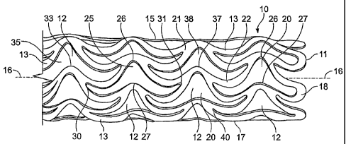
Brief Description of the Drawing
The above and still further aims, objectives, features, aspects and attendant
advantages of the present invention will become apparent to those skilled in
the art from
the following detailed description of a best mode presently contemplated of
practicing the
invention by reference to certain preferred embodiments and methods of
manufacture
thereof, taken in conjunction with the sole Figure of drawing which shows a
side view of a
preferred stent structure, for the invention (in which the far side is, not
shown for the sake of
simplicity).
Detailed Description of the Best Mode of Practicing the Invention
The sole Figure is a perspective view (not to scale) of a stent 10 in the form
of a
hollow tubular self-supporting structure composed primarily of niobium (Nb),
with a trace
amount of zirconium (Zr), titanium (Ti) or tantalum (Ta) for example,
preferably
zirconium, the trace amount preferably less than 5%, more preferably
approximately 1%,
and remainder niobium. The added trace metal improves physical characteristics
of the
stent for its intended function. Typically, the stent material used by
applicant also has
negligible amounts of tantalum (Ta, about 180 micrograms per gram ( g/g)),
iron (Fe, < 20
g/g), silicon (Si, about < gg/g), tungsten (W, < 20 g/g), nickel (Ni, < 20
g/g),
molybdenum (Mo, < 20 gg/g), hafnium (Hf, < 20 gg/g), carbon (C, about 7 g/g),
and
nitrogen (N, about 53 pg/g), as well as amounts of hydrogen (H) and oxygen (0)
primarily
introduced during the processing.
Important values of these minor elemental constituents are those of 02 and H2.
9
CA 02496551 2005-02-22
WO 2004/019822 PCT/US2003/026008
Both of these elements tend to increase brittleness of the stent material
dramatically if their
values too high. Chemical finishing (H2 source) and the vacuum annealing (02
source)
steps of applicant's process are intentionally set to keep the value of H at
less than 10 ppm
and the value of 0 at less than 80 ppm to prevent brittleness, with a desire
to keep the 0
content at less than about 35 gg/g.
The presently preferred process of fabricating the stent is performed in the
following sequence of steps: (1) tube processing from Nb-1%Zr ingots; (2)
laser cutting of
tube; (3) mechanical and chemical finishing; (4) electropolishing; (5) vacuum
annealing;
and (6) anodizing or sputtering with surface coating, preferably iridium
oxide. Anodizing
or sputtering of iridium oxide ("Irox") before vacuum annealing will increase
the 02
amount in the core material, so the Irox is preferably applied after annealing
and,
additionally, excess oxygen content is avoided by a technique to be described
presently
herein.
In the laser cutting process, the tubular stent member is provided with a
multiplicity
of through-holes or openings 12 through sidewall 15, defined and bounded by a
plurality of
struts or links 13, which enables expansion of the stent diameter when the
device is to be
deployed at a target site in a vessel, duct or tract of the human body. The
openings 12 may
be precisely cut out to form a latticework sidewall using a narrow laser beam
of a
conventional laser that follows a programmable pattern. The removed material
that
formerly occupied openings 12 is discarded following the cutting.
For example, the resulting pattern in the latticework sidewall 15 is a network
of
interconnected struts 13 which are optimized for orientation predominantly
parallel to the
CA 02496551 2005-02-22
WO 2004/019822 PCT/US2003/026008
longitudinal axis 16 of the tube 11, with none of the struts oriented
perpendicular (i.e.,
transverse) to the axis 16, so that no strut interconnecting any other struts
in the latticework
is oriented to lie completely in a plane transverse to the longitudinal axis,
without running
from one end of the stent to the opposite end. This type of structure, which
is described in
detail in applicant's USPN 6,398,805, provides a relatively very low friction
characteristic
(or coefficient of friction) of the outer surface 17 of the stent, to ease
advancement of stent
in a vessel, duct or tract to a site for deployment. The network or
latticework of struts
13 may define a series of longitudinally repeating circumferential rows 20 of
openings 12,
in which each opening has a shape which resembles the outline of a handlebar
moustache,
10 or of a Dutch winged cap, with each opening bounded by alternating links in
wavelets of
higher and lower crests in successive rows of each circumferential column
displaced along
the length of the cylindrical element. If viewed upside down, the openings
have a shape
resembling the outline of a ram's head with horns projecting at either side
upwardly from
the head and then downwardly, each opening bounded by alternating links in
wavelets of
shallower and deeper troughs in successive rows of each circumferential column
displaced
along the length of the cylindrical element.
Each pair of struts such as 21, 22 bounding an opening 12 in any given row 25
are
in the shape of circumferentially displaced wavelets with adjacent
circumferentially
aligned higher and lower crests 26, 27, respectively, in which the wavelets
intersect (30)
one another at one or both sides of the crests (30, 31). The intersection 30
of struts (or
wavelets) at one side of the adjacent circumferentially aligned crests 26, 27
of row 25 is
tangential to a crest 33 of the immediately adjacent row 35, and the
intersection 31 of struts
11
CA 02496551 2005-02-22
WO 2004/019822 PCT/US2003/026008
(or wavelets) at the other side of those crests is tangential to a crest 37 of
the immediately
adjacent row 38. Interconnecting points such as 40 between the struts may be
notched to
enhance symmetrical radial expansion of the stent during deployment thereof.
When the stent 10 is crimped onto a small diameter (low profile) delivery
balloon
(not shown), the adjacent circumferentially aligned crests of each row move
closer
together, and these portions will then fit into each other, as the pattern
formed by the
latticework of struts allows substantial nesting together of the crests and
bows, which
assures a relatively small circumference of the stent in the crimped
condition. Such a stent
is highly flexible, and is capable of undergoing bending to a small radius
corresponding to
radii of particularly tortuous coronary arteries encountered in some
individuals, without
permanent plastic deformation.
As the stent 10 is partially opened by inflation of the balloon during
deployment,
the adjacent crests begin to separate and the angle of division between struts
begins to
open. When the stent is fully expanded to its deployed diameter, the
latticework of struts
takes on a shape in which adjacent crests undergo wide separation, and
portions of the
struts take on a transverse, almost fully lateral orientation relative to the
longitudinal axis
of the stent. Such lateral orientation of a plurality of the struts enables
each fully opened
cell to contribute to the firm mechanical support offered by the stent in its
fully deployed
condition, to assure a rigid structure which is highly resistant to recoil of
the vessel wall
following stent deployment. The particular configuration of the stent
structure, while
highly desirable, is illustrative only.
The stent may be pre-opened after fabrication to relieve stresses. Pre-opening
12
CA 02496551 2005-02-22
WO 2004/019822 PCT/US2003/026008
produces a stent inner diameter that allows the stent to slide comfortably
over the
uninflated mounting balloon, for ease of crimping the stent onto the balloon.
Annealing
may be performed after pre-opening by heating the stent structure to an
appropriate
temperature for a predetermined interval of time.
The niobium/zirconium material of the stent is fabricated in any conventional
manner for producing alloys, with the zirconium amounting from 1% to 5% by
weight,
preferably about 2%, and the remainder niobium. For example, the manufacturing
process
may be performed by sintering particles or microspheres of the constituent
metals under
heat and pressure. Rather than using zirconium as the trace metal, a trace
amount (e.g.,
one to three percent) of titanium or tantalum may be alloyed with the niobium
for added
strength and other desirable physical characteristics. Other suitable
alternative additive
materials include those described in USPNs 5,472,794 and 5,679,815, for
example. The
alloy is then formed into tubing and the through holes are provided in its
side wall as
described bove.
According to the process aspect of the present invention, the principally
niobium
stent exhibits much improved performance structurally, with improved
resistance against
brittleness and thrombogenicity, by annealing the completed structure post-
fabrication in a
substantially oxygen-free atmosphere. Preferably, the environment is one of an
extreme
vacuum ranging from about 10'5 to about 10-6 millibars pressure, with less
than about 80
parts per million (ppm) of 02. The annealing is performed at a temperature
greater then
400 C, preferably at about 1100-1200 C for at least one hour, and more
preferably for
several hours.
13
CA 02496551 2005-02-22
WO 2004/019822 PCT/US2003/026008
The stent structure can be produced with a wall thickness of about 85 m,
which
offers sufficient mechanical strength to resist the natural recoil of the
blood vessel wall
following deployment of the stent, as well as excellent visibility under
fluoroscopy, but
which does not obstruct the vessel lumen to any significant extent. Since it
has none of the
distortion encountered with metallic 316L stents to MRI, use of the niobium-
based stent in
noninvasive monitoring also of cerebral and peripheral vessels is highly
beneficial.
The surface layer of iridium oxide is preferably applied post-annealing to
avoid
brittleness-producing oxygen contribution to the material . Surface
modification of the
stent to apply the preferred coating of iridium oxide, or alternatively, of
titanium nitrate is
achieved by vapor deposition, plasma deposition, or other conventional method.
Such
modification may be used to give the stent a rough surface. Alternatively, the
surface may
be anodized for oxidation of the niobium to achieve reduced immunoresponse and
less
thrombogenicity.
The most critical portion of the process currently utilized by applicant as
the best
mode for practicing that aspect of the invention is as follows:
1. Dissolve the natural oxide layer (< 2 nm thick) by placing stents for more
than 1
minute in 10% hydrofluoric (HF) acid.
2. Wrap Nb-1%Zr stents loosely in tantalum foil gathering 02 because of its
high oxygen
affinity, to further prevent undesirable contribution to oxygen content.
3. Introduce wrapped stents plus additional gather foil into recipient
chamber.
4. Heat up in 5 hrs. to 600 C maintaining vacuum < 10 -4 mbar (preferably < 10
-5 mbar,
but with recognition of considerably higher equipment cost).
14
CA 02496551 2005-02-22
WO 2004/019822 PCT/US2003/026008
5. Maintain temperature for about 2 hours.
6. Increase heating in 5 hours to 1120 C, maintaining vacuum < 10 -4 mbar.
7. Maintain set temperature for another 3 hours.
8. Cool down to 60 C while maintaining vacuum.
9. Remove stents and the 02-gather foil from recipient chamber.
Although a best mode of practicing the invention has been disclosed by
reference to
a preferred method and embodiment, it will be apparent to those skilled in the
art from a
consideration of the foregoing description that variations and modifications
may be made
without departing from the spirit and scope of the invention. Accordingly, it
is intended
that the invention be limited only by the appended claims and the rules and
principles of
applicable law.