Note: Descriptions are shown in the official language in which they were submitted.
CA 02496646 2005-02-10
50543-5
FLUID MONITORING DEVICE
Field of the Invention
This invention relates to a device to monitor the
flow of a translucent fluid. In a preferred embodiment, the
invention relates to a device to monitor administration of
intravenous fluid.
Description of the Prior Art
Intravenous fluid is generally delivered to a
patient by dripping it through an intravenous administration
set with a drip chamber and tubing, using gravity. When the
intravenous fluid container is empty, the drip chamber will
run dry and the fluid flow will eventually be stopped.
Three potential complications can occur if this termination
of fluid delivery is not detected promptly:
(1) The back flow of the blood from the venous system of the
patient will eventually cause clotting and obstruct further
fluid delivery through the intravenous catheter. This
demands the insertion of a new intravenous catheter into the
patient's extremities, which is an invasive procedure and
can be associated with complications, such as bleeding and
infection.
(2) If the intravenous fluid is used as a carrier or vehicle
for medications, such as intravenous anesthetics, completion
of fluid delivery will also stop the administration of
medications. With anesthetics, this lack of administration
over time may mean that the patient can wake up from
anesthesia during surgery and awareness can occur. With
other types of medication, the administration dosages could
suffer.
1
CA 02496646 2005-02-10
50543-5
(3) Venous air embolism (VAE) can occur spontaneously due to
mishaps with infusion fluid bags. Recently, Laskey et al.
reported that VAE occurred when a bolus of air was
unintentionally administered into a child venous system
resulting in the immediate onset of respiratory and
neurologic symptoms (Laskey AL, Dyer C, Tobias JD. Venous
air embolism during home infusion therapy. Pediatrics. 2002
Jan;109 (1):E15.). Patients will be particularly at risk of
having a VAE when the infusion of fluid is under pressure
for rapid fluid administration, such as patients in shock
following trauma.
SUMMARY OF THE INVENTION
The present invention provides a fluid monitoring
device, such as an intravenous monitor, and a method of
monitoring administration of a fluid by detecting the
presence of fluid in a chamber, such as a drip chamber,
vial, or tube, using an optical fluid sensor device. The
fluid monitoring device will indicate when the fluid level
has dropped below a predetermined level inside the chamber.
In such an event, optionally a pinch-off mechanism may
occlude a conduit, for example an intravenous tube,
connected either directly or indirectly to the chamber
thereby preventing further flow of fluid. The pinch-off
mechanism occludes the conduit distal to the optical fluid
sensor device in the direction of fluid flow through the
chamber.
This invention has particular importance in the
prevention of venous air embolism, which is caused by the
entrance of air into a patient's venous system through an
intravenous bag.
2
CA 02496646 2005-02-10
50543-5
According to one aspect of the present invention,
there is provided an optical fluid sensor device for
detecting presence of fluid in a chamber, comprising:
a radiation source;
a first sensor disposed so that the chamber is
situated between the radiation source and the first sensor,
and disposed to receive a majority of radiation emitted from
the radiation source when the portion of the chamber through
which the emitted radiation passes contains no fluid;
a second sensor disposed so that the chamber is
situated between the radiation source and the second sensor,
and disposed to receive a majority of radiation emitted from
the radiation source when the portion of the chamber through
which the emitted radiation passes contains fluid;
a logic means for controlling the radiation
source, and for detecting as a first signal an amount of
radiation falling on the first sensor, and for detecting as
a second signal an amount of radiation falling on the second
sensor, and for comparing the first and second signals; and
means for indicating when no fluid is detected in
the chamber.
Preferably, the radiation source is an infrared
(IR) emitter and the means for indicating when no fluid is
detected in the chamber is a visual and/or audible alarm.
It is also preferable to use modulated or pulsed radiation
to eliminate the interference caused by ambient light. When
the radiation source is on, each sensor is read to determine
the amount of emitted radiation reaching each sensor after
passing through the chamber, but this reading also includes
ambient light. By using modulated radiation, each sensor is
3
CA 02496646 2005-02-10
50543-5
read to determine the amount of ambient light reaching each
sensor when the radiation source is off. The readings may
then be corrected by removing the contribution due to
ambient light. The corrected readings are compared to
determine if fluid is present in the chamber.
The two-sensor system is advantageous since it
allows for ratiometric and comparative measurements, thereby
limiting the effects of inconsistent shape of the vial,
variations in the refractive index of fluids, and ambient
light.
According to another aspect of the present
invention, there is provided a pinch-off mechanism for
occluding a conduit, comprising:
a torsion spring;
means for loading the torsion spring having a
rotary pincher and a pawl; and
an electromechanical device having a lever
engaging the pawl,
whereby, when activated, the electromechanical
device disengages the lever from the pawl of the loading
means releasing stored energy in the torsion spring which
rotates the rotary pincher thereby applying pressure against
the conduit thereby occluding it.
A preferred use of the pinch-off mechanism is to
occlude a conduit, such as a tube held in a narrow channel
of an intravenous monitor. In one embodiment of said use,
the means for loading or biasing the torsion spring is a
thumbwheel. The thumbwheel is rotated to store energy in
the torsion spring and to align a flattened surface of the
rotary pincher with the side of the narrow channel to allow
4
CA 02496646 2005-02-10
50543-5
insertion of the tube. When the mechanism is activated, the
electromechanical device disengages the lever from the pawl
of the thumbwheel allowing rotation of the rotary pincher,
thereby applying pressure against the tube in the narrow
channel so as to occlude the tube. However, the pinch-off
mechanism can be used with other flow systems where
restriction of flow may be desired.
According to yet another aspect of the present
invention, there is provided a fluid monitoring device,
comprising:
(i) an optical fluid sensor device for detecting
presence of fluid in a chamber, comprising:
a radiation source;
a first sensor disposed so that the chamber is
situated between the radiation source and the first sensor,
and disposed to receive a majority of radiation emitted from
the radiation source when the portion of the chamber through
which the emitted radiation passes contains no fluid;
a second sensor disposed so that the chamber is
situated between the radiation source and the second sensor,
and disposed to receive a majority of radiation emitted from
the radiation source when the portion of the chamber through
which the emitted radiation passes contains fluid;
a logic means for controlling the radiation
source, and for detecting as a first signal an amount of
radiation falling on the first sensor, and for detecting as
a second signal an amount of radiation falling on the second
sensor, and for comparing the first and second signals; and
means for indicating when no fluid is detected in
the chamber; and
5
CA 02496646 2005-02-10
50543-5
(ii) a pinch-off mechanism for occluding a
conduit, comprising:
a torsion spring;
means for loading the torsion spring having a
rotary pincher and a pawl, wherein the pincher is located in
a position on the conduit distal to the optical fluid sensor
device in the direction of fluid flow; and
an electromechanical device having a lever
engaging the pawl,
whereby, the indicating means activates the
electromechanical device thereby disengaging the lever from
the pawl of the loading means releasing stored energy in the
torsion spring which rotates the rotary pincher thereby
applying pressure against the conduit thereby occluding it.
According to still another aspect of the present
invention, there is a method for monitoring administration
of a fluid using the fluid monitoring device as described
herein, said method comprising:
activating the radiation source;
detecting radiation using the first sensor to
produce a first signal and the second sensor to produce a
second signal;
comparing the first signal to the second signal to
detect the presence of fluid in the chamber; and
activating the pinch-off mechanism to occlude the
conduit when no fluid is present.
According to a preferred aspect of the present
invention, there is provided a use of the optical fluid
6
CA 02496646 2005-02-10
50543-5
sensor device described herein for monitoring the flow of a
fluid in an intravenous monitor. However, the optical fluid
sensor device has many other applications wherein the flow
of a translucent fluid is to be monitored, for example, the
flow in a fuel line of a motorized vehicle.
There are a number of advantages flowing from the
invention, namely:
1. A self-contained unit eliminating the complexity
of parts and attachments to the intravenous administration
set. It also eliminates protrusions, for example, wires
that can become entangled when being attached to the chamber
or when in use.
2. A pinch-off mechanism requiring the least amount
of parts and a minimal amount of area. The rotary action of
the pinch-off mechanism also delivers the maximum amount of
force to the "pinching" action on the tube. It optimizes
the force by transforming torque to lateral force at the
pinch point, through the use of a moment arm. It has the
potential of preventing a VAE from the infusion delivery
system by the "pinching" mechanism of the device when the
intravenous fluid bag is empty.
3. An optical fluid sensor using an infrared emitter
and sensors to check for the presence of a fluid (for more
detail see electronics section). This device checks for the
presence of fluid and emits a signal when the fluid vial is
empty.
4. A holding mechanism providing a tolerance fit that
attaches to the stem of the I.V. drip chamber, (which is
composed of a compressible material). This allows for a
tight, non-slip, effective connection to the intravenous
administration set. The advantage of this method is that,
7
CA 02496646 2005-02-10
50543-5
unlike other methods, it does not require any moving parts.
This will result in reduced manufacturing costs (for
instance, the example shown can be injection molded).
However, other methods of attachment could be used,
employing the same concept. These could include formed
spring devices such as wire or metal strips, or rubber-like
materials, which would have some compressive qualities. Any
of these methods could also have serrated surfaces, which
increase the holding capability.
5. A battery location and hookup providing several
advantages. The battery may be a standard nine volts DC,
which is mounted from the bottom of the device. First, it
provides quick and easy access to changing the battery,
since there is no cover to remove. Second, no cover means
less surface area, saving additional size to the unit.
Thirdly, having no battery cover means that there is one
less piece to make, ultimately reducing the total cost of
the unit. Alternatively, the battery may be a rechargeable
battery that is integral to the device, inductively charged
through the case.
6. An electronic design which is greatly simplified
since the level of the fluid is picked up by the IR sensors
in the fashion described above, thereby again reducing cost
and size of the unit (for circuit design, see electronics
section).
7. A visual and/or audible alarm ensuring timely
attention to a situation before it becomes a problem. With
reductions in nursing staff, each nurse has increased duties
and responsibilities. If the attendant nurse is busy with
another task, the alarm will alert her/him of the completion
of fluid delivery.
8
CA 02496646 2005-02-10
50543-5
BRIEF DESCRIPTION OF THE DRAWINGS
In order that the invention may be more clearly
understood, a preferred embodiment thereof will now be
described in detail by way of example, with reference to the
accompanying drawings, in which:
Fig. la and b are two assembly views of the
device;
Fig. 2 is a circuit design;
Fig. 3 is an emitter and sensor arrangement; and,
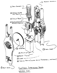
Fig. 4 is an exploded view of the device.
DETAILED DESCRIPTION OF THE PREFERRED EMBODIMENT
The following is a description of a preferred
embodiment of the invention, embodied in an intravenous
monitor. The device includes a drip chamber 1. The chamber
is a conventional drip chamber found on most I.V. sets. An
infrared emitter 2 and pick-up (sensor) is included as well.
There is also a circuit board 3. An electromechanical
device 4, preferably a solenoid, is included. Other
components such as a thermo strip could be used instead of
the solenoid depending on the reaction time required. A
thumbwheel 5 is used to manually rotate the rotary pincher 6
to the open position. This motion loads a torsion spring
(not shown) that supplies the force to the pincher. The
rotary pincher is mounted on the thumbwheel and "pinches"
the tube (stopping the flow) as it rotates 90 degrees
counterclockwise. This occurs when the signal is received
from the circuit board. A lever 7 is attached to the
electromechanical device (EMD) and is used to increase the
force of the EMD. This allows the selection of a smaller,
more cost-effective EMD. The torsion spring (not shown)
9
CA 02496646 2005-02-10
50543-5
provides the force for "pinching-off" the tube. A front
cover 9 provides protection to all internal components. As
well, it provides a "slot" to insert the drip apparatus
tubing. The slot locates the tube in a way that allows the
pincher to effectively stop the fluid flow when it is in the
closed position. A back cover 10 provides protection to all
internal components and includes the holding mechanism that
attaches the entire unit to the drip chamber. A holding
mechanism 11 being a serrated compression fitting provides
the means of attachment to the drip chamber.
The Electronics
A more detailed description of the electronics of
the flowcheck now follows. The electronics is implemented
with an embedded micro-controller to control excitation of
the infrared emitter, read the values of sensor 31 and
sensor 32 (see Figs. 2 and 3), read the battery voltage,
operate the indicator light-emitting diodes and the alarm
beeper, and operate and verify operation of the fluid
shutoff solenoid. The micro-processor includes a dedicated
real-time operating system to implement the following task
schedule:
~ every 1.3 milliseconds:
- toggle beeper - drive if beeper task enabled
every 10 milliseconds:
- beeper on/off - determine requested beeper
status
- indicator off - shut off indicators if required
time elapsed
CA 02496646 2005-02-10
50543-5
- solenoid trip task - energize solenoid until
trip sensor asserted
~ every 0.1 seconds:
- detect fluid task - turn off emitter, read
sensors 31 and 32, turn on emitter read sensors
31 and 32
- determine presence of fluid from readings -
request alarm task if vial is empty alarm task
request - red indicator LED
- shut off flow request - solenoid trip task if
vial is empty
~ every 1 second:
- indicator on task - turn on indicators if
requested (green "heartbeat", red and yellow
indicators)
- read battery volts - read battery voltage,
request low indication (yellow indicator) if
voltage is less than 7 volts
Optical Fluid Sensor
Principles of Operation
The system senses the presence of a transparent
fluid (for example, saline, Ringers, glucose, etc.) in a
cylindrical transparent vial which constitutes the drip
chamber of an intravenous system. The presence of this
fluid is sensed by determining the refractive properties of
the vial using a doubly-differential optical strategy. This
strategy also results in very low sensitivity to ambient
light.
11
CA 02496646 2005-02-10
50543-5
Determining Refraction of Filled and Empty Vials
Optical determination of the refractive properties
of the vial is performed by comparing the infrared light
received by two sensors from a single infrared emitter.
Figure 3 shows a top view of the vial 33, the infrared
emitter 34 and the two infrared sensors 31 and 32. The vial
is shown in cross section. The two infrared sensors are
positioned as shown on one side of the vial. The emitter is
on the other side such that the emitter's principal emission
direction forms a chord of both the inner and outer surfaces
of the vial as shown. Alternatively, the same principle
could be used by aligning both the infrared emitter and the
two sensors on the same side of the vial and using a
reflective surface (e.g. mirror) on the opposite side of the
vial. This alternate arrangement would take advantage of
the same refractive properties, while optimizing the
configuration of the device.
The fundamental principle of operation is as
follows. When the vial is empty, the principal beam (that
beam emitted in the emitter's principal emission direction)
is refracted twice by the air-vial interface both outside
and inside the empty vial. Since the inside and outside
walls of the vial are locally parallel, the light beam
inside the vial is parallel to the incident principal beam.
Similarly, the refracted beam is parallel to the beam inside
the vial and therefore parallel with the incident beam.
Sensor 32 is positioned so as to receive the majority of the
refracted beam when the vial is empty. The small amount of
beam energy which falls on sensor 31 is due to scattering
and the highly divergent beam from the infrared emitter.
When the vial contains a clear fluid, the optical
properties of the full vial are quite different. Since the
12
CA 02496646 2005-02-10
50543-5
refractive index of the fluid is much higher than for air
and very similar to the refractive index of the vial
material, the incident beam is refracted by the first air-
vial interface, but is not refracted significantly by the
vial-fluid interface. Thus, the light beam inside the vial
is not at all parallel to the incident beam.
Proper positioning of the infrared emitter results
in the beam in the filled vial meeting the vial wall near
sensor 31, perpendicular to the vial wall. This beam will
fall primarily on sensor 31, with very little beam energy
received at sensor 32. The situation is shown in Figure 3.
The small amount of beam energy received at sensor 32 is due
to scattering and the highly divergent beam from the
infrared emitter.
Reducing Sensitivity to Ambient Light
An optical fluid sensor typically must operate in
normal room illumination without extensive light baffles.
To cancel any effect of ambient light, two readings are made
for each sensor.
With the emitter off, each sensor is read to
determine the amount of incident illumination. These
readings are termed S1N and S2N. Then the emitter is turned
on and two more readings, S1L and S2L, are made.
The absolute differences
D1 = ~S1N-S1L
D2 - ~S2N-S2L
are formed and compared. These differences remove the
effects of illumination of the two sensors by ambient light,
13
CA 02496646 2005-02-10
50543-5
since this light is not synchronous with the operation of
the infrared emitter.
Fluid Detection
The vial is deemed to contain fluid if D1 is
greater than D2, because this situation occurs when more
light due to the infrared emitter is received at sensor 31
than sensor 32. If D2 is greater than D1, the vial is
deemed to be empty since the incident principal beam does
not undergo significant net refraction and so it falls
largely on sensor 32.
Pinch-off Mechanism
A pinch-off mechanism is also described. It was
designed to use the minimum number of parts, all of which
are designed to be very simple, to achieve the goal of
shutting off fluid flow (e. g. intravenous fluid) when
actuated. The goal was to achieve complete shutoff with a
simple, reliable and low-cost mechanism that consumes the
minimum amount of energy. The pinch-off mechanism is
suitable for use with the intravenous monitor described
herein, but is not restricted to such use. The design of
the pinch-off mechanism is described below, in a preferred
embodiment, applied to intravenous tubing.
The mechanism consists of the following parts,
with reference to Figures 1 and 4:
Thumbwheel 5 with integral rotary pincher 6, the torsion
spring (not shown) that is cocked by the thumbwheel, the
front cover 9 and specifically the narrow channel just off
the center of the front of the domed part of the cover, the
electromechanical device (solenoid by preference) 4, and the
lever 7 that is spring-loaded into the 'loaded' position by
14
CA 02496646 2005-02-10
50543-5
a scissor spring (not shown). The mechanism is configured
as follows:
1. The lever 7 is normally held by the scissor spring
to cause it to engage a pawl molded on the back of the
thumbwheel 5.
2. The electromechanical device 4 when actuated pulls
on the lever 7 against the force of the scissor spring (not
shown) so as to cause the lever to disengage from the pawl
molded on the back of the thumbwheel 5.
3. The rotary pincher 6 consists of a half-cylinder
that is integral to the thumbwheel 5 and that protrudes
through a hole in the front cover 9 and capable of rotating
in the front cover and is positioned such that at one
position its flattened surface is flush with the side of the
narrow channel that is just off center of the domed part of
the front cover and that when rotated 90 degrees from this
position it completely blocks the narrow channel.
4. A torsion spring is connected to the back cover 10
and the thumbwheel in such a manner that the thumbwheel can
be rotated so as to store energy in the torsion spring, and
that when released the thumbwheel will rotate to release the
stored energy.
The mechanism performs as follows:
1. The electromagnetic device initially is not
energized.
2. The user of the intravenous monitor cocks the
mechanism by rotating the thumbwheel 5 90 degrees counter-
clockwise when facing the front cover 9. This has the
effect of storing energy in the torsion spring (not shown)
and of rotating the rotary pincher 6 so that the narrow
CA 02496646 2005-02-10
50543-5
channel in the domed part of the front cover 9 is free of
obstruction. When fully rotated, the scissor spring (not
shown) on the lever 7 engages the pawl (not shown) on the
back of the thumbwheel 5 so as to prevent the thumbwheel
from rotating when released by the user of the invention.
3. The user of the intravenous monitor then inserts
the intravenous tube into the narrow channel by stretching
the tube slightly.
4. The user activates the intravenous monitor by
turning it on.
5. When the infrared emitter and pickup 2 sense the
absence of fluid in the drip chamber 1, the electronics on
the circuit board 3 actuate the electromechanical device
momentarily.
6. The electromechanical device then moves the lever
7 against the scissor spring (not shown) so that the lever
no longer engages the pawl on the back of the thumbwheel.
7. The thumbwheel is now rotated through 90 degrees
by the torsion spring (not shown) releasing the energy
stored therein.
8. The rotary pincher 6 being integral to the
thumbwheel 5 is also rotated through 90 degrees causing it
to exert very high localized pressure on the intravenous
tubing. This pressure is sufficiently high so as to
completely occlude the tube.
9. To ensure actuation of the mechanism, the
electronics on the circuit board 3 issues repeated,
momentary actuation signals to the electromechanical device.
This action stops at such time as the thumbwheel is
released. The release of the thumbwheel is determined by an
16
CA 02496646 2005-02-10
50543-5
optical reflective sensor mounted so as to sense the
presence of a small reflective surface (not shown) on the
back of the thumbwheel 5. This provides a positive
indication that guarantees release of the thumbwheel while
minimizing energy consumption from the battery of the '
invention.
The design of the rotary actuator, the thumbwheel,
the torsion spring and all other components has been
optimized to provide reliable pinchoff of the intravenous
tube at minimum cost and with minimum energy consumption.
It will be appreciated that the above description
relates to the preferred embodiment by way of example only.
Many variations on the invention will be obvious to those
knowledgeable in the field, and such obvious variations are
within the scope of the invention as described and claimed,
whether or not expressly described.
17