Note: Descriptions are shown in the official language in which they were submitted.
CA 02496856 2007-10-11
PATENT SPECIFICATION
TITLE: CONTINUOUS OPTOACOUSTIC MONITORING OF
HEMOGLOBIN CONCENTRATION AND HEMATOCRIT
INVENTOR: Donald Prough, Rinat Esenaliev, and Massoud Motamedi
BACKGROUND OF TAE EVVENTION
1. Field of the Invention
The present irivention relates to an apparatus'"for non-invasive, -~'real-
time;
accurate, continuous monitoring of hemoglobin concentration and hematocrit and
a
method for continuously or discretely monitoring hemoglobin 'concentration and
hematocrit.
More particularly, the present invention relates tQ. an. optoacoustic
apparatus
including a nanosecond pulsed laser, a fiber-optic delivery system and a probe
including a sensitive acoustic transducer and hardware and software for
converting a
received acoustic signal into a measurement of hemoglobin concentration and
hematocrit and.to methods for monitoring hemoglobin- concentration and
hematocrit
using the apparatus and methods for making the apparatus.
2. Description of the.Related Art
Continuous noninvasive monitoring of blood )iemoglobin concentration and
hematocrit offers great promise in the diagnosis and management of many
diseases and
life-threatening conditions, such as emergency department stabilization , of
hemorrhaging patients, management of critically ill patients in Intensive Care
Units,
and performance of extensive surgical procedures. Current techniques are
invasive,
requiring blood sampling and analysis, and cannot be performed continuously,
in real
time for extended intervals. Presently, there is no system for accurate, non-
invasive,
and continuous monitoring of hemoglobin concentration and hematocrit.
CA 02496856 2007-10-11
2
Because of the importance of hemoglobin concentration in oxygen delivery,
hematocrit and hemoglobin are among the most frequently obtained blood tests
in
both outpatients and inpatients. Current techniques for measuring hemoglobin
concentration and hematocrit require withdrawal of a blood sample from a vein
or
artery. Subsequently, the sample can e centrifuged, separating the fraction of
red cells
from plasma or chemically analyzed. These techniques are accurate but invasive
and
can result in iatrogenic anemia in patients who require frequent blood
sampling [3-7].
Continuous invasive techniques are available for monitoring hemoglobin
concentration, but these require access to an extracorporeal loop containing
circulating blood (as is present, for example, during hemodialysis) [8-12].
Although
noninvasive techniques such as pulse oximetry are available to monitor
arterial
oxygen saturation, no noninvasive technique is available to monitor hemoglobin
concentration or hematocrit.
One additional major problem with intermittent measurement of hemoglobin
concentration or hematocrit is the inevitable delay associated with withdrawal
of a
blood sample, transport to a measuring device, and processing. If the
laboratory is
remote from the site of care, the delay can be considerable. Even if the
laboratory is
in close proximity to the site of care, frequent sampling in a critically ill
patient may
occupy a substantial proportion of a technician's time, thereby increasing the
cost of
care and limiting the availability of that technician for other duties.
Thus, there is a need in the art for a non-invasive, real-time, accurate,
continuous apparatus and a method using the apparatus for monitoring
hemoglobin
concentration and hematocrit.
SUMMARY OF THE INVENTION
The present invention provides a system for measuring hemoglobin
concentrations and hematocrit comprising:
a pulsed optical source adapted to generate short optical pulses to provide
irradiation of a blood vessel or tissue site;
an optical delivery system including an optical fiber having a proximal end in
light communication with the source and a distal end out of which the optical
pulses
CA 02496856 2007-10-11
2a
exit an optical screen and an acoustic screen, where the optical delivery
system is
adapted to deliver the optical pulses to the blood vessel or tissue site,
an adjustable probe including a housing, a tip, a ring-shaped an acoustic
transducer, a backing element and an isolating layer, where the optical system
enters
the housing at its proximal end passes through a center of the piezoelectric
element
and terminates flush with the housing at the probe's tip, where the acoustic
transducer
adapted to detect pressure waves resulting from the optical pulses impinging
on the
blood vessel or tissue site and where the transducer has sufficient
sensitivity,
temporal resolution, and bandwidth to collect data from which a hemoglobin
concentration and hematocrit level can be derived and
a cable connected to the transducer at its proximal end and exiting the probe
out of the back portion of the probe, and
an electronic signal recording and processing system connected to the cable
where the signal recording and processing system includes a digital processing
unit or
computer calculating a hemoglobin concentration from the recorded optoacoustic
pressure profiles and amplitudes.
The present invention also provides a method of hemoglobin concentration
monitoring that comprises the steps of:
irradiating a blood vessel with an optical pulse resulting in a thermoelastic
optoacoustic pressure wave in said vessel or tissue site produced by an
optoacoustic
apparatus comprising:
a pulsed optical source adapted to generate short optical pulses to
provide irradiation of a blood vessel or tissue site;
an optical delivery system including an optical fiber having a proximal
end in light communication with the source and a distal end out of
which the optical pulses exit an optical screen and an acoustic screen,
where the optical delivery system is adapted to deliver the optical
pulses to the blood vessel or tissue site,
an adjustable probe including a.housing, a tip, a ring-shaped an
acoustic transducer, a backing element and an isolating layer, where
- , . ,. ,...
CA 02496856 2008-12-17
2b
the optical system enters the housing at its proximal end passes
through a center of the piezoelectric element and terminates flush with
the housing at the probe's tip, where the acoustic transducer adapted to
detect pressure waves resulting from the optical pulses impinging on
the blood vessel or tissue site and where the transducer has sufficient
sensitivity, temporal resolution, and bandwidth to collect data from
which a hemoglobin concentration and hematocrit level can be derived
and
a cable connected to the transducer at its proximal end and exiting the
probe out of the back portion of the probe, and
an electronic signal recording and processing system connected to the
cable where the signal recording and processing system includes a
digital processing unit or computer calculating a hemoglobin
concentration from the recorded optoacoustic pressure profiles and
amplitudes;
time-resolved detecting of the optoacoustic wave with the acoustic transducer;
analyzing one or both of a temporal profile and amplitude of the optoacoustic
wave with a data processing unit including software adapted to convert the
acoustic
transducer data into data representing a hemoglobin concentration or
hematocrit level
in the blood.
In one embodiment,. the present invention provides an optoacoustic apparatus
comprising:
a pulsed radiation source;
an optical system including an optical fiber, where the system is connected to
an output of the radiation source at its proximal end;
a probe including a housing, a tip, a ring-shaped piezoelectric element, a
backing element and an isolating layer, where the optical system enters the
housing at
its proximal end passes through a center of the piezoelectric element and
terminates flush
with the housing at the probe's tip;
a cable connected to the transducer at its proximal end and exiting the probe
out of the back portion of the probe; and
. . ., . . . . , .. ..... .j ..... . ...... . _....... ..,.:. ... .. , .,.....
. . ...._,.,. .
CA 02496856 2008-12-17
2c
a processing unit connected to the distal end of the cable; characterized in
that:
1) said apparatus is for monitoring hemoglobin
concentration in a blood vessel of an animal
2) said optical system includes an optical screen
and an acoustic screen; and
3) said processing unit is for converting the
transducer output into a measure of blood
hemoglobin concentration and/or hematocrit.
The present invention also provides an optoacoustic apparatus including a
nanosecond pulsed laser and a fiber-optic delivery system including a
plurality of
optical fibers, where the system is connected to an output of the laser at its
proximal
end. The apparatus also includes a probe including a piezoelectric transducer
mounted
in a front face of the probe and a back portion adapted to receive the fiber-
optic
CA 02496856 2005-02-25
WO 2004/010866 PCT/US2002/023620
-3-
delivery system. The optical fibers terminate at the front face of the probe
and are
distributed around or surround the transducer. The transducer is connected via
a cable
which exits out of the back of the probe to a processing unit that converts
the
transducer output into a continuous measure of hemoglobin concentration and
hematocrit.
The present invention also provides an optoacoustic apparatus for monitoring
hemoglobin concentration in the aorta of an animal comprising a pulsed
radiation
source; an optical system including an optical fiber, an optical screen and an
acoustic
screen, where the system is connected to an output of the radiation source at
its
proximal end; a probe including a housing, a tip, a ring-shaped piezoelectric
element,
a backing element and an isolating layer, where the optical system enters the
housing
at its proximal end passes through a center of the piezoelectric element and
terminates
flush with the housing at the probe's tip; a cable connected to the transducer
at its
proximal end and exiting the probe out of the proximal end of the probe; and a
processing unit connected to the distal end of the cable for converting the
transducer
output into a measure of aorta hemoglobin concentration and/or hematocrit.
The present invention also provides a probe including a front face having
mounted thereon a piezoelectric transducer connected to an output cable that
exits a
back portion of the probe, a plurality of optical fibers entering the probe
from the back
portion of the probe and terminating at or in the front face of the probe,
where light
from a laser is sent through the fibers and exit the probe at its front face
causing an
acoustic response which is measured by the transducer mount in the probe.
The present invention further provides a method for continuously measuring
optoacoustic monitoring of hemoglobin concentration and hematocrit including
the
step of directing radiation pulse from a laser via optical fibers into a probe
of present
invention having its front face in contact with a tissue site (blood vessel)
of an animal
including human. The light pulse leaves the probe face and enters the tissue
site
causing the production of an acoustic signal. The acoustic signal is received
by a
transducer mounted on the front face of the probe. The signal is then
transmitted to a
CA 02496856 2005-02-25
WO 2004/010866 PCT/US2002/023620
-4-
processing unit which converts the signal into a measure of hemoglobin
concentration
and hematocrit. The method can also include displaying the measurement on a
display
device. Preferably, the radiation is pulsed and particularly, the radiation is
pulsed in
a nanosecond time frame.
The present invention also provides a system for carrying out the above-stated
method including a pulsed laser system or other system capable of generating
short
optical pulses to provide irradiation of a tissue or vessel. The systems also
includes a
light communication system such as a fiber-optic system or articulated mirror
arm
optical system for delivering laser pulses to the tissue or vessel and an
acoustic
detection systems including at least one acoustic transducer for pressure
profile
detection with sufficient sensitivity, temporal resolution, and bandwidth so
that
thermoelastic optoacoustic pressure profiles of the absorbed laser energy in
the tissue
or vessel can be detected. The system also includes an adjustable holder for
the light
delivery system and the acoustic transducer(s) to provide appropriate
irradiation
conditions and acoustic contact between the investigated tissue or vessel and
the
acoustic transducer(s) and an electronic system for signal recording and
processing.
The system can also include a digital processing or computer system that
converts a
signal from the acoustic detection system into a measure the hemoglobin
concentration
of blood in a tissue or vessel.
The present invention still further provides a method for relating an acoustic
signal to an hemoglobin concentration of arterial or venous blood in a tissue
site of an
animal including a human.
DESCRIPTION OF THE DRAWINGS
The invention can be better understood with reference to the following
detailed
description together with the appended illustrative drawings in which like
elements are
numbered the saine:
Figure 1 depicts a graph of optoacoustic signals induced in blood at different
volumes;
Figure 2 depicts a graph of blood absorption coefficient calculated from
CA 02496856 2005-02-25
WO 2004/010866 PCT/US2002/023620
-5-
optoacoustic slopes at different volumes;
Figure 3 depicts a graph of blood absorption coefficient calculated from
optoacoustic slopes at different hemoglobin concentrations;
Figure 4 depicts a graph of optoacoustic signals induced in blood irradiated
through 1-cm turbid gelatin slab at different volumes;
Figure 5 depicts a graph of blood absorption coefficient calculated from
optoacoustic slopes at different blood volumes where the blood was irradiated
through
1-cm turbid gelatin slab;
Figure 6 depicts a graph of blood absorption coefficient calculated from the
- optoacoustic slopes as a function of hemoglobin concentration where the
blood was
irradiated through 1-cm turbid gelatin slab;
Figure 7 depicts a graph of optoacoustic signals induced in naphthol green
solution irradiated through 1-cm turbid gelatin slab at.different volume;
Figure 8 depicts a graph of absorption coefficient of naphthol green solution
calculated from the optoacoustic slopes at different volume. The solution was
irradiated through 1-cm turbid gelatin slab;
Figure 9 depicts a graph of absorption coefficient of naphthol green solution
calculated from the optoacoustic slopes as a function of concentration. The
solution
was irradiated through 1-cm turbid gelatin slab;
Figures l0A-C depict three preferred embodiment of an optoacoustic probe of
this invention;
Figures l OD-E depict two preferred embodiment of an optoacoustic probe of
this invention for use in the esophagus for monitoring hemoglobin
concentration in
aorta blood;
Figure 11 depicts a graph of optoacoustic signals from aorta phantom with
blood at different Hb concentrations;
Figure 12 depicts a graph of slope of optoacoustic signal recorded from aorta
phantom as a function of Hb concentration;
Figure 13 depicts a graph of optoacoustic signals recorded from 2.2-mm tube
CA 02496856 2005-02-25
WO 2004/010866 PCT/US2002/023620
-6-
with solution at different absorption coefficient;
Figure 14 depicts a graph of amplitude of optoacoustic signal recorded from
the
2.2-min tube as a function of Hb concentration;
Figure 15 depicts a graph of slope of optoacoustic signal recorded from the
2.2-
mm tube as a function of Hb concentration;
Figure 16 depicts a graph of optoacoustic signal recorded from the tube at
different axial distance between the tube and the probe;
Figure 17 depicts a graph of amplitude of optoacoustic signal recorded from
the
tube as a function axial distance between the tube and the probe;
Figure 18 depicts a graph of optoacoustic signal recorded from the tube at
different lateral distance between the tube and the probe; and
Figure 19 depicts a graph of amplitude of optoacoustic signal recorded from
the
tube as a function of lateral displacement of the prob.ewith respect to the
tube.
DETAILED DESCRIPTION OF THE INVENTION
The inventors have found that a new and efficient monitor can be constructed
for monitoring hemoglobin concentration and hematocrit using an optoacoustic
monitoring apparatus. The inventors have also found that a method using the
optoacoustic monitoring apparatus can be implemented manually or automatically
(computer) controlled and supervised for monitoring on a continuous or
discrete basis
hemoglobin concentration and hematocrit. The present invention can be used in
animals, where an animal is any member of the animal kingdom, including,
without
limitation, mammals and especially humans.
The inventors have found a novel technique that accurately monitors and
quantifies blood hemoglobin concentration and hematocrit. This technique is
based
on generation of ultrasonic (optoacoustic) waves in blood circulating in
vessels via
short optical pulses and detection of these waves by a sensitive acoustic
transducer.
The teinporal characteristics and amplitude of these waves are dependent on
hemoglobin concentration and hematocrit. Since the optoacoustic waves can
propagate
in tissues with low attenuation and distortion, this technique has high
resolution and
CA 02496856 2005-02-25
WO 2004/010866 PCT/US2002/023620
-7-
permits localization ofvessels of interest with high accuracy. This
localization permits
direct detection and measurement of signals induced in blood circulating in
vessels
without signal contamination from tissues between the transducer and blood.
The
present invention is ideally-suited for non-invasive, continuous monitoring of
hemoglobin concentration in blood by measuring induced acoustic signals in
tissues
and vessels such as the aorta, radial, femoral, carotid arteries or other
blood vessels.
The present invention relates to a method of hemoglobin concentration
monitoring that comprises the steps of: irradiating a blood vessel with at
least one
optical pulse resulting in an optoacoustic pressure wave in the vessel; time-
resolved
detecting of the optoacoustic wave with an acoustic.detector; analyzing a
temporal
profile and/or amplitude of the optoacoustic wave Nyitli a processing unit
including
computer software adapted to convert the wave data into digital data; and
calculating
a hemoglobin concentration in blood in the vessel.
The present invention relates to a system for carrying out the method of this
invention including a pulsed laser systeln or other generator of short optical
pulses to
provide irradiation of a vessel or tissue site; a fiber-optics system or an
articulated
mirror arm optical system for delivery of the radiation pulses to the vessel
or site; an
acoustic transducer for pressure wave detection with sufficient sensitivity,
temporal
resolution, and bandwidth to detect the pressure wave; an adjustable holder
for the
light delivery system and the acoustic transducer to provide appropriate
irradiation
conditions and acoustic contact between the vessel or tissue and the acoustic
transducer; an electronic system for signal recording and processing; a
computer or
digital processing unit for converting the pressure wave detected by the
transducer into
a hemoglobin concentration based on an analysis of the recorded optoacoustic
pressure
wave profile and amplitude. Preferably, the radiation source emits light in
the spectral
range from about 400 to about 2500 nm. The apparatus can include one or more
radiation sources as described in U.S. Pat. No. 5,840,023 and co-pending
application
Serial Nos. 09/179,791 and 09/633,597. Although the optical and transducer
part of
the probe can be housed in separate probes, it is preferably to have the
optical and
CA 02496856 2005-02-25
WO 2004/010866 PCT/US2002/023620
-8-
acoustic part of the apparatus in the same probe.
One preferred application of this invention is to measure a hemoglobin
concentration in blood in the aorta or other artery that is not skin
accessible. Another
preferred application of this invention is to measure hemoglobin concentration
in
arteries that can be measure by situating the probe on the skin of the patient
near the
artery such as a radial artery, a carotid artery, a brachial artery, femoral
artery or other
artery.
The method of this invention can be applied to any vessel including arteries
or
veins. The veins can be under the skin or in a hollow organ. For veins the
radiation
is preferably of wavelengths of about 548, 568, 587, and 805 nm or the
isobestic points
and in the spectral ranges from about 400 to about 640 and above 1120 nm where
absorption coefficients of oxy- and deoxygenated blood are close to each
other.
The preferred radiation sources include light derived from the first harmonic
(1064 nm) or the second harinonic (532 nm) of Nd:YAG laser or tunable lasers
such
as a Ti:Sapphire laser or a dye laser or an optical parametric generators or
mixtures or
combinations thereof.
The present invention also relates to a method wherein the above recited
method
is used for hematocrit measurements in the spectral range from 400 to 2500 nm
and
preferably in the spectral range above 1350 nm where optoacoustic signal
characteristics are more sensitive to the changes in blood scattering and,
therefore, to
changes in hematocrit. The method can be used for blood volume measurements,
for
ultrasound-guided optoacoustic monitoring of fetal anemia during pregnancy,
for
measurements of hematocrit and hemoglobin in cord blood, for hemoglobin
concentration monitoring in patients with kidney failure and dialysis.
The probe for use in this invention will generally include between 1 and 144
optical fibers, preferably, between about 6 to about 60 optical fibers,
particularly,
between about 12 and about 48 and especially between about 18 and 36, with 24
optical fibers being most preferred for probes designed to contact the skin.
For probes
designed to contact the wall of the esophagus so the Hb concentration in the
aorta can
CA 02496856 2005-02-25
WO 2004/010866 PCT/US2002/023620__
-9-
be monitored, such probes will include between 1 and about 20 optical fibers,
with
between about 1 and about 10 being preferred, and between about 1 and about 5
being
particularly preferred. The optical fibers have diameters between about 10 pm
to about
mm, preferably, between about 0.1 mm and 2 mm, particularly between about 0.2
mm and 1.5 mm. For esophagus probes (needle probes), the smaller diameter
fiber are
preferred. These needle probes can also be used to monitor Hb concentration in
various regions of the heart during bypass surgery or other myocardial
surgical
procedures.
The probes also include a sensitive acoustic transducer having a size
controlled
by the application and by design criteria. The optical fiber and transducer
are
preferably contained in a single housing to provide,stable irradiation and
detection
conditions. For skin applications, the fibers can be mounted around the
transducer,
adjacent to the transducer or in the center of a ring shaped transducer. For
aorta
monitor via the esophagus wall, the fiber(s) can mounted within a center of a
ring
shaped piezoelectric element, surrounding a disk shaped transducer or adjacent
to the
transducer.
A sensitive wide-band transducer is designed to detect optoacoustic waves from
blood circulating in a target vessel or tissue such as the aorta or other
blood vessels.
The choice of optimal designs of and materials for the piezoelectric element
and the
acoustic transducer depend on a number ofparameters: bandwidth, sensitivity,
acoustic
impedance matching to tissues, etc. For example, polyvinylidene fluoride
(PVDF)
slabs are suitable transducers for sensitive detection of optoacoustic waves
from
vessels and/or tissues. The inventors have found that a PVDF slab having a
thickiless
between about 10 in to about 1 mm thick is preferred. Other suitable
piezoelectric
materials include, without limitation, PZT, lithium niobide or other similar
piezoelectric materials. The present invention can also use other pressure
sensing
devices such as optical devices that measure the acoustic waves optically such
as
interferometric devices or other similar devices.
Importance and Significance
CA 02496856 2005-02-25
WO 2004/010866 PCT/US2002/023620
-10-
In this invention the inventors disclose a novel technique for noninvasive
continuous monitoring of hemoglobin concentration and hematocrit in blood. The
monitor can be used for (1) noninvasive measurement of hemoglobin
concentration
without blood sampling and standard blood testing and (2) continuous
measurement
of hemoglobin concentrations during surgical procedures, saline or drug
infusions,
blood infusions, and infusions of stroma-free hemoglobin.
The apparatuses andinethods ofthis invention are ideally-suited for monitoring
hemoglobin concentration in several large patient populations, including,
without
limitation, normal subjects, patients with blood diseases, and patients with a
variety of
other conditions. For example, patients who suffer hemorrhage secondary to
multisystem traulna being treated in an emergency department, typically have a
nearly
normal hemoglobin concentration because both blood and plasma have been lost.
However, as resuscitation begins with red cell -free fluids, the patient's
hemoglobin
concentration can and often does decrease rapidly.
A continuous measurement of hemoglobin concentration would permit prompt
recognition of the need to include red cell containing fluids during
resuscitation and
would also provide early evidence of continued hemorrhaging. Patients in
Intensive
Care Units often suffer from blood loss through gastrointestinal hemorrhage
and blood
loss from other sites, including blood sampling for diagnostic purposes.
A continuous, noninvasive measurement of hemoglobin concentration would
permit prompt diagnostic and therapeutic interventions and would also reduce
iatrogenic blood loss necessitated by the need to obtain blood samples for
current
hemoglobin measurements. During major surgery, particularly surgery involving
major blood vessels, the availability of a continuous measurement of
hemoglobin
concentration would permit not only prolnpt administration of needed red
cells, but
also would facilitate avoidance of unnecessary transfusion by demonstrating
that
helnoglobin exceeded an acceptable concentration.
Maintenance of adequate systemic oxygen delivery (cardiac output multiplied
by arterial oxygen content) is one of the principal clinical goals in caring
for acutely
CA 02496856 2005-02-25
WO 2004/010866 PCT/US2002/023620
-11-
traumatized patients, patients undergoing intensive care, and patients
undergoing
extensive surgery. However, many chronic diseases, such as chronic renal
failure, also
are associated with anemia and require intermittent measurement of hemoglobin
concentration or hematocrit. Because virtually all blood oxygen content
consists of
oxygen combined with hemoglobin, oxygen content bears a linear relation to
hemoglobin concentration and reduction of hemoglobin concentration requires
physiologic coinpensation, such as increased cardiac output.
Although the normal hemoglobin concentration is between about 13 and about
15 grams/dL, otherwise healthy individuals tolerate reductions of hemoglobin
to levels
as low as 7 g/dL or less, as long as their total blood volume is adequate.
However,
some patients, such as those with coronary artery disease, may develop severe
symptoms, such as angina pectoris, at hemoglobin concentrations below about 10
g/dL.
Because there are risks (e.g., transmission of viral diseases [1,2])
associated with
transfusion of blood, accurate monitoring of hemoglobin concentration or
hematocrit
facilitates both necessary transfusions and avoidance of unnecessary
transfusions.
Noninvasive monitoring of fetal anemia during pregnancy is also an ideal use
for the apparatuses and methods of this invention as well as measuring
hematocrit and
hemoglobin concentration in cord blood during pregnancy and delivery.
Additionally, continuous measurement of hemoglobin concentration using the
apparatuses and methods are useful in monitoring blood volume. In this case, a
known
small volume of saline, AV, is injected via i.v. and a decrease of hemoglobin
concentration in blood due to the injection , OC, is used to calculate the
volume of
circulating blood, V as shown in equation (1):
V = C ~ C (1)
where C represents the hemoglobin concentration in the blood.
Laser optoacoustics, is a novel technique for tissue characterization and
diagnostic imaging [13-16], and the inventors have found that the technique is
CA 02496856 2007-10-11
-12-
adaptable for hemoglobin concentration monitoring as set forth herein.
Optoacoustics
utilizes sensitive detection of laser-induced ultrasonic waves instead of the
detection
of scattered photons. An advantage of ultrasonic detection compared with
optical
detection is that propagation of acoustic waves in tissues is much less
influenced by
scattering than propagation of photon waves. Time-resolved detection of the
pressure
profiles by ultrasound transducers and analysis of the pressure signals allows
reconstruction of optoacoustic images which resemble the distribution of
optical
absorption in the irradiated tissue.
In contrast to pure optical methods in which diagnostic information about
tissue
structure is integrated over the entire optical path, the laser optoacoustic
imaging
pe n its direct reconstruction of the absorbed energy distribution from the
profile of
laser-induced pressure [13-19]. The time-resolved detection and analysis of
the laser-
induced ultrasonic waves offers a unique possibility to visualize tissue
structure at
depths as great as six centimeters with spatial resolution exceeding 0.5
millimeters in
optically turbid and opaque tissues [19-21] andto reconstruct optoacoustic
images [22,
23].
Laser optoacoustic imaging combines the merits of optical tomography (high
optical contrast) and ultrasound imaging (insignificant scattering of acoustic
waves)
to yield a noninvasive diagnostic modality with high contrast, sensitivity and
resolution. The optoacoustic imaging in tissues is disclosed in U.S. Pat. No.
5,840,023
[24] and in U.S. Application Serial No. 09/179,791 filed 27 October 1998 [25].
,The optoacoustic technique is also useful in blood
oxygenation monitoring as described in U.S. Application Serial No. 09/633,597,
filed
7 August 2000 [26], incorporated herein by reference. Recently, optoacoustic
technique was applied for noninvasive, real-time, continuous monitoring of
tissue
coagulation and temperature [27-29].
Theoretical Background
Although not intending to be bound by any theory, the magnitude of
optoacoustic pressure is proportional to the temperature rise in the
irradiated medium.
CA 02496856 2005-02-25
WO 2004/010866 PCT/US2002/023620
-13-
The temperature rise distribution, AT(r), is expressed by the following
equation (2):
OT(r) (r)(D (r)
P Cv (2)
where a(r) is the absorption coefficient in the tissue, 0(r) is the fluence
distribution
in the tissue, p is the tissue density, and C, is the heat capacity at
constant volume. The
formula shown in equation (2) is valid upon irradiation condition of heat
confinement,
which means that insignificant heat diffusion occurs during laser pulse
excitation.
If a short laser pulse irradiates the tissue, the irradiation condition of
temporal
stress confinement in the tissue volume of interest also can be satisfied. The
laser
irradiation under conditions of temporal stress confinement means
insignificant stress
(pressure) relaxation during the laser pulse excitation [30]. In a one
dimensional case,
the pressure rise distribution P(z) can be expressed as shown in equation (3):
1'(Z) = (fle2 / CP )paF = I' (z),uaF(z) = I' (z),uQFoe-UoZ (3)
where z is tissue depth in the z direction, r(z) is the efficiency of thermo-
acoustic
excitation often called the Gruneisen coefficient. The Griineisen coefficient
is a
function of three physical parameters of the irradiated sample: the thermal
expansion
coefficient, (3, the speed of sound, cs, and the heat capacity at constant
pressure, Cp as
given by equation (4):
e
R2
r=--z
Cp (4)
Irradiation conditions of temporal pressure confinement can usually be
achieved
by irradiating the sample with laser pulses having a pulse width having a
nanosecond
duration. The exponential factor exp(- az) represents the exponential
attenuation of
the optical radiation in the medium due to absorption. According to equation
(3)
optoacoustic pressure is proportional to the Gruneisen parameter, fluence, and
absorption coefficient of the medium. Equation (3) is valid for blood when the
blood
is irradiated with laser light in the visible and near-infrared spectra
because the
CA 02496856 2005-02-25
WO 2004/010866 PCT/US2002/023620
-14-
absorption coefficient of blood is greater than or close to the reduced
scattering
coefficient, 's = s(1-g), where s is the scattering coefficient and g is
the anisotropy
factor [31]. The apparatus and methods of this invention are based on the fact
that the
absorption coefficient of blood is proportional to hemoglobin concentration.
Therefore, both the amplitude and slope of the generated optoacoustic pressure
induced
in blood are dependent on hemoglobin concentration.
Since z and t are related by the simple equation:
z=cst (5)
and the spatial distribution of laser-induced pressure P(z) is detected by an
acoustic
transducer as its corresponding temporal profile P(t) as shown in equation
(6):
P(t) = I'uaFoe-(6)
Therefore, by recording and analyzing the temporal profile of optoacoustic
pressure induced in blood, one can measure the absolute value of helnoglobin
concentration with high accuracy. The high z-axial resolution of the
optoacoustic
technique permits direct measurement of hemoglobin concentration in blood
vessels,
because the signal from the blood arrives at the acoustic transducer at the
time defined
by equation (5).
Tissues are strongly scattering media. Three major optical parameters are
responsible for the distribution of light in tissues: absorption, scattering,
and the tissues
effective attenuation, eff, coefficients. The effective attenuation
coefficient is related
to a and s as shown in equation (7):
'Ueff - (Pa (Pa + Ps \i g))l1 / 2 (7)
and characterizes light penetration in tissue [31]. Light penetration depth is
defined
as 1/ eff. Absorption and scattering coefficients of tissues are low in the
near-infrared
spectral range (from about 600 to about 1300 nm) resulting in deeper
penetration of
near-infrared radiation compared with light in other parts of the
electromagnetic
spectrum. Near-infrared radiation is the preferred spectral range for the
apparatuses
CA 02496856 2005-02-25
WO 2004/010866 PCT/US2002/023620
-15-
and methods of this invention because near-infrared light allows sufficient
light
penetration into a tissue for effective optoacoustic monitoring of hemoglobin
concentration within the tissue including a blood vessel.
Another feature of near-infrared light as the excitation radiation is that
near-
infrared light induces insignificant temperature and pressure rises in the
tissue being
monitored resulting in little and probably no thermal or mechanical damage to
the
irradiated tissue.
The signal process apparatus of the present invention can comprise any analog
or digital processing unit or computer capable of converting a signal into an
output.
Such devices include, without limitation, any digital processing unit
comprising a
processing unit, lnemory, peripherals, an operating systems and communication
hardware and software. Illustrative examples include personal computers, mini-
mainframe computers, or the like.
Sites and Spectral Ranges for Monitoring Hemoglobin Concentration and
Hematocrit
The present invention is ideally suited for measuring hemoglobin (Hb)
concentrations and hematocrit in oxygenated blood or tissues having oxygenated
blood.
Oxygenated blood and especially highly oxygenated blood is ideal for
optoacoustic
monitoring because the optical properties of blood are dependent on hemoglobin
concentration and oxygen saturation.
[insert formulas]
Since arterial blood is 95 to 98 % oxygenated, the use of the optoacoustic
signals induced in arterial blood provides highly accurate hemoglobin
concentration
measurements. The most preferable arteries include, but are not limited to,
the aorta,
radial, carotid, and femoral arteries.
Hb concentration measurements in blood or tissue can be performed with high
accuracy at any wavelength within the visible and near infrared spectral
range.
Monitoring of hemoglobin concentration in the aorta can be performed by using
a
small optoacoustic probe inserted in the esophagus.
CA 02496856 2005-02-25
WO 2004/010866 PCT/US2002/023620
-16-
The aorta is the largest artery having a diameter between about 20 and about
25
mm and located in close proximity to the esophagus. Part of the aorta
(approximately
one to two inches) is in direct contact with esophagus wall. The thickness of
aorta and
esophagus is about 1 and 2-3 mm, respectively. This means that blood
circulating in
aorta represents a large optoacoustic source closely located to the
optoacoustic probe,
if the latter is inserted in the esophagus adjacent the part of the aorta in
direct contact
with the esophagus wall. The large diameter of the aorta and the short
distance
between the inner wall of the esophagus and blood circulating in the aorta
allows
substantially precise measurements of hemoglobin concentration to be obtained
using
the apparatuses and methods of this invention.
Detection of optoacoustic signals induced in the radial, carotid, and femoral
arteries also provides a highly accurate measurement of Hb in the blood
circulating
through these arteries. In this case, the optoacoustic probe can be larger and
can be
placed on the skin surface simplifying design and use of the probe. Moreover,
these
latter probes can be applied to a wider patient population.
The inventors have alsofound that optoacoustic signals induced in veins can be
used to monitor hemoglobin concentration in deoxygenated blood provided the
wavelengths at isobestic points (e.g., 548, 568, 587, and 805 nm) are applied.
At these
wavelengths oxy- and deoxyhemoglobin have equal absorption coefficients
providing
accurate measurements of hemoglobin concentration even at variation of oxygen
saturation in venous blood. High accuracy can also be obtained in the spectral
ranges
from about 400 to about 640 nm and above about 1120 nm because absorption
coefficients of oxy- and deoxygenated blood are close to each other. Suitable
lasers
for measuring hemoglobin concentrations in veins, include, without limitation,
the
second harmonic of a Nd:YAG laser (532 nm), a Ti: Sapphire, dye laser, an
Alexandrite
laser; a ruby laser, an optical parainetric generator, or any other source of
short optical
pulses in these spectral ranges.
The inventors have found that attenuation of light with a wavelength above
about 1300 to about 1350 nm in blood is dependent mostly on hematocrit, but
not on
CA 02496856 2005-02-25
WO 2004/010866 PCT/US2002/023620
-17-
hemoglobin concentration. Optoacoustic signal characteristics in this spectral
range
will be sensitive to the changes in blood scattering and therefore, to changes
in
hematocrit. Measurement of hematocrit is especially important when hematocrit
does
not follow hemoglobin concentration (e.g., during blood transfusions, etc.).
Design of Optoacoustic Probes
The inventors have found that a preferred optoacoustic monitoring system of
this invention includes an optoacoustic probe that will provide both
irradiation of blood
and detection of optoacoustic waves by an acoustic transducer. Such
optoacoustic
probe include a light delivery system (usually fiber-optic system) and a
sensitive
piezoelectric element to detect the optoacoustic waves. Different
configurations ofthe
probes are possible, depending on the site of monitoring and depth of the
blood
vessels. The optoacoustic probe can be placed on the skin surface, when
monitoring
blood in radial, femoral, carotid or other arteries or veins located
relatively close to the
skin surface, where relatively close means less than about 5 cm from the skin
surface
and preferably about 2 cm from the skin surface.
For hemoglobin monitoring in the aorta due to limitation in space in the
esophagus, small needle hydrophones are incorporated into optoacoustic probes
to
minimize the dimensions of the probe. The thickness of the needle hydrophones
is
generally about 1 mm which are incorporated into small optoacoustic probes
with the
transverse dimensions of about 2 to about 3 mm. The length of such
optoacoustic
probe is generally from about 1 to about 2 meters or more to provide delivery
of light
and signal recording by a distant optoacoustic system.
The optoacoustic probes of this invention can have an acoustic transducer(s)
surrounded by optical fiber(s) or vice versa. Moreover, the optical fiber(s)
and the
acoustic transducer(s) can be arranged in an adjacent configuration or can be
housed
in two different probes, an excitation probe and an receiving probe.
EXPERIMENTAL RESULTS
The inventors performed experiments with blood in vitro and in phantoms to
test the capability of the optoacoustic technique to monitor hemoglobin
concentration.
CA 02496856 2005-02-25
WO 2004/010866 PCT/US2002/023620
-18-
Heparinized sheep arterial blood in a plastic cuvette was irradiated by
nanosecond
Nd:YAG laser pulses (wavelength = 1064 nm). The blood was under a layer of
mineral oil to avoid contact with air. Ultrasonic gel was used to provide
acoustic
contact between the acoustic transducer and the cuvette. Pulsed laser
irradiation of
blood and detection of optoacoustic waves were performed from two opposite
surfaces
of the cuvette. Optoacoustic pressure waves induced in blood propagated to the
transducer and were recorded by a scope. The initial voluine of whole blood
with a
hemoglobin concentration of 12.4 g/dL was 30 mL. Blood dilutions were
performed
with 1-mL saline injections into the blood sample with a syringe. The
influence on
optoacoustic pressure signals due to the change in blood voluine is displayed
in Figures
1 for four (4) different volumes. As shown in Figure 1, saline injections
dramatically
chainged the amplitude and slope of the pressure signal.
Blood absorption coefficient calculated from the pressure slopes decreased
with
increasing blood volume as shown in Figure 2 due to blood dilution. Since the
initial
blood volume and volume of injected saline are known, one can calculate a
hemoglobin concentration in blood after each saline injection. The
optoacoustic signal
slope was found to be linearly dependent on hemoglobin concentration as shown
in
Figure 3.
Similar experiments and calculations were performed when blood was irradiated
through a turbid gelatin slab with the thickness of 1 cm. The gelatin slab had
optical
properties similar to that oftissues in the near infrared spectral range ( a =
0.6 cm' and
S' = 2.9 cm 1) and can be used to simulate a tissue layer in vivo. The results
presented
in Figures 4, 5, and 6 indicate that the addition of the turbid slab did not
decrease the
accuracy of the blood Hb concentration measurements. The amplitude of the
signals
is close to that recorded from blood irradiated without the gelatin slab
despite
attenuation, because scattering in the slab resulted in an increase of
irradiated blood
area and, therefore, an increase in optoacoustic signal amplitude.
Experiments were also performed with an aqueous solution colored with an
absorbing dye (naphthol green) simulating blood. The experiment demonstrated
CA 02496856 2005-02-25
WO 2004/010866 PCT/US2002/023620
-19-
similar results to the previous experiments as shown in Figures 7, 8, 9.
To monitor hemoglobin concentration and hematocrit in vivo, irradiation of
tissue by laser light and detection the laser-induced optoacoustic waves
should
generally be performed from the same side of the tissue. The inventors
designed, built,
and, tested different optoacoustic probes: (1) with a ring shape piezoelectric
element
and optical fiber in the center of the ring (see Figure 10A); (2) with optical
fibers
surrounding a disc shaped piezoelectric element (see Figure 10B); and (3) with
optical
fibers adjacent to a disc shaped piezoelectric element (see Figure l OC). Each
of these
configurations has advantages and each used a PVDF based transducer. The most
preferable probe configuration for hemoglobin monitoring includes a ring
shaped
piezoelectric elelnent with optical fibers in the center of the ring as shown
in Figure
10A. The results of tests of the probe of Figure 1 OA are presented below.
Looking at
Figures 10A-C, a probe generally 100 is shown to include a housing 102 which
can be
composed of metal or other structural material such as plastic, an optical
system 104,
a backing element 106, a piezoelectric element 108 and an isolating layer 110.
The
optical system 104 includes an optical fiber 112, an optical screen 114 and an
acoustic
screen 116. The system 104 would connect at its proximal end to a pulsed light
source
such as a laser (not shown), while its distal end 118 terminates flush with
the housing
102 at the probe's distal end 120. The probe of Figure l0A has the optical
system 104
passing through a center 122 of a ring-shaped piezoelectric element 108. The
probe
of Figure 10B has the optical system 104 distributed around a disk shaped
piezoelectric
element 108. And, the probe of Figure 10C has the optical system 104 positions
next
to (a side-by-side arrangement) the piezoelectric element 108 which can be of
any
desired shape.
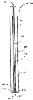
Referring now to Figure l OD and E, two preferred embodiments of esophagus
probes 200 is shown to includes a housing 202 which can be composed of metal
or
other structural material such as plastic, an optical system 204, a backing
element 206,
a piezoelectric element 208 and an isolating layer 210. The optical system 204
includes an optical fiber 212, an optical screen 214 and an acoustic screen
216. The
CA 02496856 2005-02-25
WO 2004/010866 PCT/US2002/023620
-20-
systeln 204 would connect at its proximal end to a pulsed light source such as
a laser
(not shown), while its distal end 218 terminates flush with the housing 202 at
the
probe's distal end or tip 220. The probe of Figure 10D has the optical system
204
passing through a center 222 of a ring-shaped piezoelectric element 208. The
probe
of Figure 10B also has the optical system 204 passing through the center 220
of a ring-
shaped piezoelectric element 208, but the distal end 218 of the optical system
204, the
transducer 208 and the isolating layer 210 so that the tip 220 is oriented at
a right angle
to the main body 224 of the probe 200. Of course, the tip 220 can be oriented
at any
angle relative to the main body 224 provided that the tip 220 can contact the
esophagus
wall adjacent the aorta.
Results Obtained with the Optoacoustic Probe of Figure 10A
The inventors performed experiments with whole sheep blood and absorbing
solutions in phantoms simulating aorta and radial artery. Rubber tubes having
a
diameter of about 25 mm and a length of about 50 min were filled with blood
with
different Hb concentrations (6.0, 6.2, 7.0, and 8.0 g/dL). The tubes were then
covered
with a 3-mm turbid gelatin slab to provide irradiation and detection
conditions similar
to ones for optoacoustic monitoring of hemoglobin concentration in aorta. The
optoacoustic signals induced in blood start at about 3 to about 3.5 s
depending on
hemoglobin concentration as shown in Figure 11. Looking at Figure 11, the
first two
sharp peaks are signals induced in the thin metal housing of the probe. The
flexible
tubes and gelatin slabs used to simulate a real aorta and esophagus wall with
different
thicknesses resulted in a shift in time for the signals induced in the blood
within the
tubes. Despite differences in irradiation and detection conditions for the
tubes, the
optoacoustic slope calculated from the recorded signals increased linearly
with Hb
concentration as shown graphically in Figure 12. The signals were normalized
before
the slope calculations. Calculation of the normalized signal slope provides
measurement of Hb concentration with high accuracy, e.g. about 0.5 g/dL.
Referring now to Figure 13, optoacoustic signals recorded from a phantom (2.2-
mm plastic tube in a turbid solution) simulating radial artery are shown. The
absorbing
CA 02496856 2005-02-25
WO 2004/010866 PCT/US2002/023620
-21-
solution in the tube had an absorption coefficient of about 2 to about 24
crri'. The first
peak at about 1.2 s is a signal induced in the housing of the probe and the
turbid
solution. The signals induced in the tube start at about 3 gs representing
time of flight
of the optoacoustic waves from the upper surface of the tube to the probe.
Both
amplitude and temporal profile of the signals induced in the tube are
dependent on the
solution absorption coefficient. The optoacoustic signal amplitude increases
gradually
with absorption coefficient as shown in Figure 14. The signal from the
solutions with
high absorption coefficient values has two positive peaks, while only one
positive peak
is recorded from solutions with low absorption coefficient values. The
optoacoustic
signals were norinalized and their first derivatives (slope) were calculated.
The slope
of the signals at about 5 gs is the most sensitive to changes in the
absorption
coefficient as shown in Figure 15. It is positive for solutions with high
absorptions and
negative for ones with low absorptions. The measurement and calculation of the
slope
can be used to provide accurate measurement of blood Hb concentrations.
Referring now to Figure 16, the optoacoustic signals recorded at different
axial
distance between the tube and the probe for solution with an absorption
coefficient of
about 13 cm' are shown. The variation of the distance simulated different
thicknesses
of tissue between the probe and a silnulated artery (radial, carotid, or
femoral). The
position of the signal changes with the distance indicating the depth of the
artery in the
solution, i.e., its location in a tissue. The telnporal profile of the signals
change
slightly with depth while the signal amplitude sharply decreases with
increasing depth
due to stronger attenuation of light and propagation of the optoacoustic
signals from
the cylindrical source as shown in Figure 17. Lateral displacement of the
probe with
respect to the tube changes both amplitude and profile of the signals as shown
in
Figures 18 and 19. These data indicate that lateral alignment of the probe is
ilnportant
for accurate measurement of Hb concentration. Thus, by laterally scanning the
optoacoustic probe on the skin surface, the practitioner can obtain highly
accurate Hb
concentration measurements, where the scanning is used to maximize the
measuring
process - maximize signal amplitude.
CA 02496856 2005-02-25
WO 2004/010866 PCT/US2002/023620
-22-
REFERENCES
1. Goodnough LT. Brecher ME. Kanter MH. AuBuchon JP. Transfusion lnedicine.
Second of two parts--blood conservation. New England Journal of Medicine.
340(7):525-33, 1999.
2. Goodnough LT. Brecher ME. Kanter MH. AuBuchon JP. Transfusion medicine.
First of two parts--blood transfusion. New England Journal ofMedicine. 340(6):
438-
47, 1999.
3. Silver MJ. Li YH. Gragg LA. Jubran F. Stoller JK. Reduction of blood loss
from diagnostic sampling in critically ill patients using a blood-conserving
arterial line
system. Chest. 104(6):1711-5, 1993.
4. Zimmerman JE. Seneff MG. Sun X. Wagner DP. Knaus WA. Evaluating
laboratory usage in the intensive care unit: patient and institutional
characteristics that
influence frequency of blood sampling. Critical Care Medicine. 25(5): 737-48,
1997.
5. Henry ML. Gamer WL. Fabri PJ. Iatrogenic anemia. American Journal of
Surgery. 151(3): 362-3, 1986.
6. Foulke GE. Harlow DJ. Effective measures for reducing blood loss from
diagnostic laboratory tests in intensive care unit patients. Critical Care
Medicine.
17(11):1143-5, 1989.
7. Smoller BR. Kruskall MS. Phlebotomy for diagnostic laboratory tests in
adults.
Pattern of use and effect on transfusion requirements. New England Journal of
Medicine. 314(19):1233-5, 1986.
8. Tanaka Y. Morimoto T. Watari H. Miyazaki M. Continuous monitoring of
circulating blood hematocrit. Japanese Journal of Physiology. 26(4): 345-53,
1976.
9. Kaiwa T. Mori T. Kijima T. Nogawa M. Nojiri C. Takatani S. Measurement of
blood hematocrit inside the magnetically suspended centrifugal pump using an
optical
technique: application to assessment of pump flow. Artificial Organs.
23(6):490-5,
1999.
10. Jabara AE. Mehta RL. Detennination of fluid shifts during chronic
hemodialysis using bioimpedance spectroscopy and an in-line hematocrit
monitor.
CA 02496856 2005-02-25
WO 2004/010866 PCT/US2002/023620
-23-
ASAIO Journal. 41(3): M682-7, 1995.
11. Ronco C, Brendolan A, and Bellomo R. Online monitoring in continuous renal
replacement therapies. Kidney International. 36, Suppl. 72; S-8 - S-14, 1999.
12. Maasrani M. Jaffrin MY. Boudailliez B. Continuous measurements by
impedance of haematocrit and plasma volume variations during dialysis. Medical
&
Biological Engineering & Computing. 35(3):167-71, 1997.
13. Esenaliev R.O., Oraevsky A.A., Letokhov V.S., Karabutov A.A., Malinsky
T.V.
Studies of Acoustical and Shock Waves in the Pulsed Laser Ablation of
Biotissue.
Lasers Surg. Med., 1993, v. 13, pp. 4 70-484.
14. Oraevsky A.A., Jacques S.L., Esenaliev R.O., Tittel F.K. Imaging in
layered
tissues using time-resolved detection of laser-induced stress transients. SPIE
Proc.
1994, v. 2134, pp. 122-128.
15. Oraevsky A.A., Esenaliev R.O., Jacques S.L., Tittel F.K. Laser opto-
acoustic
tomography for medical diagnostics.: principles. SPIE Proc. 1996, v. 2676, pp.
22-31.
16. Esenaliev R.O., Oraevsky A.A., Jacques S.L., Tittel F.K. Laser opto-
acoustic
tomography for medical diagnostics: Experiments wtih biological tissues. SPIE
Proc.
1996, v. 2676, pp. 84-90.
17. Oraevsky A.A., Esenaliev R.O., Jacques S.L., Tittel F.K. Laser
Optoacoustic
tomography for breast cancer diagnostics, In: "Trends in Optics and
Photonics", vol.
II, ed. by RR Alfano and JG Fujilnoto, OSA Publishing House, pp. 316-321
(1996).
18: Oraevsky A.A., Esenaliev R.O., Karabutov A.A. Optoacoustic Imaging in
Layered Tissues: Signal Processing. SPIE Proc. 1997, v. 2979, pp. 59-70.
19. Esenaliev R.O., Karabutov A.A., Tittel F.K., Fornage B.D., Thomsen S.L.,
Stelling C., Oraevsky A.A. Laser Optoacoustic Imaging for Breast Cancer
Diagnostics:
Limit of Detection and Comparison with X-ray and Ultrasound Imaging. SPIE
Proc.
1997, v. 2979, pp. 71-82.
20. Esenaliev R.O., Alma H., Tittel F.K., Oraevsky A.A. Axial resolution of
laser
optoacoustic imaging: Influence of acoustic attenuation and diffraction. SPIE
Proc.
1998, v. 3254, pp. 294-301.
CA 02496856 2005-02-25
WO 2004/010866 PCT/US2002/023620
-24-
21. Esenaliev RO, Karabutov AA, Oraevsky AA. Sensitivity of Laser Opto-
Acoustic Imaging for Detection of Early Breast Cancer. Journal of Quantum
Electronics, v.5(4), 1999, pp. 981-988.
22. Oraevsky A.A., Andreev V.G., Karabutov A.A., and Esenaliev R.O. Two-
Dimensional Opto-Acoustic Tomography Transducer Arrey and Image Reconstruction
Algorithm. SPIE Proc. 3601: 256-267, 1999.
23. Oraevsky A.A, Andreev V.G., Karabutov A.A., Fleming D.R., Gatalica Z.,
Singh H., and Esenaliev R.O. Laser Opto-Acoustic Imaging of the Breast:
Detection
of Cancer Angiogenesis. SPIE Proc. 3597, 1999, pp. 256-267.
24. A.A. Oraevsky, S.L. Jacques, R.O. Esenaliev, "Optoacoustic Imaging for
Medical Diagnostics", U.S. Pat. No. 5,840,023.
25. Esenaliev R.O., Oraevsky A.A., Motainedi M., Karabutov A.A. "Real-time
Optoacoustic Monitoring of Changes in Tissue Properties" U.S. Patent
Application
Serial No. 09/179,791 filed 27 October 1998.
26. Esenaliev R.O., Motamedi M., Prough D.S. Oraevsky A.A. "Optoacoustic
Monitoring of Blood Oxygenation" U.S. Patent Application Serial No.
09/633,597,
filed 7 August 2000.
27. Esenaliev R.O., Larin K.V., Larina I.V., Motamedi M., Oraevsky A.A.
Optical
properties of normal and coagulated tissues: Measurements using combination of
optoacoustic and diffuse reflectance techniques. SPIE Proc. 1998, v. 3726, pp.
560-
566.
28. Esenaliev R.O., Larina I.V., Larin K.V, Motamedi M, Karabutov AA, Oraevsky
AA. Laser Optoacoustic Technique for Real-Time Measurement of Thermal Damage
in Tissues. SPIE Proc. 3594, 1999, pp. 101-113.
29. Esenaliev R.O., Oraevsky A.A., Larin K.V., Larina I.V., Motamedi M. Real-
Time Optoacoustic Monitoring of Temperature in Tissues. SPIEProc. 3601:268-
275,
1999.
30. Gusev V.E., Karabutov A.A. "Laser Optoacoustics", AZP Press, New York,
1993.
CA 02496856 2005-02-25
WO 2004/010866 PCT/US2002/023620
-25-
31. Welch AJ, Van Gemert MJC, Optical-thermal response of laser-irradiated
tissue, New York: Plenum Press, 1995.
All references cited herein are incorporated by reference. While this
invention
has been described fully and completely, it should be understood that, within
the scope
of the appended claims, the invention may be practiced otherwise than as
specifically
described. Although the invention has been disclosed with reference to its
preferred
embodiments, from reading this description those of skill in the art may
appreciate
changes and modification that may be made which do not depart from the scope
and
spirit of the invention as described above and claimed hereafter.