Note: Descriptions are shown in the official language in which they were submitted.
CA 02496984 2010-06-16
j
THROMBOSPONDIN FRAGMENTS AND USES TITEREOF
IN CLINICAL ASSAYS FOR CANCER AND GENERATION
OF ANTIBODIES AND OTHER BINDING AGENTS
CROSS REFERENCE TO RELATED APPLICATIONS
FIELD OF THE INVENTION
The present invention relates to assays for blood levels of one or more
thrombospondin fragments as a diagnostic test for cancers and other diseases,
the use of such
fragments and/or derivatives thereof to generate specific antibodies and other
binding agents
and/or to use as calibrators, competitors, and/or indicators in an assay, and
to the fragments
themselves.
BACKGROUND OF THE INVENTION
Thrombospondin (TSP), also known as TSP-1, is a multimeric glycoprotein
comprised of identical monomers. The monomers migrate at an apparent molecular
weight
of approximately 185 kDa in SDS-polyacrylamide electrophoretic gels under
reducing
conditions. The predominant multimer is a trimer, which migrates at an
apparent molecular
weight of approximately 450 kDa on non-reducing gels. The molecular weights by
sedimentation equilibrium are similar, at 135 kDa for monomers and 420 kDa for
trimers.
The predicted molecular weight from just the sequence of amino acyl residues
in the
monomer is 127,524 Da, which does not include contributions from glycosylation
and 13- ,
hydroxylation. The thrombospondin glycoprotein is produced by platelets and is
released
upon platelet activation from platelet a-granules, along with many other
proteins, such as
platelet-derived growth factor, fl-thromboglobulin, fibronectin, fibrinogen,
and platelet
factor-4 (see Chapter 1, "An introduction to the thrombospondins" in The
Thrombospondin
Gene Family by JC Adams, RP Tucker, & J Lawler, Springer-Verlag: New York,
1995, pp.
1-9, but especially p. 2; and Chapter 3, "The secondary and tertiary structure
of the
thrombospondins," ibidem pp. 43-56, especially Table 3.1). Thrombospondin is
known to be
involved in biological processes such as cell adhesion, proliferation and
chemotaxis. It has
also been reported that thrombospondin may be involved in the progression of
malignant
CA 02496984 2005-02-23
WO 2004/018995
PCT/US2003/026023
tumors. Furthermore, thrombospondin has been reported to be highly expressed
in many
human malignant tissues and in surrounding stroma and/or endothelium and has
been
reported to be present in higher than normal levels in the plasma of cancer
patients, (e.g.,
Qian and Tuszynsld, Proc. Soc. Exp. Biol. Med., 212:199-207, 1996; de Fraipont
F et al.
Trends Mol. Med., 7:401-407, 2001).
Despite the foregoing, as for any potential diagnostic test, it would be
desirable to
increase the specificity and sensitivity of such tests. To that end, the
present inventor has
discovered that thrombospondin is present in the blood and blood plasma in
relatively small
amounts compared to fragments of thrombospondin, and this finding is true in
the blood and
blood plasma of cancer patients as well. This discovery provided a basis for
the present
inventions related to novel diagnostic assays that are more specific, more
sensitive, more
easily calibrated, and in some cases distinguish these thrombospondin
fragments from each
other and from thrombospondin itself.
BRIEF SUMMARY OF THE INVENTION
Important aspects of the invention are diagnostic methods and related kits
that are
based on the detection and quantification of thrombospondin fragments and/or
thrombospondin in bodily fluids, especially plasma. Foremost among those
diagnostic
methods are those that detect or monitor the status of a cancer.
Aspects of the invention closely related to the diagnostic methods are
thrombospondin fragments that are detected in the plasma, thrombospondin
fragments that
can be used to induce an antibody of interest for use in a diagnostic method
or can be used in
a competition-type or non-competitive diagnostic assay.
Thrombospondin fragments of the invention
In one aspect, the invention is a purified thrombospondin fragment that has
been
extracted from a bodily fluid, especially plasma, said fragment being one
within a molecular
weight range selected from the group consisting of 80 to 110 kDa, 40 to 60
kDa, and 20 to
kDa, wherein the size in kDa is that determined by gel electrophoresis after
disulfide
30 bond reduction. Their uses include, but are not limited to, a) the
induction of an antibody of
interest, b) induction of an antibody for a diagnostic method, c) use in a
competition-type
diagnostic assay, d) as a reference molecule in an assay for a thrombospondin
fragment or
fragments or thrombospondin of human subjects, and e) the immunization of an
animal. In a
2
CA 02496984 2005-02-23
WO 2004/018995 PCT/US2003/026023
closely related aspect, the invention is a polypeptide or modified
polypeptide, made by
recombinant and/or chemical techniques, that has the identical primary
structure as one of
said purified thrombospondin fragments or a portion thereof. Such chemical
techniques
include, but are not limited to, glycosylation, 13-hydroxylation, alkylation
and reduction.
In particular embodiments, the fragment's molecular weight is one within a
molecular
weight range selected from the group consisting of 80 to 95 kDa, 47 to 53 kDa,
and 27 to 33
kDa. Specific examples of fragment molecule weights are 85, 90, 50, and 30
kDa.
Preferably, the fragment is one found in human plasma.
In a related aspect, the invention is a purified and/or synthetic
thrombospondin
fragment or portion thereof, said fragment being one that starts between amino
acid 1-165
(just after the N12/I peptide) and V-263 (the start of the procollagen
homology domain),
inclusive (i.e., inclusive of 1-165 and V-263), and ends between amino acid K-
412 (the end
of the reported collagen type V-binding region) and 1-530 (the end of the
domain of type 1
repeats), inclusive. Prefered are such fragments that start at between N-230
and G-253,
inclusive (at or near the start of the domain of interchain disulfide bonds, 1-
241, which is the
first residue downstream [meaning towards the C-terminus of the full protein]
of a predicted
cleavage site for chymotrypsin and/or a chymotrypsin-like protease), and end
at between V-
400 and S-428, inclusive (at or near a predicted chymotrypsin cleavage site, F-
414, that falls
two residues after the end of the collagen type V-binding region), said
portion being at least
3 amino acyl acids in length (preferably at least 4 amino acyl residues in
length, more
preferably at least 6 amino acyl residues).
In a further related aspect, the invention is a purified and/or synthetic
thrombospondin fragment or portion thereof, said fragment being one that
starts between
amino acid 1-165 (just after the Ni 2/I peptide) and V-263 (the start of the
procollagen
homology domain), inclusive, and ends between amino acid 1-530 (the end of the
type 1
repeats) and R-733 (the end of the first type 3 repeat), inclusive. Preferably
such a fragment
starts between N-230 and G-253, inclusive, and ends between D-527 and S-551,
inclusive,
which is at or near a predicted chymotrypsin cleavage site, F-539, in the
first type 2 repeat;
said portion being at least 3 amino acyl acids in length (preferably at least
4 amino acyl
residues in length, more preferably at least 6 amino acyl residues).
In a still further related aspect, the invention is a purified and/or
synthetic
thrombospondin fragment or portion thereof, said fragment being one that
starts between
3
CA 02496984 2010-06-16
amino acid 1-165 (just after the N12/I peptide) and V-263 (the start of the
procollagen
homology domain), inclusive, and ends between amino acid R-792 (the end of the
third type
3 repeat) and Y-982 (the third of the predicted chymotrypsin cleavage sites in
the C-terminal
domain), inclusive. Preferably such a fragment starts between N-230 and G-253,
inclusive,
and ends between G-787 and V-811, inclusive, which is at or near a predicted
chymotrypsin
cleavage site, Y-799, in the fourth type 3 repeat; said portion being at least
3 amino acyl
acids in length (preferably at least 4 amino acyl residues in length, more
preferably at least 6
amino acyl residues). Protein molecular weights here were computed using
standard
computational aids (such aids are available, for example, at the web site of
the
Bioinformatics Organization, Inc., see Stothard,
P. 2000. The sequence manipulation suite: JavaScript programs for analyzing
and formatting
protein and DNA sequences. BioTechniques 28: 1102-1104) and adjusted upwards
to
account for post-translational modifications. Predicted cleavage sites for
chymotrypsin (and
any closely related protease) were identified using tools available from the
ExPASy (Expert
Protein Analysis System) proteomics server of the Swiss Institute of
Bioirtformatics (SIB)
and were limited to
predicted sites of at least 80% probability. The uses of said fragments and
portions include,
but are not limited to, the induction and/or screening of an antibody and/or
another binding
agent of interest in a diagnostic method and use in a diagnostic assay. In
particular
embodiments, the invention is one of the specified fragments, rather than a
portion thereof.
In additional embodiments, a fragment and/or a portion can incorporate or be
linked to a
label and/or a carrier.
Throughout, wherever reference is made to a fragment or a portion thereof (or
an
immunoreactive portion thereof), it is understood that the fragment is a
preferred
embodiment of the invention. It is also understood throughout this Application
that
immunogenic portions, immunoreactive portions, and/or epitopes are generally
six amino
acyl residues long or longer, but an occasional portion or epitope can be
shorter. Such
shorter portions or epitopes are also contemplated.
Five additional aspects are:
1) A purified and/or synthetic tkombospondin fragment, said fragment being at
least 4
6 contiguous amino acyl residues in length, and wherein the fragment comprises
a protease-
resistant core domain or a part thereof, said domain or part thereof being
selected from the
group consisting of a domain of inter-chain disulfide bonds, an
oligomerization domain, a
4
CA 02496984 2010-06-16
procollagen-like domain, a type I repeat, a type 2 repeat, and a type 3
repeat, said part being
at least 6 amino acyl residues in length.
2) A purified and/or synthetic thrombospondin fragment, said fragment being at
least
6 contiguous amino acyl residues in length, and wherein the fragment comprises
an amino
acid sequence selected from the group consisting of TEENKE (SEQ ID NO:1),
CLQDSIRKVTEENKE (which includes an N-terminal Cys added to aid conjugation)
(SEQ
ID NO:2), LQDSIRKVTEENKE (SEQ ID NO:3), EGEARE (SEQ II) NO:4),
PQMNGKPCEGEARE (SEQ ID NO:5), EDTDLD (SEQ ID NO:6),
YAGNGIICGEDTDLD (SEQ ID NO:7), CNSPSPQMNGKPCEGEAR (SEQ ID NO:8),
RKVTEENKELANELRRP (SEQ ID NO:9), CRKVTEENKELANELRRP (which includes
an N-terminal Cys added to aid conjugation) (SEQ ID NO:10), PQMNGKPCEGEAR (SEQ
' ID NO:11), CEGEAR (SEQ 11) NO:12), and RKVTEENKE (SEQ ID NO:13). (In
particular
embodiments the fragment comprises two, or even all of the foregoing
sequences).
3) a purified and/or synthetic thrombospondin fragment, said fragment being at
least
6 contiguous amino acyl residues in length, and wherein the fragment comprises
a collagen
type V binding domain or a portion thereof.
4) A purified and/or synthetic thrombospondin fragment, said fragment being at
least
6 contiguous amino acyl residues in length, and wherein the fragment comprises
an epitope
for binding at least one of the following commercially available antibodies,
each of which
recognizes a ¨450kDa (non-reduced) protein that is specifically identified as
thrombospondin (the TSP Ab numbering, e.g., "TSP Ab-2", comes from Lab Vision
Corporation, Fremont, CA;
clone designations refer to the hybridoma clone that produces a particular
monoclonal
= antibody) It is also understood that said fragment includes a fragment
that can be designed
to bind a pre-existing monoclonal antibody, through the use of peptide
scanning analysis,
competition experiments, and other methods known in the art (for an example of
such
methods, see Corada M et al. Monoclonal antibodies directed to different
regions of vascular
endothelial cadherin extracellular domain affect adhesion and clustering of
the protein and
modulate endothelial permeability. Blood. 2001 Mar 15;97(6):1679-84). It is
also
understood that the current invention includes, but is not limited to, uses of
pre-existing
antibodies independent of a purified and/or synthetic fragment, some of which
uses are also
listed below.
5
CA 02496984 2005-02-23
WO 2004/018995
PCT/US2003/026023
TSP Ab-2 (Clone D4.6): This antibody is stated to react against reduced and
non-reduced
protein, and its epitope is in the calcium-binding domain of TSP (C-terminal
50-1(Da piece of
the 120-1(Da fragment from protease digestion of Ca-replete TSP). The calcium-
binding
region is generally considered to be in the type 3 repeats (TSP residues 698-
925). For
example, it is expected that TSP Ab-2 will bind thrombospondin but not the 30-
kDa
circulating fragment. This antibody can be used to detect and/or quantify TSP
and/or a
circulating fragment; distinguish thrombospondin from a circulating fragment;
and/or
distinguish one or more fragments from each other. It shows no cross-reaction
with
fibronectin, fibrinogen, and von Willebrand factor. Its binding to
thrombospondin is
enhanced by EDTA i.e. at low [Ca2+].
TSP Ab-4 (Clone A6.1): This antibody is stated to react against reduced and
non-reduced
protein, and its epitope is in the collagen type V-binding domain. This
antibody binds
thrombospondin, and the applicant has discovered that it binds the three major
TSP
fragments in human plasma. Thus, this antibody can be used to detect and/or
quantify TSP
and/or a circulating fragment or fragments. In combination with another
antibody or binding
agent, it can be used in an assay to distinguish thrombospondin from a
circulating fragment;
and/or to distinguish one or more fragments from each other. As an example
meant to be
illustrative and not restrictive, TSP Ab-4 is used to capture TSP and
circulating fragments,
and then the other antibody or binding agent is used for detection, but is
able to distinguish
TSP from a fragment or fragments, or one fragment from another. It is
understood that TSP
Ab-4 also binds thrombospondin and thrombospondin fragments from important non-
human
sources as well, including but not limited to the dog. Thus, the use of this
antibody and/or a
similar binding agent in an assay for a thrombospondin fragment or fragments
in a sample
from a non-human source, such as dog, is contemplated. This antibody shows no
cross-
reaction with fibronectin, fibrinogen, and von Willebrand factor. This
antibody inhibits
thrombospondin-collagen interaction, and its binding to thrombospondin is
unaffected by
glycosaminoglycans (e.g. hyaluronic acid, chondroitin sulfate, and heparin).
Also, its
binding is enhanced by EDTA i.e. at low conc. of Ca2+.
TSP Ab-5 (Clone B5.2): This antibody is stated to react against reduced and
non-reduced
protein, and its epitope is in a 10-kDa fragment present at the junction of
type 2 and type 3
repeats. The junctional region is listed elsewhere as residues 674-697, but
this is only 24
6
CA 02496984 2005-02-23
WO 2004/018995
PCT/US2003/026023
residues and less than 10-kDa, so the epitope is less precisely mapped. It is
expected that
this antibody will bind TSP but not the 30-kDa circulating fragment. Thus,
this antibody can
be used to detect and/or quantify TSP and/or a circulating fragment or
fragments; distinguish
thrombospondin from a circulating fragment; and/or distinguish one or more
fragments from
each other. It shows no cross-reaction with fibronectin, fibrinogen, and von
Willebrand
factor.
TSP Ab-9 (Clone MBC 200.1): This antibody is stated to react against reduced
and non-
reduced protein, and its epitope is in the N-terminal heparin-binding domain
of
thrombospondin. Thus, it should bind to thrombospondin but not to major
circulating
fragments. In Western blotting, Ab-9 reacts with a 25kDa peptide (heparin-
binding domain)
from thermolysin digests of thrombospondin that is not disulfide bonded to any
other region
of the thrombospondin molecule. Heparin efficiently inhibits the binding of Ab-
9 to
thrombospondin. Thus, this antibody can be used to detect and/or quantify TSP;
and/or
distinguish thrombospondin from a circulating fragment or fragments. This
antibody is not
suitable for detecting all major fragments in the circulation.
TSP Ab-8 (rabbit polyclonal antibody): Recognizes a ¨450kDa (non-reduced) or
180kDa
(reduced) protein, identified as TSP. This antibody, which is a rabbit
polyclonal, can be
used in sandwich ELISAs for capture or detection and in competitive ELISAs.
The
applicant has discovered that it binds the three major TSP fragments in human
plasma.
Thus, this antibody can be used to detect and/or quantify TSP and/or a
circulating fragment
or fragments. In combination with another antibody or binding agent, it can be
used in an
assay to distinguish thrombospondin from a circulating fragment; and/or to
distinguish one
or more fragments from each other.
As an example meant to be illustrative and not restrictive, one takes the
difference
between (a) the result of an assay using an antibody or binding agent that
binds TSP and the
major circulating fragments in plasma, versus (b) the result of an assay using
an antibody or
binding agent that binds TSP but not major fragments. The antibody or binding
agent in (a)
is selected from the group consisting of TSP Ab-4, TSP Ab-8, TSP Ab-11, and an
antibody
or binding agent that binds TSP and the major circulating fragments in plasma.
The
antibody or binding agent in (b) is selected from the group consisting of TSP
Ab-3, TSP Ab-
6, TSP Ab-9, and an antibody or binding agent that binds TSP but none of the
major
7
CA 02496984 2005-02-23
WO 2004/018995 PCT/US2003/026023
circulating fragments. Said assay in (a) detects TSP plus fragments; said
assay in (b) detects
TSP; said difference, (a) minus (b), thereby gives a quantification of
fragments without TSP.
Likewise, differences can be taken between (c) the result of an assay using an
antibody or
binding agent that binds TSP and a subset of the major circulating fragments
in plasma,
TSP Ab-11 (Clones D4.6 + A6.1 + MBC 200.1): The Ab-11 cocktail is designed for
sensitive detection of thrombospondin by Western blotting. This antibody
cocktail shows no
cross-reaction with fibronectin, fibrinogen, and von Willebrand factor.
Because it is a
Other antibodies that are useful, even though they have been disclosed only as
binding non-reduced protein include, but are not limited to TSP Ab-1, TSP Ab-
3, TSP Ab-6,
TSP Ab-1 (Clone A4.1): This antibody is stated to bind the N-terminal half of
the central
stalk-like region of thrombospondin. This region is recovered as a 50kDa
fragment after
8
CA 02496984 2005-02-23
WO 2004/018995 PCT/US2003/026023
factor. It inhibits the adhesion of human melanoma G361 cells, keratinocytes,
squamous
carcinoma cells, and rat smooth muscle cells to thrombospondin. It does not
inhibit
aggregation of thrombin-induced platelets. This antibody is stated to block
the anti-
angiogenic activity of thrombospondin by inhibiting its binding to TSP-
Receptor/CD36.
TSP Ab-3 (Clone C6.7): This antibody is stated to bind the platelet or cell-
binding domain
at the extreme C-terminus of TSP and should therefore distinguish TSP from
fragments.
Thus, this antibody can be used to detect and/or quantify TSP; and/or
distinguish
thrombospondin from a circulating fragment or fragments. This antibody is not
suitable for
detecting the three major fragments in the circulation. Heparin or EDTA may
marginally
affect binding of Ab-3 to thrombospondin. Ab-3 blocks thrombospondin-mediated
agglutination of fixed red blood cells. It shows no effect on thrombospondin-
mediated
agglutination of fixed, activated platelets. It inhibits both thrombin- and
A23187-induced
aggregation of washed, live (not fixed) platelets without affecting the
secretion of serotonin.
Ab-3 inhibits adhesion of melanoma 0361 cells to thrombospondin, and blocks
the binding
of C-terminal domain to Integrin-Associated Protein (IAP)/CD47.
TSP Ab-6 (Clone A2.51: This antibody has been shown to immunoprecipitate
thrombospondin. This antibody shows no cross-reaction with fibronectin,
fibrinogen, and
von Willebrand factor. Its epitope localizes in the heparin-binding domain of
thrombospondin, and therefore, heparin efficiently inhibits the binding of Ab-
6 to
thrombospondin. Thus, this antibody can be used to detect and/or quantify TSP;
and/or
distinguish thrombospondin from a circulating fragment or fragments. This
antibody is not
suitable for detecting the three major fragments in the circulation.
Hyaluronic acid and
chondroitin sulfate show no inhibition at low concentration and only partially
inhibit over
the concentration range at which heparin abolishes the binding. Thrombospondin
binds with
high affinity to a sulfated glycolipid or sulfatide found on red cell and
platelet membranes.
Ab-6 blocks the binding of thrombospondin to sulfatides at low concentrations.
Ab-6
immunoprecipitates a 251(Da peptide (heparin-binding domain) from chymotryptic
digests of
thrombospondin that is not disulfide bonded to any other region of the
thrombospondin
molecule. This antibody inhibits the hemagglutination of trypsinized,
glutaraldehyde-fixed
human erythrocytes by purified thrombospondin. It also inhibits the
agglutination of fixed,
activated platelets by thrombospondin. It does not inhibit either thrombin- or
A23187-
9
CA 02496984 2010-06-16
induced aggregation of washed, live platelets. Ab-6 does not bind to reduced
and alkylated
thrombospondin or thrombospondin transferred to nitrocellulose membrane after
SDS-
PAGE.
TSP Ab-7 (Clone HB8432): This antibody is stated to bind type 2 repeats. Thus,
Ab-7 may
be used to detect and/or quantify TSP and/or a circulating fragment or
fragments; distinguish
thrombospondin from a circulating fragment or fragments; and/or distinguish
one or more
fragments from each other. It shows no cross-reaction with fibronectin or any
other serum
or platelet proteins except thrombospondin. Its epitope localizes in the EGF-
like repeats
(type 2) in the stalk region of human thrombospondin (disulfide-bonded core
remaining after
trypsin digestion).
All of the antibodies listed above can be purchased from Lab Vision
Corporation,
Fremont, CA See also the
published
literature such as, for TSP Ab-4, Galvin NJ et al. Interaction of human
thrombospondin with
types I-V collagen: direct binding and electron microscopy. J Cell Biol. 1987
May;104(5):1413-22). It is also understood that alternative antibodies may
also be generated
against any of the abovementioned epitopes.
5) A purified and/or synthetic thrombospondin fragment, said fragment being at
least
6 contiguous amino acyl residues in length, and wherein the fragment does not
comprise at
least one fibrinogen-binding region selected from the group consisting of (1)
a fibrinogen-
binding domain within a 210-kDa fragment of TSP, where said 210-kDa fragment
is
composed of three 70-kDa fragments that contain the region of interchain
disulfide bonds,
the procollagen homology region, and the type 1 and type 2 repeats, (2) a
fibrinogen-binding
region in the amino-terminal domain of thrombospondin, (3) a fibrinogen-
binding region in
an 18-kDa amino-terminal heparin-binding domain of thrombospondin, and (4) a
region
corresponding to synthetic peptide N12/I encompassing amino acid residues 151-
164 (I-151
to P-164) of the N-terminal domain of thrombospondin-1. In a particular
embodiment, the
fragment does not comprise any of the fibrinogen-binding regions in the group.
For each of the 5 additional aspects, the molecular weight of the
thrombospondin
fragment does not exceed 110 kDa; alternatively does not exceed 55 kDa; or
alternatively
=
does not exceed 35 kDa, wherein the size in kDa is that determined by gel
electrophoresis
after disulfide bond reduction. The fragments of the 5 additional aspects of
the invention can
CA 02496984 2005-02-23
WO 2004/018995 PCT/US2003/026023
be used to induce antibodies (and/or other binding molecules) of interest in
the diagnostic
methods or can be used in diagnostic assays, for example, as calibrators,
indicators, and/or
competitors. It is understood that a fragment can be derivatized, for example,
to incorporate
and/or be coupled to a label and/or a carrier.
A fragment that can be as little as 6 amino acyl residues in length is
preferably
immunoreactive. A typical method for immunizations comprises coupling the
peptide to a
carrier, such as keyhole limpet hemocyanin or ovalbumin. Said couplings to a
carrier are
also contemplated in the current invention.
The inclusion of the central protease-resistant core domain in the definition
of the
fragments follows from considerations discussed elsewhere herein. This domain
is
considered to comprise locations in the mature thrombospondin protein selected
from the
group consisting of: a domain of interchain disulfide bonds (around Cys-252
and Cys-256,
preferably residues 241-262); the procollagen homology domain (residues 263-
360); the
type 1 repeats (residues 361-530); the type 2 repeats (residues 531-673);
there is a short
segment (residues 674-697) between the type 2 repeat doman and the type 3
repeat domain;
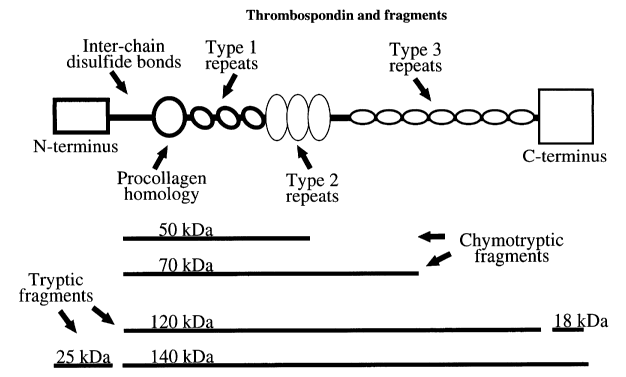
and then the type 3 repeats (residues 698-925); see Figure 1 of this
Application for examples
of protease-resistant fragments that have been reported after artificial
digestions in vitro;
Chapter 2, "The primary structure of the thrombospondins" in in The
Thrombospondin Gene
Family by JC Adams, RP Tucker, & J Lawler, Springer-Verlag: New York, 1995,
pp. 11-42,
particularly p. 12; and Chapter 6, "Mechanistic and functional aspects of the
interactions of
thrombospondins with cell surfaces," ibidem, pp. 105-157, particularly p. 115.
Interchain
disulfide bonds (in the region of residues 241-262) are often preserved in
protease-resistant
fragments. The term "mature", as used here to refer to the mature
thrombospondin protein
sequence, means without the 18- to 22-residue signal peptide sequence, here
assumed to be
18 residues, following The Thrombospondin Gene Family by JC Adams et al. 1995;
see the
full human thrombospondin sequence given below in this text; see also Figure 1
of this
application, and the discussions thereof. Nevertheless, it is understood that
GenBank file
NM 003246.1, also listed as GI:4507484, currently identifies nucleotide
residues "112..204"
as encoding the signal peptide, which implies a signal peptide of 31 amino
acyl residues).
The identification of these peptides, TEENKE (SEQ ID NO:1),
LQDSIRKVTEENKE (SEQ ID NO:3), EGEARE (SEQ ID NO:4), PQMNGKPCEGEARE
(SEQ ID NO:5), EDTDLD (SEQ ID NO:6), YAGNGIICGEDTDLD (SEQ ID NO:7),
CNSPSPQMNGKPCEGEAR (SEQ ID NO:8), RKVTEENKELANELRRP (SEQ ID NO:9),
11
CA 02496984 2005-02-23
WO 2004/018995 PCT/US2003/026023
PQMNGKPCEGEAR (SEQ ID NO:11), CEGEAR (SEQ ID NO:12), and RKVTEENKE
(SEQ ID NO:13) was achieved by computerized surveys of thrombspondin, the
surveys done
by request at commercial sources to identify immunogenic regions (epitopes),
but these
surveys identified many peptides with immunogenic regions, and so the surveys
were
followed by selection of relevant peptides and/or epitopes based on knowledge
of circulating
thrombospondin fragments. Other peptides and/or epitopes listed in this
application were
similarly identified.
A criterion that a fragment comprises an immunogenic and/or immunoreactive
portion from a collagen type V binding domain follows from the published
properties (e.g.,
Galvin NJ et al. Interaction of human thrombospondin with types I-V collagen:
direct
binding and electron microscopy. J Cell Biol. 1987 May;104(5):1413-22) of the
commercially available TSP Ab-4 antibody used below to detect thrombospondin
fragments
of interest in the plasma.
The collagen V-binding domain of thrombospondin has been mapped to the amino
acid sequence corresponding to the region between valine(333) and lysine(412)
(V-333 to K-
412, using the single-letter symbols V and K for their respective amino
acids), inclusive, of
human thrombospondin-1 (Takagi T et al. A single chain 19-kDa fragment from
bovine
thrombospondin binds to type V collagen and heparin. J Biol Chem 268:15544-
15549, 1993;
as mentioned above, numbers here refer to the mature thrombospondin protein,
that is,
without the 18- to 22-residue signal peptide sequence, here assumed to be 18
residues). This
region would include a portion of the procollagen homology region of
thrombospondin and
all or nearly all of the first type 1 repeat of thrombospondin (see Chapter 2,
"The primary
structure of the thrombospondins" in The Thrombospondin Gene Family by JC
Adams, RP
Tucker, & J Lawler, Springer-Verlag: New York, 1995, pp. 11-42, but especially
p. 24).
The criterion that the fragment comprise an epitope for binding the
commercially
available TSP Ab-4 antibody follows from the fact that the TSP Ab-4 antibody
was used
below to successfully detect thrombospondin fragments of interest in the
plasma, including
the plasma of cancer patients. Significantly, this TSP Ab-4 antibody is
described as binding
the collagen type V binding domain of thrombospondin.
For references regarding a fibrinogen-binding region within a 210-kDa fragment
of
TSP composed of three 70-kDa fragments that contain the region of interchain
disulfide
bonds, the procollagen homology region, and the type 1 and type 2 repeats, see
p.24 of
Adams et al. The Thrombospondin Gene Family; citation 53 therein, which is
Lawler J et al.
12
CA 02496984 2005-02-23
WO 2004/018995 PCT/US2003/026023
Thrombin and chymotrypsin interactions with thrombospondin. Ann N Y Acad Sci.
1986;485:273-87; and citations immediately below. Additional references for
the
fibrinogen-binding regions to be excluded include: for a region in an 18-kDa
amino-terminal
heparin-binding domain of thrombospondin (so-called TSP18), see Bonnefoy A et
al.: A
model of platelet aggregation involving multiple interactions of
thrombospondin-1,
fibrinogen, and GPIIbIlla receptor. J Biol Chem. 2001 Feb 23;276(8):5605-12.
For a region
corresponding to synthetic peptide N12/I encompassing amino acid residues 151-
164 of the
N-terminal domain of thrombospondin-1, see Voland C et al.: Platelet-
osteosarcoma cell
interaction is mediated through a specific fibrinogen-binding sequence located
within the N-
terminal domain of thrombospondin 1. J Bone Miner Res. 2000 Feb;15(2):361-368.
Citations for two fibrinogen-binding domains include p. 24 of Adams et al. The
Thrombospondin Gene Family (and citations 51-54 therein), and for the role of
the type 1
repeats include Panetti TS et al.: Interaction of recombinant procollagen and
properdin
modules of thrombospondin-1 with heparin and fibrinogen/fibrin. J Biol Chem.
1999 Jan
1 ;274 (1): 430-7 .
Thrombospondin is a glycosylated protein. Therefore, depending on which
portion
of thrombospondin is considered, the thrombospondin fragments of the invention
may be
glycosylated or non-glycosylated. Potential sites for N-linked carbohydrate
chains include
N-230 (in the N-terminal domain), N-342 (in the procollagen homology domain),
N-503 (in
the type 1 repeat domain), N-690 (in the region between the type 2 and type 3
repeat
domains), N-1033 (in the C-terminal domain), and N-1049 (in the C-terminal
domain). It is
also understood that specific C- and 0-linked saccharide attachments occur,
particularly in
the type 1 repeat domain (see Hofsteenge J, Huwiler KG, Macek B, Hess D,
Lawler J,
Mosher DF, Peter-Katalinic J: C-mannosylation and 0-facosylation of the
thrombospondin
type 1 module. J Biol Chem. 2001 Mar 2;276(9):6485-6498). It is also
understood that 13-
hydroxylation of thrombospondin can occur (such as at N-592, which is in the
type 2 repeat
domain; see Figure 2.2a in Adams JC et al. The Thrombospondin Gene Family,
1995, p. 16),
and that any of these modifications can be incorporated, or not, into
thrombospondin
fragments and/or peptides of the current invention.
Nonglycosylated entities of particular interest are synthetic peptides.
In particular embodiments, the thrombospondin fragments of the invention are
derivatized so that they comprise and/or are linked to a detectable label
and/or a carrier. In
particular embodiments, the label is selected from the group consisting of a
radioactive label,
13
CA 02496984 2005-02-23
WO 2004/018995 PCT/US2003/026023
a fluorescent label, a chemical label, a colorometric label, an enzymatic
label, a non-
fluorescent label, a non-radioactive label, a biotin moiety, and an avidin
moiety. In
particular embodiments, the carrier is selected from the group consisting of a
bead, a
microsphere, a coded microsphere, a solid matrix, a keyhole limpet hemocyanin,
an albumin,
linkage to a cross-linking agent, an epitope tag, and an epitope.
It is understood that a synthetic or purified thrombospondin fragment of the
invention
retains its identity as a fragment of the invention even if it has been
derivatized by the
addition of additional material, such as detectable label, or through
conjugation to another
molecule, or by synthesizing it as part of a chimeric protein, to name just
three of many
possible examples.
Binding agents
The detection of either thrombospondin fragments or thrombspondin usually
requires
the use of agents that will bind to them. Such agents may be multi-chain
antibodies, single-
chain antibodies, proteins that are not antibodies, non-protein molecules, or
derivatives or
combinations thereof. Polyclonal and monoclonal antibodies are normally
immunoglobulins,
i.e., multi-chain antibodies. In the case of immunoglobulin-G (IgG), each
antibody molecule
consists of a pair of heavy chains and a pair of light chains. The multichain
antibodies are
typically from mammalian or avian sources. Single-chain antibodies and non-
antbodies are
discussed below.
The term "antibodies" by itself, when not specified as being a single-chain
antibodies, refers to 4-chain antibodies, those with two heavy and two light
polypeptide
chains. By way of example, this includes but is not limited to the IgG classes
of antibodies,
but also other classes, such as ones that occur in higher multimers, such as
IgM. IgA and
IgY are also contemplated.
The term "protein" is intended to include not only molecules normally referred
to as
proteins but also those that may be referred to as polypeptides.
Methods of detecting the thrombspondin fragments while distinguishing, or not
distinguishing, from thrombospondin itself
In one such an aspect, the invention includes an assay to detect a
thrombospondin
fragment of the invention wherein the assay distinguishes the thrombospondin
fragment
from thrombospondin itself. The thrombospondin fragments of particular
interest are ones
14
CA 02496984 2005-02-23
WO 2004/018995 PCT/US2003/026023
found in humans and are within a range selected from the group consisting of
80 to 100kDa,
40 to 55 kDa and 20 to 30 kDa, wherein the size in kDa is that determined by
gel
electrophoresis after disulfide bond reduction Most preferably they are
selected from the
group consisting of an ¨ 85 kDa to 90 kDa fragment, an ¨ 50 kDa fragment, and
an ¨ 30 kDa
fragment. The assay may detect just one such fragment, or a combination of 2
or more.
In cases where the concentration of higher molecular weight forms, including
thrombospondin itself, is low in a sample (such as in the samples shown in
Figures 3 and 4,
Results of Western Blot analysis using TSP Ab-4 antibody), detection of
fragments without
necessarily excluding thrombospondin is an approach also contemplated by the
current
invention. Low concentrations of thrombospondin can be achieved in many cases
by
preventing or reducing platelet activation during sample collection and/or
storage (see below
for contemplated methods). This aspect of the current invention comprises
several
advantages over conventional detection methods that have used binding agents
against the
entire thrombospondin molecule (and these binding agents have been limited to
antibodies).
Said advantages include but are not limited to the use of binding agents that
are directed
specifically against the fragments of interest and not portions of the
thrombospondin
molecule outside of these fragments, the use of relevant peptides and/or
thrombospondin
fragments to generate said binding agents (such as antibodies), the use of
relevant peptides
and/or thrombospondin fragments as assay calibrators, and the use of relevant
peptides
and/or thrombospondin fragments as assay indicators.
Any of several acceptable approaches can be used for the assay of a
thrombospondin
fragment (or fragments) wherein the assay distinguishes it from
thrombospondin, and more
than one of these can be used in a given assay. In one approach, the assay
comprises a step
wherein the fragment is physically separated from the thrombospondin.
Generally that
approach is combined with a step in which the presence of the fragment or
thrombospondin
is shown by their reaction with a specific binding agent. In particular
embodiments, the
physical separation technique is selected from the group consisting of gel
electrophoresis,
dialysis, chromatography, size chromatography, affmity chromatography, immuno
affinity
chromatography, adsorption, immunoadsorption, isoelectric focusing, mass
spectrometry,
centrifugation, sedimentation, floatation, precipitation, immunoprecipitation,
and gel
filtration.
In a second approach, the assay distinguishes the fragment (or fragments)
based on
one or more epitopes (here "epitope" meaning a target to which a binding
agent, i.e., an
CA 02496984 2005-02-23
WO 2004/018995 PCT/US2003/026023
antibody or a non-antibody, binds) in the fragment that are not present in
thrombospondin,
including but not limited to an epitope at an end of a fragment and an epitope
that is
displayed by a fragment but is shielded in thrombospondin.
In a third approach, the assay distinguishes the fragment (or fragments) based
on one
or more epitopes in thrombospondin that are not present in the fragment. As an
illustrative
but not restrictive example, an epitope shared by thrombospondin and a
thrombospondin
fragment is used to obtain a quantitation of a total, thrombospondin plus
thrombospondin
fragment(s), from which is then subtracted a quantitation of thrombospondin
obtained using
an epitope present in thrombospondin but not present in a fragment. The
difference between
the two quantitations is a quantitation of the amount of fragment. As an
example, epitopes
in thrombospondin but not in at least one fragment from the group of an 80 to
100 kDa, a 40
to 55 kDa, or a 20 to 35 kDa fragment present in plasma can be selected from
the group
consisting of an epitope from outside the protease-resistant central core
domain, an epitope
in the N-terminal domain, an epitope in the N-terminal heparin-binding domain,
a heparin-
binding sequence in the N-terminal domain, a heparin-binding sequence in the N-
terminal
domain selected from the group consisting of residues 23-32 (RKGSGRRLVK),
residues 23-
29 (RKGSGRR), and residues 77-83 (RQMKKTR) of the mature protein (see Chapter
2,
"The primary structure of the tlarombospondins" in The Thrombospondin Gene
Family by
JC Adams, RP Tucker, & J Lawler, Springer-Verlag: New York, 1995, pp. 11-42,
but
especially p. 13 & Table 2.1; Chapter 6, "Mechanistic and functional aspects
of the
interactions of thrombospondins with cell surfaces," ibidem pp. 105-157, but
especially pp.
108 & 114; Lawler J et al. Expression and mutagenesis of thrombospondin.
Biochemistry.
1992 Feb 4;31(4):1173-80; and Cardin AD & Weintraub HJ. Molecular modeling of
protein-
glycosaminoglycan interactions. Arteriosclerosis. 1989 Jan-Feb;9(1):21-32), a
heparin-
binding sequence in the N-terminal domain selected from the group consisting
of residues
22-29 (ARKGSGRR), residues 79-84 (MKKTRG), and residues 178-189
(RLRIAKGGVNDN) of the mature protein (reviewed in the Discussion section of
Voland C
et al.: Platelet-osteosarcoma cell interaction is mediated through a specific
fibrinogen-
binding sequence located within the N-terminal domain of thrombospondin 1. J
Bone Miner
Res. 2000 Feb;15(2):361-368), an epitope in the C-terminal domain, an epitope
in the C-
terminal cell-binding domain, a thrombospondin epitope not found in a plasma
fragment, a
thrombospondin epitope not found in a plasma fragment of 80 to 100 kDa, a
thrombospondin
epitope not found in a plasma fragment of 40 to 55 kDa, and a thrombospondin
epitope not
16
CA 02496984 2005-02-23
WO 2004/018995 PCT/US2003/026023
found in a plasma fragment of 20 to 35 kDa, where all kDa molecular weights
are those after
reduction. It is understood that the absence of a strong, functional heparin-
binding domain
from a thrombospondin fragment in plasma will be a factor allowing its
accumulation in
plasma (many heparin- or heparan-binding proteins are cleared from plasma very
quickly;
see for example, Wallinder L et al. Rapid removal to the liver of
intravenously injected
lipoprotein lipase. Biochim Biophys Acta. 1979 Oct 26;575(1):166-73).
The epitopes may be divided into three Groups. Group 1: An epitope shared by
thrombospondin and a thrombospondin fragment present in plasma is preferably
one that is
contained within an amino acid sequence selected from the group consisting of
TEENKE
(SEQ ID NO:1), CLQDSIRKVTEENKE (which includes an N-terminal Cys added to aid
conjugation) (SEQ ID NO:2), LQDSIRKVTEENKE (SEQ ID NO:3), EGEARE (SEQ ID
NO:4), PQMNGKPCEGEARE (SEQ ID NO:5), EDTDLD (SEQ ID NO:6),
YAGNGIICGEDTDLD (SEQ ID NO:7), CNSPSPQMNGKPCEGEAR (SEQ ID NO:8),
RKVTEENKELANELRRP (SEQ ID NO:9), CRKVTEENKELANELRRP (SEQ ID NO:
10), PQMNGKPCEGEAR (SEQ ID NO:11), CEGEAR (SEQ ID NO:12), RKVTEENKE
(SEQ ID NO:13), or a portion at least 3 amino acyl residues in length
(preferably at least 4
amino acyl residues in length, more preferably at least 6 amino acyl residues)
of such an
amino acid sequence.
Group 2: An epitope in thrombospondin but not in an 80 to 100 kDa, 40 to 55
kDa,
and/or 20 to 35 kDa fragment present in plasma is preferably one contained
within an amino
acid sequence selected from the group consisting of TERDDD (SEQ ID NO: 24),
DFSGTFFINTERDDD (SEQ ID NO: 25), ERKDHS (SEQ ID NO: 26),
TRGTLLALERKDHS (SEQ ID NO: 27), CTRGTLLALERKDHS (SEQ ID NO: 28) (which
includes an N-terminal Cys added to aid conjugation), DDKFQD (SEQ ID NO: 29),
ANLIPPVPDDKFQD (SEQ ID NO: 30), CANLIPPVPDDKFQD (SEQ ID NO: 31) (which
includes an N-terminal Cys added to aid conjugation), DCEKME (SEQ ID NO: 32),
EDRAQLYIDCEKMEN (SEQ ID NO: 33) (although it is understood that this sequence
and
its fragments impinge on the sequence of the fibrinogen-binding N12/I
peptide),
CGTNRIPESGGDNSVFD (SEQ ID NO: 34), NRIPESGGDNSVFD (SEQ ID NO: 35),
GWKDFTAYRWRLSHRPKTG (SEQ ID NO: 36), CGWKDFTAYRWRLSHRPKTG (SEQ
ID NO: 37) (which includes an N-terminal Cys added to aid conjugation), or a
portion at
least 3 amino acyl residues in length (preferably at least 4 amino acyl
residues in length,
more preferably at least 6 amino acyl residues) of such an amino acid
sequence.
17
CA 02496984 2005-02-23
WO 2004/018995 PCT/US2003/026023
Various modifications, such as a C-terminal Cys, can be added to a peptide of
interest to allow easier conjugation to a carrier protein such as KLH,
ovalbumin, and others.
This is particularly true for the following peptides: RKVTEENKELANELRRP (SEQ
ID
NO: 9), LQDSIRKVTEENKE (SEQ ID NO: 3); TRGTLLALERKDHS (SEQ ID NO: 27),
and ANLIPPVPDDKFQD (SEQ ID NO: 30), and these modifications provide
alternative
conjugation strategies for NRIPESGGDNSVFD (SEQ ID NO: 35) and others.
In approaches related to the above, the assay can distinguish fragments from
each
other, based on physical separation methods and/or on shared and/or non-shared
binding
agent targets. Thus, for example, size-exclusion chromatography and/or SDS-
polyacrylamide gel electrophoresis can be used to separate the ¨85 to 90, ¨50-
, and ¨30-kDa
fragments from each other, for separate quantitation (an example of this is
shown in Figure
3, with the quantitation presented in Table 2). Also, for example, an epitope
(meaning a
binding agent target) in the ¨85 to 90-kDa fragment that is not contained in
the ¨50- and/or
the ¨30-kDa fragments can be used to assay it separately, and/or can be used
to subtract its
contribution from a total to obtain results reflective of the smaller
fragments.
Group 3: An additional epitope, useful as a binding agent target for
distinguishing a
fragment from full-length TSP, and/or distinguishing two fragments of
different sizes is
preferably one contained within an amino acid sequence selected from the group
consisting
of DDDDNDKIPDDRDNC (SEQ ID NO: 14), DDDDNDKIPDDRDNC[NH2] (SEQ ID
NO: 15), DDDDNDK (SEQ ID NO: 16), NLPNSGQEDYDKDG (SEQ ID NO: 17),
CNLPNSGQEDYDKDG (SEQ ID NO: 18), EDYDICD (SEQ ID NO: 19),
CPYNHNPDQADTDNNGEGD (SEQ ID NO: 20), CRLVPNPDQKDSDGD (SEQ ID NO:
21), DQKDSDGD (SEQ ID NO: 22), CPYVPNANQADHDKDGKGDA (SEQ ID NO: 23),
or a portion at least 3 amino acyl residues in length (preferably at least 4
amino acyl residues
in length, more preferably at least 6 amino acyl residues) of such an amino
acid sequence.
It is also understood that some peptides that contain an epitope shared by
thrombospondin and a first thrombospondin fragment present in plasina may
contain an
epitope that is not shared by a second thrombospondin fragment present in
plasma. Said
peptides are useful in many applications described herein, including but not
limited to
distinguishing thrombospondin from said second thrombospondin fragment,
distinguishing
said first from said second thrombospondin fragment, detecting and/or
quantitating
thrombospondin, detecting and/or quantitating said first thrombospondin
fragment, detecting
and/or quantitating said second thrombospondin fragment (in a combination
assay described
18
CA 02496984 2005-02-23
WO 2004/018995 PCT/US2003/026023
elsewhere herein), and producing a binding agent. Said peptides, which form a
subset of
Group 1, can be selected from the group consisting of EGEARE (SEQ ID NO: 4),
PQMNGKPCEGEARE (SEQ ID NO: 5), EDTDLD (SEQ ID NO: 6),
YAGNGIICGEDTDLD (SEQ ID NO: 7), CNSPSPQMNGKPCEGEAR (SEQ ID NO: 8),
PQMNGKPCEGEAR (SEQ ID NO: 11), CEGEAR (SEQ ID NO: 12), or a portion at least 3
amino acyl residues in length (preferably at least 4 amino acyl residues in
length, more
preferably at least 6 amino acyl residues) of such an amino acid sequence.
It is also understood that the current invention also includes antibody and
non-
antibody molecules that bind these peptides, other peptides of thrombospondin
specified
herein, fragments thereof, and peptides that contain fragments thereof; as
well as assays
using a reagent from this list. It is understood that an antibody or a non-
antibody that
distinguishes thrombospondin from a fragment, or one fragment from another,
can be
employed to affinity-purify thrombospondin or a fragment.
In embodiments of particular interest, a sample of material (liquid tissue,
solid tissue,
urine, perspiration, cerebrospinal fluid, a body fluid, blood or a blood
component, or stool;
most preferably blood plasma) is taken or gathered from an organism (either a
human or a
non-human, preferably a mammal or a bird in the case of non-humans) and is
subject to the
assay. The inventions disclosed herein not only apply to fragments of human
thrombospondin, but also to fragments of non-human thrombospondin. For
example, there is
a need to detect the presence of or monitor the status of disease, such as a
cancer, in
livestock, racehorses, pets, and other economically and/or emotionally
important animals.
The current inventions meet these needs.
In one set of embodiments, the assay detects the presence of, or monitors the
course
of, diseases and conditions that can affect plasma levels of thrombospondin
fragments. Such
diseases include, but are not limited to, many that in the prior art were
assumed to affect
plasma levels of thrombospondin: a cancer, renal failure, renal disease,
atopic dermatitis,
vasculitis, acute vasculitis, renal allograft, allergic asthma, diabetes
mellitus, myocardial
infarction, liver disease, splenectomy, dermatomyositis, polyarteritis nodosa,
systemic lupus
erythematosus, lupus erythematosus, Kawasaki syndrome, non-specific
vasculitis, juvenile
rheumatoid arthritis, rheumatoid arthritis, vasculitis syndrome, Henoch-
Schonlein purpura,
thrombocytopenic purpura, purpura, an inflammatory condition, a condition
associated with
clotting, a condition associated with platelet activation, a condition
associated with
intravascular platelet activation, a condition associated with consumption of
platelets,
19
CA 02496984 2005-02-23
WO 2004/018995
PCT/US2003/026023
,
heparin-induced thrombocytopenia, disseminated intravascular coagulation,
intravascular
coagulation, extravascular coagulation, a condition associated with
endothelial activation, a
condition associated with production and/or release of thrombospondin and/or a
thrombospondin fragment, urticaria, hives, angioedema, a drug reaction, an
antibiotic
reaction, an aspartame reaction, atopic dermatitis, eczema, hypersensitivity,
scleroderma,
conditions associated with plugging of vessels, a condition associated with a
cryofibrinogen,
a condition associated with a cryoglobulin, and a condition associated with an
anti-
cardiolipin antibody.
In embodiments of particular interest, the assay for thrombspondin fragments
is done
to detect the presence of, or monitor the status of, a cancer in a human
and/or in a non-
human animal. In additional embodiments of interest, the assay is done to
measure the
degree of platelet activation.
In measurements of plasma levels of the fragments, it is preferred that the
plasma is
obtained by a method that prevents or reduces platelet activation and/or
activation of a
component of the clotting cascade during sample collection and/or storage;
and/or by a
method that prevents or reduces cleavage of thrombospondin into fragments (or
fragments
into smaller fragments) during sample collection and/or storage. Platelet
activation and/or
activation of a component of the clotting cascade during sample collection
and/or storage
can result in the release of thrombospondin, but also activation of proteases
(including but
not limited to a protease of the clotting cascade) that can cleave
thrombospondin and some
thrombospondin fragments, thereby complicating the assay. To prevent or reduce
platelet
activation during sample collection and/or storage, the method may be one that
does not
comprise the use of a tournequet. Also to prevent or reduce platelet
activation and/or
activation of clotting during sample collection and/or storage, the method
may, for example,
comprise a step selected from the group consisting of: (1) use of a large-bore
needle, (2)
discarding of the initial portion of the collected blood, (3) use of a coated
needle, (4) use of a
coated tubing, (5) storage of sample between -1 C and 5 C, and (6) separation
of plasma
within 30 minutes of sample collection. Also to prevent or reduce platelet
activation and/or
protease activity during sample collection and/or storage, the method may
comprise the use
of an agent the use of an agent selected from the group consisting of a
platelet inhibitor, a
protease inhibitor, a serine protease inhibitor, an enzyme inhibitor, an
inhibitor of an enzyme
that is divalent cation dependent, a heparin, a heparin fragment, a heparan,
an anticoagulant,
a COX inhibitor, an inhibitor of a cell-adhesion molecule, an inhibitor of a
surface receptor,
CA 02496984 2005-02-23
WO 2004/018995 PCT/US2003/026023
a glycoprotein inhibitor, an inhibitor of a glycoprotein IIb/IIIa receptor, a
thrombin inhibitor,
an inhibitor of degranulation, a chelator, a citrate compound, theophylline,
adenosine, and
dipyridamole (Diatube H vacutainers containing citrate, theophylline,
adenosine, and
dipyridamole are commercially available from Becton Dickinson; see Bergseth G
et al. A
novel enzyme immunoassay for plasma thrombospondin: comparison with beta-
thromboglobulin as platelet activation marker in vitro and in vivo. Thromb.
Res. 99:41-50,
2000). Devices that minimize platelet activation and/or protease activity in a
sample are also
contemplated and include, but are not limited to, a collection tube containing
a cocktail of
platelet and/or clotting inhibitors, a collection tube containing a protease
inhibitor, a
collection tube containing an inhibitor of a protease that is or is derived
from a blood
component, and a device that discards or allows the easy discarding of the
initial portion of
collected blood. These methods can also be applied to samples of other body
fluids.
A related aspect of the invention is a combination diagnostic test (especially
for
cancer) comprising at least two types of diagnostic tests, one of said tests
being the assay for
a thrombospondin fragment (or fragments) or a portion (or portions) thereof in
plasma, the
other assay not being based on a thrombospondin fragment or portion. In one
set of
embodiments, the test not based on a thrombospondin fragment or portion
thereof is selected
from the group consisting of an imaging test, a radiographic test, a nuclear
medicine test, a
magnetic resonance imaging test, a blood test, a biopsy, a genetic test, a
guaiac test, a test for
fecal occult blood, and a test for fecal blood, a cancer test not based on a
thrombospondin
fragment or portion thereof, a disease test not based on a thrombospondin
fragment or
portion thereof, and an endoscopy. In particular embodiments of the foregoing
methods, a
thrombospondin fragment comprises a detectable label (at least during some
part of the
method).
Detection can, for example, be part of a screening process. Such a screening
could
include a comparison against a reference value, involve a comparison against a
previous
value from the same individual; and/or be done repeatedly and/or periodically
(e.g., once a
year, once every six months, or once every 2, 3, 4, 5 or 10 years.). It is
understood that
screening can be performed on humans and/or on non-human animals
The foregoing methods are assays to detect a thrombospondin fragment of the
invention wherein the assay distinguishes, or does not distinguish, a
thrombospondin
fragment from thrombospondin, or one thrombospondin fragment from another
thrombospondin fragment. In any case, such fragments can be referred to as
"target"
21
CA 02496984 2005-02-23
WO 2004/018995 PCT/US2003/026023
fragments for purposes of the assay. In many instances it is desirable to have
the method
also comprise a calibration step or procedure, in which known amounts of a
thrombospondin
fragment (such as a peptide) are subjected to the method. Such "calibration"
fragments are
optionally detectably labeled. It is possible to perform the assays in which
the target and
calibration fragments comprise different detectable labels (or where one is
detectably labeled
and the other is not).
It is understood that interference resulting from fibrinogen binding to an N-
terminal
domain of thrombospondin is unlikely to affect the detection of thrombospondin
fragments
related to the protease-resistant core domain (which lack the N-terminal
domain).
Nevertheless, assays of thrombospondin could be affected (thus, avoiding that
region of the
N-terminus when assaying thrombospondin and/or diluting, removing, inhibiting,
and/or
otherwise compensating for interfering molecules is contemplated).
Additional potentially interfering substances, inferred from reports that
these
molecules are present in plasma and that they bind TSP, are plasminogen,
histidine rich
proteins including histidine-rich glycoprotein, and fibronectin (See, for
example, Walz DA
et al., Semin Thromb Hemost. 13(3):317-025 (1987); Vanguri VK et al., Biochem
J. 2000
Apr 15; 347(Pt 2):469-73). For binding of histidine-rich glycoprotein, two
regions of
thrombospondin have been implicated: type 1 repeats (Simantov et al. J Clin
Invest. 2001
Jan, 107(1):45-52) and a TSP heparin binding domain (Vanguri VK et al., 2000).
The
heparin-binding domain of thrombospondin is expected to be absent from the
circulating
fragments.
To compensate for interfering substances in assays for thrombspondin
fragments,
diluting, removing, inhibiting, and/or otherwise compensating for interfering
molecules is
contemplated. As an illustrative, but not limiting, example, the inclusion of
an inhibitor of
thrombospondin-fibrinogen interactions is contemplated. Such an inhibitor is
selected from
the group consisting of synthetic peptide Ni 2/I encompassing amino acid
residues 151-164
of the N-terminal domain of thrombospondin-1 (see Voland C et al.: Platelet-
osteosarcoma
cell interaction is mediated through a specific fibrinogen-binding sequence
located within
the N-terminal domain of thrombospondin 1. J Bone Miner Res. 2000
Feb;15(2):361-8), and
an antibody to the cyanogen bromide cleavage fragment composed of residues 241-
476 of
the carboxyl-terminal end of the alpha chain of fibrinogen (see Tuszynsld GP
et al.: The
interaction of human platelet thrombospondin with fibrinogen. Thrombospondin
purification
and specificity of interaction. J Biol Chem. 1985 Oct 5;260(22):12240-5).
22
CA 02496984 2005-02-23
WO 2004/018995 PCT/US2003/026023
Single chain antibodies and non-antibodies
Raising conventional antibodies (also referred to herein simply as
"antibodies" as
opposed to "single chain antibodies"; and an example of a conventional
antibody is IgG,
which is composed of two heavy chains and two light chains) is merely one of a
number of
methods that are generally based on the approach of random, semi-random,
directed,
combinatorial, and/or other means for the generation of large numbers of
diverse peptides
and/or non-peptides, that is then followed by a selection procedure to
identify within this
large number those peptides and/or non-peptides that bind to a target and/or
an epitope
within a target. Selection can then be followed by methods for improving the
peptides
and/or non-peptides to achieve better affinity and/or specificity. These
diverse peptides
and/or non-peptides may be conventional multi-chain antibodies (polyclonal or
monoclonal),
single-chain antibodies, or non-antibodies, including but not limited to
peptides, products of
phage display, aptamers, DNA, RNA, or modified DNA or RNA. Also contemplated
are
thrombospondin receptors and/or binding proteins (such as a CSVTCG receptor, a
CSVTCG
binding molecule, CD36, angiocidin, 26S proteasome non-ATPase regulatory
subunit 4,
and/or anti-secretory factor).
A well-known procedure for generation of large numbers of diverse peptides is
through phage display, which is then followed by selection and can be further
refined
through other techniques such as molecular evolution (see, for example, Flores-
Flores, C. et
al, Development of human antibody fragments directed towards synaptic
acetylcholinesterase using a semi-synthetic phage display library. J Neural
Transm Suppl.
2002;(62):165-179; Qian, M.D, et al, Anti GPVI human antibodies neutralizing
collagen-
induced platelet aggregation isolated from a recombinant phage. Human.
Antibodies.
2002;11(3):97-105). scFv constructs can be made by linking variable domains of
heavy
(VH) and light (VL) chains together via a polypeptide linker (for example, see
Asvadi P et
al. Expression and functional analysis of recombinant scFv and diabody
fragments with
specificity for human RhD. J Mol Recognit 15:321-330, 2002). Peptides
generated then
selected (and then possibly improved) via this approach have been used in
ELISAs and
ELISA-like assays of their targets (e.g., see Schlattner U et al. Isoenzyme-
directed selection
and characterization of anti-creatine kinase single chain Fv antibodies from a
human phage
display library. Biochim Biophys Acta. 2002 Dec 12;1579(2-3):124-32;
Oelschlaeger P etal.
Fluorophor-linked immunosorbent assay: a time- and cost-saving method for the
23
CA 02496984 2005-02-23
WO 2004/018995 PCT/US2003/026023
characterization of antibody fragments using a fusion protein of a single-
chain antibody
fragment and enhanced green fluorescent protein. Anal Biochem. 2002 Oct
1;309(1):27;
Nathan S et al. Phage display of recombinant antibodies toward Burkholderia
pseudomallei
exotoxin. J Biochem Mol Biol Biophys. 2002 Feb;6(1):45-53; Lu D et al. Fab-
scFv fusion
protein: an efficient approach to production of bispecific antibody fragments.
J Immunol
Methods. 2002 Sep 15;267(2):213-26; Zhang W et al. Production and
characterization of
human monoclonal anti-idiotype antibodies to anti-dsDNA antibodies. Lupus.
2002;11(6):362-9; Reiche N et al. Generation and characterization of human
monoclonal
scFv antibodies against Helicobacter pylori antigens. Infect Immun. 2002
Aug;70(8):4158-
64; Rau D et al. Single-chain Fv antibody-alkaline phosphatase fusion proteins
produced by
one-step cloning as rapid detection tools for ELISA. J Immunoassay Immunochem.
2002;23(2):129-43; and Zhou B et al. Human antibodies against spores of the
genus
Bacillus: a model study for detection of and protection against anthrax and
the bioterrorist
threat. Proc Natl Acad Sci U S A. 2002 Apr 16;99(8):5241-6; Baek H et al., An
improved
helper phage system for efficient isolation of specific antibody molecules in
phage display.
Nucleic Acids Res. 2002 Mar 1; 30(5):e18).
scFv constructs can be based on antibodies, as in most of the references
above, on T-
cell receptors (e.g., Epel M et al. A functional recombinant single-chain T
cell receptor
fragment capable of selectively targeting antigen-presenting cells. Cancer
Immunol
Immunother. 2002 Dec;51(10):565-573), or on other sequences. Different phage
coat
proteins have been used to display the diverse peptides (see Gao C et al. A
method for the
generation of combinatorial antibody libraries using pIX phage display. Proc
Natl Acad Sci
USA. 2002 Oct 1;99(20):12612-6). For an example of fusion constructs, see Lu D
et al.
Fab-scFv fusion protein: an efficient approach to production of bispecific
antibody
fragments. J Immunol Methods. 2002 Sep 15;267(2):213-26.
For an example of molecular evolution to improve binding affinity, see Rau D
et al.
Cloning, functional expression and kinetic characterization of pesticide-
selective Fab
fragment variants derived by molecular evolution of variable antibody genes.
Anal Bioanal
Chem. 2002 Jan;372(2):261-7. Examples of other modifications "to improve
affinity or
avidity, respectively [include] by mutating crucial residues of
complementarity determining
regions or by increasing the number of binding sites making dimeric, trimeric
or multimeric
molecules." (the quote is from a review article, Pini A & Bracci L, Phage
display of antibody
fragments. Curr Protein Pept Sci. 2000 Sep;1(2):155-169). The initial set of
diverse
24
CA 02496984 2010-06-16
molecules can be enriched by using sequences from animals or humans exposed to
or
expressing antibodies against the target (see again Zhang W et al. Lupus 2002;
and Reiche N
et al. Infect Immun 2002).
Single chain antibodies can also be generated by using Escherichia coli (see
Sinacola
JR & Robinson AS, Rapid folding and polishing of single-chain antibodies from
Escherichia
coli inclusion bodies, Protein Expr Puff. 2002 Nov; 26(2):301-308.)
Non-antibodies also include aptamers and non-antibodies that comprise
aptamers.
Aptamers are DNA or RNA molecules that have been selected (e.g., from random
pools) on
the basis of their ability to bind to another molecule (discussed for example
at the web site of
the Ellington lab, in the Institute of Cellular and Molecular Biology, at the
University of
Texas at Austin,
wherein said molecule can be a nucleic
acid, a small organic compound, or a protein, peptide, or modified peptide
(such as
thrombospondin or a portion thereof.). An aptamer beacon is an example of a
non-antibody
that comprises an aptamer (See Hamaguchi N et al., Aptamer beacons for the
direct
detection of proteins. Anal. Biochem. 2001 Jul 15;294(2):126-131.)
Angiocidin is a CSVTCG-specific tumor cell adhesion receptor, see patent
application WO 0105968, also NCBI protein accession number CAC32386.1 and/or
CAC32387.1 (corresponding to nucleotide accession numbers AX077201 and
AX077202),
the amino acid sequences specified by those two protein accession numbers as
of the date of
filing of this application being incorporated herein by reference. It is
understood that anti-
secretory factor cDNA contains essentially identical nucleotide sequence
(e.g., accession #
U24704, 99% match by BLAST alignment) to that of arigiocidin, as does the
nucleotide
sequence for the proteasome (prosome, macropain) 265 subunit, non-ATPase, 4
(PSMD4;
e.g., accession # NM 002810, also 99% match by BLAST). Anti-secretory factor
has the
same amino acid sequence as angiocidin, except that AX077201 has a 9-bp insert
compared
to AX077202, which would mean an additional three amino acyl residues in the
encoded
protein. Thus, the terms herein are used interchangeably. The NCBI summary for
NM 002810 is as follows: "The 26S proteasome is a multicatalytic proteinase
complex with
a highly ordered structure composed of 2 complexes, a 20S core and a 19S
regulator. The
20S core is composed of 4 rings of 28 non-identical subunits; 2 rings are
composed of 7
alpha subunits and 2 rings are composed of 7 beta subunits. The 19S regulator
is composed
of a base, which contains 6 ATPase subunits and 2 non-ATPase subunits, and a
lid, which
contains up to 10 non-ATPase subunits. Proteasomes are distributed throughout
eukaryotic
CA 02496984 2005-02-23
WO 2004/018995 PCT/US2003/026023
cells at a high concentration and cleave peptides in an ATP/ubiquitin-
dependent process in a
non-lysosomal pathway. An essential function of a modified proteasome, the
immunoproteasome, is the processing of class I MHC peptides. This gene encodes
one of the
non-ATPase subunits of the 19S regulator lid. Two alternate transcripts
encoding two
different isoforms have been described. Pseudogenes have been identified on
chromosomes
and 21. Transcript Variant: This variant (1) encodes the longer protein
(isoform 1)."
Other names for the protein from the protein accession file (NP002801.1)
include
"proteasome 26S non-ATPase subunit 4 isoform 1; antisecretory factor 1; 26S
protease
subunit S5a; S5a/antisecretory factor protein; multiubiquitin chain binding
protein; 26S
10 proteasome non-ATPase regulatory subunit 4".
Methods of producing antibodies against the fragments of the invention
In another general aspect, the invention is a method of producing antibodies
against
an above-noted thrombospondin fragment and/or portion thereof, the method
comprising
administering such a fragment or portion to an organism (especially a mammal
or a bird)
capable of producing antibodies. It is understood that said antibodies may
comprise
monoclonal antibodies and/or polyclonal antibodies. For monoclonal antibodies
it is
understood that cells from the organism are typically used in the production
of hybridomas.
For production of antibodies, including monoclonal antibodies, it is
understood that any of
the thrombospondin fragments and/or portions can be conjugated to a carrier
molecule,
including but not limited to keyhole limpet hemocyanin and bovine serum
albumin, to
facilitate the antibody response.
A cell and a cell line for producing the aforementioned monoclonal antibodies
are
aspects of the invention. Examples of such cells include, but are not limited
to, hybridomas,
transfected cell lines, and infected cells.
Kits of the invention
Kits related to the above inventions are themselves aspects of the invention.
Such kits
are, for example, those that facilitate the determination of the presence of,
and/or the amount
of, and/or the concentration of, a thrombospondin fragment or fragments in a
material taken
or gathered from an organism. Such kits optionally comprise a thrombospondin
fragment or
fragments, or a portion or portions thereof, of the invention. Such kits can
comprise a
26
CA 02496984 2005-02-23
WO 2004/018995 PCT/US2003/026023
binding agent or agents specific for a thrombospondin fragment, or portion
thereof, of
interest. They optionally comprise binding agents that will react with
thrombospondin but
not a fragment or fragments, and/or a portion or portions thereof, of
interest. They
optionally comprise binding agents that distinguish between thrombospondin and
a
fragment, and/or between one fragment and another. If intended for solid
tissue, the kits
may comprise a homogenizing means for extracting a fragment into a solution,
which
optionally may also be provided. Binding agents of the current invention can
also be used
for other well-known detection methods, including but not limited to
immunohistochemistry.
Preferred binding agents are proteins, although non-proteins are also
contemplated.
Such proteins include both antibodies and nonantibodies.
Optionally, the kits comprise a means for separating or distinguishing a
fragment or
fragments (or portions thereof) from thrombospondin. The kits can also include
a
thrombospondin fragment, a peptide derived from such fragment, or a
derivatized fragment
or peptide, to facilitate detection and calibration.
In one set of embodiments, the kits are adapted for use in an automated assay,
such
as one using a clinical autoanalyzer.
Particular kit aspects of the invention can also be summarized as follows:
A kit for the determination of the presence of, and/or the amount of, and/or
the
concentration of, a thrombospondin fragment or fragments in a material taken
or gathered
from an organism, said kit comprising a thrombospondin fragment or portion
thereof.
A kit for the determination of the presence of, and/or the amount of, and/or
the
concentration of, one or more thrombospondin fragments in a material taken or
gathered
from an organism, said kit comprising a binding agent capable of binding said
one or more
fragments.
Particular embodiments are:
Such kits wherein the binding agent comprises a protein.
Such kits wherein said protein comprises an antibody.
Such kits wherein the antibody is a monoclonal antibody or a polyclonal
antibody.
Such kits wherein said protein comprises a fragment of an antibody.
Such kits wherein said protein comprises a single-chain antibody.
Such kits wherein said single chain antibody is derived from a phage display
library.
Such kits wherein said protein is a non-antibody, the non-antibody being a
protein
that is neither a multi-chain antibody nor a single-chain antibody.
27
CA 02496984 2005-02-23
WO 2004/018995 PCT/US2003/026023
Such kits wherein said protein non-antibody is selected from the group
consisting of
a thrombospondin receptor, a thrombospondin receptor that binds within a
protease-resistant
core region, a thrombospondin receptor that binds a TSP fragment present in
the plasma of a
cancer patient, a CSVTCG receptor, a CSVTCG binding molecule, a CD36 (which
reportedly binds CSVTCG; see Canon JA et al., A CD36-binding peptide from
thrombospondin-1 can stimulate resorption by osteoclasts in vitro. Biochem
Biophys Res
Commun. 2000 Apr 21;270(3):1124-7), angiocidin, anti-secretory factor, 26S
proteasome
non-ATPase regulatory subunit 4, fragments thereof that bind to their
respective targets, and
combinations, chimeras, and recombinant versions of said receptors and
fragments.
Such kits wherein said binding agent comprises a non-protein.
Such kits wherein said binding agent comprises an aptamer.
Such kits wherein said binding agent comprises angiocidin, anti-secretory
factor,
and/or 26S proteasome non-ATPase regulatory subunit 4.
Other particular kit aspects of the invention can be summarized as follows:
A kit for the determination of the presence of, and/or the amount of, and/or
the
concentration of, one or more thrombospondin fragments in a material taken or
gathered
from an organism, said kit comprising a binding agent that will react with
thrombospondin
but not with a fragment of interest. Particular embodiments are:
Such kits wherein said binding agent comprises a protein;
Such kits wherein said protein comprises an antibody;
Such kits wherein said antibody is a monoclonal antibody or a polyclonal
antibody;
Such kits wherein said protein comprises a fragment of an antibody;
Such kits wherein said protein comprises a single-chain antibody;
Such kits wherein said single chain antibody is derived from a phage display
library;
Such kits wherein the protein is a non-antibody, the non-antibody being a
protein that
is neither an antibody nor a single-chain antibody;
Such kits wherein said non-antibody is selected from the group consisting a an
integrin, an RGD receptor, an RFYVVMWK receptor, an RFYVVM receptor, an
FYVVMWK receptor, an IRVVM receptor, fragments thereof that bind to their
respective
targets, and combinations, chimeras, and recombinant versions of said
receptors, integrins,
and fragments; and
Such kits wherein said binding agent comprises an aptamer, meaning a DNA or
RNA
or related compound, that binds thrombospondin or a thrombospondin fragment.
28
CA 02496984 2005-02-23
WO 2004/018995 PCT/US2003/026023
Such kits wherein said binding agent comprises angiocidin, anti-secretory
factor,
and/or 26S proteasome non-ATPase regulatory subunit 4.
Several motifs within thrombospondin for binding to many of the receptors
referred
to above are shown in in Figure 2.2a of Adams, J.C., et al., The
thrombospondin Gene
Family, Springer Verlag, New York, 1995, p. 16. A CSVTCG receptor, a CSVTCG
binding
molecule, an angiocidin, an anti-secretory factor, a CD36, and/or fragments
and derivatives
thereof will be useful for assaying a thrombospondin fragment in a cancer
patient.
Focus on Neoplastic Disease
The invention as it pertains to the detection or monitoring of neoplastic
disease can
also be summarized as the following:
A method to detect the presence of neoplastic disease in an individual,
wherein the
method comprises the steps of:
(1) measuring the individual's plasma level of a thrombospondin fragment;
(2) utilizing the result of step (1) in a diagnosis as to whether the
individual has a
neoplastic disease; said fragment being at least 6 contiguous amino acyl
residues in length
but less than 110 kDa (preferably less than 100 kDa).
Related is such a method, where the individual referred to therein is a first
individual
and wherein the method further comprises the steps of:
(3) measuring a second individual's plasma level of the thrombospondin
fragment,
said second individual considered to not have neoplastic disease;
(4) utilizing the result of step (3) is the diagnosis of whether the first
individual has a
neoplastic disease. For example, such a method wherein the greater the extent
to which the
first individual's plasma thrombospondin fragment level exceeds the plasma
thrombospondin
level of the second individual, the more likely that the diagnosis will be
that the first
individual has a neoplastic disease and/or a neoplastic disease more advanced
than that of
the second person. It is also understood that values from the first individual
taken over time
can be compared with one another, to assess the likelihood of the appearance
of disease
and/or progression and/or regression of disease. Particular embodiments are:
Such methods wherein the fragment is selected from the group consisting of an
¨85
to 90 kDa fragment, and ¨50 kDa fragment, and an ¨ 30kDa fragment, wherein the
size in
kDa is that determined by gel electrophoresis after disulfide bond reduction;
29
CA 02496984 2005-02-23
WO 2004/018995 PCT/US2003/026023
Such methods wherein the neoplastic disease is selected from the group
consisting of
an adenoma, adenocarcinoma, carcinoma, lymphoma, leukemia, and sarcoma;
Such methods wherein the neoplastic disease is an internal cancer;
Such methods wherein the neoplastic disease is selected from the group
consisting of
a cancer of the respiratory system, a cancer of the circulatory system, a
cancer of the
musculoskeletal system, a cancer of a muscle, a cancer of a bone, a cancer of
a joint, a
cancer of a tendor or ligament, a cancer of the digestive system, a cancer of
the liver or
biliary system, a cancer of the pancreas, a cancer of the head, a cancer of
the neck, a cancer
of the endocrine system, a cancer of the reproductive system, a cancer of the
male
reproductive system, a cancer of the female reproductive system, a cancer of
the
genitourinary system, a cancer of a kidney, a cancer of the urinary tract, a
skin cancer, a
cancer of other sensory organs (such as eye, ear, nose, tongue), a cancer of
the nervous
system, a cancer of a lymphoid organ, a blood cancer, a cancer of a gland, a
cancer of a
mammary gland, a cancer of a prostate gland, a cancer of endometrial tissue, a
cancer of
mesodermal tissue, a cancer of ectodermal tissue, and a teratoma;
Such methods wherein the neoplastic disease is selected from the group
consisting of
a cancer of solid tissue, a cancer of the blood or the lymphatic system, a non-
metastatic
cancer, a premetastic cancer, a metastatic cancer, a poorly differentiated
cancer, a well-
differentiated cancer, and a moderately differentiated cancer.
Such methods wherein the measurement of a plasma thrombospondin fragment level
comprises the use of a binding agent, said binding agent being capable of
binding said
thrombspondin fragment (Such binding agents are discussed above in the context
of the kits
of the invention); and
In particular embodiments, the thrombospondin fragment is separated from
thrombospondin before said fragment is bound to the binding agent.
Such methods wherein said method comprises the use of a binding agent,
comprising
a binding agent capable of binding thrombospondin but not the thrombospondin
fragment.
Possible binding agents are discussed above in the context of kits of the
invention.
In particular embodiments, the thrombospondin fragment is separated from
thrombospondin before said fragment is bound to the binding agent.
Related inventions are:
CA 02496984 2005-02-23
WO 2004/018995 PCT/US2003/026023
A method of producing antibodies against a thrombospondin fragment, said
method
comprising administering said fragment to an organism capable of producing
antibodies;
Said method of producing antibodies wherein said fragment is at least 6 amino
acyl
residues in length but less than 110 IcDa (preferably less than 95 lcDA). A
polyclonal
antibody preparation produced by said method;
A monoclonal antibody produced by said method;
A cell line producing said monoclonal antibody; and
A method of producing a binding agent against a thrombospondin fragment, said
method comprising the use of phage display.
Said method of producing a binding agent, wherein said method comprises the
selection of a thrombospondin-binding or thrombospondin fragment-binding phage
from a
phage display.
Said method of producing a binding agent, wherein said fragment at least 6
amino
acyl residues in length.
Cancer detection method comprising measuring platelet activation
An additional general aspect of the invention is an assay for the presence of
cancer in
an organism, said method comprising measuring the extent of platelet
activation.
BRIEF DESCRIPTION OF THE DRAWINGS
Figure 1. Schematic drawing of thrombospondin.
Figure 2. Results of staining a gel with Coomasie Blue. Lanes, left to right
are in the
sequence: a lane with the molecular weight standards (Stds), followed by
samples A to G.
Figure 3. Results of Western Blot analysis using TSP Ab-4 antibody and
fluorescence detection. Lanes, left to right are in the sequence: a lane with
the molecular
weight standards (Stds), followed by samples A to G, which correspond to
aliquots of the
same samples as in Figure 2.
Figure 4. Analysis of the same samples as for Figure 3, using urea
denaturation before
electrophoresis, followed by electrophoresis through a 12% acrylamide gel and
enzymatic
colorometric detection after blotting.
31
CA 02496984 2005-02-23
WO 2004/018995 PCT/US2003/026023
DETAILED DESCRIPTION OF THE INVENTION
The terms "thrombospondin" and "thrombospondin-1" are used interchangeably
herein. It is understood that a single "band" on an electrophoresis gel may in
fact reflect the
presence of a collection of fragments that together form a population that,
during gel
electrophoresis under reducing conditions, electrophorese at similar rates.
The terms "test" and "assay" are also used interchangeably.
A "purified" fragment is for example (1) one that is found in human plasma and
that
has been purified (for example has been isolated from gels on which the plasma
has been
electrophoresed). A purified fragment is not one that is in human plasma, or
other part of a
human, and that has not undergone at least some degree of purification.
A "synthesized fragment" is, for example, one that has been synthesized in a
laboratory (e.g., by recombinant DNA technology or by chemical synthesis) so
as to have the
primary structure of such a fragment or a portion thereof.
The amino acid sequence of human thrombospondin-1 from GenBank is:
ACCESSION NM 003246 (protein_id=NP_003237 .1)
VERSION NM 003246.1 GI:4507484
MGLAWGLGVLFLMHVCGTNRIPESGGDNSVFDIFELTGAARKGSGRRLVKGPDP S S
PAFRIEDANLIPPVPDDKFQDLVDAVRAEKGFLLLASLRQMKKTRGTLLALERKDHS
GQVFSVVSNGKAGTLDLSLTVQGKQHVVSVEEALLATGQWKSITLFVQEDRAQLYI
DCEKMENAELDVPIQ SVFTRDLASIARLRIAKGGVNDNFQGVLQNVRFVF GTTPEDI
LRNKGCSS ST SVLLTLDNNVVNGS SPAIRTNYIGHKTKDLQAICGI S CDEL S SMVLEL
RGLRTIVTTLQD SIRKVTEENKELANELRRPPLCYHNGVQYRNNEEWTVDSCTECH
CQNSVTICKKVSCPIMPC SNATVPDGECCPRCWP SD SADDGWSPW SEWTS C STSCG
NGIQQRGRS CD SLNNRCEGSSVQTRTCHIQECDKRFKQDGGWSHWSPWS SCSVTCG
DGVITRIRLCNSP SPQMNGKPCEGEARETKACKKDACPINGGWGPWSPWDICSVTC
GGGVQKRSRLCNNPAPQFGGKDCVGDVTENQICNKQDCPIDGCLSNPCFAGVKCTS
YPDGSWKCGACPPGYSGNGIQCTDVDECKEVPDACFNHNGEHRCENTDPGYNCLP
CPPRF TGS QPF GQGVEHATANKQVCKPRNPCTDGTHDCNKNAKCNYLGHYSDPMY
RCECKPGYAGNGIICGEDTDLDGWPNENLVCVANATYHCKKDNCPNLPNSGQEDY
DKDGIGDACDDDDDNDKIPDDRDNCPFHYNPAQYDYDRDDVGDRCDNCPYNHNP
DQADTDNNGEGDACAADIDGDGILNERDNCQYVYNVDQRDTDMDGVGDQCDNC
PLEHNPDQLD SD SDRIGDTCDNNQDIDED GHQNNLDNCPYVPNANQADHDKD GKG
DACDHDDDNDGIPDDKDNCRLVPNPDQKD SD GD GRGDACKDDFDHD SVPDIDDIC
PENVDISETDFRRFQMIPLDPKGTSQNDPNWVVRHQGKELVQTVNCDPGLAVGYDE
FNAVDF SGTFFINTERDDDYAGFVFGYQS S SRFYVYMWKQVTQ SYWDTNPTRAQ G
YSGLSVKVVNSTTGP GEHLRNALWHTGNTPGQVRTLWHDPRHIGWKDFTAYRWR
LSHRPKTGFIRVVMYEGKKIMADSGPIYDKTYAGGRLGLFVF SQEMVFF SDLKYEC
RDP (SEQ ID NO: 38)
32
CA 02496984 2005-02-23
WO 2004/018995 PCT/US2003/026023
The underlined N in the first line of the sequence above refers to amino acid
number
1 of the mature protein (i.e., without the 18- to 22-residue signal peptide
sequence, here
assumed to be 18 residues; see p. 13 and Figure 1 in Adams JC et al. The
Thrombospondin
Gene Family, 1995).
Here is a partially annotated version of the human TSP-1 sequence from
GenBank,
broken into domains, and including indications of some of the functional
regions that have
been identified in the literature.
MGLAWGLGVLFLMHVCGT (SEQ ID NO: 39) [The
signal peptide is considered to be
18-22 residues long (18 residues assumed here, following The Thrombospondin
Gene
Family by JC Adams et al. 1995)]
NRI PESGGDNSVFDI FELTGAARKGSGRRLVKGPDPS S PAFRI E DANL I PPVPDDKFQDLVD
AVRAEKGFLLLASLRQMKKTRGTLLALERKDHSGQVFSVVSNGKAGTLDLSLTVQGKQHVVS
VEEALLATGQWKS IT LFVQEDRAQLYI DCEKMENAELDVPIQSVFTRDLAS IARLRIAKGGV
NDNFQGVLQNVRFVFGTT PEDILRNKGCS SSTS VLLT LDNNVVNGS S PAI RTNY(SEQ ID
NO: 40) [N-terminal domain (1-240). The underlined N at the beginning of this
domain
refers to amino acid number 1 of the mature protein (i.e., without the 18- to
22-residue signal
peptide sequence, here assumed to be 18 residues; see p. 13 and Figure 1 in
Adams JC et al.
The Thrombospondin Gene Family, 1995). Two apparent heparin-binding regions
are
double-underlined. Finally, the last underlined region in this domain
corresponds to
"synthetic peptide N12/I encompassing amino acid residues 151-164 of the N-
terminal
domain of TSP-1", which was reported to bind fibrinogen.]
IGHKTKDLQAICGI SCDELSSM (SEQ ID NO: 41)[Domain of inter-chain disulfide bonds
(241-262)]
VLELRGLRT IVTTLQD S I RKVTEENKELANELRRP PL CYHNGVQYRNNEEWTVDS CTECHC
QNSVT I CKKVS C P IMPCSNATVPDGECCPRCWPSDSA [ (SEQ ID NO: 42) [Procollagen
homology domain (263-360). Notice that the collagen V-binding region
(valine[333] to
lysine[412]), which is double underlined here, is partly in this domain and
partly in the first
type 1 repeat, which immediately follows this domain.]
DDGWS PWS EWTS CS TS CGNGIQORGRS CDS LNNRCEGS SVOTRTCHIOECDKRFKQ
DGGWSHWS PWSSCSVTCGDGVITRIRLCNS PS PQMNGKPCEGEARETKACKKDACP I
33
CA 02496984 2005-02-23
WO 2004/018995 PCT/US2003/026023
NGGWGPWS PWD I CSVT CGGGVQ KRS RL CNNPAP QFGGKD CVGDVT ENQ I CNKQDCP I (SEQ
ID NO: 43) [Domain of type 1 repeats (361-530). This domain consists of three
type 1
repeats. The double-underlined segment at the beginning of this domain is the
continuation
of the collagen V-binding region (valine[333] to lysine[412]).]
DGCLSNPCFAGVKCTSYPDGSWKCGACPPGYSGNGI QCTDV
DECKEVPDACFNHNGEHRCENTDPGYNCL P CP PRFTGS Q P FGQGVEHATANKQVCKPR
NP CTDGTHDCNKNAKCNYLGHY SD PMYRCE CKP GYAGNG I I CGE (SEQ ID NO: 44)
[Domain of type 2 repeats (531-673). This domain consists of three type 2
repeats.]
DTDLDGWPNENLVCVANATYHCKK (SEQ ID NO: 45) [Region between the type 2 and the
type 3 repeat (674-697)]
DNCPNLPNSGQEDYDKDGIGDACDDDDDNDKI PDDR (SEQ ID NO: 46)
DNCPFHYNPAQYDYDRDDVGDRC (SEQ ID NO: 47)
DNCPYNHNPDQADTDNNGEGDACAADIDGDGI LNER (SEQ ID NO: 48)
DNCQYVYNVDQRDTDMDGVGDQC (SEQ ID NO: 49)
DNCPLEHNPDQLDSDSDRIGDTCDNNQDIDEDGHQNNL (SEQ ID NO: 50)
DNCPYVPNANQADHDKDGKGDACDHDDDNDGI PDDK (SEQ ID NO: 51)
DNCRLVPNPDQKDS DGDGRGDACKDDFDHDSVPD ID (SEQ ID NO: 52) [Domain of type
3 repeats (698-925). This domain consists of seven type 3 repeats.]
DI C PENVD I SETDFRRFQMI PLDPKGTSQNDPNWVVRHQGKELVQTVNCDPGLAVGYDEFN
AVDFSGTFFINTERDDDYAGFVFGYQSSSRFYVVMWKQVTQSYWDTNPTRAQGYSGLSVKV
VNS TTGPGEHLRNALWHTGNTPGQVRTLWHDPRHIGWKDFTAYRWRLSHRPKTGF I RVVMY
EGKKIMADSGP IYDKTYAGGRLGLFVFSQEMVFFSDLKYECRDP (SEQ ID NO: 53)
[C-terminal domain (926-1152)]
It is understood that genetic variants of thrombospondin exist, including but
not
limited to human polymorphisms (e.g., see dbSNP: 2229364, db SNP:2228261,
dbSNP:2292305, dbSNP:2228262, and dbSNP:2228263 for variants in the coding
region;
and dbSNP:1051442, dbSNP:3743125, dbSNP:3743124, dbSNP:1051514, dbSNP:1131745,
and dbSNP:11282 for 3' UTR variants). The current invention contemplates
assays that
34
CA 02496984 2005-02-23
WO 2004/018995 PCT/US2003/026023
detect polymorphic variants as well as common types involving the coding
region, either
through the use of an antibody or antibodies or other binding molecule or
molecules that
recognize variant and common peptide sequences, and/or through the use of
sequences that
are not polymorphic. It is understood that A-505 [alanine(505)] in the GenBank
sequence
NM 003246 is instead given as a T [threonine(505)] in Figure 2.2a of Chapter
2, "The
primary structure of the thrombospondins" in The Thrombospondin Gene Family by
JC
Adams, RP Tucker, & J Lawler, Springer-Verlag: New York, 1995, p. 16.
It is believed that the collagen type V binding domain corresponds to the
region
extending from valine(333) and lysine(412) of thrombospondin-1 (Takagi T et
al. J Biol
Chem 268:15544-15549, 1993; here, the residue numbers refer to the mature
protein). Thus,
the collagen type V-binding region would include a portion of the procollagen
homology
region of thrombospondin and all or nearly all of the first type 1 repeat of
thrombospondin
(see Chapter 2, "The primary structure of the thrombospondins" in The
Thrombospondin
Gene Family by JC Adams, RP Tucker, & J Lawler, Springer-Verlag: New York,
1995, pp.
11-42, but especially p. 24). See Figure 1 of this application, as well as the
annotated TSP
sequence, above. As indicated on the Figure 1 of this application, the
leftmost rectangle
represents the N-terminal domain (mature residues 1 to ¨240), which contains
heparin-
binding sequence; the short vertical lines represent Cys(252) and Cys(256) of
human
thrombospondin-1, which are involved in inter-chain disulfide bonds, to form
trimers; the
first oval represents the procollagen homology domain (residues 263-360); the
three slanted
ovals represent the three type 1 repeats (residues 361-530), which resemble
properidin and a
malarial protein; the three tall ovals represent the three type 2 repeats
(residues 531-673),
which show similarities to the epidermal growth factor (EGF) repeat; there is
a short
sequence (residues 674-697) separating type 2 and type 3 repeats; the seven
ovals represent
the seven type 3 repeats (residues 698-925), which are rich in aspartic acid
and resemble the
calcium-binding pocket of parvalbumin or calmodulin; and right-hand square
represents the
C-terminal cell-binding domain (residues 926 to the end, that is, Proline-
1152; see Figure
2.2a in Adams JC et al. The Thrombospondin Gene Family, 1995, p. 16). The two
chymotryptic fragments (70- and 50-kDa), and to some extent the 120-kDa
tryptic fragment,
indicated schematically on Figure 1, correspond to the protease-resistant
central core domain
of thrombospondin.
Examples of cancers that can be detected using assays for the thrombospondin
fragments include but are not limited to: adenoma, adenocarcinoma, carcinoma,
lymphoma,
CA 02496984 2005-02-23
WO 2004/018995 PCT/US2003/026023
leukemia, sacrcoma, solid cancer, liquid cancer, metastatic cancer, pre-
metastatic cancer,
non-metastatic cancer, a cancer with vascular invasion, internal cancer, skin
cancer, cancer
of the respiratory system, cancer of the circulatory system, cancer of the
musculoskeletal
system, cancer of a muscle, cancer of a bone, cancer of a joint, cancer of a
tendor or
ligament, cancer of the digestive system, cancer of the liver or biliary
system, cancer of the
pancreas, cancer of the head, cancer of the neck, cancer of the endocrine
system, cancer of
the reproductive system, cancer of the male reproductive system, cancer of the
female
reproductive system, cancer of the genitourinary system, cancer of a kidney,
cancer of the
urinary tract, cancer of a sensory system, cancer of the nervous system,
cancer of a lymphoid
organ, a blood cancer, cancer of a gland (for example but not limited to
cancer of a
mammary or a prostate gland), cancer of an endometrial tissue, cancer of a
mesodermal
tissue, cancer of an ectodermal tissue, cancer of an endodermal tissue, a
teratoma, a poorly-
differentiated cancer, a well-differentiated cancer, and a moderately
differentiated cancer.
One of the options for tests for the presence of thrombospondin fragments is
to
fractionate the material (e.g., plasma) into fractions (e.g., positions on an
electrophoresis gel,
or chromatographic elution samples) collected by a technique capable of
separating the
fragments from thrombospondin (e.g., by electrophoresis, size-dependent
chromatography,
and/or affinity chromatography) and to detect the fragments in the fractions
where such
fragments would be expected to appear. Another of the various additional known
options for
assays is to test the ability of plasma to inhibit the binding of
thrombospondin fragments or
portions thereof to compounds (e.g., antibodies) that specifically bind to
them.
The thrombospondin fragments of primary interest in the diagnostic tests are
ones
that have apparent molecular weights of'-' 85 kDa (or ¨ 90 kDA), ¨ 50 kDa, and
¨ 30 kDa as
determined by SDS-PAGE electrophoresis after reduction (see Figures 3 and 4).
Preferred
conditions for determining the molecular weights are those referred to below
as "Standard
Gel Electrophoresis Protocol." The assignment of a number such as 50 kDa to
the size of a
fragment reflects its approximate molecular weight as determined using the
Standard Gel
Electrophoresis Protocol.
It is believed that the ¨85 kDa, ¨50 kDa, and ¨30 kDa fragments all contain an
immunogenic portion of "collagen type V-binding domain" of thrombospondin. In
a
preferred aspect of the invention, the fragments are detected by antibody that
binds to such a
domain, as is believed to be the case for the TSP Ab-4 monoclonal antibody
referred to
below. Because the collagen V-binding domain is relatively small (-19 kDa; see
Takagi et
36
CA 02496984 2005-02-23
WO 2004/018995 PCT/US2003/026023
al. JBC 1993), it is concluded from the apparent molecular weights of these
fragments,
which are substantially greater than 19kDa, that additional portions of the
thrombospondin
molecule must also be present in these fragments (multimers of the 19-kDa
region are not a
plausible explanation for the higher molecular weights, because the 19-kDa
region does not
comprise the region of inter-chain disulfide bonds, plus the fact that the
gels in Figures 3 and
4 were run under reducing conditions). It is believed that additional portions
come from the
protease-resistant central core domain of thrombospondin, which can be
selected from the
group of thrombospondin domains consisting of the region of inter-chain
disulfide bonds, the
procollagen-like domain, a type 1 repeat, and to some extent a type 2 repeat
and a type 3
repeat (see Prater CA et al. The properdin-like type 1 repeats of human
thrombospondin
contain a cell attachment site. J Cell Biol. 1991 Mar;112(5):1031-40; Schultz-
Cherry S et al.
The type 1 repeats of thrombospondin 1 activate latent transforming growth
factor-beta. J
Biol Chem. 1994 Oct 28;269(43):26783-8; Figure 6.2 in Adams JC et al. The
Thrombospondin Gene Family, 1995, p. 107; and chymotryptic and tryptic
fragments of
thrombospondin indicated schematically in Figure 1 of this application). See
also the
sequence ranges given earlier in this Application. Note that several
aforementioned
peptides, such as, CNSPSPQMNGKPCEGEAR (residues 444-461),
RKVTEENKELANELRPP residues 281-297); PQMNGKPCEGEAR (residues 449-461);
CEGEAR (residues 456-461; and RKVTEENKE (residues 281-289) are within the
protease-
resistant central core domain. An antibody against a region outside of a
collagen V-binding
domain, but present in a thrombospondin fragment present in a cancer patient,
is also
preferred.
In competition assays, a sample of material (e.g., plasma) that contains
thrombospondin fragment(s) and/or thrombospondin is tested for its ability to
interfere with
the binding of one (or more) of the fragments to a fragment-specific binding
agent,
preferably an antibody, such as a monoclonal antibody. Under optimal
conditions, the
ability of the sample to interfere with the binding of the fragment increases
monotonically in
relation to the amount of similarly binding fragments in the sample.
Thrombospondin will
also interfere with the binding, but the present inventor has discovered that
thrombospondin
is present in plasma in significantly smaller amounts than the fragments. In
addition,
competition assays are easily standardized through the use of known quantities
of fragments,
synthetic or otherwise, and/or through the use of molecules, such as peptides,
that contain an
epitope recognized by the binding agent. In one scenario, assay detection is
accomplished
37
CA 02496984 2005-02-23
WO 2004/018995
PCT/US2003/026023
through the use of labeled fragments and/or peptides, and addition of a sample
that contains
a thrombospondin fragment or addition of known quantities of an unlabeled
thrombospondin
fragment (as a standard) results in competition with the binding of the
labeled fragments
and/or peptide to the binding agent. Loss of signal upon addition of known
quantities of
unlabeled or differently labeled thrombospondin fragments is used to
standardize the assay.
In addition to an assay of thrombospondin fragments, other examples of
platelet
activation assays include but are not limited to: a thromboxane assay, a B2
assay, a beta-
thromboglobulin (BTG) assay, a platelet-derived growth factor assay, a
fibronectin assay, a
fibrinogen assay, and a platelet factor 4 assay. Each of these can be assayed
by antibody-
based assays, such as an ELISA or a competive ELISA, as is well-known in the
art. Platelet
activation, including the formation of platelet thrombi, is also indicated by
markers that
include membrane constituents, such as P selectin (which can be assayed, for
example, as
soluble P-selectin, which is generated as an alternatively spliced form or is
proteolytically
released from membrane-bound P-selectin), gpV, and glycocalicin (see Gurney D
et al.: A
reliable plasma marker of platelet activation: Does it exist? Am J Hematol.
2002
Jun;70(2):139-44; glycocalicin is the extracellular domain of GP Ibalpha,
which can be
released from Gp IbN/IX complexes on platelets, see Baglia FA et al.: Factor
XI binding to
the platelet glycoprotein lb-IX-V complex promotes factor XI activation by
thrombin. J Biol
Chem. 2002 Jan 18;277(3):1662-8), as well as platelet microparticles (see
Michelson AD &
Furman MI: Laboratory markers of platelet activation and their clinical
significance. Curr
Opin Hematol. 1999 Sep;6(5):342-8; Nomura S et al.: Relationship between
platelet
activation and cytokines in systemic inflammatory response syndrome patients
with
hematological malignancies. Thromb Res. 1999 Sep 1;95(5):205-13; Nomura S et
al.:
Function and clinical significance of platelet-derived microparticles. Int J
Hematol. 2001
Dec;74(4):397-404) and certain prostanoids. Assays of these are also well-
known in the art.
Detection of thrombospondin fragments by Western Blot analysis
The following protocol (Sections I, II, and III) is referred to herein as the
"Standard
Gel Electrophoresis Protocol" and is preferred for determining whether the
size of a
fragment is ¨ 85 kDa, ¨ 50 kDa, ¨ 30 kDa or another size. Nevertheless,
suitable
alternatives for fractionating and detecting molecules and molecular fragments
are well-
known in the art (see numerous methods articles and texts, as well as
protocols from
38
CA 02496984 2005-02-23
WO 2004/018995
PCT/US2003/026023
commercial sources) and are readily applied to the current situation with
appropriate
modifications.
I. Sample preparation
Protease inhibitors added:
1 1 of leupeptin solution (1mg/m1 in sterile water) is added per ml plasma
IA of PMSF solution (1.74 mg /m1 in isopropanol) is added per ml plasma
4X sample buffer:
10 dH20 4.0 ml! 0.5M tris-HC11.0 ml / glycerol 0.8 ml / 10% SDS 1.6 ml / 2-
mercaptoethanol
0.4 ml / 0.05 % bromophenol blue 0.2 ml
5 1 plasma samples are diluted with 20 1 distilled water, and 25 1 2X
sample buffer is
added, followed by heating (to aid disulfide bond reduction).
10 1 of each sample mixture is then run on the gel.
In an example of an alternative to the Standard Gel Electrophoresis Procedure,
to aid
reduction and denaturation, blood plasma is mixed with 5% fresh
mercaptoethanol and 4-6
M fresh urea and boiled for at least 5 minutes in a fume hood.
II. Electrophoresis
Gel electrophoresis is done on SDS-polyacrylamide gels (4% stacking, 10%
running
gel) in tris/glycine/SDS buffer (see running buffer below, pH 8.3) at 200 V/ 7-
8cm at 25 C
for 34 minutes. Alternative electrophoretic set-ups and procedures are well-
known in the art
and can be used (e.g., using gels of about 8%-12% acrylamide; omission of the
stacking gel),
but should reliably separate 185 kDa, 85 kDa, 50 kDa, and 30 kDa (these are
the
approximate apparent weights on a reducing gel of thrombospondin and of the
three major
thrombospondin fragments in plasma). Molecular weight standards were: 184 kDa,
121
kDa, 86 kDa, 67 kDa, 52 kDa, 40 kDa, 28 kDa, and 22 kDa (Figure 3). Other
molecular
weight markers are suitable as well, but should include markers near to 185
kDa (the
approximate weight of thrombospondin on reducing gels) and near to 85, 50, and
30 kDa
(the approximate weights on reducing gel of the major thrombospondin fragments
present in
39
CA 02496984 2010-06-16
plasma). Suitable molecular weight standards are purchasable from a variety of
commercial
sources, such as Invitrogen Life Technologies (http://www.invitrogen.com/).
v
5X running buffer pH 8.3: Tris Base 15 g / Glycine 72 g / SDS 5 g / distilled
water to
1 liter
The -85-kDa thrombospondin fragment electrophoreses close to the 86 kD
standard.
The --50-kDa thrombospondin fragment electrophoreses close to the 52 kD
standard.
The --30-kDa thrombospondin fragment electrophoreses close to the 28-kDa
standard.
ILE. Detection of the fragments on the gels
The fragments may be detected by the Western Blot procedure using antibodies
that
react with the 85 kDa, 50 kDa, and 30 kDa fragments. TSP Ab-4 antibodies from
Lab Vision
Corporation (Fremont, CA) can be used for this purpose (as
primary antibody), as can polyclonal anti-TSP antibodies (such as Ab-8, a
rabbit polyclonal
antibody from Lab Vision). Following standard protocols, proteins from the
polyacrylamide
gel are transferred to a suitable membrane, unoccupied protein-binding sites
of the
membrane are then blocked (e.g., by incubation with skim milk), and the
membrane is
exposed to primary antibody. The presence of TSP Ab-4 antibodies that have
bound to
thrombospondin or thrombospondin fragments on the membrane can be detected by
reacting
those antibodies with fluorophore-labeled antibodies against mouse IgG
(secondary
antibody, i.e., that themselves react with the TSP Ab-4 antibodies), followed
by subsequent
fluorescence-based scanning of the membrane. Detection of polyclonal anti-TSP
antibodies
is performed similarly, using appropriate secondary antibodies. Other systems
for detection
of primary antibody are well-known in the art, including but not limited to
other systems for
labeling a secondary antibody, such as conjugation to an enzyme, such as
horseradish
peroxidase. Biotin-avidin systems are also well-known in the art, as .are
radioactive labeling
methods.
Determination of albumin concentration in _plasma samples for purposes of
normalizing the
Western Blot results.
Gels are run under the same conditions as for the Western Blot, but then
stained with
CA 02496984 2005-02-23
WO 2004/018995
PCT/US2003/026023
Coomasie Blue. The major band (which is near the 67-kDa standard) is albumin,
which is
quantified by densitometric scanning.
Illustrative, but not restrictive, examples of quantitative assays for TSf
(i.e., a
thrombospondin fragment or fragments):
Enzyme-linked immunoabsorbant assays (ELISA) and related approaches are well-
known in the art (for an example of an ELISA of thrombospondin, but not
directed towards
thrombospondin fragments, see Tuszynski, G.P., Switalska, H.I., and Knudsen,
K.: Modem
Methods in Pharmacology in "Methods of Studying Platelet-Secreted Proteins and
the
Platelet Cytoskeleton," Vol. 4, Alan R. Liss, Inc., New York, p.267-286,
1987). Two types
of ELISAs are competitive ELISAs, which require only one anti-TSf antibody,
and sandwich
ELISAs, which can require two anti-TSf antibodies. Essentially identical
assays are also
contemplated, in which a binding agent other than an antibody is used.
For a competitive ELISA or ELISA-like assay, two sets of wells can be used,
one a
set of reaction wells and the other a set of pre-mix wells. In the reaction
wells, antigen is
bound to a surface, such as a plate or a bead (for simplicity, the rest of
this description refers
to such a surface as a plate or a well, but it is understood that other
surfaces can also be
used). Here, the antigen would be based on a thrombospondin fragment present
in a cancer
patient. Said antigen could take a form selected from the group consisting
of
thrombospondin (TSP) itself, a TSP fragment found in a cancer patient, a TSP
fragment that
contains a TSP fragment found in a cancer patient, a TSP fragment that is
contained within a
TSP fragment found in a cancer patient, a peptide that contains an epitope
from a TSP
fragment in a cancer patient (where said peptide can be synthetic), and a
derivatized peptide
and/or fragment. The essential requirement for the fragment, protein or
peptide coated on
the walls is that it can compete with the TSP fragment of interest (for
example a fragment in
a patient's plasma) for binding to a binding agent, such as an antibody, used
in the ELISA.
As an illustration, TSP itself can be used, as stated above. TSP can be
prepared by
activating platelets in vitro (which then release TSP-1), followed by
purification of this TSP
from the platelet-conditioned medium; if standard 96-well microtiter plates
are used, 75 ng
of TSP-1 in 200 iaL of phosphate-buffered saline can be added per well.
Corresponding
amounts (molar or mass) of TSP fragments and/or peptides can be used instead,
and are
preferable, based on ease of preparation and standardization. After binding
the antigen to
the immobilized surface, additional binding sites on the surface are blocked
by standard
41
CA 02496984 2005-02-23
WO 2004/018995
PCT/US2003/026023
protocols (for example, incubation with bovine serum albumin then Tween, both
in
phosphate-buffered saline).
The premix wells are prepared with no antigen, but then blocked (e.g., with
BSA
then Tween). These premix wells can be used as convenient reaction vessels for
the initial
binding of anti-TSf antibody with either known amounts of antigen in solution
(for a
standard curve) or unknown amounts of antigens in a sample to be tested (see
the next two
paragraphs).
In order to generate a standard curve, to the pre-mix wells are added
different
concentrations of a standard antigen in solution. The standard antigen might
(as described
elsewhere herein) be selected so as to quantify the amount of thrombosopondin
fragments of
the invention, the amount of a subset of thrombospondin fragment or fragments,
the amount
of thrombsopondin, or their combined total. The antigen may be synthetic,
isolated from a
cancer patient, isolated from an individual without cancer, or isolated from
any other
appropriate source, including but not limited to recombinant material. As
indicated above,
the immobilized antigen in the reaction wells and the antigen in solution in
the pre-mix wells
do not have to be the same, but they should both react with ¨ and thereby
eventually
compete for ¨ the binding agent (such as a primary antibody) used in the
assay. As an
illustrative example, if TSP-1 itself is the standard antigen in solution in
the premix wells, 0,
2, 5, 10, 20, 40, 60, and 80 ng can be added per well, in PBS-Tween, in volume
of 110 uL
per microtiter well. Corresponding amounts (molar or mass) of TSP fragments or
peptides
can be used instead, and are preferable, based on their ease of preparation
and
standardization. These wells will be used to generate a standard curve.
Unknowns (i.e., samples in which it is desired to quantify the concentration
of a TSP
fragment) are also added, to separate pre-mix wells. For plasma samples, it is
typical to
dilute them beforehand, say, with PBS-Tween. This can be important, to bring
the amount
of TSf down into the range of the standard curve, and also to dilute
potentially interfering
substances in plasma (one such interfering substance may be fibrinogen, which
can bind TSP
and some TSP fragments). Total volume should be the same as for the soluble
antigen
standards. Diluted binding agent, such as an antibody (e.g., in 110 uL), that
reacts against a
TSP fragment found in a cancer patient is then added. Note that the antigen
immobilized in
the reaction wells and the antigen in solution in the pre-mix wells must be
chosen to also
react against this binding agent. An incubation is performed, to allow antigen-
antibody
binding (or target-binding agent binding) to occur in the pre-mix wells.
42
CA 02496984 2005-02-23
WO 2004/018995 PCT/US2003/026023
An aliquot (e.g., 200 uL) of liquid from each premix well (standards and
unknowns)
is then transferred to an antigen-coated reaction well, followed by an
incubation (as a blank,
some wells can receive buffer only, such as PBS-Tween). After this incubation,
liquid is
removed from the antigen-coated reaction wells, and the wells are washed. If a
primary
antibody is used as the binding agent, enzyme-conjugated secondary antibody
(specific
against the primary antibody) is then added to the wells, followed by an
incubation to allow
it to bind to whatever primary antibody has bound to the immobilized TSf on
the plate. This
step is followed by detection (for example, if the secondary antibody is
conjugated to
alkaline phosphatase, detection can be accomplished with Sigma phosphatase
substrate
followed by absorbance readings at 405 rim). A standard curve is plotted, and
quantities of a
TSf in the unknown samples are calculated based on the standard curve. Note
that higher
amounts of TSf in the sample will result in less primary antibody bound to the
immobilized
antigen on the well, and hence less signal from the secondary antibody.
Similar detection
methods are used if the binding agent is a non-antibody.
Sandwich ELISAs and ELISA-like assays are also contemplated. In this case, a
first
anti-TSf antibody (or a first non-antibody binding agent that binds TSf) is
immobilized on a
plate, a bead, or another surface, and it is used to capture the TSf in a
standard or unknown
sample. The first antibody is often polyclonal, but this is not a requirement.
Detection of
captured material is then accomplished with a second anti-TSf antibody. The
second
antibody is often monoclonal, but this is not a requirement. As is well-known
in the art, the
first and second antibodies should not substantially interfere with each
other's access to the
captured material. Detection can be accomplished with an enzyme-linked
antibody specific
to the second anti-TSf antibody. Again, if the first (capturing) binding agent
and/or the
second (detecting) binding agent is a non-antibody, similar methods are used.
Many variants well-known in the art are contemplated for these competitive and
sandwich ELISAs and ELISA-like assays. For example, non-enzymatic methods,
such as
radioactive, fluorescent, biotin-avidin-based methods, and other methods to
detect the anti-
TSf antibody are contemplated.
Also, automated assays, such as ones performed on a
clinical autoanalyzer, are contemplated. Also, various anti-TSf antibodies are
contemplated,
including but not limited to polyclonal antibodies, monoclonal antibodies,
anti-peptide
antibodies, antibodies against a TSP fragment present in a cancer patient,
antibodies against
a TSP fragment generated in vitro, and antibodies against a TSP fragment
generated in vitro
by proteolysis. Single-chain antibodies are also contemplated, as are non-
antibodies.
43
CA 02496984 2010-06-16
For the sandwich ELISA, one option is the use of color-coded microbeads
(microspheres) with immobilized anti-TSf antibody to capture, then a
fluorescent second
anti-TSf antibody to detect. The detection apparatus reads each bead, one at a
time, assaying
for bead color as well as the signal from the second anti-TSf antibody. The
advantage here
is that several different analytes can be assayed at once, such as one group
of beads for full-
length TSP (or an epitope outside of what circulates in substantial
concentration in a cancer
patient) and another group of beads, of a different color, for a TSP fragment.
Or, one group
of beads to assay an epitope present in the ¨85-kDa TSP fragment that is not
present in the
¨50- or ¨30-kDa fragments, and another group of beads to asspy an epitope
present in the
¨50-kDa fragment but not the ¨30-kDa fragment (this is followed by a numerical
calculation
= to yield the amounts of ¨85-IcDa fragment and of ¨50-kDa fragment
separately). An example
of this use of color-coded beads can be found at the web site for Linco
Research, Inc.
Mother option for analyzing multiple analytes is SearchLightTM Proteome
Arrays,
which are multiplexed sandwich ELISAs, currently adapted for the quantitative
measurement of two to 16 proteins per well. It is understood herein that the
method can also
be used for protein fragments, such as one or more thrombospondin fragments.
Using a
spotting technique, 2 to 16 target-specific antibodies are bound to each well
of a microplate,
although it is understood that this number can be expanded with improved
spotting
techniques and/or larger wells. Following a standard sandwich ELISA procedure,
luminescent signals are imaged with a cooled CCD (charged coupled device)
camera. The
image is then analyzed using Array Vision Thl software. The amount of signal
generated at
each spot is related to the amount of target protein in the original standard
or sample. Values
for specific proteins and/or protein fragments can be calculated based on the
position of the
spots and comparison of density values for unknowns to density values for
known standards
(and TSP fragments or peptides can be used as standards). The SearchLightTM
technology is
available through Pierce Boston Technology Center)
including customized arrays using proprietary reagents from outside Pierce or
assay targets
not currently available at Pierce .
Other assay methods are also contemplated. They include but are not limited to
immunoblotting, dot-blotting and immunoturbidimetric assays (for a detailed
example of this
44
CA 02496984 2005-02-23
WO 2004/018995
PCT/US2003/026023
last approach with another plasma protein, see Levine, D.M. and Williams,
K.J.: Automated
measurement of mouse apolipoprotein B: convenient screening tool for mouse
models of
atherosclerosis. OM. Chem. 43:669-674, 1997), as well as blotting and/or
turbidimetric
assays that use binding agents in general. Other competitive assays are also
contemplated,
such as ones in which material in a standard and an unknown competes with one
or more
labeled peptides, one or more labeled TSP fragments, and/or labeled TSP for
binding to an
agent that binds TSf, such as an anti-TSf antibody (the label is then used for
detection and
hence quantification). One example of this approach is to immobilize an anti-
TSf antibody,
and then add sample mixed with labeled peptide, labeled TSP fragments, or
labeled TSP, so
that sample and labeled material compete for binding to the immobilized
antibody (note that
this approach requires only one anti-TSf antibody). Binding of labeled
material is then
quantified. It is understood that any of these assays, including immune-based
and non-
immune-based assays, can be automated. It is also understood that potentially
interfering
substances in unknown samples can be diluted, removed, inhibited, avoided (for
example, in
the case of fibrinogen, by using epitopes away from a fibrinogen-binding
region of TSP),
and/or compensated for.
Use of thrombospondin fragments as immunogens to generate fragment-specific
antibodies:
A purified preparation of fragments (e.g., by elution of fragments from the
gel, by
immunoprecipitation or antibody column or other immune-based purification
methods, by
recombinant DNA techniques, by chemical synthesis, or by a combination of
synthesis
and/or purification methods) is injected into a rabbit or rabbits with any of
the standard
adjuvants used with peptide immunogens and antibodies are collected using
protocols well
known in the art. For small peptides, linkage to a carrier, such as keyhole
limpet
hemocyanin or bovine serum albumin, is well-known in the art. Injection into
other animals
is also well-known, including but not limited to a goat, sheep, chicken,
turkey, donkey, dog,
cat, rat, and mouse. Monoclonal antibodies can be prepared using antibody-
producing cells
obtained from any immunized animal, but the technology is most easily
available for the
mouse (for example, antibody-producing cells from an immunized animal are
fused with an
immortal cell, then clones of fused cells are screened for their production of
antibody against
one or more thrombospondin fragments of interest).
CA 02496984 2005-02-23
WO 2004/018995 PCT/US2003/026023
It is understood that the methods disclosed herein are readily applied to
other
members of the thrombospondin gene family, including but not limited to TSP-2
(for a
description of the thrombospondin gene family, see The Thrombospondin Gene
Family by
JC Adams, RP Tucker, & J Lawler, Springer-Verlag: New York, 1995; de Fraipont
F et al.
Trends Mol. Med., 7:401-407, 2001; and elsewhere). It is also understood that
the methods
disclosed herein are readily applied to other animal species of economic
and/or emotional
importance, including but not limited to pets, animals used in breeding,
racehorses, and
racing dogs.
EXAMPLES
Western Blot analysis of plasma samples from cancer patients
Electrophoresis was done according to the Standard Gel Electrophoresis
Protocol
described above.
Table I shows plasma and serum samples obtained for analysis.
Sample Plasma/Serum! Cancer Stage/Grade C Age/Sex Comment
A plasma colon T2 1/G2 57/F 1 Ascending
_
_
plasma colon T3 II/G2 71/M Ascending
plasma prostate II/Gleason 6 71/M DRE-
abnormal
plasma prostate II/Gleason 5 63/M DRE-
abnormal
plasma lung T2 LB/G2 67/M Squamous
..*
serum TSP is
released from
platelets during clotting,
and proteases are
aactivated during clotting.
G plasma no cancer N/A I F lichen
planus, a non-
! cancerous
inflammatory
disorder
Table 1
46
CA 02496984 2005-02-23
WO 2004/018995
PCT/US2003/026023
The results are shown in Figures 2 and 3, and the quantitative data are
summarized in
Table 2.
Applox MVV A IBC ID E
(Da) Colon-
11 Colon-2 1Prostate-J Prostate-LmIlu_nk I Serum No cancer
851 0.5721 0.847 1.175! 1.29_2_1 1.142 1.434:
0526
108.8%. 161.1% 223.6% 245.7rd 217.4% 272.9% 100.0%
______________ sof 0.534 0.666 1.037 1.416,1 1.8091
2.7221 0.596
89..7% 111.8%T16% 237.7.% 303.6%1 456.9%1 100.0%
TTLT3oio. 1A01 1.687 1.5931 ________ 1.9881, 7.351L1 1.424
1 85.0%1 98.4% 118.5% 111.9%: 139.6% 516.3%1 100.0%
r
Total Abell 2.31A 2.914i __ 3.898 4.301 4.9391 11.5071
2.545
signal 91.0%, 114.5%1 153.2% 169.0% 194.1%L. 100.0%
Albumin signal 240201 26723 25187 __ 27073 __ 23888 4359_
26110
above bkg1 1 1
Table 2: Quantitation of thrombospondin fragments, normalized for sample
loading
Numbers refer to the strengths of TSf signal from the Western blot (Figure 3),
normalized to
the albumin signal from Coomassie staining (Figure 2 and final row of numbers
in this
Table). Percentages indicate the ratio to the no-cancer sample (sample G).
The results summarized in Table 2 represent data generated by densitometric
scanning of the photographic film generated by fluorescent staining of the TSP
Ab-4
Western Blot (See Figure 3). Thus, for very dark signals, such as the band or
group of bands
around 30 IcDa, the fact that the signals on film saturate when very strong
means that
increases compared to the no-cancer control sample are seriously under-
estimated. This is
not particularly evident in the serum sample, which served as the positive
control for
increased signal, owing to platelet activation (much less serum was loaded, as
is evident
from the albumin signal; so even though it generated a strong normalized
signal, it did not
saturate the film nearly as much).
To obtain the data for Table 2, the signal (above background) for the Western
Blot
was determined and that signal was normalized to the albumin signal (above
background) for
the gel shown in Figure 2. Table 2 shows the normalized signal (e.g., 0.572)
with the
47
CA 02496984 2005-02-23
WO 2004/018995
PCT/US2003/026023
percentage (e.g., 108.8%) underneath the normalized signal being the
percentage of the "no-
cancer" signal.
The molecular weight standards used were 184 kDa, 121 kDa, 86 kDa, 67 kDa, 52
kDa, 40 kDa, 28 kDa, and 22 kDa. Based on the given molecular weights and the
relative
positions of the standard bands versus the TSP Ab-4 bands and groups of bands,
it was
concluded that the TSP Ab-4 signals were in three general bands or groups of
bands, at
approximately 85, 50, and 30 kDa (see Figure 3). Notice, for example, the
relative strength
of signals at around 185 kDa (thrombospondin) versus around 85, 50, and 30 kDa
(fragments). It is clear that there is overwhelmingly more signal from the
fragments than
from thrombospondin itself Thus, detection of specific fragments, as disclosed
in the
current inventions, is preferred over detection of the TSP molecule itself, or
general
detection of epitopes that occur throughout the whole TSP molecule, or
detection of epitopes
outside of those contained within the specific fragments demonstrated herein.
The plasma samples from cancer patients (lanes A-E) came from Golden West
Biologicals, Inc.of Temecula, CA. The serum sample (lane F) was from a non-
cancerous
individual. The no-cancer control plasma (lane G) came from an individual with
lichen
planus, a non-cancerous but inflammatory skin condition.
The serum sample (Lane F) was prepared by deliberately clotting the blood.
Protease
inhibitors were not added to sample F until after clotting had been completed
and the serum
had been harvested. Ideally for the current invention, however, blood is not
allowed to clot
during sample collection (activation of platelets during clotting causes
release of
thrombospondin, which was used here on purpose to increase the signal from
sample F), and
protease inhibitors are added promptly during sample collection (not done for
sample F
because the clotting cascade involves activation of proteases).
The predominance of thrombospondin fragments, as opposed to thrombospondin
itself, in sample F is consistent with a) platelet activation and release of
thrombospondin,
plus b) activation of proteases of the clotting cascade, which evidently
cleaved the newly
released thrombospondin.
Plasma samples from Golden West Biologicals were samples from individuals with
relatively early disease. The first colon cancer sample (lane A) was from an
individual with
stage I, grade G2 disease. All other cancer samples (lanes B-E) came from
individuals with
stage II disease (except for lane E, which was stage TB). Plasma from patients
with such
relatively early stage cancers would be expected to have a lower concentration
of
48
CA 02496984 2005-02-23
WO 2004/018995
PCT/US2003/026023
thrombospondin fragments than plasma from patients with more advanced cancers.
Nevertheless, the robustness of the technique is demonstrated by the fact that
(1) increased
levels were found with the earlier stage cancers, and (2) gel scanning was
done under
conditions in which portions of the detecting film were saturated or nearly
saturated.
All cancer samples show an increased signal from the 85-kDa band (or group of
similarly electrophoresing bands). All but the stage I sample show increased
signal from the
50-kDa band (or group of bands), as well as increased total Ab-4 signal. All
but the two
early colon cancer samples show increased signal from the 30-kDa band (or
group of bands).
Thus, the detection and quantitation of specific thrombospondin fragments
works to
distinguish even relatively early cancer patients from a no-cancer control who
has a non-
cancerous disease. These thrombospondin fragments are well-suited for
diagnostic assays
because (a) they have specific molecular weights (or molecular weight ranges);
and (b) they
contain specific epitopes. The present results provide validation for other
fragment-based
approaches, including (but not limited to) non-competitive ELISA and ELISA-
like assays,
and competition assays.
Figure 4 shows the results of an independent gel analysis of the samples. The
samples were denatured then run on a 12% gel, transblotted, and then stained
with the same
TSP Ab-4 that we used before. Unlike the blot shown in Figure 3, the
denaturation step here
included urea, and detection used an enzymatic colorometric method that is
based on
horseradish peroxidase conjugates and the BioRad Opti-4CN substrate kit (see
http://www.discover.bio-rad.corn/), not fluorescence as before. Along the left
edge of lane
1, there are from top to bottom, the following handwritten numbers evident: 1,
97, 66, 45,
30, 20, and 14, respectively. With the exception of 1, the numbers correspond
to the
positions where standard proteins of corresponding molecular weights (in kDa)
had
electrophoresed.
In Figure 4, Lanes 2 through 6 correspond to patients A though E,
respectively, in
Table 1. Lanes 1 and 7 through 9 show the electrophoresis patterns of
thrombospondin. The
results confirm that:
a) there is virtually no TSP in the plasma samples (the first plasma lane
shows some
TSP at its appropriate monomer molecular weight, but this is certainly spill-
over from the
vastly overloaded first sample lane);
b) there are indeed TSP fragments in the plasma samples; and
49
CA 02496984 2005-02-23
WO 2004/018995
PCT/US2003/026023
c) the fragments have molecular weights of about 28, 50, and a faint band
around 90
kDa. In this blot, the TSf bands are very sharp, implying well-defined
molecular weight
fragments (presumably a purely technical improvement, owing to better
denaturation in the
presence of urea). As in Figure 3, there are a number of less prominent
fragment bands at
other molecular weights. It is understood that a thrombospondin fragment in
any of these
bands can also be assayed and used in diagnosis and screening and in kits.
CA 02496984 2005-09-08
SEQUENCE LISTING
<110> Kevin J. Williams
Williams, Kevin J.
<120> Thrombospondin Fragments and Uses Thereof In Clinical Assays for
Cancer and Generation of Antibodies and Other Binding Agents
<130> 30710-0204
<140> 2,496,984
<141> 2003-04-21
<160> 53
<170> PatentIn version 3.2
<210> 1
<211> 6
<212> PRT
<213> Artificial Sequence
<220>
<223> Thrombospondin Region
<400> 1
Thr Glu Glu Asn Lys Glu
1 5
<210> 2
<211> 15
<212> PRT
<213> Artificial Sequence
<220>
<223> Thrombospondin Region which includes an N-terminal Cys added to
aid conjugation
<400> 2
Cys Leu Gin Asp Ser Ile Arg Lys Val Thr Glu Glu Asn Lys Glu
1 5 10 15
<210> 3
<211> 14
<212> PRT
<213> Artificial Sequence
<220>
<223> Thrombospondin Region
<400> 3
Leu Gin Asp Ser Ile Arg Lys Val Thr Glu Glu Asn Lys Glu
1 5 10
1 of 23
CA 02496984 2005-09-08
<210> 4
<211> 6
<212> PRT
<213> Artificial Sequence
<220>
<223> Thrombospondin Region
<400> 4
Glu Gly Glu Ala Arg Glu
1 5
<210> 5
<211> 14
<212> PRT
<213> Artificial Sequence
<220>
<223> Thrombospondin Region
<400> 5
Pro Gin Met Asn Gly Lys Pro Cys Glu Gly Glu Ala Arg Glu
1 5 10
<210> 6
<211> 6
<212> PRT
<213> Artificial Sequence
<220>
<223> Thrombospondin Region
<400> 6
Glu Asp Thr Asp Leu Asp
1 5
<210> 7
<211> 15
<212> PRT
<213> Artificial Sequence
<220>
<223> Thrombospondin
<400> 7
Tyr Ala Gly Asn Gly Ile Ile Cys Gly Glu Asp Thr Asp Leu Asp
1 5 10 15
<210> 8
<211> 18
<212> PRT
<213> Artificial Sequence
2 of 23
CA 02496984 2005-09-08
=
w
<220>
<223> Thrombospondin Region
<400> 8
Cys Asn Ser Pro Ser Pro Gin Met Asn Gly Lys Pro Cys Glu Gly Glu
1 5 10 15
Ala Arg
<210> 9
<211> 17
<212> PRT
<213> Artificial Sequence
<220>
<223> Thrombospondin Region
<400> 9
Arg Lys Val Thr Glu Glu Asn Lys Glu Leu Ala Asn Glu Leu Arg Arg
1 5 10 15
Pro
<210> 10
<211> 18
<212> PRT
<213> Artificial Sequence
<220>
<223> Thrombospondin Region which includes an N-terminal Cys added to
aid conjugation
<400> 10
Cys Arg Lys Val Thr Glu Glu Asn Lys Glu Leu Ala Asn Glu Leu Arg
1 5 10 15
Arg Pro
<210> 11
<211> 13
<212> PRT
<213> Artificial Sequence
<220>
<223> Thrombospondin Region
<400> 11
3 of 23
CA 02496984 2005-09-08
. =
4
Pro Gin Met Asn Gly Lys Pro Cys Glu Gly Glu Ala Arg
1 5 10
<210> 12
<211> 6
<212> PRT
<213> Artificial Sequence
<220>
<223> Thrombospondin Region
<400> 12
Cys Glu Gly Glu Ala Arg
1 5
<210> 13
<211> 9
<212> PRT
<213> Artificial Sequence
<220>
<223> Thrombospondin Region
<400> 13
Arg Lys Val Thr Glu Glu Asn Lys Glu
1 5
<210> 14
<211> 15
<212> PRT
<213> Artificial Sequence
<220>
<223> Thrombospondin Region
<400> 14
Asp Asp Asp Asp Asn Asp Lys Ile Pro Asp Asp Arg Asp Asn Cys
1 5 10 15
<210> 15
<211> 15
<212> PRT
<213> Artificial Sequence
<220>
<223> Thrombospondin Region additional NH2 group
<220>
<221> MOD RES
<222> (15T..(15)
<223> AMIDATION
4 of 23
CA 02496984 2005-09-08
4
<400> 15
Asp Asp Asp Asp Asn Asp Lys Ile Pro Asp Asp Arg Asp Asn Cys
1 5 10 15
<210> 16
<211> 7
<212> PRT
<213> Artificial Sequence
<220>
<223> Thrombospondin Region
<400> 16
Asp Asp Asp Asp Asn Asp Lys
1 5
<210> 17
<211> 14
<212> PRT
<213> Artificial Sequence
<220>
<223> Thrombospondin Region
<400> 17
Asn Leu Pro Asn Ser Gly Gin Glu Asp Tyr Asp Lys Asp Gly
1 5 10
<210> 18
<211> 15
<212> PRT
<213> Artificial Sequence
<220>
<223> Thrombospondin Region plus N-terminal Cys to aid
<400> 18
Cys Asn Leu Pro Asn Ser Gly Gin Glu Asp Tyr Asp Lys Asp Gly
1 5 10 15
<210> 19
<211> 6
<212> PRT
<213> Artificial Sequence
<220>
<223> Thrombospondin Region
<400> 19
Glu Asp Tyr Asp Lys Asp
of 23
CA 02496984 2005-09-08
=
=
=
=
1 5
<210> 20
<211> 19
<212> PRT
<213> Artificial Sequence
<220>
<223> Thrombospondin Region
<400> 20
Cys Pro Tyr Asn His Asn Pro Asp Gin Ala Asp Thr Asp Asn Asn Gly
1 5 10 15
Glu Gly Asp
<210> 21
<211> 15
<212> PRT
<213> Artificial Sequence
<220>
<223> Thrombospondin Region
<400> 21
Cys Arg Leu Val Pro Asn Pro Asp Gin Lys Asp Ser Asp Gly Asp
1 5 10 15
<210> 22
<211> 8
<212> PRT
<213> Artificial Sequence
<220>
<223> Thrombospondin Region
<400> 22
Asp Gin Lys Asp Ser Asp Gly Asp
1 5
<210> 23
<211> 20
<212> PRT
<213> Artificial Region
<220>
<223> Thrombospondin Region
<400> 23
Cys Pro Tyr Val Pro Asn Ala Asn Gin Ala Asp His Asp Lys Asp Gly
6 of 23
CA 02496984 2005-09-08
1 5 10 15
Lys Gly Asp Ala
<210> 24
<211> 6
<212> PRT
<213> Artificial Sequence
<220>
<223> Thrombospondin Region
<400> 24
Thr Glu Arg Asp Asp Asp
1 5
<210> 25
<211> 15
<212> PRT
<213> Artificial Sequence
<220>
<223> Thrombospondin Region
<400> 25
Asp Phe Ser Gly Thr Phe Phe Ile Asn Thr Glu Arg Asp Asp Asp
1 5 10 15
<210> 26
<211> 6
<212> PRT
<213> Artificial Sequence
<220>
<223> Thrombospondin Region
<400> 26
Glu Arg Lys Asp His Ser
1 5
<210> 27
<211> 14
<212> PRT
<213> Artificial Sequence
<220>
<223> Thrombospondin Region
<400> 27
Thr Arg Gly Thr Leu Leu Ala Leu Glu Arg Lys Asp His Ser
7 of 23
CA 02496984 2005-09-08
1 5 10
<210> 28
<211> 15
<212> PRT
<213> Artificial Sequence
<220>
<223> Thrombospondin Region plus N-terminal Cys
<400> 28
Cys Thr Arg Gly Thr Leu Leu Ala Leu Glu Arg Lys Asp His Ser
1 5 10 15
<210> 29
<211> 6
<212> PRT
<213> Artificial Sequence
<220>
<223> Thrombospondin Region
<400> 29
Asp Asp Lys Phe Gin Asp
1 5
<210> 30
<211> 14
<212> PRT
<213> Artificial Sequence
<220>
<223> Thrombospondin Region
<400> 30
Ala Asn Leu Ile Pro Pro Val Pro Asp Asp Lys Phe Gin Asp
1 5 10
<210> 31
<211> 15
<212> PRT
<213> Artificial Sequence
<220>
<223> Thrombospondin Region plus N-terminal Cys
<400> 31
Cys Ala Asn Leu Ile Pro Pro Val Pro Asp Asp Lys Phe Gin Asp
1 5 10 15
<210> 32
8 of 23
CA 02496984 2005-09-08
<211> 6
<212> PRT
<213> Artificial Sequence
<220>
<223> Thrombospondin Region
<400> 32
.Asp Cys Glu Lys Met Glu
1 5
<210> 33
<211> 15
<212> PRT
<213> Artificial Sequence
<220>
<223> Thrombospondin Region
<400> 33
Glu Asp Arg Ala Gin Leu Tyr Ile Asp Cys Glu Lys Met Glu Asn
1 5 10 15
<210> 34
<211> 17
<212> PRT
<213> Artificial Sequence
<220>
<223> Thrombospondin Region
<400> 34
Cys Gly Thr Asn Arg Ile Pro Glu Ser Gly Gly Asp Asn Ser Val Phe
1 5 10 15
Asp
<210> 35
<211> 14
<212> PRT
<213> Artificial Sequence
<220>
<223> Thrombospondin Region
<400> 35
Asn Arg Ile Pro Glu Ser Gly Gly Asp Asn Ser Val Phe Asp
1 5 10
<210> 36
9 of 23
CA 02496984 2005-09-08
4
<211> 19
<212> PRT
<213> Artificial Sequence
<220>
<223> Thrombospondin Region
<400> 36
Gly Trp Lys Asp Phe Thr Ala Tyr Arg Trp Arg Leu Ser His Arg Pro
1 5 10 15
Lys Thr Gly
<210> 37
<211> 20
<212> PRT
<213> Artificial Sequence
<220>
<223> Thrombospondin Region plus N-terminal Cys
<400> 37
Cys Gly Trp Lys Asp Phe Thr Ala Tyr Arg Trp Arg Leu Ser His Arg
1 5 10 15
Pro Lys Thr Gly
<210> 38
<211> 1170
<212> PRT
<213> Human
<400> 38
Met Gly Leu Ala Trp Gly Leu Gly Val Leu Phe Leu Met His Val Cys
1 5 10 15
Gly Thr Asn Arg Ile Pro Glu Ser Gly Gly Asp Asn Ser Val Phe Asp
20 25 30
Ile Phe Glu Leu Thr Gly Ala Ala Arg Lys Gly Ser Gly Arg Arg Leu
35 40 45
Val Lys Gly Pro Asp Pro Ser Ser Pro Ala Phe Arg Ile Glu Asp Ala
50 55 60
Asn Leu Ile Pro Pro Val Pro Asp Asp Lys Phe Gin Asp Leu Val Asp
65 70 75 80
10 of 23
CA 02496984 2005-09-08
Ala Val Arg Ala Glu Lys Gly Phe Leu Leu Leu Ala Ser Leu Arg Gin
85 90 95
Met Lys Lys Thr Arg Gly Thr Leu Leu Ala Leu Glu Arg Lys Asp His
100 105 110
Ser Gly Gin Val Phe Ser Val Val Ser Asn Gly Lys Ala Gly Thr Leu
115 120 125
Asp Leu Ser Leu Thr Val Gin Gly Lys Gin His Val Val Ser Val Glu
130 135 140
Glu Ala Leu Leu Ala Thr Gly Gin Trp Lys Ser Ile Thr Leu Phe Val
145 150 155 160
Gin Glu Asp Arg Ala Gin Leu Tyr Ile Asp Cys Glu Lys Met Glu Asn
165 170 175
Ala Glu Leu Asp Val Pro Ile Gin Ser Val Phe Thr Arg Asp Leu Ala
180 185 190
Ser Ile Ala Arg Leu Arg Ile Ala Lys Gly Gly Val Asn Asp Asn Phe
195 200 205
Gin Gly Val Leu Gin Asn Val Arg Phe Val Phe Gly Thr Thr Pro Glu
210 215 220
Asp Ile Leu Arg Asn Lys Gly Cys Ser Ser Ser Thr Ser Val Leu Leu
225 230 235 240
Thr Leu Asp Asn Asn Val Val Asn Gly Ser Ser Pro Ala Ile Arg Thr
245 250 255
Asn Tyr Ile Gly His Lys Thr Lys Asp Leu Gin Ala Ile Cys Gly Ile
260 265 270
Ser Cys Asp Glu Leu Ser Ser Met Val Leu Glu Leu Arg Gly Leu Arg
275 280 285
Thr Ile Val Thr Thr Leu Gin Asp Ser Ile Arg Lys Val Thr Glu Glu
290 295 300
Asn Lys Glu Leu Ala Asn Glu Leu Arg Arg Pro Pro Leu Cys Tyr His
305 310 315 320
11 of 23
CA 02496984 2005-09-08
Asn Gly Val Gin Tyr Arg Asn Asn Glu Glu Trp Thr Val Asp Ser Cys
325 330 335
Thr Glu Cys His Cys Gin Asn Ser Val Thr Ile Cys Lys Lys Val Ser
340 345 350
Cys Pro Ile Met Pro Cys Ser Asn Ala Thr Val Pro Asp Gly Glu Cys
355 360 365
Cys Pro Arg Cys Trp Pro Ser Asp Ser Ala Asp Asp Gly Trp Ser Pro
370 375 380
Trp Ser Glu Trp Thr Ser Cys Ser Thr Ser Cys Gly Asn Gly Ile Gin
385 390 395 400
Gin Arg Gly Arg Ser Cys Asp Ser Leu Asn Asn Arg Cys Glu Gly Ser
405 410 415
Ser Val Gin Thr Arg Thr Cys His Ile Gin Glu Cys Asp Lys Arg Phe
420 425 430
Lys Gin Asp Gly Gly Trp Ser His Trp Ser Pro Trp Ser Ser Cys Ser
435 440 445
Val Thr Cys Gly Asp Gly Val Ile Thr Arg Ile Arg Leu Cys Asn Ser
450 455 460
Pro Ser Pro Gin Met Asn Gly Lys Pro Cys Glu Gly Glu Ala Arg Glu
465 470 475 480
Thr Lys Ala Cys Lys Lys Asp Ala Cys Pro Ile Asn Gly Gly Trp Gly
485 490 495
Pro Trp Ser Pro Trp Asp Ile Cys Ser Val Thr Cys Gly Gly Gly Val
500 505 510
Gin Lys Arg Ser Arg Leu Cys Asn Asn Pro Ala Pro Gin Phe Gly Gly
515 520 525
Lys Asp Cys Val Gly Asp Val Thr Glu Asn Gin Ile Cys Asn Lys Gin
530 535 540
Asp Cys Pro Ile Asp Gly Cys Leu Ser Asn Pro Cys Phe Ala Gly Val
545 550 555 560
12 of 23
CA 02496984 2005-09-08
Lys Cys Thr Ser Tyr Pro Asp Gly Ser Trp Lys Cys Gly Ala Cys Pro
565 570 575
Pro Gly Tyr Ser Gly Asn Gly Ile Gln Cys Thr Asp Val Asp Glu Cys
580 585 590
Lys Glu Val Pro Asp Ala Cys Phe Asn His Asn Gly Glu His Arg Cys
595 600 605
Glu Asn Thr Asp Pro Gly Tyr Asn Cys Leu Pro Cys Pro Pro Arg Phe
610 615 620
Thr Gly Ser Gln Pro Phe Gly Gln Gly Val Glu His Ala Thr Ala Asn
625 630 635 640
Lys Gln Val Cys Lys Pro Arg Asn Pro Cys Thr Asp Gly Thr His Asp
645 650 655
Cys Asn Lys Asn Ala Lys Cys Asn Tyr Leu Gly His Tyr Ser Asp Pro
660 665 670
Met Tyr Arg Cys Glu Cys Lys Pro Gly Tyr Ala Gly Asn Gly Ile Ile
675 680 685
Cys Gly Glu Asp Thr Asp Leu Asp Gly Trp Pro Asn Glu Asn Leu Val
690 695 700
Cys Val Ala Asn Ala Thr Tyr His Cys Lys Lys Asp Asn Cys Pro Asn
705 710 715 720
Leu Pro Asn Ser Gly Gln Glu Asp Tyr Asp Lys Asp Gly Ile Gly Asp
725 730 735
Ala Cys Asp Asp Asp Asp Asp Asn Asp Lys Ile Pro Asp Asp Arg Asp
740 745 750
Asn Cys Pro Phe His Tyr Asn Pro Ala Gln Tyr Asp Tyr Asp Arg Asp
755 760 765
Asp Val Gly Asp Arg Cys Asp Asn Cys Pro Tyr Asn His Asn Pro Asp
770 775 780
Gln Ala Asp Thr Asp Asn Asn Gly Glu Gly Asp Ala Cys Ala Ala Asp
785 790 795 800
13 of 23
CA 02496984 2005-09-08
'
Ile Asp Gly Asp Gly Ile Leu Asn Glu Arg Asp Asn Cys Gin Tyr Val
805 810 815
Tyr Asn Val Asp Gin Arg Asp Thr Asp Met Asp Gly Val Gly Asp Gin
820 825 830
Cys Asp Asn Cys Pro Leu Glu His Asn Pro Asp Gin Leu Asp Ser Asp
835 840 845
Ser Asp Arg Ile Gly Asp Thr Cys Asp Asn Asn Gin Asp Ile Asp Glu
850 855 860
Asp Gly His Gin Asn Asn Leu Asp Asn Cys Pro Tyr Val Pro Asn Ala
865 870 875 880
Asn Gin Ala Asp His Asp Lys Asp Gly Lys Gly Asp Ala Cys Asp His
885 890 895
Asp Asp Asp Asn Asp Gly Ile Pro Asp Asp Lys Asp Asn Cys Arg Leu
Yin 905 910
Val Pro Asn Pro Asp Gin Lys Asp Ser Asp Gly Asp Gly Arg Gly Asp
915 920 925
Ala Cys Lys Asp Asp Phe Asp His Asp Ser Val Pro Asp Ile Asp Asp
930 935 940
Ile Cys Pro Glu Asn Val Asp Ile Ser Glu Thr Asp Phe Arg Arg Phe
945 950 955 960
Gin Met Ile Pro Leu Asp Pro Lys Gly Thr Ser Gin Asn Asp Pro Asn
965 970 975
Trp Val Val Arg His Gin Gly Lys Glu Leu Val Gin Thr Val Asn Cys
980 985 990
Asp Pro Gly Leu Ala Val Gly Tyr Asp Glu Phe Asn Ala Val Asp Phe
995 1000 1005
Ser Gly Thr Phe Phe Ile Asn Thr Glu Arg Asp Asp Asp Tyr Ala
1010 1015 1020
Gly Phe Val Phe Gly Tyr Gin Ser Ser Ser Arg Phe Tyr Val Val
1025 1030 1035
14 of 23
CA 02496984 2005-09-08
= ,
Met Trp Lys Gin Val Thr Gin Ser Tyr Trp Asp Thr Asn Pro Thr
1040 1045 1050
Arg Ala Gin Gly Tyr Ser Gly Leu Ser Val Lys Val Val Asn Ser
1055 1060 1065
Thr Thr Gly Pro Gly Glu His Leu Arg Asn Ala Leu Trp His Thr
1070 1075 1080
Gly Asn Thr Pro Gly Gin Val Arg Thr Leu Trp His Asp Pro Arg
1085 1090 1095
His Ile Gly Trp Lys Asp Phe Thr Ala Tyr Arg Trp Arg Leu Ser
1100 1105 1110
His Arg Pro Lys Thr Gly Phe Ile Arg Val Val Met Tyr Glu Gly
1115 1120 1125
Lys Lys Ile Met Ala Asp Ser Gly Pro Ile Tyr Asp Lys Thr Tyr
1130 1135 1140
Ala Gly Gly Arg Leu Gly Leu Phe Val Phe Ser Gin Glu Met Val
1145 1150 1155
Phe Phe Ser Asp Leu Lys Tyr Glu Cys Arg Asp Pro
1160 1165 1170
<210> 39
<211> 18
<212> PRT
<213> Artificial Sequence
<220>
<223> Thrombospondin Region
<400> 39
Met Gly Leu Ala Trp Gly Leu Gly Val Leu Phe Leu Met His Val Cys
1 5 10 15
Gly Thr
<210> 40
<211> 240
<212> PRT
<213> Artificial Sequence
15 of 23
CA 02496984 2005-09-08
<220>
<223> Thrombospondin Region plus N-terminal domain
<400> 40
Asn Arg Ile Pro Glu Ser Gly Gly Asp Asn Ser Val Phe Asp Ile Phe
1 5 10 15
Glu Leu Thr Gly Ala Ala Arg Lys Gly Ser Gly Arg Arg Leu Val Lys
20 25 30
Gly Pro Asp Pro Ser Ser Pro Ala Phe Arg Ile Glu Asp Ala Asn Leu
35 40 45
Ile Pro Pro Val Pro Asp Asp Lys Phe Gin Asp Leu Val Asp Ala Val
50 55 60
Arg Ala Glu Lys Gly Phe Leu Leu Leu Ala Ser Leu Arg Gin Met Lys
65 70 75 80
Lys Thr Arg Gly Thr Leu Leu Ala Leu Glu Arg Lys Asp His Ser Gly
85 90 95
Gin Val Phe Ser Val Val Ser Asn Gly Lys Ala Gly Thr Leu Asp Leu
100 105 110
Ser Leu Thr Val Gin Gly Lys Gin His Val Val Ser Val Glu Glu Ala
115 120 125
Leu Leu Ala Thr Gly Gin Trp Lys Ser Ile Thr Leu Phe Val Gin Glu
130 135 140
Asp Arg Ala Gin Leu Tyr Ile Asp Cys Glu Lys Met Glu Asn Ala Glu
145 150 155 160
Leu Asp Val Pro Ile Gin Ser Val Phe Thr Arg Asp Leu Ala Ser Ile
165 170 175
Ala Arg Leu Arg Ile Ala Lys Gly Gly Val Asn Asp Asn Phe Gin Gly
180 185 190
Val Leu Gin Asn Val Arg Phe Val Phe Gly Thr Thr Pro Glu Asp Ile
195 200 205
Leu Arg Asn Lys Gly Cys Ser Ser Ser Thr Ser Val Leu Leu Thr Leu
210 215 220
16 of 23
CA 02496984 2005-09-08
,
A
Asp Asn Asn Val Val Asn Gly Ser Ser Pro Ala Ile Arg Thr Asn Tyr
225 230 235 240
<210> 41
<211> 22
<212> PRT
<213> Artificial Sequence
<220>
<223> Thrombospondin Region plus Domain of inter-chain disulfide bonds
<400> 41
Ile Gly His Lys Thr Lys Asp Leu Gln Ala Ile Cys Gly Ile Ser Cys
1 5 10 15
Asp Glu Leu Ser Ser Met
<210> 42
<211> 98
<212> PRT
<213> Artificial Sequence
<220>
<223> Thrombospondin Region plus Procollagen homology domain
<400> 42
Val Leu Glu Leu Arg Gly Leu Arg Thr Ile Val Thr Thr Leu Gln Asp
1 5 10 15
Ser Ile Arg Lys Val Thr Glu Glu Asn Lys Glu Leu Ala Asn Glu Leu
20 25 30
Arg Arg Pro Pro Leu Cys Tyr His Asn Gly Val Gln Tyr Arg Asn Asn
35 40 45
Glu Glu Trp Thr Val Asp Ser Cys Thr Glu Cys His Cys Gln Asn Ser
50 55 60
Val Thr Ile Cys Lys Lys Val Ser Cys Pro Ile Met Pro Cys Ser Asn
65 70 75 80
Ala Thr Val Pro Asp Gly Glu Cys Cys Pro Arg Cys Trp Pro Ser Asp
85 90 95
Ser Ala
17 of 23
CA 02496984 2005-09-08
. ,
' =
<210> 43
<211> 170
<212> PRT
<213> Artificial Sequence
<220>
<223> Thrombospondin Region plus Domain of type 1 repeats
<400> 43
Asp Asp Gly Trp Ser Pro Trp Ser Glu Trp Thr Ser Cys Ser Thr Ser
1 5 10 15
Cys Gly Asn Gly Ile Gin Gin Arg Gly Arg Ser Cys Asp Ser Leu Asn
20 25 30
Asn Arg Cys Glu Gly Ser Ser Val Gin Thr Arg Thr Cys His Ile Gin
35 40 45
Glu Cys Asp Lys Arg Phe Lys Gin Asp Gly Gly Trp Ser His Trp Ser
50 55 60
Pro Trp Ser Ser Cys Ser Val Thr Cys Gly Asp Gly Val Ile Thr Arg
65 70 75 80
Ile Arg Leu Cys Asn Ser Pro Ser Pro Gin Met Asn Gly Lys Pro Cys
85 90 95
Glu Gly Glu Ala Arg Glu Thr Lys Ala Cys Lys Lys Asp Ala Cys Pro
100 105 110
Ile Asn Gly Gly Trp Gly Pro Trp Ser Pro Trp Asp Ile Cys Ser Val
115 120 125
Thr Cys Gly Gly Gly Val Gin Lys Arg Ser Arg Leu Cys Asn Asn Pro
130 135 140
Ala Pro Gin Phe Gly Gly Lys Asp Cys Val Gly Asp Val Thr Glu Asn
145 150 155 160
Gin Ile Cys Asn Lys Gin Asp Cys Pro Ile
165 170
<210> 44
<211> 143
<212> PRT
<213> Artificial Sequence
18 of 23
CA 02496984 2005-09-08
õ
L
<220>
<223> Thrombospondin Region plus Domain of type 2 repeats
<400> 44
Asp Gly Cys Leu Ser Asn Pro Cys Phe Ala Gly Val Lys Cys Thr Ser
1 5 10 15
Tyr Pro Asp Gly Ser Trp Lys Cys Gly Ala Cys Pro Pro Gly Tyr Ser
20 25 30
Gly Asn Gly Ile Gin Cys Thr Asp Val Asp Glu Cys Lys Glu Val Pro
35 40 45
Asp Ala Cys Phe Asn His Asn Gly Glu His Arg Cys Glu Asn Thr Asp
50 55 60
Pro Gly Tyr Asn Cys Leu Pro Cys Pro Pro Arg Phe Thr Gly Ser Gin
65 70 75 80
Pro Phe Gly Gin Gly Val Glu His Ala Thr Ala Asn Lys Gln Val Cys
85 90 95
Lys Pro Arg Asn Pro Cys Thr Asp Gly Thr His Asp Cys Asn Lys Asn
100 105 110
Ala Lys Cys Asn Tyr Leu Gly His Tyr Ser Asp Pro Met Tyr Arg Cys
115 120 125
Glu Cys Lys Pro Gly Tyr Ala Gly Asn Gly Ile Ile Cys Gly Glu
130 135 140
<210> 45
<211> 24
<212> PRT
<213> Artificial Sequence
<220>
<223> Thrombospondin Region plus Region between the type 2 and the type
3 repeat
<400> 45
Asp Thr Asp Leu Asp Gly Trp Pro Asn Glu Asn Leu Val Cys Val Ala
1 5 10 15
Asn Ala Thr Tyr His Cys Lys Lys
19 of 23
CA 02496984 2005-09-08
<210> 46
<211> 36
<212> PRT
<213> Artificial Sequence
<220>
<223> Thrombospondin Region
<400> 46
Asp Asn Cys Pro Asn Leu Pro Asn Ser Gly Gin Glu Asp Tyr Asp Lys
1 5 10 15
Asp Gly Ile Gly Asp Ala Cys Asp Asp Asp Asp Asp Asn Asp Lys Ile
20 25 30
Pro Asp Asp Arg
<210> 47
<211> 23
<212> PRT
<213> Artificial Sequence
<220>
<223> Thrombospondin Region
<400> 47
Asp Asn Cys Pro Phe His Tyr Asn Pro Ala Gin Tyr Asp Tyr Asp Arg
1 5 10 15
Asp Asp Val Gly Asp Arg Cys
<210> 48
<211> 36
<212> PRT
<213> Artificial Sequence
<220>
<223> Thrombospondin Region
<400> 48
Asp Asn Cys Pro Tyr Asn His Asn Pro Asp Gin Ala Asp Thr Asp Asn
1 5 10 15
Asn Gly Glu Gly Asp Ala Cys Ala Ala Asp Ile Asp Gly Asp Gly Ile
20 25 30
Leu Asn Glu Arg
20 of 23
CA 02496984 2005-09-08
" .
/
<210> 49
<211> 23
<212> PRT
<213> Artificial Sequence
<220>
<223> Thrombospondin Region
<400> 49
Asp Asn Cys Gin Tyr Val Tyr Asn Val Asp Gin Arg Asp Thr Asp Met
1 5 10 15
Asp Gly Val Gly Asp Gin Cys
<210> 50
<211> 38
<212> PRT
<213> Artificial Sequence
<220>
<223> Thrombospondin Region
<400> 50
Asp Asn Cys Pro Leu Glu His Asn Pro Asp Gin Leu Asp Ser Asp Ser
1 5 10 15
Asp Arg Ile Gly Asp Thr Cys Asp Asn Asn Gin Asp Ile Asp Glu Asp
20 25 30
Gly His Gin Asn Asn Leu
<210> 51
<211> 36
<212> PRT
<213> Artificial Sequence
<220>
<223> Thrombospondin Region
<400> 51
Asp Asn Cys Pro Tyr Val Pro Asn Ala Asn Gin Ala Asp His Asp Lys
1 5 10 15
Asp Gly Lys Gly Asp Ala Cys Asp His Asp Asp Asp Asn Asp Gly Ile
20 25 30
21 of 23
CA 02496984 2005-09-08
' = .
4.
,
Pro Asp Asp Lys
<210> 52
<211> 36
<212> PRT
<213> Artificial Sequence
<220>
<223> Thrombospondin Region plus Domain of type 3 repeats
<400> 52
Asp Asn Cys Arg Leu Val Pro Asn Pro Asp Gin Lys Asp Ser Asp Gly
1 5 10 15
Asp Gly Arg Gly Asp Ala Cys Lys Asp Asp Phe Asp His Asp Ser Val
20 25 30
Pro Asp Ile Asp
<210> 53
<211> 227
<212> PRT
<213> Artificial Sequence
<220>
<223> Thrombospondin Region plus C-terminal domain
<400> 53
Asp Ile Cys Pro Glu Asn Val Asp Ile Ser Glu Thr Asp Phe Arg Arg
1 5 10 15
Phe Gin Met Ile Pro Leu Asp Pro Lys Gly Thr Ser Gin Asn Asp Pro
20 25 30
Asn Trp Val Val Arg His Gin Gly Lys Glu Leu Val Gin Thr Val Asn
35 40 45
Cys Asp Pro Gly Leu Ala Val Gly Tyr Asp Glu Phe Asn Ala Val Asp
50 55 60
Phe Ser Gly Thr Phe Phe Ile Asn Thr Glu Arg Asp Asp Asp Tyr Ala
65 70 75 80
Gly Phe Val Phe Gly Tyr Gin Ser Ser Ser Arg Phe Tyr Val Val Met
85 90 95
22 of 23
CA 02496984 2005-09-08
,
4
Trp Lys Gin Val Thr Gin Ser Tyr Trp Asp Thr Asn Pro Thr Arg Ala
100 105 110
Gin Gly Tyr Ser Gly Leu Ser Val Lys Val Val Asn Ser Thr Thr Gly
115 120 125
Pro Gly Glu His Leu Arg Asn Ala Leu Trp His Thr Gly Asn Thr Pro
130 135 140
Gly Gin Val Arg Thr Leu Trp His Asp Pro Arg His Ile Gly Trp Lys
145 150 155 160
Asp Phe Thr Ala Tyr Arg Trp Arg Leu Ser His Arg Pro Lys Thr Gly
165 170 175
Phe Ile Arg Val Val Met Tyr Glu Gly Lys Lys Ile Met Ala Asp Ser
180 185 190
Gly Pro Ile Tyr Asp Lys Thr Tyr Ala Gly Gly Arg Leu Gly Leu Phe
195 200 205
Val Phe Ser Gin Glu Met Val Phe Phe Ser Asp Leu Lys Tyr Glu Cys
210 215 220
Arg Asp Pro
225
, w
23 of 23