Note: Descriptions are shown in the official language in which they were submitted.
CA 02496999 2005-02-25
Device for Measuring Parameters in the Brain
It is known prior art to implant probes epidurally or subdurally for measur-
ing brain pressure or other parameters in the human brain. These probes are
equipped with measurement sensors that measure the brain pressure, con-
vert it into electric signals and transmit it over a cable connection to a pa-
tient monitor.
There, the measured values are processed and displayed in the form of nu-
merical readings and graphically as curves.
The cable connections between probes having measurement sensors and
the patient monitors, however, can be created only with significant effort
since the patient monitors have differently designed sockets and are prone
to defects in their operation. Additionally, almost every diagnostic measur-
ing element requires a special cable so that especially in the intensive care
unit patients are connected to a confusing amount of cables, which results
in complications in patient care and presents a risk for the patient.
This applies especially if the patient must be treated in stressful situations
or is being transported.
Faulty measurements or the total failure of the measurement probes may be
possible, with the consequence that new measurement probes must be im-
planted.
Overall, the use of cable connections is thus expensive and in particular
cases it is associated with high risks for the patient.
Probes of this type with integrated measurement sensors for implantation
are produced, for example, by firms REHAU AG + Co., Johnson & John-
son, Camino, Medtronic.
IIY 112 _INIS AfASO N111159H'OFP
CA 02496999 2005-02-25
-2-
The subject of DE 43 29 898 A1 is a wireless medical diagnosing and
monitoring system, for example also for neuromonitoring. The system
comprises an evaluator station and one or more electrodes that are attached
to the surface of the patient's skin.
The electrodes comprise a digital transmitting unit with antenna, optionally
a receiving unit, a power supply unit, as well as at least one semiconductor
sensor. The semiconductor sensors may be used, among other things, for
the detection of EEG or EKG signals.
This solution has the shortcoming that only electrodes can be used that are
attached to the patient's skin surface.
The attempt to implement, either permanently or on an outpatient basis,
brain pressure measurements in shunt systems for the treatment of hydro-
cephali has produced combinations of implanted measuring probes with
sensors whose measuring signals are telemetrically linked to the given
evaluator unit.
DE 197 OS 474 A1, for example, describes an implantable measuring unit
for measurements, among other things, of brain pressures. The sensor ele-
ment and telemetry unit therein are affixed on a flexible film. The teleme-
try unit has an external coil whereby the implanted circuit board is powered
inductively, additionally the data measured in the transmitter element is
inductively transmitted to the evaluator unit.
The shortcoming is that such an inductive wireless transmission of data or
power works only across a very short distance - a few millimeters - so that
only epidural and possibly also subdural measurements are possible. DE 43
41 903 A 1 describes a particularly small, implantable device whose outer
dimensions are smaller than 1.0 x 1.5 x 0.6 mm and which is suitable for
continuous measuring of the pressure and/or flow and/or temperature in
bodies or organs of humans or animals. This device transmits network val-
CA 02496999 2005-02-25
-3-
ues or measuring signals percutaneously, without cabling system, to a re-
ceiver located outside the body that processes the measuring signals and
brings them to display.
Such sensor-telemetry-unit systems that are integrated on a chip, i.e.,
tightly coupled, are not suitable for measuring the desired parameters (for
example brain pressure, temperature) at the locations that are optimal for
the indication.
The reason is that they can be implanted problem-free only epidurally, as
well as possibly also subdurally. Their implantation into the locations that
are much more suitable for the measurements, namely into the parenchyma
or the ventricles, is not possible.
In these regions the external power supply by means of induction or HF
fields is also virtually impossible, as a result of which the measuring and
transmitter unit are functional only for a short time.
Additionally, the often necessary additional use of imaging processes, such
as magnetic resonance imaging, leads to malfunctions of the implanted
control and regulation technology or to inductive currents in the circuit sys-
tem, and last but not least to the heating of and damage to the tissue sur-
rounding the implants.
In general, it may be stated with respect to the described prior art in con-
nection with the telemetric transmission of signals from implanted sensors,
that no reports have been available up to now regarding their successful
practical implementation.
Given that, in addition to the design of the sensitive and specific sensors,
the measuring locations in particular are crucial for the correct measure-
ment of physiological data in the human brain, the object of the present
CA 02496999 2005-02-25
-4-
invention therefore presented itself to provide a device for measuring pa-
rameters in the brain that has the following features:
- Measuring of the desired parameters is possible at the usual = classic,
medically accepted locations, namely in the parenchyma and/or in the
ventricles; if required, the epidural or subdural measurements shall re-
main possible as well.
- The transmission and processing of patient data takes place digitally
and via telemetry.
- A modular system is available whereby the measuring device - depend-
ing on the given requirement - can be assembled tailor-made.
- The electronics unit is reusable after sterilization thereof.
This object has been met with the invention in such a way that
- the sensors are arranged in a catheter of polymeric materials, which op-
tionally incorporates at least one lumen for the drainage of fluid
- the electronics unit is received in an enclosed assembly of preferably
annular design
- the catheter is fixed solidly and tightly but removably in the centrical
cutout of the base plate by means of an annular fastening element
- the sensor unit and electronics unit are connected to one another by
means of a micro plug
- the measuring unit with the catheter and sensor, and the electronics unit
that is mounted on the base plate with the power supply and above the
same a removable cover is placed completely under the scalp on the
skull bone and fully enclosed toward the outside.
CA 02496999 2005-02-25
- 5 -
The invention shall now be explained in more detail below:
The base plate is semi-flexible, it comprises a centrical cutout with connec-
tion piece and integrated annular fastening element. Alternatively, a ball
housing with a valve may be provided in its place that is suitable for cathe-
ters of at least two different sizes and that also permits the slanted seat of
the catheter in the base plate.
The base plate, after its completion with the electronics unit and catheter,
is
provided for implantation purposes with a flexible, tight-fitting and remov-
able cover.
The sensor unit comprises a catheter having one or more sensors for meas-
uring, for example, brain pressure, temperature, C02 saturation, or pH, etc.
For fluid drainage, at least one lumen may also be integrated in the cathe-
ter. The catheter has at its proximal end a micro plug that creates the con-
nection to the electronics unit, so that the measuring signals can be ac-
quired and relayed to the evaluator unit.
The electronics unit that is disposed underneath the semi-flexible cover is
resterilizable and thus reusable after disassembly from the base plate and
decoupling of the catheter by unplugging of the micro plug.
It is a particular advantage of the inventive device that, due to the modular
design, the components can be assembled based on the application at hand.
For example, a short catheter with a diameter of CH 3 may be used for the
measurement in the parenchyma, a short catheter with a diameter of CH 6
for measurements in the ventricle region with fluid drainage.
CA 02496999 2005-02-25
-6-
In accordance with the invention it is also particularly advantageous that
the sensor unit and electronics unit are initially separate from one another.
The catheter containing the sensor/sensors can therefore be placed mini-
mally invasively at the optimal measuring locations, namely the ventricles
or the parenchyma, in the usual manner, for example - after opening of the
scalp and placement of a bore in the skull bone - by means of a sleeve and
mandrin.
The proximal end of the catheter is subsequently tightly screwed with the
centrical cutout of the base plate over the fastening element and connected
to the electronics unit by means of the micro plug. Lastly, the base plate
that has been completed in this manner is tightly connected to a semi-
flexible cover and the scalp is reclosed.
The embedding of the sensor and connecting wire into a catheter and into a
non-metallic sensor housing prevents heat build-up in the surrounding tis-
sue and dislocation at the measuring location and thus the appearance of
artifacts during the measurement and application of the imaging diagnos-
tics, especially in magnetic resonance imaging (MRI).
If rechargeable batteries are used, an inductive thermo-electric or HF-field
charging is ensured in this manner. Their function may be protected by
means of a shielding of the components or by switching off the sensor unit
during the MRI exam.
In the case of catheters that incorporate measurement sensors and a lumen
for fluid drainage, a connection piece is integrated on the base plate, which
leads the lumen away from the measuring unit, near the patient, and cou-
ples it to a catheter that leads into the patient's chest cavity or abdominal
cavity. A connection to a shunt valve is possible as well.
CA 02496999 2005-02-25
Every system assembly can therefore in principle be designed as a closed
system.
An interesting inventive option for the power supply for the implanted sys-
tem is as follows:
If the brain pressure is measured in the ventricle, the flow of the fluid can
be used for power generation by means of a miniaturized dynamo. To this
end, the sensor housing has integrated into it a chamber with an inflow and
an outflow opening, between which a turbine with a connected dynamo is
mounted.
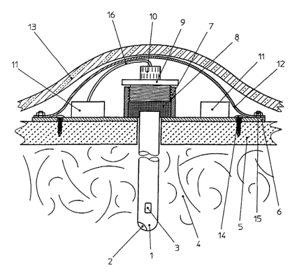
The invention shall now be explained in detail in an example embodiment;
in amplification thereof please see the explanatory illustration in Fig. 1.
Example Embodiment:
The implanted device in modular-system design consists of a catheter 1,
which comprises at its distal end a temperature sensor 2 and a pressure sen-
sor 3 and extends through the skull bone 5 into the brain tissue 4.
The base plate 6 that is fastened on the skull bone S by means of a screw 14
comprises an electronics unit and an integrated fastening element 7 with
internal thread. By means of this thread the screw 9 exerts a force onto the
seal 8 whereby the space between the semi-flexible cover 12 and base plate
6 is closed tightly relative to the brain tissue 4 and at the same time the
catheter 1 is secured on the base plate 6 and thus on the skull bone 5.
The micro plug 10 that is located on the proximal end of the catheter 1 is
connected via a line 16 to the electronics unit 11.
CA 02496999 2005-02-25
_ g
A radio signal is now used to test the device for functionality. Afterwards
the semi-flexible cover 12 is tightly but removably connected to the base
plate 6 by means of the screws 15.
The scalp 13 stretches over and protects the implanted device.
- Followed by Patent Claims -