Note: Descriptions are shown in the official language in which they were submitted.
CA 02497493 2005-02-17
22422 -MI
Adsorption of nucleic acids to a solid phase
The present invention is directed to a method for adsorbing, i.e. non-
covalently
binding, a nucleic acid to a solid phase. Furthermore, the present invention
pertains
to a method for isolating a nucleic acid from a biological sample.
Many biological substances, especially nucleic acids, present special
challenges in
terms of isolating them from their natural environment. On the one hand, they
are
often present in very small concentrations and, on the other hand, they are
often
found in the presence of r.nany other solid and dissolved substances e.g.
after lysis of
cells. This makes them difficult to isolate or to measure, in particular in
biospecific
assays which allow the detection of specific nucleic acids, or the detection
of specific
properties of a nucleic acid. Such biospecific assays play a major role in the
field of
diagnostics and bioanalytics in research and development. Examples for
biospecific
assays are hybridisation assays, immuno assays and receptor-ligand assays.
Hybridisation assays use the specific base-pairing for the molecular detection
of
nucleic acid analytes e.g. RNA and DNA. Hence, oligonucleotide probes with a
length of 18 to 20 nucleotides may enable the specific recognition of a
selected
complementary sequence e.g. in the human genome. Another assay which entails
the selective binding of two oligonucleotide primers is the polymerase chain
reaction (PCR) describeci in US 4,683,195. This methud allows the selective
amplification of a specific nucleic acid region to detectable levels by a
thermostable
polymerase in the presence of desoxynucleotide triphosphates in several
cycles.
As described above, before the nucleic acids may be analyzed in one of the
above-
mentioned assays or used for other processes, they have to be isolated or
purified
from biological samples containing complex mixtures of different components as
e.g. proteinaceous and no;n-proteinaceous components. Often, for the first
steps,
processes are used which allow the enrichment of the component of interest,
i.e. the
nucleic acids. Frequently, these are contained in a bacterial cell, a fungal
cell, a viral
particle, or the cell of a more complex organism, such as a human blood cell
or a
plant cell. Nucleic acids as a component of interest can also be called a
"target
component".
To release the contents of said cells or particles, they may be treated with
enzymes
or with chemicals to dissolve, degrade or denature the cellular walls and
cellular
CA 02497493 2005-02-17
-2-
membranes of such organisms. This process is commonly referred to as lysis.
The
resulting solution containing such lysed material is referred to as lysate. A
problem
often encountered duririg the lysis is that other enzymes degrading the target
component, e.g. desoxyribonucleases or ribonucleases degrading nucleic acids,
come into contact with the target component during lysis. These degrading
enzymes may also be present outside the cells or may have been spatially
separated
in different cellular compartiments before the lysis and come now into contact
with
the target component. Other components released during this process may be
e.g.
endotoxins belonging to the family of lipopolysaccharides which are toxic to
cells
and can cause problems for products intended to be used in human or animal
therapy.
In the next steps of the sample preparation which follow on the lysis step,
the
nucleic acids are further enriched. Nucleic acids are normally extracted from
the
complex lysis mixtures before they are used in a probe-based assay. There are
several methods for the extraction of nucleic acids. Sequence-dependent or
biospecific methods include, e.g., affinity chromatography or hybridisation to
immobilised probes. Sequence-independent or physico-chemical methods include,
e.g., liquid-liquid extraction with phenol-chloroform, precipitation with pure
ethanol or isopropanol, extraction with filter paper, extraction with micelle-
forming agents as cetyl-trimethyl-ammonium-bromide, binding to immobilized,
intercalating dyes such as acridine derivatives, adsorption to substrates such
as silica
gel or diatomic earths, adsorption to magnetically attractable glass particles
(MGP)
or organo silane particles under chaotropic conditions. Direct binding of the
nucleic acids to a substrate such as a material with a silica surface is
preferred
because among other reasons the nucleic acids do not have to be modified and
even
native nucleic acids can be bound.
Particularly interesting for extraction purposes is the adsorption of nucleic
acids to
a glass surface although other surfaces are possible.
Nucleic acids which are set free, e.g. by way of cell lysis and/ or lysis of
cellular
organelles such as mitochondria, plastids, nuclei or other nucleic acid-
containing
organelles, can be purified by way of binding to a solid phase such as a
mineral
substrate, washing said mineral substrate with the bound nucleic acids and
releasing
said nucleic acids from said mineral substrate.
CA 02497493 2005-02-17
-3-
Adsorption of nucleic acids to glass particles or silica particles in the
presence of
chaotropic salts is known, to the art (Vogeistein, B., and Gillespie, D.,
Proc. Natl.
Acad. Sci. USA 76 (1979) 615-619) and provide the basis for chromatographic
purification and separation processes for nucleic acids. Also known to the art
are
methods to isolate and purify RNA and DNA from lysates using high
concentrations of chaotropic salts, e.g. sodium iodide, sodium perchlorate and
guanidine thiocyanate (Boom, R., et al., J. Clin. Microbiol. 28 (1990) 495-
503;
Yamada, 0., et al., J. Virol. Methods 27 (1990) 203-209). The purification of
plasmid DNA from bacteria on glass dust in the presence of sodium perchlorate
is
described in Marko, M.A., et al., Anal. Biochem. 121 (1982) 382-387. In
DE 37 24 442, the isolation of single-stranded M13 phage DNA on glass fiber
filters
by precipitating phage particles using acetic acid and lysis of the phage
particles
with perchlorate is described. The nucleic acids bound to the glass fiber
filters are
washed and then eluted vvith a methanol-containing Tris/EDTA buffer. A similar
procedure for purifying DNA from lambda phages is described in Jakobi, R., et
al.,
Anal. Biochem. 175 (1988) 196-201. The procedure entails the selective binding
of
nucleic acids to glass surfaces in chaotropic salt solutions and separating
the nucleic
acids from contaminants such as agarose, proteins or cell residue. To separate
the
glass particles from the contaminants, the particles may be either centrifuged
or
fluids are drawn through glass fiber filters. This is a limiting step,
however, that
prevents the procedure from being used to process large quantities of samples.
The use of magnetic particles to immobilize nucleic acids after precipitation
by
adding salt and ethanol is more advantageous and described e.g. in: Alderton,
R.P.,
et al., Anal. Biochem. 201 (1992) 166-169 and WO 91/00212. In this procedure,
the
nucleic acids are agglutinated along with the magnetic particles. The
agglutinate is
separated from the origina.l solvent by applying a magnetic field and
performing a
wash step. After one wash step, the nucleic acids are dissolved in a Tris
buffer. This
procedure has a disadvantage, however, in that the precipitation is not
selective for
nucleic acids. Rather, a variety of solid and dissolved substances are
agglutinated as
well. As a result, this procedure can not be used to remove significant
quantities of
any inhibitors of specific enzymatic reactions that may be present. Magnetic,
porous glass is also available on the market that contains magnetic particles
in a
porous, particular glass matrix and is covered with a layer containing
streptavidin.
This product can be used to isolate biological materials, e.g., proteins or
nucleic
acids, if they are modified in a complex preparation step so that they bind
CA 02497493 2008-04-17
c- . , .
-4-
covalently to biotin. Magnetizable particular adsorbents proved to be very
efficient
and suitable for automatic sample preparation. Ferrimagnetic and ferromagnetic
as
well as superparamagnetic pigments are used for this purpose. The most
preferred
MGPs are those described in WO 01/37291.
Purification of a nucleic acid by way of adsorbing the same to a substrate
such as a
mineral substrate in the presence of high concentration of salts is also
applied to
other complex mixtures. Examples therefor are known to the person skilled in
the
art of molecular biology and include reaction mixtures following, e.g., in-
vitro
synthesis of nucleic acids such as PCR, restriction enzyme digestions,
ligation
reactions, etc.. In Vogelstein, B., and Gillespie, D., Proc. Nat1. Acad. Sci.
USA 76
(1979) 615-619, for instance, a procedure for binding nucleic acids from
agarose
gels in the presence of sodium iodide to ground flint glass is proposed.
Another
application for purification of a nucleic acid by way of adsorbing the same to
a
substrate such as a mineral substrate in the presence of a high concentration
of salts
is the removal of pyrogenic contaminants which may have copurified with the
nucleic acid.
The mechanism by which nucleic acids bind to the mineral support in the
presence
of chaotropic agents is not entirely clear. It is hypothesized that the
interaction
between the nucleic acids and the solvent is influenced such that the nucleic
acids
adsorb to the mineral support and denaturate. In the presence of high
concentrations of chaotropic agents the reaction is almost quantitative. The
adsorbed nucleic acids can be eluted by applying to the mineral support
buffers of
low ionic strength.
US 5,808,041 discloses methods for isolating nucleic acids with lengths
greater than
about 50 bases from certain biological samples. An aqueous lysate containing
chaotropic ions at a concentration above about 2 M is produced and the nucleic
acids are adsorbed from the lysate to silica material (also referred to as
"binding").
A slurry or resin comprising silica material and chaotropic salts is added to
the
biological material thus resulting in a one-step lysis and binding procedure.
The
methods for lysing a biological sample disclosed in the document include lysis
of
bacteria using alkali hydroxide and SDS, lysis of M13 or lambda phages by
incubating the phages in the presence of 2.8 M guanidinium, and lysis of fresh
or
CA 02497493 2008-04-17
~ ... :...., -5-
frozen tissue by incubating the tissue in the presence of 2.8 M guanidinium.
Additionally, N-lauryl-sarcosine is used to aid lysing.
EP 0 389 063 and EP 0 819 696 disclose the method of purifying a nucleic acid
by
way of mixing in a liquid phase material containing the nucleic acid with a
chaotropic substance and a nucleic acid binding solid phase. Thus, the
procedures
disclosed in the documents also represent one-step lysis and binding
procedures.
Following lysis and binding, the solid phase with bound nucleic acid is
separated
from the liquid phase. Following a washing step the nucleic acid is eluted
from the
solid phase. The documents disclose a lysis buffer containing about 10 M
guanidinium thiocyanate, about 2% Triton X100, about 0.1 M Tris salt, and
about
50 M EDTA. Another lysis buffer further contains 40% weight by volume
dextrane
sulfate. Another lysis buffer contains about 10 M guanidinium thiocyanate and
about 50 M EDTA. Other lysis buffers are disclosed in the documents that
contain
as a chaotropic substance potassium iodide or sodium iodide at a concentration
of
about 3 M, or potassium or sodium iodide in combination with 1 M or 8 M urea.
Lysis of a biological sample is effected by way of incubating the sample with
a lysis
buffer, whereby 50 volume parts of the biological sample were mixed with 900
volume parts of a lysis buffer and 40 volume parts of a silica coarse. As an
alternative to using silica coarse, other procedures are described wherein
silica filter
material is used.
EP 0 658 164 describes methods for the chromatographic purification of nucleic
acids by way of chromatographic purification. Particularly, a two-step
procedure
comprising a first lysis step and a second binding step is described. In the
first step
(lysis), the biological sample is mixed with a chaotropic agent whereby the
concentration of the chaotropic agente in the mixture is between about 2 M and
about 4 M. Optionally, the mixture additionally contains phenol, chloroform or
ether. Optionally, the mixture additionally contains a detergent. A protease
is added
and the mixture is incubated. In the second step (binding), an alcohol is
added and
the resulting mixture is contacted with the nucleic acid-binding solid phase.
The methods of the state of the art have certain disadvantages. Therefore, it
was the
object of the present invention to provide an alternative method of preparing
a
biological sample containing a nucleic acid and adsorbing from the preparation
the
nucleic acid to a solid phase. It was another object of the invention to
overcome the
CA 02497493 2008-04-17
----~ ,
-6-
need for alcohol during the adsorption step as alcohol is a flammable
substance and
therefore it is desired to restrict its use.
In the present document it is understood that-the term "a nucleic acid"
denotes at
least one nucleic acid. Furhermore, the term "a nucleic acid" also may
indicate a
mixture of nucleic acids. The terms "solid phase" and "substrate" denotes a
substance which is substantially insoluble in an aqueous solution and on which
a
nucleic acid in an aqueous solution of high ionic strength can adsorb when the
substance is added. Examples therefore are porous or non-porous mineral
particles
such as silica, glass, quartz, zeolites or mixtures thereof. Also, the term
"substrate"
encompasses magnetically attractable particles coated with silica, glass,
quartz, or
zeolites. Further, it is understood that a substrate in the form of "powder"
or
"powdered" material refers to finely divided material which, when dispersed in
a
liquid phase such as a liquid organic compound or an aqueous solution,
produces a
suspension. The term "powder" or "powdered" material is intended to include
tablets, in which the powdered material has been aggregated, but still yields
a
suspension when combined with a liquid phase such as an aqueous solution.
Further, it is understood that the terms "high ionic strength". and "high
concentration" mean the ionic strength or concentration in an aqueous solution
that results from dissolved salts in, concentrations equal to or greater than
about
1 M. Preferred are chaotropic salts in concentrations of 1 to 10 M.
The inventors surprisingly found that performing a two-step lysis and binding
procedure is advantageous, whereby in the first step lysis is effected by
mixing the
biological sample with an aqueous lysis buffer containing a chaotropic agent
and
incubating the mixture; in the second step, the concentration of the
chaotropic
agent in the mixture is increased and the mixture is contacted with a solid
phase
capable of binding nucleic acids, whereby the nucleic acid in the liquid phase
is
adsorbed to the solid phase.
A first embodiment of the invention is therefore a method for adsorbing a
nucleic
acid from a biological sample to a solid phase, comprising the steps of (a)
providing
an aqueous lysis buffer that contains a chaotropic agent; (b) mixing the lysis
buffer
with the biological sample, whereby the concentration of the chaotropic agent
in
the mixture is between 1 M and 4 M, and incubating the mixture; (c) dissolving
an
additional amount of chaotropic agent in or adding an aqueous solution
containing
CA 02497493 2008-04-17
-7-
additional chaotropic agent to the mixture of step (b), thereby increasing the
concentration of the chaotropic agent in the mixture by more than 0.5 M; (d)
contacting the mixture of step (c) with the solid phase, thereby adsorbing the
nucleic acid from the mixture to the solid phase.
A second embodiment of the invention is a method to isolate a nucleic acid
from a
biological sample, comprising the steps of (a) providing an aqueous lysis
buffer that
contains a chaotropic agent; (b) mixing the lysis buffer with the biological
sample,
whereby the concentration of the chaotropic agent in the mixture is between 1
M
and 4 M, and incubating the mixture; (c) dissolving an additional amount of
chaotropic agent in or adding an aqueous solution containing additional
chaotropic
agent to the mixture of step (b), thereby increasing the concentration of the
chaotropic agent in the mixture by more than 0.5 M; (d) providing a solid
phase
and contacting the mixture of step (c) with the solid phase, thereby adsorbing
the
nucleic acid from the mixture to the solid phase; (e) separating the solid
phase from
the liquid phase; (f) optionally washing the solid phase of step with a
washing
buffer; (g) eluting the nucleic acid from the solid phase thereby isolating
the nucleic
acid; (h) optionally precipitating the nucleic acid of step (g) from the
eluate and
isolating the precipitated nucleic acid.
Many procedures for isolating nucleic acids from their natural environment
have
been proposed in recent years by the use of their binding behavior to
substrates
such as glass surfaces. It is common to use chaotropic agents such as, e.g.,
guanidine
thiocyanate under high salt conditions. A high concentration of a chaotropic
agent
changes the bulk properties of water (Cacace, M.G., et al. Quarterly Review of
Biophysics (1997) 30:241-277).
Certain ions in water will tend to increase hydrophobic interactions, while
other
ions will decrease hydrophobic interactions. Which ions have a tendency to
which
effect is described by what is called a Hofmeister series. The series is as
follows:
Cations:
NH4+ > Rb+ > K+ > Na+ > Cs+ > Li+ > Mg2+ > Ca2+ > Ba2+ > guanidinium
Anions:
CA 02497493 2008-04-17
-8-
PO43 > S042 > HP042 > acetate > citrate > tartrate > Cl > Br > N03 > C103 >
C104" > I- > SCN
Ions on the left are said to be "kosmotropic" and increase the strength of
hydrophobic interactions and thus will precipitate or "salt out" proteins at a
high
concentrations. Ions on the right are "chaotropic" and tend to weaken
hydrophobic
interactions. The Hofineister series explains why guanidinium is a protein
denaturant. It weakens hydrophobic interactions causing proteins to denature.
In
contrast, (NH4)2SO4 will dissociate into NH4+ and S042" ions. Both of these
ions are
kosmotropic, and the effect of each is independent and additive. This makes
(NH4)2SO4 a versatile precipitant which is widely used in protein purification
procedures. NaCI is in the middle of the series, that is it is neither
kosmotropic nor
chaotropic.
It was found by the inventors that for preparing a biological sample
containing a
nucleic acid and adsorbing from the preparation the nucleic acid to a solid
phase
advantageously a two-step lysis and binding method is applied. In the first
step
(lysis) the concentration of the chaotropic agent is lower than in the second
step
(binding). During the lysis step, that is to say in the mixture of the
biological
sample and the lysis buffer the preferred concentration of the chaotropic
agent is
between 1 M and 4 M. A first preferred embodiment of the invention is
therefore a
method for adsorbing a nucleic acid from a biological sample to a solid phase,
comprising the steps of (a) providing an aqueous lysis buffer that contains a
chaotropic agent; (b) mixing the lysis buffer with the biological sample,
whereby
the concentration of the chaotropic agent in the mixture is between 1 M and 4
M,
and incubating the mixture; (c) dissolving an additional amount of chaotropic
agent in or adding an aqueous solution containing additional chaotropic agent
to
the mixture of step (b), thereby increasing the concentration of the
chaotropic
agent in the mixture by more than 0.5 M; (d) contacting the mixture of step
(c)
with the solid phase, thereby adsorbing the nucleic acid from the mixture to
the
solid phase.
It is preferred that the chaotropic agent is a chaotropic salt. It is more
preferred that
the chaotropic agent is selected from the group consisting of guanidinium
hydrochloride, guanidinium thiocyanate, guanidinium isothiocyanate, an alkali
iodide, and an alkali perchlorate. Preferably, the alkali iodide is KI or NaI.
CA 02497493 2005-02-17
-9-
Preferably, the alkali perchlorate is NaC1O4 or KC1O4. Mixtures of these
compounds as well as mixtures of these compounds with urea are also possible.
Depending on the chaotropic agent used, the optimal concentration thereof in
the
mixture of step (b) may vary. E.g., to obtain a comparable chaotropic effect
using
guanidinium hydrochloride or guanidinium thiocyanate different concentrations
in
the indicated range of between 1 M and 4 M must be selected. The principle
underlying this difference is that the guanidinium thiocyanate salt when
dissolved
in water dissociates to result in two chaotropic ions, whereas dissociated
guanidinium hydrochloride results in only one chaotropic ion. It is therefore
preferred that in the mixture of step (b) the concentration of the chaotropic
agent is
between 1 M and 2 M. Ii: is more preferred that in the mixture of step (b) the
concentration of the chaotropic agent is between 1.5 M and 2 M. It is even
more
preferred that in the mixture of step (b) the concentration of the chaotropic
agent is
about 2 M. Depending on the chaotropic agent used it is also preferred that in
the
mixture of step (b) the concentration of the chaotropic agent is between 2 M
and
4 M. It is more preferred that in the mixture of step (b) the concentration of
the
chaotropic agent is between 2.5 M and 3 M. It is even more preferred that in
the
mixture of step (b) the concentration of the chaotropic agent is about 3 M.
In this regard it is the gerieral understanding of the skilled artisan that
the word
"about" in combination with a numerically quantified value means that this
value
may be subject to vartiation, whereby the desired technical effect which is
described
or defined by the value remains unchanged. Generally, "about" is understood to
imply a variation of 5%. For example, a value of about 100 comprises the
values
between 95 and 105.
Guanidinium and thiocyanate ions are classified in the Hofmeister series as
having
enhanced chaotropic properties over, e.g. potassium or sodium cations, or
iodide or
chlorate anions, respectively. It is thus also preferred that in the mixture
of step (b)
the concentration of the chaotropic agent is between 2 M and 4 M.
In the second step (binding) the concentration of chaotropic agent in the
mixture is
increased by adding additional chaotropic agent to the mixture of step (b). As
indicated for step (c) there are several ways to achieve the desired increase.
It is
possible to add the chaotropic agent as solid matter to the mixture of step
(b) and
CA 02497493 2008-04-17
-10-
dissolve the chaotropic agent. Alternatively, an aqueous solution of the
chaotropic
agent is prepared and added to the mixture of step (b). Preferably, the
aqueous
solution contains further ingredients such as a buffer salt, a detergent or
both. An
example for such an aqueous solution is the binding buffer described in
Examples 2, 3 and 4.
The concentration of the chaotropic agent in the mixture of step (c) is
increased by
more than 0.5 M. It is preferred, therefore, that step (c) comprises
dissolving an
additional amount of chaotropic agent in or adding an aqueous solution
containing
additional chaotropic agent to the mixture of step (b), thereby increasing the
concentration of the chaotropic agent in the mixture to a value between 1.5 M
and
10 M. It is noted that additional chaotropic agent can be added as solid
matter to
the mixture of step (b) until saturation in the mixture is reached. Thus, it
is further
preferred that step (c) comprises dissolving an additional amount of
chaotropic
agent in the mixture of step (b), thereby saturating the mixture with the
chaotropic
agent. It is preferred that in step (c) the concentration of the chaotropic
agent in the
mixture is increased by between 0.5 M and 6 M. It is more preferred that in
step (c)
the concentration of the chaotropic agent in the mixture is increased by
between
0.5 M and 4 M. It is even more preferred that in step (c) the concentration of
the
chaotropic agent in the mixture is increased by between 0.5 M and 3 M. It is
even
more preferred that in step (c) the concentration of the chaotropic agent in
the
mixture is increased by between 0.5 M and 2 M. It is even more preferred that
in
step (c) the concentration of the chaotropic agent in the mixture is increased
by
between 0.5 M and 1.5 M. It is even more preferred that in step (c) the
concentration of the chaotropic agent in the mixture is increased by between
0.5 M
and 1 M.
In detail, the procedure for binding a (at least one) nucleic acid (also
referred to as
target nucleic acid) to a substrate such as, e.g., silica particles, silica
fibers, glass filter
or glass particles can be described as follows. According to the invention it
is
performed in the presence of chaotropic salts with a concentration of between
1.5 M and 10 M, and preferably between 2 M and 6 M.
DNA or RNA bind to material with a glass surface under these conditions i.e.
in the
presence of certain concentrations of a chaotropic agent. To bring the lysate
in
contact with the substrate, i.e. the material with an affinity to nucleic
acids, the
CA 02497493 2005-02-17
-11-
lysate is mixed with the substrate and incubated for a period of time
sufficient for
the binding to occur. In case the substrate is a filter comprising e.g. glass,
silica, or
quartz fibers, the lysate (i.e. the mixture of step (c)) can be passed through
the filter
by gravitational pull, by applying pressure or by applying suction. While
passing
through the filter, the nucleic acid in the liquid phase comes in contact with
the
solid phase and is adsorbed thereto. Experts are usually familiar with the
duration
of the incubation of the liquid phase and the solid phase. The incubation can
be
optimized by determining the quantity of immobilized biological material on
the
surface at different points in time. Incubation times of between 10 seconds
and 30
minutes can be appropriate for nucleic acids.
It is further preferred to use in the procedures for binding a nucleic acid to
a solid
phase or for isolating a:nucleic acid from a biological sample an enzyme with
proteolytic activity when lysing a biological sample in order to set free the
nucleic
acids. The term "enzyme with proteolytic activity" is understood to encompass
a
protease or a mixture of proteases to rapidly degrade in the biological sample
nucleic acid degrading enzymes or other unwanted proteins. In the present
context,
the term "protease" is intended to mean any hydrolase, peptidase, proteinase
or
enzyme having proteolytic activity (i.e. hydrolases acting on peptide bonds)
as
comprised in EC 3.4-3.11 and any modification thereof, which modification have
retained the activity of the enzyme. The enzyme with proteolytic activity may
be
isolated from animal tissue, plant tissue, a microorganism, or may be obtained
by
recombinant means.
Therefore, it is preferred that the lysis buffer of step (a) contains an
enzyme with
proteolytic activity. It is more preferred that the enzyme with proteolytic
activity is
selected from the proteases as comprised in EC 3.4-3.11. It is even more
preferred
that the enzyme with proteolytic activity is selected from the group
consisting of
Achromopeptidase, Aminopeptidase, Ancrod, Angiotensin Converting Enzyme,
Bromelain, Calpain, Calpain I, Calpain II, Carboxypeptidase A,
Carboxypeptidase
B, Carboxypeptidase G, Carboxypeptidase P, Carboxypeptidase W,
Carboxypeptidase Y, Caspase, Caspase 1, Caspase 2, Caspase 3, Caspase 4,
Caspase
5, Caspase 6, Caspase 7, Caspase 8, Caspase 9, Caspase 10, Caspase 13,
Cathepsin B,
Cathepsin C, Cathepsin D, Cathepsin G, Cathepsin H, Cathepsin L, Chymopapain,
Chymase, alpha- Chymotrypsin, Clostripain, Collagenase, Complement Clr,
Complement Cls, Complement Factor D, Complement factor I, Cucumisin,
CA 02497493 2005-02-17
-12-
Dipeptidyl peptidase IV, leukocyte Elastase, pancreatic Elastase,
Endoproteinase
Arg-C, Endoproteinase Asp-N, Endoproteinase Glu-C, Endoproteinase Lys-C,
Enterokinase, Factor Xa, Ficin, Furin, Granzyme A, Granzyme B, HIV Protease,
IGase, Kallikrein tissue, Leucine Aminopeptidase, cytosolic Leucine
aminopeptidase, microsomal Leucine aminopeptidase, Matrix metalloprotease,
Methionine Aminopeptidase, Neutrase, Papain, Pepsin, Plasmin, Prolidase,
Pronase
E, Prostate Specific Antigen, Alkalophilic protease from Streptomyces griseus,
Protease from Aspergillus, Protease from Aspergillus saitoi, Protease from
Aspergillus sojae, Alkaline protease from B. licheniformis, Alcalase from B.
licheniformis, Protease from Bacillus polymyxa, Protease from Bacillus sp,
Protease
from Bacillus sp (Esperase), Protease from Rhizopus sp., Protease S,
Proteasomes,
Proteinase from Aspergillus oryzae, Proteinase 3, Proteinase A, Proteinase K,
Protein C, Pyroglutamate aminopeptidase, Renin, Rennin, Streptokinase,
Subtilisin,
Thermolysin, Thrombin, Tissue Plasminogen Activator, Trypsin, Tryptase, and
Urokinase. It is even more preferred that the enzyme with proteolytic activity
is
selected from the group consisting of a Caspase, Proteinase K, Pronase E,
Protease
from Bacillus sp (Esperase), and Subtilisin. A mixture of at least two
different
proteases as comprised in EC 3.4-3.11 is also preferred. With regard to the
selection
of a protease experts are usually familiar with the optimization of lysis
buffers. As
described in Example 1, the skilled artisan will test for proteolytic activity
of a
selected protease in a buffer containing a chaotropic agent at different
concentrations. A protease active under the chaotropic conditions of a mixture
according to step (b) is preferably selected. Optimization of the duration of
the
incubation with the protease as well as the optimization of the incubation
temperature can be perforined by the expert. As a parameter, the skilled
artisan will
determine the quantity of nucleic acid(s) set free from the biological sample
into
the liquid phase and capable of being bound to the substrate.
It is highly preferred that the lysis buffer of step (a) contains Proteinase
K.
Preferably, the activity of Proteinase K in the mixture of step (b) is between
0.1 U/ml and 10 U/ml. It is more preferred that the activity of Proteinase K
in the
mixture of step (b) is between 1 U/ml and 6 U/ml. It is even more preferred
that the
activity of Proteinase K in the mixture of step (b) is between 2 U/ml and 4
U/ml. It
is even more preferred that: the activity of Proteinase K in the mixture of
step (b) is
about 3 U/ml. In this regard it is understood that the recited activity values
of
Proteinase K reflect activity values determined as described in Example 1.
CA 02497493 2008-04-17
-13-
When lysing a biological sample in order to set free the nucleic acids or when
binding the nucleic acid to the solid phase it is further preferred to use a
detergent
in the procedures, that is to say an anionic, cationic, zwitterionic or non-
ionic
detergent,. Such detergents are well known to the person skilled in the art.
Generally, a "detergent" is a surface active agent, also known as a
surfactant. A
detergent is capable of lowering the surface tension of the medium in which it
is
dissolved, and/or the interfacial tension with other phases, and, accordingly,
is
positively adsorbed at the liquid/vapour and/or at other interfaces. Thus,
detergents
are amphipathic molecules with polar (water soluble) and nonpolar
(hydrophobic)
domains. They are capable of binding to hydrophobic molecules or molecular
domains to confer water solubility. Depending on its ionic characteristics, a
detergent can be categorized as an ionic detergent, a non-ionic detergent, and
a
zwitterionic detergent. Ionic detergents can be further classified into either
cationic
detergents such as SDS (Sodium dodecyl sulfate), LiDS (Lithium dodecyl
sulfate),
or Cetyltrimethylammoniumbromide (CTAB), and anionic detergents such as
Deoxycholic acid, or Sodium lauroyl sarcosine. Thus, these are usually highly
protein denaturant. Non-ionic detergents such as are less protein denaturant.
This
is also true for zwitterionic detergents such as CHAPS or Sulphobetaine 14.
Zwitterionic compounds, also known as zwitterions, inner salts or dipolar ions
are
neutral compounds having formal unit electrical charges of opposite sign.
Therefore, it is preferred that the lysis buffer of step (a) contains a
detergent. It is
more preferred that the lysis buffer of step (a) contains an anionic,
cationic,
zwitterionic or non-ionic detergent. It is even more preferred that the
detergent in
the lysis buffer of step (a) is selected from the group consisting of Sodium
dodecyl
sulfate, Lithium dodecyl sulfate, Cet T1Mrimethylammoniumbromide, Deoxycholic
TM
acid, Sodium lauroyl sarcosine, Triton-X100, Tween 20, Octyl beta-D-glucoside,
Nonidet P40, CHAPS or Sulphobetaine 14. However, other detergents are
possible.
Generally, when using the combination of a chaotropic agent, a detergent and a
protease for lysing a biological sample, the skilled artisan selects a
detergent and its
concentration in the mixture of step (b) on the basis that proteolytic
activity is
preserved.
Preferably, the solid phase comprises a porous or non-porous mineral substrate
selected from the group consisting of silica gel, glass fibers, quartz fibers,
and
zeolites. Also preferred, the solid phase comprises a porous or non-porous
mineral
CA 02497493 2008-04-17
-14-
substrate selected from the group consisting of metal oxides, and/ or metal
mixed
oxides, alumina, titania, zirconia, and materials predominantly consisting of
glass.
It is also preferred that the solid phase comprises a mineral substrate with a
particle
size of 0.1 m to 1,000 m. It is also preferred that the solid phase
comprises porous
mineral support materials with a pore size of from 2 to 1,000 nm. More
preferred,
porous or non-porous support materials, especially zeolites, are in the form
of loose
packings. Even more preferred, the solid phase consists of filter sheets in
the form
of glass, quartz or ceramic filter sheets, and/ or a membrane containing
silica gel
and/ or particles or fibers of mineral supports and fabrics of quartz or glass
wool. It
is also preferred that the solid phase comprises magnetically attractable
particles.
More preferred, the magnetically attractable particles are coated with a
mineral
substrate selected from the group consisting of silica gel, glass, quartz, and
zeolites.
Even more preferred, the substrate comprises magnetic glass particles.
A further embodiment of the invention is a method to isolate a nucleic acid
from a
biological sample, comprising the steps of (a) providing an aqueous lysis
buffer that
contains a chaotropic agent; (b) mixing the lysis buffer with the biological
sample,
whereby the concentration of the chaotropic agent in the mixture is between 1
M
and 4 M, and incubating the mixture; (c) dissolving an additional amount of
chaotropic agent in or adding an aqueous solution containing additional
chaotropic
agent to the mixture of step (b), thereby increasing the concentration of the
chaotropic agent in the mixture by more than 0.5 M; (d) providing a solid
phase
and contacting the mixture of step (c) with the solid phase, thereby adsorbing
the
nucleic acid from the mixture to the solid phase; (e) separating the solid
phase from
the liquid phase; (f) optionally washing the solid phase of step with a
washing
buffer; (g) eluting the nucleic acid from the solid phase thereby isolating
the nucleic
acid; (h) optionally precipitating the nucleic acid of step (g) from the
eluate and
isolating the precipitated nucleic acid.
It is preferred that the solid phase comprises a porous or non-porous mineral
substrate selected from the group consisting of silica gel, glass fibers,
quartz fibers,
and zeolites. It is also preferred that the solid phase comprises magnetic
glass
particles.
It is very much preferred that the chaotropic agent is a chaotropic salt. It
is more
preferred that the chaotropic agent is selected from the group consisting of
CA 02497493 2005-02-17
- 15-
guanidinium hydrochloride, guanidinium thiocyanate, guanidinium
isothiocyanate, an alkali iodide, and an alkali perchlorate.
It is also preferred that: the lysis buffer of step (a) contains an enzyme
with
proteolytic activity and a detergent. It is very much preferred that the lysis
buffer of
step (a) contains an enzyme with proteolytic activity. It is more preferred
that the
enzyme with proteolytic activity is selected from the proteases as comprised
in EC
3.4-3.11. It is even more preferred that the enzyme with proteolytic activity
is
selected from the group consisting of a Caspase, Proteinase K, Pronase E,
Protease
from Bacillus sp (Esperase), and Subtilisin. A mixture of at least two
different
proteases as comprised in EC 3.4-3.11 is also preferred. It is also very much
preferred that the lysis buffer of step (a) contains an anionic, cationic,
zwitterionic
or non-ionic detergent. l:t is even more preferred that the detergent in the
lysis
buffer of step (a) is selected from the group consisting of Sodium dodecyl
sulfate,
Lithium dodecyl sulfate, Cetyltrimethylammoniumbromide, Deoxycholic acid,
Sodium lauroyl sarcosine, Triton-X100, Tween 20, Octyl beta-D-glucoside,
Nonidet P40, CHAPS or Sulphobetaine 14.
It is preferred that the pH value of the mixture of step (b) is between 8.5
and 4. It is
more preferred that the pI-I value of the mixture of step (b) is between 7.5
and 5. It
is even more preferred that the pH value of the mixture of step (b) is between
7 and
6. It is even more preferreci that the pH value of the mixture of step (b) is
about 6.
It is very much preferred that the mixture of step (b) contains the biological
sample,
guanidinium thiocyanate at a concentration between 1.5 M and 3 M, Tris salt at
a
concentration of between 20 mM and 40 mM, Triton-X100 at a concentration of
between 5% and 20% volume by volume, and Proteinase K, whereby the proteolytic
activity of Proteinase K in the mixture is between 1 U/ml and 5 U/rnl, and
whereby
the pH of the mixture is between 8.5 and 6Ø In this regard it is understood
that the
activity of Proteinase K is determined as described in Example 1.
It is highly preferred that the mixture of step (b) contains the biological
sample,
guanidinium thiocyanate at a concentration between 1.5 M and 2.5 M, Tris salt
at a
concentration of between 20 mM and 30 mM, Triton-X100 at a concentration of
between 5% and 15% voluine by volume, and Proteinase K, whereby the
proteolytic
CA 02497493 2005-02-17
-16-
activity of Proteinase K in the mixture is between 2 U/ml and 4 U/ml, and
whereby
the pH of the mixture is between 8.5 and 6Ø
It is highly preferred that the mixture of step (b) contains the biological
sample,
guanidinium thiocyanate at a concentration of about 2 M, Tris salt at a
concentration of about 25 mM, Triton-X100 at a concentration of about 10%
volume by volume, aiid Proteinase K, whereby the proteolytic activity of
Proteinase K in the mixture is about 3 U/ml, and whereby the pH of the mixture
is
about 6. Even more preferred is a pH of 6. An example therefor is the lysis
mixture
(i.e. the mixture according to step (b)) used in Experiment 4 of Example 2.
It is also highly preferred that the mixture of step (b) contains the
biological
sample, guanidinium hyclrochloride at a concentration of about 2.7 M, urea at
a
concentration of about 5 mM, Tris salt at a concentration of about 5 mM,
Triton-
X100 at a concentration of about 9% volume by volume, and Proteinase K,
whereby
the proteolytic activity of Proteinase K in the mixture is about 3 U/ml, and
whereby
the pH of the mixture is between 4.4 and 6.5.
It is also highly preferred that the mixture of step (b) contains the
biological
sample, guanidinium hydrochloride at a concentration of about 2.4 M, urea at a
concentration of about 1.6 mM, Tris salt at a concentration of about 85 mM,
EDTA
at a concentration of about 88 mM, NaCI at a concentration of about 8 mM,
Triton-X100 at a concentration of about 9% volume by volume, and Proteinase K,
whereby the proteolytic activity of Proteinase K in the mixture is about 3
U/ml, and
whereby the pH of the mixture is between 4.4 and 6.5.
It is further preferred that the mixture of step (b) including a protease is
incubated
for a certain amount of time and at ambient conditions that allow proteolytic
activity. It is preferred that the mixture of step (b) is incubated for 10 min
to 30
min at a temperature between 20 C and 75 C, whereby the mixture is agitated.
Preferred agitation means is a roller mixer or a thermomixer. The terms
"agitation"
and "to agitate" are understood as moving the test tube containing the mixture
of
step (b) as to invert the test tube once a second. The terms also include
movements
having an equivalent effect with regard to causing turbulence in the mixture
of step
(b). The degree of agitation can also be influenced by the size of the nucleic
acid(s)
to be isolated. Too much agitation can lead to shearing forces resulting in
size
CA 02497493 2005-02-17
-17-
restriction of the nucleic acid molecules in the mixture. Therefore, slower
agitation
may be selected by the expert.
It is more preferred that the mixture of step (b) is incubated for 30 min at
room
temperature, whereby the mixture is agitated using a roller mixer. It is even
more
preferred that the mixture of step (b) is incubated for about 10 to 20 min at
a
temperature between 50 C and 75 C, whereby the mixture is agitated using a
thermomixer. It is even more preferred that the mixture of step (b) is
incubated for
min to 20 min at about 56 C, whereby the mixture is agitated using a roller
mixer or a thermomixer. It is even more preferred that the mixture of step (b)
is
10 incubated for 15 min at 56 C, whereby the mixture is agitated using a
roller mixer
or a thermomixer. It is also very much preferred that the mixture of step (b)
is
incubated for 10 min to 20 min at about 72 C, whereby the mixture is agitated
using a roller mixer or a thermomixer. It is even more preferred that the
mixture of
step (b) is incubated for 10 min at 72 C, whereby the mixture is agitated
using a
roller mixer or a thermomixer.
After adsorbing the nucleic acid(s) from the mixture of step (c) to the solid
phase,
bound nucleic acid(s) is separated from the liquid phase. This may be achieved
in
general by gravity or in the convenient case of nucleic acids bound to
magnetic glass
particles by separating the material bound to the magnetic particles by
applying a
magnetic field. For instance, the magnetic particles can be pulled to the wall
of the
vessel in which incubation was performed. The liquid containing the sample
contents that were not bound to the magnetic particles can then be removed.
The
removal procedure used depends on the type of vessel in which incubation was
performed. Suitable steps include removing the liquid via pipetting or
aspiration.
In case the solid phase is a filter (such as a filter comprising quartz or
glass fibers)
the mixture of step (c) is preferably passed through the filter. Separation of
the
liquid phase from the solid phase is then effected by by gravitational pull,
by the
application of pressure or by the application of suction.
The solid phase with the bound DNA or RNA may then be washed. This step is
optional, depending on the nature and the amount of undesired material in the
biological sample as well as on the desired purity of the isolated nucleic
acid. If a
washing step is desired, it is preferred that the solid phase is washed at
least once,
CA 02497493 2008-04-17
-18-
e.g. with a mixture of 1-90% volume by volume of an alcohol such as isopropyl
alcohol or ethanol. Examples for wash solutions are the "Inhibitor Removal
Buffer"
and the "Wash Buffer" described in Example 2 (A). Generally, a wash solution
is
used that does not cause the nucleic acid(s) to be released from the surface
of the
solid phase but that washes away the undesired contaminants as thoroughly as
possible. This wash step preferably takes place by incubating the material
with the
bound nucleic acid(s) with the wash solution. The solid phase is preferably
resuspended during this step. In case the solid phase is a filter it is
preferred that the
wash solution is passed through the filter. The contaminated wash solution is
preferably removed just as in the step described above for binding the nucleic
acids.
It is even more preferred to perform two consecutive washing steps, whereby
the
first washing step is performed using inhibitor removal buffer and the second
washing step is performed using wash buffer. An inhibitor removal buffer is
characterized in that it contains a chaotropic agent. Washing with the
inhibitor
removal buffer removes toxic substances or inhibitors that can interfere with
enzymatic reactions such as, e.g. reactions performed by restriction enzyme or
polymerases. Washing with washing buffer removes residual chaotropic agent
from
the bound nucleic acids. After the last wash step, the material can be dried
briefly in
a vacuum, or the fluid can be allowed to evaporate. A pretreatment step using
acetone may also be performed.
Afterwards, the conditions may be reversed, e.g. the salt concentration is
decreased
to elute the DNA or RNA bound to the material. Elution buffers are known from
DE 37 24 442 and Jakobi, R., et al., Anal. Biochem. 175 (1988) 196-20 1.
Elution
buffers with a low salt content are in particular buffers with a content of
less than
0.2 M. Preferably, the elution buffer contains the substance Tris for
buffering
purposes. It is very much preferred that the elution buffer is an aqueous
solution
containing a Tris salt, whereby the concentration of the Tris salt is 50 mM.
More
preferred is an aqueous solution containing a Tris salt, whereby the
concentration
of the Tris salt is 50 mM and the pH is between 6.5 and 8.5. Even more
preferred is
a pH of 7.5. Also preferred, the elution buffer is demineralized water. The
solution
containing purified DNA or RNA can now be used for other reactions.
Optionally,
the nucleic acid(s) can be precipitated from the solution using, e.g., ethanol
or
isopropanol. The precipitate can also be subjected to further washing steps.
Methods of this kind are well known to the skilled artisan and are described
in
CA 02497493 2005-02-17
-19-
detail in Sambrook, Fritsch & Maniatis, Molecular Cloning, A Laboratory
Manual,
3rd edition, CSHL Press, 2001.
The target nucleic acid(s) can be detected and determined. The above-described
purification method is preferred, followed by a determination or detection
step or
purification methods followed by an amplification and determination or
detection
step. The target nucleic acid or nucleic acids of interest may be contained in
a
matrix of non-target nucleic acids, and may even be a minor component in said
mixture of specific nucleic acids. Suitable DNA detection methods are known to
the
skilled artisan and are described in standard textbooks as Sambrook, Fritsch &
Maniatis, Molecular Cloning, A Laboratory Manual, 3rd edition, CSHL Press,
2001;
and Ausubel et al., Current Protocols in Molecular Biology, J. Wiley and Sons,
NY,
1987.
There may be also further purification steps before the DNA detection step is
carried out as e.g. a precipitation step. The detection methods may include
but are
not limited to the bindirig or intercalating of specific dyes as
ethidiumbromide
which intercalates into the double-stranded DNA and changes its fluorescence
thereafter. The purified DNA may also be separated by electrophoretic methods,
optionally after a restricticin digest, and visualized thereafter. There are
also probe-
based assays which exploit the oligonucleotide hybridization to specific
sequences
and subsequent detection of the hybrid. It is also possible to sequence the
DNA
after further steps known to the skilled artisan. Other methods apply a
diversity of
DNA sequences to a silicon chip to which specific probes are bound and yield a
signal when a complementary sequences bind.
The invention also encompasses the mixture of non-proteinaceous and
proteinaceous components comprising nucleic acids whereby the nucleic acids
comprise DNA or RNA or both.
The invention also encompasses biological samples, from which nucleic acids
are
purified, comprising viruses or bacterial cells, as well as isolated cells
from
multicellular organisms as e.g. human and animal cells such as leucocytes, and
immunologically active low and high molecular chemical compounds such as
haptens, antigens, antibodies and nucleic acids, blood plasma, cerebral fluid,
sputum, stool, biopsy specimens, bone marrow, oral rinses, blood serum,
tissues,
CA 02497493 2005-02-17
-20-
urine or mixtures thereof. The present invention also encompasses biological
samples such as a fluid from the human or animal body; preferably the
biological
sample is whole blood, blood plasma, blood serum or urine. The whole blood
sample is preferably EDT'A blood, heparin or citrate blood. The blood plasma
is
preferably EDTA, heparin or citrate blood plasma. In an embodiment of the
invention the biological sample comprises bacterial cells, eukaryotic cells,
viruses or
mixtures thereof.
It is also preferred that the mixture of nucleic acids and proteinaceous
material
comprises desoxyribonucleic acid (DNA) or ribonucleic acid (RNA) or both,
preferably the DNA or RNA or both is derived from a virus or a (at least one)
microorganism. The virus can be hepatitis A virus (HAV), hepatitis B virus
(HBV),
hepatitis C virus (HCV), the human immunodeficiency virus (HIV), the human
papilloma virus (HPV) or parvovirus B19.
It is also preferred that a target nucleic acid component and the other
nucleic acids
are purified essentially as described above. Then the target nucleic acid
component
is further manipulated and detected, i.e. it is amplified with the polymerase
chain
reaction which specifically amplifies target sequences to detectable amounts.
Other
possible amplification reactions are the ligase Chain Reaction (LCR, Wu, D.Y.,
and
Wallace, R.B., Genomics 4 (1989) 560-569, and Barany, F., Proc. Natl. Acad.
Sci.
USA 88 (1991) 189-193); Polymerase Ligase Chain Reaction (Barany, F., PCR
Methods and Applic. 1(1991) 5-16); Gap-LCR (WO 90/01069); Repair Chain
Reaction (EP 0 439 182), 3SR (Kwoh, D.Y., et al., Proc. Natl. Acad. Sci. USA
86
(1989) 1173-1177; Guatelli, J.C., et al., Proc. Natl. Acad. Sci. USA 87 (1990)
1874-
1878; WO 92/08800), and NASBA (US 5,130,238). Further, there are strand
displacement amplification (SDA), transciption mediated amplification (TMA),
and Q-beta-amplification (for a review see e.g. Whelen, A.C., and Persing,
D.H.,
Annu. Rev. Microbiol. 50 (1996) 349-373; Abramson, R.D., and Myers, T.W.,
Curr.
Opin. Biotechnol. 4 (1993) 41-47).
Particularly preferred is the TaqMan detection method disclosed in WO
92/02638
and the corresponding US patents US 5,210,015; US 5,804,375; US 5,487,972.
This
method exploits the exonuclease activity of a polymerase to generate a signal.
In
detail, the target nucleic acid component is detected by a process comprising
contacting the sample with an oligonucleotide containing a sequence
CA 02497493 2005-02-17
-21-
complementary to a region of the target nucleic acid component and a labelled
oligonucleotide containing a sequence complementary to a second region of the
same target nucleic acid component sequence strand, but not including the
nucleic
acid sequence defined by the first oligonucleotide, to create a mixture of
duplexes
during hybridization conditions, wherein the duplexes comprise the target
nucleic
acid annealed to the first oligonucleotide and to the labelled oligonucleotide
such
that the 3'-end of the first oligonucleotide is adjacent to the 5'-end of the
labelled
oligonucleotide. Then this mixture is treated with a template-dependent
nucleic
acid polymerase having a 5' to 3' nuclease activity under conditions
sufficient to
permit the 5' to 3' nuclease activity of the polymerase to cleave the
annealed,
labelled oligonucleotide and release labelled fragments. The signal generated
by the
hydrolysis of the labelled oligonucleotide is detected and/ or measured.
TaqMan
technology eliminates the need for a solid phase bound reaction complex to be
formed and made detectable. In more general terms, a procedure for the
purification of a target nucleic acid component followed by a detection step
is
disclosed wherein the amplification and/ or detection reaction is a
homogeneous
solution-phase.
The following examples, references, and figures are provided to aid the
understanding of the present invention, the true scope of which is set forth
in the
appended claims. It is understood that modifications can be made in the
procedures set forth without departing from the spirit of the invention.
Description of the Figures
Figure 1 Activity of Proteinase K at t= 0 min in relation to varying
concentrations of guanidinium thiocyanate (0 min values). The x
axis inciicates the molar concentration of guanidinium
thiocyanate, the y axis indicates the activity of Proteinase K in
M.
Figure 2 Activity of Proteinase K at t = 15 min in relation to varying
concentrations of guanidinium thiocyanate (15 min values). The
x axis indicates the molar concentration of guanidinium
thiocyanate, the y axis indicates the activity of Proteinase K in
[%].
CA 02497493 2005-02-17
-22-
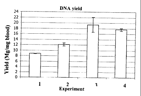
Figure 3 Yield of' DNA in pg per ml blood, according to Example 2 and
Table 3. The x axis indicates the respective experiment, the y axis
indicates the DNA yield in g per ml blood. Also indicated are the
standard deviations.
Figure 4 Yield of'DNA in pg per ml blood, according to Example 3 and
Table 5. The x axis indicates the respective experiment, the y axis
indicates the DNA yield in g per ml blood. Also indicated are the
standard deviations.
Figure 5 Yield of DNA in pg per ml blood, according to Example 4 and
Table 7. The x axis indicates the respective experiment, the y axis
indicates the DNA yield in g per ml blood. Also indicated are the
standard deviations.
am le 1
(A) Incubation of Proteinase K in the presence of a chaotropic agent
10 l of a 20 mg/ml stock solution of Proteinase K (Roche Diagnostics GmbH,
Mannheimm, catalogue no. 745723; 90 mg dissolved in 4.5 ml water) was mixed
with a chaotropic buffer containing 50 mM Tris-HCl pH 6.0, 1% DTT, 20%
Triton-X100 and x M Guanidinum thiocyanate; x = 0, 1, 2, 3, 4, 5, 6). Directly
after
mixing the Proteinase K activity was measured in a first aliquot (10 1) of
the
mixture using the assay described below (0 min activity value). 15 min after
mixing
the Proteinase K activity was measured in a second aliquot (10 1) of the
mixture
using the assay described below (15 min activity value).
(B) Assay to determine Proteinase K activity
10 1 of the Proteinase K in chaotropic buffer (see above, (A)) were mixed
with
assay buffer, that is to say 980 10.2 M Triethanolamin, 0.05% (weight by
volume)
PEG 6000, 0.1 M Calcium chloride, and 10 l of a 200 mM substate solution. The
substrate was Suc-Ala-Ala-Ala-p-Nitroanilid. The assay buffer was provided in
a
cuvette. Immediately after mixing the cuvette was placed in a Photometer.
CA 02497493 2005-02-17
-23-
Measurements (absorption) were taken over 15 min at 25 C. The activity of
Proteinase K in the assay buffer was calculated from the kinetics as indicated
by a
change in absorption at 405 nm.
Table 1 lists the 0 min values of the activity of Proteinase K in the presence
of
guanidinium thiocyanate at the different concentrations given in (A). Table 2
lists
the 15 min values of the activity of Proteinase K in the presence of
guanidinium
thiocyanate at the differerit concentrations given in (A) as well as the
control value
(no chaotropic agent present). The values are expressed in relation to the 0
min
activity value of Proteinase K in the control buffer without chaotropic agent
(see
(A)), corresponding to 0.5 U/ml; this value was set as 100%. The data of Table
I
and Table 2 are graphically represented in Figure 1 and Figure 2.
Table 1
Activity of Proteinase K at t= 0 min in relation to varying concentrations of
guanidinium thiocyanate (0 min values)
concentration [M] Activity [a/o]
0 81
1 94
2 100
3 66
4 1
5 0
6 0
Table 2
Activity of Proteinase K at t= 15 min in relation to varying concentrations of
guanidinium thiocyanate (15 min values)
concentration [M] Activity [%]
0 105
L 1 95
CA 02497493 2005-02-17
-24-
2 95
3 1
4 0
0
6 0
Example 2
(A) Reagents
a) Proteinase K stock solution: recombinant Proteinase K, PCR grade, 50 U/ml
5 b) Lysis Buffer: 4 M guanidinium thiocyanate, 50 mM Tris-HCI, 20% Triton-
X100, pH 6.0
c) Binding Buffer: 4 M guanidinium thiocyanate, 50 mM Tris-HCI, 20% Triton-
X100, pH 6.0
d) Inhibitor Removal Buffer: 5 M guanidinium hydrochloride, 20 mM Tris-HCI,
38% ethanol, pH 6.6
e) Wash Buffer: 20 mM NaCI, 2 mM Tris-HCI, 80% ethanol, pH 7.5
f) Elution Buffer: 50 mM Tris-HC1, pH 8.2
g) Ethanol (100%)
Additionally necessary: Commercially available spin columns containing a
silica
membrane, e.g. NucleoSpin Blood L, distributed by Machery & Nagel.
(B) Experiment 1: one-step procedure without Proteinase K treatment
1. pipette 1,000 l EDTA blood into a 15 ml Falcon tube
2. add 1,000 l Lysis Buffer, vortex gently
3. incubate for 15 min at room temperature on a roller mixer, agitate
4. put a spin column in a new Falcon tube
5. transfer the mixture of steps 1.-2. (about 2,000 l) to the spin column
6. centrifuge for 3 min at 1,900 x g.
7. add 1,000 l Inhibitor Removal Buffer
8. centrifuge for 2 min at 4,500 x g.
9. add 2,000 l Wash Buffer
10. centrifuge for 10 min at 4,500 x g.
11. discard flowthrough and put filter column in a new Falcon tube
CA 02497493 2005-02-17
- 25 -
12. elute with 300 l pre.-heated (70 C) Elution Buffer
13. incubate for 5 min at room temperature
14. centrifuge for 2 min at 4,500 x g.
15. OD measurement of the eluate at 260, 280 and 320 nm
(C) Experiment 2: one-step procedure including Proteinase K treatment
1. pipette 125 l Proteirrase K into a 15m1 Falcon tube
2. add 1,000 l EDTA blood, vortex gently
3. add 1,000 l Lysis Buffer, vortex gently
4. incubate and shake at 56 C for 15 min (e.g. by using a thermomixer),
agitate
5. put a spin column in a new Falcon tube
6. transfer the mixture of steps l.-3. (about 2,125 l) to the spin column
7. centrifuge for 3 min at 1,900 x g
8. add 1,000 l Inhibitor Removal Buffer
9. centrifuge for 2 min at 4,500 x g
10. add 2,000 l Wash Buffer
11. centrifuge for 10 miri at 4,500 x g.
12. discard flowthrough and put filter column in a new Falcon tube
13. eluate with 300 l pre-heated (70 C) Elution Buffer
14. incubate for 5 min at room temperature
15. centrifuge for 2 min at 4,500 x g.
16. OD measurement of the eluate at 260, 280 and 320 nm
(D) Experiment 3: two-step procedure including ethanol
1. pipette 125 l Proteinase K into a 15m1 Falcon tube
2. add 1,000 l EDTA blood, vortex gently
3. add 1,000 1 Lysis Buffer, vortex gently
4. incubate and shake at 56 C for 15 min (e.g. by using a thermomixer),
agitate
5. add 1,000 l Ethanol, vortex gently
6. put a spin column in a new Falcon tube
7. transfer the mixture of steps 1.-5.(about 3,125 l) to the spin column
8. centrifuge for 3 min at 1,900 x g.
9. add 1,000 l Inhibitor Removal Buffer
10. centrifuge for 2 min at 4,500 x g
CA 02497493 2005-02-17
-26-
11. add 2,000 l Wash Buffer
12. centrifuge for 10 miri at 4,500 x g
13. discard flowthrough and put filter column on a new Falcon tube
14, eluate with 300 l pre-heated (70 C) Elution Buffer
15. incubate for 5 min at room temperature
16. centrifuge for 2 min at 4,500 x g.
17. OD measurement of the eluate at 260, 280 and 320 nm
(E) Experiment 4: two-step procedure including Binding Buffer
1. pipette 125 l Proteinase K into a 15m1 Falcon tube
2. add 1,000 l EDTA blood, vortex gently
3. add 1,000 l Lysis Buffer, vortex gently
4. incubate and shake at 56 C for 15 min (e.g. by using a thermomixer),
agitate
5. add 1,000 pi Binding :Buffer, vortex gently
6. put a spin column in a new Falcon tube
7. transfer the mixture of the steps 1.-5. (about 3,125 l) to the spin column
8. centrifuge for 3 min at 1,900 x g
9. add 1,000 l Inhibitor Removal Buffer
10. centrifuge for 2 min at 4,500 x g.
11. add 2,000 l Wash Buffer
12. centrifuge for 10 min at 4,500 x g.
13. discard flowthrough and put filter column on a new Falcon tube
14. eluate with 300 l pre-heated (70 C) Elution Buffer
15. incubate for 5 min at room temperature
16. centrifuge for 2 min at 4,500 x g.
17. OD measurement of the eluate at 260, 280 and 320 nm
CA 02497493 2005-02-17
-27-
Table 3
Yield of human DNA, in [ g/ml blood]
Experiment 1 Experiment 2 Experiment 3 Experiment 4
(a) 8.72 11.78 21.23 17.96
(b) 8.81 12.57 17.43 17.30
Mean 8.77 12.18 19.33 17.63
SD 0.06 0.55 2.68 0.46
(a) and (b) indicate data from replicate experiments; SD: standard deviation
Table 4
DNA purity as determineci by 260 nm/280 nm ratios with 320 nm correction
Experiment 1 Experiment 2 Experiment 3 Experiment 4
(a) 1.95 1.92 1.87 1.88
(b) 1.96 1.85 1.88 1.84
(a) and (b) indicate data fi-om replicate experiments (see Table 3)
With regard to purity of DNA, a 260 nm/280 nm ratio (including 320 nm
correction) of 1.8 0.1 is regarded as being acceptable. The data show that
the
method according to the invention, that is to say the method of Experiment 4
produces equally pure (if not a DNA of higher purity) compared with the the
method of Experiment 3.
Example 3
(A) Reagents
a) Lysis buffer / binding Buffer, chaotropic (7M [lysis buffer] / 4M [binding
buffer] guanidinium thiocyanate, 50mM Tris-HCI, 20% Triton-X100, pH 6.0)
b) Inhibitor Removal Buffer (5M guanidinium HCI, 20mM Tris-HCI, 38%
ethanol, pH 6.6)
c) Washing Buffer (20mM NaCI, 2mM Tris-HCI, 80% ethanol, pH 7.5)
CA 02497493 2005-02-17
-28-
d) Elution Buffer (50mM Tris, pH 8.1)
e) Ethanol (absolute)
Additionally necessary: Glass fibre filter columns ( with silica membrane,
e.g.
NucleoSpin Blood L, commercially available from Macherey-Nagel)
(B) Experiment 5, one-step procedure: Lysis Buffer: 7M, no binding buffer
1,000 l EDTA blood was pipetted into a 15 ml Falcon tube, 1,000 l Lysis
buffer
(7 M) was added and the tube was vortexed. The mixture was incubated for 15
min
at 56 C on a Thermo Mixer. A new Falcon tube with a glass fibre filter column
was
provided and the whole sample preparation (about 2,000 l; chaotrope
concentration 3.5 M) was transferred to the filter column. After
centrifugation at
1,900 x g for 3 min 1,000 l Inhibitor Removal Buffer was transferred to the
column, followed by another centrifugation step at 4,200 x g for 2 min. After
that,
2,000 l Washing Buffer was transferred to the column, followed by
centrifugation
at 4,200 x g for 10 min. T'he flowthrough was discarded and the filter column
was
put on a new Falcon tube. Bound nucleic acids were eluted using 500 l pre-
heated
(70 C) Elution Buffer. T'he Elution Buffer was transferred to the column and
incubated for 5 min at room temperature. After centrifugation at 4,200 x g for
2
min the flowthrough was analyzed by optical density measurement at 230, 260,
280
and 320 nm.
(C) Experiment 6, two-step procedure: Lysis Buffer: 7M, Binding Buffer: 4M
1,000 l EDTA blood was pipetted into a 15 ml Falcon tube, 1,000 l Lysis
buffer
(7 M) was added and the tube was vortexed. The mixture was incubated for 15
min
at 56 C on a Thermo Mixer. 500 1 Binding Buffer (4 M) was added mixed by
vortexing. A new Falcon tube with a glass fibre filter column was provided and
the
whole sample preparation (about 2,500 l; chaotrope concentration 4.5 M) was
transferred to the filter column. After centrifugation at 1,900 x g for 3 min
1,000 l
Inhibitor Removal Buffer was transferred to the column, followed by another
centrifugation step at 4,200 x g for 2 min. After that, 2,000 l Washing
Buffer was
transferred to the column, followed by centrifugation at 4,200 x g for 10 min.
The
flowthrough was discarded and the filter column was put on a new Falcon tube.
Bound nucleic acids were eluted using 500 l pre-heated (70 C) Elution Buffer.
The
Elution Buffer was transferred to the column and incubated for 5 min at room
CA 02497493 2005-02-17
-29-
temperature. After centr=ifugation at 4,200 x g for 2 min the flowthrough was
analyzed by optical density measurement at 230, 260, 280 and 320 nm.
Table 5
Yield of human DNA, in I g/ml blood]
Experiment 5 Experiment 6
(a) 5.95 11.35
(b) 11.35 13.02
Mean 8.65 12.18
SD 3.82 1.18
(a) and (b) indicate data from replicate experiments; SD: standard deviation
Table 6
DNA purity as determineci by 260 nm/280 nm ratios with 320 nm correction
Experiment 5 Experiment 6
(a) 1.92 1.94
(b) 1.88 1.81
mean 1.90 1.88
(a) and (b) indicate data fi-om replicate experiments (see Table 5)
Example 4
(A) Reagents
a) Lysis buffer / binding Buffer, chaotropic (7M [lysis buffer] / 5M [lysis
buffer] /
4M [binding buffer] guanidinium thiocyanate, 50mM Tris-HCI, 20% Triton-
X100, pH 6.0)
b) Inhibitor Removal Buffer (5M guanidinium HCI, 20mM Tris-HCI, 38%
ethanol, pH 6.6)
c) Washing Buffer (20mM NaC1, 2mM Tris-HCI, 80% ethanol, pH 7.5)
d) Elution Buffer (50mM Tris, pH 8.1)
e) Ethanol (absolute)
CA 02497493 2005-02-17
-30-
Additionally necessary: Glass fibre filter columns ( with silica membrane,
e.g.
NucleoSpin Blood L, commercially available from Macherey-Nagel)
(B) Experiment 7, one-step procedure: Lysis Buffer: 7M, no binding buffer
1,000 l EDTA blood was pipetted into a 15 ml Falcon tube, 1,000 l Lysis
buffer
(7 M) was added and the tube was vortexed. The mixture was incubated for 15
min
at 56 C on a Thermo Mixer. A new Falcon tube with a glass fibre filter column
was
provided and the whcile sample preparation (about 2,000 l; chaotrope
concentration 3.5 M) was transferred to the filter column. After
centrifugation at
1,900 x g for 3 min 1,000 l Inhibitor Removal Buffer was transferred to the
column, followed by another centrifugation step at 4,200 x g for 2 min. After
that,
2,000 l Washing Buffer was transferred to the column, followed by
centrifugation
at 4,200 x g for 10 min. T'he flowthrough was discarded and the filter column
was
put on a new Falcon tube. Bound nucleic acids were eluted using 500 l pre-
heated
(70 C) Elution Buffer. T'he Elution Buffer was transferred to the column and
incubated for 5 min at room temperature. After centrifugation at 4,200 x g for
2
min the flowthrough was analyzed by optical density measurement at 230, 260,
280
and 320 nm.
(C) Experiment 8, two-step procedure: Lysis Buffer: 5M, Binding Buffer: 4M
1,000 l EDTA blood was pipetted into a 15 ml Falcon tube, 1,000 l Lysis
buffer
(5 M) was added and the t:ube was vortexed. The mixture was incubated for 15
min
at 56 C on a Thermo Mixer. 500 1 Binding Buffer (4 M) was added mixed by
vortexing. A new Falcon tube with a glass fibre filter column was provided and
the
whole sample preparatioii (about 2,500 l; chaotrope concentration 3.5 M) was
transferred to the filter column. After centrifugation at 1,900 x g for 3 min
1,000 l
Inhibitor Removal Buffer was transferred to the column, followed by another
centrifugation step at 4,200 x g for 2 min. After that, 2,000 1 Washing
Buffer was
transferred to the column, followed by centrifugation at 4,200 x g for 10 min.
The
flowthrough was discarded and the filter column was put on a new Falcon tube.
Bound nucleic acids were eluted using 500 l pre-heated (70 C) Elution Buffer.
The
Elution Buffer was transferred to the column and incubated for 5 min at room
temperature. After centrifugation at 4,200 x g for 2 min the flowthrough was
analyzed by optical density measurement at 230, 260, 280 and 320 nm.
CA 02497493 2005-02-17
-31-
Table 7
Yield of human DNA, in [ g/ml blood)
Experiment 7 Experiment 8
(a) 5.95 7.16
(b) 11.35 12.20
Mean 8.65 9.68
SD 3.82 3.57
(a) and (b) indicate data from replicate experiments; SD: standard deviation
Table 8
DNA purity as determineci by 260 nm/280 nm ratios with 320 nm correction
Experiment 7 Experiment 8
(a) 1.92 1.91
(b) 1.88 1.92
mean 1.90 1.92
(a) and (b) indicate data fi=om replicate experiments (see Table 7)
CA 02497493 2005-02-17
-32-
List of References
Abramson, R.D., and Myers, T.W., Curr. Opin. Biotechnol. 4 (1993) 41-47
Alderton, R.P., et al., Anal. Biochem. 201 (1992) 166-169
Ausubel et al., Current Protocols in Molecular Biology, J. Wiley and Sons, NY,
1987
Barany, F., PCR Methods and Applic. 1(1991) 5-16
Barany, F., Proc. Natl. Acad. Sci. USA 88 (1991) 189-193
Boom, R., et al., J. Clin. Microbiol. 28 (1990) 495-503
Cacace, M.G., et al. Quarterly Review of Biophysics (1997) 30:241-277
DE 37 24 442
EP 0 439 182
EP0389063
EP 0 658 164
EP 0 819 696
Guatelli, J.C., et al., Proc. Natl. Acad. Sci. USA 87 (1990) 1874-1878
Jakobi, R., et al., Anal. Biochem. 175 (1988) 196-201
Kwoh, D.Y., et al., Proc. Natl. Acad. Sci. USA 86 (1989) 1173-1177
Marko, M.A., et al., Anal. :Biochem. 121 (1982) 382-387
Sambrook, Fritsch & Maniatis, Molecular Cloning, A Laboratory Manual, 3rd
edition, CSHL Press, 2001
US 4,683,195
US 5,130,238
US 5,210,015
US 5,487,972
US 5,804,375
US 5,808,041
Vogelstein, B., and Gillespie, D., Proc. Natl. Acad. Sci. USA 76 (1979) 615-
619
Whelen, A.C., and Persing, D.H., Annu. Rev. Microbiol. 50 (1996) 349-373
WO 01/37291
WO 90/01069
WO 91/00212
WO 92/02638
WO 92/08800
Wu, D.Y., and Wallace, R.B., Genomics 4 (1989) 560-569
Yamada, 0., et al., J. Virol. Methods 27 (1990) 203-209