Note: Descriptions are shown in the official language in which they were submitted.
CA 02497815 2011-09-01
-1-
MEASURING PROPERTIES OF AN ANATOMICAL BODY
BACKGROUND
Injection of a liquid such as a drug into a human patient or an agriculture
animal is performed in a number of ways. One of the easiest methods for drug
delivery is through the skin which is the outermost protective layer of the
body. It is
composed of the epidermis, including the stratum corneum, the stratum
granulosum,
the stratum spino sum, and the stratum basale, and the dermis, containing,
among
other things, the capillary layer. The stratum comeum is a tough, scaly layer
made
of dead cell tissue. It extends around 10-20 microns from the skin surface and
has
no blood supply. Because of the density of this layer of cells, moving
compounds
across the skin, either into or out of the body, can be difficult.
The current technology for delivering local pharmaceuticals through the skin
includes methods that use needles or other skin piercing devices. Invasive
procedures, such as use of needles or lances, effectively overcome the barrier
function of the stratum comeum. However, these methods suffer from several
major
disadvantages: local skin damage, bleeding, and risk of infection at the
injection site,
and creation of contaminated needles or lances that must be disposed. Further,
when
these devices are used to inject drugs in agriculture animals, the needles
break off
from time to time and remain embedded in the animal.
Needleless injection devices have been proposed to overcome the problems
associated with needles, but the proposed devices present different problems.
For
example, some needleless injection devices rely on spring actuators that offer
limited
CA 02497815 2005-03-04
WO 2004/021882 PCT/US2003/027907
-2-
control. Others use solenoids, compressed air or hydraulic actuators also
offer
limited control.
SUMMARY
Skin sensor apparatus and methods described herein use specially tailored
stimulation to effectively measure one or more properties of the surface of an
anatomical body, such as the compliance gain and/or stiffness of skin.
A medical device includes a sensor configured to measure a property of an
outer layer of an anatomical body surface. The sensor includes a source probe
configured stimulate a local surface of the outer layer of an anatomical body
surface.
The sensor also includes a detector configured to measure a response of the
outer
layer resulting from the source probe stimulation. Further, the device
includes a
controller coupled to the sensor. The controller drives the source probe using
a
tailored stochastic sequence. The controller then determines the property of
the outer
layer using the measured response received from the detector.
The body surface can be the skin of a subject, or an internal body surface.
The body surface can be modeled as a second order mechanical system. Further,
the
property of the outer layer can be determined using system identification
techniques.
The source probe can include a voice coil for stimulating the local surface of
the outer layer. For example, the voice coil can be coupled to the outer layer
and
driven at a frequency to displace the surface. The detector measures
displacement of
the body surface, for example, using an accelerometer. In one embodiment, the
detector includes a linear differential variable transducer detecting
displacement of
the body surface. In some embodiments, the detector further includes a strain
gauge
for measuring a static displacement of the body surface.
The medical device can be a drug injection device. The drug injection device
is coupled to the sensor and injects a drug into an anatomical body in
response to the
determined property of the outer layer. For example, the device can include a
servo-
controller coupled to a delivery device for delivering a pharmaceutical. The
servo-
controller adjusts the delivery characteristics of the delivery device based
on the
CA 02497815 2005-03-04
WO 2004/021882 PCT/US2003/027907
-3-
surface properties. In one embodiment, the drug injection device is a
needleless
injector.
A device for injecting drug into a biological body includes a drug injector
for
holding the drug to be delivered to the body. The device also includes a skin
sensor
that measures skin properties of the body and a servo-controller coupled to
the drug
injector and the skin sensor. The servo-controller adjusts the injection
pressure of
the drug injector to selectively deliver the drug to the body based on the
skin
properties. In some embodiments, the skin sensor measures the properties of
the
body using a tailored stochastic sequence.
BRIEF DESCRIPTION OF THE DRAWINGS
The foregoing and other objects, features and advantages of the invention
will be apparent from the following more particular description of preferred
embodiments of the invention, as illustrated in the accompanying drawings in
which
like reference characters refer to the same parts throughout the different
views. The
drawings are not necessarily to scale, emphasis instead being placed upon
illustrating
the principles of the invention.
FIG. lA is a perspective view of a drug delivery device in accordance with
the invention.
FIG. 1B is a side view of the drug delivery device of FIG. 1A.
FIG. 1C is an end view of the drug delivery device taken along the line 1C-
1C of FIG. 1B.
FIG. 2 is a perspective view of the drug delivery device of FIG. lA with a
controller and energy source.
FIG. 3A is a graph of the time response of a shape memory alloy fiber of the
drug delivery device of FIG. lA for a high strain.
FIG. 3B is a graph of the time response of the shape memory alloy fiber of
the drug delivery device of FIG. 1A when the fiber is subjected to a potential
as a
quick pulse.
FIGs. 4A-4C are respectively side, front, and top views of a hand-held drug
delivery device.
WO 2004/021882 CA 02497815 2005-03-04PCT/US2003/027907
-4-
FIG. 4D is a perspective view of the drug delivery device shown in
FIGs. 4A-4C.
FIG. 5A is a cross-sectional view of the drug delivery device taken along the
line 5A-5A of FIG. 1C prior to delivery of a drug.
FIG. 5B is a cross-sectional view of the drug delivery device of FIG. lA
during drug delivery.
FIG. 6A is a perspective view of an alternative embodiment of the drug
delivery device in accordance with the invention.
FIG. 6B is a side view of the drug delivery device of FIG. 6A.
FIG. 6C is top view of the drug delivery device taken along the line 6C-6C of
FIG. 6B.
FIG. 6D is front view of the drug delivery device taken along the line 5D-5D
of FIG. 6B.
FIG. 7A is a perspective view of a drug vile for the drug delivery device of
FIG. 6A.
FIG. 7B is a cross-sectional view of the drug vile of FIG. 7A.
FIG. 8 is a perspective view of the drug delivery device of FIG. 6A with a
controller and energy source.
FIG. 9A is a cross-sectional view of the drug delivery device taken along the
line 9A-9A of FIG. 6D prior to delivery of a drug.
FIG. 9B is a cross-sectional view of the drug delivery device during drug
delivery.
FIG. 10 is cross-sectional view of another alternative embodiment of the
drug delivery device in accordance with the invention.
FIG. 11 illustrates the drug delivery device of FIG. 10 with a protective
sterile ribbon in accordance with the invention.
FIGs. 12A and 12B illustrate yet another alternative embodiment of the drug
delivery device in accordance with the invention.
FIG. 13 illustrates the drug delivery device with a sensor used to detect
properties of the skin in accordance with the invention.
CA 02497815 2005-03-04
WO 2004/021882 PCT/US2003/027907
-5-
FIG. 14 is a block diagram of an alternative embodiment of the sensor used
to detect properties of the skin in accordance with the invention.
DETAILED DESCRIPTION OF THE INVENTION
A description of preferred embodiments of the invention follows.
Referring to FIGs. 1A-1C, there are shown various views of a drug delivery
device used to inject a liquid formulation of an active principle, for
example, a drug,
into biological body such as an agriculture animal or human being. The
delivery
device is generally identified as 10 in the illustrated embodiment as well as
in other
embodiments described later. The drug is initially contained in a chamber 12
(FIG.
5A) and is injected out through an orifice or output port 14 into the body.
A nozzle is typically used to convey the drug to the skin at the required
Speed
and diameter to penetrate the skin as required. The nozzle generally contains
a flat
surface, such as the head 17 that can be placed against the skin and an
orifice 14. It
is the inner diameter of the orifice 14 that controls the diameter of the drug
stream.
Additionally, the length of an aperture, or tube, defining the orifice 14 also
controls
the injection pressure. In some embodiments, a standard hypodermic needle is
cut to
a predetermined length and coupled to the head. One end of the needle is
flush, or
slightly recessed, with respect to the surface of the head 17 that contacts
the skin to
avoid puncturing the skin during use. The internal diameter of the needle
(e.g., 100
[Lin) defines the diameter of the aperture, and the length of the needle
(e.g., 5 mm)
together with the aperture dimension controls the resulting injection
pressure, for a
given applicator pressure. In other embodiments, a hole can be drilled
directly into
the head 17 to reduce assembly steps. In general, the length of the orifice is
selectable, for example ranging from 500 um to 5 mm, while its diameter can
range
from 80 um to 200 um.
The device 10 includes a guide tube 16 in which a piston 18 is positioned.
An interchangeable head 17 is attached at an enlarged end 19 of the tube 16
with a
set of screws 21. One end of the piston 18, along with the inside of the
enlarged end
19 and head 17 define the chamber 12, and a push block 22 is attached at the
other
end of the piston 18. Although the piston 18 forms a clearance seal with the
tube 16,
CA 02497815 2005-03-04
WO 2004/021882
PCT/US2003/027907
-6-
a seal ring can be placed about the piston 18 to prevent drug from escaping
from the
chamber 12 between the piston 18 and the tube 16. Attached on the outside of
the
push block 22 is an electrical contact plate 24. Another contact plate 26 is
positioned between the interchangeable head 17 and the enlarged end 19.
In some embodiments, the guide tube 16 includes linear bearings to reduce
the friction of the piston 18. Preferably, the piston 18 is rigid to avoid
buckling
under the force exerted by the actuator. Further, the piston 18 is light
weight to
reduce its inertia ensuring a rapid acceleration upon activation. In one
embodiment,
the piston 18 is formed from a hollow aluminum rod. Other parts can also be
advantageously constructed of light weight materials. For example, the push
block
22 can be formed from a machinable poly acetal.
In addition to the contact plates 24 and 26, an actuator 28 includes one to
six
or more wires 30 positioned about the tube 16 and parallel to one another. One
end
32 of each wire 30 is attached to the contact plate 24 through the push block
22, and
another end 34 of the wire 30 is attached to a respective capstan 36. The
capstan 36,
and the contact plates 24 and 26 are electrically conductive. Hence, the ends
32 and
34 of the wires 30 are electrically connected to each other through the
contact plates
24 and 26, respectively. An insulating collar 38 positioned about the guide
tube 20
helps guide the wires 30 through the holes 39 between the enlarged region 19
and
the push block 22.
To apply the appropriate tension to the wires 30 and to define the volume of
the chamber 12, a coiled spring 37 is positioned about the piston 18 between
the end
of the tube 16 and the push block 22, and the capstans 36 are turned
accordingly,
much like adjusting the tension in guitar strings. The wires 30 are wrapped
around
the respective capstans 36 one or more times. As such, the strain near the
terminal
ends 34 of the wires 30 attached to the capstans 36 are significantly less
than the
strain along the remainder of the length of the wires 30. For example, the
strain near
the terminal end 34 may be about 1% while that of the remainder of the wire
may be
about 15%.The wires 30 can be secured to the contact plate 24 with capstans,
as well.
Alternatively, the wires 30 can be attached to one or both contact plates 24
and 26 by
= CA 02497815 2011-09-01
-7-
other techniques, for example, by electrodeposition as described in U.S.
Patent
No. 5,641,391.
Alternatively, each wire 30 can be twisted with a respective electrically
conductive wire made of, for example, copper or iron. The twisted segment is
then
bent back, and partially twisted forming a loop, with the partially twisted
segment
formed of two strands of the wire 30 and two strands of the copper wire. The
formed loop can be placed on a pin, for example, or it can be fully twisted
and then
bent back and partially twisted forming another loop, with the partially
twisted
segment formed of four strands of the wire 30 and four strands of the copper
wire.
Again, the formed loop can be placed on a pin to secure the wire 30 to the
contact
plate 24 and/or 26.
More generally, the wires 30 can be formed from a shape memory material
that changes from a first stable state to a second stable state upon
excitation. For
example, the shape memory material can be a shape memory polymer.
Alternatively, or in addition, the shape memory material can be an alloy. In
some
embodiments, a phase change of the shape memory material occurs when the
material is heated. For example, a shape metal alloy can exist with one of two
different lattice structures, such that a phase change from one lattice
structure to
another occurs responsive to the application and/or removal of thermal energy.
The wires 30 are made of a suitable material that contracts when heated and
can be used as an actuation method. Heating can be accomplished by passing a
current through the wire 30, known as Joule heating. Thus, the current is
conducted
within the wires 30 after a potential is applied across them. A class of
materials that
contract when a potential is applied to them includes piezoelectric materials
and
shape memory alloys. While piezoelectric crystals contract about 1%, shape
memory alloys are able to contract approximately 15% or more. The larger
contraction of shape memory alloys makes them desirable for the illustrated
embodiment. Accordingly, the wires 30 are made of shape memory alloy such as,
for example, Ni-Ti (also known as Nitinol), available from Shaped Memory
Applications Inc., of San Jose, CA, and from Dynalloy Inc. of Costa Mesa, CA,
under the Trade Mark FLEXINOL. When a potential is applied across the wires 30
CA 02497815 2011-09-01
-8-
via the contact plates 24 and 26 the wires 30 heat up. As the wires 30 heat
up, a
phase transformation of the wire material occurs, namely, the wire changes
phase
from martensite to austenite. This phase transformation causes the wires 30 to
contract such that the piston 18 is pushed towards the orifice 14, thereby
forcing the
drug from the chamber 12 out the orifice 14. Preferably, the shape memory
alloy is
fast acting to provide a sudden force suitable for injecting,a drug into a
patient's skin
without using a needle. A more detailed description of shape memory alloys and
their use is described in U.S. Patent No. 5,092,901.
To use the device 10, the device is connected to a controller 50 with a pair
of
leads 52, and the controller in turn in connected to a capacitor bank 54 with
another
pair of leads 56, as illustrated in FIG. 2. The controller 50 can be a simple
microprocessor, or alternatively a personal computer with multifunction
capabilities.
The capacitors of the bank 54 are energized through a power source in the
controller
50 or by an. external power source. Once energized, the capacitors, under the
direction of the controller 50, discharge to apply a potential across the
wires 30 via
the plates 24 and 26 through the leads 52. In this manner, the wires 30 are
connected
together in a parallel configuration, the supply potential being applied
equally across
the ends of each of the multiple wires 30. In another embodiment, the wires 30
are
connected together in a series configuration. Still other arrangements can be
used to
apply the potential across the wires 30, for example, as describe in U.S.
Application
No. 10/200,574 filed July 19, 2002, by Angel and Hunter.
Although any capacitor can be used in the bank 54, a super capacitor has the
advantageous feature. of providing a large energy density in a small physical
size.
Hence the capacitors of the bank 54 can be super capacitors 53 that have a
volume
from 1.5 ml to 30 ml, preferably 3 ml, and an energy output of 10 J to 1 KS,
preferably 100 J. The current applied to the wires 30 is approximately 100
mAmps
to 5 Amps, and the voltage applied to the wires 30 is between about 1 volt to
10
volts. In one embodiment, the applied current is 1 Amp, and the applied
voltage is 5
volts. To heat the wires 30 quickly, larger currents of 25 to 100 Amps can be
CA 02497815 2005-03-04
WO 2004/021882 PCT/US2003/027907
-9-
applied. As fast action is required, the power source must also be able to
switch
large currents with millisecond timing.
The amount of force per area generated by the wires 30 is about 235 MN/m2.
In the illustrated embodiment, the volume of drug initially contained in the
chamber
12 is about 200 p,L to 250 iL, and the orifice 14 has a diameter of between
about 50
pm to 500 pm. In some embodiments, the drug volume is up to 500 tiL. The drug
injection velocity is about 150 m/s with a 150 p,m orifice 14. Generally, an
injection
velocity of 100 m/s or greater is required for successful skin penetration
(e.g.,
penetrating skin to a depth of 2 mm) in a stream having a diameter of 100
Advantageously, the stream diameter of the needleless injector can be
substantially
smaller than a typical 24 gauge needle having a diameter of 450 p,m.
The device 10 has a length, L1, of approximately 150 mm, and the wires 30
contract about 7 mm when a potential is applied across them. The wires 30 can
have
circular cross section, in which case each wire 30 has a diameter of
approximately
0.025 mm to 2 mm, preferably 380 tim. Alternatively, each fiber can have a
flat
ribbon shape with a thickness approximately in the range 0.025 mm to 0.5 mm
and a
width of approximately 0.75 mm to 10 mm. Other suitable shape memory alloys
include Ag-Cd, Au-Cd, Au-Cu-Zn, Cu-Al, Cu-Al-N, Cu-Zn, Cu-Zn-Al, Cu-Zn-Ga,
Cu-Zn-Si, Cu-Zn-Sn, Fe-Pt, Fe-Ni, In-Cd, In-Ti, and Ti-Nb.
Referring now to FIGs. 3A and 3B, there are shown graphs of the time
response of wires 30 made from Ni-Ti. Shown in FIG. 3A is the response of a
wire
subjected to a strain of nearly 5%. As can be seen, the contraction time for
this wire
is about 10 ms. By way of contrast, FIG. 3B illustrates a wire subjected to
faster
pulse than that applied to the wire of FIG. 3A. With the faster pulse, the
fiber
experiences a strain of about 1%, with a contraction time of about 1 ms.
In use, the device 10 is typically mounted within an applicator that is held
by
an operator. The applicator can be shaped as a pistol, cylinder or any other
suitable
geometry. An exemplary applicator is shown in FIGS. 4A through 4D. In one
embodiment, referring to FIG. 4A, a pistol shaped applicator 400 includes a
barrel
405 configured to house the device 10. The barrel 405 can be a hollow tube or
rectangle having a cavity sized to accept the device 10. Referring to FIG. 4B,
the
CA 02497815 2005-03-04
WO 2004/021882 PCT/US2003/027907
-10-
barrel 405 includes an aperture 420 at one end sized to accept the head 17 of
the
device 10. The head 17 protrudes through the aperture 420 to facilitate
contact with
an animal's skin. Further, the applicator 400 includes a handle 410 configured
to be
grasped by an operator. The handle 410 is coupled at one end to the barrel
405.
Additionally, the applicator 400 can include a base 415 coupled to another end
of the
handle 410. The base 415 can be configured to house other parts of the
needleless
injector, such as the power source and/or control unit. The handle 410 can be
similarly configured (e.g., hollowed out) to also house parts of the
needleless
injector. Further, the applicator 400 can include a switch 420. The switch 420
can
be controlled by an operator to operate the device 10 to initiate an injection
and/or a
filling of the device with a drug.
Referring to FIGs. 5A and 5B, as well as to FIG. 1A, the operator positions
the applicator to place a surface 60 of the head 17 against the skin, S, of
the
biological body. Prior to the placement of the head 17 against the skin, or
while the
head 17 is positioned against the skin, the capacitor bank 54 is energized as
described above. The operator then triggers the device 10 through the
controller 50
to discharge the capacitor bank 54, thereby applying a potential across the
wires 30
which causes them to contract. As the wires 30 contract, they pull the push
block
22, which pushes the piston 18 towards the head 17 to force the drug, D, from
the
chamber 12 through the orifice 14 into the body. The injection pressure can be
as
low as 1 MPa or lower or as high as 300 MPa. For comparison, a minimum local
pressure of approximately 1.91 MPa is required for piercing skin to a depth of
2mm
using a 100 p.m diameter needle After the energy in the capacitor bank is
depleted,
the potential across the wires 30 is removed which causes the wires 30 to
extend to
their original length as the coiled spring 37 pushes the push block 22 away
from the
head 17. The chamber 12 can then be refilled if desired with additional drug
to be
injected into another body or the same body.
Turning now to FIGs. 6A-6D, there are shown various views of an
alternative embodiment of the drug delivery device 10, where like features are
identified by like numerals. Here, the device 10 includes two base portions 70
and
72. The piston 18 extends through the base portion 72 and through part of the
base
CA 02497815 2005-03-04
WO 2004/021882 PCT/US2003/027907
-11-
portion 70, as shown, for example, in FIG. 9A. As before, the piston 18 is
attached
at one end to the push block 22, which slides back and forth over a surface 76
of the
base portion 72, such that the piston slides back and forth in the base
portions.
Referring also to FIGs. 7A and 7B, a removable and/or disposable vial 80 is
mounted in the base portion 70. For example, the vial 80 can be screw mounted
to
the base portion 70. The vial 80 is provided with a nozzle, as described
above, at
one end defining the orifice 14. The vial 80 also includes a plunger 82 that
moves
back and forth in the chamber 12 defined within the vial 80. The plunger 82
abuts
the teuninal end 84 of the piston 18. As such, as the piston 18 moves towards
the
orifice 14, drug, D, contained in the chamber 12 is expelled through the
orifice 14.
In some implementations, the orifice of the drug vial, or the chamber of the
embodiment of FIG. 1A, is sealed with a suitable material prior to use. The
seal may
be manually removed, or it may be removed by the injection pressure of the
drag as
it ejects from the vial or chamber.
A single length wire 30 is positioned on each side of the base portions 70 and
72 and attached at one end to a lead capstan 90a, wrapped sequentially around
intermediate capstans 90b, 90c, 90d, and attached at the other end to a
terminal
capstan 90e. To apply the appropriate tension to the wires 30, the coiled
spring 37 is
positioned about the piston 18 between the base portion 72 and the push block
22,
and a rachet mechanism 92 is employed to adjust the tension in the wires 30.
The
capstans 90a, 90c, and 90e are electrically conductive, and are coupled to
respective
conductive bars 94 and 96. The capstans 90b and 90d are also electrically
conductive, and are electrically coupled to respective conductive plates 98
and 100.
The plates 98 and 100 in tarn are electrically connected to each other through
the
push block 22, but electrically insulated from the piston 18 and base portion
72. The
two bars 94 and 96 are electrically insulated from the base portion 70. As
such,
when a potential is applied across the conductive bars 94 and 96, the
potential is also
applied across the four segments of each wire 30.
In one implementation, the device 10 of FIG. 6A is connected to the
controller 50 with the pair of leads 52, and the controller in turn in
connected to the
capacitor bank 54 with another pair of leads 56, as illustrated in FIG. 8. As
WO 2004/021882 CA 02497815 2005-03-04
PCT/US2003/027907
-12-
mentioned above, the capacitors of the bank 54 are energized through a power
source in the controller 50 or by an external power source. Once energized,
the
capacitors, under the direction of the controller 50, discharge to apply a
potential
across the wires 30 via the conductive bars 94 and 96 through the leads 52.
The
wires 30 heat up and contract such that the piston 18 is pushed towards the
orifice
14, thereby forcing the drug D from the chamber 12 of the vial 80 out the
orifice 14.
Although shown as blocks, the base portions 70 and 72 can have any suitable
geometry which facilitates the use of the device 10 of FIG. 6A in a particular
application. As mentioned before, the device can be mounted within an
applicator
that is held by an operator.
Referring to FIGs. 9A and 9B, as well as to FIG. 6A, to use the device 10, the
operator positions the applicator such that a surface 101 of the vial 80 is
placed
against the skin, S, of the body. Prior to the placement of the surface 101
against the
skin, or while the surface 101 is positioned against the skin, the capacitor
bank 54 is
energized, as described earlier. The operator then triggers the device 10
through the
controller 50 to discharge the capacitor bank 54, thereby applying a potential
across
the wires 30 which causes them to contract. As the wires 30 contract, they
pull the
push block 22 which pushes the piston 18, which in turn pushes the plunger 82
towards the orifice 14 to force the drug, D, from the chamber 12 through the
orifice
14 into the body. After the energy in the capacitor bank is depleted, the
potential
across the wires 30 is removed which causes the wires 30 to extend to their
original
length as the coiled spring 37 pushes the push block 22 away from the vial 80.
The
chamber 12 can then be refilled if desired with additional drug to be injected
into
another body.The device 10 of FIGs. 1A or 5A can be used as a single-use
device or for
multiple uses. When used as a multiuse device, the cycle time between uses can
be
0.5 seconds or less.
For example, there is shown in FIG. 10 the device 10 of FIG. 1A coupled to a
reservoir 100 that supplies the chamber 12 with a sufficient amount of drug,
D, for
each injection, and holds enough drug for approximately 20 to 200 or more
injections. Alternatively, individual doses may be provided in a plurality of
CA 02497815 2005-03-04
WO 2004/021882 PCT/US2003/027907
-13-
reservoirs sequentially coupled to the delivery device 10. A valve 102 is
associated
with a tube 103 connecting the reservoir 100 with an inlet port 104 of the
chamber
12. The valve 102 is opened and closed -under the direction of the controller
50, or
an additional controller, to allow the desired amount of drug into the chamber
12 for
each injection. The device 10 of FIG. 6A can also be coupled to a similar
reservoir
that is operated in the manner just described.
When the device 10 of FIG. 10 is in use, the controller 50 instructs the valve
102 to open to allow the drug to flow from the reservoir 100 through the inlet
port
104 into the chamber 12, and, after a prescribed period of time, the
controller 50
directs the valve 102 to close so that a desired amount of the drug is held in
the
chamber 12 for a single injection.
Next, or while the chamber 12 is being filled with drug, the operator
positions the applicator to place the surface 60 of the head 17 against the
skin, S, of
the body. Meanwhile, the capacitor bank 54 is energized as described above.
The
operator then triggers the device 10 through the controller 50 to discharge
the
capacitor bank 54, thereby applying a potential across the wires 30 which
causes
them to contract. As the wires 30 contract, they pull the push block 22 which
pushes
the piston 18 towards the head 17 to force the drug, D, from the chamber 12
through
the orifice 14 into the body. After the energy in the capacitor bank is
depleted, the
potential across the wires 30 is removed which causes the wires 30 to extend
to their
original length as the coiled spring 37 pushes the push block 22 away from the
head
17. The controller 50 then instructs the valve 102 to open to refill the
chamber 12
with additional drug from the reservoir 100 to be injected into another body.
When the device 10 is intended for multiple uses, it may be desirable to
provide some type of protective sterile barrier between the head 17 and the
skin of
the body to eliminate or at least minimize exposing a subsequent body with
contaminants from a previous body.
For example, there is shown in FIG. lithe device 10 provided with a supply
of ribbon from a supply roller 110 mounted to the device 10 with a support
112. A
sheet of ribbon 111 passes between the face 60 (see, e.g., FIG. 1A) and the
skin, S,
of the body. After use, the ribbon 111 is spooled onto a take-up roller 114
that is
CA 02497815 2005-03-04
WO 2004/021882 PCT/US2003/027907
-14-
mounted to the device 10 with a support 116. The ribbon 111 is wide enough to
cover the face 60 such that none of the face 60 makes contact with the skin,
S. The
ribbon 111 is made of any suitable material that prevents cross-contamination
between biological bodies, such as a non-porous flexible material.
The operation of the take-up roller 114, and, optionally, the supply roller
110, can be controlled by the controller 50, or an additional controller.
Thus, when
in use, the device 10 ejects drug from the orifice 14 through the ribbon 111
into the
body. After the drug has been injected into the body, additional drug can be
supplied from the reservoir 100 according to the techniques described above,
while
the controller 50 instructs the roller 114 to take up a sufficient amount of
ribbon 111
in the direction A, so that the next body is exposed only to a new sterile
portion of
the ribbon 111 during the injection procedure.
In other implementations, a new sterile head 17 is positioned on the device
10 after an injection, while the previous head 17 is disposed in a suitable
manner.
Referring now to FIGs. 12A and 12B, there is shown another embodiment of
the device 10 suitable for multiuse operations. The device 10 is provided with
a
series of vials 80 connected together, for example, with a flexible web 120.
Enlarged regions 122 and 124 (see, e.g., FIG. 7A) of the vials 80 engage with
a slot
126 of the base portion 70. Thus, after each injection, a driver 200, separate
from or
integral with the device 10, pulls the web 120, and hence the vials 80, in the
direction B until a vial filled with drug and fed from the top of the base 70
is suitably
coupled with the piston 18 for the next injection. The injection procedure
proceeds
as described earlier, for example, for the embodiment of FIG. 6A. As such, the
device 10 can be used in a "machine-gun" like manner, with new vials being fed
through the top of the base 70, while depleted vials are pulled out from the
bottom of
the base 70. The driver 200 can be under the control of the controller 50 or
another
controller. The vials 80 could be fed and removed from the side of the base
portion
70. Moreover, such an automated arrangement could be implemented with the
device 10 of FIGs. 1-4.
In some implementations, the controller 50 is coupled with a sensor that
detects skin properties. This information can be used to servo-control the
actuator
CA 02497815 2005-03-04
WO 2004/021882
PCT/US2003/027907
-15-
28 to tailor the injection pressure, and, therefore, the depth of penetration
of drug
into the skin for a particular application. For instance, when the device 10
is used on
a baby, the sensor detects the softness of the baby's skin, and the controller
50 uses
the properties of the baby's skin and consequently reduces the injection
pressure.
The injection pressure can be adjusted, for example, by controlling the
current
amplitude applied to the wires 30 and/or the current pulse rise time and/or
duration.
When used on an adult or someone with sun damaged skin, the controller may
increase the injection pressure. The injection pressure may be adjusted
depending
on location of the skin on the body, for example, the face versus the at
iii of the
patient. The injection pressure can also be tailored to deliver the drug just
underneath the skin or deep into muscle tissue. Moreover, the injection
pressure
may be varied over time. For instance, in some implementations, a large
injection
pressure is initially used to pierce the skin with the drug, and then a lower
injection
pressure is used to deliver the drug. A larger injection may also be used to
break a
seal that seals the chamber or vial.
Skin is a non-linear, viscoelastic material. Microscopic changes in cellular
mechanical properties or adhesion between tissue can be observed as
macroscopic
changes in static or dynamic mechanical tissue properties. These factors
combine to
determine the behavior of skin in response to outside stimulants. For small
force
perturbations about an applied static force, the skin mechanical dynamics can
be
approximated as a linear mechanical system relating the applied force F(t) to
skin
defolination x(t) as:
F (t) = I d2 x(t) + B dx(t) + Kx(t) , dt2
dt
(1)
where I is the inertia in kg, B is the viscosity in kg/s, and K is the
stiffness in N/m of
skin. After taking the Laplace transform of equation (1), the equivalent
transfer
function representing the mechanical compliance of the skin as a function of
frequency, co, is:
CA 02497815 2011-09-01
x(co)2 G con
F(co) co2 2cCOnC0 CO2õ 2
where
G = 1 K (3)
W n (4)
and
1 B
= 2 = (5)
A Bode plot (gain vs. freq.) can be obtained for the above mechanical
system, illustrating a decrease in compliance with increase skin stiffness. A
tailored
stochastic sequence can also be performed by tuning F(t) to pull out the
relevant
parameters. As such, skin properties can be determined with system
identification
techniques. Such techniques are described in the article "The Identification
of
Nonlinear Biological Systems: Volterra Kernel Approaches,÷ by Michael J.
Korenberg and Ian W. Hunter, Annals of Biomedical Engineering, Vol. 24, pp.
250-
269, 1996.
Refen-ing now to FIG. 13, there is shown a skin property sensor 200
associated with the drug delivery device 10. The sensor 200 includes an
electromagnetically driven voice coil 202 coupled to a force transducer 206
with a
flexure 204. The force transducer 206 in tam is coupled to a linear variable
differential transducer (LVDT) 208 with a sensor tip 201. In the
implementation
shown, the voice coil 202, the force transducer 206, and the LVDT 208 are
connected to a controller such as the controller 50, which drives the sensor
200 as
well as receives signals from the sensor 200. The sensor 200 can be integrated
with
the device 10, or it can be a separate unit. As shown, the sensor is
positioned within
CA 02497815 2005-03-04
WO 2004/021882 PCT/US2003/027907
-17-
the device 10, with the sensor tip 201 located near the orifice 14 (see also
FIGs. 1A,
5A, and 6A).
Accordingly, when the device 10 is used with the sensor 200, the device 10 is
initially placed against the skin, S, of the body such that the sensor tip 201
also rests
against the skin. The controller 50 then drives the voice coil 202, for
example, up to
20 kHz, to perturb the skin, while the force transducer 202 detects the force
the tip
201 applies to the skin, and the LVDT 208 detects the displacement of the
skin.
This data is fed back to the controller 50 which then evaluates the skin
properties
with the system identification techniques described earlier. Based on the
detected
skin properties, the controller 50 directs the actuator 28 to eject the drug,
D,
contained in the chamber 12, through the orifice 14 with the desired injection
pressure. Alternatively, a body portion 210 in which the chamber 12 is defined
can
function as the sensor tip 201. In such implementations, the body portion 210
would
be coupled to the LVDT 208 and force sensor 206 so that the chamber 12, body
portion 210, and sensor 200 would be positioned in line.
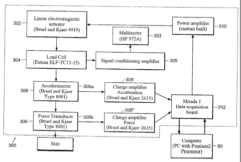
Other skin property sensor arrangements can also be used with the device 10,
such as the sensor configuration 300 shown as a block diagram in FIG. 14. The
sensor 300 includes a linear electromagnetic actuator 302 (e.g., model no.
4910,
available from Bruel and Kjaer) vertically mounted to a rigid frame. A strain
gauge
type load cell 304 (e.g., model no. ELF-TC13-15, available from Entran, of
Fairfield, NJ) is mounted to the actuator platform for the purpose of
measuring the
DC offset of the system corresponding to the static loading, as measured with
a
multimeter 303 (e.g., model no. HP 972A, available from Hewlett Packard, or
Palo
Alto, CA) via a signal conditioning amplifier 305. Below the load cell 304 is
an
impedance head 306 (Bruel and Kjaer model no. 8001) consisting of a
piezoelectric
accelerometer 306a and a piezoelectric force transducer 306b. The two outputs
from
the accelerometer record the force applied to the skin and its resulting
acceleration.
Two charge amplifiers 308', 308" (generally 308) (Bruel and Kjaer model no.
2635)
transform the force to a proportional voltage and doubly integrate the
acceleration to
give the skin displacement. The actuator 302 is driven by an algorithm, such
as a
Visual BASIC program, that simulates a Dynamic Signal Analyzer through a power
CA 02497815 2011-09-01
amplifier 310. The algorithm outputs a swept sinusoidal signal within a range
of
pre-determined frequencies. This modulation is a small perturbation on top of
an
initial static load, which is determined from the output voltage of the load
cell 304.
The measured force and displacement of the actuator are then input to two
separate
channels of a data acquisition board 312 and used to calculate the compliance
transfer function gain and phase with a computer or the controller 50. In one
implementation, there is a 50 kHz per channel of the data acquisition board,
which
can be increased to 100 kHz per channel when multiplexed. The AID is 18 bits
with
4.5 V, while the D/A is 18 bits with 3.0 V. Like that shown in FIG. 13 for
the
sensor 200, the sensor 300 is preferably associated with the device 10 through
the
controller 50. Accordingly, properties of the skin are analyzed by the
controller 50
based on the data from the sensor 300. The controller 50 then directs the
device 10
to eject drug into the body with the appropriate injection pressure.
Although the sensors 200 and 300 are shown in combination with the device
10, the sensors can be combined with other types of medical devices. For
example,
the sensor can be combined with other types of needleless injectors such as
those
using magnetic, chemical, hydraulic, and spring actuators, and those described
in
U.S. Application No. 10/200,574 filed July 19, 2002. Additionally, the sensor
can be
combined with injectors that use needles, such as microneedle injectors, and
those
described in U.S. Application Nos. 10/238,844 filed September 9, 2002 and
10/278,049 filed October 21, 2002. Advantageously, the sensed properties can
be
used to control the depth and/or insertion force of the needles.
Furthermore, the sensors 200, 300 can be used to measure skin properties of
a subject, as described above, or they can be used, to measure properties of
other
body surfaces. For example, the sensor can be used to measure properties of
the
internal anatomy of subject, such as the surface of an internal cavity or
organ during
a surgical procedure.
In some embodiments, the sensors 200 and 300 can be configured as stand
alone units. Thus, the system components discussed in relation to FIGs. 13 and
14
CA 02497815 2005-03-04
WO 2004/021882 PCT/US2003/027907
-19-
can be packaged within a single housing. The housing can be tethered to an
external
power source, or can include an internal power source, such as a battery.
Additionally, a stand alone unit can be configured as a wearable device that
can
attach to a patient's body using a bandage, or an adhesive. For example, a
small
force transducer and an accelerometer can be packaged in an adhesive bandage
that
is placed on the skin. The transducer first resonates at a resonant frequency
(e.g., 50
Hz) for a period of time (e.g., several seconds). The transducer stimulates
the local
skin and the accelerometer detects the displacement of the skin. A controller
can
then record the resulting skin displacement in a memory and calculate the
compliance gain of the skin. The controller can further determine the
mechanical
behavior of the skin (e.g., stiffness) using the calculated compliance gain.
Still
further, the controller can further identify the type of skin using its
calculated
mechanical behavior and/or compliance gain (e.g., that of a baby or of an
adult).
The sensor can ultimately generate a signal or command used as an indicator to
an
operator and/or a control signal to a medical device.
While this invention has been particularly shown and described with
references to preferred embodiments thereof, it will be understood by those
skilled in
the art that various changes in form and details may be made therein without
departing from the scope of the invention encompassed by the appended claims.
For
example, contractile polymers, or any other suitable contracting material, can
be
used instead of the shape memory alloy. The device 10 may include multiple
chambers or may accommodate multiple drug vials. Thus, the device 10 is able
to
deliver drug sequentially or simultaneously. For example, the device 10 is
able to
deliver two or more drugs at once to the body.